A paracrine network regulates the cross-talk between human lung stem cells and the stroma
14
ARTICLE Received 16 Aug 2013 | Accepted 20 Dec 2013 | Published 16 Jan 2014 A paracrine network regulates the cross-talk between human lung stem cells and the stroma E. Josue Ruiz 1 , Feride Oeztuerk-Winder 1 & Juan-Jose Ventura 1 The signals that regulate stem cell self-renewal and differentiation in the lung remain elusive. Lung stem cells undergo self-renewal or lineage commitment to replenish tissue, depending on cross-talk with their environment. This environment, also known as the niche, includes mesenchymal and endothelial tissues. Here we define molecular mechanisms involved in the interaction between human lung Lgr6 þ stem cells (LSCs) and fibroblasts in a functional microenvironment. We reveal a central role for p38a MAPK in establishing and maintaining such cross-talk, acting in both cell types. In LSCs, p38a induces the expression of SDF-1, which activates the stroma. p38a is essential for fibroblast activation and induction of cytokine expression, in particular TNFa . This paracrine network induces a hierarchical activation leading to the recruitment of endothelium, establishing a functional micro- environment. Disruption of this cross-talk abrogates proper LSC differentiation in vivo and may lead to lung dysfunction and disease. DOI: 10.1038/ncomms4175 OPEN 1 SCI (Wellcome Trust-Medical Research Council Stem Cell Institute, University of Cambridge), Tennis Court Road, Cambridge, CB2 1QR, UK. Correspondence and requests for materials should be addressed to J.-J.V. (email: [email protected]). NATURE COMMUNICATIONS | 5:3175 | DOI: 10.1038/ncomms4175 | www.nature.com/naturecommunications 1 & 2014 Macmillan Publishers Limited. All rights reserved.
-
Upload
independent -
Category
Documents
-
view
2 -
download
0
Transcript of A paracrine network regulates the cross-talk between human lung stem cells and the stroma

ARTICLE
Received 16 Aug 2013 | Accepted 20 Dec 2013 | Published 16 Jan 2014
A paracrine network regulates the cross-talkbetween human lung stem cells and the stromaE. Josue Ruiz1, Feride Oeztuerk-Winder1 & Juan-Jose Ventura1
The signals that regulate stem cell self-renewal and differentiation in the lung remain elusive.
Lung stem cells undergo self-renewal or lineage commitment to replenish tissue, depending
on cross-talk with their environment. This environment, also known as the niche, includes
mesenchymal and endothelial tissues. Here we define molecular mechanisms involved in the
interaction between human lung Lgr6þ stem cells (LSCs) and fibroblasts in a functional
microenvironment. We reveal a central role for p38a MAPK in establishing and maintaining
such cross-talk, acting in both cell types. In LSCs, p38a induces the expression of SDF-1,
which activates the stroma. p38a is essential for fibroblast activation and induction of
cytokine expression, in particular TNFa. This paracrine network induces a hierarchical
activation leading to the recruitment of endothelium, establishing a functional micro-
environment. Disruption of this cross-talk abrogates proper LSC differentiation in vivo and
may lead to lung dysfunction and disease.
DOI: 10.1038/ncomms4175 OPEN
1 SCI (Wellcome Trust-Medical Research Council Stem Cell Institute, University of Cambridge), Tennis Court Road, Cambridge, CB2 1QR, UK. Correspondenceand requests for materials should be addressed to J.-J.V. (email: [email protected]).
NATURE COMMUNICATIONS | 5:3175 | DOI: 10.1038/ncomms4175 | www.nature.com/naturecommunications 1
& 2014 Macmillan Publishers Limited. All rights reserved.

The stromal microenvironment plays a fundamental role inthe regulation of tissue homeostasis and the promotion ofpathological processes. The stem cell ‘niche’ allows the
maintenance of multipotency and regulates cell division1. Theniche is a microenvironment that includes the extracellularmatrix, cell contacts and a broad number of autocrine andparacrine signals and hormones2,3. Altogether, these contributorsregulate the stem cells to either remain in the niche, dividesymmetrically or asymmetrically, or migrate from the niche anddifferentiate into either transient progenitors or terminallydifferentiated cells4.
In the lung, several niches have been described that harbourdifferent adult multipotent stem cells involved in the turnover ofdistinct anatomical areas of the lung. There are different types ofstem cells in the trachea (submucosal gland stem cell), bronchi(basal cell) and bronchioles (neuroendocrine bodies)5. Recently, agroup of cells has been reported as putative progenitors for themouse bronchioalveolar area, with the potential to differentiateinto Clara or Alveolar (type 1 or 2) cells6.
We previously isolated a population of mouse bronchioalveolarcells based on negative selection for non-epithelial markers andsorting for Sca-1/E-Cad-positive cells7. Furthermore, we haverecently characterized a clonally derived population of humanlung Lgr6þ stem cells (LSCs) from the distal lung, which expressE-Cadherin and Lgr6, but not endothelial, mesenchymal orhematopoietic markers (CD34� /CD73� /CD45� /PECAM� ).The stem cell potential of these cells has been confirmed usingdifferent in vitro and in vivo assays, such as kidney capsuleengraftments and culture of lung explants8. In kidney grafts, LSCsare able to recapitulate a bronchioalveolar epithelium and also torecruit connective (Vimentinþ ) and endothelial (CD73þ ) cellsto create a functional environment8.
The role of paracrine signals in the maintenance of stem cellniches is well known. Activation of stromal cells, and speciallyfibroblasts, to induce their migration and production of otherparacrine signals plays an essential role in niche formationin cancer and homeostasis9,10. Simultaneously, molecular signalsreleased by the stroma control cell division and fate deter-mination in stem cells11. Stromal regulation is pivotal for properlung homeostasis and the existence of a niche is necessary tocreate a functional adult tissue with a turnover potential12.
The balance in cross-talk between signals from the stem cellsand signals from the stroma may also be a determinant for theproper regeneration of the bronchioalveolar epithelium afterinjury13. Failure to maintain the right balance may lead to patho-logical processes (for example, lung fibrosis, cancer metastasis),in which inflammatory signalling promotes expansion of thestromal compartment while preventing epithelial differentiationand functional tissue repair14.
Here, we delineate how the molecular interactions of aparacrine signalling circuit of cytokines and chemokines, releasedby LSCs and stromal fibroblasts, create a self-maintainedfunctional microenvironment both in vitro and in vivo. At thecentre of this circuit, acting as intracellular mediators of paracrinecues in both LSCs and fibroblasts, is the p38a MAPK signallingpathway. This kinase pathway co-ordinates the release andresponse to cytokines in both cell types, and it is necessary forLSCs to establish and maintain a bronchioalveolar environmentin vivo.
ResultsLSCs interact with their environment. We have described aclonally derived enriched human Lgr6þ population with thepotential to differentiate in vitro and in vivo into all bronch-ioalveolar mature cell types, named as Lgr6þ stem cells
(LSCs from here on). These cells are able to produce a bronch-ioalveolar epithelium in an alien environment when injectedunder the kidney capsule of nude mice8. This epithelium containsconnective and endothelial tissue (Fig. 1a). Interestingly, weobserved that a discrete population of LSCs is surrounded byfibroblasts (Vimentinþ ) mimicking a niche, suggesting thatLSCs are able to recruit stromal cells to create their ownmicroenvironment (Fig. 1a).
Reciprocal modulation between LSCs and fibroblasts. Tofunctionally test the ability of LSCs to recruit stromal cells, weused lung slices treated with bleomycin, to remove all kind ofcellular constituents, including bronchiolar, alveolar, mesenchy-mal and endothelial cell types (Supplementary Fig. 1 and 8). Exvivo bleomycin has been used for many years in lung explants as amodel of lung toxicity in vitro15,16. As a DNA-damaging andROS-induced apoptotic agent17, it kills most cell types, withoutany inflammatory response, producing a physiological scaffoldthat allows studying the process followed by LSCs to promote therecruitment of stromal cells. We injected differentially labelledLSCs and fibroblasts at different sites to test the range of distance-dependent paracrine cross-talk. The distance was determined bymonitoring the locations and relative positions of individual LSCand fibroblast cells over a period of 7 days, allowing us to quantifythe recruitment of fibroblasts to the proximity of LSCs (Fig. 1b–eand Supplementary Movie 1). Cell tracking was performed foreach cellular type within the region of analysis with an ImageJsoftware complemented with a MtrackJ plugin. We observed amaximum distance of 103 mm for the paracrine signals to engageLSC recruitment of fibroblasts (Supplementary Fig. 2A), in eitherbleomycin-treated or non-treated lung explants (SupplementaryFig. 2B). The dynamics of the recruitment varies, and it maydepend on paracrine factor gradients around different LSCs(Fig. 1d,e). Proliferation of LSCs and fibroblasts is promoted onlyafter they get in close proximity (Fig. 1d, and SupplementaryFig. 2A). Fibroblasts migrate to LSCs vicinity but do not engage indirect contact at early time points (Fig. 1d). However, at latertime points LSCs are surrounded by fibroblasts (Fig. 1d andSupplementary Fig. 2B), recapitulating the kidney graft results(see Fig. 1a).
Thus, these findings suggest that there is a paracrine cross-talkbetween LSCs and fibroblasts inducing the recruitment of thelatter, and recruited fibroblasts enclosing LSCs could function asputative niche cells.
To begin defining the mechanisms involved in the paracrinesignalling induced as part of the interplay between LSCs and lungfibroblasts, we used Boyden chambers. A comparative analysis ofthe factors released into the medium using a cytokine arrayshowed a number of changes in the paracrine signal profilereleased by LSCs and fibroblasts when cultured either alone or inclose proximity to each other (Fig. 2a, Supplementary Fig. S2Cand Supplementary Table 1 and 2). LSCs co-cultured withfibroblasts were considered as activated-LSCs (ALSCs). Cytokinesfound to be released differentially by ALSCs were involved in theinflammatory response (TNFa), vascularization (IL8, MIP2) orchemoattraction (SDF-1) (Fig. 2a and Supplementary Fig. 2C).These variations in cytokine release were transcriptionallyregulated, as cytokine mRNA expression correlated with proteinlevels (Fig. 2b). Especially interesting was the increase in SDF-1levels induced in ALSCs. This chemokine is essential inmaintaining the hematopoietic stem cell niche, although usuallyfrom mesenchymal or immune cell origin18. Furthermore, in theco-cultures, activated fibroblasts (AFs) showed an increased in theexpression of both SDF-1 receptors, CXCR4 and CXCR7(Supplementary Fig. 2D).
ARTICLE NATURE COMMUNICATIONS | DOI: 10.1038/ncomms4175
2 NATURE COMMUNICATIONS | 5:3175 | DOI: 10.1038/ncomms4175 | www.nature.com/naturecommunications
& 2014 Macmillan Publishers Limited. All rights reserved.

1 320
600
800
Rel
ativ
e po
sitio
n(p
ixel
es)
400
200
4 65 7Day
Fib. 1LSC 1 600
800
Rel
ativ
e po
sitio
n(p
ixel
es) 700
500
Day
Fib. 2LSC 2
1 32 4 65 7
+Bleomycin
Lung slides(human/mouse)
48h
Tracking
1–7 days
0 21
0
600
900
Rel
ativ
e po
sitio
n (p
ixel
es)
700
500
400
300
200
100
3 54 6
Day
7
Fib.
LSC
800
LSC 4
Fib.5
LSC 6
LSC 2
Fib.3
Fib.1
100
600
800
Rel
ativ
e le
ngth
(pi
xele
s) 700
500
400
300
200
100
2 43 5
Day
Fib.1
Fib.5Fib.3
LSC 6
LSC 2LSC 4
LSC
Fib.
LSC
Fib.
LSC-eGFP + fibroblasts-Dsred2
0 m 330 m270 m 420 m 480 m 24 h
Injection ofLSC and
E Day 3 Day 5Day 2Day 1
LSC
Fibroblasts
EC
LSC
Fib.
21
21
34
62
1
5
Figure 1 | Analysis of stromal cell recruitment by lung stem cells. (a) LSCs (GFP-labelled) engraft in the kidney capsule and recruit fibroblasts
(Vimentinþ ) and endothelial cells (CD73þ ) (red). Note that fibroblasts surrounded the LSCs. Scale bars: 100mm. (b) General strategy to track the
movement of the cells. Schematic tracing of three pairs of LSC-fibroblasts injected in ex vivo lung tissue from different distances at starting point (arrows).
Relative length graph showing the spatial location of the LSC-fibroblasts injected during a 5-day period. (c) Graphs showing the relative positioning and
schematic tracing of single pairs of LSC-fibroblast cells from different distances at starting point (arrows). (d) Real-time tracing of LSCs (green)
and fibroblasts (red) over a 24 h (upper panel, scale bars: 20mm) or 5 days period (lower panel, scale bars: 200mm). (e) Average of the relative
positioning between injected fibroblasts and LSCs. N¼ 35, from 4–5 experiments±s.e. of the mean s.e.m.
NATURE COMMUNICATIONS | DOI: 10.1038/ncomms4175 ARTICLE
NATURE COMMUNICATIONS | 5:3175 | DOI: 10.1038/ncomms4175 | www.nature.com/naturecommunications 3
& 2014 Macmillan Publishers Limited. All rights reserved.

IL8 SDF1CCL20
8
12
mR
NA
rel
ativ
e le
vels LSC
ALSC10
6
4
2P<0.01
P=0.01 P=0.02
P<0.01
TNFα
LSC
AF
ALSC+
LSC ALSC
IL8
CCL2
SDF1
MIP2
M-CSF
Control
TNFα IL8
CCL2
SDF1
MIP2
M-CSF
Control
TNFα
LSC-eGFP + fibroblasts-DsRed2
Day 1 Day 2 Day 3 Day 4
SDF1CXCR4DAPI
CXCR4DAPI
SDF1DAPI
SDF1DAPI
CXCR4DAPI
SDF1CXCR4DAPI
Control0
40
CMLSC
shControl
CMLSC-KD
CMALSC
shControl
60
80
Mig
rate
d fib
rob
last
spe
r fie
ld
20
CMALSC
shControl+anti-SDF1
CMALSC-KD
P<0.01 P<0.01
P<0.01
P<0.01P<0.01 P<0.01
P=0.8
CMALSC-KD
+SDF1
P<0.01
Figure 2 | SDF-1 released by activated LSCs is the key paracrine factor to recruit stromal fibroblasts. (a) Protein array showing differential cytokine
release by activated LSCs (ALSC) when co-cultured with fibroblasts. (b) The mRNA relative levels of different cytokines confirm their upregulation in
ALSCs. (c) Time-lapse tracing of fibroblast (red) recruitment by LSCs (green) in lung explants. Scale bars, 200mm. (d) Serial sections images captured at
day 4 showing expression of SDF-1 (white) and CXCR4 (purple) by LSCs and fibroblasts, respectively. Scale bars, 50mm. (e) Fibroblast migration induced
by conditioned medium (CM) from ALSCs (expressing control shRNA) (line 4) is prevented by addition of an anti-SDF-1 antibody (line 5) or by using
CM from LSCs lacking SDF-1 (ALSC-KD) (line 6). Addition of SDF-1 recombinant protein into CM from ALSC-KD cells rescued their potential to
recruit fibroblasts (line 7). Scale bar, 200mm. All results (b and e) are the mean±s.e.m. of 4–5 triplicate experiments. Po0.01 values were defined
as statistically significant, as analysed by one-way ANOVA.
ARTICLE NATURE COMMUNICATIONS | DOI: 10.1038/ncomms4175
4 NATURE COMMUNICATIONS | 5:3175 | DOI: 10.1038/ncomms4175 | www.nature.com/naturecommunications
& 2014 Macmillan Publishers Limited. All rights reserved.

SDF-1 is the key paracrine factor to recruit stromal cells. Tobegin defining whether SDF-1 is required for fibroblast recruit-ment, LSCs and fibroblasts were injected into human lungexplants, and then we analysed the expression of SDF-1 in LSCsand CXCR4 expression in fibroblasts. Serial sections of theexplants were captured by immunofluorescent microscopy for theunambiguous identification of both proteins. Importantly andconsistent with the above results, when fibroblasts are recruitedby LSCs (Fig. 2c), the stem cells expressed SDF-1 and the fibro-blasts expressed the CXCR4 receptor (Fig. 2d).
Using transwell chambers, we also observed that conditionedmedium (CM) from ALSCs induced the migration of fibroblastsmore strongly than CM from LSCs cultured alone (Fig. 2e,compare lines 2 and 4). Migration was specifically activated,independently of proliferation, which was not affected. To testwhether SDF-1 released by ALSCs is responsible to inducefibroblast migration, CM from ALSCs was incubated with aneutralizing antibody for SDF-1. We found that blocking SDF-1activity prevented the ability of the ALSCs to induce fibroblastmigration (Fig. 2e, see lines 4 and 5). To confirm this observation,LSCs expressing short hairpins for SDF-1 (LSC-KD) weregenerated (Supplementary Fig. 2E) and the CM from eitherLSC-KD cultured alone or co-cultured with fibroblasts (ALSC-KD) was used as a chemoattractant. As expected, knockingdown SDF-1 in ALSCs (Supplementary Fig. 2F) also inhibitedfibroblast migration (Fig. 2e, see lines 4 and 6). Finally,confirmation of the chemoattractant role of SDF-1 was shownby rescuing recruitment of fibroblasts by the addition ofrecombinant SDF-1 protein to the CM from ALSC-KD (Fig. 2e,see lines 4, 6 and 7).
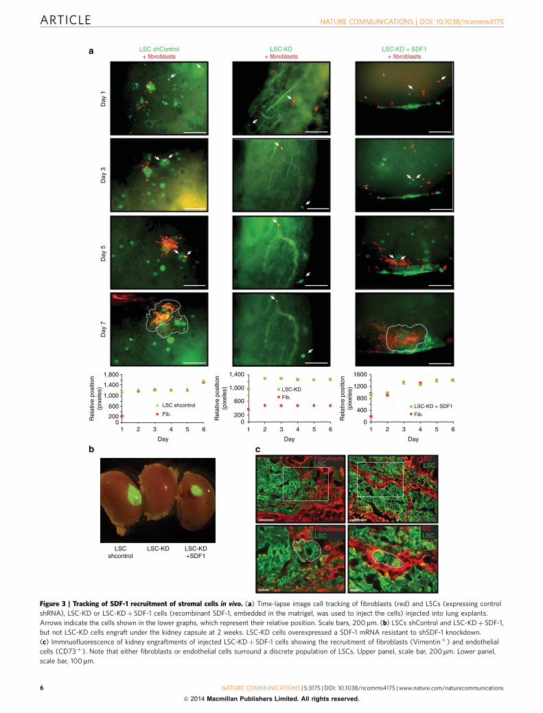
On the basis of this evidence, we hypothesized that theinhibition of SDF-1 would result in the impairment of fibroblastmigration in lung explants. To test this hypothesis, we injectedfibroblasts together with either LSCs expressing a short hairpincontrol or LSC-KD into human lung explants. In agreement withprevious results, LSCs expressing a shControl were able to recruitfibroblasts (Fig. 3a). However, the injection of LSCs lacking SDF-1 expression confirmed the absence of the necessary paracrinecross-talk to recruit fibroblasts via SDF-1 (Fig. 3a, middle panel,and Supplementary Fig. 3A, B). Fibroblasts recruitment couldbe rescued by overexpression of a SDF-1 mRNA resistant toshSDF-1 knockdown or embedding SDF-1 protein in thematrigel carrying the LSC-KD cells (Fig. 3a, right panel, andSupplementary Fig. 3C,D). Furthermore and consistent with theabove results, recruited fibroblasts are able to enclose LSCs.
Finally, kidney capsule engraftments were used to test, in vivo,the observed in vitro role of SDF-1 in the recruitment of stromalfibroblasts. We found that LSCs lacking SDF-1 (LSC-KD)failed to produce grafts under the kidney capsule (Fig. 3b andSupplementary Fig. 3E). However, again it could be rescuedby overexpression of shSDF-1 resistant mRNA (Fig. 3b andSupplementary Table 3) or embedding SDF-1 recombinantprotein in the matrigel carrying the LSC-KD cells(Supplementary Fig. 3E,F). The inability of the LSC-KD cells toengraft was independent of their proliferative potential(Supplementary Fig. 3G). Interestingly, the engraftments showedthat LSC-KD cells, rescued by shSDF-1 resistant mRNA, wereable to recruit stromal cells from the host tissue (Fig. 3c).Furthermore, we also found that a small population of LSCs wassurrounded by fibroblasts (Vimentinþ ) or endothelial cells(CD73þ ) recapitulating a niche (Fig. 3c). The mouse origin ofthe stroma in the graft was confirmed as it did not express GFPand was negative for an anti-human mitochondrial antibody(Supplementary Fig. 3H).
Together, these data suggest that SDF-1 released by LSCs hasan essential role in the recruitment of stromal cells.
Regulation of SDF-1 expression and secretion by TNFa andTGFb. Having demonstrated the essential role of SDF-1 to recruitstromal cells, we next sought to understand how SDF-1 expres-sion is regulated by stromal fibroblasts. It has been reported that,depending on cellular context, either TGFb or TNFa signallingpathways can regulate SDF-1 expression19,20. Thus, SDF-1regulation in LSCs by TNFa and TGFb pathways was tested.Although TNFa was able to induce SDF-1 expression, TGFb wasa much stronger activator in LSCs (Fig. 4a). Interestingly, TGFblevels were increased in ALSCs (Fig. 4b), but not in AFs(Supplementary Fig. 4A), so TGFb was excluded as the putativeparacrine signal used by the fibroblasts to promote SDF-1expression in LSCs. Nevertheless, AFs expressed and releasedmore TNFa (Fig. 4g, upper panel, and Supplementary Fig. 4B).This cytokine was considered as a candidate to induce theparacrine activation of LSCs. As TGFb was not of fibroblasticorigin, a possible autocrine regulation of SDF-1 expression inALSCs was considered. To test this possibility, ALSCs wereincubated with a neutralizing antibody for TGFb. We found thatblocking TGFb activity inhibited the ability of the ALSCs toexpress SDF-1, confirming the autocrine role of TGFb(Supplementary Fig. 4C).
TGFb expression can be induced by TNFa through c-Junactivation21. Indeed, LSCs responded to exogenous TNFatreatment by activating the JNK/AP-1 pathway (SupplementaryFig. 4D). In addition, we observed a correlation between TNFa-induced TGFb and SDF-1 expression in LSCs (SupplementaryFig. 4E). The specificity of this correlation was tested using aninhibitor (SP600125) of the JNK pathway, which abrogated bothc-Jun phosphorylation and activation (Supplementary Fig. 4F).These results showed that AP-1 activation was required forTNFa-induced SDF-1 expression (Supplementary Fig. 4G), butnot for TGFb expression, suggesting that other pathway controlsTNFa-induced TGFb expression. However, we confirmed thatTGFb expression in ALSCs was completely dependent onTNFa released by fibroblasts, as it could be prevented using aneutralizing anti-TNFa antibody (Fig. 4c).
These results suggest that activated fibroblasts (AFs) releaseTNFa, which as a paracrine signal induces TGFb autocrine loopthat enhances SDF-1 production in ALSCs.
p38a regulates LSCs and fibroblasts activation. As a well-known regulator of cytokine production, the role of p38a in thecross-talk between LSCs and the stromal fibroblasts was investi-gated using two different approaches. p38a signalling wasinhibited in LSCs using a shRNA (SH3) against this kinase8.LSC-SH3 cells express very low levels of p38a protein and did notactivate the p38a pathway (Supplementary Fig. 4H). UnlikeALSCs, cells lacking p38a (ALSC-SH3) did not respond tofibroblasts stimulation by either releasing SDF-1 protein (Fig. 4d)or expressing SDF-1 mRNA (Supplementary Fig. 4I). In addition,recombinant TNFa failed to induce TGFb expression in LSC-SH3cells (Supplementary Fig. 4J), suggesting that in ALSCs p38a isessential to activate the TGFb autocrine loop induced byparacrine TNFa. Furthermore, the direct role of p38a in SDF-1expression induced by autocrine TGFb was confirmed, asexogenous TGFb was insufficient to promote SDF-1 mRNAexpression in LSC-SH3 cells (Fig. 4e). The activation of the TGFbcanonical Smad pathway is not affected in LSC-SH3 cells(Supplementary Fig. 4K).
In fibroblasts, the p38a pathway was inhibited using over-expression of a dominant negative form of p38 (p38DN)22. p38activity, but not cJun, was induced in fibroblasts after 6 h ofco-culture, and, as expected, it was absent in p38DN fibroblasts(Fib.-DN) (Fig. 4f). Moreover, fibroblasts lacking p38a signallinghad reduced levels of both TNFa protein and mRNA compared
NATURE COMMUNICATIONS | DOI: 10.1038/ncomms4175 ARTICLE
NATURE COMMUNICATIONS | 5:3175 | DOI: 10.1038/ncomms4175 | www.nature.com/naturecommunications 5
& 2014 Macmillan Publishers Limited. All rights reserved.
0Rel
ativ
e po
sitio
n(p
ixel
es)
1,400
1,000
600
200
51 2 3 4 6
Day
Fib.
LSC-KD
Fib.
LSC shcontrol
0
1,400
Rel
ativ
e po
sitio
n(p
ixel
es)
1,000
600
51 3 4 6
Day
200
2
1,800
21 3 4 60
1200
Rel
ativ
e po
sitio
n(p
ixel
es)
800
400
5
Day
Fib.
LSC-KD + SDF1
1600
LSC shControl+ fibroblasts
Day
1
LSC-KD+ fibroblasts
Day
5D
ay 7
Day
3
LSC-KD + SDF1+ fibroblasts
LSCshcontrol
LSC-KD
LSCECLSC
FibroblastsLSC
ECLSC
LSC-KD+SDF1
Fibroblasts
a
b c
Figure 3 | Tracking of SDF-1 recruitment of stromal cells in vivo. (a) Time-lapse image cell tracking of fibroblasts (red) and LSCs (expressing control
shRNA), LSC-KD or LSC-KDþ SDF-1 cells (recombinant SDF-1, embedded in the matrigel, was used to inject the cells) injected into lung explants.
Arrows indicate the cells shown in the lower graphs, which represent their relative position. Scale bars, 200mm. (b) LSCs shControl and LSC-KDþ SDF-1,
but not LSC-KD cells engraft under the kidney capsule at 2 weeks. LSC-KD cells overexpressed a SDF-1 mRNA resistant to shSDF-1 knockdown.
(c) Immnuofluorescence of kidney engraftments of injected LSC-KDþ SDF-1 cells showing the recruitment of fibroblasts (Vimentinþ ) and endothelial
cells (CD73þ ). Note that either fibroblasts or endothelial cells surround a discrete population of LSCs. Upper panel, scale bar, 200mm. Lower panel,
scale bar, 100mm.
ARTICLE NATURE COMMUNICATIONS | DOI: 10.1038/ncomms4175
6 NATURE COMMUNICATIONS | 5:3175 | DOI: 10.1038/ncomms4175 | www.nature.com/naturecommunications
& 2014 Macmillan Publishers Limited. All rights reserved.
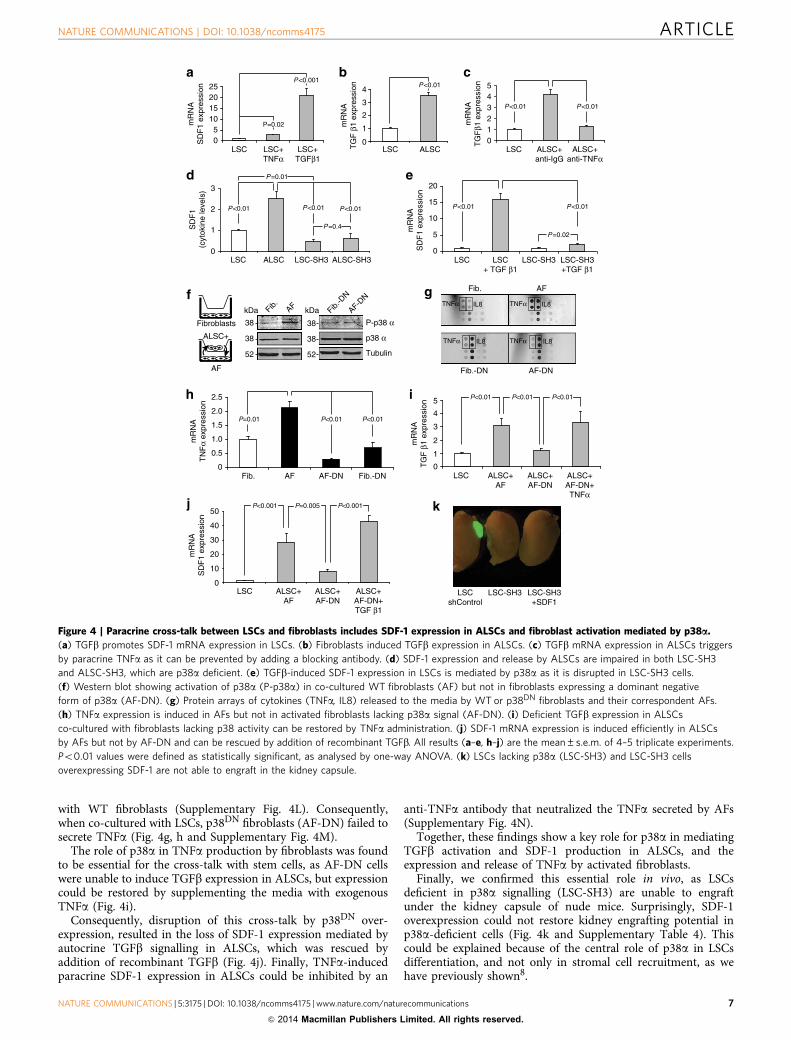
with WT fibroblasts (Supplementary Fig. 4L). Consequently,when co-cultured with LSCs, p38DN fibroblasts (AF-DN) failed tosecrete TNFa (Fig. 4g, h and Supplementary Fig. 4M).
The role of p38a in TNFa production by fibroblasts was foundto be essential for the cross-talk with stem cells, as AF-DN cellswere unable to induce TGFb expression in ALSCs, but expressioncould be restored by supplementing the media with exogenousTNFa (Fig. 4i).
Consequently, disruption of this cross-talk by p38DN over-expression, resulted in the loss of SDF-1 expression mediated byautocrine TGFb signalling in ALSCs, which was rescued byaddition of recombinant TGFb (Fig. 4j). Finally, TNFa-inducedparacrine SDF-1 expression in ALSCs could be inhibited by an
anti-TNFa antibody that neutralized the TNFa secreted by AFs(Supplementary Fig. 4N).
Together, these findings show a key role for p38a in mediatingTGFb activation and SDF-1 production in ALSCs, and theexpression and release of TNFa by activated fibroblasts.
Finally, we confirmed this essential role in vivo, as LSCsdeficient in p38a signalling (LSC-SH3) are unable to engraftunder the kidney capsule of nude mice. Surprisingly, SDF-1overexpression could not restore kidney engrafting potential inp38a-deficient cells (Fig. 4k and Supplementary Table 4). Thiscould be explained because of the central role of p38a in LSCsdifferentiation, and not only in stromal cell recruitment, as wehave previously shown8.
LSC0
ALSC
3
2
ALSC-SH3LSC-SH3
1
P<0.01P<0.01P<0.01
P=0.01
P=0.4SD
F1
(cyt
okin
e le
vels
)
012345
P<0.01 P<0.01
05
10152025
mR
NA
SD
F1
expr
essi
on
P<0.001
P=0.02
TNFα IL8 TNFα IL8
TNFα IL8IL8TNFα
AFFib.
AF-DNFib.-DN
Fib. AF
P=0.01 P<0.01
0
0.5
1.5
2.0
2.5
1.0
Fib.-DNAF-DN
P<0.01
LSC0
10
20
30
50
40
ALSC+AF-DN
P=0.005P<0.001 P<0.001
LSC0
1
2
ALSC
3
4P<0.01
0
1
2
3
5
4
P<0.01P<0.01P<0.01
P<0.01 P<0.01
0
15
20
10
5 P=0.02
mR
NA
SD
F1
expr
essi
on
LSCshControl
LSC-SH3+SDF1
LSC-SH3
AF
ALSC+
Tubulin
Fib. AF Fib.-D
N
AF-DN
P-p38 α
p38 α
52
38
38
kDa
52
38
38
kDa
mR
NA
TN
Fα
expr
essi
on
mR
NA
TG
F β
1 ex
pres
sion
LSC ALSC+AF
ALSC+AF-DN
ALSC+AF-DN+TNFα
ALSC+AF
ALSC+AF-DN+TGF β1
mR
NA
SD
F1
expr
essi
on
Fibroblasts
LSC LSC-SH3 LSC-SH3+TGF β1
LSC+ TGF β1
LSC LSC+TGFβ1
LSC+TNFα
mR
NA
TG
F β
1 ex
pres
sion
mR
NA
TG
Fβ1
exp
ress
ion
LSC ALSC+anti-TNFα
ALSC+anti-IgG
Figure 4 | Paracrine cross-talk between LSCs and fibroblasts includes SDF-1 expression in ALSCs and fibroblast activation mediated by p38a.
(a) TGFb promotes SDF-1 mRNA expression in LSCs. (b) Fibroblasts induced TGFb expression in ALSCs. (c) TGFb mRNA expression in ALSCs triggers
by paracrine TNFa as it can be prevented by adding a blocking antibody. (d) SDF-1 expression and release by ALSCs are impaired in both LSC-SH3
and ALSC-SH3, which are p38a deficient. (e) TGFb-induced SDF-1 expression in LSCs is mediated by p38a as it is disrupted in LSC-SH3 cells.
(f) Western blot showing activation of p38a (P-p38a) in co-cultured WT fibroblasts (AF) but not in fibroblasts expressing a dominant negative
form of p38a (AF-DN). (g) Protein arrays of cytokines (TNFa, IL8) released to the media by WT or p38DN fibroblasts and their correspondent AFs.
(h) TNFa expression is induced in AFs but not in activated fibroblasts lacking p38a signal (AF-DN). (i) Deficient TGFb expression in ALSCs
co-cultured with fibroblasts lacking p38 activity can be restored by TNFa administration. (j) SDF-1 mRNA expression is induced efficiently in ALSCs
by AFs but not by AF-DN and can be rescued by addition of recombinant TGFb. All results (a–e, h–j) are the mean±s.e.m. of 4–5 triplicate experiments.
Po0.01 values were defined as statistically significant, as analysed by one-way ANOVA. (k) LSCs lacking p38a (LSC-SH3) and LSC-SH3 cells
overexpressing SDF-1 are not able to engraft in the kidney capsule.
NATURE COMMUNICATIONS | DOI: 10.1038/ncomms4175 ARTICLE
NATURE COMMUNICATIONS | 5:3175 | DOI: 10.1038/ncomms4175 | www.nature.com/naturecommunications 7
& 2014 Macmillan Publishers Limited. All rights reserved.
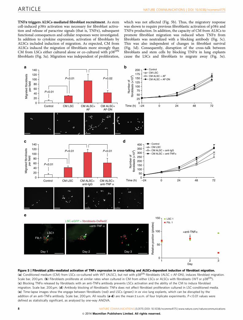
TNFa triggers ALSCs-mediated fibroblast recruitment. As stemcell-induced p38a activation was necessary for fibroblast activa-tion and release of paracrine signals (that is, TNFa), subsequentfunctional consequences and cellular responses were investigated.In addition to cytokine expression, activation of fibroblasts byALSCs included induction of migration. As expected, CM fromALSCs induced the migration of fibroblasts more strongly thanCM from LSCs either cultured alone or co-cultured with p38DN
fibroblasts (Fig. 5a). Migration was independent of proliferation,
which was not affected (Fig. 5b). Thus, the migratory responsewas shown to require previous fibroblastic activation of p38a andTNFa production. In addition, the capacity of CM from ALSCs topromote fibroblast migration was reduced when TNFa fromfibroblasts was neutralized with a blocking antibody (Fig. 5c).This was also independent of changes in fibroblast survival(Fig. 5d). Consequently, disruption of the cross-talk betweenfibroblasts and stem cells by blocking TNFa in lung explantscause the LSCs and fibroblasts to migrate away (Fig. 5e).
025
100125150175
7550
200
050
200250300350
150100
400
–24 0 24 48 72Time (h)
–24 0 24 48 72Time (h)
CM LSCCM ALSC + anti-IgGCM ALSC + anti-TNFα
LSC 1
Fib. 1
31 2
Rel
ativ
e po
sitio
n (p
ixel
es)
0
150
100
50
Day
Day 3
LSC1
Fib.1
Day 1 Day 2
0
20
80
100
120
140
60
40P<0.01
P<0.01 P=0.02
P<0.01
P<0.01
0
20
80
100
120
140
60
40
P<0.01
+anti-TNFα
LSC-eGFP + fibroblasts-DsRed2
Mig
rate
d fib
robl
asts
per
field
Control CM LSC CM ALSC+anti-TNF α
CM ALSC+anti-IgG
Num
ber
of
fibro
blas
ts (
×10
3 )
Control
CM LSC CM ALSC+AF
CM ALSC+AF-DN
Control
Mig
rate
d fib
robl
asts
per
field
Num
ber
of
fibro
blas
ts (
×10
3 )
ControlCM LSCCM ALSC + AFCM ALSC + AF-DN
+anti-TNF α
Figure 5 | Fibroblast p38a-mediated activation of TNFa expression in cross-talking and ALSCs-dependent induction of fibroblast migration.
(a) Conditioned medium (CM) from LSCs co-cultured with WT (ALSC), but not with p38DN fibroblasts (ALSCþAF-DN), induces fibroblast migration.
Scale bar, 200mm. (b) Fibroblasts proliferate at similar rates when cultured in CM from either LSCs or ALSCs with fibroblasts (WT or p38DN).
(c) Blocking TNFa released by fibroblasts with an anti-TNFa antibody prevents LSCs activation and the ability of the CM to induce fibroblast
migration. Scale bar, 200mm. (d) Antibody blocking of fibroblastic TNFa does not affect fibroblast proliferation cultured in LSC-conditioned media.
(e) Time-lapse images show the engage between fibroblasts (red) and LSCs (green) in ex vivo lung explants, which can be disrupted by the
addition of an anti-TNFa antibody. Scale bar, 200mm. All results (a–d) are the mean±s.e.m. of four triplicate experiments. Po0.01 values were
defined as statistically significant, as analysed by one-way ANOVA.
ARTICLE NATURE COMMUNICATIONS | DOI: 10.1038/ncomms4175
8 NATURE COMMUNICATIONS | 5:3175 | DOI: 10.1038/ncomms4175 | www.nature.com/naturecommunications
& 2014 Macmillan Publishers Limited. All rights reserved.

The effect from TNFa blocking is consistent with the previouslyshown decrease in SDF-1 levels in ALSCs under this condition(Supplementary Fig. 4M).
Together, these data suggest that activation of LSCs byparacrine TNFa is essential for the subsequent ability of ALSCsto recruit fibroblasts.
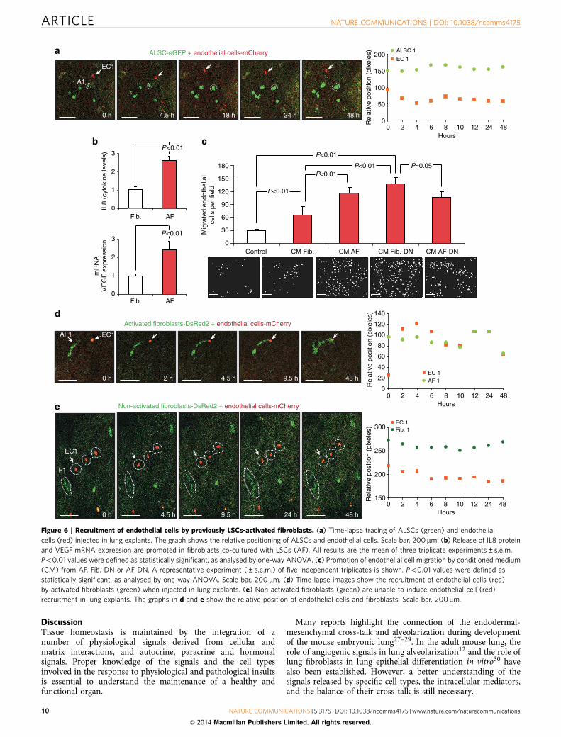
Sequential fibroblast activation and EC migration led by LSCs.Previous studies have suggested that stem cells can have thepotential to cross-talk with vascular cells9. After showing theparacrine activation of fibroblasts by LSCs and taking intoaccount the results from kidney engraftments, the potential directrecruitment of endothelial cells (EC) by ALSCs was investigated.Surprisingly and unlike fibroblasts, ALSCs fail to recruitendothelial cells in lung explants and to migrate in vitro(Fig. 6a and Supplementary Fig. 5A).
Interestingly, increased expression and release of the angio-genic factors IL8 and VEGF in AFs was observed (Fig. 4g, Fig. 6band Supplementary Fig. 5B), suggesting a putative role of thesemolecules in endothelial cell recruitment. This expression wasindependent of p38a activity, as Fib.-DN also showed increasedexpression of angiogenic factors (Fig. 4g and SupplementaryFig. 5C) and responded to ALSC induction (Fig. 4g andSupplementary Fig. 5D).
Inhibition of p38a activity may result in a constitutiveactivation of the JNK pathway7. Indeed, Fib.-DN had aconstitutively activated JNK/AP-1 pathway, which was alsopresent in AFs following 12 h of co-culture (SupplementaryFig. 5E). AP-1 has been shown to regulate the expression of bothVEGF and IL8 (refs 23–26). The direct role of AP-1 wasconfirmed, as inhibition of JNK activity by a small moleculeinhibitor (SP600125) prevented the induction of both IL8 andVEGF expression in AFs by ALSCs (Supplementary Fig. 5F).Consequently, CM from AFs, Fib.-DN or AF-DN, induceda higher migration of endothelial cells than CM fromWT fibroblasts (Fig. 6c), independently of proliferation(Supplementary Fig. 5G). Importantly and consistent with theabove results, only AFs (Fig. 6d and Supplementary Fig. 5H) butnot regular fibroblasts (Fig. 6e and Supplementary Fig. 5I) wereable to recruit endothelial cells in lung explants.
Therefore, endothelial migration and recruitment is mediatedby angiogenic signals, induced by the JNK/AP-1 pathway in AFs,but not by direct paracrine signals from ALSCs.
SDF-1 induces TNFa and angiogenic factors in fibroblasts.We have shown that there are two signalling pathways (p38aand JNK/AP-1) activated in fibroblasts only when they areco-cultured with LSCs, suggesting that those fibroblasts are alsoregulated by paracrine factors from LSCs. We hypothesized thatSDF-1 could be the factor responsible to activate both pathways.To test this hypothesis, either shControl LSCs or LSCs lackingSDF-1 (LSC-KD) were co-cultured with fibroblasts. We foundthat fibroblast activation was dependent on SDF-1. LSCs lackingSDF-1 did not induce p38a at 6 h (Supplementary Fig. 6A), norJNK/AP-1 activation at 12 h (Supplementary Fig. 6B), correlatingwith the reduced expression of TNFa and angiogenic factors (IL8,VEGF), respectively (Supplementary Fig. 6C,D). However, acti-vation of both pathways and expression of those cytokines couldbe rescued by addition of recombinant SDF-1 (SupplementaryFig. 6E).
Moreover, we observed that low levels of SDF-1 induced earlyexpression of TNFa, but only high levels could induce theexpression of VEGF and IL8 in fibroblasts (SupplementaryFig. 6F). Thus, SDF-1 promoted early activation of p38a, whichcorrelates with TNFa expression, and later JNK/AP-1 activationthat correlated with IL8 and VEGF expression in AFs.
Interestingly, real-time qPCR analysis showed that TNFa andVEGF were upregulated in kidney engraftments in comparisonwith mouse fibroblasts, giving the former cytokines an in vivo rolein the engrafting and microenvironment process initiated byLSCs (Supplementary Fig. 6G).
Finally and consistent with the above results, we confirmedthe essential cross-talk between LSCs and fibroblasts toregulate endothelial cell migration. We found that CM fromfibroblasts co-cultured with SDF-1-deficient stem cells(AFþ LSC-KD) failed to enhance endothelial migration(Supplementary Fig. 6H), but it did not affect their proliferation(Fig. 6i).
Altogether, these findings are consistent with the results fromkidney grafts, in which LSCs lacking SDF-1 failed to recruitstromal cells as a result of the disruption of the TNFa-TGFb-p38a-SDF-1 network.
The results allow us to delineate a model that can explain howlung stem cells promote the formation of their own environment,a necessary contributor to generating a lung epithelium. LSCsengage in cross-talk with fibroblasts. Basal levels of SDF-1 fromLSCs can induce both the recruitment and priming of fibroblasts.‘Low activated’ fibroblasts release TNFa, leading to subsequentactivation of a TGFb/p38a autocrine loop in LSCs (ALSCs) andfurther promotion of SDF-1 expression. Higher levels of SDF-1induce a second and enhanced expression of TNFa (throughp38a) in fibroblasts (AFs). High SDF-1 levels are also able toinduce expression of angiogenic factors (through the JNK/AP-1pathway) in fibroblasts. IL8 and VEGF released by ‘highlyactivated’ fibroblasts then recruit endothelial cells, thuspromoting angiogenesis (Fig. 7f).
Fibroblast-dependent control of stem cell differentiation. Wehave begun to identify the paracrine signals required to maintaina functional cross-talk between stem and stromal cells. Indeed,this cross-talk is essential because, in kidney engraftments, res-cued LSC-KD cells by SDF-1 overexpression are able not only torecruit fibroblasts and endothelial cells to the graft (see Fig. 3c)but also to differentiate into bronchiolar (Fig. 7a) and alveolar(Fig. 7b) cells. These results suggest that SDF-1 released by LSCsis essential to recruit stromal cells and also to allow engraftmentand—consequently—differentiation of LSCs into the kidneycapsule. Why the stromal cells are necessary to develop a LSCmicroenvironment is still unknown. We have previously reportedthat grafts from single LSCs injections harboured small pools ofLgr6þ undifferentiated cells, which retain self-renewal ability8.We hypothesized that stromal cells are required to maintain thispotential.
To test this hypothesis, we injected LSCs and fibroblasts inhuman lung explants to allow cell recruitment. After 10 days weanalyzed the expression of several differentiation markers in serialsections by immunofluorescent microscopy. Consistent with ourhypothesis, now we provided unambiguous evidence that LSCslocated away from fibroblasts are allowed to start lineagecommitment as they engraft into the epithelial tissue differentiat-ing into AT2 (SPCþ ), Clara (CC10þ ) or AT1 (AQ5þ ) cells(Fig. 7c, d, and Supplementary Fig. 7A,B). In contrast, LSCs closerto fibroblasts remain in an undifferentiated state, expressing thestem cell marker Lgr6 but not the lung differentiation markersSPC (Surfactant protein C), CC10 (Clara cell 10) or AQ5(Aquaporin 5) (Fig. 7e).
These findings suggest that LSCs induce the recruitment ofstromal cells to create a functional microenvironment to ensureself-renewal capacity. Once this capability is ensured, LSCs canstart the differentiation process towards bronchiolar or alveolarlineage commitment.
NATURE COMMUNICATIONS | DOI: 10.1038/ncomms4175 ARTICLE
NATURE COMMUNICATIONS | 5:3175 | DOI: 10.1038/ncomms4175 | www.nature.com/naturecommunications 9
& 2014 Macmillan Publishers Limited. All rights reserved.
DiscussionTissue homeostasis is maintained by the integration of anumber of physiological signals derived from cellular andmatrix interactions, and autocrine, paracrine and hormonalsignals. Proper knowledge of the signals and the cell typesinvolved in the response to physiological and pathological insultsis essential to understand the maintenance of a healthy andfunctional organ.
Many reports highlight the connection of the endodermal-mesenchymal cross-talk and alveolarization during developmentof the mouse embryonic lung27–29. In the adult mouse lung, therole of angiogenic signals in lung alveolarization12 and the role oflung fibroblasts in lung epithelial differentiation in vitro30 havealso been established. However, a better understanding of thesignals released by specific cell types, the intracellular mediators,and the balance of their cross-talk is still necessary.
Fib.0
AF
3
mR
NA
VE
GF
exp
ress
ion
2
1
P<0.01
Rel
ativ
e po
sitio
n (p
ixel
es)
100
200
150
Hours
0
50
ALSC 1
EC 1
0 h
A1
EC1
4.5 h 18 h 24 h 48 h
ALSC-eGFP + endothelial cells-mCherry
EC 1Fib. 1
Rel
ativ
e po
sitio
n (p
ixel
es)
2 64
200
300
8 10Hours
1504824120
2 64 8 10Hours
4824120
2 64 8 10 4824120
250
Non-activated fibroblasts-DsRed2 + endothelial cells-mCherry
Activated fibroblasts-DsRed2 + endothelial cells-mCherry
0 h
F1
EC1
4.5 h 9.5 h 24 h 48 h
Rel
ativ
e po
sitio
n (p
ixel
es)
60
140
100
0
40EC 1AF 1
120
80
202 h 4.5 h 9.5 h 48 h0 h
AF1 EC1
Fib.0
AF
3
2
1
IL8
(cyt
okin
e le
vels
)
P<0.01
Control0
90
CM Fib.-DN
120
150
180
Mig
rate
d en
doth
elia
lce
lls p
er fi
eld
60
30
P<0.01
P<0.01
P<0.01
P=0.05P<0.01
CM AF-DNCM Fib. CM AF
Figure 6 | Recruitment of endothelial cells by previously LSCs-activated fibroblasts. (a) Time-lapse tracing of ALSCs (green) and endothelial
cells (red) injected in lung explants. The graph shows the relative positioning of ALSCs and endothelial cells. Scale bar, 200mm. (b) Release of IL8 protein
and VEGF mRNA expression are promoted in fibroblasts co-cultured with LSCs (AF). All results are the mean of three triplicate experiments±s.e.m.
Po0.01 values were defined as statistically significant, as analysed by one-way ANOVA. (c) Promotion of endothelial cell migration by conditioned medium
(CM) from AF, Fib.-DN or AF-DN. A representative experiment (±s.e.m.) of five independent triplicates is shown. Po0.01 values were defined as
statistically significant, as analysed by one-way ANOVA. Scale bar, 200mm. (d) Time-lapse images show the recruitment of endothelial cells (red)
by activated fibroblasts (green) when injected in lung explants. (e) Non-activated fibroblasts (green) are unable to induce endothelial cell (red)
recruitment in lung explants. The graphs in d and e show the relative position of endothelial cells and fibroblasts. Scale bar, 200mm.
ARTICLE NATURE COMMUNICATIONS | DOI: 10.1038/ncomms4175
10 NATURE COMMUNICATIONS | 5:3175 | DOI: 10.1038/ncomms4175 | www.nature.com/naturecommunications
& 2014 Macmillan Publishers Limited. All rights reserved.

CC10GFPDAPI
CC10GFP
CC10DAPI
GFPDAPI
SPCGFP
SPCDAPI
GFPDAPI
GFPDAPI
SPCDAPI
SPCGFP
GFPDAPI
CC10DAPI
CC10GFP
DsRedGFPDAPI
DsRedLgr6DAPI
DsRedGFPDAPI
DsRedCC10DAPI
DsRedGFPDAPI
DsRedGFPLgr6DAPI
SDF-1
LSCs
Activation andrecruitment
Fibroblasts Endothelialcells
IL8/VEGF
Migration andangiogenesis
Stemness
TNFαActivation
DsRedSPCDAPI
SPCGFPDAPI
Figure 7 | Analysis of LSC differentiation controlled by stromal fibroblasts. In kidney capsule engraftments, LSC-KD cells (GFP-labelled)
overexpressing a shSDF-1 resistant mRNA can differentiate either in (a) bronchiolar Clara cells (CC10þ ) or in (b) alveolar AT2 cells (SPCþ )
(see also Fig. 3b). (c,d) Ex vivo lung explants showing the ability of LSCs (GFP-labelled) to differentiate in (c) alveolar AT2 cells (SPCþ ) and
(d) in bronchiolar Clara (CC10þ ) cells. (e) Immunofluorescence images of serial sections of lung explants. LSCs (GFP-labelled), which are in
contact with fibroblasts (DsRed-labelled), express the stem cell marker Lgr6 but not the lung differentiation markers SPC or CC10. (f) A model
that summarizes the paracrine cross-talk between stromal cells and LSCs. Scale bar, 50mm.
NATURE COMMUNICATIONS | DOI: 10.1038/ncomms4175 ARTICLE
NATURE COMMUNICATIONS | 5:3175 | DOI: 10.1038/ncomms4175 | www.nature.com/naturecommunications 11
& 2014 Macmillan Publishers Limited. All rights reserved.

Here we establish the importance of the paracrine cross-talkbetween an enriched LSC population and fibroblasts in therecruitment of stromal cells and in the posterior maintenance ofthe stem cell potential of LSCs. LSCs closer to fibroblasts remainin an undifferentiated state, ensuring self-renewal capacity. Oncethis ability is ensured, LSCs can leave their microenvironmentand respond to differentiation signals leading to lineagecommitment.
In this manuscript, we have revealed the potential of seriallypropagated, clonally derived enriched Lgr6þ /E-Cadþ (LSC)populations to recruit stromal cells, which influence the potentialof LSC to maintain their status (that is, stemness) or differentiateinto mature bronchioalveolar epithelial cell lineages. However,it is important to note that these serially propagated Lgr6þ /E-Cadþ cells are heterogeneous. While their ability to generatebronchioalveolar epithelium when injected under the kidneycapsule, and to differentiate into alveolar or bronchiolar cells inex vivo lung explants, is consistent with the enrichment of LSCactivity, it does not prove that the inoculum comprises individualcells with homogeneous functional attributes. Although the assaysused are considered the best available to test self-renewal anddifferentiation potential of human cells, we acknowledge theirlimitations. Consequently, although we believe our data areconsistent with the interpretation that Lrg6þ (LSCs) are the keycells that mediate the cross-talk with fibroblasts, we cannotexclude the possibility that other Lrg6þ cells with diminishedor no regenerative potential also participate in the cross-talkwith fibroblasts to maintain LSC status in a functionalmicroenvironment.
We have provided new elements to delineate a functional andtemporal hierarchy, for stem and stromal cell activation in thehuman adult lung. In this circuit, LSCs released basal levels ofSDF-1 that moderately activated lung fibroblasts leading to theproduction of paracrine signals, and in particular TNFa. Thiscytokine in a positive feedback loop activates LSCs (ALSCs),inducing a TGFb autocrine loop that enhances the expression ofSDF-1. High SDF-1 levels promoted the recruitment and furtheractivation of fibroblasts (AFs). ALSCs exploited AFs forendothelial cell recruitment. Only AFs primed by high SDF-1levels, expressed the angiogenic factors VEGF and IL8, needed forendothelial chemoattraction.
Intracellularly, the p38a MAPK pathway is key to maintain theauto-regulation of this circuitry. In fibroblasts, p38a is involved inresponding to SDF-1 and induce cytokine expression, includingTNFa. Lack of p38a MAPK signal resulted in deficient cytokinerelease and abrogation of the feedback loop to activate LSCs.Furthermore, p38a signal in ALSCs mediated TGFb-inducedSDF-1 expression. The secretion of this chemoattractant isessential for LSCs potential to promote their own niche. Thisproperty of LSCs is crucial to generate a lung-like epitheliumin the kidney capsule, and lack of p38a or SDF-1 avoidsengraftment. In vitro and in vivo rescue by SDF-1 overexpressionor addition of the recombinant protein confirmed that it isessential in the circuit. The role of p38a guaranteeing thedifferentiation potential relies in the previous establishment of aniche, but this is not sufficient, as shown by overexpression ofSDF-1, which cannot rescue the lack of p38a in promoting LSCsbronchioalveolar differentiation.
Paracrine cross-talk and activation of stromal fibroblasts areessential for lung stem cells to initiate and maintain a functionaltissue. Many efforts have been made in the past years trying tounderstand the signals controlling lung homeostasis and theirinvolvement in pathological processes. We have begun to define acircuitry of intracellular and extracellular components involved inthe setting and maintenance of the system8. We now provide newtargets that include soluble molecules and intracellular pathways
that can be targeted in specific cell types to modify or restoreproper regeneration of the human lung alveolar epithelium, or totarget metastatic lung adenocarcinoma cells.
MethodsCell culture and isolation of human lung cells. Human umbilical veinendothelial cells (HUVEC) were purchased from Lonza and cultured in EGMBulletKitt medium (Lonza, #CC-3124). Fibroblasts, obtained from human lungtissue, were negatively sorted for CD45� /CD31� /E-Cadherin� /Lgr6� to avoidhematopoietic, endothelial, epithelial and stem cell contamination. Negative sortedcells were then put in culture in DMEM medium containing 10% fetal bovineserum (FBS). An aliquot of these cells was fixed, permeabilized and labelled withantibodies to Vimentin, confirming that 99% of the cells were Vimentinþ .
Human lung stem cells were isolated as describe previously8. Human lung tissuewas obtained from patients undergoing lung resection at Papworth Hospital, UK.All subjects gave informed consent. Normal lung specimens were finely minced,resuspended in DMEM containing a mix of collagenase (0.5–3 mg ml� 1,Whorthington)/dispase (1 mg ml� 1, Invitrogen) and incubated for 30–45 min at37 �C in a shaking incubator. The suspension was spin for 5 min at 1,200 r.p.m. andthe supernatant removed. The pellet was resuspended in fresh DMEM containing0.1 mg ml� 1 DNase (optional) and incubated for further 5–10 min. The suspensionwas washed with PBS, filtered through cell strainers (100 mm and 70mm, BDFalcon) and treated with red blood cell lysis buffer (Roche Applied Science).Following further filtration (40 mm mesh) and centrifugation (5 min at1,200 r.p.m.), the isolated cells were cultured in RHB-A medium containing 2%FCS, with additional insulin (5 mg ml� 1, Pepro Tech), EGF (10 ng ml� 1, PeproTech) and FGF2 (20 ng ml� 1, Pepro Tech) for 2 days. Cells were then sorted in aflow cytometer. Cells were first negatively sorted for Lyn- (CD45� /CD73� /CD31� /CD34� /CD33� ) to avoid mesenchymal, hematopoietic and endothelialcell contamination. Negative sorted cells were then double sorted forE-Cadherinþ /Lgr6þ and then put in culture or used for in vivo assays. Cells inculture were maintained in serum-free medium containing EGF and FGF2 (37 �Cin a 7% humidified CO2 incubator). All the experiments were performed with lungstem cells derived from single cell clones as we previously described8. Single cellswere seeded into 96-well plates by limited dilutions and maintained in stem cellrestriction medium RHB-A containing EGF and FGF2. After 14 days, the numberof wells with colonies was counted. Every assay was repeated four times.
Tracking cells in Bleomycin-treated lung explants. Human or adult mouse lungswere cultured as slices (600–900 mm thickness) and exposed to bleomycin(5 U kg� 1 in 100ml PBS) for 2–3 days in vitro. As a control, lung slides wereincubated for 2–3 days in N2B27 medium containing 2–5% FBS. After injury,EGFP-labelled LSCs, DsRed2-labelled fibroblasts or mCherry-labelled endothelialcells were microinjected into the lungs and cultured for 7–10 days.
For tracking of LSCs and fibroblasts, EGFP-labelled LSCs (1� 106 cells) andDsRed2-labelled fibroblasts (3� 105 cells) were resuspended in 1 ml of PBS andthen microinjected (50–100ml) into the lung slides and cultured for 7–10 days inN2B27 medium containing 2–5% FBS.
For tracking of LSCs and endothelial cells, EGFP-labelled LSCs (3� 105 cells)and mCherry-labelled endothelial cells (3� 105 cells) were resuspended in 1 ml ofEGM medium (containing 5% FBS) and then microinjected (50–100 ml) into thelung slides and cultured in the same medium.
For tracking of fibroblasts and endothelial cells, DsRed2-labelled fibroblasts(activated or non-activated) (3� 105 cells) and mCherry-labelled endothelial cells(3� 105 cells) were resuspended in 1 ml of EGM medium (containing 5% FBS) andthen microinjected (50–100ml) into the lung slides and cultured in the samemedium.
For cell tracking a Leica Confocal microscope was used. Pictures of selectedregions were taken every day over a period of 7 days. Manual cell tracking wasperformed for each cellular type that stayed within the region of analysis withImageJ software using the MtrackJ plugin. For live imaging a Leica Confocalmicroscope was used. Pictures of selected regions were taken every 20–30 min overa period of 48 h.
For rescue experiments, LSCs knockdown for SDF-1 were co-injected withrecombinant human SDF-1a/CXCL12 (200 ng ml� 1) (R&D Systems, #350-NS-010) and matrigel. Alternatively, LSCs knockdown for SDF-1 were transduced withviruses expressing an shSDF-1 resistant mRNA. For inhibition experiments,1 mg ml� 1 of anti-TNFa (R&D Systems, #MAB4101) was added to each well. Lungslides were fixed for 1–2 h with 4% paraformaldehyde at room temperature. Tissueswere then processed for cryosectioning.
Co-culture assay and preparation of conditioned medium. For co-cultureexperiments, cells were cultured at a seeding ratio of 1:3 (LSCs:fibroblasts) inBoyden chambers (BD Falcon, #353091). A total of 200,000 fibroblasts were seededin six-well plates 24 h before the start of the assay. Fibroblasts were then washedwith PBS and 70,000 LSCs were added into each of the upper chambers intriplicates. Human lung stem cells were co-cultured with fibroblasts in insertshaving a 3.0 mm porous membrane in serum-free DMEM medium. Thus, thetwo cell types shared the same culture medium but did not physically contact
ARTICLE NATURE COMMUNICATIONS | DOI: 10.1038/ncomms4175
12 NATURE COMMUNICATIONS | 5:3175 | DOI: 10.1038/ncomms4175 | www.nature.com/naturecommunications
& 2014 Macmillan Publishers Limited. All rights reserved.

each other. After 48 h of co-culture, the conditioned medium generated by eachcellular type was collected and filtered for its analysis or used it for in vitro assays.LSCs and fibroblasts were processed for western blotting or quantitative RT–PCR.For antibody experiments, 1 mg ml� 1 of anti-TNFa (R&D Systems, #MAB4101) oranti-TGFb (R&D Systems) was added to each well of the lower or the upperchamber in triplicates, respectively. For inhibition experiments, fibroblasts wereincubated with 5–10 mM of JNK inhibitor SP600125 (Sigma), and the assay wasallowed to proceed for 24 h.
For rescue experiments, after 24 h of co-culture, recombinant human TGFb1(10 ng ml� 1) (Cell Signalling, #8915) or recombinant human TNFa (100 ng ml� 1)(Cell Signaling, #8902) was added to the upper chamber and incubated for 6 h or12 h, respectively.
Cytokine assays. Cytokine Array Panel A (R&D Systems, #ARY006) was used todetermine the cytokines present in the medium of co-cultured LSCs and fibro-blasts. After 48 h of co-culture, the conditioned medium was transferred to thearray, and the analysis was done according to the manufacturer’s instructions. Thesignals were detected and quantified using the Odyssey Infrared Imaging System(Li-Cor, Biosciences).
Flow cytometry. Single-cell flow cytometry was performed (Fortesa/BDBiosciences) following fixation and incubation with Muc5AC (1/1,000, Abcam,ab3649), CGRP (1/1,000, Abcam, ab 81887), SPC (1/500, Santa Cruz, sc-7706),CC10 (1/1,000, Santa Cruz, sc-25555) and Vimentin (1/1,000, BD Pharmigen,#550513) primary antibodies, using a BD-Fortessa machine. Secondary antibody(1/5,000, Alexa Fluor 488 and/or 1/5,000, Alexa Fluor 555, 1/5,000, Alexa Fluor 647secondary antibodies, Invitrogen) incubation took place for 30 min at RT. Datawere processed using FlowJo.
Cellular treatments. To analyse SDF-1 expression, LSCs were treated withrecombinant human TGFb1 (10 ng/ml) (Cell Signaling, #8915) for 6 h or withrecombinant human TNFa (100 ng/ml) (Cell Signaling, #8902) for 12 h. Forwestern blotting, LSCs were treated with recombinant human TNFa (10 ng ml� 1)for 15 min. For inhibition experiments, LSCs were pre-incubated with a JNKinhibitor SP600125 (5–50 mM) 60 min before TNFa treatment.
For JNK and p38aMAPK activation, fibroblasts were seeded in six-well plates24 h before the start of the assay. Then, the medium was changed to DMEMcontaining 2% FBS. After 6 h the medium was replaced with DMEM containing0.5% FBS and recombinant human SDF-1a/CXCL12 (200 ng ml� 1)(R&D Systems, #350-NS-010) was added to each well. The same samples wereprocessed for real-time qPCR to measure TNFa, VEGF and IL8 expression.
Fibroblasts and HUVEC migration assay. The in vitro migration assays werecarried out in Boyden chambers of 8.0 mm porous size (BD Falcon, #353097) usingthe protocol described by ref. 31. A total of 100,000 HUVEC cells or fibroblastswere resuspended in 1 ml of 0.2 or 0.5% FBS DMEM medium, respectively. A0.5 ml of the re-suspension was then added into each of the upper chambers intriplicates. Cells were then stimulated with 0.8–1 ml conditioned medium added tothe lower chamber. After 16–20 h of incubation, the non-migrated cells on theupper side of the chamber membranes were removed. The migrated cells to thebasal side of the chamber membranes were fixed with methanol for 10 min atroom temperature. Migrated cells were visualized with DAPI (40 ,6-diamidino-2-phenylindole) and counted in 5–10 fields per membrane using ImageJ.
For rescue experiments, conditioned medium was supplemented withrecombinant human SDF-1a/CXCL12 (30 ng ml� 1) (R&D Systems, #350-NS-010).
For fibroblasts recruitment assays that required preincubation with antibodies,conditioned medium was preincubated with 5 mg ml� 1 of anti-humanSDF-1/CXCL12 antibody (R&D Systems, #AF-310-NA) or control IgG antibodyfor 60 min and then added to each well.
Fibroblast and HUVEC proliferation. A total of 30,000 fibroblasts or 25,000HUVEC cells were seeded in triplicate into 24-well plates 24 h before the start ofthe proliferation assay. Fibroblasts or HUVEC cells were then washed with PBSand 1 ml conditioned medium was added to each well. After 48 h, the conditionedmedium was replaced with another 1 ml of conditioned medium. Every 24 h, thecells were trypsinized and counted using a haemocytometer.
shRNA-mediated messenger RNA knockdown. A commercial shRNA (Sigma,NM_000609.4-247s21c1) was used to knockdown SDF-1/CXCL12 expression:‘50-CCGGCAAACTGTGCCCTTCAGATTGCTCGAGCAATCTGAAGGGCACAGTTTGTTTTTG-30 ’ (underline letters indicate the target region). Lentiviralparticles were produced by co-transfecting 293T cells with pLKO.1-puro, pCMVD8.9 and VSV-G. Culture supernatants were collected 48 h after transfection andfiltered through 0.45 mM membranes. The supernantans were concentrated bycentrifugation at 28,000 r.p.m. (2 h at 4 �C) and resuspended in BSA 1%. LSCswere then transduced with lentiviruses for 16 h in the presence of 8 mg ml� 1
polybrene (hexadimethrine bromide, Sigma).
Retroviral vector expressing RNAi-resistant SDF-1. A commercialpBabe-SDF-1a (Addgene, x12270) vector was used to generate an RNAi-resistantSDF-1. Introducing six silent mutations within shRNA target region generatedthe RNAi-resistant SDF-1. The target sequence of SDF-1 mRNA was50-CAAACTGTGCCCTTCAGATTG-30 , and the target sequence of RNAi-resistantSDF-1 mRNA was 50-CCA AAT TGC GCG CTG CAA ATA G-30 (bold lettersindicate the silent mutations).
Western blotting. Cells were lysed in lysis buffer (50 mM Tris-HCl pH 7.5,150 mM NaCl, 1% (v/v) NP-40, 5 mM EDTA pH 8.0, 5 mM EGTA pH 8.0, 20 mMNaF, 0.1 mM PMSF, 0.1 mM NaVO3, plus complete protease inhibitor cocktail(Roche)) and cellular lysates were separated by SDS–PAGE and transferred toPVDF membranes. The following antibodies were used for protein detection: p38aMAPK (1/1,000, Cell Signaling, #9228), phospho-p38 (Thr180/Tyr182, 1/1,000,Cell Signaling, #9215), phospho-c-Jun (Ser63, 1/1,000, Cell Signaling, #9161), c-Jun(1/1,000, Cell Signaling, #L70B11), phospho-JNK (Thr183/Tyr185, 1/1,000, CellSignaling, #9255), JNK (1/1,000, Cell Signalling, #9258), phospho-Smad2(Ser465/Ser467, 1/1,000, Millipore, #AB3849), Smad2 (1/1,000, Cell Signalling,#3103), human CXCL12/SDF-1 antibody (1/1,000, R&D Systems, #AF-310-NA),tubulin (Sigma). For detection, we used Alexa Fluor 680- (1/5,000, MolecularProbes) or Li-Cor IRDye 800- (Rockland) labelled antibodies with the OdysseyInfrared Imaging System (Li-Cor).
Total RNA isolation and quantitative RT-PCR. Total RNA was extracted usingTrizol (Invitrogen) and treated with RNase-free DNAse I (Promega). Onemicrogram RNA was reverse transcribed (Biorad), according to the manufacturer’sinstructions. Quantitative real-time PCR (qPCR) was used to determine theexpression levels of the different genes using specific primer pairs (Eppendorf,Realplex2) (Supplementary Table 5). Reaction conditions for amplification were asfollows: first step of 95 �C 20 s, then 40 cycles of three-step 95 �C 3 s, 60 �C 30 s and68 �C 20 s with 2 ml of cDNA per reaction in 10ml SYBR Green PCR Master Mix(Applied Biosystems). Specificity of PCR products was tested by dissociationcurves. Threshold cycles of primer probes were normalized to a housekeeping gene(GAPDH or b-actin) and relative values calculated.
Kidney capsule engraftments. All mouse experiments were performed accordingto UK Home Office Regulations, with the approval of the Ethics Committee atCambridge University. CD-1 nude mice (Charles River) were maintained understandard pathogen-free conditions. Six- to eight-week-old male mice wereanaesthetized with isoflurane (0.5–2%). LSCs WT or LSCs overexpressing a shRNAto knockdown SDF-1/CXCL12 were disassociated with accutase to generate asingle-cell suspension (0.5–1� 105 cells in 10 ml PBS), and this suspension wasinjected under the kidney capsule. Mice were killed 2, 3 and 4 weeks later and thekidneys were collected to examine in vivo differentiation of the injected cells or toanalyse the recruitment of stromal cells. Grafts were removed and prepared forimmunofluorescent microscopy.
For rescue experiments, LSCs overexpressing a shRNA to knockdownSDF-1/CXCL12 (LSC-KD) were co-injected with recombinant humanSDF-1a/CXCL12 (40–50 ng ml� 1) (R&D Systems, #350-NS-010) and matrigelunder the kidney capsule. Alternatively, LSC-KD cells expressing a shSDF-1resistant mRNA were injected under the renal capsule. Grafts were removed after2 and 4 weeks and prepared for immunofluorescent microscopy.
Histology and immunostaining. Kidney capsule grafts or lung slides were fixedwith 4% paraformaldehyde and embedded in OCT. Samples were sectioned at6–12 mm sections. The following primary antibodies were used: anti-human CC10(1/1,000, Santa Cruz, sc-365992), anti-human SPC (1/1,000, Santa Cruz, sc-7705),anti-human AQP5 (1/500, Santa Cruz, sc-9890), anti-human LGR6 (1/1,000,Santa Cruz, SC-48236), anti-human SDF-1 (1/500, Santa Cruz, sc-6193),anti-human CXCR4 (1/500, Santa Cruz, sc-9046), anti-GFP (1/1,000, Abcam,ab-13970), anti-RFP (1/1,000, Abcam, ab-62341), anti-CD73 (1/1,000, Abcam,ab-54217), anti-mouse Vimentin (1/1,000, BD Pharmigen, #550513), anti-humanNuclei antibody (1/1,000, Millipore, MAB1281). Sections were incubated inblocking buffer (PBS, 4% donkey serum, 1% Triton) for 1 h at room temperature.Primary antibodies were incubated overnight at 4 �C. Sections were rinsed threetimes in PBS and incubated with secondary antibodies diluted at 1:1,000 for 1 h atroom temperature. Slides were mounted in Vectashield mounting media withDAPI (40 ,6-diamidino-2-phenylindole).
References1. Scadden, D. T. The stem-cell niche as an entity of action. Nature 441,
1075–1079 (2006).2. Watt, F. M. & Hogan, B. L. Out of Eden: stem cells and their niches. Science
287, 1427–1430 (2000).3. Moore, K. A. & Lemischka, I. R. Stem cells and their niches. Science 311,
1880–1885 (2006).4. Korkaya, H., Liu, S. & Wicha, M. S. Breast cancer stem cells, cytokine networks,
and the tumor microenvironment. J. Clin. Invest. 121, 3804–3809 (2011).
NATURE COMMUNICATIONS | DOI: 10.1038/ncomms4175 ARTICLE
NATURE COMMUNICATIONS | 5:3175 | DOI: 10.1038/ncomms4175 | www.nature.com/naturecommunications 13
& 2014 Macmillan Publishers Limited. All rights reserved.

5. Liu, X. & Engelhardt, J. F. The glandular stem/progenitor cell niche in airwaydevelopment and repair. Proc. Am. Thorac. Soc. 5, 682–688 (2008).
6. Kim, C. F. et al. Identification of bronchioalveolar stem cells in normal lungand lung cancer. Cell 121, 823–835 (2005).
7. Ventura, J. J. et al. p38alpha MAP kinase is essential in lung stem andprogenitor cell proliferation and differentiation. Nat. Genet. 39, 750–758(2007).
8. Oeztuerk-Winder, F., Guinot, A., Ochalek, A. & Ventura, J. J. Regulation ofhuman lung alveolar multipotent cells by a novel p38alpha MAPK/miR-17-92axis. EMBO J 31, 3431–3441 (2012).
9. Hsu, Y. C. & Fuchs, E. A family business: stem cell progeny join the niche toregulate homeostasis. Nat. Rev. Mol. Cell. Biol. 13, 103–114 (2012).
10. Wang, L. D. & Wagers, A. J. Dynamic niches in the origination anddifferentiation of haematopoietic stem cells. Nat. Rev. Mol. Cell. Biol. 12,643–655 (2011).
11. Bautch, V. L. Stem cells and the vasculature. Nat. Med. 17, 1437–1443 (2011).12. Ding, B. S. et al. Endothelial-derived angiocrine signals induce and sustain
regenerative lung alveolarization. Cell 147, 539–553 (2011).13. Giangreco, A. et al. Stem cells are dispensable for lung homeostasis but restore
airways after injury. Proc. Natl. Acad. Sci. USA 106, 9286–9291 (2009).14. Andersson-Sjoland, A., Nihlberg, K., Eriksson, L., Bjermer, L. &
Westergren-Thorsson, G. Fibrocytes and the tissue niche in lung repair.Respir. Res. 12, 76 (2011).
15. Moseley, P. L., Shasby, D. M., Brady, M. & Hunninghake, G. W. Lungparenchymal injury induced by bleomycin. Am. Rev. Respir. Dis. 130,1082–1086 (1984).
16. Fisher, G. L. & Placke, M. E. In vitro models of lung toxicity. Toxicology 47,71–93 (1987).
17. Sikic, B. I. Biochemical and cellular determinants of bleomycin cytotoxicity.Cancer. Surv. 5, 81–91 (1986).
18. Avecilla, S. T. et al. Chemokine-mediated interaction of hematopoieticprogenitors with the bone marrow vascular niche is required forthrombopoiesis. Nat. Med. 10, 64–71 (2004).
19. Kojima, Y. et al. Autocrine TGF-beta and stromal cell-derived factor-1 (SDF-1)signaling drives the evolution of tumor-promoting mammary stromalmyofibroblasts. Proc. Natl. Acad. Sci. USA 107, 20009–20014 (2010).
20. Tamura, M., Sato, M. M. & Nashimoto, M. Regulation of CXCL12 expressionby canonical Wnt signaling in bone marrow stromal cells. Int. J. Biochem. CellBiol. 43, 760–767 (2011).
21. Xie, S. et al. Regulation of TGF-beta 1-induced connective tissue growth factorexpression in airway smooth muscle cells. Am. J. Physiol. Lung. Cell. Mol.Physiol. 288, L68–L76 (2005).
22. Raingeaud, J. et al. Pro-inflammatory cytokines and environmental stress causep38 mitogen-activated protein kinase activation by dual phosphorylation ontyrosine and threonine. J. Biol. Chem. 270, 7420–7426 (1995).
23. Bancroft, C. C. et al. Effects of pharmacologic antagonists of epidermal growthfactor receptor, PI3K and MEK signal kinases on NF-kappaB and AP-1activation and IL-8 and VEGF expression in human head and neck squamouscell carcinoma lines. Int. J. Cancer 99, 538–548 (2002).
24. Bezzerri, V. et al. Mapping the transcriptional machinery of the IL-8 gene inhuman bronchial epithelial cells. J. Immunol. 187, 6069–6081 (2011).
25. Lee, C. C. et al. Hyperbaric oxygen induces VEGF expression through ERK,JNK and c-Jun/AP-1 activation in human umbilical vein endothelial cells.J. Biomed. Sci. 13, 143–156 (2006).
26. Li, J. et al. Regulation of human airway epithelial cell IL-8 expression by MAPkinases. Am. J. Physiol. Lung Cell Mol. Physiol. 283, L690–L699 (2002).
27. Cardoso, W. V. Molecular regulation of lung development. Annu. Rev. Physiol.63, 471–494 (2001).
28. Metzger, R. J., Klein, O. D., Martin, G. R. & Krasnow, M. A. The branchingprogramme of mouse lung development. Nature 453, 745–750 (2008).
29. White, A. C., Lavine, K. J. & Ornitz, D. M. FGF9 and SHH regulatemesenchymal Vegfa expression and development of the pulmonary capillarynetwork. Development 134, 3743–3752 (2007).
30. McQualter, J. L., Yuen, K., Williams, B. & Bertoncello, I. Evidence of anepithelial stem/progenitor cell hierarchy in the adult mouse lung. Proc. NatlAcad. Sci. USA 107, 1414–1419 (2010).
31. Png, K. J., Halberg, N., Yoshida, M. & Tavazoie, S. F. A microRNA regulon thatmediates endothelial recruitment and metastasis by cancer cells. Nature 481,190–194 (2012).
AcknowledgementsWe thank D. Sanchez-Tatay, P. Humphreys, C-E. Dumeau, W. Mansfield, M. McLeishand N. Miller for technical help. D. Winder and A. Smith for critically reading themanuscript. Grants from the Medical Research Council (MRC) (RG51968/RG57589) andCancer Research UK (RG52191) funded this work. E.J.R. was supported by an EMBOlong-term fellowship.
Author contributionsE.J.R. performed and designed experiments, analysed results and wrote the manuscript.F.O-.W. performed and designed experiments. J-.J.V. designed experiments, analysedresults and wrote the manuscript.
Additional informationSupplementary Information accompanies this paper at http://www.nature.com/naturecommunications
Competing financial interests: The authors declare no competing financial interests.
Reprints and permission information is available online at http://npg.nature.com/reprintsandpermissions/
How to cite this article: Ruiz, E. J. et al. A paracrine network regulates the cross-talkbetween human lung stem cells and the stroma. Nat. Commun. 5:3175 doi: 10.1038/ncomms4175 (2014).
This article is licensed under a Creative Commons Attribution 3.0Unported Licence. To view a copy of this licence visit http://
creativecommons.org/licenses/by/3.0/.
ARTICLE NATURE COMMUNICATIONS | DOI: 10.1038/ncomms4175
14 NATURE COMMUNICATIONS | 5:3175 | DOI: 10.1038/ncomms4175 | www.nature.com/naturecommunications
& 2014 Macmillan Publishers Limited. All rights reserved.