Cranial Irradiation Alters the Brain’s Microenvironment and Permits CCR2+ Macrophage Infiltration
10
Cranial Irradiation Alters the Brain’s Microenvironment and Permits CCR2 + Macrophage Infiltration Josh M. Morganti 1,2 , Timothy D. Jopson 1,2 , Sharon Liu 3 , Nalin Gupta 3 , Susanna Rosi 1,2,3 * 1 Brain and Spinal Injury Center, University of California San Francisco, San Francisco, California, United States of America, 2 Departments of Physical Therapy and Rehabilitation Science, University of California San Francisco, San Francisco, California, United States of America, 3 Neurological Surgery, University of California San Francisco, San Francisco, California, United States of America Abstract Therapeutic irradiation is commonly used to treat primary or metastatic central nervous system tumors. It is believed that activation of neuroinflammatory signaling pathways contributes to the development of common adverse effects, which may ultimately contribute to cognitive dysfunction. Recent studies identified the chemokine (C-C motif) receptor (CCR2), constitutively expressed by cells of the monocyte-macrophage lineage, as a mediator of cognitive impairments induced by irradiation. In the present study we utilized a unique reporter mouse (CCR2 RFP/+ CX3CR1 GFP/+ ) to accurately delineate the resident (CX3CR1 + ) versus peripheral (CCR2 + ) innate immune response in the brain following cranial irradiation. Our results demonstrate that a single dose of 10Gy cranial c-irradiation induced a significant decrease in the percentage of resident microglia, while inducing an increase in the infiltration of peripherally derived CCR2 + macrophages. Although reduced in percentage, there was a significant increase in F4/80 + activated macrophages in irradiated animals compared to sham. Moreover, we found that there were altered levels of pro-inflammatory cytokines, chemokines, adhesion molecules, and growth factors in the hippocampi of wild type irradiated mice as compared to sham. All of these molecules are implicated in the recruitment, adhesion, and migration of peripheral monocytes to injured tissue. Importantly, there were no measureable changes in the expression of multiple markers associated with blood-brain barrier integrity; implicating the infiltration of peripheral CCR2 + macrophages may be due to inflammatory induced chemotactic signaling. Cumulatively, these data provide evidence that therapeutic levels of cranial radiation are sufficient to alter the brain’s homeostatic balance and permit the influx of peripherally-derived CCR2 + macrophages as well as the regional susceptibility of the hippocampal formation to ionizing radiation. Citation: Morganti JM, Jopson TD, Liu S, Gupta N, Rosi S (2014) Cranial Irradiation Alters the Brain’s Microenvironment and Permits CCR2 + Macrophage Infiltration. PLoS ONE 9(4): e93650. doi:10.1371/journal.pone.0093650 Editor: Eric Y. Chuang, National Taiwan University, Taiwan Received October 2, 2013; Accepted March 8, 2014; Published April 2, 2014 Copyright: ß 2014 Morganti et al. This is an open-access article distributed under the terms of the Creative Commons Attribution License, which permits unrestricted use, distribution, and reproduction in any medium, provided the original author and source are credited. Funding: This work was supported by the NIH-NCI R01 CA133216 (SR) and the Pediatric Brain Tumor Foundation (NG). The funders had no role in study design, data collection and analysis, decision to publish, or preparation of the manuscript. Competing Interests: The authors have declared that no competing interests exist. * E-mail: [email protected] Introduction Ionizing radiation is commonly used to treat both primary and metastatic brain tumors and can cause a number of late effects including progressive cognitive dysfunction [1]. Specifically, irradiation of the temporal lobe can profoundly affect the cellular structures mediating learning and memory [2–4]. Ionizing radiation has been consistently shown to affect multiple neuroin- flammatory signaling cascades [5–7] ultimately causing disruptions in hippocampal function [3–5,8,9]. Importantly, broad-spectrum anti-inflammatory treatment has been shown to abrogate certain aspects of radiation-induced hippocampal functional deficits [4,9]. We have recently shown that radiation-induced disruption in neuronal networks associated with learning and memory as well as inflammatory response are blunted in CCR2-deficient mice following cranial radiation [10]. Previously, CCR2 has been shown to be expressed on neurons and glia [11,12], although more recent evidence suggests that CCR2 is expressed predominantly on blood-born monocytes and macrophages rather than resident cells in the central nervous system (CNS) [13–17]. Monocytes are important mediators of innate immune function given their ability to differentiate into tissue macrophages [18–20]. Recent work has demonstrated that monocytes can be divided into two distinct subpopulations based upon expression of specific cell surface antigens. Notably, these are termed ‘inflammatory’ (Ly- 6C hi CCR2 + CX3CR1 2 ) and ‘circulating’ (Ly- 6C l uCCR2 2 CX3CR1 + ) monocytes. Monocytes expressing the chemokine receptor CCR2 (i.e. inflammatory monocytes) are able to migrate from bone marrow, infiltrate injured tissues where they become macrophages, and produce high levels of pro-inflamma- tory cytokines [18,21–23]. It has been shown that the role of these subpopulations may differ between various models of disease [24,25]. Interestingly, blocking CCR2 signaling has been shown to be both neuroprotective or neurotoxic in various animal models of neurodegenerative disease [16,21,26–32]. Multiple studies using different animals models have shown that CNS resident microglia do not express CCR2 in vivo [14–17]. Moreover, in a mouse model of stroke using CCR2-deficient mice, there is no difference in resident microglia activation, suggesting that CCR2 does not regulate resident microglial response [33]. Additionally, in vitro studies have shown that isolated mouse microglia lack significant expression of CCR2 mRNA [34,35]. However, CCR2 depletion has been shown to affect the emigration of bone-marrow derived monocytes into circulation, PLOS ONE | www.plosone.org 1 April 2014 | Volume 9 | Issue 4 | e93650
Transcript of Cranial Irradiation Alters the Brain’s Microenvironment and Permits CCR2+ Macrophage Infiltration

Cranial Irradiation Alters the Brain’s Microenvironmentand Permits CCR2+ Macrophage InfiltrationJosh M. Morganti1,2, Timothy D. Jopson1,2, Sharon Liu3, Nalin Gupta3, Susanna Rosi1,2,3*
1 Brain and Spinal Injury Center, University of California San Francisco, San Francisco, California, United States of America, 2Departments of Physical Therapy and
Rehabilitation Science, University of California San Francisco, San Francisco, California, United States of America, 3Neurological Surgery, University of California San
Francisco, San Francisco, California, United States of America
Abstract
Therapeutic irradiation is commonly used to treat primary or metastatic central nervous system tumors. It is believed thatactivation of neuroinflammatory signaling pathways contributes to the development of common adverse effects, whichmay ultimately contribute to cognitive dysfunction. Recent studies identified the chemokine (C-C motif) receptor (CCR2),constitutively expressed by cells of the monocyte-macrophage lineage, as a mediator of cognitive impairments induced byirradiation. In the present study we utilized a unique reporter mouse (CCR2RFP/+CX3CR1GFP/+) to accurately delineate theresident (CX3CR1+) versus peripheral (CCR2+) innate immune response in the brain following cranial irradiation. Our resultsdemonstrate that a single dose of 10Gy cranial c-irradiation induced a significant decrease in the percentage of residentmicroglia, while inducing an increase in the infiltration of peripherally derived CCR2+ macrophages. Although reduced inpercentage, there was a significant increase in F4/80+ activated macrophages in irradiated animals compared to sham.Moreover, we found that there were altered levels of pro-inflammatory cytokines, chemokines, adhesion molecules, andgrowth factors in the hippocampi of wild type irradiated mice as compared to sham. All of these molecules are implicated inthe recruitment, adhesion, and migration of peripheral monocytes to injured tissue. Importantly, there were no measureablechanges in the expression of multiple markers associated with blood-brain barrier integrity; implicating the infiltration ofperipheral CCR2+ macrophages may be due to inflammatory induced chemotactic signaling. Cumulatively, these dataprovide evidence that therapeutic levels of cranial radiation are sufficient to alter the brain’s homeostatic balance andpermit the influx of peripherally-derived CCR2+ macrophages as well as the regional susceptibility of the hippocampalformation to ionizing radiation.
Citation: Morganti JM, Jopson TD, Liu S, Gupta N, Rosi S (2014) Cranial Irradiation Alters the Brain’s Microenvironment and Permits CCR2+ MacrophageInfiltration. PLoS ONE 9(4): e93650. doi:10.1371/journal.pone.0093650
Editor: Eric Y. Chuang, National Taiwan University, Taiwan
Received October 2, 2013; Accepted March 8, 2014; Published April 2, 2014
Copyright: � 2014 Morganti et al. This is an open-access article distributed under the terms of the Creative Commons Attribution License, which permitsunrestricted use, distribution, and reproduction in any medium, provided the original author and source are credited.
Funding: This work was supported by the NIH-NCI R01 CA133216 (SR) and the Pediatric Brain Tumor Foundation (NG). The funders had no role in study design,data collection and analysis, decision to publish, or preparation of the manuscript.
Competing Interests: The authors have declared that no competing interests exist.
* E-mail: [email protected]
Introduction
Ionizing radiation is commonly used to treat both primary and
metastatic brain tumors and can cause a number of late effects
including progressive cognitive dysfunction [1]. Specifically,
irradiation of the temporal lobe can profoundly affect the cellular
structures mediating learning and memory [2–4]. Ionizing
radiation has been consistently shown to affect multiple neuroin-
flammatory signaling cascades [5–7] ultimately causing disruptions
in hippocampal function [3–5,8,9]. Importantly, broad-spectrum
anti-inflammatory treatment has been shown to abrogate certain
aspects of radiation-induced hippocampal functional deficits [4,9].
We have recently shown that radiation-induced disruption in
neuronal networks associated with learning and memory as well as
inflammatory response are blunted in CCR2-deficient mice
following cranial radiation [10]. Previously, CCR2 has been
shown to be expressed on neurons and glia [11,12], although more
recent evidence suggests that CCR2 is expressed predominantly
on blood-born monocytes and macrophages rather than resident
cells in the central nervous system (CNS) [13–17].
Monocytes are important mediators of innate immune function
given their ability to differentiate into tissue macrophages [18–20].
Recent work has demonstrated that monocytes can be divided into
two distinct subpopulations based upon expression of specific cell
surface antigens. Notably, these are termed ‘inflammatory’ (Ly-
6ChiCCR2+CX3CR12) and ‘circulating’ (Ly-
6CluCCR22CX3CR1+) monocytes. Monocytes expressing the
chemokine receptor CCR2 (i.e. inflammatory monocytes) are able
to migrate from bone marrow, infiltrate injured tissues where they
become macrophages, and produce high levels of pro-inflamma-
tory cytokines [18,21–23]. It has been shown that the role of these
subpopulations may differ between various models of disease
[24,25]. Interestingly, blocking CCR2 signaling has been shown to
be both neuroprotective or neurotoxic in various animal models of
neurodegenerative disease [16,21,26–32].
Multiple studies using different animals models have shown that
CNS resident microglia do not express CCR2 in vivo [14–17].
Moreover, in a mouse model of stroke using CCR2-deficient mice,
there is no difference in resident microglia activation, suggesting
that CCR2 does not regulate resident microglial response [33].
Additionally, in vitro studies have shown that isolated mouse
microglia lack significant expression of CCR2 mRNA [34,35].
However, CCR2 depletion has been shown to affect the
emigration of bone-marrow derived monocytes into circulation,
PLOS ONE | www.plosone.org 1 April 2014 | Volume 9 | Issue 4 | e93650

ultimately reducing the infiltration into CNS tissues in various
models of neurological disease [14,17,21,33,36]. Cumulatively,
these studies suggest that the expression of CCR2 is predominantly
attributed to bone-marrow derived circulating monocytes.
Cranial irradiation induces pleiotropic neuroinflammatory
signaling cascades in multiple rodent models [7,37], which can
be abrogated by broad spectrum anti-inflammatory treatments
[4,9], implicating the innate immune response in the brain.
However, the effect of cranial radiation on microglia, the brain’s
resident tissue macrophage, remains ambiguous in the context of
short-term alterations after irradiation. Moreover, the role of
CCR2+ macrophage infiltration following cranial irradiation
remains unclear.
In the present study we examined the response of resident
microglia after cranial irradiation in addition to multiple signaling
factors associated with neuroinflammation to determine if
irradiation induces a permissive environment for infiltrating
CCR2+ macrophages. To separate resident microglia from
Figure 1. Cranial irradiation alters myeloid cell population over time. A. 10 Gy cranial irradiation was associated with a decrease in theproportion of CD11b+ cells (ANOVA; F(3,12) = 12.07, wwwp= 0.0006) at 7 (Tukey’s HSD; *p,0.05) and 14 (Tukey’s HSD; **p,0.01) days post irradiation.The percentage of CD11b+ cells returned to sham levels by day 28 (Tukey’s HSD; p.0.05). B. Overall, cranial irradiation induced a significant increasein the percentage of CD11b+F4/80+ macrophages at 7 days (ANOVA; F(3,12) = 4.048, wp= 0.0335; Tukey’s HSD; *p,0.05). C. Concomitant with theradiation induced increase of percentage of F4/80+ macrophages, we observed a trend for decreased F4/802 stained cells. D. Representative plotsshowing the 10 Gy radiation induced shift in the percentage of CD11b+ cells that stained for F4/80 as a function of time. Average proportion of F4/80+ is given.doi:10.1371/journal.pone.0093650.g001
Cranial Irradiation and Peripheral Macrophages
PLOS ONE | www.plosone.org 2 April 2014 | Volume 9 | Issue 4 | e93650
macrophages derived from circulating monocytes in vivo, we
utilized a unique reporter mouse (CCR2RFP/+CX3CR1GFP/+) to
accurately delineate cells based on their expression of either
CX3CR1+ or CCR2+ following cranial irradiation. The genotype
of these mice allows for characterization by flow cytometry of
macrophages derived from a peripheral source (CCR2+) from
those derived from cells residing in the CNS (CX3CR1+). Using
our established animal model [5,10] in conjunction with flow
cytometry and multiple biochemical analyses, our results demon-
strate that cranial irradiation (10 Gy) alters the pool of resident
microglia as well as multiple pro-inflammatory mediators to create
a permissive environment for the infiltration of CCR2+ macro-
phages into the brain parenchyma.
Methods
AnimalsAll experiments were conducted in accordance with the
National Institutes of Health Guide for the Care and Use of
Laboratory Animals and were approved by the Institutional
Animal Care and Use Committee of the University of California
(San Francisco, CA). Three-month-old male C57BL/6J (WT) and
CCR2RFP/+CX3CR1GFP/+ mice were used for the proceeding
experiments. WT mice were purchased from The Jackson
Laboratory. CCR2RFP/+CX3CR1GFP/+mice were generated as
previously described [16] and genotype was confirmed using a
commercially available service (Transnetyx; Cordova, TN). Mice
were group housed in environmentally controlled conditions
(12:12 h light:dark cycle at 2161uC) and provided food and
water ad libitum.
Radiation ProcedureAnimals were randomly assigned to receive either cranial
radiation or sham procedures, as we have previously described
[10]. The eyes and body were shielded by a lead collimator, which
limited the beam to a width of 1.0 cm. An additional lead shield
was positioned to block exposure to the trachea. Irradiated
animals received a beam exposure to both hemispheres at 5 Gy
each, for a total of 10 Gy dose to the head. Sham animals were
exposed to identical procedures, however radiation was omitted.
Lectin ProcedureSeven days following sham or irradiation procedures a small
cohort of mice were injected with 100 uL of 5 mg/mL rhoda-
mine-conjucated lectin (Vector #RL-1082) via the tail vein.
Twenty minutes following injection, animals were lethally
overdosed with an i.p. injection of ketamine/xylazine to allow
transcardial perfusion. Mice were transcardially perfused with
buffered saline followed by 4% paraformaldehyde in buffered
saline.
Biochemical Analyses Tissue PreparationSeven days following radiation exposure WT mice were killed
via cervical dislocation. Brain tissues encompassing the hippo-
campus (HPC) were dissected and rapidly frozen in dry ice chilled
(270uC) isopentane before final storage at 280uC. Hippocampi
were allocated for either Western blot and ELISA or quantitative
reverse transcription PCR (qRT-PCR) analyses. For Western blot
and ELISA analyses tissues were prepared as previously described
[38]. For qRT-PCR analyses, tissues were homogenized in Qiazol
reagent (Qiagen #79306) and RNA was isolated using RNEasy
mini-columns (Qiagen #74106) following manufacturer’s suggest-
ed protocol. One microgram of total RNA was reverse-transcribed
using the High-Capacity cDNA Reverse Transcription Kit
(Applied Biosystems #12574035).
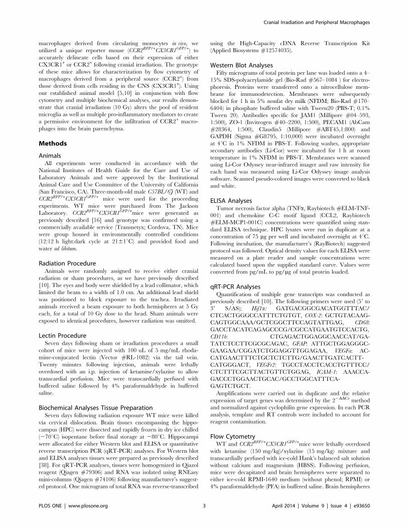
Western Blot AnalysesFifty micrograms of total protein per lane was loaded onto a 4–
15% SDS-polyacrylamide gel (Bio-Rad #567–1084 ) for electro-
phoresis. Proteins were transferred onto a nitrocellulose mem-
brane for immunodetection. Membranes were subsequently
blocked for 1 h in 5% nonfat dry milk (NFDM; Bio-Rad #170–
6404) in phosphate buffered saline with Tween20 (PBS-T; 0.1%
Tween 20). Antibodies specific for JAM1 (Millipore #04–593,
1:500), ZO-1 (Invitrogen #40–2200, 1:500), PECAM1 (AbCam
#28364, 1:500), Claudin5 (Millipore #ABT45,1:800) and
GAPDH (Sigma #G8795, 1:10,000) were incubated overnight
at 4uC in 1% NFDM in PBS-T. Following washes, appropriate
secondary antibodies (Li-Cor) were incubated for 1 h at room
temperature in 1% NFDM in PBS-T. Membranes were scanned
using Li-Cor Odyssey near-infrared imager and raw intensity for
each band was measured using Li-Cor Odyssey image analysis
software. Scanned pseudo-colored images were converted to black
and white.
ELISA AnalysesTumor necrosis factor alpha (TNFa, Raybiotech #ELM-TNF-
001) and chemokine C-C motif ligand (CCL2, Raybiotech
#ELM-MCP1-001C) concentrations were quantified using stan-
dard ELISA technique. HPC lysates were run in duplicate at a
concentration of 75 mg per well and incubated overnight at 4uC.
Following incubation, the manufacturer’s (RayBiotech) suggested
protocol was followed. Optical density values for each ELISA were
measured on a plate reader and sample concentrations were
calculated based upon the supplied standard curve. Values were
converted from pg/mL to pg/mg of total protein loaded.
qRT-PCR AnalysesQuantification of multiple gene transcripts was conducted as
previously described [10]. The following primers were used (59 to
39 S/AS); Hif1a: GATGACGGCGACATGGTTTAC/
CTCACTGGGCCATTTCTGTGT, COX-2: GCTGTACAAG-
CAGTGGCAAA/GCTCGGCTTCCAGTATTGAG, CD68:
GACCTACATCAGAGCCCG/CGCCATGAATGTCCACTG,
CD11b: CTGAGACTGGAGGCAACCAT/GA-
TATCTCCTTCGCGCAGAC, GFAP: ATTGCTGGAGGGC-
GAAGAA/CGGATCTGGAGGTTGGAGAA, VEGFa: AC-
CATGAACTTTCTGCTCTCTTG/GAACTTGATCACTT-
CATGGGACT, VEGFr2: TGCCTACCTCACCTGTTTCC/
CTCTTTCGCTTACTGTTCTGGAG, ICAM-1: AAACCA-
GACCCTGGAACTGCAC/GCCTGGCATTTCA-
GAGTCTGCT.
Amplifications were carried out in duplicate and the relative
expression of target genes was determined by the 22DDCt method
and normalized against cyclophilin gene expression. In each PCR
analysis, template and RT controls were included to account for
reagent contamination.
Flow CytometryWT and CCR2RFP/+CX3CR1GFP/+mice were lethally overdosed
with ketamine (150 mg/kg)/xylazine (15 mg/kg) mixture and
transcardially perfused with ice-cold Hank’s balanced salt solution
without calcium and magnesium (HBSS). Following perfusion,
mice were decapitated and brain hemispheres were separated to
either ice-cold RPMI-1640 medium (without phenol; RPMI) or
4% paraformaldehyde (PFA) in buffered saline. Brain hemispheres
Cranial Irradiation and Peripheral Macrophages
PLOS ONE | www.plosone.org 3 April 2014 | Volume 9 | Issue 4 | e93650

in RMPI were used for microglia/macrophage isolation following
standard procedures [39], while 4% PFA post-fixed tissues were
used for sectioning and imaging (see below). Fc receptor blocking
was performed before all staining procedures using an anti-CD16/
32 antibody (BD Pharmigen #553142). The following reagents
were used for labeling isolated microglia/macrophage: 7AAD
(Sigma-Aldrich #A9400), CD11b Alexafluor 700 (BD Pharmigen
#557960), F4/80 APC (Invitrogen #MF48005). Mandibular
blood draws from naıve CCR2RFP/RFPCX3CR1GFP/GFP mice were
used as positive controls for RFP and GFP expression. Addition-
ally, naıve WT isolated microglia/macrophages served as negative
control for RFP and GFP expression. Spectral compensation was
achieved using polystyrene microparticles (BD Pharmigen
#552845) in combination with each of the above listed conjugated
antibodies following manufacturer’s suggested protocol. Standard
staining procedures were conducted as previously described [39]
before analysis on FACSAria III cell sorter (BD Biosciences). All
samples were diluted 1:10 and run in duplicate.
Brain Tissue Sectioning and ImagingAll brain tissue used for fluorescence imaging was sectioned as
previously described [38]. 40 mm free-floating sections were
mounted onto Superfrost Plus slides (Fisher #12-550-15) and
allowed to dry overnight. Slides were rinsed in buffered saline
solution before counterstaining with 49,6-diamidino-2-phenylin-
dole (DAPI; Invitrogen #D1306) followed by coverslipping in
Vectashield fluorescent mounting medium (Vector #H1000). All
imaging was achieved using a Zeiss Imager.Z1 Apotome
microscope controlled by ZEN software (Zeiss 2012).
Data AnalysisAll data were analyzed using Prism software (v6.0, GraphPad;
La Jolla, CA) and are presented as the mean 6 standard error of
the mean (SEM). Statistical analyses were performed using
ANOVA or Student’s t-test. Pairwise comparisons within ANOVA
were assessed by Tukey’s HSD post hoc multiple comparisons test.
Throughout, p values of ,0.05 were considered significant.
Results
Cranial Irradiation Induces a Protracted Decrease inCD11b+ Myeloid Cells in the Brain
We characterized the response of myeloid cells at 7, 14, and 28
days following ionizing radiation. All mice tolerated cranial
radiation (10 Gy) dosage and gained weight normally over the
duration of the studies (data not shown) as we have previously
reported [5,10]. A single 10 Gy dose of radiation was sufficient to
decrease (F(3,12) = 12.07, p = 0.0006) the proportion of CD11b+
myeloid cells at 7 (p,0.05) and 14 (p,0.01) days after radiation
compared to sham, however this deficit returned to sham levels by
day 28 (Fig. 1A). Interestingly, although there was a decrease in
the overall population of myeloid cells, we observed a significant
increase in the percentage of CD11b+ cells that were F4/80+, a
marker of activated macrophages (F(3,12) = 4.048, p = 0.0335), after
radiation exposure. A pairwise comparison revealed significance
only at the 7-day time point (p,0.05) compared to sham
(Fig. 1B,D). Commensurately, we observed a trend for downreg-
ulation of CD11b+ cells that were F4/802 (Fig. 1C,D).
10 Gy Cranial Irradiation is Sufficient to Induce theMigration of F4/80+CCR2+ Peripheral Macrophages intothe Brain
Recent evidence using a microglia ‘‘death signal’’ model,
wherein all CNS microglia are systematically depleted, suggests
that peripherally derived macrophages infiltrate into the CNS and
replace resident microglia [40]. As defined by the expression of
CCR2, our studies examined if peripherally derived macrophages
infiltrate the CNS following cranial irradiation. Seven days after
irradiation in CCR2RFP/+CX3CR1GFP/+ reporter mice, our data
showed that there is a depletion of CD11b+GFP+ cells (Fig. 2A, p,
0.05); similar to what was observed in WT mice. There was no
change in percentage of activated (F4/80+) CX3CR1+ cells in
irradiated mice compared to sham (Fig. 2B). Additionally, while
there were no differences in the proportion of infiltrated
macrophages (F4/80+CCR2+) in response to cranial irradiation
(Fig. 2C), there was a significant increase (p,0.05) in the
percentage of cells that expressed both CCR2 and CX3CR1
(Fig. 2D,E) seven days after cranial irradiation. Notably, these
double-labeled cells are visible in the dorsal HPC by immunoflu-
orescence (Fig. 2F).
Cranial Radiation Alters Microglia and Astrocyte GeneExpression in the Hippocampus
The hippocampus has been shown to be especially vulnerable to
radiation-induced damage. Because of the observed changes in the
myeloid cell population, we next examined HPC gene expression
for markers of microglia and astrocyte reactivity. In accordance
with our flow cytometry data in both WT and CCR2RFP/
+CX3CR1GFP/+ mice, we observed a significant decrease in
CD11b mRNA expression compared to sham (Fig. 3A, p,0.05).
However, there were no changes induced by radiation exposure to
CD68, a lysosomal marker associated with phagocytic microglia/
macrophages. We next examined CD45, which is a haemotopoetic
marker of monocytes/macrophages but found no difference
initiated by irradiation, although there was a trend for a reduction.
Astrocytes have been shown to contribute to innate immune
response by releasing multiple pro-inflammatory cytokines and
chemokines [41]. Relative gene expression for glial fibrilarly acid
protein (GFAP) was significantly induced in the HPC of irradiated
mice compared to sham (Fig. 3B). However, when we examined
vimentin and S100b as other markers associated with astrocyte
activation we found no differences induced by irradiation.
Cranial Irradiation Induces Pro-inflammatory Response inthe Hippocampus of WT Mice
Given our data showing multiple alterations in both the cell
types and markers associated with innate immune function, we
next examined two pro-inflammatory factors classically associated
with these responses. Specifically, we measured the concentrations
of TNFa and CCL2 from the hippocampus of WT mice seven
days after cranial irradiation. Induction of TNFa has pleiotropic
effects in the CNS, most notably protracted expression of this pro-
inflammatory cytokine has been shown to be neurotoxic in a
variety of animals models of disease. Moreover, increased
expression of the pro-inflammatory chemokine CCL2 has been
shown to chemotactically recruit CCR2+ macrophages to sites of
inflammation [13,42–45]. Herein, our data show that 10 Gy
radiation exposure significantly upregulates the production of both
TNFa (p,0.01) and CCL2 (p,0.001; Fig. 4). Interestingly, we did
not observe any changes in mRNA gene expression of CCL7,
CCL8, or CCL12 in the HPC of irradiated mice compared to
sham (data not shown).
Cranial Irradiation and Peripheral Macrophages
PLOS ONE | www.plosone.org 4 April 2014 | Volume 9 | Issue 4 | e93650

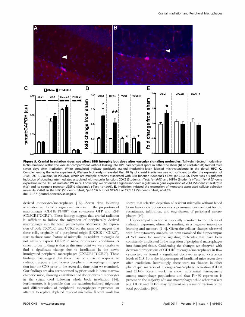
10 Gy Cranial Radiation does not Alter BBB Integrity butdoes Affect Multiple Signaling Cascades Associated withEndothelial Function in the HPCIn vitro and in vivo data suggest that high doses of ionizing
radiation can disrupt the physiological properties of the blood-
brain barrier especially in light of increased CCL2 expression [46–
51]. However, the effect of cranial radiation in vivo is not well
defined. This dosage and time point failed to reveal any vascular
leaking of tail-vein delivered lectin-B4 into the HPC parenchymal
compartment (Fig. 5A/B). We also analyzed multiple molecular
markers associated with the maintenance of BBB tight junctions,
junction adhesion molecule 1 (JAM1), zona occludins 1 (ZO-1),
claudin5, and platelet endothelial cellular adhesion molecule 1
(PECAM), using Western blot technique. At this dosage and time
point there were no significant differences induced by radiation
among any targets (Fig. 5C). Next, we examined multiple signaling
molecules associated with inflammation and altered vascular
response. Specifically, there was a significant induction of
cyclooxygenase 2 (COX2) mRNA in the hippocampus of
irradiated mice compared to sham (Fig. 5D, p,0.05); a similar
increase was found in the expression of hypoxia inducible factor 1
alpha (HIF1a, p,0.01). Importantly, these signaling molecules
have been implicated in the regulation of vasculogenesis [52–56]
by altering the expression of vascular endothelial growth factor
(VEGF) and its cognate receptor (VEGFr2). Our results demon-
strate a significant decrease in both VEGF (p,0.05) and VEGFr2
(p,0.05) mRNA in the hippocampus of irradiated mice compared
to sham. Ionizing radiation and inflammation have been shown to
alter the expression of endothelial-associated adhesion molecules
(e.g. integrins and chemokines), which may influence the arrest,
adhesion, and migration of circulating immune cells (e.g.
macrophages) into tissue compartments [57–59]. Lastly, we
examined the expression of multiple molecules associated with
the recruitment of peripheral macrophages into CNS parenchyma
[58]. Irradiation resulted in a significant increase (p,0.05) in the
expression of ICAM1 in the HPC compared to sham (Fig. 5E).
However, no radiation-induced changes were observed for
Figure 2. Cranial irradiation induces the infiltration of peripherally derived macrophages at seven days. A. The percentage ofCD11b+GFP+ microglial cells was significantly decreased in CCR2RFP/+CX3CR1GFP/+ irradiated mice compared to sham (Student’s t-Test; *p,0.05). B.Cranial irradiation does not affect the percentage of resident macrophages (CD11b+F4/80+CX3CR1+CCR22, Student’s t-Test; p.0.05), C. nor theproportion of peripheral macrophages (CD11b+F4/80+CX3CR12CCR2+, Student’s t-Test; p.0.05); D. but is associated with a significant increase in thepercentage of infiltrated peripherally derived cells that had differentiated into microglia-like macrophages (CD11b+F4/80+CX3CR1+CCR2+, Student’s t-Test; *p,0.05). E. Representative plots showing the radiation-induced accumulation of peripherally derived macrophages into the brain parenchyma.F. Moreover, these cells can be seen in the dentate gyrus of the dorsal hippocampal formation following irradiation. RFP and GFP single pseudo-colored image is converted to black and white to discriminate location in the color co-localized panel. Black arrowheads point to individual positivelylabeled cells for their respective fluorchrome, while white arrowheads show the colocalized cells in the pseudo-colored image.doi:10.1371/journal.pone.0093650.g002
Cranial Irradiation and Peripheral Macrophages
PLOS ONE | www.plosone.org 5 April 2014 | Volume 9 | Issue 4 | e93650

VCAM1 or CXCL12, which are both implicated in differential
recruitment of peripheral monocytes [57,58,60].
Discussion
Recently, we demonstrated that CCR2-deficient mice have
reduced levels of irradiation-induced inflammatory response in the
hippocampus, and improved cognitive function as compared to
wild type animals. These results suggest that CCR2 plays an
important role in radiation-induced hippocampal neuronal
dysfunction [10], either directly or through the modulation of
other pathways. Based on these results, we hypothesized that
cranial irradiation may alter the brain’s microenvironment
sufficiently to permit the infiltration of peripherally derived, pro-
inflammatory CCR2+ macrophages. To test this hypothesis we
used both WT and CCR2RFP/+CX3CR1GFP/+ reporter mice [16] to
examine the effects of cranial irradiation on the brain’s myeloid
cell population; central and peripherally derived. Further, we
examined the expression of multiple inflammation-related signal-
ing molecules in the hippocampus to determine if these changes
may mediate the infiltration of CCR2+ macrophages from
systemic circulation following cranial radiation.
Overall, radiation exposure produced a persistent decrease in
the percentage of CD11b+ microglia in WT mice, which returned
to sham levels by 28 days after irradiation. Previous work in a rat
model of lesioned spinal cord has shown that 7 days following
25 Gy exposure was sufficient to reduce the number of microglia
in the lesioned, non-lesioned, and sham tissues exposed to
radiation [61]. However, to the authors’ best knowledge, these
are the first data showing that cranial irradiation is also capable of
altering the brain’s resident microglia population within a
relatively short time period. Although radiation induced an overall
decrease in the proportion of resident microglia seven days
following exposure, at this time point we observed a significant
increase in the percentage expressing F4/80, a classic marker of
macrophage activation [62]. Given these unexpected results at the
seven-day time point, we extended these parameters to the
CCR2RFP/+CX3CR1GFP/+ mice to be able to fully delineate the
effect of cranial irradiation upon both resident (GFP+) versus
infiltrating (RFP+) microglia/macrophages. Corroborating the
effects we observed in WT mice, we again found a significant
decrease in the percentage of CD11b+GFP+ resident microglia in
the irradiated mice at the seven-day time point.
By using CCR2RFP/+CX3CR1GFP/+ mice, it is possible to
distinguish resident microglia/macrophages from peripherally
Figure 3. Cranial irradiation alters microglia and astrocytemolecular markers of activation in the hippocampus of WTmice. A. Irradiation induced a significant decrease of CD11b geneexpression compared to sham (Student’s t-Test; *p,0.05) but nochanges in the phagocytic or haematopoetic markers CD68 or CD45(Student’s t-Test; p.0.05). B. GFAP expression was significantlyincreased compared to sham (Student’s t-Test; *p,0.05). Conversely,there were no changes in gene expression for alternate markersassociated with astrocyte activation: vimentin and S100b (Student’s t-Test; p.0.05).doi:10.1371/journal.pone.0093650.g003
Figure 4. Cranial irradiation is associated with a significantincrease of pro-inflammatory signaling molecules in thehippocampus. Standard ELISA analysis revealed a significant induc-tion in TNFa (Student’s t-Test; **p,0.01) and CCL2 (Student’s t-Test;***p,0.001) levels in the HPC of WT irradiated mice compared to sham.doi:10.1371/journal.pone.0093650.g004
Cranial Irradiation and Peripheral Macrophages
PLOS ONE | www.plosone.org 6 April 2014 | Volume 9 | Issue 4 | e93650
derived monocytes/macrophages [16]. Seven days following
irradiation we found a significant increase in the proportion of
macrophages (CD11b+F4/80+) that co-express GFP and RFP
(CX3CR1+CCR2+). These findings suggest that cranial radiation
is sufficient to induce the migration of peripherally derived
macrophages into the brain parenchyma. Moreover, the expres-
sion of both CX3CR1 and CCR2 on the same cell suggest that
these cells, originally of a peripheral origin (CX3CR12CCR2+),
start to share some feature of microglia, as resident microglia do
not natively express CCR2 in naıve or diseased conditions. A
caveat to our findings is that at this time point we were unable to
find a significant change due to irradiation in the newly
immigrated peripheral macrophages (CX3CR12CCR2+). These
findings may suggest that there may be an acute response to
radiation exposure that induces peripheral macrophage immigra-
tion into the CNS prior to the seven-day time point we examined.
Our findings are also corroborated by prior work in bone marrow
chimeric mice, showing engraftment of donor-derived monocytes
in the spinal cord following whole body irradiation [14].
Furthermore, it is possible that the radiation-induced migration
and differentiation of peripheral macrophages represents an
attempt to replace depleted resident microglia. Recent work has
shown that selective depletion of resident microglia without blood
brain barrier disruption creates a permissive environment for the
recruitment, infiltration, and engraftment of peripheral macro-
phages [40].
Hippocampal function is especially sensitive to the effects of
radiation exposure, ultimately resulting in a negative impact on
learning and memory [2–4]. Given the cellular changes observed
with flow cytometry analysis, we next examined the hippocampus
of WT mice for multiple signaling molecules that have been
consistently implicated in the migration of peripheral macrophages
into damaged tissue. Confirming the changes we observed with
decreased proportions of CD11b+ microglia/macrophages in flow
cytometry, we found a significant decrease in gene expression
levels of CD11b in the hippocampus of irradiated mice seven days
after irradiation. Interestingly, there were no changes in other
phenotypic markers of microglia/macrophage activation (CD68
and CD45). Recent work has shown substantial heterogeneity
among macrophage populations and that F4/80 expression is
present on the majority of tissue macrophages while other markers
(e.g. CD68 and CD45) may represent only a minor fraction of the
total population [63].
Figure 5. Cranial irradiation does not affect BBB integrity but does alter vascular signaling molecules. Tail-vein injected rhodamine-lectin remained within the vascular compartment without leaking into HPC parenchymal space in either the sham (A) or irradiated (B) treated miceseven days after irradiation. White arrowhead indicate positively stained rhodamine-lectin labeled microvasculature in the dorsal HPC. C.Complementing the lectin experiment, Western blot analysis revealed that 10 Gy of cranial irradiation was not sufficient to alter the expression ofJAM1, ZO-1, Claudin5, or PECAM1, which are multiple proteins associated with BBB function (Student’s t-Test; p.0.05). D. There was a significantinduction of signaling intermediates associated with vascular function: COX2 (Student’s t-Test; *p,0.05) and HIF1a (Student’s t-Test; **p,0.05) geneexpression in the HPC of irradiated WT mice. Conversely, we observed a significant down regulation in gene expression of VEGF (Student’s t-Test; *p,0.05) and its cognate receptor VEGFr2 (Student’s t-Test; *p,0.05). E. Irradiation induced the expression of monocyte associated cellular adhesionmolecule ICAM1 in the HPC (Student’s t-Test; *p,0.05) but not VCAM1 or CXCL12 (Student’s t-Test; p.0.05).doi:10.1371/journal.pone.0093650.g005
Cranial Irradiation and Peripheral Macrophages
PLOS ONE | www.plosone.org 7 April 2014 | Volume 9 | Issue 4 | e93650

Astrocyte activation has been consistently shown to play a role
in the innate immune response associated with various models of
neurodegenerative disease [41,59,64–66]. Our study revealed a
significant increase in GFAP gene expression in the hippocampus
of irradiated mice, but not in vimentin or S100b as other markers
of astrocytosis. Interestingly, we did observe a trend for decreased
expression of S100b in irradiated mice. This decline may be due to
the expression of S100b on oligodendrocytes and their precursors
[67], which undergo apoptosis following irradiation [68].
Brain irradiation can induce the local expression of multiple
cytokines in the rodent brain [7]. In accordance with previous
findings, the present results show a significant induction of the pro-
inflammatory cytokine TNFa and chemokine CCL2 in the
hippocampus of irradiated mice. TNFa expression has pleio-
trophic effects in the CNS, notably it has been shown to
concomitantly induce the expression of CCL2 in astrocytes
[34,69–71]. Moreover, increased expression of CCL2 has consis-
tently been shown to alter the integrity of the blood brain barrier
[48,50,51,72]. In the present study, we did not observe any
radiation-induced effects on the expression of multiple proteins
associated with endothelial tight junction integrity in the
hippocampus. However, others have demonstrated vasculature
vulnerability and rarefaction following irradiation [73–75] and
these changes may be sufficient to induce multiple factors
associated with the recruitment of systemic macrophages into
damaged tissues [57,59,76,77]. Specifically, we observed a
significant increase in ICAM1 hippocampal gene expression.
However, other markers associated with endothelial mediated
macrophage recruitment were not altered at this time point. These
findings are in agreement with the lack of change in the
percentage of peripheral macrophages (CX3CR12CCR2+) at this
time point and may further suggest that the elevated levels of
ICAM1 are a residual response.
Next, we examined upstream signaling mediators of the altered
inflammatory and endothelial response. In the hippocampus of
irradiated WT mice, there was a significant increase in the
expression of two early response genes COX-2 and HIF1a.
Prostaglandins, a COX-2 dependent by-product, have been shown
to induce the expression of HIF1a in normoxic conditions,
perpetuating inflammatory response [78]. Moreover, these two
mediators have been shown to alter the expression of VEGF, a
potent angiogenic factor for endothelial cells [53,79]. Herein, we
showed that both VEGF and its cognate receptor (VEGFr2) gene
expression are significantly downregulated in the irradiated
hippocampus. These findings are in agreement and extend
previous work in a rat model of cranial irradiation that showed
similar reductions in multiple early time points [54]. Taken
together, the increased expression of pro-inflammatory early
response genes COX-2 and HIF1a alongside the concomitant
increase in TNFa and CCL2 may create a feed-forward pro-
inflammatory loop ultimately resulting in hippocampal microvas-
culature rarefaction following radiation exposure. Consequently,
this in turn could explain the increase cellular adhesion molecules
driving the increased migration of peripherally derived macro-
phages to the brain parenchyma.
In conclusion, the present study demonstrates that of the brain’s
innate immune response is particularly vulnerable to cranial
irradiation. Alterations of the brain’s local microenvironment may
be responsible for the recruitment and immigration of peripherally
derived macrophages in an effort to replace apoptotic or
dysfunctional resident microglia. Moreover, the infiltration of
peripheral macrophages seems to be due to chemotactic signaling
and not dependent on the disruption of the BBB. It remains
unclear if peripherally derived macrophages can functionally
substitute for resident microglia. Cumulatively, radiation-induced
alterations in the production of cytokines, chemokines, and growth
factors may contribute to altered endothelial function and
upregulation of adhesion molecules implicated in the recruitment
of peripheral macrophages from systemic circulation. All together,
these data provides novel insight into a potential molecular
mechanism that may contribute to radiation brain-injury. Further
work is still needed to define the role of these infiltrating
macrophages in response to radiotherapy.
Author Contributions
Conceived and designed the experiments: JM NG SR. Performed the
experiments: JM TJ SL. Analyzed the data: JM NG SR. Contributed
reagents/materials/analysis tools: SR NG. Wrote the paper: JM NG SR.
References
1. Abayomi OK (1996) Pathogenesis of irradiation-induced cognitive dysfunction.
Acta Oncol 35: 659–663.
2. Rola R, Raber J, Rizk A, Otsuka S, VandenBerg SR, et al. (2004) Radiation-
induced impairment of hippocampal neurogenesis is associated with cognitive
deficits in young mice. Experimental neurology 188: 316–330.
3. Mizumatsu S, Monje ML, Morhardt DR, Rola R, Palmer TD, et al. (2003)
Extreme Sensitivity of Adult Neurogenesis to Low Doses of X-Irradiation.
Cancer research 63: 4021.
4. Monje ML, Mizumatsu S, Fike JR, Palmer TD (2002) Irradiation induces neural
precursor-cell dysfunction. Nat Med 8: 955–962.
5. Rosi S, Andres-Mach M, Fishman KM, Levy W, Ferguson RA, et al. (2008)
Cranial irradiation alters the behaviorally induced immediate-early gene arc
(activity-regulated cytoskeleton-associated protein). Cancer Res 68: 9763–9770.
6. Moravan MJ, Olschowka JA, Williams JP, O’Banion MK (2011) Cranial
irradiation leads to acute and persistent neuroinflammation with delayed
increases in T-cell infiltration and CD11c expression in C57BL/6 mouse brain.
Radiation research 176: 459–473.
7. Linard C, Marquette C, Mathieu J, Pennequin A, Clarencon D, et al. (2004)
Acute induction of inflammatory cytokine expression after gamma-irradiation in
the rat: effect of an NF-kappaB inhibitor. Int J Radiat Oncol Biol Phys 58: 427–
434.
8. Rosi S, Ferguson R, Fishman K, Allen A, Raber J, et al. (2012) The polyamine
inhibitor alpha-difluoromethylornithine modulates hippocampus-dependent
function after single and combined injuries. PLoS One 7: e31094.
9. Monje ML, Toda H, Palmer TD (2003) Inflammatory blockade restores adult
hippocampal neurogenesis. Science 302: 1760–1765.
10. Belarbi K, Jopson T, Arellano C, Fike JR, Rosi S (2013) CCR2 deficiency
prevents neuronal dysfunction and cognitive impairments induced by cranial
irradiation. Cancer Res 73: 1201–1210.
11. Banisadr G, Gosselin RD, Mechighel P, Rostene W, Kitabgi P, et al. (2005)
Constitutive neuronal expression of CCR2 chemokine receptor and its
colocalization with neurotransmitters in normal rat brain: functional effect of
MCP-1/CCL2 on calcium mobilization in primary cultured neurons. J Comp
Neurol 492: 178–192.
12. Stamatovic SM, Shakui P, Keep RF, Moore BB, Kunkel SL, et al. (2005)
Monocyte chemoattractant protein-1 regulation of blood-brain barrier perme-
ability. J Cereb Blood Flow Metab 25: 593–606.
13. Semple BD, Bye N, Rancan M, Ziebell JM, Morganti-Kossmann MC (2010)
Role of CCL2 (MCP-1) in traumatic brain injury (TBI): evidence from severe
TBI patients and CCL22/2 mice. J Cereb Blood Flow Metab 30: 769–782.
14. Mildner A, Schmidt H, Nitsche M, Merkler D, Hanisch UK, et al. (2007)
Microglia in the adult brain arise from Ly-6ChiCCR2+ monocytes only under
defined host conditions. Nat Neurosci 10: 1544–1553.
15. Varol C, Yona S, Jung S (2009) Origins and tissue-context-dependent fates of
blood monocytes. Immunology and cell biology 87: 30–38.
16. Saederup N, Cardona AE, Croft K, Mizutani M, Cotleur AC, et al. (2010)
Selective Chemokine Receptor Usage by Central Nervous System Myeloid Cells
in CCR2-Red Fluorescent Protein Knock-In Mice. PloS one 5: e13693.
17. Mizutani M, Pino PA, Saederup N, Charo IF, Ransohoff RM, et al. (2012) The
fractalkine receptor but not CCR2 is present on microglia from embryonic
development throughout adulthood. J Immunol 188: 29–36.
18. Auffray C, Fogg D, Garfa M, Elain G, Join-Lambert O, et al. (2007) Monitoring
of blood vessels and tissues by a population of monocytes with patrolling
behavior. Science 317: 666–670.
Cranial Irradiation and Peripheral Macrophages
PLOS ONE | www.plosone.org 8 April 2014 | Volume 9 | Issue 4 | e93650

19. Geissmann F, Jung S, Littman DR (2003) Blood monocytes consist of two
principal subsets with distinct migratory properties. Immunity 19: 71–82.
20. Swirski FK, Libby P, Aikawa E, Alcaide P, Luscinskas FW, et al. (2007) Ly-6Chi
monocytes dominate hypercholesterolemia-associated monocytosis and give rise
to macrophages in atheromata. The Journal of clinical investigation 117: 195–
205.
21. Prinz M, Priller J (2010) Tickets to the brain: role of CCR2 and CX3CR1 in
myeloid cell entry in the CNS. J Neuroimmunol 224: 80–84.
22. Getts DR, Terry RL, Getts MT, Muller M, Rana S, et al. (2008) Ly6c+‘‘inflammatory monocytes’’ are microglial precursors recruited in a pathogenic
manner in West Nile virus encephalitis. J Exp Med 205: 2319–2337.
23. Serbina NV, Pamer EG (2006) Monocyte emigration from bone marrow during
bacterial infection requires signals mediated by chemokine receptor CCR2. Nat
Immunol 7: 311–317.
24. Auffray C, Sieweke MH, Geissmann F (2009) Blood monocytes: development,
heterogeneity, and relationship with dendritic cells. Annu Rev Immunol 27:
669–692.
25. Gordon S, Taylor PR (2005) Monocyte and macrophage heterogeneity. Nat Rev
Immunol 5: 953–964.
26. Naert G, Rivest S (2012) Age-related changes in synaptic markers and monocyte
subsets link the cognitive decline of APP(Swe)/PS1 mice. Front Cell Neurosci 6:
51.
27. Mildner A, Mack M, Schmidt H, Bruck W, Djukic M, et al. (2009) CCR2+Ly-
6Chi monocytes are crucial for the effector phase of autoimmunity in the central
nervous system. Brain 132: 2487–2500.
28. Westin K, Buchhave P, Nielsen H, Minthon L, Janciauskiene S, et al. (2012)
CCL2 is associated with a faster rate of cognitive decline during early stages of
Alzheimer’s disease. PLoS One 7: e30525.
29. Naert G, Rivest S (2011) CC chemokine receptor 2 deficiency aggravates
cognitive impairments and amyloid pathology in a transgenic mouse model of
Alzheimer’s disease. J Neurosci 31: 6208–6220.
30. Naert G, Rivest S (2012) Hematopoietic CC-chemokine receptor 2 (CCR2)
competent cells are protective for the cognitive impairments and amyloid
pathology in a transgenic mouse model of Alzheimer’s disease. Mol Med 18:
297–313.
31. El Khoury J, Toft M, Hickman SE, Means TK, Terada K, et al. (2007) Ccr2
deficiency impairs microglial accumulation and accelerates progression of
Alzheimer-like disease. Nat Med 13: 432–438.
32. Mildner A, Schlevogt B, Kierdorf K, Bottcher C, Erny D, et al. (2011) Distinct
and non-redundant roles of microglia and myeloid subsets in mouse models of
Alzheimer’s disease. J Neurosci 31: 11159–11171.
33. Schilling M, Strecker J-K, Ringelstein EB, Schabitz W-R, Kiefer R (2009) The
role of CC chemokine receptor 2 on microglia activation and blood-borne cell
recruitment after transient focal cerebral ischemia in mice. Brain research 1289:
79–84.
34. Zuurman MW, Heeroma J, Brouwer N, Boddeke HWGM, Biber K (2003) LPS-
induced expression of a novel chemokine receptor (L-CCR) in mouse glial cells
in vitro and in vivo. Glia 41: 327–336.
35. Olah M, Amor S, Brouwer N, Vinet J, Eggen B, et al. (2012) Identification of a
microglia phenotype supportive of remyelination. Glia 60: 306–321.
36. Prinz M, Mildner A (2011) Microglia in the CNS: immigrants from another
world. Glia 59: 177–187.
37. Kim SH, Lim DJ, Chung YG, Cho TH, Lim SJ, et al. (2002) Expression of
TNF-alpha and TGF-beta 1 in the rat brain after a single high-dose irradiation.
J Korean Med Sci 17: 242–248.
38. Morganti JM, Nash KR, Grimmig BA, Ranjit S, Small B, et al. (2012) The
soluble isoform of CX3CL1 is necessary for neuroprotection in a mouse model
of Parkinson’s disease. J Neurosci 32: 14592–14601.
39. Cardona AE, Huang D, Sasse ME, Ransohoff RM (2006) Isolation of murine
microglial cells for RNA analysis or flow cytometry. Nat Protoc 1: 1947–1951.
40. Varvel NH, Grathwohl SA, Baumann F, Liebig C, Bosch A, et al. (2012)
Microglial repopulation model reveals a robust homeostatic process for replacing
CNS myeloid cells. Proc Natl Acad Sci U S A 109: 18150–18155.
41. Ransohoff RM, Brown MA (2012) Innate immunity in the central nervous
system. J Clin Invest 122: 1164–1171.
42. Andres RH, Choi R, Pendharkar AV, Gaeta X, Wang N, et al. (2011) The
CCR2/CCL2 interaction mediates the transendothelial recruitment of intra-
vascularly delivered neural stem cells to the ischemic brain. Stroke 42: 2923–
2931.
43. Chen M, Zhao J, Luo C, Pandi SP, Penalva RG, et al. (2012) Para-
inflammation-mediated retinal recruitment of bone marrow-derived myeloid
cells following whole-body irradiation is CCL2 dependent. Glia 60: 833–842.
44. Szmydynger-Chodobska J, Strazielle N, Gandy JR, Keefe TH, Zink BJ, et al.
(2012) Posttraumatic invasion of monocytes across the blood-cerebrospinal fluid
barrier. J Cereb Blood Flow Metab 32: 93–104.
45. Semple BD, Kossmann T, Morganti-Kossmann MC (2010) Role of chemokines
in CNS health and pathology: a focus on the CCL2/CCR2 and CXCL8/
CXCR2 networks. J Cereb Blood Flow Metab 30: 459–473.
46. Nordal RA, Wong CS (2005) Molecular targets in radiation-induced blood-brain
barrier disruption. Int J Radiat Oncol Biol Phys 62: 279–287.
47. Fauquette W, Amourette C, Dehouck MP, Diserbo M (2012) Radiation-induced
blood-brain barrier damages: an in vitro study. Brain Res 1433: 114–126.
48. Dimitrijevic OB, Stamatovic SM, Keep RF, Andjelkovic AV (2006) Effects of
the chemokine CCL2 on blood-brain barrier permeability during ischemia-
reperfusion injury. J Cereb Blood Flow Metab 26: 797–810.
49. Stamatovic SM, Sladojevic N, Keep RF, Andjelkovic AV (2012) Relocalization
of junctional adhesion molecule A during inflammatory stimulation of brain
endothelial cells. Mol Cell Biol 32: 3414–3427.
50. Roberts TK, Eugenin EA, Lopez L, Romero IA, Weksler BB, et al. (2012) CCL2
disrupts the adherens junction: implications for neuroinflammation. Lab Invest
92: 1213–1233.
51. Schellenberg AE, Buist R, Del Bigio MR, Toft-Hansen H, Khorooshi R, et al.
(2012) Blood-brain barrier disruption in CCL2 transgenic mice during pertussis
toxin-induced brain inflammation. Fluids and Barriers of the CNS 9: 10.
52. Wu G, Luo J, Rana JS, Laham R, Sellke FW, et al. (2006) Involvement of COX-
2 in VEGF-induced angiogenesis via P38 and JNK pathways in vascular
endothelial cells. Cardiovasc Res 69: 512–519.
53. Ferrara N, Gerber HP, LeCouter J (2003) The biology of VEGF and its
receptors. Nat Med 9: 669–676.
54. Lee WH, Cho HJ, Sonntag WE, Lee YW (2011) Radiation attenuates
physiological angiogenesis by differential expression of VEGF, Ang-1, tie-2
and Ang-2 in rat brain. Radiat Res 176: 753–760.
55. Kioi M, Vogel H, Schultz G, Hoffman RM, Harsh GR, et al. (2010) Inhibition
of vasculogenesis, but not angiogenesis, prevents the recurrence of glioblastoma
after irradiation in mice. The Journal of clinical investigation 120: 694–705.
56. Lerman OZ, Greives MR, Singh SP, Thanik VD, Chang CC, et al. (2010) Low-
dose radiation augments vasculogenesis signaling through HIF-1-dependent and
-independent SDF-1 induction. Blood 116: 3669–3676.
57. Shi C, Pamer EG (2011) Monocyte recruitment during infection and
inflammation. Nature reviews Immunology 11: 762–774.
58. Takeshita Y, Ransohoff RM (2012) Inflammatory cell trafficking across the
blood-brain barrier: chemokine regulation and in vitro models. Immunol Rev
248: 228–239.
59. Zlokovic BV (2008) The blood-brain barrier in health and chronic neurode-
generative disorders. Neuron 57: 178–201.
60. Man S, Tucky B, Cotleur A, Drazba J, Takeshita Y, et al. (2012) CXCL12-
Induced Monocyte-Endothelial Interactions Promote Lymphocyte Transmigra-
tion Across an in Vitro Blood-Brain Barrier. Science Translational Medicine 4:
119ra114–119ra114.
61. Gilmore S (2003) Radiation-induced modulation of the microglial population in
the normal and injured mature spinal cord. Experimental neurology 182: 169–
179.
62. Austyn JM, Gordon S (1981) F4/80, a monoclonal antibody directed specifically
against the mouse macrophage. Eur J Immunol 11: 805–815.
63. Lloyd CM, Phillips ARJ, Cooper GJS, Dunbar PR (2008) Three-colour
fluorescence immunohistochemistry reveals the diversity of cells staining for
macrophage markers in murine spleen and liver. Journal of immunological
methods 334: 70–81.
64. Farina C, Aloisi F, Meinl E (2007) Astrocytes are active players in cerebral innate
immunity. Trends Immunol 28: 138–145.
65. Ridet JL, Malhotra SK, Privat A, Gage FH (1997) Reactive astrocytes: cellular
and molecular cues to biological function. Trends Neurosci 20: 570–577.
66. Lee SC, Liu W, Dickson DW, Brosnan CF, Berman JW (1993) Cytokine
production by human fetal microglia and astrocytes. Differential induction by
lipopolysaccharide and IL-1 beta. J Immunol 150: 2659–2667.
67. Steiner J, Bernstein HG, Bogerts B, Gos T, Richter-Landsberg C, et al. (2008)
S100B is expressed in, and released from, OLN-93 oligodendrocytes: Influence
of serum and glucose deprivation. Neuroscience 154: 496–503.
68. Panagiotakos G, Alshamy G, Chan B, Abrams R, Greenberg E, et al. (2007)
Long-Term Impact of Radiation on the Stem Cell and Oligodendrocyte
Precursors in the Brain. PloS one 2: e588.
69. Brambilla R, Bracchi-Ricard V, Hu WH, Frydel B, Bramwell A, et al. (2005)
Inhibition of astroglial nuclear factor kappaB reduces inflammation and
improves functional recovery after spinal cord injury. J Exp Med 202: 145–156.
70. Babcock AA, Kuziel WA, Rivest S, Owens T (2003) Chemokine expression by
glial cells directs leukocytes to sites of axonal injury in the CNS. J Neurosci 23:
7922–7930.
71. Semple BD, Frugier T, Morganti-Kossmann MC (2010) CCL2 modulates
cytokine production in cultured mouse astrocytes. J Neuroinflammation 7: 67.
72. Mahad D, Callahan MK, Williams KA, Ubogu EE, Kivisakk P, et al. (2006)
Modulating CCR2 and CCL2 at the blood-brain barrier: relevance for multiple
sclerosis pathogenesis. Brain 129: 212–223.
73. Ljubimova NV, Levitman MK, Plotnikova ED, Eidus LK (1991) Endothelial cell
population dynamics in rat brain after local irradiation. The British journal of
radiology 64: 934–940.
74. Nguyen V, Gaber MW, Sontag MR, Kiani MF (2000) Late effects of ionizing
radiation on the microvascular networks in normal tissue. Radiation research
154: 531–536.
75. Roth NM, Sontag MR, Kiani MF (1999) Early effects of ionizing radiation on
the microvascular networks in normal tissue. Radiation research 151: 270–277.
76. Wu K-L, Tu B, Li Y-Q, Wong CS (2010) Role of Intercellular Adhesion
Molecule-1 in Radiation-Induced Brain Injury. International Journal of
Radiation Oncology*Biology*Physics 76: 220–228.
77. Ransohoff RM, Cardona AE (2010) The myeloid cells of the central nervous
system parenchyma. Nature 468: 253–262.
Cranial Irradiation and Peripheral Macrophages
PLOS ONE | www.plosone.org 9 April 2014 | Volume 9 | Issue 4 | e93650

78. Stasinopoulos I, O’Brien DR, Bhujwalla ZM (2009) Inflammation, but not
hypoxia, mediated HIF-1alpha activation depends on COX-2. Cancer biology &
therapy 8: 31–35.
79. Lund EL, H g A, Olsen MWB, Hansen LT, Engelholm SA, et al. (2004)
Differential regulation of VEGF, HIF1a and angiopoietin-1, -2 and -4 byhypoxia and ionizing radiation in human glioblastoma. International Journal of
Cancer 108: 833–838.
Cranial Irradiation and Peripheral Macrophages
PLOS ONE | www.plosone.org 10 April 2014 | Volume 9 | Issue 4 | e93650