Presence of Tumor DNA in Plasma of Breast Cancer Patients: Clinicopathological Correlations1
Upload
khangminh22Category
view
2download
0
LUND UNIVERSITY
PO Box 117221 00 Lund+46 46-222 00 00
Growth factor signaling in the breast tumor microenvironment
Bocci, Matteo
2018
Document Version:Publisher's PDF, also known as Version of record
Link to publication
Citation for published version (APA):Bocci, M. (2018). Growth factor signaling in the breast tumor microenvironment. Lund University: Faculty ofMedicine.
Total number of authors:1
General rightsUnless other specific re-use rights are stated the following general rights apply:Copyright and moral rights for the publications made accessible in the public portal are retained by the authorsand/or other copyright owners and it is a condition of accessing publications that users recognise and abide by thelegal requirements associated with these rights. • Users may download and print one copy of any publication from the public portal for the purpose of private studyor research. • You may not further distribute the material or use it for any profit-making activity or commercial gain • You may freely distribute the URL identifying the publication in the public portal
Read more about Creative commons licenses: https://creativecommons.org/licenses/Take down policyIf you believe that this document breaches copyright please contact us providing details, and we will removeaccess to the work immediately and investigate your claim.

Growth factor signaling in the breast tumor microenvironmentMATTEO BOCCI
DEPARTMENT OF LABORATORY MEDICINE | FACULTY OF MEDICINE | LUND UNIVERSITY

Department of Laboratory Medicine
Lund University, Faculty of Medicine Doctoral Dissertation Series 2018:129
ISBN 978-91-7619-697-7 ISSN 1652-8220 9
789176
196977
Prin
ted
by M
edia
-Try
ck, L
und
2018
N
ORD
IC S
WA
N E
CO
LABE
L 3
041
0903

Growth factor signaling in the breast tumor microenvironment
Matteo Bocci
DOCTORAL DISSERTATION by due permission of the Faculty of Medicine, Lund University, Sweden.
To be defended at the main lecture hall, Medicon Village, Lund.
Friday, 16th November at 09:30 AM.
Faculty opponent Professor Morag Park
Rosalind & Morris Goodman Cancer Research Centre Departments of Biochemistry, Medicine and Oncology
McGill University, Montreal, Canada

Organization LUND UNIVERSITY
Document name Doctoral dissertation
Faculty of Medicine Translational Cancer Research
Date of issue 16 Novmber, 2018
Author: Matteo Bocci Sponsoring organization: NA
Title and subtitle Growth factor signaling in the breast tumor microenvironment Abstract Cancer represents a collection of malignancies characterized by an aberrant expansion of cells. This unrestrained growth is the result of the acquisition of several pro-survival features and the evasion of cellular fail-safe mechanisms, collectively known as the hallmarks of cancer. In the clinical setting, disease management has heavily relied on the sole targeting of malignant cells but, except for rare cases, monotherapy regimens showed insufficient antitumor activity. Indeed, translational and clinical studies revealed that cancer cells almost invariably adapt to treatment, mainly through acquisition of additional (epi)mutations and/or clonal diversification and activation of bypass signaling pathways. In parallel, characterization of the malignant mass exposed the existence of other (non-transformed) cell types and non-cellular constituents, with specialized functions and potentially different origins, jointly reffered to as the tumor stroma. This local microenvironment is educated by and coevolves with the cancer cells by engaging in an intricate network of communication that plays a fundamental role in the establishment, progression and malignization of a tumor, as well as modulating the response to treatment. The stroma comprises the endothelial cells and pericytes that compose the vasculature, fibroblasts, immune cells and the extracellular matrix. Therefore, the genetic make-up of polyclonal tumors and the composition of the microenvironment define the genomic, spatial and functional diversity of each tumor, also at the metastatic site. In agreement with this, the concept of intratumoral heterogeneity denotes a key aspect that has been increasingly recognized, although not fully implemented, in personalized medicine. Moreover, recent efforts have started to address the systemic changes instigated by the tumor mass –including metabolism– and how these influence the survival/dormancy, the colonization and the metastatic growth of disseminated cancer cells. In the papers included in this thesis, we made use of experimental breast cancer models to deepen our understanding of the tumor milieu and its clinical implications. Paper I reports the results of the preclinical trials of a compound that was designed to block activin receptor-like kinase (ALK)1, a protein involved in the formation of the blood vessels. Experimental models showed promising inhibition of tumor growth and marked reduction of the metastatic disease. In paper II, we analyzed how ALK1 communicates in different tumors in order to determine a set of characteristics that might help to predict which patients could benefit from ALK1-blocking therapy. Moreover, we discovered that the presence of ALK1 in tumor blood vessels influences the presence and function of the immune cells. In paper III, we define a novel therapeutic opportunity for the basal subtype of breast cancer, for which only surgery, radio- and chemotherapy are currently available. We identified the specific role of PDGF-C, that is released by tumor cells to activate fibroblasts. This communication loop maintains the tumor cells in a more aggressive state and makes them resistant to treatment. Thus, by blocking PDGF-C, tumor cells transform to a less aggressive luminal type and become sensitive to endocrine therapy, which can be used to limit the development of the tumor mass. Finally, paper IV gives us information about the diversity of the cells within the fibroblast population. By using a state of the art technology, we increased the resolution at which we are able to distinguish the function of each individual fibroblast isolated from a tissue, and match it with a specific cell-of-origin. Taken together, the use of mouse models of cancer allows us to reproduce the complexity of human tumors, and delineate how these cellular relationships are shaped and maintained during tumor development. Our data illustrate the value of impinging on the crosstalk between tumor cells and other components of the tumor mass to develop novel therapeutic strategies for the clinical management of breast cancer. Key words: Breast cancer, tumor microenvironment, angiogenesis, cancer-associated fibroblast, PDGF-C, ALK1
Classification system and/or index terms (if any): NA
Supplementary bibliographical information: NA Language: English
ISSN and key title: 1652-8220 Lund University, Faculty of Medicine Doctoral Dissertation series 2018:129
ISBN: 978-91-7619-697-7
Recipient’s notes: NA Number of pages Price: NA
Security classification: NA
I, the undersigned, being the copyright owner of the abstract of the above-mentioned dissertation, hereby grant to all reference sources permission to publish and disseminate the abstract of the above-mentioned dissertation.
Signature Date

Growth factor signaling in the breast tumor microenvironment
Matteo Bocci

Cover photo by Inês Bravo: Multiculturalism and integration - cancer edition.
Copyright Matteo Bocci
Faculty of Medicine Department of Laboratory Medicine
Lund University, Faculty of Medicine, Doctoral Dissertation Series 2018:129 ISBN 978-91-7619-697-7 ISSN 1652-8220
Printed in Sweden by Media-Tryck, Lund University, Lund 2018

To Roberta, “il mio turchese”
“And once the storm is over, you won’t even remember how you made it through, how you managed to survive. You won’t even be sure, whether the storm is really over. But one
thing is certain. When you come out of the storm, you won’t be the same person who walked in. That’s what this storm’s all about”.
Haruki Murakami – Kafka on the shore
“Some natural tears they dropped, but wiped them soon; The world was all before them, where to choose
their place of rest, and Providence their guide They, hand in hand, with wandering steps and slow,
Through Eden took their solitary way”. John Milton – Paradise lost


Table of Contents
List of original papers ............................................................................................... 9 Abbreviations ......................................................................................................... 11 Acknowledgements ................................................................................................ 17 Popular science summary ....................................................................................... 23 Sintesi scientifica a scopo divulgativo ................................................................... 25 Abstract .................................................................................................................. 27 The tumor microenvironment ................................................................................. 29
Mesenchymal cells ....................................................................................... 31 Cancer-associated fibroblasts .............................................................. 31 Pericytes .............................................................................................. 36
Vasculature ................................................................................................... 40 Endothelial cells .................................................................................. 40 Tumor angiogenesis ............................................................................ 41
Immune cells ................................................................................................ 46 Cancer and immunoescape .................................................................. 46
Targeting the microenvironment .................................................................. 48 Antiangiogenic therapy ....................................................................... 48 Immunotherapy ................................................................................... 50
Growth factor signaling .......................................................................................... 53 Transforming growth factor β ...................................................................... 53
Role of the TGF-β family signaling in homeostasis and cancer ......... 55 ALK1 and vascular development ........................................................ 56 Development of ALK1-blocking agents ............................................. 59
Platelet-derived growth factor ...................................................................... 61 Role of PDGF signaling ...................................................................... 63 PDGF-CC ............................................................................................ 65
Breast cancer .......................................................................................................... 67 Epidemiology and etiology .......................................................................... 67
Risk factors .......................................................................................... 67

Breast development ...................................................................................... 68 Mammary stem cells ........................................................................... 70
Classification ................................................................................................ 71 Histopathological analysis ................................................................... 71 Tumor grade and staging ..................................................................... 73 Intrinsic molecular subtyping .............................................................. 74
Treatment ..................................................................................................... 76 Surgery and radiation therapy ............................................................. 76 Chemotherapy ..................................................................................... 77 Endocrine therapy ............................................................................... 79 Targeted therapy .................................................................................. 80 Novel therapeutic opportunities .......................................................... 83
Mouse models of breast cancer .............................................................................. 85 Environmentally-induced models ................................................................ 85 Transplantable models ................................................................................. 86
Syngeneic models ................................................................................ 87 Xenogeneic models ............................................................................. 87 Humanized mice .................................................................................. 89
Genetically engineered mouse models ......................................................... 89 Conventional GEMMs ........................................................................ 90 Conditional and inducible GEMMs .................................................... 92 Genome editing ................................................................................... 94
Present investigation ............................................................................................... 97 Paper I .......................................................................................................... 97 Paper II ......................................................................................................... 98 Paper III ........................................................................................................ 98 Paper IV ..................................................................................................... 100 Discussion .................................................................................................. 101
Paper I and II ..................................................................................... 101 Paper III and IV: ................................................................................ 107
Conclusions and future perspectives .......................................................... 113 References ............................................................................................................ 117

9
List of original papers
This thesis is based on the following papers, which are referred to in the text by Roman numerals.
I. Endothelial ALK1 is a therapeutic target to block metastatic disseminationof breast cancer.Cunha SI, Bocci M, Lövrot J, Eleftheriou NM, Roswall P, Cordero E,Lindström L, Bartoschek M, Haller BK, Pearsall RS, Mulivor AW, KumarR, Larsson C, Bergh J, Pietras K.Cancer Res. 2015 Jun 15;75(12):2445-56.
II. Activin receptor-like kinase 1 is associated with immune cell infiltrationand regulates CLEC14A transcription in cancer.Bocci M, Sjölund J, Kurzejamska E, Lindgren D, Marzouka NA,Bartoschek M, Höglund M, Pietras K.Angiogenesis , 2018 Aug 21 (Epub ahead of print)
III. Microenvironmental control of breast cancer subtype elicited throughparacrine platelet-derived growth factor-CC signaling.Roswall P*, Bocci M*, Bartoschek M*, Li H*, Kristiansen G, Jansson S,Lehn S, Sjölund J, Reid S, Larsson C, Eriksson P, Anderberg C, Cortez E,Saal LH, Orsmark-Pietras C, Cordero E, Haller BK, Häkkinen J,Burvenich IJG, Lim E, Orimo A, Höglund M, Rydén L, Moch H, ScottAM, Eriksson U, Pietras K.Nat Med. 2018 May;24(4);463-473.
IV. Spatially and functionally distinct subclasses of breast cancer-associatedfibroblasts revealed by single cell RNA sequencing.Bartoschek M, Oskolkov N, Bocci M, Lövrot J, Larsson C, Sommarin M,Madsen CD, Lindgren D, Karlsson G, Rignér M, Bergh J, Björklund Å,Pietras K.Manuscript resubmitted to Nature Communications.
The star (*) indicates equal contribution
Reprints were made with permission from the publishers

10
Papers not included in the thesis
1. Compound genetically engineered mouse models of cancer reveal dualtargeting of ALK1 and endoglin as a synergistic opportunity to impingeon angiogenic TGF-β signalling.Eleftheriou NM, Sjölund J, Bocci M, Cortez E, Lee SJ, Cunha SI, PietrasK.Oncotarget. 2016 Dec 20;7(51):84214-84325.
2. Targeting tumor vasculature by inhibiting activin receptor-like kinase(ALK1) function.De Vinuesa AG*, Bocci M*, Pietras K, Ten Dijke P.Biochem Soc Trans. 2016 Aug 15;44(4):1142-9. (Review)
3. Functional malignant cell heterogeneity in pancreatic neuroendocrinetumors revealed by targeting of PDGF-DD.Cortez E, Gladh H, Braun S, Bocci M, Cordero E, Björkström NK,Miyazaki H, Michael IP, Eriksson U, Folestad E, Pietras K.Proc Natl Acad U S A, 2016 Feb 16 ;113(7):E864-73.
The star (*) indicates equal contribution

11
Abbreviations
ACTA2 Actin, alpha 2, smooth muscle, aorta ACVRL1 Activin A receptor like type 1 ADAM A disintegrin and metalloproteinase domain ADF Adipocyte-derived fibroblast AKT AKT serine/threonine kinase 1 AI Aromatase inhibitor ALK Activin receptor-like kinase ANG Angiopoietin APC Antigen presenting cell AR Androgen receptor ASCL1 Achaete-scute homolog 1 ASLV-A Avian sarcoma-leukosis virus subgroup A ATM Ataxia telangiectasia mutated serine/threonine kinase ATP Adenosine triphosphate BBB Blood-brain barrier BEC Blood endothelial cell BM Basement membrane BMP Bone morphogenetic protein BRCA1/2 Breast cancer susceptibility gene 1/2 CAA Cancer-associated adipocyte CAF Cancer-associated fibroblast CAR Chimeric antigen receptor CAS9 CRISPR associated protein 9 CAV Caveolin CCL Chemokine (C-C motif) ligand CD Cluster of differentiation CDH Cadherin CDK Cyclin-dependent kinase CDKN2A/B CDK inhibitor 2A/B CDX Cell-derived xenograft CHEK2 Checkpoint kinase 2 ChIP-seq Chromatin immunoprecipitation with parallel DNA sequencing CNS Central nervous system COL Collagen

12
COUP-TFII Chicken ovalbumin upstream promoter transcription factor 2 c/pCR Clinical/pathological complete response CRISPR Clustered regularly interspaced short palindromic repeats CRE Causes recombination/Cyclization recombinase CSC Cancer stem cell CSPG4 Chondroitin sulfate proteoglycan 4 CTLA-4 Cytotoxic T lymphocyte antigen 4 CUB Complement C1r/C1s, Uegf, Bmp1 CXC(L/R) Chemokine (C-X-C motif) ligand/receptor c-MET MET proto-oncogene, receptor tyrosine kinase DCIS Ductal carcinoma in situ DCN Decorin DLL4 Delta-like protein 4 DMBA Dimethylbenz[a]anthracene DNA Deoxyribonucleic acid DSB Double strand break EBF2 Early B cell factor 2 ECM Extracellular matrix EEndT Epithelial-to-endothelial transition EGF(R) Epidermal growth factor (receptor) EMA European medicines agency EMT Epithelial-to-mesenchymal transition EndMT Endothelial-to-mesenchymal transition EN1 Engrailed homeobox 1 EPC Endothelial progenitor cell EpCAM Epithelial cell adhesion molecule ER Estrogen receptor ERBB2 Erb-b2 receptor tyrosine kinase 2 (HER2) ERE Estrogen responsive element ERK Extracellular signal-regulated kinase ESC Embryonic stem cell ESR1 Estrogen receptor 1 FAP Fibroblast activation protein FDA Food and drug administration FGF-2 Fibroblast growth factor 2 (bFGF) FIH Factor inhibiting HIF FKBP12 FK506-binding protein 12 FLP Flippase FOX Forkhead box FRT Flp recombinase target FSP-1 Fibroblast specific protein 1 F4/80 EGF-like module-containing mucin-like hormone-receptor-like 1

13
GAG Glycosaminoglycan GATA3 GATA binding protein 3 GEMM Genetically engineered mouse model GoF Gain-of-function GM-CSF Granulocyte-macrophage colony stimulating factor GPR77 G-protein coupled receptor 77GSEA Gene set enrichment analysisHDR Homology-directed repairHER2 Human epidermal growth factor receptor 2HGF Hepatocyte growth factorHHT Human hereditary telangiectasiaHIF-1α Hypoxia inducible factor 1 subunit αHLA Human leukocyte antigenHR Hormone receptorH-RAS Harvey rat sarcoma virus oncogeneHSC Hematopoietic stem cellID Inhibitor of differentiationIFN InterferonIFP Interstitial fluid pressureIGF Insulin-like growth factorIGFBP IGF binding proteinIHC ImmunohistochemistryIL InterleukinISH In situ hybridizationJAG1 Jagged 1JARID1 Jumonji, AT rich interactive domain 1 (lysine demethylase 5B)LCIS Lobular carcinoma in situLEC Lymphatic endothelial cellsLoF Loss-of-functionLOX Lysyl oxidaseLOXP Locus of X-over P1LSD1 Lysine-specific demethylase 1LTR Long terminal repeatLYVE-1 Lymphatic vessel endothelial hyaluronan receptor 1L1CAM L1 cell adhesion moleculeMAPK Mitogen-activated protein kinaseMDSC Myeloid-derived suppressor cellmiRNA MicroRNAMMP Matrix metalloproteinaseMMTV Mouse mammary tumor virusMPA Medroxyprogesterone acetateMSC Mesenchymal stem cell

14
mTOR Mammalian target of rapamycin NACT Neoadjuvant chemotherapy NBN Nibrin NF-κB Nuclear factor κ-light-chain-enhancer of activated B cells NF1 Neurofibromin 1 NG2 Nerve/Glial antigen 2 NHEJ Non-homologous end joining NHG Nottingham histological grade NMU N-Nitroso-N-methylureaNOD Non-obese diabeticNOS Nitric oxide synthaseNSG/NRG NOD scid/rag gamma ORF Open reading frame OS Overall survival t/uPA Plasminogen activator (tissue/urokinase) PAI-1 Plasminogen activator inhibitor-1 PALB2 Partner and localizer of BRCA2 PAM Protospacer-adjacent motif PARP Poly ADP-ribose polymerase PBMC Peripheral blood mononuclear cell PECAM-1 Platelet and endothelial cell adhesion molecule 1 PD(L) Programmed death (ligand) PDGF(R) Platelet-derived growth factor (receptor) PDX Patient-derived xenograft PFS Progression-free survival PFT Pericyte-to-fibroblast transition PIK3CA Phosphatidylinositol-4,5-bisphosphate 3-kinase catalytic subunit α PLC-γ Phospholipase C γ1 PlGF Placental growth factor PP2A Protein phosphatase 2A PR Progesterone receptor PRKDC Protein kinase, DNA-activated, catalytic polypeptide PTEN Phosphatase and tensin homolog PTHrP Parathyroid hormone-related protein PyMT Polyoma virus middle T antigen RAG1 Recombinant activated gene 1 RCAS Replication-competent ASLV LTR with splice acceptor RMCE Recombinase-mediated cassette exchange RNA Ribonucleic acid RT-qPCR Quantitative reverse transcription polymerase chain reaction SCID Severe combined immunodeficiency SERD Selective estrogen receptor degrader/down-regulator

15
SERM Selective estrogen receptor modulator SDE Significantly differentially expressed SDF-1 Stromal cell-derived factor 1 sgRNA Single guide RNA SHC Src homology 2 domain-containing adaptor protein SHH/PTC Sonic hedgehog/Patch-1 SMAD Mothers against decapentaplegic homolog STAT Signal transducer and activator of transcription STC-1 Stanniocalcin 1 STK11 Serine/threonine kinase 11 SMA Smooth muscle actin SNV Single nucleotide variant TAGLN Transgelin (SM22α) TALEN Transcription activator-like effector nuclease TAM Tumor-associated macrophage TCR T cell receptor TDLU Terminal duct lobular unit TGF-β Transforming growth factor β TIE Tyrosine kinase with immunoglobulin-like and EGF-like domains TIL Tumor infiltrating lymphocyte TIMP Tissue inhibitor of metalloproteinases TNBC Triple negative breast cancer TNF Tumor necrosis factor TP53 Tumor protein p53 TSP Tissue-specific promoter TVA Tumor virus A t-SNE T-distributed stochastic neighbor embeddingVCAM Vascular cell adhesion moleculeVE Vascular endothelialVEGF(R) Vascular-endothelial growth factor (receptor)VHL Von Hippel-Lindau tumor suppressorVM Vascular mimicryVP16 Herpes simplex virus protein vmw65vSMC Vascular smooth muscle cellWWTR1 WW domain containing transcription regulator 1 (TAZ)ZFN Zinc finger nuclease

16

17
Acknowledgements
Foreword This thesis is dedicated to you, zia Roberta. This is my best chance to let everyone know the great woman and fighter you were. You taught me about the dignity of life and the existential quest for survival. You showed me what it means to live every single day with a purpose, without letting the pain and the fear take away the beauty of true happiness and love. It is now two years since you slipped away, and I so wish I could still hear your voice. No matter how I hard I try to accept all this, you took away with you the magic of words and left me here with a bucket of emotions that I cannot describe. Your enormous legacy is still nothing compared to the void you have left me with. I love you.
Every story has its beginning, and none of this would have been possible without you, Kristian. The first time I met you, I did my best to hide how ill at ease I was feeling about science, after a disastrous attempt at it left me shattered inside. However, after ten minutes in your office, talking with you was all I wanted to do. Thank you for giving me this second chance almost six years ago, you restored my faith in science. Yet today, you make it fun to come to work (almost) every single morning. Thank you for including me into your own vision of the future and for taking us to Lund, it is indeed the Promised Land you have been advertising for when we were still living in Stockholm. It feels incredibly rewarding to call this place home, and a great deal of this satisfaction comes with the realization of the “perfect” working environment you have created, where people come together for a greater good (whether it is discussing about the latest scientific advancement or a newly-inaugurated hipster bakery –I’ll leave this to you!). Your door has always been open for me, and you have tirelessly reassured me and infused me with the confidence I needed to go through this (bumpy) road, especially in the last two years. You have spoiled me with care, attention and genuine optimism. I will never be able to fully show you my gratitude for trusting in my capabilities, and for helping me grow into a “young” scientist through the endless opportunities you have given me since the very first day. I feel deeply indebted, and at the same time proud, of what we have achieved together. You are the person I look up to, both at a professional and a personal level. Should I become a PI one day, I will try to put into practice all you have taught me about it.

18
Past and current members of the KP lab, each of you has shaped our environment to what we have today. From the joyful laughter of Pernilla, to the “ciao cacao” of Sara, all the way to Lotta and her messy lab bench, thank you for the few months we have spent together in Stockholm, they were formative but most of all fun! Eugenia, a good chunk of this PhD is yours. You have been my best companion during our endless “surgery days”, and I could write another gospel with all the things you taught me about mice (rule #1: mice are better than people) and life in general. I stick to my words once again: we would be lost without you. Sebastian, you are the best of two worlds, or maybe three, if we still divide Germany in West and East! You have a piutiful soul. Clara, you brought so much fresh blood in the group, together with your candid “super cool” and “oh, shit!” during our lab meetings. By the way, you totally deserve a PhD ad honorem in screenplay, directing and video editing! Steve, you are the piece we did not know we were missing. You are witty and bright, a true British gentleman! Thank you for answering all my questions about English grammar and for keeping your cool every single time I said “but in Italian we say it like this”. Jonas, thank you for being such a well of knowledge, and for emanating a very relaxed aura. Sophie, you are just so sweet, thank you for the early morning talks and for illuminating me about the many unknown aspects of clinical research. Ewa, it was short but intense! You have been a constant reminder about how efficient a day at work can be! Good luck in Stockholm, but do not forget about us here in the sunny South! Pia, I am happy you always stop by the office to say “buongiorno”!
My dearest Micha, I lived in hope that one day I would be just as brilliant as you are. I accept defeat. But what I am most grateful for is to have you in my life. Unlike you, I did like you from the first moment we met. Thank you for being my closest friend here and one essential person in my life. Thank you for being so fantastically unique and talented, and for putting down your armor to let your heart of gold shine when we are together. I love your personal take on life and how you constantly manage to capture so much joy and share it with the world. Thank you for the amazing people you have brought into my life, especially Ralf and Melanie. Shantay you stay, I am not ready for goodbye.
Eliane, I kept my promise of not letting anyone else sit at your desk, I look at it in veneration, still hoping you will come back one day, just like in a true revelation! There are so many things I miss about our daily life together, but most of all our endless, deepest, terribly convoluted conversations and thoughts about the future. I hope you are still going around with your purple robe when at home, wearing secretary glasses and keeping you hair in a high bun! By the way, your prediction of my thesis defense in January 2019 was not that far-fetched! Beijinho.
Nik, mr. frank! Our friendship developed slowly over the years, but I am so happy of what we have right now! Next batch of pumpkin pie or pear gelato, please send

19
some my way! Hideki, I have learnt a lot from your pathologist’s perspective. Thank you so much for the fantastic day we spent together in Tokyo! Lisa, you have the kindest heart. I wish you had decided to stay, we would have turned TCR into a all-year-round Eurovision Song Contest! I miss you dearly, but I always keep at hand the tin of fresh alpine air that makes me think of you! Mandus, thank you for being such a gifted student. You brought up philosophical aspects of science that helped me understand my own limitations. You taught me a lot, I am really grateful we had the opportunity to work together.
TCR, you have been the safest place all these years. Thanks to each and every one of you for the inclusive and cheerful environment you have created. Tina, you and your wonderful family have been one of the highlights of my life here. Thank you for your generosity and care. To the best lunch buddies Gjendine, Renee and Vasiliki, I think there is not a single topic we have not covered during our meals! Christina and Margareta, I cannot think of a single day you have not greeted me with the kindest of smiles: thank you for bringing so much serenity and motherly love to our corridor. Yasmin, and Wonde, my fantastic lab mates! Thank you Håkan, for our conversations in the office and for your pretty relaxed attitude. Anson, you are a good laugh! Thank you for always finding time for a nice chat. Kristin and Elisabeth, you are directing everything so meticulously in the backstage, to make most things incredibly easy for us. Alissa and Noémie, you made the first year in Lund a truly memorable one.
MBB at Karolinska Institutet has been the fun house for good six months. Thank you Ulf, Annelie, Annika, Hanna and everyone else in the vascular biology division for making me feel part of the family since the very beginning.
A special thanks to Verånika and Isadora for helping me out with the mouse colonies, your support has been invaluable. Kristina Lövgren and Anna Ebbesson, you cleared all my doubts about staining procedures and the use of laser capture microdissection! Thanks to Timmy and to all the members of the Departments of Immunotechnology, Oncology and Pathology for contributing to such a friendly environment.
To all the authors and co-authors of the papers included in this book, thank you for your invaluable contribution and for teaching me so many things. Amaya, it has been a true pleasure to work with you. David, Nour and Matthias, thank you for coping with my embarrassingly silly questions about gene signatures and the wonders of bioinformatics.
Mattias Belting, we had our first and only conversation the day I asked you to sign my application for the PhD defense. Thank you for your wise words, maybe I should have turned to you earlier than that.

20
I would like to sincerely express my gratitude to Calle Heldin, for transforming each TGF-β conference into a friendly and almost familial meeting. An equally big thanks to Aristidis Moustakas, Peter ten Dijke, Petra Knaus and Sabine Bailly, for adding to such generous environment with our inspiring conversations.
Valentina, I am so grateful our paths have crossed. You rescued me after my “kamikaze mission” in Stockholm more than three years ago, and since then we have been celebrating life one Aperol Spritz at a time. Thank you for being a quintessentially Italian certainty in my life. Together with Vittorio, you are my home away from home. Ti (e vi) voglio bene.
Elena, Alexia, Daniela and Melania, you are the living testament of our glorious years in Bologna. Claudia, thank you for your honest and loyal friendship over the years. It all started in Bologna, it continued in Stockholm and even now, despite the Atlantic Ocean in between, we find time for each other.
Ann-Kathrin, Bas, Hannah, Lorenz, Moritz, Sarah and Sòley, the gang: you are the most precious gift Stockholm has given me. It is now almost ten years since we have first met and seven since we have started crossing Europe’s skies to spend quality time together. Your friendship is a safe haven.
David, thank you for your noble heart. You are one of the most determined and humble people I know, I am so honored that I can call you friend. Peter, you are my very first Swedish friend! Stockholm is a better place because of you two. Anna, you were gone too soon. Losing you was unfair and tragic, but I am blessed to have memories I will cherish forever. I truly miss you.
Adriana, you are a party-warrior and a classy aperitivo lady all rolled into one. Whenever we are together, I am so glad I am not the loudest one! Rita, thank you for your discreet and ever so sincere friendship, and also for introducing me to the heavenly pão de Deus. Solange and Anna Chiara, you are the emblem of spontaneity and selflessness. Thank you for keeping an eye on me despite the distance! Inês, thank you for the wonderful cover you have created and that gave value to this book. You are amazing! Sepideh and Haneen, thank you for never missing a chance to smile. Christina, our latest members of the fun crew! Federico, thanks for holding on tight to the thread that keeps our friendship together. Jocelyn, I am so grateful we have met at the Jackson Lab! Thank you for your support and for hosting me when I visited in Australia! Eva, you are one of the strongest and most cheerful women I know. Thank you for opening your home to me and for being around when I needed it.
Lund International Students Choir, you made my dream of singing come true. You have been my voice when I did not have one. You took me in with a broken heart and healed me with the power of music. Gösta and Anna, thank you for giving me the opportunity to open our Christmas concerts with a solo.

21
Michele, we have come a long way. We were like one, then we parted for years, and we found each other again, with a lot more experience, wisdom, and genuine care. You survived cancer and I was there, waiting with open arms to welcome you back to life. And ever since, despite the distance, it has been just great. You are a superhero dad to Filippo, a great partner to Valentina and an indispensable friend to me. Ti voglio un mondo di bene. Mi manchi sempre.
There is a hotchpotch of personalities and talents that waits for me every time I go back to my hometown. Elena and Paolo, you are the only friends who attended all my three graduation days! Literally, you are always with me! Giulia, you have such a special place in my heart! Elena, my Springsteen partner and so much more! Francesca, we have known each other for more than twenty years, thank you for everything we have shared! Paola, you are the reassuring constant.
Mamma and papà, I will be forever grateful for your support and unconditional love. Moving to Sweden has been the toughest decision I have ever made, although for the better: nine years ago I took a leap of faith, and only recently I realized that your encouragement, excitement and hope made it look almost effortless. You know how terribly hard it is to be far away, the distance that has become a constant in our lives is indeed the saddest part of this otherwise amazing journey. Eleonora, thank you for being the best sister one could ask for. It is simply impossible to imagine my life without you, even more so now with Alfredo and baby Antonio.
Chiara, zia Renata and zia Sabrina, you are the roots of our family, and with you I know I can always celebrate the memories of our beloved nonno Gino and zia Memena. Thanks to you, my childhood has been an explosion of love. Livia, Yngve, Dennis, Cristina, Kristi and my Lego-mate Leo, thank you for accepting me in your life, I am lucky to have such an extended family.
Last but definitely not least, my own precious family. Isak, thank you for all the hope you have brought into my life and for clearly revealing me its true meaning. Thank you for endowing me with probably the most important and most fun role of a lifetime, being a papà is and will be a daily adventure. As you become more and more inquisitive, I cannot wait for the day we will start discovering the world together. Carol, thank you for standing next to me, and for bearing with me while I was writing this seemingly endless book: in the last two months, I brought home stress and frustration, and all I have received from you was more love and comprehension. Thank you for rocking my life, for complementing me and for letting me see the beauty of the present through your eyes. I finally belong somewhere and that is where I find you. As I sing to you, “finally, you and me are the lucky ones this time”. I love you, my beautiful two, every day a little more.

22

23
Popular science summary
A cancer can be defined as an ecosystem in which different types of cells organize and interact with each other to promote the growth of the tumor mass. Together with the cancer cells, this local microenvironment contributes to the development of a tumor, and it also plays a fundamental role in the response to therapy. In this microenvironment, we can identify blood and lymphatic vessels, the cells of the immune system and the fibroblasts. Fibroblasts provide a scaffold mainly by producing the extracellular matrix, a series of molecules that offer structural and biochemical support.
In the clinic, most drugs aim to kill the cancer cells, although patients have started to benefit from the application of compounds that are specifically hitting the other cells making up the microenvironment. However, the responses to cancer therapy vary from patient to patient and in most cases, after an initial positive response, tumors tend to become insensitive to the treatment and continue to grow. In addition, tumor cells that are able to escape to other organs, affecting their normal function, greatly reduce the survival of patients. Therefore, effective and new treatments remain an urgent necessity.
The work collected in this thesis takes advantage of mouse models of breast cancer to increase our understanding of how malignant cells communicate with other cells in their local surroundings. The aim is to translate our findings to the human disease and therefore improve patient care.
Paper I reports the results of the preclinical trials of a compound that was designed to block ALK1, a protein involved in the formation of the blood vessels that deliver oxygen and nutrients to the cancer cells. In breast cancer patients, high production of ALK1 is associated with worse survival and increased risk of spread to distant organs. When given to mice, this drug reduced the tumor growth, increased the effect of chemotherapy and, most importantly, provided a protection against the escape of tumor cells to the lungs.
Despite these promising results, when this compound was tested in humans, it showed very disappointing activity and its development was stopped. This is the starting point of the investigation assembled in paper II. Here, we tried to understand the causes of this disappointing result. We analyzed how ALK1 operates in different types of tumors and we determined a set of characteristics

24
that might help to predict which patients could benefit from the drug blocking ALK1. We discovered that the presence of ALK1 in tumor blood vessels influences the presence and function of the immune cells. Of note, immune cells are the targets of many novel drugs that are either currently being tested or already employed in the clinic, and this opens to the possibility to re-consider the development of the ALK1-blocking agent by combining it with other therapies to limit the growth of solid tumors.
In paper III, we define a novel promising therapy for a subtype of breast cancer for which only surgery, radio- and chemotherapy are currently available. These tumors are usually more aggressive and they are more likely to reappear. Our study led to the identification of a specific role for the PDGF-CC protein, which is released by the tumor cells to activate the local fibroblasts. In response to this stimulation, fibroblasts produce other factors that affect the tumor cells. In such a way, a communication loop maintains the tumor cells in a more aggressive state and makes them hard to treat. By blocking PDGF-CC, tumor cells transform to a less aggressive type and become sensitive to drugs that are used for other types of breast cancer and that can be used to slow down the development of the tumor mass.
Finally, paper IV gives us information about the diversity of the cells within the fibroblast population. By using a novel and advanced technology, we were able to distinguish each individual fibroblast isolated from the tumor of a mouse, and to group them in four different subclasses according to their specific function. Moreover, by visualizing how these fibroblasts are distributed in the tissue, we could propose a distinct origin for each of the subclasses.
Taken together, the use of mouse models of cancer allows us to reproduce the complexity of human tumors, and delineate how these cellular relationships are shaped and maintained during tumor development. Our data illustrate the value of affecting the crosstalk between tumor cells and other components of the tumor mass to develop novel therapeutic strategies for the clinical management of breast cancer.

25
Sintesi scientifica a scopo divulgativo
Un tumore può essere definito come un ecosistema in cui tipi differenti di cellule si organizzano e interagiscono fra di loro per promuovere la crescita della massa cancerosa. Insieme alle cellule maligne, questo microambiente contribuisce allo sviluppo del tumore, oltre a giocare un ruolo fondamentale nella risposta alle terapie farmacologiche. In questo microambiente possiamo identificare i vasi sanguigni e quelli linfatici, le cellule del sistema immunitario e i fibroblasti. Questi ultimi forniscono un’impalcatura producendo la matrice extracellulare, ovvero una serie di molecole che offre supporto strutturale e biochimico per la crescita del tumore.
In ambito clinico, la maggior parte delle terapie si prefigge di eliminare le cellule tumorali, sebbene nuovi farmaci in grado di colpire in modo specifico le altre cellule del microambiente tumorale siano stati recentemente commercializzati e utilizzati a beneficio dei pazienti. Nonostante tutto, le risposte a queste cure variano da paziente a paziente e in molti casi, dopo una inziale risposta positiva, i tumori tendono a diventare insensibili al trattamento e continuano a crescere. Inoltre, le cellule tumorali che sono in grado di diffondersi in altri organi influenzandone la normale funzione, limitano la sopravvivenza dei pazienti. Di conseguenza, nuove ed efficaci terapie rimangono una necessità urgente.
Il lavoro racchiuso in questa tesi sfrutta modelli animali di tumore al seno per aumentare la comprensione di come le cellule maligne possano comunicare con le altre cellule dell’ambiente circostante. Lo scopo è di sfruttare le nostre scoperte a livello clinico e di conseguenza migliorare le cure ad oggi disponibili contro questa forma tumorale.
L’articolo I riporta i risultati dei test preclinici di un farmaco sperimentale ideato per bloccare ALK1, una proteina coinvolta nella formazione dei vasi sanguigni, che trasportano ossigeno e sostanze nutrienti alle cellule tumorali. Nei pazienti diagnosticati con tumore al seno, un’alta produzione di ALK1 è associata ad una bassa probabilità di sopravvivenza e ad un maggior rischio di diffusione delle cellule tumorali in altri organi. Quando somministrato ai topi, questo farmaco sperimentale riduce la crescita tumorale, aumentando l’effetto della chemioterapia e, fondamentalmente, garantendo una protezione contro lo spargimento delle cellule tumorali negli altri organi.

26
Nonostante questi incoraggianti risultati, questo farmaco non ha mostrato un’azione soddisfacente quando testato sui pazienti e il suo sviluppo è stato interrotto. Questo rappresenta il punto di partenza della ricerca inclusa nell’articolo II. Qui, abbiamo cercato di capire le cause di questi risultati deludenti. Abbiamo analizzato come ALK1 opera in diverse tipologie tumorali e abbiamo cercato di determinare una serie di caratteristiche che permettano di predire quali pazienti possano beneficiare della terapia che blocca ALK1. Abbiamo scoperto che la presenza di ALK1 nei vasi sanguigni tumorali influenza la presenza e l’attività delle cellule immunitarie. In particolare, le cellule del sistema immunitario sono i bersagli di molte nuove terapie sperimentali che sono in via di sviluppo o che sono state recentemente approvate per l’uso medico: ciò apre la possibilità di potere riconsiderare lo sviluppo dei farmaci che bloccano ALK1, combinandoli appunto con altri trattamenti che arrestano la crescita dei tumori.
Nell’articolo III, abbiamo definito una promettente terapia sperimentale per un tipo di tumore al seno per il quale le uniche cure attualmente disponibili sono la chirurgia, la radio- e la chemioterapia. Questi tumori sono in genere più aggressivi e hanno anche una percentuale più alta di ricaduta. Il nostro studio ha portato ad identificare il ruolo di una proteina chiamata PDGF-CC, la quale viene rilasciata dalle cellule tumorali per attivare i fibroblasti. In risposta a questa stimolazione, i fibroblasti producono altre sostanze che agiscono sulle cellule tumorali. In questo modo, un circolo vizioso mantiene le cellule in una condizione che le rende insensibili alle terapie farmacologiche. Dai nostri esperimenti si evince che bloccando la funzione di PDGF-CC, le cellule tumorali sono meno aggressive e diventano ricettive ad una classe di farmaci usata in altre tipologie di tumore al seno, che potrebbe quindi essere utilizzata per limitare la crescita della massa tumorale.
Infine, il manoscritto IV fornisce informazioni sulla diversità delle cellule all’interno della popolazione di fibroblasti. Utilizzando una tecnologia all’avanguardia, siamo in grado di distinguere ogni singolo fibroblasto ottenuto da un tumore di un topo e di suddividere queste cellule in quattro gruppi diversi in base alle loro funzioni specifiche. Inoltre, visualizzando come questi fibroblasti sono distribuiti all’interno di un tumore, abbiamo potuto ipotizzare le origini dei quattro gruppi.
Nel complesso, l’uso di modelli animali ci permette di riprodurre la complessità dei tumori umani per evidenziare come si formano le relazioni fra le cellule e come esse siano mantenute durante l’evoluzione di un tumore. I nostri dati illustrano l’opportunità di influenzare la comunicazione fra le cellule maligne e gli altri elementi della massa tumorale al fine di ideare nuove strategie di cura contro il tumore al seno.

27
Abstract
Cancer represents a collection of malignancies characterized by an aberrant expansion of cells. This unrestrained growth is the result of the acquisition of several pro-survival features and the evasion of cellular fail-safe mechanisms, collectively known as the hallmarks of cancer. In the clinical setting, disease management has heavily relied on the sole targeting of malignant cells but, except for rare cases, monotherapy regimens have showed insufficient antitumor activity. Indeed, translational and clinical studies revealed that cancer cells almost invariably adapt to treatment, mainly through acquisition of additional (epi)mutations and/or clonal diversification and activation of bypass signaling pathways.
In parallel, characterization of the malignant mass exposed the existence of other (non-transformed) cell types and non-cellular constituents, with specialized functions and potentially different origins, jointly referred to as the tumor stroma. This local microenvironment is educated by and coevolves with the cancer cells by engaging in an intricate network of communication that plays a fundamental role in the establishment, progression and malignization of a tumor, as well as in modulating the response to treatment. The stroma comprises the endothelial cells and pericytes that compose the vasculature, fibroblasts, immune cells and the extracellular matrix. Therefore, the genetic make-up of polyclonal tumors and the composition of the microenvironment define the genomic, spatial and functional diversity of each tumor, also at the metastatic site. In agreement with this, the concept of intratumoral heterogeneity denotes a key aspect that has been increasingly recognized, although not fully implemented, in personalized medicine.
Moreover, recent efforts have started to address the systemic changes instigated by the tumor mass –including metabolism- and how these influence the survival/dormancy, the colonization and the metastatic growth of disseminated cancer cells.
In the papers included in this thesis, we made use of experimental breast cancer models to deepen our understanding of the tumor milieu and its clinical implications. Specifically, I have focused my interest on angiogenesis and on cancer-associated fibroblasts.

28

29
The tumor microenvironment
A reductionist view of cancer defines a tumor mass as a conglomerate of transformed cells that have gained the potential to grow in an indefinite fashion as a result of genetic mutations. Undeniably, cell-autonomous mechanisms endow tumor cells with the ability to evade apoptosis, as well as acquiring immortalization and autocrine signaling to further promote proliferation and elude growth-inhibitory stimuli1. At the same time, malignant cells cannot sustain their growth without the specialized functions of other cell types that are either residing in the tissue where the cancer arises, or that are recruited to endorse disease progression. In keeping with this, a nascent tumor mass has been compared to a wound, instigating an inflammatory response that is not tightly regulated and resolved, but that is maintained chaotic and disorderly2. This local microenvironment encompasses the endothelial cells and pericytes that compose the vasculature, cancer-associated fibroblasts, cells of the immune system, and the extracellular matrix.
Among the many crosstalk mechanisms that have been detailed, cancer cells can instigate angiogenesis, as blood vessels are required for the delivery of oxygen and nutrients, and they represent a route for tumor cells to detach from the primary site and disseminate into the circulation to distant organs1. Moreover, tumors devise strategies to reduce their immunogenicity and the risk of being rejected from the host, and this can be achieved directly by reducing their immune recognition or by educating the local immune system to tolerance3. Similarly, cancer cells require a scaffold to ensure physical and biochemical stability, and this is provided by mesenchymal fibroblasts depositing the extracellular matrix, in which cytokines and other factors required for intercellular communication are dispersed and accumulate. Of crucial importance, these interactions are not unidirectional, as cells of the tumor microenvironment equally contribute to the initiation and evolution of a cancer4. Finally, cancers cause systemic effects, not only due to their altered metabolic profiles, but also because of impaired functionality of secondary organs in which metastases have grown1,3.
For these reasons, tumors should be regarded as communicating organs, where different cell types co-evolve and determine the properties of the individual disease (Figure 1). The concept of intratumoral heterogeneity highlights the very dynamic and tissue-specific interactions (both positive and negative) that

30
determine the fitness of individual malignant clones within a single tumor mass5,6, as a consequence of the communication with their local environment. Of note, polyclonal tumors have a greater ability to adapt to the changes in their composition, and this generally translates to entities that are harder to treat7-10.
Figure 1. The tumor ecosystem. Cancer cells adapt to their local microenvironment, evolving into three indpendent clones (pink, orange and green). An additional group (gray) is located at a hypoxic/necrotic area. Perictyes loosely cover blood endothelial cells, facilitating the dissemination of circulating tumor cells through blood and lymphatic vessels. Myeloid cells represent tumor-associated machrophages (TAMs), myeloid-derived suppressor cells (MDSCs), eosinphils, basophils and neutrophils of the innate immunity. CAFs are found at the leading edge and interspersed within the tumor. MSC: mesenchymal stem cell; CAF: cancer-associated fibroblast; ECM: extracellular matrix. Orignal image courtesy of Professor Frances Balkwill, modified by Clara Oudenaarden and Mats Öberg.
CA
B lymphocErythrocyte
E
B
Cancer cells
MSC CAF Pericyte Adipocyte
Mesenchymal cells
B T NK
Lymphocytes
Blood Lymphatic
Endothelial cells
Myeloid cell ECM Erythrocyte Dendritic cell

31
Strikingly, this heterogeneity is not restricted to the cancer cells, but it is affecting all the other components of the tumor microenvironment. However, functional diversity in these non-transformed cells has not been thoroughly dissected so far.
In this chapter, I will introduce the different cell types that constitute a tumor, with a specific emphasis on cancer-associated fibroblasts and the tumor vasculature.
Mesenchymal cells
Cancer-associated fibroblasts
Normal fibroblasts In their quiescent state, normal fibroblasts are non-migratory spindle-like shaped cells embedded within a fibrillar extracellular matrix (ECM). Upon activation, fibroblasts assume a stellate or cruciform morphology, gain motility and contractile features, and become metabolically active to sustain ECM deposition. The matrisome identifies the different components of the ECM, such as structural proteins (collagens, proteoglycans and glycoproteins), anchored and secreted factors, and a series of molecules that are involved in the regulation of the ECM, including crosslinking and proteolytic enzymes11. Type I, II, III and V are the main fibrillar collagens present in the ECM, and together with laminins, fibronectin and glycosaminoglycans (GAG) generate a hydrated, elastic and resilient scaffold that adapts to the anatomical architecture of the organ12.
Fibroblasts and cancer During tumorigenesis, incipient tumors necessitate a supportive stroma to grow and expand. In this context, fibroblasts are able to integrate signals from their local environment and hence, can be “educated” by the tumor cells13. This activation is sustained throughout time and links the functionality of these cancer-associated fibroblasts (CAFs) to almost all processes required for tumor progression. Prominent CAF-derived factors are metalloproteinases (MMPs)14,15, disintegrin and metalloproteinases (ADAMs) and their tissue inhibitors (TIMPs) required for the degradation of the matrix; crosslinking enzymes like the lysyl oxidase (LOX), angiogenic mediators, mitogenic factors such as CXCL1416, and modulators of the immune cell compartment, including IL-6, CXCL3 and the pleiotropic transforming growth factor (TGF)-β. Conspicuous levels of these factors are constantly released in the interstitial stroma, as other cell types in the tumor microenvironment also produce them, including cancer cells for the promotion of local invasion and distant metastasis.

32
Normal dermal fibroblasts can be corrupted and activated to CAFs by a nascent tumor mass through interleukin (IL)-1β secreted from CD45+ immune cells17. Consequently, this signaling pathway triggered the action of NF-κB to drive tumor growth via macrophage recruitment and angiogenesis. Moreover, this molecular cue is reinforced by IL-1β secretion from macrophages and cancer cells during tumor progression17. Recently, another study demonstrated that CAFs protect cancer cells from the immune system by inducing apoptosis of cytotoxic lymphocyte through expression of Fas and programmed death (PD)-1 ligands18.
Altered signaling in the resident fibroblasts can also lead to tumorigenesis, as shown in the mammary gland upon loss of stromal PTEN: neoplastic growth is initiated through up-regulation of ETS2, a transcription factor that recruits macrophages and increased the deposition of collagen by directing the transcription of Mmp9 and Ccl3, respectively19. Further characterization of this model led to the discovery that the genomic stability of the epithelium is strongly influenced by stromal PTEN, since its deletion engages paracrine EGFR/Erbb2 oncogenic signaling that sensitizes pre-neoplastic epithelial cells to radiation20.
The composition of the ECM links CAFs to tumor cell dissemination and metastasis as well. For instance, CAFs specifically secretes CXCL12 and insulin-like growth factor (IGF)-1 to select a population of SRChigh tumor cells with a hyperactive downstream PI3K/AKT signaling, resulting in bone metastasis in triple negative breast cancer (TNBC)21. Moreover, the release of the crosslinking enzyme LOX by tumor cells is associated with hypoxic conditions22, metastatic niche priming23 and bone tropism of disseminated estrogen receptor (ER) negative breast cancer cells24. LOX-mediated invasion involves increased focal adhesion kinase activity downstream of integrin engagement25. In turn, integrins induce miRNA-18a, which down-regulates the phosphatase and tensin homolog (PTEN) in tumor cells, thereby promoting local aggressiveness and metastasis26. In addition, by determining the stiffness of the stromal scaffold, LOX activity differentially affects the tissue architecture, including the vascular tree: in physiological conditions (400 Pa), the matrix allows the formation of a capillary-like network, which transforms to a poorly-branched bed with large vessels and broad lumens in case of excessive stiffness (> 20 kPa). A recent piece of work showed that a rigid scaffold caused by tumor growth affected the secretome of endothelial cells, and in particular the secretion of the CCN1 protein, which in turn led to an increase of N-cadherin levels27. As N-cadherin localizes with vascular endothelial (VE)-cadherin at cell-cell junctions, malignant cells exploited its enhanced expression for transendothelial migration. Notably, CAFs can directly stimulate angiogenesis by releasing CXCL12 that mobilizes endothelial progenitor cells (EPCs) from the bone marrow and recruit them to the tumor site28, or by secreting fibroblast growth factor (FGF)-2 and osteopontin29,30.

33
Finally, in light of the desmoplastic response typical of activated fibroblasts, CAFs interspersed in the ECM can act as physical barrier to protect the tumor cells from the effects of therapy: it was recently disclosed that breast cancer cells became insensitive to lapatinib when in spatial proximity to alpha-smooth muscle actin (α-SMA)+ fibroblasts, through a mechanism involving the hyaluronan in the ECM31.
Notwithstanding the ever-increasing list of CAF-derived factors contributing to the intra- and intertumor heterogeneity typical of human cancers, conserved features of CAFs bear prognostic capability, as ascertained by a milestone study that generated a 26-gene predictor of clinical outcome from laser capture microdissected breast cancer stroma: the good prognosis signature included genes mediating antigen presenting cell functions, as well as activation and persistent infiltration of cytotoxic lymphocytes and their effectors, such as granzymes32. Conversely, the poor outcome gene set reflected the hypoxic state of the tumor tissue, the induction of angiogenesis, and immune evasion, encompassing genes like osteopontin and CXCL1432. Similarly, another investigation established stromal gene sets that could distinguish between pre-malignant and malignant esophageal carcinoma, and that held prognostic potential33. In addition, stromal features can be equally predictive of therapeutic response, as emerged from a study that generated a 50-gene signature from TNBC patients that were treated with chemotherapy34.
CAF markers and subtypes Normal activated fibroblast and CAFs show a very dynamic expression of markers of the mesenchymal lineage, including fibroblast activation protein (FAP), fibroblast specific protein (FSP)-1, α-SMA, platelet-derived growth factor receptor (PDGFR)-α and -β, but none of them is neither specific nor all-encompassing35. Comparably, markers of quiescence and metabolic inactivity are missing: the low expression levels of markers otherwise found in the activated stroma usually identify normal (resident) fibroblasts. Therefore, identification of fibroblasts and CAFs is based on a combination of markers and localization within the tissue. These general markers have been extensively investigated, highlighting the context-dependency of the interactions between the stroma and the malignant epithelium. In agreement with this, FSP1+ fibroblasts showed both pro-tumorigenic and anti-tumorigenic functions, by increasing the recruitment of macrophages in skin cancer36, and by secreting collagen that sequestered chemical carcinogens37, respectively. The role of FAP+ mesenchymal cells in the fibrotic reaction and wound healing is mirrored in tumorigenesis, where FAP-positive CAFs increased aggressiveness of pancreatic ductal adenocarcinomas by differentially modeling the ECM38. Moreover, these CAFs are apparently involved in the immune modulation of the environment, as their ablation led to interferon (IFN)-γ and tumor necrosis factor (TNF)-α-mediated tumor rejection39. The

34
expression of PDGFR-β delineates a metastatic-prone population of mesenchymal cells with vascular smooth muscle features40. Signaling through this receptor in perivascular cells and CAFs has been associated to interstitial fluid pressure (IFP, see “pericyte” section)41 and increased metastatic dissemination. On the contrary, activation of stromal PDGFR-α was associated to pro-angiogenic stimuli (e.g. vascular endothelial growth factor VEGF-A, FGF-2)29,42, pro-proliferative cues, increased matrix deposition and stromal abundance.
Recently, four independent studies attempted to dissect CAF heterogeneity in cancer. Öhlund and colleagues made use of a murine model of pancreatic ductal adenocarcinoma and stained tissue sections by immunofluorescence for FAP and α-SMA, which labeled a set of myofibroblasts engaging in juxtacrine signaling with the malignant epithelium. A second population was instead characterized by the expression of FAP and IL-6 at the expense of α-SMA-mediated contractility: this inflammatory phenotype was spatially distinct and relied on paracrine signaling to influence the microenvironment43. Importantly, these populations were mutually exclusive but could revert to one another. Two major types were also described when analyzing single cell transcriptomes of whole human colorectal cancer samples, but given the disconcertingly small size of the fibroblast population, these results are rather inconclusive. Nonetheless, the authors reported a myofibroblastic group bearing differentially expressed transcripts for ACTA2, TAGLN and PDGFRA, as well as a subclass with ECM remodeling properties, in light of MMP2, DCN and COL1A2 expression44. A third study detected a specific group of CD10+GPR77+ CAFs by means of immunohistochemistry (IHC), which secreted IL-6 and IL-8 to mediate enrichment of cancer stem cells (CSCs) and chemoresistance in both human breast and lung carcinomas45. Breast cancer was also the focus of the investigation led by Costa and colleagues. Following a negative selection of the stromal compartment from human samples, a panel of six proteins (FAP, integrin β1/CD29, α-SMA, FSP-1, PDGFR-β and CAV-1) was employed for FACS analysis and further clustering of four independent groups. Regrettably, this experimental design biased the definition of subtypes only based on known markers and limited the potential output of this study. Moreover, out of the four subgroups, the authors did not address the biological relevance of two of them and proposed immune-related functions for the two remaining myofibroblast populations, labeled S1 and S4: the former was found in association with TNBC enriched in FOXP3+ regulatory T cells, whereas the latter in TNBC with a distinct infiltration of CD8+ lymphocytes46.
It is striking to observe that many of the factors secreted by CAFs overlap with the senescence-associated secretory phenotype that has been described in the literature following DNA damage-mediated irreversible growth arrest47. It is currently not certain whether there is a direct relationship between these phenomena, but this

35
might indicate that the tumor-suppressive function of the stroma in the first phases of tumorigenesis goes through the induction of cellular senescence, independently of DNA damage. Only at later stages, when tumor cells are also releasing considerable amounts of different mediators in the intercellular space, the overload of stimuli could result in a switch to tumor-promoting functions.
Origin of CAFs Injury and wound healing epitomize fibroblast activation and the tightly regulated cascade of events that lead to the resolution of the inflammatory response and the formation of scar tissue. Normal fibroblasts can be stimulated and mobilized by a plethora of signaling molecules, including PDGFs48, FGF-2, TGF-β, IFN-γ and TNF-α. Unlike normal fibroblasts in acute responses, the proximity to cancer cells provides a chronic exposure to a large set of cytokines and modulators, leading to a reactive stroma that is persistently activated49,50. Experimental models ascribed a pivotal role for many signaling pathways in prompting the activation of resident fibroblasts. For example, the loss of the TGF-β receptor (TGFBR)2 in FSP1+ fibroblasts led to hyperplasia and tumor initiation in the prostate and in the forestomach, together with an abundant desmoplastic response. The cause of this proliferation was attributed to the up-regulation of hepatocyte growth factor (HGF) in the stroma, and the concomitant binding to its cognate c-MET receptor in the epithelial cells51.
However, the activation of resident fibroblasts is just one of the many putative sources that have been proposed for the generation of CAFs52. A range of studies have shed light on the multiple potential origins of these mesenchymal cells, although future investigations will have to show compelling evidence that a different origin commits to a specific function in a determined subpopulation, as there is currently no clear indication of this relationship. One of the most characterized mechanisms for the genesis of fibroblasts and CAFs is the epithelial-to-mesenchymal transition (EMT), which was initially described in developmental chick embryogenesis53. Cancer cells can equally initiate this program when signaling molecules like TGF-β and PDGF trigger a transcriptional modification that results in the acquisition of mesenchymal features (N-cadherin, FSP1, α-SMA) at the expense of epithelial markers (cytokeratins, and down-regulation of E-cadherin through induction of Snail, Slug and Twist transcriptional factors)51,54-
56. Of note, this transition can be transient or stable and it displays as a continuum, denoting that there are many intermediate states characterized by an altered epithelial/mesenchymal phenotype. Similarly, the two major cellular components of the vascular compartment can generate CAFs through similar processes, namely pericyte-to-fibroblast transition (PFT) and endothelial-to-mesenchymal transition (EndMT)57. To date, PFT in carcinogenesis has been directly addressed only in one study, which portrayed a PDGF-BB-dependent detachment of pericytes from

36
the vascular bed, the acquisition of FSP1 and α-SMA expression, collectively resulting in an augmented dissemination of tumor cells58. However, some of the claims made by the authors arise from a dubious interpretation of the data, thus requiring further validation of this mechanistic model. On the contrary, the dynamics of the EndMT process have been reported in several instances, and it is commonly regarded as a specialized type of EMT59-61, although specific mechanisms cannot be excluded.
Bone marrow mesenchymal stem/progenitor cells (MSCs) can integrate in the intratumoral stroma and secrete factors like CCL5 to promote breast cancer growth62, or can be recruited by tumor cell-derived osteopontin to instigate the malignant outgrowth at secondary sites63. Moreover, murine MSCs possess the ability to differentiate into multiple lineages (including fibroblasts) through expression of the early B cell factor (EBF)2 transcription factor64. In addition, patients with metastatic sarcomas displayed elevated levels of circulating fibrocytes, a subset of myeloid-derived suppressor cells (MDSCs), which expressed α-SMA, collagen I and V, and promoted angiogenesis as well as suppressed T-cell proliferation65.
Finally, adipocytes have been indicated as a potential source of CAFs in cancer. Their relevance is even greater in the context of breast tumorigenesis, considering the high fat content of the mammary gland. Tumor cells can release Wnt-3, which triggers the activation of the Wnt/β-catenin pathway in mature adipocytes, causing their de-differentiation to adipocyte-derived fibroblasts (ADFs) through an intermediate cancer-associated adipocyte (CAA) phenotype66. On the one hand, ADFs are FSP1+α-SMA- cells with enhanced migratory properties, which are able to synthesize fibronectin and collagen I. On the other hand, CAAs are identified by delipidation and by their ability to support tumor cell growth via the release of adipokines (leptin and adiponectin), collagen VI, as well as MMP-11, HGF and IL-667. The loss of lipid content is a peculiar feature of CAAs, as the free fatty acids released by the mature adipocytes are incorporated by the malignant cells for the generation of ATP through β-oxidation68,69.
Pericytes
Pericyte recruitment and function Pericytes represent one of the cell types encompassed in the broader category of mural cells, and are mainly found in association with blood, but not lymphatic, microvasculature, i.e. arterioles, venules and capillaries70 The name of this population of mesenchymal cells derives from their close proximity to the blood vessels. On the contrary, the expression of α-SMA confers contractile properties

37
and characterizes vascular smooth muscle cells (vSMCs), which are usually covering large caliber vessels like arteries70.
During angiogenesis, nascent vascular sprouts release PDGF-B that attracts PDGFR-β-expressing pericytes71. The essential role of this molecular cue for the integrity and the stability of the vessels was deciphered in a mouse model, wherein PDGF-B was deprived of the retention motif (Pdgfbret/ret) required to generate a chemoattractant gradient. In addition to the abnormal structural organization of the blood vessels, adult animals were affected by the onset of retinal degeneration and sclerotic tissue in the kidney, as well as proteinuria72,73. Together with the fibroblasts, pericytes are responsible for the deposition of the basement membrane (BM), a specialized compact and less porous ECM mainly composed of type IV collagen, fibronectin, laminin, heparan sulfate and nidogen-1/212. The association with endothelial cells is facilitated by N-Cadherin74 and this interaction also regulates the production of the BM75,76, which embeds the mature perivascular cells and the endothelium to form a vascular unit. In a resting state, the BM separates the two cell types, except for the discrete sites where the cytoplasmic processes of the pericytes directly contact the endothelial cells: the number of these sites depends on tissue-specific vascularization, but it can reach up to 1000 hotspots per individual endothelial cell70. Besides this peg-socket type of communication77, adhesion plaques and chemo-mechanical stimulation were also described as signaling routes between the layers of the vasculature. Blood vessels have specialized roles that reflect the function of the different organs, like the almost sealed blood brain barrier (BBB) in the central nervous system or the selective permeability typical of the renal glomeruli. The density of the perivascular sheets that cover the abluminal surface of vessels indeed determines the interstitial fluid pressure (IFP, discussed in the “growth factor signaling” chapter) by regulating the vascular tone: in physiological conditions, the ratio between pericytes and endothelial cells ranges between 1:1 and 10:171,78. A specific paracrine crosstalk is apparently at the basis of the control of quiescence and permeability, with endothelial cells expressing the receptor Tie-2, whereas pericytes are the main source of the Ang-1 ligand.
Markers and origin The identification of pericytes is not a trivial task, as molecular markers are often shared with other mesenchymal cell types, and similarly, the elongated morphology is not a unique feature of the perivascular cells50. Hence, together with the localization within the tissue, these two characteristics guide their discrimination. Despite a very dynamic pattern of expression, markers that are commonly used to recognize these cells in tissues include PDGFR-β, α-SMA, chondroitin sulfate proteoglycan 4 (CSPG4/NG2), regulator of G protein signaling (RGS)-5 and desmin.

38
An increasing body of evidence suggests that pericytes bear properties of putative mesenchymal stem cells, given their ability to differentiate into vSMCs, osteoblasts and adipocytes. Adult pericytes could even commit to the neural cell lineage and generate neurons through cellular reprogramming mediated by two transcription factors, namely Ascl1 and Sox279. Interestingly, MSCs express several pericyte markers, such as PDGFR-β, α-SMA and NG2. Nonetheless, several developmental studies converge on the idea that pericytes derive from many different cell types depending on the organ in which they are found: for example, in organs stemming from the mesoderm –including lungs, liver, gastrointestinal tract and coronary arteries– pericytes seem to differentiate from the mesothelium, an epithelial membrane that lines the internal cavities of the body80,81. Conversely, thymus and brain pericytes are thought to have a neural crest derivation82. Intriguingly, recent findings revealed that during embryogenesis, CD31+F4/80+ hematopoietic cells committed to lymphocytic lineage by expressing CD206 and CD11b, adhered to the subventricular vascular plexus where they transdifferentiated to NG2+PDGFR-β +desmin+ pericytes83,84.
Pericytes in tumor biology Many of the properties of the tumor vasculature can be ascribed to an impaired perivascular structure. First of all, tumor pericytes are usually detached from the endothelial monolayer, with many cytoplasmic protrusions invading the surrounding stroma instead of stabilizing the endothelial cells. Moreover, most solid cancers display a drastic reduction in the number and in the density of pericytes, contributing to the increased vascular permeability and “leakiness” seen in this pathological setting.
Noteworthy, different subgroups of pericytes have been implicated in specific aspects of tumor progression. The expression of PDGFR-β denotes an immature population that is able to differentiate to more mature types85. More than its essential role in endothelial cell survival through interaction with PDGF-B, PDGFR-β is tightly associated to intratumoral IFP86. Conversely, NG2+ perivascular cells promote a more sealed vasculature, as their knockout exacerbated hypoxia and dissemination of tumor cells87. In addition, chronic levels of VEGF-A further negatively regulate the functionality of the mural cells. In connection with this, tumors refractory to anti-VEGFR2 therapy revealed an increased expression of α-SMA+ pericytes88, which were also implicated in vascular co-option89, suggesting that these cells might derive from the adjoining normal tissue. Desmin-positive pericytes seemed equally insensitive to PDGFR-β and anti-VEGF agents90, but exhibited a tighter association to the vasculature compared to other tumor perivascular cells91. Finally, recruitment of RGS5-expressing pericytes was associated with a blunted trafficking of immune cells, whereas their ablation normalized the vasculature, increased the presence of NG2+

39
and α-SMA+ pericytes, and boosted the influx of immune effectors92. Finally, tumor pericytes can additionally acquire the expression of markers that might confer a biological advantage to the tumor: a peculiar example is the pro-metastatic function of endosialin93, which was found to promote extravasation of cancer cells without affecting the growth of the tumor mass at the primary site94.
Targeting pericytes in preclinical models Given these premises, targeting of pericytes might hold promising therapeutic significance. Surprisingly, crossing Pdgfbret/ret mice with a transgenic model of pancreatic neuroendocrine tumorigenesis (RIP1-TAg2) did not show any effect on the growth of primary tumors95, unlike observed in two different breast cancer models where NG2+ or PDGFR-β+ pericytes were depleted87,96. Nevertheless, all these studies unequivocally displayed an enhanced metastatic phenotype, caused by exacerbated tumor hypoxia that promoted EMT and spread through compensatory mechanisms, including expression of c-Met and Ang-2. Dual inhibition of PDGF and VEGF signaling through soluble receptors or receptor tyrosine kinase (RTK) inhibitors elicited a more potent tumor growth inhibition97-
100, likely by ameliorating excessive IFP. It is important to stress that RTK inhibitors concomitantly target multiple kinases in different cell types, and affect autocrine as well as paracrine signaling. Administration of these compounds suppressed vascular density, matched with a generally favorable anti-metastatic activity, although several studies highlighted hypoxia-induced malignization and tumor growth rebound at distant sites101-103. Similarly, resistance to BRAF inhibitors was recently credited to pericytes in papillary thyroid carcinoma, through the specification of a thrombospondin-1/TGF-β1 axis104.
However, one of the ways tumor pericytes can confer resistance to therapy is by acting as shields to protect endothelial cells from anti-angiogenic therapy. Indeed, pericytes could induce endothelial cell survival through integrin αV-mediated intracrine up-regulation of VEGF-A signaling and of the anti-apoptotic protein Bcl-w105. In order to obtain durable tumor responses and marked improved survival, a “chemo-switch” approach was devised in RIP1-TAg2 mice. Here, the transitory effects of maximum tolerated doses of chemotherapy were followed by a metronomic regimen of continuous but low-dose exposure to the same agent. In this context, inhibition of PDGF signaling via the RTK inhibitor imatinib disrupted the vascular supportive functions of pericytes, eventually sensitizing endothelial cells to VEGF therapy and potentiating the metronomic schedule directed against proliferating cells (including the vasculature)106.
Resistance to therapy has also been associated to reactivation of slowly proliferating stem cells that could repopulate the tumor parenchyma. In light of the multipotent features displayed by pericytes in a diverse range of experimental models, additional data showed that tissue resident stem cells are often detected in

40
perivascular niches, suggesting a specific role for pericytes in the maintenance of progenitor cell populations. In parallel, in brain tumor models, characterization of the interaction dynamics in the niche revealed that endothelial cells and CSCs controlled self-renewal properties and the differentiation of tumor-initiating cells107. Moreover, paracrine CXCL12/CXCR-4 signaling, followed by lineage commitment instigated by TGF-β, promoted differentiation of glioblastoma stem cell to pericytes108. This concept was recently challenged by a lineage tracing study demonstrating that Tbx18+ mural cells did not contribute to other cell types during aging or following injury109. Such studies highlight the limitation of our ability to detect and follow a single population, and its role in a specific context. Whether Tbx18 truly marks all mural cells or it might simply identify a large proportion of them remains to be clarified.
Collectively, these data point towards plasticity as an intrinsically defining feature of perivascular cells, but at the same time leave many aspects pending –among all, whether pericytes can really contribute to tissue homeostasis, as well as specialized pro-tumorigenic support in cancer, remain elusive. More defiantly, a tempting hypothesis is that perivascular cells, by virtue of their multipotent status, could replenish the malignant epithelial compartment during tumorigenesis and in response to treatment. On the one hand, the main caveat to this proposition is clearly embodied by genetic mutations that are acquired and that contribute to the intratumor heterogeneity. On the other hand, epigenetic reprograming could intervene in this context and promote a full mesenchymal-to-epithelial transition.
Vasculature
The presence of a circulatory system is an essential feature of most multi-cellular macro-organisms. The formation of a network of vessels is initiated during embryogenesis and the adult vascular tree is maintained throughout life, in a constant homeostatic balance. Through regulation of the permeability of the endothelium, blood vessels transport oxygen, hormones and nutrients, while they are aided by the lymphatic system to ensure the removal of byproducts of the respiration and to drain fluid, as well as for the mobilization of white blood cells. These functions are carried out during physiological and pathological conditions, including wound healing, chronic inflammation and cancer.
Endothelial cells During embryogenesis, the Wnt, Notch and bone morphogenetic protein (BMP) pathways coordinately activate the transcription factor Etv2 in a set of

41
mesenchymal progenitor cells in the mesoderm to give rise to a differentiating angioblast110,111. Since Etv2 regulates the expression of early vascular markers, like Vegfr2 and Pecam1, angioblasts can further commit to endothelial cells by responding to VEGF signaling, and assemble into primitive vascular cords. Subsequently, arterial and venous differentiation is achieved through a tight balance between two pathways: activation of Notch establishes an arterial fate, whereas its inhibition by the COUP-TFII transcription factor dictates commitment to the venous lineage. The role of COUP-TFII is equally important at a later step, when venous endothelial cells acquire Sox18 expression and become competent for lymphatic cell specification112. At this point, COUP-TFII and Sox18 cooperation to induce Prox1 is sufficient and necessary for the final specification of the lymphatic lineage113. Following the expression of VEGFR3, lymphatic endothelial cells (LECs) become susceptible to mesenchymal VEGF-C gradients for the development of the lymphatic system.
As a direct consequence of this hierarchical determination, blood endothelial cells (BECs) and LECs have specific molecular identities, with VEGFR2 and neuropilin-1 identifying BECs, whereas LECs prominently express VEGFR3114, as well as the surface proteins podoplanin115 and LYVE-1116,117. Interestingly, several markers initially thought to be specific for BECs118 –including PECAM-1, VE-cadherin, Tie-2 and endoglin– are actually expressed (although potentially at different levels) also by LECs. These findings might signify that many endothelial genes are specified very early in developmental angiogenesis. Zebrafish models also indicate that the dorsal aorta and the axial vein are differentially regulated by pro-angiogenic factors: the former is controlled by VEGF-A gradients, while the latter responds to BMP2-mediated activation of activin receptor-like kinases (ALK)2 and ALK3 of the TGF-β superfamily119. Moreover, the expansion of the vascular plexus was shown to mainly rely on vein-derived tip cells. This dependence was attributed to a specific signaling allowing CXCR4-expressing tip cells to collectively migrate towards CXCL12 accumulated in the fin ray bones120. Interestingly, the CXCL12/CXCR4 molecular cue has recently regained attention as the main determinant of angiogenic sprouting (see next section).
Tumor angiogenesis Oxygen diffusion is critically linked to the presence of a functional vascular bed. In most healthy tissues, angiogenesis intimately shadows organogenesis, so that a physiological oxygen tension (5%) is maintained within 150 μm from the nearest capillary121. This is generally true for solid tumors, which cannot grow larger than 1-2 mm3 before engaging in an angiogenic-switch to cope with the expanding mass122,123. One of the central characteristics of the tumorigenic state is the chronic VEGF signals wherein the entire microenvironment is bathed in. Sources of VEGF

42
are disparate and deregulated, contributing to the abnormal morphology and altered functionality of the tumor vasculature. First and foremost, the malignant epithelium itself releases VEGF to sustain its increasing metabolic needs through neovascularization. This growth indirectly reinforces VEGF signaling in two additional ways. First, the proliferative pressure promotes a hypoxic state, which fuels the induction of vessel formation to restore the original equilibrium. Next, as tumor cells undergo a metabolic switch to increase the biomass incorporation needed for proliferation (“Warburg effect” theory)124,125, the local tumor space is replenished with lactic acid, whose accumulation exacerbates the secretion of VEGF, independently of hypoxia, via ERK1/2 signaling126,127.
So far, six mechanisms of blood vessel recruitment have been described in the literature: sprouting angiogenesis, vasculogenesis, intussusception, vessel co-option, vascular mimicry and tumor (stem cell)-to-endothelial cell differentiation.
Sprouting angiogenesis Proangiogenic factors orchestrate a process of tissue remodeling of the quiescent endothelium to allow the activation of endothelial cells. One of the earliest events in this cascade is the detachment of the perivascular cells, mediated by the secretion of Ang-2 by the endothelium, followed by the degradation of the basement membrane110. A proangiogenic cytokine gradient is sensed by the filopodia extensions of highly migratory tip cells, which invade the extracellular matrix through a balanced activity of MMPs (including MMP-1 and MMP-14) and plasmin activator inhibitor (PAI)-1128. Cells in a stalk position trail tip endothelial cells and elongate the growing sprouts through proliferation. Eventually, newly formed sprouts meet, connect and fuse through anastomosis. Stabilization of this structure ensues from the expression of tight junction proteins like VE-Cadherin, a transient VEGF-C/VEGFR3 signaling129-131, as well as pericyte recruitment through endothelial cell-derived PDGF-B. Finally, perfusion of the vessel promotes endothelial quiescence and a phalanx phenotype: endothelial cells assume a typical cuboidal morphology, deposit new basement membrane and are invested by mature pericytes.
Initially, it was believed that tip-stalk specification was dependent on a tightly regulated cross talk between VEGF-A/VEGFR2 and Dll4/Notch signaling. In this model, the gradient of VEGF-A allowed the degradation of the basement membrane and the promotion of migratory features in a discrete number of endothelial cells, which would compete for the tip cell position based on the relative levels of Vegfr1 and Vegfr2132. As a consequence of this tip cell selection, the neighboring cells are maintained in a stalk position through a mechanism of lateral inhibition. Indeed, activation of VEGFR2 mediated the expression of Dll4 in tip cells, triggering paracrine Notch signaling in the stalk cells, in turn dampening the VEGF signaling through increased levels of VEGFR1. Alongside,

43
the up-regulation of Jag1 -another ligand of the Notch family- antagonizes Dll4 signaling in the sprout-leading cells, contributing to the maintenance of a tip phenotype. Another dynamic regulation of the tip-stalk phenotype is exerted by the orphan receptor Tie-1 and its ability to modulate Tie-2 function: in tip cells, high levels of Tie-1 prevent Tie-2 localization to the cell membrane, while during remodeling it cooperates with Tie-2 in stalk cells133.
This view has recently been challenged by a more dynamic hierarchy, which redefined the role of Notch signaling in sprouting angiogenesis. Plausibly, three independent studies coupled Notch activity with the capacity to form arteries to cope with the demands of a growing tissue. Through computational modeling of in vivo observations in zebrafish, Costa and colleagues revealed that the division of tip cells is asymmetrical, generating daughter cells of different size and with differential VEGFR activity that do not necessitate Notch signaling for the tip/stalk cell specification134. Concordant results derived from another investigation in the same model system, which additionally focused on the dual property of Notch to regulate the expression of its target gene Cxcr4. At earlier stages, transient expression of Cxcr4 directs tip cells and the nascent sprout towards pre-existing arterial vasculature, as opposed to later stages, where Cxcr4 repression prevents EC hypersprouting135. Accordingly, in a mouse retinal model, Cxcr4 function was tied to VEGF activity, whose expression was limited by Notch to thwart excessive endothelial cell proliferation136.
An increasing body of evidence supports a role for TGF-β signaling in sprouting angiogenesis. By employing retinal angiogenesis models, it was shown that ALK1-mediated induction of SMAD1/5 synergized with Notch to curb VEGF response and to potentiate the transcription of Notch targets in stalk cells137. Moreover, the VEGF co-receptor Neuropilin-1 could inhibit ALK1 and ALK5 downstream signaling in tip cells. In parallel, sustained activation of Notch in stalk cells represses Neuropilin-1, resulting in higher levels of SMAD effectors and reinforcement of the stalk cell identity138. Seemingly, this crosstalk between Notch and BMPs further synchronizes lateral branching through a Notch-induced ALK1-independent SMAD6-mediated tuning of the signals, thereby postulating the existence of stalk cells with a heterogeneous responsiveness to BMPs. In this model, SMAD6 integrates stimuli from persistent Notch signaling and from BMP2/6 activation of SMAD1/5/8. Of note, SMAD6 is not only regulated by Notch, but it is also a direct transcriptional target of ALK1139. SMAD6 determines the threshold for tip vs. stalk cell specification, by regulating the intracellular levels of phospho-SMAD1/5 that shuttle to the nucleus. Therefore, increasing the ligand availability allows stalk cells to reach the threshold and assume a tip phenotype140. The pro-angiogenic roles of BMP2 and BMP6 have been additionally characterized in vitro: BMP2 signals through ALK3 and contributes to the tip-cell phenotype via SMAD-independent p38-mediated cell migration and

44
by inducing the expression of DLL4 and VEGFR2, whereas BMP6 enhances canonical SMAD1/5/8 signaling by binding ALK2 and endows stalk cell competence141.
Vasculogenesis Vasculogenesis refers to the de novo formation of a vascular plexus from progenitor cells residing in a stem cell niche in the bone marrow. Here, CXCL12 attracts endothelial progenitor cells (EPCs) expressing CXCR4, and retains them in a concerted action with VCAM-1 and integrin α4β1
142,143. It is important to note at this stage that tumors with high content of stem cell-like cells express higher levels of CXCL12 and VEGF, whose converging signaling induce mobilization of EPCs. This event is mediated by MMP-9-dependent proteolytic activation of the membrane-bound stem cell factor Kit, which induces the migration and the release of EPCs into the circulation144. Once the progenitors have reached their location, they are incorporated in the vessel sprout and further stimulate vascularization by secreting pro-angiogenic factors.
Intussusception This modality is defined as the generation of a septum that split a pre-existing vessel. Endothelial cells from opposite walls form intra-luminal bridges, which are stabilized by collagen bundles from the connective tissue that integrate in this primitive structure145. The local degradation of the basement membrane is also required for the retraction of endothelial cells from the interstitial wall to allow the formation of two distinct lumens. Finally, the process is resolved through remodelling operated by the joint activity of pericytes and myofibroblasts: the former invade the newly-formed pillar providing stability, while the latter deposit connective tissue to reinforce the structure146. Conceivably, major endothelial cell proliferation is not required, making intussusception a faster and less metabolically demanding process than sprouting angiogenesis. In consideration of this, it was suggested that intussusceptive growth might be promoted following anti-angiogenic therapy or in VEGF-poor environments128.
Vessel co-option Despite a generally disorganized developmental pattern, in specific instances tumor cells can supersede the healthy counterpart without affecting the architecture of the organ. This peculiar growth pattern is ostensibly not dependent on angiogenesis (and VEGF), so that the tumor mass can incorporate the host’s natural capillary bed and “hijack” its intact structure and highly functional flow to thrive and develop. This type of non-angiogenic growth has only been observed in very vascularized tissues, like the liver, the lungs and the brain128. Nevertheless, tumor cells that co-opt resident vessels still have the potential to give rise to a very

45
aggressive disease, as a consequence of a deregulated balance between VEGF and tumor cell-derived Ang-2 levels. Indeed, Ang-2 can antagonize Ang-1 (from paracrine pericyte signaling) by binding to the same Tie-2 receptor to induce vascular remodeling and destabilization, which can progress to vascular regression in the absence of angiogenic stimuli from VEGF. The massive cell death in the epithelial compartment induced by vascular ablation promotes a potent spike in the expression of VEGF and Ang-2: in the context of tumor development, coordinated action of these two cytokines leads to neovascularization147. Interestingly, in a model of colorectal cancer with liver metastasis, it was shown that the actin-related protein 2/3 complex is implicated in vessel co-option, and that its targeting in combination with anti-angiogenic therapy significantly reduces the size of the hepatic lesions148.
Vascular mimicry and tumor-to-endothelial cell differentiation Tumor cells possess the repertoire to assemble into channel-like structures for the transport of oxygen, thereby mimicking endothelial cell function and actively contributing to circulation149. According to the characterization of this phenomenon in melanoma, the malignant cells are not simply assuming the functionality of endothelial cells, but they are actually reverting to a more plastic phenotype, by virtue of expression of some prototypical endothelial markers, like e.g. vascular endothelial (VE)-cadherin. Successively, VE-cadherin triggers a cascade of events culminating in the proteolysis of laminin-5γ2-chain by MMP-2 and MMP-14. In this scenario, tumor cells are in direct contact with the bloodstream, facilitating invasion and metastatic dissemination. Williamson and colleagues showed that vascular mimicry (VM) increased the delivery of cisplatin, but reduced its in vivo effect150. In addition, Serpine2 and secretory leukocyte protease inhibitor (Slpi) were shown to be the cellular drivers for the VM program in a breast cancer model, which were also found to be overexpressed in human patients with lung metastases151. Serpine2 belongs to the family of serine protease inhibitors (serpins), and the serpine 1 member (also known as PAI-1) was also implicated in facilitating the infiltration of breast cancer to the brain: not only did serpine1 shield tumor cells from the lethal effects of plasmin, but it also allowed the retention of L1CAM for vessel co-option152. It was recently clarified that tumor cells use L1CAM to crawl and spread on blood vessels, thereby displacing pericytes and permitting the infiltration of the metastatic organ153.
Vascular mimicry has also been associated with the ability of cancer stem cells to differentiate into endothelial cells. Although stem cells have not been unequivocally defined, a striking finding in a mouse model of brain tumorigenesis lends support to this theory. Indeed, a proportion of endothelial cells within a glioblastoma mass harbored the same genetic mutation that characterized the tumor cells –in this case a specific amplification of the EGF receptor154,155. In a

46
similar fashion, the same molecular cues that control the mobilization of EPCs to the site of angiogenesis allow endothelial cells to recruit stem cells to further differentiate into pericytes under the influence of TGF-β.
Immune cells The fundamental role of the immune system is to protect our body from foreign and harmful pathogens and allergens, as well as generating and maintaining tolerance to self-antigens. The innate immunity is based on a rapid, relatively unspecific response to intracellular and extracellular pathogens156. Cells of the innate immunity commonly reside in specialized anatomical barriers (e.g. epithelium, mucosa, blood-brain barrier), where they are continuously engaged in the recognition of infectious agents, whose clearance is dependent on the recruitment of leukocytes. This first line of defense activates subsets of leukocytes specifically recognizing intracellular or extracellular pathogens: eosinophils and basophils release granules containing toxic proteins and free radicals, while mast cells secrete chemokines and other factors to recruit macrophages and neutrophils, thereby initiating an inflammatory response. Moreover, neutrophils and macrophages first ingest the foreign particles, and then create an intracellular vesicle where the microbe is destroyed by oxidizing agents and free oxygen radicals. On the contrary, natural killer (NK) cells can trigger programmed cell death by releasing perforin and granzymes that kill infected cells via apoptosis. Additionally, macrophages and dendritic cells (both differentiating from circulating monocytes) are specialized antigen presenting cells (APCs): indeed, upon engulfing the pathogens, APCs expose foreign antigens on their plasma membrane to recruit another set of leukocytes, the lymphocytes of the adaptive immunity157. Depending on the type of surface molecule used for the recognition of the foreign antigen (major histocompatibility complex MHC, class I or II), effector (CD8+) or helper (CD4+) T cells mediate the adaptive response: the former act similarly to NK cells, whereas the latter aid mature B cell to differentiate into antibody-producing plasma cells in the lymph nodes. The onset of this adaptive response is slower but very specific, and it also generates memory cells for faster and more potent future immune reactions to a previous exogenous pathogen.
Cancer and immunoescape The immune system plays a dual role in tumor initiation and progression, as postulated in the theory of immune-editing158. In the early phases, the host’s immune system is engaged in a vigorous and effective anti-tumor activity: the pro-inflammatory environment caused by the growing tumor is enhanced by the

47
additional release of immune modulators from other components of the local microenvironment. Ultimately, the recruitment of CD8+ cytotoxic lymphocytes and NK cells proficiently eliminate most of the malignant cells. However, the cells that have survived display a low immunogenic phenotype, and a constant equilibrium between the malignant cells and the local milieu is established throughout tumor development. Eventually, tumor cells acquire different mechanisms through which they are able to elude the immune surveillance, and proliferate to drive cancer progression158. Typical examples of this escape are achieved through the loss of tumor-associated antigens or MHC class I, or by up-regulating negative modulators of the immune system, all in all contributing to an immunosuppressive state.
As can be evinced from this hypothesis, the composition of the immune cell compartment parallels the evolution of the malignant epithelium. In agreement with this, a recent paper described how the interactions between immune cells and breast cancer cells are shaped during the progression of a localized lesion to invasive disease in the breast159. Accordingly, the infiltration of different subpopulations of immune cells was shown to be involved in the clinical performance of patients160. For example, the presence of CD8+ cells (both effectors and memory cells) has been associated to good prognosis, together with the TH1 cells that promote the activity of cytotoxic lymphocytes by releasing IL-2 and IFN-γ161,162. On the contrary, a different subtype of T cells, known as TH2, is generally linked to a worse prognosis. The tumor-promoting properties of TH2 cells derive from their ability to release IL-4, IL-5 and IL-13, all implicated in tumor cell proliferation and metastasis163,164. Similarly, by virtue of their pro-inflammatory functions, persistent activation of TH17 and increased levels of IL-17, IL-21165 and IL-22 can lead to autoimmune diseases and favor oncogenesis through chronic inflammation166-168. Yet another population of T cells, with suppressive functions (Tregs), has been shown to largely contribute to block CD8+- and NK cell-mediated killing by expressing TGF-β, IL-10 and IL-35169. Moreover, Tregs can suppress dendritic cell function directly through LAG3170, or inhibit the interaction between dendritic cell and effector lymphocytes via cytotoxic T lymphocyte-associated protein (CTLA)-4. Finally, regulatory T cells can release granzyme B to induce apoptosis in effector cells. For these reasons, Tregs infiltration is associated with poorer patient survival171-173.
The large spectrum of sources for macrophage activation reflects the opposing functions of different subtypes. Although some pro-inflammatory and phagocytic macrophages are involved in tumor rejection, the vast majority accumulates in hypoxic and necrotic regions, where these cells release anti-inflammatory IL-10 and IL-12, with a definite role for Tie-2-expressing monocytes/macrophages in stimulating angiogenesis174-176. In support of this notion, tumor-associated macrophages (TAMs) have been shown to be involved in invasion177,178, early

48
dissemination179, and metastasis180. In addition, tumor hypoxia is also one of the leading causes of defective dendritic cell function in many cancer types. Other fundamental inhibitory cues originate from myeloid-derived suppressor cells (MDSCs), as they induce Tregs and TAMs via nitric oxide synthase (NOS)2 and arginase. Finally, neutrophils represent the largest component of circulating leukocytes in normal condition, and their occurrence has been linked to both anti- and pro-tumorigenic functions181: neutrophils can indeed induce angiogenesis, promote ECM degradation and contribute to the priming of metastatic lesions182-
184, but they are also able to directly eliminate disseminated tumor cells185.
Targeting the microenvironment The backbone of most therapeutic regimens is mainly directed at the elimination of the cancer cells. On the one hand, the genetic aberrations of the malignant cells make them a logical and relatively easy target for the development of anti-neoplastic agents. On the other hand, inhibiting specific signals in individual cell types has been shown to provide very short-lived responses, after which tumors activate alternative mechanisms to drive proliferation. However, as introduced in this chapter, the non-transformed components of a malignant mass are obligatory partners for the onset and progression of a tumor, underlying that by defining the tumor-specific dependence on signaling networks, it could be possible to achieve clinical benefit. In this context, the tumor microenvironment offers many actionable opportunities, although drug development has so far been focused on angiogenesis186-188 and immune infiltration. For example, therapies specifically targeting CAFs are not currently available for carcinomas, although patients with mesenchymal tumors like soft tissue sarcomas have benefitted from the approval of olaratumab, a monoclonal antibody against PDGFR-α189.
Antiangiogenic therapy Anti-angiogenic compounds have a long history, as vascularization of tumor tissues has been explored since the 1950s190,191. In the early 1970s, Judah Folkman gathered these observations and postulated that indolent tumors required angiogenesis in order to grow123. Folkman extracted a soluble molecule that was named tumor-angiogenesis factor (TAF), stimulating the proliferation of endothelial cells and the formation of capillaries122. Along this line, the excessive extravasation of plasma fibrinogen and accumulation of ascites fluid in experimental models of carcinoma was attributed to the hyperpermeability of the vessels caused by a vascular permeability factor (VPF), which could be purified from tumor cell-conditioned medium192. Only in 1989, two independent lines of

49
investigation identified VPF as a highly diffusible endothelial cell-specific mitogen, i.e. vascular endothelial growth factor (VEGF)193,194. The development of a monoclonal antibody directed against VEGF-A elicited a strong anti-angiogenic response in glioblastoma, leiomyosarcoma and rhabdomyosarcoma models, as blockade of VEGF-A reduced the vascular density and the permeability of the vessels, which also had a less tortuous morphology. Clinical trials with the neutralizing antibody bevacizumab195 showed encouraging anti-tumor effects, and in 2004, FDA granted the approval for bevacizumab as a first-line treatment for metastatic colorectal cancer in combination with chemotherapy196. Nonetheless, the addition of bevacizumab to the standard of care improved the progression free survival (PFS), but did not affect the overall survival (OS) in breast cancer patients197, who often displayed more severe side effects as well. Therefore, in consideration of these results, FDA revoked its recommendation for the use of bevacizumab in breast cancer in early 2011. The main limitation of the targeted inhibition of VEGF is the acquisition of resistance to therapy, which involved –at least in preclinical models– malignant rebound fueled by the exacerbated hypoxic state102,103,198, a phenotype likely reflecting the ubiquitous pattern of expression of VEGFR2.
Since then, the search for alternative ways to impinge on tumor angiogenesis has expanded to other regulators of this process, although the VEGF pathways remained a pivotal target of many investigational agents: two additional compounds that have been approved in a range of solid tumors encompass the anti-VEGFR2 ramucirumab, and the decoy receptor aflibercept, which traps VEGF-A, VEGF-B and PlGF and prevents their binding to VEGFRs128. Other specific therapies have been admitted or are under consideration for the targeting of angiogenesis, including anti-integrin α5β1 and αvβ3 (expressed by endothelial cells to stabilize their growth), as well as ang-1/2 blocking agents, usually administered together with chemotherapy-based regimens. Conversely, RTK inhibitors are also used as mainstay monotherapies in many malignancies, including hematological cancers. These small molecules are designed to directly obstruct the ATP binding pocket of the catalytic domain of the kinase or to cause allosteric hindrance, which results in a conformational change that inactivates the signaling through the receptor. Tyrosine kinase domains are largely conserved in extracellular receptors, therefore the specificity of the inhibitors largely depends on their own structure, with each molecule bearing differential affinity to receptor families: VEGFRs are common targets of most kinase inhibitors, such as sunitinib, pazopanib, sorafenib, regorafenib and axitinib128. Due to their broad spectrum and multi-targeted activity, the clinical performance of these compounds is quite variable, as it is hard to determine the contribution of each individual receptor to the inhibition, especially in consideration of the pivotal role of other common targets –such as EGFRs, PDGFRs, FGFRs, c-KIT– in the biology of a tumor.

50
In light of a more restricted pattern of expression to the activated endothelium, members of the TGF-β family have received considerable attention as potential anti-angiogenic targets. Murine studies highlighted essential functions for the activin receptor-like kinase (ALK)1 and endoglin in vascular development. For a detailed analysis of ALK1 and the clinical development of its inhibitors, the reader can refer to the “growth factor signaling” chapter. As for endoglin, this co-receptor has been implicated in the crosstalk between VEGF and TGF-β signaling, given its ability to bind to VEGFR2 in vitro199. In the context of tumorigenesis, endoglin has been associated with refractoriness to VEGF therapy, as mice heterozygous for endoglin displayed prolonged sensitivity to anti-VEGF agents, although the vasculature also appeared more permissive to tumor cells, increasing the incidence of metastasis101. The encouraging results of the fully monoclonal human antibody against endoglin (TRC105/carotuximab) in preclinical200 and early first-in-human tests201-205 illustrate that this might be a viable opportunity to further explore205. In agreement with this, TRC105 is currently included in a total of nine clinical trials in carcinomas and sarcomas, in combination with chemotherapy, other angiogenesis inhibitors, as well as immunotherapy.
Immunotherapy Different immune cell types and related mechanisms have been exploited for the generation of effective anti-cancer treatments. Accordingly, many strategies have been developed and are approved for specific indications or are currently being tested and await clearance for clinical use. For example, infusion of cytokines like IFN-α to inhibit the growth of tumor cells and stimulate effector T cells is used in renal cell carcinoma, melanoma, multiple myeloma and different types of leukemia206. Similarly, activation of lymphocytes through direct injection of recombinant IL-2 is approved in kidney cancer207. Stimulation of the host’s immune system can also be achieved through vaccination, with tumor-specific antigens, DNA or whole cells, whereas the inoculation of a weakened form of the BCG bacillus that causes tuberculosis is also approved as a means of triggering an immune response in non-invasive bladder cancer208. Moreover, effective elimination of tumor cells can also be the consequence of the infection with oncolytic viruses: for instance, an engineered herpes simplex virus is the backbone of the T-Vec therapy in melanoma209. This agent also produces granulocyte-macrophage colony stimulating factor (GM-CSF), which allows dendritic cell maturation and education of CD8+ T cells, in turn targeting and enhancing the killing of cancer cells210. Finally, dendritic cells can be educated ex vivo by exposing them to the prostatic antigen phosphatase, and re-infused in patients where they can activate effector T cells in androgen-independent prostate cancer211-213. A comparable strategy forms the basis for adoptive T cell transfer,

51
where the patient’s autologous tumor infiltrating lymphocytes (TILs) are isolated, cultivated in vitro, expanded by exposing them to IL-2, and eventually re-infused into the host214-217. The effectiveness of this treatment is based on the antigen specificity, so in order to enhance the reaction elicited by the TILs, these cells can be modified to express improved versions of their native T cell receptor (TCR)218 or engineered with a synthetic chimeric antigen receptor (CAR)219. So far, CAR-T therapy has been successfully implemented in hematological malignancies, with two drugs targeting specifically CD19 in acute lymphoblastic leukemia220 and diffuse large B cell lymphoma221. These approaches have shown unprecedented durable responses in patients, although cytokine release syndrome and targeted elimination of antibody-producing B cells have been reported as serious and threatening adverse effects222.
The clinical application of inhibitors directed against the immune checkpoints CTLA-4 and programmed death (PD)-1 represented a turning point in the treatment of many solid cancers. These proteins were identified in the late 1980s223,224 and early 1990s225, although their precise function was clarified only later in time226-231. CTLA-4 competes with the co-stimulatory receptor CD28 for the binding of the common ligand B7, which is usually expressed by professional APCs. Conversely, PD ligand (PD-L)1 and PD-L2 are found in APCs but also in tumor cells226,232. This pattern of expression highlights different mechanisms of actions: CTLA-4 is believed to regulate the priming phase of T cell activation in lymphoid tissues, whereas PD-1 exerts its role in the later effector phase in peripheral tissues233. As a result of the expression of immune checkpoints, activated T cells reduce their proliferation, glucose metabolism, cytokine production and thus, survival. In 1996, it was first reported that the inhibition of CTLA-4 could be exploited in the context of tumorigenesis234. The rationale behind this translational study was that by thwarting the negative regulation of the immune activation, a strong response could be unleashed against tumor cells. Indeed, not only did the administration of an antibody blocking CTLA-4 regress established colon carcinoma grafts, but it also conferred immunity against a secondary exposure to the same tumor cells234. Fifteen years later, ipilimumab was the first anti-CTLA-4 antibody approved for the management of melanoma. In 2014, PD-1-blocking antibodies pembrolizumab and nivolumab were granted clearance for clinical application in melanoma. Strikingly, pembrolizumab was also the first agent that received tissue-agnostic approval, as FDA indicated the use of this antibody in any solid tumor type characterized by high microsatellite instability or mismatch repair deficient. This also goes well in line with findings uncovering a greater response to such therapies in tumors with high mutational load235-237. Similarly, agents impinging on the ability of PD-L1 to bind its cognate receptor, such as avelumab and atezolizumab, have also been officially accepted for Merkel cell carcinoma, bladder and non-small cell lung carcinoma.

52
Remarkably, and in agreement with different roles in dampening the immune response, the combination of both ipilimumab and nivolumab strongly improved the OS in melanoma patients, compared to either treatment alone238-240. The epochal importance of these seminal findings was recognized with the Nobel Prize in Physiology or Medicine in 2018. However, despite these encouraging results, the lack of adequate biomarkers still accounts for the wide clinical presentation of side effects241, potentially reflecting a suboptimal selection of the patients.

53
Growth factor signaling
Inter- and intracellular communications rely on signal transduction, defined as the transformation of a stimulus into a biochemical cue. Stimuli include mechanical forces as well as physic-chemical sources, such as light, temperature and osmolarity. Nevertheless, the actual interaction between ligands and their cognate receptors evokes the vast majority of intracellular responses. For plasma membrane receptors, upon binding of the primary messenger, the transmembrane complex integrates the external stimulus and propagates it to its intracellular portion: this ligand-mediated activation triggers the release of second messengers, which regulate stimulatory and inhibitory cellular responses by functioning as chemical relays.
In this chapter, I will concisely introduce the transforming growth factor (TGF)-β and platelet-derived growth factor (PDGF) families of ligands and receptors, with an in-depth description of the functions of ALK1 and PDGF-CC. Both pathways are crucial in physiological conditions, not only for the development and homeostasis of the epithelial cell compartment, but also for the crosstalk with the surrounding stroma, including specific processes like fibrosis and angiogenesis. Given these pleiotropic actions, it is not surprising that these signaling pathways are commonly hijacked and deregulated in cancer.
Transforming growth factor β This family comprises more than thirty cytokines, encompassing three different isoforms of TGF-β, activins and inhibins, bone morphogenetic proteins (BMPs), growth and differentiation factors (GDFs), NODAL, and the anti-Müllerian hormone242. Ligands are usually synthesized as inactive precursors (pre-pro-polypeptides), which are then cleaved by proprotein convertases like furin to generate mature peptides that form dimers via disulphide bonds243,244. Dimers are retained by components of the ECM prior to their activation, and signal through at least seven different type I activin-like receptor serine/threonine kinases (ALK) and five constitutively active type II receptors (TGFBR2, BMPR2, ActR2A/B and AMHR2)245.

54
The basic signaling unit is composed of a heterotetrameric structure of two type I and two type II receptors, which can be aided by auxiliary type III receptors (endoglin and β-glycan) for specialized interactions in certain cell types246. The establishment of this signaling complex is very context- and ligand-dependent: generally speaking, TGF-β isoforms bind to the constitutively active TGFBR2 and bring the type I dimer to the complex, which is then phosphorylated247 (Figure 2), while BMPs preferentially recognize type I receptors, alone or in a preformed complex with type II receptors248,249. Binding of BMPs to the type I receptor allows the recruitment of the type II receptor and the oligomerization to a functional signaling complex250.
Figure 2. Assembly of the TGF-β/TGFBR2/ALK5 signaling complex. Homodimeric TGF-β binds to the membrane-bound type II receptor TGFBR2. The type I receptor ALK5 is recruited to the complex and it is phosphorylated by the constitutively active TGFBR2. The final complex is composed of two type I and type II receptors, and ALK5 phosphorylates SMAD2/3 to modulate the transcription of genes such as PDGFB and Serpine1 (gene encoding for PAI-1). Image by Michael Bartoschek.
Once the machinery is assembled, ALKs activate defined sets of SMAD family members to modulate downstream gene expression. It is now largely accepted that TGF-βs, activins and NODAL act through receptor-regulated R-SMAD2/3 following the binding to TGFBR1/ALK5, ALK4 or ALK7, whereas BMPs and GDFs utilize ALK1, ALK2, ALK3 and ALK6 to dictate R-SMAD1/5/8 recruitment251,252. Both R-SMAD branches partner with Co-SMAD4 and translocate to the nucleus, where they assemble larger complexes of DNA-binding factors to selectively regulate gene transcription246,253. Finally, inhibitory I-SMADs are responsible for the repression of R/Co-SMADs function, with SMAD6 and SMAD7 impinging on the BMP254 and TGF-β255 signaling arm, respectively. Silencing of R-SMADs function is elicited at many different levels, from competitive binding of I-SMADs to type I receptors, to targeting of R-SMADs for ubiquitin-mediated degradation, as well as preventing the docking of R/Co-SMADs to the DNA.

55
These canonical pathways are complemented by a multitude of SMAD-independent non-canonical molecular cascades, through which cytokines of the TGF-β family further cross-regulate pathways like PI3K/AKT, MAPK/JNK/p38 and ERK256, IFN-γ, NF-κB, TNF-α and EGF257,258. For example, TGF-β and BMP signaling are implicated in the SMAD4-independent maturation of miRNA-21 –which functions as a repressor of the vascular smooth muscle contractility– by recruiting downstream effectors and other components of the DROSHA complex to the pre-miRNA-21259.
Role of the TGF-β family signaling in homeostasis and cancer
Embryonic development and homeostasis The large number of ligands compared to the relatively restricted number of receptors, and the further downstream bottleneck due to signals converging to two groups of SMAD effectors, indicate redundant signals and the possibility for other factors to compensate for the paucity of other primary messengers. Thus, the bioavailability and the localization of all the components of the machinery –including the diffusion gradient and accessibility of the ligands– determine highly contextual signaling cascades with agonistic and antagonistic purposes, which are indispensable during specific steps of the embryonic development and adult homeostasis. For example, TGF-β is a master regulator of the wound healing and tissue repair process, guiding mesenchymal cell proliferation and differentiation, collagen synthesis, deposition of ECM and immunosuppression245. TGF-β is also inducing cell cycle arrest and apoptosis in epithelial and hematopoietic cells260-262, and EMT during gastrulation and in the genesis of several organ structures. BMPs are required for the formation and patterning of the three germinal layers during gastrulation, as well as for organogenesis and hematopoietic, vascular and neuronal homeostasis263-265, with GDFs specifically controlling chondrogenesis in developing limbs and skeletal muscle growth264,266,267. NODAL instead governs the anterior-posterior axis of the embryo, including the left-right symmetry, and dorsalin is involved in the regulation of cell differentiation in the neural tube264,268,269. Furthermore, NODAL maintains pluripotency and stemness at the blastocyst stage270. Finally, activins and inhibin regulate the follicle-stimulating hormone production and erythroid cell differentiation263,271.
Duality in cancer Tumorigenesis is a multi-step progression from an indolent lesion to a systemic disease. Resembling the homeostatic equilibrium, in the pre-malignant state, TGF-β signaling enforces cytostasis via the cyclin-dependent kinase (CDK) inhibitors p15272, p21273 and p27274, and through repression of c-MYC275. Additionally, TGF-β promoted differentiation to a less proliferative state by contrasting BMP-

56
dependent activation of ID1-3276,277, and limited the mitogenic interactions between epithelium and stroma, e.g. paracrine c-MET/HGF signaling51. The TGF-β superfamily exerts its tumor suppressive effects by curtailing excessive inflammatory responses of the innate and adaptive immunity, and by triggering apoptosis. However, during malignant progression, the abundance of TGF-β in the local milieu prompts the functional switch to a tumor-promoting response. This shift comes as a result of inactivating (epi)mutations in different core components of the signaling cascade or due to the deactivation of the tumor-suppressive functions of this pathway278,279. The gradual loss of responsiveness to TGF-β allows evasion of the immune surveillance through the induction of B-cell apoptosis280, the repression of cytolytic factors in CD8+ T-cells281 and pro-apoptotic cytokines: this inhibition is also potentiated by the TGF-β-mediated stimulation of different populations of regulatory cells, like Tregs
282 and TH17283, which further constrain the effector cells and the innate immune function. Moreover, cytokines of the TGF-β family boost the production of autocrine pro-proliferative factors (like PDGF-B in gliomas284), and empower tumor cells with motile and invasive properties through EMT285,286. TGF-β also affects the local as well as the metastatic microenvironment, by committing mesenchymal cells to myofibroblasts differentiation and through organ-specific reshaping of the local microenvironment, e.g. osteoclast mobilization in the bone287. Finally, when malignant cells disseminate to distant organ, colonization is enabled by favoring extravasation288, and by priming cancer cells for metastases, with defined roles for TGFBR1289 and TGF-β2
290 in metabolic reprogramming and dormancy, respectively.
ALK1 and vascular development
Murine developmental studies The first evidence for a pivotal role of ALK1 in vascular development came from a murine model of systemic genetic ablation of Acvrl1 (gene encoding for ALK1), which led to embryonic lethality at E11.5291. Embryos appeared severely distorted, with enlarged pericardium and angiogenic defects culminating in the avascular yolk sac, a feature common to the knock-out of other TGF-β superfamily members, including Tgfb1292,293, Tgfbr1294 and Tgfbr2295. At the molecular level, Acvrl1-/- mutants showed a marked increase in the expression of components of the plasminogen-plasmin pathway –involved in the proteolysis of the perivascular matrix during angiogenesis– including tissue-type plasminogen activator (PA), urokinase-type (uPA) and PAI-1, as well as VEGF and Ang-2. Moreover, impaired perivascular localization and delayed differentiation of SM22α+/transgelin vSMCs were also observed. This profound phenotype led the authors to the conclusion that ALK1 was involved in the resolution phase of the

57
angiogenic process: upon TGF-β binding, ALK1 could signal via SMAD1/5 to block the proteases and proangiogenic factors activated by the ALK5 signaling branch296. Similar conclusions were drawn in two papers, the former describing an increased number of endothelial cells in an alk1-deficient zebrafish model, and the latter by means of adenoviral-mediated expression of a constitutively active ALK1 construct in cultured cells297,298.
In total contrast, another group showed that ALK1 could instead promote migration and proliferation of endothelial cells by up-regulating ID1299, which in turn repressed thrombospondin-1, a negative regulator of angiogenesis300. The same authors further indicated that ALK5 was required for optimal ALK1 activation by TGF-β, speculating on the potential existence of a tetrameric complex composed of an ALK1/ALK5 heterodimer and a TGFBR2 homodimer 301. An additional level of regulation of the balance between ALK5 and ALK1 signals derives from the activity of endoglin. This co-receptor, predominantly expressed in endothelial cells, lacks the intracellular kinase domain, and it was deemed essential to tip the TGF-β-mediated signaling in favor of ALK1302.
Developmental studies have sought to unravel the controversy on the role of ALK5 and ALK1 in endothelial cells. First, it was proposed that α-SMA+ vSMCs expressed ALK5, considering the absence of a vascular phenotype in the otherwise embryonic lethal targeted disruption of Tgfbr1303. Later, by employing more sophisticated cell type-specific deletion of Acvrl1, Tgfbr1 and Tgfbr2, another investigation uncoupled ALK1 from ALK5 and TGFBR2 functions. Indeed, on the one hand, specific ablation of ALK1 in endothelial cells still led to embryonic lethality, although at a later developmental stage (E18.5) compared to the global deletion (E11.5): together with arteriovenous malformations (AVMs), this model recapitulated the initial findings of impaired (peri)vascular development, with arteries being more similar to veins in light of their increased lumen diameter and reduced thickness304. The expression of ALK1 was found to be restricted to arteries and to a subset of capillary beds, specifically alveolar, glomerular and in the neural tube. Notably, ALK1 expression decreased in adulthood except at sites of active wound healing and pathological angiogenesis303. On the other hand, mice lacking ALK5 and TGFBR2 expression in the endothelium were viable, implying a dispensable role in ALK1-mediated activity304. Although these data are difficult to reconcile, an alternative murine model revealed a lethal phenotype when ablating ALK5 activity specifically in endothelial cells305,306.
Only in 2007 were the high-affinity ligands for ALK1, BMP9 and BMP10 first described307, further separating TGF-β/ALK5 activity from that of ALK1. The liver is believed to be the main source of circulating BMP9, whereas BMP10 is synthesized by cardiomyocytes during embryonic development, with a more specific postnatal production limited to the right atrium. In keeping with its

58
essential role in cardiac growth and chamber maturation, genetic deletion of BMP10 results in embryonic lethality308,309. However, homozygous mutants for Gdf2 (gene encoding for BMP9) are viable and fertile and only present lymphatic defects, with abnormal development of the lymph nodes and impaired lymph draining310. In physiological conditions, a heterodimer of BMP9 and BMP10 was proposed as the major form of biologically active ligand for ALK1 in the circulation311. Notwithstanding, the exact role of these ligands in the stimulation of ALK1 still remains unclear, with reports describing properties of quiescence factors in angiogenesis312-314 contrasting the pro-angiogenic role suggested for BMP9315.
ALK1 is able to interact with three different type II receptors, namely ActRIIa, ActRIIb and BMPRII: according to structural models, BMP9 but not BMP10 specifically recognizes ActRIIb as a cognate type II receptor for the interaction with ALK1316. Recently, Saito and colleagues elucidated the dynamics of this ternary complex (Figure 3), showing that ALK1 and endoglin are able to bind non-competitively and with comparable affinity to BMP9, while the type II receptor ActRIIb competes with endoglin for the ligand, leading to a mutually exclusive recognition pattern.
Figure 3. Assembly of the BMP9/ActRIIb/ALK1 signaling complex Circulating BMP9 interacts with endoglin and, in turn, ALK1 is recruited and binds to the dimer. In order to trigger the activation of the complex, endoglin is displaced by ActRIIb, which phosphorylates ALK1. In the final heterotetrameric complex, ALK1 initiates the canonical SMAD1/5/8 signal transduction for the transcription of ID1-3 and other genes. Image by Michael Bartoschek.
This study confirmed a model in which the dimeric BMP9 is retained by the endoglin homodimer, which in turn recruits ALK1. Consequently, endoglin is displaced to allow the docking of the constitutively active type II receptor that is brought to the complex. ActRIIb phosphorylates different residues within the GS domain of ALK1 and de-represses its kinase activity, therefore initiating the signal transduction through activation of SMAD1/5317.

59
In contrast, endoglin can bind to TGF-β1,3 only in conjunction with TGFBR2318, whereas TGF-β2 recognizes the co-receptor betaglycan expressed in neural cells during development319.
ALK1 in lymphatic vessels As already discussed in the previous chapter, ALK1 is implicated in the specification of the tip/stalk phenotype through an active crosstalk with VEGF and Notch signaling pathways, with additional regulation coming from other BMP/ALK complexes. Importantly, ALK1 signaling is not only limited to blood vessels, as data from different studies support a role for ALK1 in lymphatic vessel development and function320. Indeed, ALK1 is expressed in LECs and it is responsible for maturation and remodeling of the vessels321. This work further revealed that ALK1 exerted its function only in developing vessels, since pre-existing mature vessels were not affected321. These results are in agreement with the defective lymph node development and draining displayed by BMP-9 knock-out mice310,322,323. Mechanistically, BMP9 limited BEC-to-LEC differentiation by inhibiting the Prox1 gene, required for LEC identity322.
Development of ALK1-blocking agents The inadequate clinical response to anti-VEGF therapy impelled the search for alternative targets to contrast neo-angiogenesis. Given its more restricted expression pattern to the activated endothelium, ALK1 emerged as a promising candidate for drug discovery. Acceleron Pharma developed a decoy receptor by fusing the extracellular portion of the human ALK1 (residues 1-99) to the Fc fragment of the human immunoglobulin (Ig)G1324. Therefore, ACE-041/dalantercept (and its mouse equivalent RAP-041) traps the high-affinity ligands BMP9/10 and prevents their binding to ALK1 in endothelial cells, although sparing low-affinity ligands that are still able to activate the same receptor. In contrast, Pfizer’s fully human IgG2 monoclonal antibody PF-03446962 blocks the binding of the ligands to the ALK1 receptor325, consequently altering the ratio of the ligands that are available to modulate the signaling by interacting with different receptor complexes326.
Preclinical models Pharmacological inhibition of ALK1 by RAP-041 reduced the growth of primary tumors in a range of murine models of solid cancers, and established evidence of vessel normalization (in terms of increased pericyte coverage and decreased leakiness) and reduced microvascular density in vivo327,328. Moreover, RAP-041 increased the efficiency of chemotherapeutic agents like cisplatin and doxorubicin328. In a different study, the ligand trap was administered together with

60
the RTK inhibitor sunitinib: not only did the treatment with dalantercept and sunitinib achieve greater tumor growth inhibition (paired with increased tumor necrosis and a further decrease in vascular density), but also the combination was able to restore sensitivity to tumors that were progressing after being challenged with sunitinib monotherapy329. In contrast, data from our lab on combination treatment in the MMTV-PyMT mouse model of breast cancer revealed that administration of RAP-041 with either DC101 (anti-mouse VEGFR2) or Herceptin (anti-HER2) did not show any added effect on tumor growth (Sara Cunha, unpublished observation). Furthermore, ACE-041 elicited a hypoxic response and grossly altered the vasculature in a xenograft model of VHL-deficient renal cell carcinoma329. Arguably, the choice of this tumor model might pose a bias in the assessment of the induction of hypoxia mediated by ALK1 inhibition, in consideration of the genetic make-up of the cells. In an opposite fashion –more than limiting the growth of primary tumors– the monoclonal antibody engineered by Pfizer was shown to disrupt the bevacizumab-induced normalization of the vascular phenotype, partially contributing to the resistance mechanisms to anti-VEGF therapy325.
Interestingly, a recent study reported differential outcomes of tumor growth in ligand-deficient environments. In the context of the Gdf2 null background, mammary tumors grew bigger, and their associated vasculature was less perfused and mature, resulting in an increase of metastatic foci in the lungs330. However, genetic ablation of Gdf2 in a transgenic model of pancreatic neuroendocrine tumorigenesis led to a reduced volume of the primary lesions, paired with hyperbranched vasculature and increased metastatic dissemination323. On the contrary, loss of BMP10 did not affect any of these characteristics compared to wildtype controls. Furthermore, double mutants did not show a more exacerbated phenotype compared to tumors established in BMP9 knock-out mice330. Importantly, the experiments included in this work were performed in six-week old mice. Considering that the mammary gland development is still not completed at this stage, these results require further validation before re-evaluating the current strategies exploited for ALK1 inhibition.
Clinical trials Motivated by these promising results, phase I clinical trials were commenced for dalantercept331 and PF-03446962332,333, with the three studies reaching their common primary end point, i.e. determining the maximum tolerated dose. Anti-tumor activity was detected already at this stage, and it included partial responses as well as prolonged stable disease. In support of this objective outcome, decreased blood flow, perfusion and microvascular density were reported as signs of on-target effects of ALK1 inhibition. Of note, some patients developed visible focal skin redness due to altered vessel structure and subsequent blood leakage,

61
further corroborating the specificity of these investigational compounds. Indeed, such telangiectasia-like lesions mirror the known causative role for endoglin and ALK1 haploinsufficiency in the onset of the human hereditary telangiectasia (HHT)-1 and 2, respectively334,335. Importantly, the clinical data showed distinct safety profiles from that of VEGF therapy, likely emphasizing the differential expression of these two markers in the vasculature.
In the absence of validated markers for patient selection and clinical response, a series of monotherapy expansion trials in specific tumor types were initiated, but no objective response was observed in recurrent ovarian cancer336, metastatic head and neck squamous cell carcinoma337 and persistent endometrial cancer338. The clinical development of dalantercept was eventually halted in 2017 following the discouraging results of the phase II “DART” trial in combination with axitinib in patients with advanced renal cell carcinoma. In this setting, dalantercept failed to improve PFS compared to axitinib alone339. In parallel, the (multiple ascending dose) phase Ib “DASH” study was carried out in advanced hepatocellular carcinoma, in which dalantercept was added to the standard of care sorafenib340. Despite labeled as “completed” (NCT02024087), the results of this trial have not been disclosed. Likewise, the phase Ib trial of PF-03446962 and the VEGFR2/TIE-2 RTK inhibitor regorafenib in colorectal cancer (NCT02116894) were suspended due to “reprioritization of the PF (Pfizer) development program”, without any further comment on its activity and effect341. The monoclonal antibody did not show efficacy as a monotherapy in malignant pleural mesothelioma342 and urothelial cancer343, although in an exploratory trial in hepatocellular carcinoma, 50% of the patients presented a stabilized disease, accompanied by increased BMP9 serum levels and higher expression of c-MET in tumor cells344.
Platelet-derived growth factor This family signals through four different polypeptide chains that assemble into disulphide-bond-stabilized dimers to generate PDGF-AA, -BB, -CC, -DD and -AB ligands, which bind to and activate homo- and hetero-receptor tyrosine kinases, i.e. PDGFR-αα, PDGFR-ββ and PDGFR-αβ345-348.
The individual PDGF chains share a highly conserved growth factor domain of approximately 100 amino acid residues (which is also common to the VEGF family) –the PDGF/VEGF homology domain. This sequence is essential and sufficient for dimerization, binding and activation of the different receptors. On the one hand, PDGF-A and PDGF-B are secreted in their active form349. Moreover, PDGFA can be differentially spliced in a short and long form, the latter

62
characterized by a C-terminal retention motif, which anchors the ligand to ECM components –mainly heparan sulfate proteoglycans– to generate diffusion gradients 350. Of note, the retention motif also characterizes the structure of PDGF-B351-353. On the other hand, PDGF-C and PDGF-D are synthesized as latent precursors, which require additional processing for their activation 354-356. Different enzymes were shown to have the ability to cleave the CUB domain of the two cytokines, from the broadly specific plasmin357, to tPA357,358 and matriptase359 (specific for PDGF-C), and uPA360,361 (for PDGF-D).
The environmental availability of the components of the signaling complex determines the specificity of the interaction (Figure 4): PDGF-AA and -DD show unique affinity for PDGFR-αα and -ββ, respectively, whereas PDGF-BB can activate all the different receptor combinations. The presence of type-α-containing receptors is instead required for PDGF-AB and -CC.
Figure 4. Ineractions between PDGF ligands and their receptors. Homodimeric PDGF-AA and -DD signal exclusively through PDGFR-αα and -ββ, respectively. PDGF-BB is the only dimer that is able to bind to all the different combinations of receptors. The PDGF-AB heterodimer requires at least one receptor-α chain for the activation of the signal transduction. A similar pattern characterizes PDGF-CC, although the binding to PDGFR-αβ (dashed line) has been only observed in vitro. TK: tyrosine kinase. Image by Michael Bartoschek.
The two PDGF receptors are structurally related membrane proteins that share a high degree of homology by virtue of five extracellular immunoglobulin (Ig)-like domains and a split tyrosine kinase domain in the intracellular portion: ligand dimers bind the Ig-like domains 2 and 3362 of two receptors at the same time, while the domain 4 is implicated in complex stability358. Successively, dimerization-induced proximity favors trans auto-phosphorylation and activation of the receptors. This event generates docking sites for many adaptor proteins with enzymatic activity –e.g. Src kinases, phospholipase C-γ and SHP-2 phosphatases– or signal transducer and activator of transcription (STAT) proteins, which can
63
further translocate to the nucleus to modulate gene expression363. In addition, the complex can bind the regulatory unit of the PI3K pathway, as well as Grb2, leading to the activation of Ras and ERK/MAPK pathways.
Role of PDGF signaling
Developmental biology The members of the PDGF family are essential in embryonic development, during which they act as potent mitogenic factors and by inducing EMT. In physiological conditions, this signaling mediates epithelial-mesenchymal interactions, since the ligands are secreted by the endothelium and the epithelium, whereas the receptors are almost exclusively found in mesenchymal cells. Except for Pdgfd364, a series of murine models shed light on indispensable and nonredundant roles for all the other components of the PDGF family. A broad pattern of lethality emerged in the absence of Pdgfa, which was required for lung myofibroblast development and alveologenesis, progressing to deadly lung emphysema in living pups365,366. A specific role in neural crest development and somite patterning was attributed instead to Pdgfra367, which was also required, together with PDGF-A, for the morphogenesis of the intestinal villi368. Pdgfb369 and Pdgfrb370 knock-outs exhibited very similar perinatally lethal phenotypes, characterized by impaired kidney development and severe vascular abnormalities, in line with the critical role of PDGF-B/PDGFR-β in the recruitment of perivascular cells72. Loss of Pdgfc had a differential impact depending on the mouse strain that was used, leading to embryonic lethality in 129S1 mice due to cleft palate and spina bifida occulta371. On the contrary, a milder phenotype of combined vascular and cerebral impaired development did not affect birth and survival in the C57Bl/6 background372. The lack of a lethal phenotype for the Pdgfd knock-out mouse is likely due to a compensatory PDGF-B signaling via PDGFR-β, although mutant mice still display a mild vascular phenotype of increased blood pressure and disorganized pericyte structure in cardiac vessels. This might indicate that PDGF-D is regulating “minor”, yet specific, vascular functions connected to occurrence of these features. In agreement with this, a later report indicated that PDGF-D could bind and activate Neuropilin-1 in endothelial cells, independently of PDGFR-β, and thereby affecting the availability of this receptor to participate to VEGF-mediated signaling373. Moreover, PDGF-D stimulation could regulate the formation of a PDGFR-β/Neuropilin-1 complex in trans373, opening the possibility of specific downstream signals compared to the traditional recruitment of PDGFR-β alone.
PDGF system in cancer Pro-proliferative PDGF signaling is a common feature of most solid and hematological cancers374. Overexpression and activating mutations of the

64
PDGFRA gene are instrumental for the onset of a proportion of a specific type of gastrointestinal stromal tumors (GIST) that arise in the cells of the autonomic nervous system375 and glioblastoma376,377. Alternatively, gene fusions have been reported in chronic myelomonocytic leukemia and in chronic eosinophilic leukemia: the former produces a TEL-PDGFR-β378 fusion, while the latter generates chimeric FIP1L1-PDGFR-α379 to drive tumor growth. The activity of PDGFR-β is also linked to stem cell properties in glioma stem cells380: in this context, overexpression of PDGF-A and PDGF-B was sufficient to instigate glioblastoma formation381,382. Besides, paracrine PDGF crosstalk mediated pro-angiogenic and growth-promoting signals in CAFs, by inducing FGF-2 and FGF-7 in experimental cervical cancer, respectively383. In addition, a recent paper showed how a small population of pancreatic neuroendocrine tumors expressed PDGFR-β to maintain intratumor heterogeneity specifically in response to environmental PDGF-DD: this communication axis promoted a more malignant phenotype, as PDGFR-β+ clones were enriched in hepatic metastases in the RIP1-TAg2 mouse model384. Coordinately, ectopic autocrine PDGF-D/PDGFR-β signaling is also commonly activated in primary and metastatic prostate cancer cells385,386. In spite of these general tumor-promoting functions, tumor-derived PDGF-DD was linked to the binding and activation of NKp44, a low-affinity receptor in NK and innate lymphoid cells that mediate anti-tumor activity and decrease the spreading of tumor cells by inducing the production of IFN-γ and TNF-α387. The segregation of the receptors was additionally investigated in breast cancer: albeit PDGFR-α was predominantly associated to the stromal compartment and HER-2 and Ki-67 positivity388, epithelial expression of PDGFR-α correlated with lymph node metastasis389, high histological grade and hormone receptor negativity390.
PDGF-B/PDGFR-β axis and interstitial fluid pressure As already discussed, PDGF signaling can evidently confer important properties to the tumor mass, contributing to its maintenance and progression. For example, the leaky phenotype typical of the tumor-associated vasculature is due to an impaired recruitment of mural cells and altered maturation signaling. Likewise, an increased IFP caused by the PDGF-B/PDGFR-β-dependent activation of PI3K generated a barrier against optimal transvascular exchange391. From a therapeutic perspective, increased IFP limits the drug-uptake and thus, it hampers the efficiency of the treatment in the clinical setting. Hence, by restoring the functionality of the vessels, appropriate delivery of therapeutics should be reinstated as well. Experimental evidence of the feasibility of this intervention comes from the results of PDGF inhibition –either with imatinib or an inhibitory PDGF-BB-specific aptamer– which relieved the IFP stress and “normalized” the flow in a rat model of colon cancer392. In later investigations, the anti-PDGF-based IFP-targeting approach was combined with different chemotherapeutic agents in two rodent models of solid malignancies: the expected reduction of the IFP augmented the

65
tumor growth inhibitory effect by increasing tumor-uptake of chemotherapy but not through improved sensitivity to anti-cancer drugs. Importantly, the enhanced apoptotic rates and decreased proliferation rate did not affect the endothelial cell compartment, as there was no evidence of anti-angiogenic activity393,394. Conversely, the addition of a combined regimen of PDGF and VEGF inhibition to chemotherapy induced an anti-angiogenic response but did not ameliorate the efficacy of taxol, reflecting the different regulation of the IFP process by the two signaling pathways395.
PDGF-CC Initially identified in 2000, PDGF-CC acts as a PDGFR-α agonist to induce proliferation of fibroblasts. In the same study, elevated levels of Pdgfc transcripts were detected in the developing heart, liver, pancreas and ovary, as well as in the kidney. Especially in this latter site, a strong induction of Pdgfc guided the mesenchymal-to-epithelial conversion of the embryonic structure that is required for the tubular formation in the nephron356. A specific role for PDGF-CC was also described in the brain, where it is required for the development of the meningeal basement membrane around the cerebral cortex396. A concordant expression of PDGF-CC in the adult brain and in the testis, kidney, liver and heart was also reported397.
The mitogenic function of PDGF-CC has been thoroughly characterized in several studies. Persistent activity of PDGF-CC in the heart caused cardiac fibrosis, hypertrophy and cardiomyopathy398, whereas macrophage-derived PDGF-CC activated fibroblasts in the dermis399. Similarly, in the liver, PDGF-CC induced the transition of hepatic stellate cells to myofibroblasts, which in turn initiated and maintained a chronic inflammation state –including steatosis and fibrosis400– ultimately promoting hepatic carcinoma initiation401. Remarkably, the proliferation of the hepatic stellate cells and the production of collagen were dependent on PDGF-CC-mediated activation of another prominent inducer of fibrosis, TGF-β, and specifically the downstream effector SMAD3402.
The role of PDGF-CC in cancer is also based on its ability to alter the composition of the local microenvironment and thus favor tumor initiation and progression. For example, in an experimental model of melanoma, PDGF-CC recruited PDGFR-α-expressing fibroblasts to promote tumor growth. At the molecular level, PDGF-CC stimulated the expression of FGF-2, a mediator of angiogenesis, as well as the soluble matrix protein osteopontin. This seemingly direct cross-talk actually involved different subpopulations of stromal cells, as evinced by the identification of FSP-1+ CAFs as the source of osteopontin29. In parallel to this paracrine signaling, PDGF-CC could engage in autocrine stimulation of human melanoma

66
cells expressing Neuropilin-1 to induce an invasive phenotype403,404. This body of work highlights the highly contextual nature of the regulation of this signaling and, more in general, of the multitude of (discordant) stimuli that are incessantly cramming the tumor ecosystem. One such example is the hypoxic state caused by the fast proliferation of the malignant epithelium, and that guide neoangiogenesis to compensate for the low oxygen tension. In osteosarcoma, overexpression of the factor inhibiting HIF-1α (FIH) increased the pericyte coverage and consequently reduced the leakiness of the tumor vessels. Expression of PDGF-CC overlapped with the enhanced maturation of the vessels, which became more competent to sustain tumor growth405,406. Virtually identical results were observed in glioblastoma, where the deposition of a thicker basement membrane further conferred insensitivity to the anti-VEGFR2 inhibitor DC101407. Interestingly, resistance to anti-VEGF therapy itself could prompt the expression of PDGF-CC by tumor-associated macrophages to overcome VEGF inhibition and endorse a compensatory angiogenic response42.
These somewhat counterintuitive findings clearly recapitulate the two sides of the coin of altered vascular morphology in cancer. Albeit on the one hand better sealed vessels might reduce hypoxia, metastatic spread and improve drug delivery, data indicate that PDGF-B/PDGF-D-dependent recruitment of pericytes reduced the ability to sensitize tumor cells to the action of anti-cancer agents, for example by impairing tumor cell apoptosis408. On the other hand, in colorectal and pancreatic xenografts, overexpression of PDGF-B and subsequent pericyte investment of the blood vessels produced opposite results, as mural cells slowed down the proliferation of endothelial cell and hence, angiogenesis-dependent tumor expansion409.

67
Breast cancer
Epidemiology and etiology According to the latest statistics, breast cancer is the most common cancer type in women in the Western world410. In Sweden, the Board of Health and Welfare reported 7558 new cases –corresponding to 30% of all the cancer diagnoses in women– and 1391 deaths in 2016411. In a slow but steady trend, the incidence has increased on average by 1.7% every year for the past twenty years, mainly due to the longer life expectancy and implementation of mammographic prevention screening programs at the national level412. In an opposite fashion, the breast cancer death rate is decreasing, showing a striking drop of about 38% since the early 1970s in industrialized countries like the United States of America413. This drastic reduction of deceased cases is attributable to improved diagnostic tools that allow an early diagnosis, usually matched with a more favorable clinical outcome, improved therapeutic strategies, as well as increased awareness of actionable changes in lifestyle for cancer prevention. As a result, the five-year and ten-year cumulative breast cancer survival rates peak at 92% and 86,2%, respectively412.
Risk factors Environmental and genetic determinants have been linked to an increased risk of developing breast cancer during a woman’s lifespan. Age is by far the most important endogenous factor, as cancer is typically a disease of the elderly. Early menarche and late menopause onset are also associated to augmented risk414. In terms of race, Caucasian women have a higher likelihood of developing breast cancer compared to Hispanic and black ethnicities, but the latter tend to develop more aggressive tumors at a younger age415. Moreover, high breast density is listed as an intrinsic hazard component416, together with personal history of breast cancer417 or a previous diagnosis of carcinoma in situ418.
An increasingly long list of other risk factors is mainly connected to lifestyle habits. Alcohol consumption419, tobacco smoke420, obesity421,422 and lack of physical activity423,424, use of oral contraceptives425 and hormone replacement therapy426, nulliparity427 or giving birth at an older age428 are all detrimental factors, whereas having carried a pregnancy to term and breastfeeding have been

68
associated with a protective function against breast cancer insurgence427-429. Despite large cohort multicenter studies, there is still controversy on the role of exposure to specific chemicals, radiation and other environmental factors and their relationship with the onset of breast cancer.
Approximately 5-10% of the breast cancer cases are linked to family history, and just a fraction of them depends on the genetic predisposition through autosomal dominant inheritance of mutations in specific genes430. Inactivating mutations in the breast cancer susceptibility genes (BRCA)1/2, which play a fundamental role in DNA repair431, dramatically increase the lifelong risk of breast cancer development, with a cumulative hazard by the age of 70 of 64% and 45% for BRCA1 and BRCA2, respectively432. These patients are more likely to develop contralateral and de novo ipsilateral breast cancer, with BRCA1-mutated tumors more likely to be associated with the most aggressive and least treatable form of breast cancer433. For these reasons, special early-on prophylactic care has been implemented in the clinic for families carrying BRCA mutations. Furthermore, inheritance of genes encoding truncating variants of four additional proteins involved in DNA repair, PALB2, CHEK2, ATM and NBN, has also been strongly tied to an augmented likelihood of breast cancer events434. Similarly, germline mutations in TP53, CDH1, PTEN, STK11 and NF1 cause different cancer syndromes, with a spectrum also including breast cancer.
Somatic mutations in other genes occur with an incidence above 10% in breast cancer, with enrichment in specific subtypes, TP53 being mostly associated to basal-like disease, and GATA3, PIK3CA and MAPK largely restricted to the luminal A subtype435,436. Importantly, the segregation of such mutations within specific subgroups offers the opportunity to therapeutically exploit them in the context of personalized medical care (see “treatment” section).
Breast development The human breast is composed of a series of simple mammary glands embedded in a fat-rich stroma. The development of its characteristic branched architecture is established during embryogenesis and only completed postnatally, following a very hierarchical tissue expansion and organization437.
In the embryo, the Wnt, FGF and PTHrP signaling pathways coordinate the invasion of the surrounding mesoderm by the epithelial buds of ectodermal origin, giving rise to the very primitive ductal tree presented at birth437. After parturition, it is already possible to distinguish a laminin-containing basement membrane that separates the stroma from the epithelial compartment: the basement membrane is in direct contact with a layer of myoepithelial cells, in turn juxtaposed with the

69
luminal cells that are facing the hollow lumen of the channel where the milk will be secreted (Figure 5). Myoepithelial cells express cytokeratin (CK)5/14, as well as p63 and α-SMA, whereas CK8/18 identify the luminal compartment438.
Figure 5. Schematic representation of the human mammary gland. Segmental ducts connect the terminal duct lobular units (TDLUs) to the lactiferous sinus, where milk accumulates. From this reservoir, milk flows through the collecting duct that opens at the nipple. In each TDLU, ductules are specialized milk-prducing glandular structures contained within a basement membrane (BM). Luminal alveolar cells secrete milk proteins and lipids into the lumen of the duct, whereas the contractile myoepithelial cells facilitate the flowing of the milk towards larger ducts. Image by Mats Öberg.
In the pubertal stage, estrogen, progesterone and somatotropin drive ductal morphogenesis, during which the tips of the primordial primary ducts acquire temporary features of mesenchymal cells (e.g. loss of adhesion molecules and gain of motility) to elongate, bifurcate and invade the mammary stroma437. A finely-tuned balance of inhibitory signals from endogenous TGF-β and positive morphogenic stimuli of fibroblast-derived HGF orchestrates the lateral secondary branching, resulting in the extensive budding structure of terminal duct lobular units (TDLUs) of the virgin and sexually mature stage439,440. These lobules are clusters of specialized glandular acini that are responsible for the production and
BM
Luminal
Cell layers
Ductule
TDLU
Lactiferous sinus
Segmental duct Collecting duct
Lumen
Myoepithelial

70
secretion of milk lipids and proteins441. TDLUs are connected to excretory terminal ducts, eventually converging to the nipple via the milk duct.
During pregnancy, estrogen and progesterone (released from the corpus luteum in the ovary), as well as somatotropin (from the placenta), prolactin and adrenocorticoids (from the pituitary and adrenal gland, respectively) direct a considerable reshaping of the tissue, and trigger a burst of proliferation of the alveoli and their differentiation into milk-producing cells 437,442. In response to prolactin stimulation, the luminal epithelial layer produces milk and secretes it in the lumen of the ducts. In parallel, the release of oxytocin from the suckling infant stimulates the contractile myoepithelial cells to generate a flow of milk along the ducts and towards the milk reservoirs (lactiferous sinus) beneath the nipple437.
The gradual loss of lactation stimuli typical of the weaning process provokes a massive involution of the tissue, with up to 80% of the glandular epithelial cells eliminated through apoptosis. Concomitantly, ECM remodeling and degradation of the basement membrane favor the reduction of the lobular structures, eventually restoring an overall architecture corresponding to the virgin quiescent state443.
Mammary stem cells
Stem cells in development and homeostasis The ability of the mammary epithelium to cyclically undergo growth and regression implies the existence of a pool of cells with high regenerative potential. Already in the 1960s, repopulation studies showed the ability of epithelial cells to fully reconstitute a fat pad previously cleared of its rudimental ductal tree442,444. Although a number of studies identified multipotent/bipotent mammary stem cells in experimental models445-447, a consensus is yet to be reached.
Indeed, several reports converged on the idea that, during postnatal development, multipotent progenitor cells become lineage-restricted to ensure homeostasis in adulthood448-450. Intriguingly, a population with “mixed” characteristics, which might precede the transition to the luminal cell fate, was also described451. Recently, different investigations have posited this determination to take place as early as during embryogenesis: mammary multipotent/progenitor cells could commit to luminal and basal lineage through a Notch1-mediated switch and sustained p63 expression, respectively452,453. Otherwise, another study proposed the long-term retention of a luminal unipotent embryonic stem cell with specific functions in ductal morphogenesis and pregnancy-related alveologenesis454. Finally, additional data are consistent with a differentiation continuum of the luminal lineage in mice, where a common progenitor was able to generate hormone positive luminal cells and hormone negative milk-secreting alveolar

71
cells455. An analogous hierarchy was also defined in human breast epithelial cells, but in this case each group of mature clusters also contained a small proliferating population to maintain these differentiated cell types456.
Breast cancer and cell of origin In a related fashion, the unresolved conundrum of the cell of origin extends to breast cancer. The inability to discriminate whether the different subtypes of this disease derive from a common ancestor poses a remarkable hurdle to our understanding of the cellular plasticity, eventually affecting the clinical care.
Available data suggest that cell fate is highly contextual: according to one study, most of the tumors, both luminal and basal, originate from luminal EPCAM+ progenitors, with the exception of metaplastic cancers that arise from myoepithelial CD10+ cells457. This is in line with findings indicating that most basal tumors actually lack CD10 expression458,459. Likewise, a luminal origin has also been ascribed to aggressive BRCA1-deficient basal tumors460. BRCA1 has an established role in DNA repair, but it was also shown to regulate the differentiation of mammary epithelial cells461. Moreover, knock-out of BRCA1 reduced the luminal phenotype in favor of cells with stem properties462. Counterintuitively, human BRCA1-mutated tumors displayed an expanded pool of luminal progenitors463, but further characterization led to the discovery that, in the context of BRCA1 haploinsufficiency, luminal progenitors reacquired stem cell properties and de-differentiated to basal cells through up-regulation of SLUG464: not only does SLUG inhibit luminal differentiation, but it also promotes stem cell transition by recruiting the chromatin modifier LSD1465. On the contrary, sustained expression of FOXA1466 and TAZ467 or epigenetic regulators, such as EN1468 and JARID1B469, permitted the transdifferentiation of lineage-committed progenitors.
Classification
The diagnosis associated to the detection of a tumor derives from the multidisciplinary evaluation of the distinguishing features of the cancer cells.
Histopathological analysis
Histological examination This analysis reports on the anatomical location of the malignant mass and its local invasiveness. Approximately 75% of the diagnoses represent invasive carcinoma of no special type (NST), formerly recognized as invasive ductal

72
carcinoma470. The remaining 25% comprises a more heterogeneous group of special subtypes, in which the lobular carcinoma is the most common, making up to 15% of all breast cancers (characterized by infiltrating single rows of cells), followed by the more rare tubular carcinoma, carcinoma with medullary features (“fleshy” tumors lacking a fibroblastic component) and metaplastic carcinoma. As a common and general feature, these tumors have breached the anatomical structure wherein they arise and invaded the surrounding tissue. When cancer cells are instead contained within the basement membrane, this earliest detectable lesion is called ductal/lobular carcinoma in situ (DCIS/LCIS). Considering the (usually) small size and confined nature of DCIS and LCIS, considerable attention has been given to their respective aptitude to progress to invasive tumors. Although not fatal, DCIS should be treated to preclude its progression to a stage I breast cancer471. Conversely, LCIS is now considered a non-cancerous condition with low malignant potential that confers a higher risk of developing invasive breast cancer.
Immunohistochemical staining The clinical subtypes used for treatment stratification are generated from the protein expression patterns of two hormone receptors (HR) –namely Estrogen (ER) and Progesterone (PR)– the human epidermal growth factor receptor (HER)2, and the proliferation marker Ki-67.
Despite standardized immunohistochemistry (IHC) protocols, the pathological evaluation is very operator dependent and can lead to substantial bias if the positivity is not striking. The international guidelines set the threshold for HR positivity to 1%, whereas in Sweden pathological routine agreed on arbitrary 10% and 20% cut-off levels for ER and PR, respectively. Similarly, the proliferative capability of tumor cells follows a gradient of low, intermediate and high (≥ 20%) Ki-67472 (Figure 6).
The assessment of HER2 is more elaborate, as this receptor is found amplified in a specific group of tumors. The IHC results are scored with a 0-3 grading system and, for borderline cases (2+), a further in situ hybridization (ISH) –based on the signal ratio between HER2 and the centromere-specific probe for chromosome (CEP)17473– is performed to confirm the overexpression.
This classification identifies luminal tumors (characterized by ER positivity, and divided in A and B as a result of differential expression of PR), HER2-enriched (with or without additional luminal features) and triple negative breast cancers (TNBC, lacking the expression of the three receptors). In keeping with their receptor status, each cancer subtype is matched with a specific treatment protocol (discussed in the section “Treatment”), which is further adjusted to fit other

73
fundamental biological and clinical patient parameters, including age, menopausal status and any other potential medical condition.
Figure 6. Swedish guidelines for the classification of distinct breast cancer subtypes472. ER: estrogen receptor; HER2: human epithelial growth factor receptor; PR: progesterone receptor; HG: histological grade. Image adapted, courtesy of Maria Ekholm.
Tumor grade and staging
Nottingham histological grading (NHG) The NHG system is indicative of the tissue differentiation in the tumor specimen474. Specifically, it assigns a score (1-3) to three morphological parameters: tubule and gland formation (extent of normal breast duct structures), nuclear atypia (variation of the nuclear size and shape) and mitotic count (number of dividing cells).
Well-differentiated grade 1 (G1) tumors receive a final score of 3-5; followed by intermediate/moderately differentiated (G2, score 6 and 7) and high/poorly differentiated (G3, score 8 and 9) cancers.
Importantly, the NHG score is routinely used in the clinical practice and it has an established prognostic value475. Indeed, in the latest revision of the immunohistochemical classification, the histological grade is also considered for the final subtype definition472.
Tumor staging This grading scale assesses how advanced a tumor is, based on the tumor size (T), involvement of regional lymph nodes (N) and detection of distant metastasis (M). Each category is further divided into subclasses to guide a precise assignment:
ER+ HER2-
Ki-67 status Intermediate High
PR status < 20% ≥ 20%
Luminal A
HER2+/ Luminal HER2+ Triple
negative
ER+ ER-
HER2+ ER-
PR-
HER2-
Luminal B
Low

74
• T0-4: length of the greatest dimension of the tumor, and potential extension to the chest wall or skin, as well as presence of satellite nodules.
• N0-3: extent of tumor cell infiltration and number of lymph nodes that are involved, anatomical location (axillary, internal mammary and supraclavicular) and clinical features (movable or fixed);
• M0-1: evidence of tumor cells in distant organs or in non-regional nodes.
By intersecting the TNM results with the biological features of the tumor, such as the grade, ER, PR and HER2 status, a total of four main stages and up to three subclasses (I a/b, II a/b, III a/b/c and IV) determine the prognostic group of a tumor476. In a recent meta-analysis, the risk of distant recurrence and death in ER+ breast cancer patient that were disease-free after five years of endocrine treatment correlated with the increasing size of the tumor and the number of metastatic lymph nodes477.
Intrinsic molecular subtyping The histopathological analysis of ER, PR, HER2, Ki-67 and NHG are currently used to stratify patients and select the appropriate treatment option. Nevertheless, in the era of -omics, a comprehensive genomic and transcriptional characterization of the tumors is a feasible complement. However, the concordance between the clinicopathological properties and molecular subtypes is not absolute –including intraindividual discordance between primary tumor and relapse478– and the genomic features are far from being fully implemented in the clinical care, despite offering specific actionable opportunities for treatment improvement479-482.
The cornerstone study of the genomic classification of breast cancer was published almost twenty years ago, and it included four groups: normal breast-like, luminal epithelial/ER+, Erb-B2+, and basal-like483. Since then, this classification has been refined and improved, thanks to a broader range of large-scale RNA-sequencing (RNA-seq) platforms with increased coverage, and different methods for the clustering and the analysis of the data435,484-487. RNA-seq proved especially useful in regards to TNBC, as gene expression profiles were exploited to dissect TNBC in a set of different subtypes, each of them with specific features480,488-490.
The latest detailed fingerprinting of tumors includes six distinct molecular subtypes: luminal A and B, HER2-amplified, basal, claudin-low and normal-like.
Luminal A and B Luminal A tumors are the most common type of breast malignancies, encompassing 50-60% of all the diagnoses. They are characterized by the high expression of hormone receptors, a low proliferative grade and the lack of meaningful levels of HER2. The expression of PR might be the underlying cause

75
of the low grade and the very favorable prognosis of this subtype491, since progesterone acts as a break on estrogen-dependent cell growth and division492. In line with this, many tumors tend to lose PR expression during progression and in metastasis493. Conversely, 10-20% of the luminal tumors display increased proliferative rate and usually decreased levels of PR. Tumors included in this luminal B category are generally considered of higher grade, and 20% of them also retain expression of HER2. Importantly, BRCA2-LoF mutations tend to cluster in this group433. Despite these more aggressive features, the prognosis of the luminal B fraction is still relatively favorable but inferior to that of the luminal A494.
HER2 Between 15-20% of all tumors bear a distinctive high expression or amplification of the ERBB2 oncogene, although this does not always translate to a detectable overexpressed protein level. Despite the development and availability of targeted therapy against HER2, the prognosis of this group is worse than the luminal subtypes495. A detrimental factor associated to the lower survival rate observed is the high proportion (one third) of patients that develop brain metastasis496.
Basal-like and claudin-low Basal-like cancers are a very heterogeneous group of malignancies that constitutes approximately 15% of all the diagnoses. This subtype is enriched for TNBC, as approximately 70% of these tumors lack expression of HRs and HER2. Basal tumors are usually associated with high genomic instability, expression of proliferation genes, basal CK5/6 and/or EGFR458,497. The clinical performance of this group is usually poor494, as targeted therapy is not currently available and treatment modalities only rely on surgery, radio- and chemotherapy491. Another type of basal tumors is named after the salient low expression of the tight junctions related genes claudin 3/4/7489. The claudin-low signature also displays high enrichment for mesenchymal, EMT and stem cell-like processes489,498. Histologically, this group is mainly associated with metaplastic triple negative phenotypes464, and shows intrinsic response to chemotherapy between basal and luminal subtypes.
Normal-like This group is largely uncharacterized, as it is still debated whether it represents an actual clinical entity or an artifact499, as a result of its appreciable adipocyte signature495. As the name suggests, this subtype presents characteristics of the normal breast, with variable expression of HR but negativity for HER2 and basal CKs, a low grade and proliferative capacity as measured by Ki-67. Despite these properties, the prognosis is intermediate between luminal and basal subtypes.

76
Treatment
The objective of the categorical classification of breast cancer provided by the guidelines is to identify common modalities in the clinical setting for the dosage, timing, route of administration and length of the treatment.
The first line of therapy (also called induction therapy) can have a curative intent, or it is administered to increase the effectiveness of subsequent treatments. The outcome of this primary therapy is very diverse, with a broad range including complete and partial response, durable regression, stabilized disease, and intrinsic refractoriness. When the treatment shows limited efficacy, the tumor starts progressing or the load of the side effects becomes unacceptable, the transition to a second line of treatment aims to tame the growth of the tumor with a different strategy. Importantly, if approved treatments are not available, healthcare professionals might be able to recruit the patient to a specific clinical trial. Finally, in case of terminal disease, when the tumor progresses unrestrained irrespectively of the treatment and the patient shows a rapid decline of the general health, a transition to palliative care focuses on symptom relief and improvement of the quality of life.
Whether at diagnosis or due to progression, the presence of clinically detectable cancer cells in distant organs defines advanced/metastatic disease. In the context of breast cancer, the most common organs for dissemination are the lungs, liver, brain and bones500. Each site comes with its own set of symptoms, all of them considerably affecting the quality of life of the patients: for example, metastases in the bone are reported to be the most common cause of cancer-related pain, whereas brain metastasis can cause dizziness and seizures.
With an average survival of two to four years, metastatic breast cancer remains an incurable disease. As a consequence, patients are subjected to an additional and grave psychological burden: the focus of the treatment is to delay the progression of the tumor as much as possible, and at the same time to preserve the quality of the life of the patients501. De facto, adequate pain management and supportive care are included in the treatment schedule that is especially designed for the metastatic setting.
Surgery and radiation therapy The type of surgery is heavily dependent on the tumor size and its location. The surgical excision can be a partial lumpectomy (also known as breast-conserving surgery) or a full mastectomy, where the entire breast with the nipple and the lining of the chest muscle are removed: in this case, reconstructive surgery with implants or tissue flaps can be offered to patients.

77
Although the physical intervention alone still represents the best approach with curative intent, lumpectomy is generally followed by localized radiation therapy, which reduces locoregional recurrence, overall recurrence and mortality in patients with N1/N2 (up to three and between four and nine metastatic lymph nodes, respectively) disease502. On the contrary, ionizing radiations can be a complement for patients that underwent full mastectomy and that also had up to three metastatic axillary lymph nodes. Moreover, stereotactic methodologies can be used directly in the breast164, brain (in this case with a specific Gamma knife technology) and extra-cranial metastases, with a very precise and circumscribed delivery of high-doses of radiation.
Importantly, the surgical removal of the tumor is used as a landmark to identify the type of therapeutic intervention, with the neoadjuvant and adjuvant regimens identifying any treatment administered prior to and after the surgery, respectively.
Chemotherapy Unlike localized radiation therapy, the use of chemotherapy aims to actively eradicate the primary tumor and to eliminate tumor cells that have already spread systemically. Chemical compounds can either kill the cell as a consequence of toxicity (alkylating agents and cytotoxic antibiotics), or they can impede the growth and replication of the cancer cells (cytostatic antimetabolites, topoisomerase and mitotic inhibitors): within each class, every molecule comes with a specific safety profile and associated side effects. In most cases, common short-term adverse effects include fatigue, anemia, immunosuppression, hair thinning or loss, gastrointestinal distress, nausea and vomiting. More severe and long-term consequences comprise infertility, secondary neoplasms and organ damage.
Depending on the tumor characteristics and its stage at diagnosis, breast cancer guidelines suggest different combinations of compounds. In addition, taking into account the vast availability of drugs, treatment modalities and schedules are further tailored to include hormone and/or targeted therapy.
Recently, the benefit of neoadjuvant chemotherapy (NACT) has been questioned by a group of clinicians503, sparking the discussion in the field. The rationale behind the use of NACT is at least twofold: first, NACT will quickly expose the characteristics of the treatment-naïve cancer, offering the opportunity to evaluate its intrinsic responsiveness to therapy and allow the assessment of the residual cancer burden, which is a strong indicator of distant relapse-free survival504; second, NACT can be offered to patients with larger tumors with the intention of reducing the primary tumor size and consequently increasing the chances of breast-conservative surgery. In accordance with this, a recent meta-analysis

78
revealed that following a NACT schedule, more than two thirds of the patients experienced clinical/pathological complete response (CR, defined as the “disappearance of all signs of cancer in response to treatment”505), matched with increased rate of breast-conserving surgery, and unaltered fifteen-year risk of distant recurrence mortality506. Arguably, the same investigation highlighted an intensification of local recurrence events associated to NACT regimens, but this could be clinically managed through optimized post-operative radiation therapy.
Multi-gene test and early HR+ breast cancer The heterogeneity of early breast cancer is mirrored by the variable manifestation of distant recurrent disease. As of today, a multi-gene test can be performed to further clarify the prognostic stage and devise a personalized treatment plan for the patient. The development of these predictive platforms followed a retrospective-study validation approach to provide scores for the long-term ten-year risk of distant recurrence, usually in early-stage ER+ HER2- N0/N1 tumors507-509.
The genomic tests are either based on RT-qPCR (Oncotype DX508,510, EndoPredict511 and Breast Cancer Index, BCI512,513) or microarray (Prosigna486 and MammaPrint514). While Oncotype DX and EndoPredict assess the expression level of a set of cancer-related genes, the BCI also generates a ratio between two complementary gene signatures of the ER signaling pathway. The Prosigna microarray (based on the PAM50 predictor) is indicated only for postmenopausal women and the recurrence score is calculated after incorporating the molecular intrinsic subtype, tumor size, nodal status and proliferation grade. Finally, the distinctive feature of the MammaPrint test is instead its broader inclusion criteria, as the microarray assay offers readout for Stage I/II, N0/N1 tumors, irrespective of the ER status.
By implementing molecular prognostic signatures in the daily practice, the application of such tests has quickly revolutionized the ability to guide treatment decisions. For example, low-risk patients are usually maintained under endocrine therapy only, whereas high-risk women are placed on a combined chemoendocrine regimen515,516. Of groundbreaking relevance, the “Microarray In Node negative Disease may Avoid ChemoTherapy” (MINDACT) trial and the “Trial Assigning IndividuaLized Options for treatment (Rx)” (TAILORx)517 have proven the power for two multigene platforms to predict the magnitude of benefit from chemotherapy for cases with intermediate risk (Oncotype DX) or with discordant clinical and genomic risk (MammaPrint), consequently sparing unnecessary treatment and side effects to a potentially large group of patients. In addition, the “PROspective study of MammaPrint in breast cancer patients with an Intermediate recurrence Score” (PROMIS) trial aimed to assess the concordance between the 21-gene assay (Oncotype DX) and the 70-gene signature (MammaPrint), and how re-evaluation of uncertain cases led to a shift in the treatment decision518.

79
Endocrine therapy Treatment aimed against the activity of estrogen receptor represents the mainstay of the adjuvant setting in HR+ tumors. Three classes of drugs are currently approved and routinely used in the clinic: selective ER modulators (SERM), aromatase inhibitors (AI) and selective ER degraders/down-regulators (SERD). Despite the different strategies, mechanisms of resistance share common bases: indeed, tumors can acquire resistance to endocrine therapy via down-regulation/loss of ER expression, selection of ESR1 mutants519,520 and hyperactive gene fusions521, epigenetic regulation522, as well as compensatory signaling pathways like PI3K/AKT/mTOR, MAPK/ERK523, CDK4/6524-526, IGFR, HER2 and EGFR. Moreover, HIF-1α, a fundamental sensor of microenvironmental oxygen supply, is a direct transcriptional target of ER and its expression was associated to poor sensitivity to antiestrogen therapy527.
Selective estrogen receptor modulators Tamoxifen represents the most widely used SERM in the clinic. By competitively binding to ER, it determines a conformational change of the receptor, which consecutively inhibits the estrogen-dependent cell growth528. This antagonistic function in the breast is opposed by its agonistic role in the endometrium. For this reason, when prescribed to premenopausal women, tamoxifen is usually matched with ovarian suppression to reduce the risk of endometrial hyperplasia. Likewise, tamoxifen was also shown to increase the mineralization of the bone529.
According to the guidelines, tamoxifen can be administered both as neoadjuvant and adjuvant therapy. The “Adjuvant Tamoxifen: Longer Against Shorter” (ATLAS) study determined the effect of extended tamoxifen administration to ten years, compared to the recommended five-year schedule: follow-up of patients for fifteen years revealed that death rate decreased of about one third, especially after discontinuation of the treatment at the ten-year mark530. Similarly, the “adjuvant Tamoxifen – To offer more” (aTTom) trial revealed the positive influence of prolonged tamoxifen intake at the expense of increased toxicity, menopausal symptoms, pulmonary embolus and endometrial cancer events531.
Aromatase inhibitors These compounds directly impinge on the function of the aromatase enzyme, which is responsible for the conversion of androgen into estrogen. The inhibition can be reversible (binding to the heme moiety of the enzyme) or irreversible (active hindrance of the substrate-binding pocket). The range of side effects is similar to that of tamoxifen and ovarian suppression is equally required for premenopausal women, although AIs are more prone to cause arthralgia and osteoporotic events to the bones532. In light of this, bisphosphonates are a standard addition to AI therapy: this class of drugs binds to calcium ions and prevents bone

80
resorption by inducing apoptosis of the osteoclasts. Bisphosphonates can reduce the recurrence and improve the survival of patients, but the benefit seems limited to postmenopausal women533. Alternatively, the monoclonal antibody denosumab inhibits osteoclast maturation and bone remodeling by targeting RANKL, but its adjuvant use in early disease did not improve bone metastasis-free survival534.
In terms of clinical response, a recent investigation confirmed that letrozole was superior to tamoxifen in postmenopausal women535, as recurrence was reduced by 30% during the treatment period of five years, and mortality was decreased by 15% after the treatment was completed536.
Selective estrogen receptor degraders/down-regulators The only SERD that has received FDA- and EMA-clearance for clinical use is fulvestrant. Fulvestrant binds to the ER generating a conformational change that increases the surface hydrophobicity, causing protein destabilization and leading to its degradation537. Unlike tamoxifen, the use of fulvestrant is only recommended in the second line of treatment for patients that have progressed after previous endocrine therapy, usually in combination with CDK4/6 inhibitors538 (see next section). Furthermore, the lack of oral bioavailability for fulvestrant prompted the development of novel SERD molecules: of note, the investigational compound RAD1901/elacestrant also showed the ability to cross the BBB, indicating the potential to target breast cancer brain metastasis539,540.
Targeted therapy Molecular characterization of human breast cancer unveiled the heterogeneity of this disease, with a relatively high frequency of recurrent somatic mutations in specific subtypes436. Hence, this offers the opportunity to design compounds directed against specific proteins that are expressed by the tumor cells.
HER2 inhibitors In 1998, trastuzumab became the first ever monoclonal antibody approved for therapeutic use against solid tumors, specifically breast cancer. Despite its cardiotoxicity, trastuzumab intravenous infusions still represent the therapy backbone for HER2+ breast cancer. Its inhibitory activity is based on the capability to block HER2 homodimerization, which is required for signal transduction, further potentiated by antibody-dependent cell-mediated toxicity (ADCC)541. Similarly, the monoclonal antibody pertuzumab inhibits HER2/HER3 heterodimerization. Following the “CLinical Evaluation Of Pertuzuamb And TRAstuzumab” (CLEOPATRA)542,543 and the “Adjuvant Pertuzumab and Herceptin IN Initial TherapY” (APHINITY)544 investigations, a combined adjuvant regimen of trastuzumab, pertuzumab and chemotherapy is now approved

81
as a first line of treatment for metastatic and early HER2+ breast cancer, respectively. Moreover, trastuzumab has been conjugated with a strong chemotherapeutic agent, resulting in Ado-trastuzumab emtansine (TDM-1), which is used in the metastatic setting for tumors that have progressed after trastuzumab and chemotherapy545,546.
The dual inhibition of the RTKs HER2 and EGFR distinguishes lapatinib, which is used in the metastatic setting and can be administered in combination with trastuzumab as a first line option for metastatic breast cancer547, or in combination with a chemotherapeutic agent after progression548. Importantly, this combination seems to be very effective against brain metastases, since lapatinib is able to cross the BBB549. A similar RTK inhibitor, neratinib, has been instead approved in the USA for the treatment of early HER2+ breast cancer.
The strategies to inhibit HER2 signaling highlight the different mechanisms of resistance that tumors acquire after adjusting to anti-HER2 therapy: activation of bypass transduction signals (e.g. HER2/HER3), selection of mutant isoforms that can activate the downstream signaling in a dimerization-independent fashion, as well as hyperactivation of downstream regulators of the HER2 pathway550. Interestingly, a recent mechanistic study showed that the tumor microenvironment instigated distinct resistance mechanism to lapatinib in luminal/HER2 (L-HER2+) and HER2-overexpressing (HER2E) tumors, and that treatment sensitivity could be restored by blocking the respective paracrine signals: HGF mediated the refractoriness in HER2E, whereas neuregulin1-β1 contrasted the RTK inhibition by binding to the HER3 receptor551. In addition, other studies reported up-regulation of CDK4/6552, activating mutations of PIK3CA553 or loss of PTEN554 in refractory tumors.
CDK4/6 inhibitors Upon binding to cyclin D1, cyclin-dependent kinases (CDKs) 4/6 are responsible for the phosphorylation of the retinoblastoma (Rb) tumor suppressor protein in the G1 phase of the cell cycle. This allows cells to cross the restriction point, after which phosphorylated-RB (pRb)-mediated de-repression of the EF300 transcription factor determines the progression to the S-phase and the commitment to the mitotic cascade. Many tumor cells have hijacked this regulation, leading to the unrestrained and uncontrolled proliferation of the malignant cells, and elevated levels of CDK4/6 are commonly found following RAF, EGFR and PI3K inhibition555. A series of selective inhibitors –including palbociclib, ribociclib and abemaciclib– are characterized by their high affinity for the ATP cleft of these kinases and have been tested in ER+ advanced breast cancer. The rationale behind the selection of patients with HR+ disease is embodied by the 20% of tumors that display overexpression of cyclin D1 in this group. Moreover, cyclin D1 is a direct target of ER signaling, and resistance to endocrine therapy is also associated to up-

82
regulation of CDK4/6. Given the positive results of the MONALEESA556,557, PALOMA538,558,559 and MONARCH560,561 trials, these agents are indicated as first line of treatment for metastatic breast cancer in combination with AIs, as well as with fulvestrant if the tumors have stopped responding to endocrine therapy.
Different mechanisms can mediate resistance to CDK inhibitors, including de novo loss of Rb or high levels of CDKN2A (gene encoding for the endogenous CDK4/6 inhibitor p16), overexpression of CDK6562, cyclin D1 (also required for non-canonical activation of CDK2) and E1563 (it binds to CDK2 to guide G1/S-phase transition), and compensatory up-regulation of MAP/MEK564 signaling.
mTOR inhibitors Protein kinases are fundamental regulators of cellular processes, both in physiological and pathological conditions. The PI3K/AKT/mTOR pathway is one of the most commonly deregulated signaling cascades in cancer, and, PIK3CA activating mutations are generally associated with HR+ breast tumors. The mammalian target of rapamycin (mTOR) arranges the assembly of two different complexes (mTORC1 and mTORC2) that control a plethora of cellular processes -among others cell growth, proliferation, nutrient uptake, metabolism, cytoskeletal organization and angiogenesis. Everolimus is an allosteric inhibitor of mTOR and its primary target is FKBP12, a receptor required for the mTORC1 complex. The everolimus-bound FKBP12 is still able to bind to mTOR, but it prevents the further recognition of other components that are necessary to induce the downstream kinase activity, thereby inhibiting the signaling565. Everolimus is approved for the second line of treatment of treatment-resistant metastatic ER+ HER2- tumors in combination with AI. Pending the clinical development of supposedly more effective dual mTORC1/2 and mTOR/PI3K blocking agents, resistance to current mTOR inhibition relies on the activation of upstream pathways, like PI3K and AKT (via mTORC2-dependent phosphorylation), EGFR and MEK/ERK566, as well as mutations in the kinase and FKBP-rapamycin binding domains of mTOR567.
PARP inhibitors The Achilles’ heel of tumors with germline BRCA mutations is the defective DNA damage response, since BRCA1/2 are involved in the resolution of double-strand DNA breaks (DSB), whereas the poly ADP-ribose polymerase (PARP) is recruited to single-strand DNA breaks and coordinate the assembly of the machinery necessary for the repair. Interestingly, PARP inhibitors have a dual activity, as they abrogate the catalytic function by averting the binding of NAD+, as well as actively sequestering PARP proteins on the DNA568. During replication, PARP-DNA complexes cause the stalling of the replication fork, its collapse and the accumulation of DSB that –in the context of BRCA deficiency– cannot be

83
repaired, ultimately leading to synthetic lethality and apoptosis. Recent data also suggest that BRCA-deficient tumors might be more susceptible to chemotherapeutic agents, compared to their competent counterpart569,570.
Olaparib is a PARP inhibitor that is used as a monotherapy in BRCA-deficient metastatic breast cancer patients that have been previously treated with chemotherapy571. New BRCA1 isoforms572, replication fork stabilization in BRCA2-mutant cells573, c-MET-mediated phosphorylation of PARP1574, sustained activation of drug efflux pumps575, as well as the AKT/mTOR576 and NF-κB pathways577, can mediate the insensitivity to olaparib. Surprisingly, a series of studies revealed how resistance was achieved through partial restoration of homologous recombination activity: indeed, loss of 53BP1578, epigenetic re-expression of BRCA1 mediated by promoter hypomethylation579, additional mutations in the BRCA2 gene that reinstated the ability to translate functional truncated proteins from new ORFs580, or up-regulation of miRNA622581 conferred refractoriness to PARP inhibition.
Novel therapeutic opportunities The incessant and more profound insights of the tumor composition, its heterogeneity and evolution, as well as the almost invariable acquisition of resistance to treatment, have boosted the development of investigational compounds to implement and/or complement current treatment strategies. Intuitively, this translates to a seemingly endless list of clinical tests, ranging from pharmacovigilance evaluation of tolerability and toxicity, all the way to trials assessing efficacy (ideal condition) and effectiveness (actual clinical condition) of these compounds.
As described in the previous sections, many drugs were recently granted approval for their use in the metastatic settings, prompting several studies to justify the feasibility of the same regimen in early breast cancer, like for capecitabine and TDM-1 in HER2-driven cancers. Additionally, other investigations are also examining whether a neoadjuvant setting would benefit early breast cancer patients, and this is the case for PARP inhibitors and chemotherapy in the context of TNBC and BRCA-deficient tumors. Similarly, optimized delivery of localized radiations is being scrutinized, with state-of-the-art modalities including brachytherapy165 (intra-tumor release) or intra-operative radiation therapy582.
Likewise, compounds impinging on the PI3K/AKT, MAPK and EGFR pathways, as well as other specific targets are currently under development for different subtypes of breast cancer583. Moreover, modulation of epigenetic regulators584 and miRNAs585 have emerged as promising therapeutic targets in different cancer types, but warrant further validation before approval for their clinical use.

84
Immunotherapy generated unprecedented excitement in the cancer arena. As already described previously, different approaches have been already authorized for other cancer types, but they are still under exploration for breast cancer586. The magnitude of this massive undertaking is clearly exemplified by the list of 181 entries when querying the clinicaltrials.gov website specifically for breast cancer and immunotherapy at the time of writing (October 2018). Although breast cancer is generally considered low immunogenic, immune checkpoint blockade was proposed as a treatment strategy for BRCA-1 mutated tumors, in light of their increased mutational load compared to other subtypes587. Moreover, another preclinical model revealed that CDK4/6 inhibition promoted anti-tumor immunity that could be exploited by adding a PD-L1-neutralizing antibody588, indicating that it might be possible to turn “cold” tumors “hot”. Nonetheless, the tumor microenvironment offers other opportunities that can be therapeutically exploited, angiogenesis being a chief candidate process. Despite that bevacizumab approval for breast cancer was withdrawn in 2011, the clinical development of novel VEGF/VEGFR inhibitors has expanded to other recognized players –including PDGF/PDGFR, FGF/FGFR, Ang/TIE– fueled by promising experimental and clinical data suggesting a pivotal role for angiogenic mediators in modulating immune cell trafficking589-591.
Innovative therapeutic options might also come from drug repurposing. This is the case of androgen receptor inhibitors currently administered in prostate cancer, which are now probed in breast cancer patients592,593. Androgen receptor gained attention in breast cancer as this receptor is expressed by the majority of HR+ positive tumors and –definitely more interesting from the therapeutic perspective– by a group of basal tumors with TNBC features, luminal gene signature and predicted low response to chemotherapy488.
In parallel, diagnostic, predictive and prognostic markers remain a largely unmet need. Biomarker discovery is indeed essential in each and every step of the pipeline, from translational studies to trials in humans, as well as for the actual management of therapeutics in the clinic. The effort of the scientific community is focusing on ways to monitor response and progression of tumors, and predict their distant recurrence with companion tools like gene signatures, circulating tumor cells and other serum-based assays (e.g. liquid biopsies).

85
Mouse models of breast cancer
“All models are wrong, but some models are useful”. – George E. P. Box
Despite the increasing availability of specific in vitro co-culturing systems, the inherent limitation of growing cells in a dish restricts the number of cell populations that can be followed and hinders long-term investigations594. Hence, the use of laboratory animals still represents a fundamental tool for modeling complex systems.
In this chapter, I will describe different types of murine models of cancer and include specific information about the in vivo approaches we have employed in our work.
Environmentally-induced models
The use of ionizing radiation and chemicals can be exploited to induce tumorigenesis in mice. Carcinogens like dimethylbenz[a]antracene (DMBA) and N-metyl-N-nitrosourea (NMU) are very well documented in the literature on breast cancer models595-598. One of the most commonly used protocols is based on the combination of the hormone medroxyprogesterone acetate (MPA) and DMBA: a single pretreatment with MPA, followed by the implantation of a DMBA-releasing pellet, was sufficient to half the time-to-onset compared with mice that were treated with DMBA alone, possibly through a mechanism increasing the rate of H-Ras mutations induced by the carcinogen599.
Importantly, the genetic background of the host might alter the sensitivity and susceptibility to these environmental mutagens. Moreover, these methods usually display a much more heterogeneous disease penetrance and phenotype, as well as longer latencies, compared to other models. In agreement with this, sublethal exposure of mice to gamma- and X-rays augmented the frequency of tumors, but did not affect the onset of the disease itself600,601. In addition, given the total body irradiation, there is a potential risk of neoplastic growth in other organs more than the one of interest.

86
Transplantable models
Cell lines and tumor fragments can be implanted to generate models that are able to grow as primary tumors and generally have the capacity to disseminate to distant organs.
Mice possess a total of 10 mammary fat pads that are easily accessible for orthotopic (i.e. in the original place) procedures: the presence of the native molecular cues that regulate the physiological state of the mammary gland makes this approach a more natural set-up for cell engraftment and tumor development (Figure 7). Moreover, as the glands develop after birth, fat pads can be “cleared” to allow repopulation and organogenesis with donor cells602,603. The transplantation is mainly performed in the abdominal and in the lower thoracic mammary glands, whereas the inguinal, cervical and upper thoracic fat pads are usually spared. Conversely, subcutaneous pockets are the recommended sites for ectopic grafts. Finally, tail intravenous and intracardiac injection methods have been developed to study systemic and metastatic disease.
Figure 7. Common transplantation sites in murine models of breast cancer. The lower thoracic and abdominal fat pads are the common mammary glands used for the orthotopic transplantation of cell lines and tumor pieces. Subcutaneous pockets are instead preferred for ectopic procedures. Tail vein and intracardiac injections are suitable for studies on metastatic dissemination and colonization. Image by Mats Öberg.
Intracardiac injection (left ventricle)
Intravenous injection
Subcutaneous pocket
Cervical
Upper thoracic
Lower thoracic
Abdominal
Inguinal
Fat p
ads

87
Orthotopic techniques have been further refined to preserve the intrinsic transcriptional profile and the genetic features of the cell line that is injected: one such example is the development of the intraductal transplantation for luminal breast cancer cells, that spares the hormone supplementation otherwise necessary for the same cell type to grow within the fat pad604. Importantly, disease latencies are much longer and the invasive/metastatic growth is more controlled than the standard fat pad transplantation approach.
In the context of experimental breast cancer, the single malignant lesion can also be surgically resected. Thus, grafting models are versatile and suitable for preclinical drug testing in the adjuvant setting or, more in general, to address specific questions about residual disseminated tumor cells and metastatic disease.
Syngeneic models The use of murine cell lines allows the transplantation into fully immunocompetent recipient mice with identical genetic background, where the tumor-host interactions also extend to the resident and infiltrated immune cells. Even though these systems permit the control of the inoculation to define an optimal window of opportunity for observation and/or treatment, the major drawbacks are the fast cell engraftment and the aggressive growth of the primary tumor.
Together with the cell lines and tumor pieces that were generated from the MMTV-PyMT model (see “genetically engineered mouse models” section), we made use of two murine cell lines for our orthotopic approaches. E0771 cells were isolated from a spontaneous medullary breast adenocarcinoma in a C57BL/6 mouse, with documented potential to metastasize to the lungs605. Although initially referred to as ER+, a recent immunohistological characterization of this line did not reveal detectable expression of this luminal marker, whereas genomic assessment of single nucleotide variants (SNV) revealed several mutations, including Kras, Map2k4 and Trp53606. 4T1 cells represent a more aggressive model of p53 null TNBC with the ability to seed metastases to several distant organs, including lymph nodes, lung, liver, brain and bone in BALB/c host mice607-609. Furthermore, 4T1 and E0771 cells carry a homozygous deletion of Cdkn2b (gene encoding for p15, an endogenous CDK4/6 inhibitor)606.
Xenogeneic models Human cell lines (including organoids) can be injected to create cell-derived xenografts (CDXs), but this method requires hosts lacking functional arms of the immune system to prevent graft rejection. As a consequence, the absence of a full

88
immune response poses a limit to the type of study these experimental systems can be employed for. The severity of the immune deficiency identifies several murine strains that are commercially available and suitable for the generation of CDXs.
Immunocompromised hosts A mutation in the pleiotropic forkhead box (Fox)n1 transcription factor results in the hairless phenotype specific of the nude mice: this strain is athymic and lacks mature T lymphocytes, but still preserves an intact innate immunity610,611. A homozygous mutation in the recombinant activated gene (Rag)1 is instrumental for the systemic loss of B cells, and also confers high tolerance to both radiations and chemotherapy612. Similarly, severe combined immunodeficiency (scid) mice are characterized by the inactivation of Prkdc, a gene involved in DNA damage repair and VDJ recombination in early B- and T-cell development. Thus, scid mice display no functional B and T cells, but they are also sensitive to radiation613. A drastic reduction of the innate immune compartment can be achieved by crossing scid mice with non-obese diabetic (NOD) counterparts: the resulting NOD-scid phenotype presents residual NK cell activity matched with defective macrophages and dendritic cells614. An additional mutation in the interleukin (Il)2 γ-chain depletes the system of NK cells, resulting in the NOD scid gamma (NSG)615,616 and NOD rag gamma (NRG)617, respectively.
Cell-derived xenografts In our work (papers I, III and IV), we used scid, NOD-scid and NSG mice for the orthotopic transplantation of two human cell lines, both originally isolated from pleural effusions. MCF-7 are hormone responsive cells that require estradiol supplementation to be tumorigenic in vivo. These cells are characterized by the expression of ER, but no meaningful expression of PR transcripts and protein has been reported618. Moreover, MCF-7 cells retain wild-type p53 activity, but lack a functional Caspase-3 for the execution of apoptosis. In contrast, MDA-MB-231 cells do not express ER, PR and HER2 and also bear p53 mutations619. As a consequence, these cells tend to be more aggressive and display a distinct metastatic pattern620-623. Expression profiling highlighted the enrichment for genes associated with EMT, stemness and immune infiltration485,624. Hence, MDA-MB-231 can be regarded as a claudin-low TNBC line.
Patient-derived xenografts Given the heterogeneity of human cancers, patient-derived xenografts (PDXs) have been increasingly used in translational research. In PDXs, the tumor fragment or the cell suspension is obtained directly from a patient and transplanted into an immunodeficient host. Different studies highlighted the potential of PDX models to re-create experimental avatars to guide personalized therapeutic

89
decision-making625-629. Moreover, passaging of PDXs did not modify the features of the malignant cells, as later generations retained the genetic aberrations of the original malignant mass630,631. However, due to the variable time-to-development, the progressive loss of the human-derived stroma, as well as the risk of host-driven PDX tumor evolution632, the full potential of this approach is yet to be established.
In paper III, we have used tumor fragments from two different patients diagnosed with TNBC, and implanted them in NSG mice: the PDX 12.58 originated from a treatment-resistant BRCA2-mutated liver metastasis, whereas the 14.32 pieces represented a treatment-naive primary tumor.
Humanized mice In order to overcome the intrinsic shortcomings of xenograft models for preclinical testing, NSG mice have been used as a platform for the development of in vivo systems with a partially reconstituted human immune compartment. These humanized mice can be engrafted with human CD34+ hematopoietic stem cells (HSCs) to generate functional myeloid progenitor cells, B cells, as well as cytotoxic cells, regulatory and helper T cells. Moreover, T cells can be educated via transgenic expression of human HLA633. Alternatively, human PBMCs can be transferred in hosts further engineered to lack the major histocompatibility complex class I and II, in order to prevent the rejection of the implant and the insurgence of severe graft-versus-host disease634. Importantly, both approaches have shown translational relevance in preclinical trials for immunotherapy when mice were implanted with PDXs635,636. Despite these encouraging results, the absence of a full native human microenvironment –including fibroblasts, blood vessels and ECM– precludes a comprehensive investigation of tumor-stroma interactions: in this direction, the possibility of using patient-derived mesenchymal stem cells (MSCs) to stably repopulate tumors with host-derived stromal infiltration has been proposed637, but thus far reported only with short-term success625,627.
Genetically engineered mouse models
In terms of genetics, mice and humans are very similar organisms and many pathologies share analogous underlying genetic origins in both species. Therefore, genetically engineered mouse models (GEMMs) have proven paramount to decipher the function of many genes and their association to a specific disease.

90
Conventional GEMMs Traditional GEMMs rely on the pronuclear injection of exogenous DNA and the subsequent implantation of the fertilized egg in a pseudopregnant foster mother638-
641. The resulting pups are screened for the expression of the transgene and positive germline founders are bred to propagate the new strain. With such a strategy, transgenes, putative oncogenes and dominant-negative tumor suppressors could be (over)expressed to drive transformation and neoplastic growth642-645. Following the development of homologous recombination-based gene-targeting technology in embryonic stem cells (ESCs)646, tumor suppressor genes could also be systemically inactivated647,648, opening the way for more complex and realistic platforms to study cancer.
A series of promoters has been exploited to drive breast tumorigenesis in mice. These transcription initiators can be expressed during certain stages of development and with different extent of specificity, as summarized in Table 1.
Table 1. Common promoters used in murine models of breast cancer. Promoter Name Tissue expression Activating stimulus Reference
WAP Whey acidic protein ME Lactation 649 BLG Bovine β-lactoglobulin ME, SG Pregnancy, lactation 650 MMTV Mouse mammary tumor virus ME, SG, Te, Sp Steroid hormones 651 K14 Cytokeratin 14 Sk, ME, SG, Th Development 652
ME: mammary epithelium; SG: salivary glands; Te: testis; Sp: spleen; Sk: skin; Th: thymus.
The MMTV-PyMT model First described in 1992643, this spontaneous GEMM represented the backbone of our in vivo investigations in papers I, III and IV. Tumorigenesis is instigated by the expression of a cDNA sequence that encodes the polyoma middle T (PyMT) antigen, placed under the transcriptional control of the long terminal repeat (LTR) of the MMTV promoter. PyMT is a membrane-associated protein that exerts its transforming activity by assembling a large interacting complex for the activation of different oncogenic pathways, thereby promoting cellular division and growth. The viral oncogene serves as a scaffold for the protein phosphatase 2A (PP2A)653,654, which in turn allows the docking of several protein tyrosine kinases (PTKs) of the Src family655,656. Upon binding, PTKs phosphorylate three specific tyrosine (Tyr) residues to recruit modulators of different signaling transduction cascades: Shc/MAPK (Tyr250)657,658, PI3K/AKT (Tyr315)659 and PLC-γ (Tyr322)660. Unlike other tumor viruses, like the small and large T, PyMT does not interfere directly with p53 activity, although increased levels of c-Myc were associated with the expression of the viral protein661. Moreover, the strong PyMT-mediated oncogenic pressure does not require any secondary mutations during the progression of the disease, thereby curbing the clonal heterogeneity that is instead typical of human (breast) cancers.

91
One of the major advantages of this model is its synchronicity, as the appearance of multifocal carcinomas follows a stepwise progression. Hyperplastic growth can start as early as three weeks of age in the main milk duct at the level of the nipple, but lesions usually become detectable by palpation when mice are seven weeks old. As cells proliferate, they engulf acini and ducts of the glands, leading to the formation of adenomas that are still confined within the basement membrane (BM). From week ten, the breaching and gradual disintegration of the BM mark the progression to carcinoma, and multiple foci in distant ducts begin to proliferate and eventually fuse with the primary nodule to form large carcinomas by week fourteen. At this stage, 94% of the mice also display metastasis to the lungs. At the molecular level, this GEMM progressively loses the expression of ER and PR, as well as integrin β1, which was shown to be necessary for the orientation of the mammary epithelium. Conversely, tumor cells acquire positivity for ErbB2 and also display increased expression of cyclin D1662,663.
Notably, the background strain of the mouse can alter the features of this model, as FVB/NJ and C57Bl/6J exhibit different latencies and disease progression, which ultimately affects the penetrance of the disease664.
Pdgfc knock-out mouse In paper III, we made use of a Pdgfc-deficient mouse in the framework of PyMT-driven carcinogenesis. The null allele was generated by inserting a mutated exon 2 containing a LacZ cassette in the Pdgfc gene371. In the 129S1 background, the knock-out phenotype was perinatally lethal, with most mice dying within one day post-birth and failing to reach the weaning stage. Analysis of the embryos revealed defective palatogenesis –causing feeding and respiratory difficulties– as well as the appearance of spina bifida occulta. In contrast, the same genetic ablation did not lead to a lethal phenotype in the C57Bl/6 strain, albeit gross malformations at the cerebral level still persisted, including vascular abnormalities and asymmetric development of the ventricles372. In our experimental setting, back-crossing of Pdgfc-/- mutants into the FVB/N strain generated an intermediate phenotype, with an incomplete penetrance of the genetic defects resulting in smaller litters of pups carrying a homozygous deletion of PDGF-C, which survived to adulthood and were able to mate.
Tumors from the MMTV-PyMT and the compound MMTV-PyMT; Pdgfc-/- models were used to obtain tumor fragments for orthotopic transplantations. Furthermore, different cell lines were established from age-matched tumors for in vitro studies and transplantation purposes. Interestingly, and despite a reprograming to a luminal state, MMTV-PyMT;Pdgfc-/- cells did not require estrogen supplementation for their growth, likely reflecting the strong addiction to the PyMT oncogene.

92
Conditional and inducible GEMMs The conventional GEMMs are nowadays widely used alongside with more sophisticated inducible models, characterized by the opportunity to control tumorigenesis in a temporal- and spatial-dependent fashion.
Cre-loxP and Flp-FRT systems The Cre-loxP665-667 and Flp-FRT668,669 methodologies have been extensively used (even in combination) for this spatiotemporal modulation. Briefly, loxP/FRT sites are inserted at the flanks of the genomic segment of interest. These exogenous sequences are recognized and cleaved by a Cre/Flp recombinase, which also determines the tissue-specificity of the experimental set-up: configurations range from ubiquitous transgenic Cre/Flp mice to enzyme-expressing viruses all the way to recombinases placed under the transcriptional control of a tissue (or cell-type) specific promoter (TSP)670. The resulting recombination can generate a knock-out652 or allow the expression of a constitutively active (knock-in) mutant671,672, as well as producing inversions and translocations. A further temporal control can be achieved by generating fusion proteins containing a TSP-Cre/Flp attached to an estrogen response element (ERE): when tamoxifen is administered, this antagonist can bind to the ERE, allowing its translocation to the nucleus, where Cre/Flp can exert their function.
It is worth noting that this approach more closely resembles the stochastic nature of human oncogenesis, as recombination will occur only in a proportion of cells within a tissue: this mosaicism is also a pivotal feature of virus-mediated systems (see below). However, it is fundamental to evaluate the “leakiness” of the enzymatic activity, as some studies reported such events even in the absence of tamoxifen673-675. Moreover, the generation of conditional multi-allelic models to recapitulate the features of human cancers is a time-consuming (approximately 20 months to fully generate a single-mutant colony), labor-intensive and onerous undertaking.
RCAS/TVA system This virus-based method enables comparable outcomes to the approaches described previously. Mammalian cells have to be first engineered to express the receptor tumor virus A (TVA). Then, TVA-expressing cells become susceptible to the infection with the avian sarcoma-leukosis virus (ASLV) bearing the mutated insert676.
On the one hand, major benefits of this technology are its postnatal application, the ability to control the viral titer for the infection of the target cells, the possibility to combine it with other GEMMs677 and editing technologies678, as well as the concomitant infection with viruses containing different exogenous

93
sequences679. On the other hand, two significant weaknesses of this methodology are the limited infection rate and the size of the insert carried by the virus.
Tet systems Upon induction, all the models described so far are irreversible. In order to answer specific biological questions about gene dependencies, a reversible approach based on the environmental availability of tetracycline (Tet) has been established. Given its higher stability, the Tet analogue doxycyclin (Dox) has been used in experimental settings. This system is composed of two different parts, namely a Tet operator promoter (TetO), that controls the expression of the gene of interest, and a transactivator. In the Tet-off680 framework, the Tet-transactivator (tTA) is bound to TetO and Dox is employed to sequester TetO and prevent gene expression. Conversely, in the Tet-on681 approach, the reverse tTA (rtTA) requires Dox in order to bind TetO and initiate transcription. Furthermore, tTA and rtTA can be regulated by a TSP to ensure spatial control of the transcriptional activity682-685. It is important to consider that these models are not as fast-reacting as other GEMMs, as they depend on the transcription and translation of the tTA and rtTA elements. In addition, tTa- and rtTA-dependent cytotoxicity, potentially due to overexpression of the VP16 transactivation domain, has been reported686.
Non-germline GEMM-ESC In order to overcome the drawbacks of the conditional/inducible models, a technology based on ex-vivo modification of embryonic stem cells (ESCs) isolated from GEMMs has been developed, resulting in the generation of non-germline chimeras in as little as 6 months687-689. First, ESCs are isolated from the blastocyst of a GEMM, maintained in culture to preserve their pluripotency and modified at specific loci with site-specific sequences to accept recombinase-mediated cassette exchange (RMCE) constructs. In a second phase, a donor vector containing the insert (e.g. GoF/LoF mutations, shRNA, tet system, reporter gene) is introduced in the RMCE locus and the cells are injected in a blastocyst implanted in a pseudopregnant female. The chimeric mice are directly available for investigational use without any additional breeding. A fundamental advantage of this platform is the opportunity to modify existing GEMMs with clinically relevant mutations690,691, and GEMM-ESC models are also compatible with the genome editing technology (see following section). Moreover, by exploiting RMCE, this approach ensures the correct and controlled insertion of the donor constructs, overcoming the risk of random integration at multiple loci that is observed in classical transgenic methods.
Interestingly, the Tet, RCAS/TVA and GEMM-ESC platforms are all suitable for the delivery of short hairpin and other non-coding RNAs, at the heart of the RNA interference (RNAi) technology: the degradation of RNA molecules allows the

94
reversible silencing of a target of interest without modifying the genome of the host692-694.
Genome editing The common feature of this class of tools is the employment of restriction enzymes with nuclease activity in order to engineer the genome of an organism at specific sites. Four main technologies are currently available: meganucleases (MNs)695, zinc fingers proteins (ZFNs)696,697, transcription activator-like effector nucleases (TALENs)698,699, and clustered regularly interspaced short palindromic repeats (CRISPR)700-702.
The nucleases act like “molecular scissors” to generate double-strand DNA breaks (DSB), that can be resolved in two ways, the non-homologous end joining (NHEJ) and the homology-directed repair (HDR). Thus, the endogenous DNA repair response pathways can be harnessed to attain precise editing of the target genome: NHEJ produces a frameshift insertion or deletion of bases (indels) that can disrupt the gene product. Otherwise, a single-stranded oligonucleotide or a donor vector template (containing e.g. a knock-in mutation, a gene correction, a reporter) can be incorporated at the DSB site following HDR695,703.
MNs were the first to be developed and, together with ZFNs and TALENs, they recognize the target sequence based on direct protein-DNA interaction. While MNs integrate the binding and nuclease activity –a feature that endows the very high specificity of the recognition, but a rather cumbersome engineering– ZFNs and TALENs require an additional linking to the catalytic domain of the Fok1 restriction enzyme. Therefore, ZFNs and TALENs are easier to engineer and the specificity is mainly ensured by the compulsory dimerization of Fok1 for the activation of the nuclease activity704. Nonetheless, Fok1-mediated off-target mutations are still possible705. ZFNs and TALENs differ in the length of the recognition sequence, being 3-base pair (bp) for the former and 1 single base for the latter. Of note, in order to obtain high specificity and good modularity, the hybrid MegaTAL architecture –containing the MN catalytic domain and the TAL effector domain– was established706.
Although the production costs progressively decreased from MNs to TALENs, the major caveat of these tools is their multiplexed application. As a consequence, flexibility is what earned CRISPR its popularity: indeed, this technology is unique in that it recognizes the target DNA by means of single guide (sg)RNAs, which are easy and economical to design, customize and deliver. At the same time, the sgRNAs are not able to provide the same high-specificity of protein-based recognition systems, leading to some of the main drawbacks of CRISPR, e.g. unintended off-target effects707, potential large DNA deletions and

95
rearrangements708, as well as high rate of random integration of the DNA template and activation of NHEJ instead of HDR. In order to overcome this last issue, which is also common to other endonucleases modalities709, the nuclease domains can be modified to induce single-strand breaks (nicks) instead of DSB: these nickase-coupled platforms have shown a reduction in toxicity and off-target cleavage, as well as an increase in HDR710-714. Lastly, in light of the relative simplicity of use and the accessible costs, a recent report of CRISPR-mediated correction of a pathogenic mutation in human embryos opened the debate on the use of this technology and its related ethical limitations715.
Another fundamental aspect to consider is the in vivo delivery of the components required for gene editing, both in terms of size and immunogenicity. On the one hand MNs, MegaTALs and ZFNs are relatively small and can be transferred by lipid nanoparticles, electroporation, transduction and viral vectors (integrase-deficient lenti-, adeno- and adeno-associated virus). On the other hand, TALENs are fairly big, and thus far could only be delivered via adenoviral or RNA-based gene transfer716. Similarly, Cas9 and sgRNAs can be delivered by retro- and adenoviral particles717,718, virtually expanding their application to any mouse strain. Since Cas9 is relatively large, in order to be packed into adeno-associated viruses, orthologs from other bacteria should be used. Compared to GEMMs, CRISPR allows the injection of several sgRNAs and Cas9 mRNA directly in mouse embryos, in principle allowing a cheaper and quicker generation of mice bearing multiple mutations719 that can be propagated in different backgrounds, further eliminating the need for backcrossing. Moreover, availability of systemic Cre-dependent Cas9 mice supports postnatal genome editing, following the administration of a viral vector containing both the recombinase and the sgRNA720.
The efficiency of the genetic editing is very variable within each system, but different reports suggest higher rates for MegaTALs, ZFNs and TALENs compared to CRISPR. In agreement with this, two independent studies recently documented that Cas9-induced DSBs triggered a p53-dependent DNA damage response that significantly limited the efficiency of the sequence replacement, altogether posing a concern about the (long-term) risk of cancer insurgence if selecting cells without a functional p53 activity721,722.

96

97
Present investigation
Paper I Endothelial ALK1 is a therapeutic target to block metastatic dissemination of breast cancer
Aim: To investigate the utility of ALK1 inhibition as an anti-angiogenic therapy in preclinical models of metastatic breast cancer.
Results: Long-term inhibition of ALK1 through a ligand-trap that sequesters the high-affinity ligands BMP-9/10 (RAP-041) reduced tumor growth, impaired metastatic dissemination and prolonged the survival in a mouse model of pancreatic neuroendocrine tumorigenesis. In this context, although administration of RAP-041 reduced the vascular density, it did not elicit a hypoxic response. Irrespective of the tumor stage, RAP-041 monotherapy blocked the progression of both early and fully developed experimental MMTV-PyMT breast carcinomas and equally limited the vessel area and the metastatic colonization to the lungs. These results were confirmed in E0771 syngrafts and in MDA-MB-231 xenografts. Moreover, combination of RAP-041 with the chemotherapeutic agent docetaxel further controlled disease development, with a synergistic exacerbation of the effects in all the parameters previously considered, i.e. tumor volume, vessel density and extent of lung metastases.
Computational analysis of a population-based nested case-control study encompassing more than 750 breast cancer patients revealed that ALK1 was an independent biomarker for metastatic recurrence in human breast cancer, both in a univariable and multivariable analysis after adjusting for clinically relevant parameters, such as tumor size, lymph node status and HER2 expression. Validation of these results in the TCGA repository further established that the relative expression of ALK1 in the endothelium –determined through a ratio between ALK1 and the endothelial metagene corresponding to the normalized average expression of PECAM1, CDH5, and CD34– was an independent prognostic factor for poor survival in human breast cancer.

98
Paper II Activin receptor-like kinase 1 is associated with immune cell infiltration and regulates CLEC14A transcription in cancer
Aim: To determine the genetic network and the biological processes connected with the expression of ALK1 in human cancers.
Results: We assessed the expression of the components of ALK1 signaling in a panel of 14 solid cancers in the TCGA database. This initial analysis confirmed that ACVRL1 (gene encoding for ALK1) and the co-receptor endoglin were a common feature of the vasculature in solid malignancies, although generally displaying a lower expression than that of healthy tissues.
Comparative analysis of ranked lists of ACVRL1-correlated genes led to the generation of a conserved signature of eight genes, which could be used as a proxy for stromal infiltration in a cohort of bladder cancer patients. The gene with the highest median expression was CLEC14A, a c-type lectin containing domain protein that mediates cell-cell adhesion. By scrutinizing publicly available ChIP-seq data of human endothelial cells stimulated with different BMPs, we determined that ALK1 downstream effectors SMAD1/5 could directly bind the CLEC14A promoter. This regulation was corroborated in vitro by assessing the expression of CLEC14A transcripts following BMP9 stimulation of HUVECs and by means of dual RNAscope in situ hybridization in human breast cancer specimens.
Finally, ranked lists of ACVRL1-correlated genes were queried for gene set enrichment analysis, uncovering an association with processes regulating the composition of the tumor microenvironment, including a distinct emphasis on immune modulation.
Paper III Microenvironmental control of the molecular subtype of breast cancer elicited through paracrine PDGF-CC signaling
Aim: To elucidate the role of PDGF-CC-mediated tumor-stroma interactions in the specification of the molecular subtype of breast cancer.

99
Results: The expression of PDGF-CC was evaluated in two cohorts of breast cancer patients, revealing a notable accumulation at the border between the malignant epithelium and the surrounding stroma, whereas the cognate PDGF receptors α and β were uniformly present in the stroma. Regression analysis showed that epithelial PDGF-CC expression was associated with poor survival in breast cancer, both in a univariable and a multivariable analysis.
This PDGF-CC/PDGFR interaction was modeled by knocking out PDGF-CC in the context of MMTV-PyMT tumorigenesis. Genetic ablation of Pdgfc resulted in smaller tumors with lower grade, displaying increased necrosis and hypoxic areas and reduced ECM deposition. Transcriptional analysis unveiled a potent upregulation of the transcription factor Foxa1 in the absence of PDGF-CC signaling. In agreement with this, FOXA1 was found to be associated with non-basal cancers in the TCGA database. The inverse correlation between FOXA1 and PDGFC was confirmed in a panel of 50 human breast cancer cell lines and by the expression of ER-α in our mouse model. Moreover, by intersecting RNA-sequencing data from our experimental system with available ChIP-seq of human cell lines, we determined a luminal reprogramming instigated by Pdgfc deficiency.
We further identified the secreted factors that mediate the microenvironmental response, and in vitro experiments validated the ability of HGF, IGFBP-3 and STC-1 to curb the expression of luminal markers and reduce the sensitivity to tamoxifen (Figure 8). Finally, genetic ablation or pharmacological inhibition of PDGF-CC sensitized a range of preclinical models of triple negative/basal breast cancer (MMTV-PyMT, MDA-MB-231 xenografts and human PDXs) to the action of tamoxifen, resulting in a significant reduction of the total tumor burden.
Figure 8. Schematic representation of the PDGF-CC-mediated crosstalk. Basal-like tumor cells release PDGF-C that is taken up by PDGFR-α-expressing CAFs. In turn, the stromal cells secrete HGF, IGFBP-3, STC-1 to maintain the basal phenotype and suppress the expression of luminal markers, making tumor cells intrinsically refractory to tamoxifen. By blocking PDGF-C function, this loop is disrupted, leading to the switch to a luminal-like state, chracterized by ER-α positivity, that can be exploited for therapeutic intervention. Image created by using Servier Medical Art according to a Creative Commons Attribution 3.0 Unported License.
Basal-like Tumor cell
PDGFRα-positive CAF
PDGF-CC
HGF IGFBP3
STC1 Luminal-like Tumor cell
PDGF-CC
ER/PR negative Tamoxifen unresponsive
ER/PR positive Tamoxifen responsive

100
Paper IV Diverse origins and functions of breast cancer-associated fibroblast subclasses revealed by single cell RNA sequencing
Aim: To identify and characterize the different subtypes of cancer-associated fibroblasts in a murine model of breast cancer.
Results: A negative selection FACS strategy was employed to isolate a population of 768 EPCAM-CD31-CD45-NG2- cells from suspensions obtained from two MMTV-PyMT tumors. These cells were processed for single-cell RNA-sequencing based on the Smart-Seq2 protocol. Following quality control, a total of 716 single-cell transcriptomes were subjected to dimensionality reduction by principal component analysis and t-SNE. This conversion highlighted the presence of four distinct populations of CAFs with specific transcriptional profiles, and significantly differentially expressed (SDE) genes were used to infer the functional properties of each subclass. Immunostaining was employed to corroborate the existence of each subgroup in MMTV-PyMT tumors, as well as 4T1 and E0771 syngrafts, MDA-MB-231 xenografts and human breast cancer biopsies. Furthermore, we deduced the potential origin of the different subtypes by scrutinizing the expression patterns in tissues from different stages of PyMT-driven tumorigenesis.
The SDE genes of the largest subgroup were enriched in processes involved in angiogenesis and vascular development, earning them the vCAF nomenclature. Initially found in close proximity to vessels, vCAFs lost this tight association and infiltrated the malignant epithelium at later stages of tumor progression. Given the overlap between the vCAF signature and core components of pericyte identity, we attributed pericyte-to-fibroblast transition (PFT) as the putative origin for vCAFs. A related population shared identical features, but additionally showed an exclusive expression of genes associated with cell cycle regulation: this small group of cells indeed represented the proliferative portion of the vascular CAFs, and was hence termed cycling cCAF. Enrichment for ECM identified the SDE genes of the matrix mCAFs, which were usually located at the tumor edge. In light of their activated status, we postulated that mCAFs derived from resident fibroblasts that were educated by tumor cells. Finally, development and differentiation genes denoted the dCAF class, which showed a scattered pattern of expression, both in the epithelium and in the stroma. Moreover, the PyMT oncogene was found expressed in dCAFs, suggesting EMT as the source for this subgroup (Figure 9).

101
Figure 9. Proposed classification of CAFs subtypes. The different subclasses (v: vascular; c: cycling; m: matrix, and d: developmental) of cancer-associated fibroblasts (CAFs) have different origins, functions and are spatially differentiated. PFT: pericyte-to-fibroblast transition; EMT: epithelial-to-mesenchymal transition; ECM: extracellular matrix. Image adapted, courtesy of Michael Bartoschek.
By generating specific signatures for vCAFs and mCAFs, we confirmed their conserved expression in human tumors. Lastly, we further addressed the clinical relevance of these CAF populations, with mCAFs and vCAFs bearing prognostic capability for metastatic dissemination in breast cancer.
Discussion
Paper I and II Paper I and II revolve around the role of the TGF-β superfamily member ALK1 in vascular development and its therapeutic targeting in neoplastic growth. Specifically, paper I explores and depicts the activity of a decoy receptor (RAP-041/dalantercept) in experimental metastatic breast cancer, whereas paper II focuses on delineating common features of ALK1 expression in human solid tumors that could be exploited for therapeutic gain.
More than what has already been discussed in the individual publications, my aim is to articulate arguments around the following points:
• Why is ALK1 inhibition anti-metastatic? • What can we learn from ALK1 expression in human cancer? • Is there a future for dalantercept from a biological standpoint?
The activity of the ALK1 ligand-trap was dissected in mouse models of breast and prostate cancer, melanoma, head and neck squamous cell carcinoma, renal cell

102
carcinoma and pancreatic neuroendocrine tumors327-329, but the attention was mainly circumscribed to the effects on the growth at the primary tumor site as readout of the on-target anti-angiogenic effects of RAP-041. Paper I expands this characterization by focusing on a chief aspect of tumor progression that is the metastatic disease. We therefore queried relevant preclinical models of metastatic cancer and treated them for four weeks with RAP-041. In line with what is already described in the literature, RAP-041 restrained the growth of primary tumors, pruned the associated vascular tree and increased its pericyte coverage. Strikingly, in the RIP1-TAg2 model of pancreatic neuroendocrine tumorigenesis, the anti-angiogenic effect of the agent did not elicit a hypoxic response (as measured by HIF-1α and carbonic anhydrase (CA)-IX expression), and severely reduced the number of hepatic lesions compared to short-term treatment, eventually resulting in prolonged survival of the mice. These observations denote a very different action of ALK1 compared to conventional anti-angiogenic therapy based on VEGF inhibition, which is usually accompanied by a short-lived response, adaptation and a more malignant progression, due to exacerbated hypoxic conditions102,103,198. In addition, the lack of hypoxic induction differs from what is already shown in another preclinical model following exposure to ACE-041/dalantercept329 (the human equivalent of RAP-041), but the human cells that were used in this xenograft are deficient for VHL (a master regulator of the activity of HIF-1α), somewhat limiting the significance of this observation. Nonetheless, the phenotype we detected in our experimental setting is not completely surprising, if we take into account the more restricted pattern of expression of ALK1 to the activated endothelium, compared to the otherwise ubiquitous VEGFR2.
Our results in the transgenic MMTV-PyMT breast cancer model also indicate that the RAP-041 regimen is efficacious independently of the timing of its administration, as metastatic growth was severely impeded in early as well as in advanced disease. Conceivably, a suppressed vascular density lowered the probability for cells to abandon the primary tumor and successfully enter the circulation to disseminate, but ALK1 inhibition seemed to offer a long-term protection against metastatic disease. An explanation for this phenomenon might ensue from the altered signaling ratios within the TGF-β family. First, RAP-041 impaired the levels of the downstream effector of canonical ALK1 activation ID-1. In an analogous fashion, blocking ID1 by means of an inducible shRNA inhibited metastatic growth in a mouse model of lung carcinoma723. In keeping with a role for this factor in metastatic progression, ID1 belongs to a predictive signature for breast cancer metastasis to the lungs623. Second, RAP-041 also blunted the TGF-β/ALK5 cascade that is believed to balance the pro-angiogenic role of ALK1. Interestingly, TGF-β1-mediated regulation of SMAD2 was shown to toggle between collective and single cell motility in breast cancer724 and, in the context of

103
ALK1 inhibition, the hampered ALK5 signaling arm would endorse collective migration, although authors suggested that this phenomenon would mainly rely on lymphatic vessel dissemination. Nonetheless, coupling this phenotype with vessels that are less permissive to intravasation due to improved pericyte coverage, could generate the effects we reported.
Ligands of the TGF-β family are recognized potent inducers of EMT. In light of our results, it is tempting to speculate that the reduced metastatic load following anti-ALK1 therapy could be ascribed to an active angiocrine inhibition of the EMT process. In keeping with this proposition, it has been shown that the tumor vasculature possesses a specific transcriptional profile compared to the normal endothelium725, raising the possibility of a specific circuitry that is interrupted following RAP-041 administration. Given that the expression of ACVRL1 is limited to the endothelium, EMT could refer to the specialized endothelial-to-mesenchymal transition (EndMT). Indeed, a partial EndMT via induction of Slug was reported in the context of sprouting angiogenesis726. Similarly, endothelial cells heterozygous for Eng showed increased levels of Twist accompanied by EndMT in experimental pancreatic neuroendocrine tumorigenesis101, supporting the notion that a weakened endothelial cell barrier can facilitate the escape of tumor cells. Yet, the proficient colonization of distant organs implies the ability of tumor cells to escape the primary tumor site. As described in the introduction, malignant cells can acquire temporary features of the endothelium through vascular mimicry (VM). The resulting cancer cell-lined vascular structures are regularly perfused, hence enabling tumor cells that underwent this epithelial-to-endothelial transition (EEndT) direct access to the blood stream. Once at the level of the secondary organ, a reverse endothelial-to-epithelial conversion might allow the invasion of a distant organ, devoid of a mesenchymal transition. Although it has not been established whether the mechanism governing EMT equally instigates EEndT or if a specific program is required, a study revealed that Notch-4-dependent activation of NODAL mediated in vitro VM formation and the more aggressive phenotype displayed by melanoma cells727. Similarly, γ-secretase and NOTCH-1 are implicated in the differentiation of CD133+ glioma stem cells to endothelial progenitors155, whereas CD133+ breast cancer stem cells were found to be closely associated to VM sites to facilitate the assembly of epithelial-derived vessels by increasing expression of VE-cadherin, MMP-2 and MMP-9728.
However, the requirement of EMT for metastatic dissemination has recently been challenged by two independent reports. Strikingly, the deletion of Snail or Twist in pancreatic adenocarcinoma did not limit the rate of metastatic disease in the colon, lung and liver of mice, although it increased the proliferation of the malignant cells, which became sensitive to chemotherapy by displaying an increased expression of nucleoside transporters729. Similar conclusions were drawn when following the fate of malignant cells with a switchable fluorescent marker

104
activated by acquisition of FSP positivity through EMT in breast cancer models, which confirmed that metastases only contained cells with epithelial features. In agreement with a role in therapeutic resistance, the authors further determined that following chemotherapy most metastatic cells underwent EMT and gained refractoriness to the regimen, which could be abrogated by inhibiting miRNA-200730. Arguably, the authors drew very generalized conclusions about EMT by following the specific transition program instigated by three individual drivers, whereas the whole program is a multi-step and multi-gene process. Thus, although these studies shed light on some aspects of EMT, its role in metastasis cannot be completely ruled out.
Our investigation also addressed the relevance of ALK1 expression in human breast cancer. Analysis of a nested case-control study identified that ACVRL1 is an independent prognostic factor for metastatic recurrence, even after adjusting for clinically relevant parameters such as the tumor size, the lymph node involvement and HER2 status. Similar outcomes were obtained when we interrogated the human data for SMAD6, a direct transcriptional target of ALK1. As a complement, we further extended our analysis to the breast cancer cohort of The Cancer Genome Atlas initiative. In this setting, we generated an endothelial metagene based on the average expression of the prototypical markers PECAM1, CDH5 and CD34. Then, we created a ratio between ACVRL1 and the metagene, revealing that the relative expression of ACVRL1 correlated with poor prognosis in breast cancer. The strong correlation between ALK1 expression and tumor recurrence justifies further exploration of the role of this receptor in the metastatic niche and how it can affect the local microenvironment. Several experimental models, based on transplantation of tumor cells directly in the mammary fat pad, can imitate the clinical setting wherein the treatment is commenced only after removal of the primary malignant mass. Adjuvant inhibition of ALK1 could elicit a twofold anti-metastatic effect: first, by impairing the ability of circulating tumor cells to extravasate, and second, by directly abrogating the angiogenic-switch of small avascular foci.
Our preclinical data have been confirmed by the promising results of the phase I clinical trial of dalantercept in advanced cancers. The drug was well tolerated and, despite the general and unrefined criteria of inclusion, some patients exhibited partial responses and prolonged stable disease. Moreover, following the outcome of combination therapy with VEGF inhibitors in models of renal carcinoma, clinical trials assessing the safety and tolerability of such multi-targeted regimen were initiated in clear cell carcinoma and hepatocellular carcinoma patients. Regrettably, dalantercept addition to the standard care only resulted in a modest activity that failed to prolong the PFS in patients. These dismal results came after a series of clinical investigations invariably disclosing insufficient efficacy of dalantercept as a monotherapy. As a consequence, the development of this

105
inhibitor was halted. All these studies were limited by the absence of a predictive marker that could be used to assess the response to dalantercept and to help select the right cohort of patients. In paper II, we delineated the common features of ALK1 activation in a collection of 14 solid malignancies, most of which corresponded to those included in the above-mentioned clinical trials. The cross-cancer evaluation of the expression status of the different components of the signaling complex centered around ALK1 revealed that both the receptor and the co-receptor endoglin were a shared feature of the vasculature of all the different tumor types, reinforcing the common knowledge about the role of this pathway in the regulation of angiogenesis. Accordingly, the production of the ligands required for the activation of the cascade was not hijacked by tumor cells, as the expression levels of BMP9 and BMP10 only peaked in the healthy liver, where BMP9 is indeed usually synthesized307,308.
The gene set enrichment analysis (GSEA) results contained in paper II emphasize how ACVRL1 expression is associated to angiogenesis and properties of the tumor microenvironment, thereby stressing the relationship between tissue vascularization and tumor growth. Although the coordinated expression of ACVRL1 induced similar responses in most tumor types, glioblastoma (GBM) and clear cell kidney carcinoma (KIRC) revealed distinct patterns. GBM showed a unique positive enrichment for all the hallmarks comprised in the GSEA, which may be attributable to its non-epithelial origin. As for KIRC, the enrichment scores followed the common tendencies of the other tumor types, but to a lower extent. Indeed, genes co-expressed with ACVRL1 were also specifically negatively associated with metabolic processes, potentially reflecting the energetic rewiring instigated by the loss of VHL731. Moreover, genes co-expressed with ACVRL1 were also enriched in EMT, a feature that cells acquire in order to become motile53 and that was already discussed in connection to the anti-metastatic features of the ALK1 blocking agent in paper I. The distinctive trait that emerged from the GSEA report was the set of processes related to the immune regulation. Many of the pathways that fell in this category have already been characterized in different contexts as necessary for regulatory and peripheral T cell development732,733, as well as contributing to the immune evasion during tumor progression734,735. Future work will have to assess whether a combined treatment of RAP-041 and immunotherapeutics could improve the disease progression in experimental models, in this way offering a second chance for the clinical development of dalantercept. To support this rationale, the parallel administration of anti-VEGF agents and immunomodulatory compounds has recently met with promising results in different mouse models589,591. Although the efforts have been limited to a restricted number of receptors and ligands (discussed in the introduction), one of the biggest successes with immunotherapy is the shift in the treatment modalities and the long-sought recognition of the power of the microenvironment in shaping

106
the natural and acquired properties of a neoplastic lesion. In accordance with this, a recent clinical trial combining the selective VEGFR1-3 inhibitor axitinib with the PD-1 checkpoint inhibitor pembrolizumab in patients with treatment-naïve advanced renal cell carcinoma showed good tolerability and encouraging signs of antitumor activity590. Similarly, the addition of the anti-PD-L1 atezolizumab to a regimen of bevacizumab and chemotherapy significantly prolonged both PFS and OS when administered as a first line of treatment in metastatic non-squamous cell lung carcinoma736.
As already stated, the paucity of precise biomarkers for anti-ALK1 activity severely hampered the clinical application of dalantercept. We challenged this critical point by generating a small signature of eight genes that were coherently conserved in all the tumor types, which could be used –upon validation of its biological meaningfulness– to stratify patients and to monitor treatment responses. Interestingly, the ACVRL1 signature was informative of stromal infiltration in bladder cancer and this is well in line with GSEA results of negative association between ACVRL1 expression and cell cycle-related processes. Stromal signatures have also been shown to have a predictive value in breast cancer32,34,737, although condensed gene lists are not routinely used in the clinical practice yet. For this reason, we refined our list to define a single gene, whose expression could be followed as a readout marker of ALK1 activity: CLEC14A was the member with highest median expression in all the datasets and it has been recently described as a tumor-specific endothelial marker738. Notably, CLEC14A expression was enriched in the differentiation of progenitors to endothelial-committed cells. The same trend was observed for ENG, and the co-receptor TGFBR2, whereas TGFBR1 was down-regulated739. Since TGF-β isoforms can signal through ALK1 in presence of TGFBR2, it is fascinating to surmise a specific role for TGFBR2 as the limiting factor in the regulation of endothelial cell proliferation, but this will have to be tested.
Previous results from our lab established that the expression of ALK1 follows the traditional pattern of other genes involved in vessel formation, peaking in the angiogenic stage and subsequently declining327, partially reflecting the increasing ratio between tumor cells and vasculature. Nonetheless, ALK1 remained measurable in the mature vasculature during angiogenesis. On the contrary, Clec14a was detected only in more established vessels in full-blown tumors740. Although not perfectly aligned, these results should not be interpreted as antithetical. Expression of ALK1 in mature vessels does not exclude a peak in another phase: indeed, most studies focus only on healthy vs. tumor, missing information on intermediates stages. The discrepancy between reports might actually suggest that ALK1 reaches its highest expression on the angiogenic phase to balance proliferation and ALK5/ALK1 signaling. Then, only at a later stage, ALK1 orchestrates the maturation of the vessel, e.g. by inducing the transcription

107
of CLEC14A for endothelial cell adhesion. Hence, expression of CLEC14A reflects a more mature vessel, given its specific biological function. Interestingly, it was also shown that multimerin-2 is a common ligand for both CLEC14A (expressed on the endothelial cells) and endosialin (on the pericytes)741, the latter recently implicated in the promotion of metastatic dissemination in breast cancer94. In light of the strong correlation between ALK1 expression and metastatic recurrence, it would be interesting to determine whether this feature could be partially ascribed to CLEC14A-dependent “recruitment” of endosialin.
Along with these notions, c-type lectin domain proteins other than CLEC14A are also expressed in endothelial742 and in different subtypes of immune cells742-744, raising the possibility of a direct physical interaction and regulation between the endothelium and the immune cell compartment. While the literature reflects on the expression of CLECs in inflammation and sepsis, the suspicious similarities between the inflammatory response and tumor progression2,745 give a notable relevance to these observations. Collectively, one may speculate about a pro-active role of ALK1 signaling in promoting trafficking of immune cells across the endothelium via CLEC14A; further studies in this direction are warranted.
Finally, evaluation of copy number alteration and mutations within the coding sequence of ACVRL1 interestingly highlighted the occurrence of several point mutations that have been already linked to the onset of the human hereditary telangiectasia746-749, an autosomal dominant genetic vascular disorder caused by mutations in ACVRL1. The impaired vascular integrity typical of these LoF mutations would likely result in an association to a reduced metastatic dissemination and altered sensitivity to therapy. In this direction, a recent study disclosed for the first time enrichment for germline disruptive ALK1 mutations in non-aggressive vs. metastatic prostate cancer cases750. This is also in agreement with our findings in paper I, indicating that ALK1 is an independent biomarker for tumor recurrence in human breast cancer, and empiric evidence that these mutations, albeit rare, can dilute the overall therapeutic effects when intrinsic non-responders are included in a non-selected cohort of patients. However, the limited depth of our screen in paper II does not allow to speculate more about the actual cell-of-origin of these alterations. A different technical approach based on a higher resolution (e.g. single cell DNA sequencing) to dissect the cellular heterogeneity of a tumor will be necessary in order to assess the extent of mutations, if any, in the endothelial cell compartment for appropriate patient stratification.
Paper III and IV: Paper III and IV build on the concept of tumor heterogeneity and how the composition of a malignant mass affects –from the biological perspective– the

108
clinical practice. In paper III, we rewire an active crosstalk between the malignant epithelium and the stroma in basal-like cancer to sensitize tumor cells to a class of drugs that have shown great efficacy in the management of other subtypes of breast cancer. Paper IV tackles the heterogeneity of the fibroblasts derived from a mouse model of breast cancer to define a new taxonomy, based on the specific inferred functions and proposed origin of four different subclasses identified through single cell RNA sequencing.
The results collected in these manuscripts are somewhat complementary, but several questions still need to be addressed:
• Which subgroup of patients would benefit from PDGF-C inhibition? • Is the resident stroma involved in basal vs. luminal specification? • What are the biological and clinical implications of CAFs heterogeneity?
In the model system presented in paper III, genetic depletion of Pdgfc is sufficient to instigate a drastic change in the morphology as well as in the molecular features of mammary tumors. In agreement with the mechanistic characterization we propose, PDGFC was found to be associated to a basal phenotype in human cell lines, although to different extent. A closer inspection of this panel of cells also reveals that MDA-MB-231 cells have the highest expression of PDGFC within the basal subgroup. In terms of transcriptional data, MDA-MB-231 cells are classified as claudin-low, corresponding to a disease actually characterized by enrichment for EMT and stem cell features. It is therefore appealing to postulate that PDGFC expression might be a peculiar property of a “stem” state. In two public datasets that were generated to study murine and human mammary populations751,752, Pdgfc expression was noticeably increased in the stem cell-enriched and luminal progenitor groups, compared to the mature and the stromal clusters (Jonas Sjölund, unpublished observations). This points towards a PDGF-C signaling that could promote or maintain stemness, either through paracrine signaling with PDGFR-αα or –hypothetically– through autocrine activation of a PDGFR-αβ heterodimer. Indeed, even though levels of PDGFR-α were heterogeneous within the different populations, expression of PDGFR-β was highly enriched in the stroma (as expected) and in the stem population, the latter a finding reminiscent of PDGFR-β+ MSCs. Since PDGF-CC-mediated activation of PDGFR-αβ was only shown in vitro753, further characterization of the actual physiological interaction is required to establish the plausibility of this speculation. Strikingly, PDGFC belongs to a pro-metastatic network of nine genes whose functions are repressed in luminal tumor cells by a GATA3-miRNA-29 axis: VEGFA and ANGPTL4 regulated angiogenesis and endothelial cell-cell junctions, while LOX and MMP9 were involved in ECM remodeling. In addition, TGFB2 and TGFB3 affected epithelial plasticity via EMT, and finally ITGA6 and ITGB1 maintained stemness754. Taken together, these findings might also imply that patients

109
diagnosed with basal-like claudin-low breast cancer are the designated responders to therapies impinging on the activity of PDGF-C. However, it remains to be clarified whether the “PDGF-C switch” is specific to the mammary tissue or if it represents a more common feature of progenitor-like cancer cells.
Our working hypothesis is based on the assumption that the cancer-associated fibroblasts respond to a signal emitted by the malignant epithelium and, altogether, a balanced crosstalk maintains the basal phenotype in the tumor cells. The data presented in paper III suggest that ER-α negativity is a direct consequence of the paracrine signaling, as the permissive stroma secretes HGF, IGFBP-3 and STC-1 to feed this loop. This could also explain the appearance of distinct foci of ER-α cells that cluster and arrange in a glandular fashion in PDXs following inhibition of PDGF-C with a neutralizing antibody. However, this phenotype does not reflect the dramatic increase of Foxa1 following genetic depletion of Pdgfc in the MMTV-PyMT mouse model. Although these results were generated in two different mouse models and with two different approaches, they might suggest a more complex regulation of genotype and phenotype. Notwithstanding, this has a clear clinical implication, as tumors are routinely classified based on histopathological assessment of a biopsy. As demonstrated by Prat and colleagues, a large heterogeneity afflicts the TNBC group: approximately 78% of TNBC corresponds to basal-like tumors and, conversely, only 68% of basal tumors have a true TNBC phenotype490. It is therefore possible that part of the discrepancy we observed could be due to tumors with a luminal transcription profile but a minimal (almost absent) expression of ER-α, which could also represent an interesting clinical target.
From a therapeutic perspective, the aim is to maximize the efficiency of this basal-to-luminal conversion in order to subsequently hit as many cells as possible. Hence, a coherent way to achieve this would be to push the function of the Foxa1-mediated luminal transcription program. Transcription factors have notoriously been considered “undruggable” targets, in light of their context-dependent recruitment of cofactors that drives the transcription of specific gene sets. Although modulating the activity of a transcription factor is undoubtedly more difficult than suppressing its function, targeting epigenetic regulators might represent a viable strategy to see whether we could enhance the downstream effectors of the PDGF-C-mediated reprogramming. Albeit not mediated by paracrine interaction, an additional proof of concept about the feasibility of this type of “switch” intervention was recently exposed in androgen receptor (AR)-independent prostate cancer. When cells were engineered to inactivate the function of TP53 and RB1, elevated levels of the transcription factor SOX2 (another typical stem cell marker755) reprogrammed the cells to an androgen-dependent growth, and thus enabled sensitivity to anti-AR agents756. Importantly, the PDGF-C neutralizing antibody (6B3) that was used in our investigation will require

110
additional validation in vivo before entering the clinical trials for in-human use757. At the same time, we demonstrated that RTK inhibitors like imatinib could attain similar sensitization to endocrine therapy, potentially speeding up the approval of a specific “switch” regimen in combination with endocrine therapy (author’s unpublished observation).
In order for the stroma to actively determine the onset and development of a luminal tumor, the level of regulation should be pushed upwards in the differentiation cascade from progenitors to luminal-committed cells. Despite a general state of non-activation, resident fibroblasts could secrete factors that act in a paracrine fashion or that could diffuse to force a luminal phenotype by tuning the levels of PDGF-C. A similar molecular cue could then be preserved upon instigation of tumorigenesis by recruiting appropriate CAF populations. Despite being strictly speculative, this conjecture integrates well in the present view of luminal tumors originating from basal progenitor cells and it also confirms the observations made on PDGFC expression and stemness. Two recent papers corroborate the role of the tumor microenvironment in shaping the properties of the malignant cells in breast cancer. Similarly to our hypothesis, Brechbuhl and colleagues shed light on a population of CD146- CAFs as the mediator of ER-independent growth and endocrine therapy resistance through activation of RTK pathways758. A more intricate model is instead based on the extracellular vesicle-mediated mitochondrial DNA transfer from CAFs to HR sensitive cells in order to activate metabolic processes and escape from dormancy, in turn determining HR refractoriness759. This type of regulation is even more intriguing, as it does not strictly require spatial proximity.
The concerted action of HGF, IGFBP-3 and STC-1 appears key to preserve the basal state of the tumor cells. Individually, the role of each factor has already been described in the literature, although the trends did not show consistent directionality in the regulation. In agreement with our proposed role, the glycoprotein STC-1 was associated to tumor growth and metastasis in breast760,761 –by activating the JNK/c-Jun and PI3K pathways– and colorectal cancer40,762. Conversely, the activity of the same factor limited the growth of hepatocellular carcinomas763 and cervical cancer cells764. Despite a positive correlation between IGFBP-3 and common cancers in a meta-analysis765, tumor-specific roles of IGFBP-3 remain somewhat unclear. On the one hand, by binding circulating IGFs with a much higher affinity than IGFRs, this factor restrains the mitogenic function of the IGF pathway. In agreement with this, low levels of IGFBP-3 correlated with larger tumor sizes and increased risk of progression in ovarian cancer766, metastatic disease in prostate cancer767 and gastric adenocarcinoma768, as well as lung cancer susceptibility in smokers769. On the other hand, two independent studies reported increased levels of IGFBP-3 in breast cancer770 and, specifically, in TNBC771. As for HGF, the interactions with its cognate c-MET

111
receptor have been implicated in many cancer types and invariably associated to angiogenesis, metastasis and stem cells. Autocrine stimulation of c-MET, as well as its amplification, have also been highlighted in cancer. In breast cancer, c-MET drives luminal-to-basal differentiation of luminal progenitor cells and its overexpression is also a specific feature of basal subtypes772,773. Moreover, in combination with p53 loss, TNBC tumors further resembled the claudin-low subtype, including reduced expression of miRNA-200774. Of note, in paper III we restricted the validation of the secretome to soluble proteins that were up-regulated by PDGF-C stimulation, but we did not assess the function and the potential contribution to the phenotype of down-regulated factors.
When including the characterization of the CAFs from paper IV in our working model for PDGF-CC signaling, mCAFs appear to be the putative responder population in this loop, by virtue of Pdgfra expression. Moreover, mCAFs are also responsible for the synthesis of collagen and other components of the extracellular matrix. This specific function might also be related to PDGF-C signaling, given the striking remodeling of the matrix in the MMTV-PyMT;Pdgfc-/- mouse model presented in the paper. Characterization of the effect of the 6B3 antibody in the PDX12.58 revealed an equally altered ECM structure, although in this specific case producing fewer but thicker intratumoral collagen streaks (author’s unpublished observations). In terms of secreted proteins, mCAFs showed the highest and lowest amount of transcripts for Igfbp3 and Stc1, respectively, whereas Hgf was expressed at low levels by all the CAF populations (Michael Bartoschek, unpublished observations). Two main reasons can be attributed to these patterns: first, the MMTV-PyMT model does not display full features of basal tumors, as the gradual loss of luminal markers during tumor progression is counteracted by an increased expression of Cyclin D1 and Her2. Therefore, this might indicate that the tumors from which CAFs were isolated did not have a strong PDGF-C signaling, whether because of the size or the specific developmental stage. Second, the CAF2 cells775 we have used for the stimulation with PDGF-CC is a heavily manipulated fibroblast line which is generically labeled as activated, nullifying the functional diversity we have highlighted in the four populations. Thus, we cannot exclude that the different factors might come from the coordinated action of different subgroups of CAFs, but this is a hypothesis that will have to be tested.
Several studies have attempted to dissect the heterogeneity of CAFs43-46. Substantial bias in this systematic classification is introduced by the method employed to discern the potential features of different subpopulations. Although single cell RNA sequencing is a relatively novel technology, different methodologies have been developed. As of today, there are two fundamental technical caveats that limit the field, i.e. sequencing depth (and length) and size of the population. On the one hand, the Smart-Seq2 technology used in paper IV

112
permitted us a depth that is unprecedented, but is limited to relatively few cells by the platform design. Conversely, the 10X genomics technology exploits massive parallel sequencing at the expense of the quality of the reads. This method was recently engaged in a paper investigating the heterogeneity of endothelial cells and other stromal compartments following anti-angiogenic therapy. Despite the relevance of the biological question that was being addressed, the study was hindered by the shallow resolution of the system: as a consequence, endothelial cells were merely classified according to the established tip and stalk phenotypes, with an additional transition state. Upon treatment with anti-VEGF agents, authors only determined an altered ratio of the different populations. Similarly, tumor-associated fibroblasts were classified into decorin-expressing fibroblast-like, ACTA2-positive smooth muscle cells and desmin+ pericytes776. In paper IV, we set the basis for the functional characterization of the four subclasses that were identified through an unbiased approach. A future endeavor to compare the effects of different therapeutic regimens on the composition of the CAFs will also provide us with critical information about the emergence of potential new subgroups, granted that the platform could capture a different transcriptome. Moreover, we should also confirm the existence of these four subgroups in other solid malignancies and test whether their function is equally preserved. Interestingly, Öhlund and colleagues showed that their two populations (dichotomized by differential expression of α-SMA) were plastic in that they could revert to one another in their pancreatic ductal adenocarcinoma model43, potentially implying a common origin. The use of the transgenic MMTV-PyMT model also allowed us to longitudinally study the presence of the different CAF populations and infer a differential origin for each of them. It remains to be established whether our four clusters could also be re-educated in vivo, although we will have to account for a generalized homogenization of the transcriptional profile of the CAFs when grown in vitro. One might argue that distinct origins would impinge on the ability of CAFs to “transition” from one subtype to the other, unless all these four populations first converged into a single “primordial” CAF. Future studies, possibly by taking advantage of lineage tracing, could answer this unresolved concern.
Although based on association, the condensed gene signatures generated for the vCAF and the mCAF populations point towards a pro-tumorigenic role of these subclasses. Similar conclusions could be drawn for dCAFs, in light of their strong EMT derivation. Nonetheless, before moving forward to clinical application, caution should be exercised when promoting the targeting of such cell types, especially in consideration that stromal cells exert different functions during tumor development. In agreement with this, depletion of stromal components, such as FAP and FSP-1 (which are not specific to any of our four clusters) led to very contrasting results in different experimental models. The tumor-suppressive role of

113
fibroblasts and immune cells in early tumorigenesis was confirmed in a model of pancreatic cancer, following the blocking the Shh signaling pathway: despite a reduction in the stromal content, tumors appeared more vascularized and more proliferative777. Similar results were obtained in a second model of pancreatic cancer when depleting α-SMA+ cells, both at an early and late developmental stage778. In an opposite fashion, when eliminating FSP-1+ stromal cells in hepatocellular carcinoma, the onset of the disease was not affected, but reduced the expression of stem cell markers and steatosis779: even though the authors attributed this latter phenotype to a decreased inflammation, they did not elaborate on whether this is a fibroblast-dependent effect or if it is a direct consequence of the potential depletion of other cell types, as this marker was found to be expressed by a specific macrophage population in the liver780. Finally, targeting of FAP+ stromal cells counteracted the pro-tumorigenic desmoplastic response in lung and pancreatic models781.
Conclusions and future perspectives The clinical management of breast cancer has been incredibly successful against primary tumor masses. Nonetheless, the growth of metastatic tumors is still poorly understood, especially in consideration that luminal tumors –notoriously associated with a better outcome– display a much longer time to recurrence compared to the initially more aggressive basal like tumors, and therefore exposing patients to an increased risk for disease recurrence in the long-term. Eventually, the unpredictable nature of metastatic recurrence majorly affects the survival of patients, as most patients succumb to the systemic complications instigated by secondary cancers.
The views on the metastatic process are very controversial. Recent in vivo modeling782,783 and in silico prediction platforms784-786 generally converge on the proposition that tumor colonization is an early event, although several studies have reported on the existence of polyclonal metastases, thereby implying sequential waves of seeding of clones that over time appeared at the primary tumor site787,788. A more modern interpretation instead suggests that polyclonal secondary tumors originate from cells clusters that detach from the tumor and reach the metastatic site, where they can organize and thrive if the environment is supportive789-791. Another overlooked possibility is that metastatic lesions are themselves the source of new disseminating tumor cells, adding to the heterogeneity of the different metastases in light of a previous exposure to a novel microenvironment. A similar concept of re-seeding of the primary tumor was observed in different experimental models of melanoma, breast and colon cancer792. So far, most of the hypotheses generated from the analysis of human specimens have been confounded in that the

114
clonal heterogeneity of the secondary site could not be disconnected from the likely genetic pressure and selection caused by the treatment. Only recently, a study brought together primary tumors and matched treatment-naïve metastatic biopsies from patients diagnosed with a range of different solid malignancies: mathematical modeling of the different parameters included to evaluate the tumor evolution revealed that the founder clone of the primary tumor could seed all the metastases, and that metastatic heterogeneity negatively correlates with the growth pattern of the primary tumor793. This knowledge complements a field where many factors have been linked to the tropism of the metastasis and their reactivation after a period of dormancy, including exosomal particles that prepare the fertile soil to later act as attracting beacons for disseminated tumor cells794,795.
Although formally halted in terms of clinical development, the modus operandi of dalantercept might still offer an opportunity to tame tumor vascularization and our results in paper I indicate that ALK1 might be a viable therapeutic opportunity to adopt against metastatic dissemination, not as a single therapy but rather in combined regimens targeting other components of the microenvironment, potentially immunomodulators. Preclinical platforms will be strategic in modeling the way this alternative anti-angiogenic approach modifies the environment in secondary organs, with the goal of finding common or tissue-specific partners for candidate drug discovery. Validation of the signature reflecting the activation status of ALK1 pathway or even the single gene CLEC14A composed in paper II are imperative to justify further advance, as one of the flaws of current clinical testing is the paucity of reliable predictive markers. In this respect, drug testing itself should be revisited, in light of the increasing amount of investigational compounds, and even more in consideration that monotherapies have shown little or no effect in many tumor settings.
The signaling loop instigated by PDGF-CC and the heterogeneity of the CAFs that we revealed in the last two papers also represent a starting point to scrutinize the clinical significance and utility of these findings. In paper III, there was little emphasis on the metastatic dissemination, but it will certainly be important to understand how this “sensitizing” regimen would affect dormant cells or the progression of indolent metastatic foci. Moreover, if PDGFC is truly associated to a stem cell-cell phenotype, the opportunity to act on the differentiation status of the tumor cells will be an invaluable tool in the clinic. However, the data included in paper IV delineate that many processes within a tumor rely on the strategic spatial heterogeneity of the malignant mass. Functional studies will be compulsory to confirm the inferred properties of the CAF populations, which might also result from their diverse origin. The issue with the origin equally afflicts the tumor cell counterpart, and the elusive nature of cancer stem cells, their markers, locations and properties represent a major focus in the field. For example, a recent piece of work highlighted how different origins influenced tumor evolution and the
115
metastatic progression in small cell lung cancer796. Although our data point towards detrimental and pro-tumorigenic functions of all four CAF subclasses, different pharmacological approaches might be required depending on the tumor type and their developmental stage, in breast cancer, as well as in other malignancies.
Collectively, the work included in this thesis directly addresses some aspects of the metastatic dissemination, but only deeper comprehension of the molecular mechanism driving basic processes at the primary site will help us fathom how to prevent the spread of tumor cells. First and foremost, it will be pivotal to understand whether the microenvironment of the secondary tumor reflects the composition of the primary tumor. Only in this case, strategies commonly used for the management of the original cancer could be extended to the metastatic site. Moreover, it will be equally important to define a therapeutic window, as the two main signaling pathways presented in this thesis are differentially regulated in early and late stage of tumor development, adding to the complexity of the tumor ecosystem. Finally, to really implement the concept of personalized medicine, the technological advancement will have to provide tools to systematically identify and stratify eligible patients, as well as monitoring therapeutic responses in a reliable and affordable way.

116

117
References
1. Hanahan D, Weinberg RA. The hallmarks of cancer. Cell. 2000;100(1):57-70.
2. Dvorak HF. Tumors: wounds that do not heal. Similarities between tumor stroma generation and wound healing. The New England journal of medicine. 1986;315(26):1650-1659.
3. Hanahan D, Weinberg RA. Hallmarks of cancer: the next generation. Cell. 2011;144(5):646-674.
4. Pietras K, Ostman A. Hallmarks of cancer: interactions with the tumor stroma. Experimental cell research. 2010;316(8):1324-1331.
5. Marusyk A, Tabassum DP, Altrock PM, Almendro V, Michor F, Polyak K. Non-cell-autonomous driving of tumour growth supports sub-clonal heterogeneity. Nature. 2014;514(7520):54-58.
6. Tabassum DP, Polyak K. Tumorigenesis: it takes a village. Nature reviews Cancer. 2015;15(8):473-483.
7. Jamal-Hanjani M, Wilson GA, McGranahan N, et al. Tracking the Evolution of Non-Small-Cell Lung Cancer. The New England journal of medicine. 2017;376(22):2109-2121.
8. McGranahan N, Swanton C. Clonal Heterogeneity and Tumor Evolution: Past, Present, and the Future. Cell. 2017;168(4):613-628.
9. Mengelbier LH, Karlsson J, Lindgren D, et al. Intratumoral genome diversity parallels progression and predicts outcome in pediatric cancer. Nature communications. 2015;6:6125.
10. Turajlic S, Xu H, Litchfield K, et al. Deterministic Evolutionary Trajectories Influence Primary Tumor Growth: TRACERx Renal. Cell. 2018;173(3):595-610 e511.
11. Naba A, Clauser KR, Hoersch S, Liu H, Carr SA, Hynes RO. The matrisome: in silico definition and in vivo characterization by proteomics of normal and tumor extracellular matrices. Molecular & cellular proteomics : MCP. 2012;11(4):M111 014647.
12. Hoye AM, Erler JT. Structural ECM components in the premetastatic and metastatic niche. American journal of physiology Cell physiology. 2016;310(11):C955-967.
13. Bhowmick NA, Neilson EG, Moses HL. Stromal fibroblasts in cancer initiation and progression. Nature. 2004;432(7015):332-337.
14. Hu M, Yao J, Carroll DK, et al. Regulation of in situ to invasive breast carcinoma transition. Cancer cell. 2008;13(5):394-406.

118
15. Sternlicht MD, Lochter A, Sympson CJ, et al. The stromal proteinase MMP3/stromelysin-1 promotes mammary carcinogenesis. Cell. 1999;98(2):137-146.
16. Allinen M, Beroukhim R, Cai L, et al. Molecular characterization of the tumor microenvironment in breast cancer. Cancer cell. 2004;6(1):17-32.
17. Erez N, Truitt M, Olson P, Arron ST, Hanahan D. Cancer-Associated Fibroblasts Are Activated in Incipient Neoplasia to Orchestrate Tumor-Promoting Inflammation in an NF-kappaB-Dependent Manner. Cancer cell. 2010;17(2):135-147.
18. Lakins MA, Ghorani E, Munir H, Martins CP, Shields JD. Cancer-associated fibroblasts induce antigen-specific deletion of CD8 (+) T Cells to protect tumour cells. Nature communications. 2018;9(1):948.
19. Trimboli AJ, Cantemir-Stone CZ, Li F, et al. Pten in stromal fibroblasts suppresses mammary epithelial tumours. Nature. 2009;461(7267):1084-1091.
20. Sizemore GM, Balakrishnan S, Thies KA, et al. Stromal PTEN determines mammary epithelial response to radiotherapy. Nature communications. 2018;9(1):2783.
21. Zhang XH, Jin X, Malladi S, et al. Selection of bone metastasis seeds by mesenchymal signals in the primary tumor stroma. Cell. 2013;154(5):1060-1073.
22. Erler JT, Bennewith KL, Nicolau M, et al. Lysyl oxidase is essential for hypoxia-induced metastasis. Nature. 2006;440(7088):1222-1226.
23. Erler JT, Bennewith KL, Cox TR, et al. Hypoxia-induced lysyl oxidase is a critical mediator of bone marrow cell recruitment to form the premetastatic niche. Cancer cell. 2009;15(1):35-44.
24. Cox TR, Rumney RMH, Schoof EM, et al. The hypoxic cancer secretome induces pre-metastatic bone lesions through lysyl oxidase. Nature. 2015;522(7554):106-110.
25. Levental KR, Yu H, Kass L, et al. Matrix crosslinking forces tumor progression by enhancing integrin signaling. Cell. 2009;139(5):891-906.
26. Mouw JK, Yui Y, Damiano L, et al. Tissue mechanics modulate microRNA-dependent PTEN expression to regulate malignant progression. Nature medicine. 2014;20(4):360-367.
27. Reid SE, Kay EJ, Neilson LJ, et al. Tumor matrix stiffness promotes metastatic cancer cell interaction with the endothelium. The EMBO journal. 2017;36(16):2373-2389.
28. Orimo A, Gupta PB, Sgroi DC, et al. Stromal fibroblasts present in invasive human breast carcinomas promote tumor growth and angiogenesis through elevated SDF-1/CXCL12 secretion. Cell. 2005;121(3):335-348.
29. Anderberg C, Li H, Fredriksson L, et al. Paracrine signaling by platelet-derived growth factor-CC promotes tumor growth by recruitment of cancer-associated fibroblasts. Cancer research. 2009;69(1):369-378.

119
30. Leali D, Dell'Era P, Stabile H, et al. Osteopontin (Eta-1) and fibroblast growth factor-2 cross-talk in angiogenesis. Journal of immunology. 2003;171(2):1085-1093.
31. Marusyk A, Tabassum DP, Janiszewska M, et al. Spatial Proximity to Fibroblasts Impacts Molecular Features and Therapeutic Sensitivity of Breast Cancer Cells Influencing Clinical Outcomes. Cancer research. 2016;76(22):6495-6506.
32. Finak G, Bertos N, Pepin F, et al. Stromal gene expression predicts clinical outcome in breast cancer. Nature medicine. 2008;14(5):518-527.
33. Saadi A, Shannon NB, Lao-Sirieix P, et al. Stromal genes discriminate preinvasive from invasive disease, predict outcome, and highlight inflammatory pathways in digestive cancers. Proceedings of the National Academy of Sciences of the United States of America. 2010;107(5):2177-2182.
34. Farmer P, Bonnefoi H, Anderle P, et al. A stroma-related gene signature predicts resistance to neoadjuvant chemotherapy in breast cancer. Nature medicine. 2009;15(1):68-74.
35. Cortez E, Roswall P, Pietras K. Functional subsets of mesenchymal cell types in the tumor microenvironment. Seminars in cancer biology. 2014;25:3-9.
36. Zhang J, Chen L, Xiao M, Wang C, Qin Z. FSP1+ fibroblasts promote skin carcinogenesis by maintaining MCP-1-mediated macrophage infiltration and chronic inflammation. The American journal of pathology. 2011;178(1):382-390.
37. Zhang J, Chen L, Liu X, Kammertoens T, Blankenstein T, Qin Z. Fibroblast-specific protein 1/S100A4-positive cells prevent carcinoma through collagen production and encapsulation of carcinogens. Cancer research. 2013;73(9):2770-2781.
38. Lee HO, Mullins SR, Franco-Barraza J, Valianou M, Cukierman E, Cheng JD. FAP-overexpressing fibroblasts produce an extracellular matrix that enhances invasive velocity and directionality of pancreatic cancer cells. BMC cancer. 2011;11:245.
39. Kraman M, Bambrough PJ, Arnold JN, et al. Suppression of antitumor immunity by stromal cells expressing fibroblast activation protein-alpha. Science. 2010;330(6005):827-830.
40. Pena C, Cespedes MV, Lindh MB, et al. STC1 expression by cancer-associated fibroblasts drives metastasis of colorectal cancer. Cancer research. 2013;73(4):1287-1297.
41. Falcon BL, Pietras K, Chou J, et al. Increased vascular delivery and efficacy of chemotherapy after inhibition of platelet-derived growth factor-B. The American journal of pathology. 2011;178(6):2920-2930.
42. Crawford Y, Kasman I, Yu L, et al. PDGF-C mediates the angiogenic and tumorigenic properties of fibroblasts associated with tumors refractory to anti-VEGF treatment. Cancer cell. 2009;15(1):21-34.

120
43. Ohlund D, Handly-Santana A, Biffi G, et al. Distinct populations of inflammatory fibroblasts and myofibroblasts in pancreatic cancer. The Journal of experimental medicine. 2017;214(3):579-596.
44. Li H, Courtois ET, Sengupta D, et al. Reference component analysis of single-cell transcriptomes elucidates cellular heterogeneity in human colorectal tumors. Nature genetics. 2017;49(5):708-718.
45. Su S, Chen J, Yao H, et al. CD10(+)GPR77(+) Cancer-Associated Fibroblasts Promote Cancer Formation and Chemoresistance by Sustaining Cancer Stemness. Cell. 2018;172(4):841-856 e816.
46. Costa A, Kieffer Y, Scholer-Dahirel A, et al. Fibroblast Heterogeneity and Immunosuppressive Environment in Human Breast Cancer. Cancer cell. 2018;33(3):463-479 e410.
47. Coppe JP, Desprez PY, Krtolica A, Campisi J. The senescence-associated secretory phenotype: the dark side of tumor suppression. Annual review of pathology. 2010;5:99-118.
48. Lynch SE, Nixon JC, Colvin RB, Antoniades HN. Role of platelet-derived growth factor in wound healing: synergistic effects with other growth factors. Proceedings of the National Academy of Sciences of the United States of America. 1987;84(21):7696-7700.
49. Gascard P, Tlsty TD. Carcinoma-associated fibroblasts: orchestrating the composition of malignancy. Genes & development. 2016;30(9):1002-1019.
50. LeBleu VS, Kalluri R. A peek into cancer-associated fibroblasts: origins, functions and translational impact. Disease models & mechanisms. 2018;11(4).
51. Bhowmick NA, Chytil A, Plieth D, et al. TGF-beta signaling in fibroblasts modulates the oncogenic potential of adjacent epithelia. Science. 2004;303(5659):848-851.
52. Kalluri R. The biology and function of fibroblasts in cancer. Nature reviews Cancer. 2016;16(9):582-598.
53. Hay ED. An overview of epithelio-mesenchymal transformation. Acta anatomica. 1995;154(1):8-20.
54. Petersen OW, Nielsen HL, Gudjonsson T, et al. Epithelial to mesenchymal transition in human breast cancer can provide a nonmalignant stroma. The American journal of pathology. 2003;162(2):391-402.
55. Zeisberg M, Yang C, Martino M, et al. Fibroblasts derive from hepatocytes in liver fibrosis via epithelial to mesenchymal transition. The Journal of biological chemistry. 2007;282(32):23337-23347.
56. Zeisberg M, Neilson EG. Biomarkers for epithelial-mesenchymal transitions. The Journal of clinical investigation. 2009;119(6):1429-1437.
57. Dejana E, Hirschi KK, Simons M. The molecular basis of endothelial cell plasticity. Nature communications. 2017;8:14361.
58. Hosaka K, Yang Y, Seki T, et al. Pericyte-fibroblast transition promotes tumor growth and metastasis. Proceedings of the National Academy of Sciences of the United States of America. 2016;113(38):E5618-5627.

121
59. Xiao L, Kim DJ, Davis CL, et al. Tumor Endothelial Cells with Distinct Patterns of TGFbeta-Driven Endothelial-to-Mesenchymal Transition. Cancer research. 2015;75(7):1244-1254.
60. Zeisberg EM, Potenta S, Xie L, Zeisberg M, Kalluri R. Discovery of endothelial to mesenchymal transition as a source for carcinoma-associated fibroblasts. Cancer research. 2007;67(21):10123-10128.
61. Zeisberg EM, Tarnavski O, Zeisberg M, et al. Endothelial-to-mesenchymal transition contributes to cardiac fibrosis. Nature medicine. 2007;13(8):952-961.
62. Karnoub AE, Dash AB, Vo AP, et al. Mesenchymal stem cells within tumour stroma promote breast cancer metastasis. Nature. 2007;449(7162):557-563.
63. McAllister SS, Gifford AM, Greiner AL, et al. Systemic endocrine instigation of indolent tumor growth requires osteopontin. Cell. 2008;133(6):994-1005.
64. Qian H, Badaloni A, Chiara F, et al. Molecular characterization of prospectively isolated multipotent mesenchymal progenitors provides new insight into the cellular identity of mesenchymal stem cells in mouse bone marrow. Molecular and cellular biology. 2013;33(4):661-677.
65. Zhang H, Maric I, DiPrima MJ, et al. Fibrocytes represent a novel MDSC subset circulating in patients with metastatic cancer. Blood. 2013;122(7):1105-1113.
66. Bochet L, Lehuede C, Dauvillier S, et al. Adipocyte-derived fibroblasts promote tumor progression and contribute to the desmoplastic reaction in breast cancer. Cancer research. 2013;73(18):5657-5668.
67. Dirat B, Bochet L, Dabek M, et al. Cancer-associated adipocytes exhibit an activated phenotype and contribute to breast cancer invasion. Cancer research. 2011;71(7):2455-2465.
68. Duong MN, Geneste A, Fallone F, Li X, Dumontet C, Muller C. The fat and the bad: Mature adipocytes, key actors in tumor progression and resistance. Oncotarget. 2017;8(34):57622-57641.
69. Nieman KM, Kenny HA, Penicka CV, et al. Adipocytes promote ovarian cancer metastasis and provide energy for rapid tumor growth. Nature medicine. 2011;17(11):1498-1503.
70. Armulik A, Mae M, Betsholtz C. Pericytes and the blood-brain barrier: recent advances and implications for the delivery of CNS therapy. Therapeutic delivery. 2011;2(4):419-422.
71. Gaengel K, Genove G, Armulik A, Betsholtz C. Endothelial-mural cell signaling in vascular development and angiogenesis. Arteriosclerosis, thrombosis, and vascular biology. 2009;29(5):630-638.
72. Lindblom P, Gerhardt H, Liebner S, et al. Endothelial PDGF-B retention is required for proper investment of pericytes in the microvessel wall. Genes & development. 2003;17(15):1835-1840.
73. Stratman AN, Schwindt AE, Malotte KM, Davis GE. Endothelial-derived PDGF-BB and HB-EGF coordinately regulate pericyte recruitment during

122
vasculogenic tube assembly and stabilization. Blood. 2010;116(22):4720-4730.
74. Gerhardt H, Wolburg H, Redies C. N-cadherin mediates pericytic-endothelial interaction during brain angiogenesis in the chicken. Developmental dynamics : an official publication of the American Association of Anatomists. 2000;218(3):472-479.
75. Stratman AN, Malotte KM, Mahan RD, Davis MJ, Davis GE. Pericyte recruitment during vasculogenic tube assembly stimulates endothelial basement membrane matrix formation. Blood. 2009;114(24):5091-5101.
76. Stratman AN, Saunders WB, Sacharidou A, et al. Endothelial cell lumen and vascular guidance tunnel formation requires MT1-MMP-dependent proteolysis in 3-dimensional collagen matrices. Blood. 2009;114(2):237-247.
77. Caruso RA, Fedele F, Finocchiaro G, et al. Ultrastructural descriptions of pericyte/endothelium peg-socket interdigitations in the microvasculature of human gastric carcinomas. Anticancer research. 2009;29(1):449-453.
78. Armulik A, Genove G, Mae M, et al. Pericytes regulate the blood-brain barrier. Nature. 2010;468(7323):557-561.
79. Karow M, Camp JG, Falk S, et al. Direct pericyte-to-neuron reprogramming via unfolding of a neural stem cell-like program. Nature neuroscience. 2018;21(7):932-940.
80. Que J, Wilm B, Hasegawa H, Wang F, Bader D, Hogan BL. Mesothelium contributes to vascular smooth muscle and mesenchyme during lung development. Proceedings of the National Academy of Sciences of the United States of America. 2008;105(43):16626-16630.
81. Wilm B, Ipenberg A, Hastie ND, Burch JB, Bader DM. The serosal mesothelium is a major source of smooth muscle cells of the gut vasculature. Development. 2005;132(23):5317-5328.
82. Bergwerff M, Verberne ME, DeRuiter MC, Poelmann RE, Gittenberger-de Groot AC. Neural crest cell contribution to the developing circulatory system: implications for vascular morphology? Circulation research. 1998;82(2):221-231.
83. Yamamoto S, Muramatsu M, Azuma E, et al. A subset of cerebrovascular pericytes originates from mature macrophages in the very early phase of vascular development in CNS. Scientific reports. 2017;7(1):3855.
84. Yamazaki T, Nalbandian A, Uchida Y, et al. Tissue Myeloid Progenitors Differentiate into Pericytes through TGF-beta Signaling in Developing Skin Vasculature. Cell reports. 2017;18(12):2991-3004.
85. Song S, Ewald AJ, Stallcup W, Werb Z, Bergers G. PDGFRbeta+ perivascular progenitor cells in tumours regulate pericyte differentiation and vascular survival. Nature cell biology. 2005;7(9):870-879.
86. Rodt SA, Ahlen K, Berg A, Rubin K, Reed RK. A novel physiological function for platelet-derived growth factor-BB in rat dermis. The Journal of physiology. 1996;495 ( Pt 1):193-200.

123
87. Cooke VG, LeBleu VS, Keskin D, et al. Pericyte depletion results in hypoxia-associated epithelial-to-mesenchymal transition and metastasis mediated by met signaling pathway. Cancer cell. 2012;21(1):66-81.
88. Franco M, Paez-Ribes M, Cortez E, Casanovas O, Pietras K. Use of a mouse model of pancreatic neuroendocrine tumors to find pericyte biomarkers of resistance to anti-angiogenic therapy. Hormone and metabolic research = Hormon- und Stoffwechselforschung = Hormones et metabolisme. 2011;43(12):884-889.
89. Helfrich I, Scheffrahn I, Bartling S, et al. Resistance to antiangiogenic therapy is directed by vascular phenotype, vessel stabilization, and maturation in malignant melanoma. The Journal of experimental medicine. 2010;207(3):491-503.
90. Hasumi Y, Klosowska-Wardega A, Furuhashi M, Ostman A, Heldin CH, Hellberg C. Identification of a subset of pericytes that respond to combination therapy targeting PDGF and VEGF signaling. International journal of cancer. 2007;121(12):2606-2614.
91. Morikawa S, Baluk P, Kaidoh T, Haskell A, Jain RK, McDonald DM. Abnormalities in pericytes on blood vessels and endothelial sprouts in tumors. The American journal of pathology. 2002;160(3):985-1000.
92. Hamzah J, Jugold M, Kiessling F, et al. Vascular normalization in Rgs5-deficient tumours promotes immune destruction. Nature. 2008;453(7193):410-414.
93. Simonavicius N, Robertson D, Bax DA, Jones C, Huijbers IJ, Isacke CM. Endosialin (CD248) is a marker of tumor-associated pericytes in high-grade glioma. Modern pathology : an official journal of the United States and Canadian Academy of Pathology, Inc. 2008;21(3):308-315.
94. Viski C, Konig C, Kijewska M, Mogler C, Isacke CM, Augustin HG. Endosialin-Expressing Pericytes Promote Metastatic Dissemination. Cancer research. 2016;76(18):5313-5325.
95. Xian X, Hakansson J, Stahlberg A, et al. Pericytes limit tumor cell metastasis. The Journal of clinical investigation. 2006;116(3):642-651.
96. Keskin D, Kim J, Cooke VG, et al. Targeting vascular pericytes in hypoxic tumors increases lung metastasis via angiopoietin-2. Cell reports. 2015;10(7):1066-1081.
97. Bergers G, Song S, Meyer-Morse N, Bergsland E, Hanahan D. Benefits of targeting both pericytes and endothelial cells in the tumor vasculature with kinase inhibitors. The Journal of clinical investigation. 2003;111(9):1287-1295.
98. Bill R, Fagiani E, Zumsteg A, et al. Nintedanib Is a Highly Effective Therapeutic for Neuroendocrine Carcinoma of the Pancreas (PNET) in the Rip1Tag2 Transgenic Mouse Model. Clinical cancer research : an official journal of the American Association for Cancer Research. 2015;21(21):4856-4867.
99. Erber R, Thurnher A, Katsen AD, et al. Combined inhibition of VEGF and PDGF signaling enforces tumor vessel regression by interfering with

124
pericyte-mediated endothelial cell survival mechanisms. FASEB journal : official publication of the Federation of American Societies for Experimental Biology. 2004;18(2):338-340.
100. Sennino B, Kuhnert F, Tabruyn SP, et al. Cellular source and amount of vascular endothelial growth factor and platelet-derived growth factor in tumors determine response to angiogenesis inhibitors. Cancer research. 2009;69(10):4527-4536.
101. Anderberg C, Cunha SI, Zhai Z, et al. Deficiency for endoglin in tumor vasculature weakens the endothelial barrier to metastatic dissemination. The Journal of experimental medicine. 2013;210(3):563-579.
102. Ebos JM, Lee CR, Cruz-Munoz W, Bjarnason GA, Christensen JG, Kerbel RS. Accelerated metastasis after short-term treatment with a potent inhibitor of tumor angiogenesis. Cancer cell. 2009;15(3):232-239.
103. Paez-Ribes M, Allen E, Hudock J, et al. Antiangiogenic therapy elicits malignant progression of tumors to increased local invasion and distant metastasis. Cancer cell. 2009;15(3):220-231.
104. Prete A, Lo AS, Sadow PM, et al. Pericytes elicit resistance to vemurafenib and sorafenib therapy in thyroid carcinoma via the TSP-1/TGFbeta1 axis. Clinical cancer research : an official journal of the American Association for Cancer Research. 2018.
105. Franco M, Roswall P, Cortez E, Hanahan D, Pietras K. Pericytes promote endothelial cell survival through induction of autocrine VEGF-A signaling and Bcl-w expression. Blood. 2011;118(10):2906-2917.
106. Pietras K, Hanahan D. A multitargeted, metronomic, and maximum-tolerated dose "chemo-switch" regimen is antiangiogenic, producing objective responses and survival benefit in a mouse model of cancer. Journal of clinical oncology : official journal of the American Society of Clinical Oncology. 2005;23(5):939-952.
107. Calabrese C, Poppleton H, Kocak M, et al. A perivascular niche for brain tumor stem cells. Cancer cell. 2007;11(1):69-82.
108. Cheng L, Huang Z, Zhou W, et al. Glioblastoma stem cells generate vascular pericytes to support vessel function and tumor growth. Cell. 2013;153(1):139-152.
109. Guimaraes-Camboa N, Cattaneo P, Sun Y, et al. Pericytes of Multiple Organs Do Not Behave as Mesenchymal Stem Cells In Vivo. Cell stem cell. 2017;20(3):345-359 e345.
110. Potente M, Gerhardt H, Carmeliet P. Basic and therapeutic aspects of angiogenesis. Cell. 2011;146(6):873-887.
111. Oliver G, Srinivasan RS. Endothelial cell plasticity: how to become and remain a lymphatic endothelial cell. Development. 2010;137(3):363-372.
112. Srinivasan RS, Geng X, Yang Y, et al. The nuclear hormone receptor Coup-TFII is required for the initiation and early maintenance of Prox1 expression in lymphatic endothelial cells. Genes & development. 2010;24(7):696-707.

125
113. Wigle JT, Oliver G. Prox1 function is required for the development of the murine lymphatic system. Cell. 1999;98(6):769-778.
114. Lymboussaki A, Olofsson B, Eriksson U, Alitalo K. Vascular endothelial growth factor (VEGF) and VEGF-C show overlapping binding sites in embryonic endothelia and distinct sites in differentiated adult endothelia. Circulation research. 1999;85(11):992-999.
115. Kriehuber E, Breiteneder-Geleff S, Groeger M, et al. Isolation and characterization of dermal lymphatic and blood endothelial cells reveal stable and functionally specialized cell lineages. The Journal of experimental medicine. 2001;194(6):797-808.
116. Banerji S, Ni J, Wang SX, et al. LYVE-1, a new homologue of the CD44 glycoprotein, is a lymph-specific receptor for hyaluronan. The Journal of cell biology. 1999;144(4):789-801.
117. Makinen T, Veikkola T, Mustjoki S, et al. Isolated lymphatic endothelial cells transduce growth, survival and migratory signals via the VEGF-C/D receptor VEGFR-3. The EMBO journal. 2001;20(17):4762-4773.
118. Hirakawa S, Hong YK, Harvey N, et al. Identification of vascular lineage-specific genes by transcriptional profiling of isolated blood vascular and lymphatic endothelial cells. The American journal of pathology. 2003;162(2):575-586.
119. Wiley DM, Kim JD, Hao J, Hong CC, Bautch VL, Jin SW. Distinct signalling pathways regulate sprouting angiogenesis from the dorsal aorta and the axial vein. Nature cell biology. 2011;13(6):686-692.
120. Xu C, Hasan SS, Schmidt I, et al. Arteries are formed by vein-derived endothelial tip cells. Nature communications. 2014;5:5758.
121. McKeown SR. Defining normoxia, physoxia and hypoxia in tumours-implications for treatment response. The British journal of radiology. 2014;87(1035):20130676.
122. Folkman J, Merler E, Abernathy C, Williams G. Isolation of a tumor factor responsible for angiogenesis. The Journal of experimental medicine. 1971;133(2):275-288.
123. Folkman J. Tumor angiogenesis: therapeutic implications. The New England journal of medicine. 1971;285(21):1182-1186.
124. Cairns RA, Harris IS, Mak TW. Regulation of cancer cell metabolism. Nature reviews Cancer. 2011;11(2):85-95.
125. Warburg O. On the origin of cancer cells. Science. 1956;123(3191):309-314.
126. Shi Q, Le X, Wang B, et al. Regulation of vascular endothelial growth factor expression by acidosis in human cancer cells. Oncogene. 2001;20(28):3751-3756.
127. Xu L, Fukumura D, Jain RK. Acidic extracellular pH induces vascular endothelial growth factor (VEGF) in human glioblastoma cells via ERK1/2 MAPK signaling pathway: mechanism of low pH-induced VEGF. The Journal of biological chemistry. 2002;277(13):11368-11374.

126
128. Ronca R, Benkheil M, Mitola S, Struyf S, Liekens S. Tumor angiogenesis revisited: Regulators and clinical implications. Medicinal research reviews. 2017;37(6):1231-1274.
129. Kubo H, Fujiwara T, Jussila L, et al. Involvement of vascular endothelial growth factor receptor-3 in maintenance of integrity of endothelial cell lining during tumor angiogenesis. Blood. 2000;96(2):546-553.
130. Tammela T, Zarkada G, Nurmi H, et al. VEGFR-3 controls tip to stalk conversion at vessel fusion sites by reinforcing Notch signalling. Nature cell biology. 2011;13(10):1202-1213.
131. Valtola R, Salven P, Heikkila P, et al. VEGFR-3 and its ligand VEGF-C are associated with angiogenesis in breast cancer. The American journal of pathology. 1999;154(5):1381-1390.
132. Jakobsson L, Franco CA, Bentley K, et al. Endothelial cells dynamically compete for the tip cell position during angiogenic sprouting. Nature cell biology. 2010;12(10):943-953.
133. Savant S, La Porta S, Budnik A, et al. The Orphan Receptor Tie1 Controls Angiogenesis and Vascular Remodeling by Differentially Regulating Tie2 in Tip and Stalk Cells. Cell reports. 2015;12(11):1761-1773.
134. Costa G, Harrington KI, Lovegrove HE, et al. Asymmetric division coordinates collective cell migration in angiogenesis. Nature cell biology. 2016;18(12):1292-1301.
135. Hasan SS, Tsaryk R, Lange M, et al. Endothelial Notch signalling limits angiogenesis via control of artery formation. Nature cell biology. 2017;19(8):928-940.
136. Pitulescu ME, Schmidt I, Giaimo BD, et al. Dll4 and Notch signalling couples sprouting angiogenesis and artery formation. Nature cell biology. 2017;19(8):915-927.
137. Larrivee B, Prahst C, Gordon E, et al. ALK1 signaling inhibits angiogenesis by cooperating with the Notch pathway. Developmental cell. 2012;22(3):489-500.
138. Aspalter IM, Gordon E, Dubrac A, et al. Alk1 and Alk5 inhibition by Nrp1 controls vascular sprouting downstream of Notch. Nature communications. 2015;6:7264.
139. Ota T, Fujii M, Sugizaki T, et al. Targets of transcriptional regulation by two distinct type I receptors for transforming growth factor-beta in human umbilical vein endothelial cells. Journal of cellular physiology. 2002;193(3):299-318.
140. Mouillesseaux KP, Wiley DS, Saunders LM, et al. Notch regulates BMP responsiveness and lateral branching in vessel networks via SMAD6. Nature communications. 2016;7:13247.
141. Benn A, Hiepen C, Osterland M, Schutte C, Zwijsen A, Knaus P. Role of bone morphogenetic proteins in sprouting angiogenesis: differential BMP receptor-dependent signaling pathways balance stalk vs. tip cell competence. FASEB journal : official publication of the Federation of American Societies for Experimental Biology. 2017;31(11):4720-4733.

127
142. Ceradini DJ, Kulkarni AR, Callaghan MJ, et al. Progenitor cell trafficking is regulated by hypoxic gradients through HIF-1 induction of SDF-1. Nature medicine. 2004;10(8):858-864.
143. Jin H, Su J, Garmy-Susini B, Kleeman J, Varner J. Integrin alpha4beta1 promotes monocyte trafficking and angiogenesis in tumors. Cancer research. 2006;66(4):2146-2152.
144. Heissig B, Hattori K, Dias S, et al. Recruitment of stem and progenitor cells from the bone marrow niche requires MMP-9 mediated release of kit-ligand. Cell. 2002;109(5):625-637.
145. Caduff JH, Fischer LC, Burri PH. Scanning electron microscope study of the developing microvasculature in the postnatal rat lung. The Anatomical record. 1986;216(2):154-164.
146. Paku S, Dezso K, Bugyik E, et al. A new mechanism for pillar formation during tumor-induced intussusceptive angiogenesis: inverse sprouting. The American journal of pathology. 2011;179(3):1573-1585.
147. Holash J, Maisonpierre PC, Compton D, et al. Vessel cooption, regression, and growth in tumors mediated by angiopoietins and VEGF. Science. 1999;284(5422):1994-1998.
148. Frentzas S, Simoneau E, Bridgeman VL, et al. Vessel co-option mediates resistance to anti-angiogenic therapy in liver metastases. Nature medicine. 2016;22(11):1294-1302.
149. Maniotis AJ, Folberg R, Hess A, et al. Vascular channel formation by human melanoma cells in vivo and in vitro: vasculogenic mimicry. The American journal of pathology. 1999;155(3):739-752.
150. Williamson SC, Metcalf RL, Trapani F, et al. Vasculogenic mimicry in small cell lung cancer. Nature communications. 2016;7:13322.
151. Wagenblast E, Soto M, Gutierrez-Angel S, et al. A model of breast cancer heterogeneity reveals vascular mimicry as a driver of metastasis. Nature. 2015;520(7547):358-362.
152. Valiente M, Obenauf AC, Jin X, et al. Serpins promote cancer cell survival and vascular co-option in brain metastasis. Cell. 2014;156(5):1002-1016.
153. Er EE, Valiente M, Ganesh K, et al. Pericyte-like spreading by disseminated cancer cells activates YAP and MRTF for metastatic colonization. Nature cell biology. 2018;20(8):966-978.
154. Ricci-Vitiani L, Pallini R, Biffoni M, et al. Tumour vascularization via endothelial differentiation of glioblastoma stem-like cells. Nature. 2010;468(7325):824-828.
155. Wang R, Chadalavada K, Wilshire J, et al. Glioblastoma stem-like cells give rise to tumour endothelium. Nature. 2010;468(7325):829-833.
156. Palucka AK, Coussens LM. The Basis of Oncoimmunology. Cell. 2016;164(6):1233-1247.
157. Marshall JS, Warrington R, Watson W, Kim HL. An introduction to immunology and immunopathology. Allergy, asthma, and clinical

128
immunology : official journal of the Canadian Society of Allergy and Clinical Immunology. 2018;14(Suppl 2):49.
158. Dunn GP, Bruce AT, Ikeda H, Old LJ, Schreiber RD. Cancer immunoediting: from immunosurveillance to tumor escape. Nature immunology. 2002;3(11):991-998.
159. Gil Del Alcazar CR, Huh SJ, Ekram MB, et al. Immune Escape in Breast Cancer During In Situ to Invasive Carcinoma Transition. Cancer discovery. 2017;7(10):1098-1115.
160. Balkwill FR, Capasso M, Hagemann T. The tumor microenvironment at a glance. Journal of cell science. 2012;125(Pt 23):5591-5596.
161. Fridman WH, Pages F, Sautes-Fridman C, Galon J. The immune contexture in human tumours: impact on clinical outcome. Nature reviews Cancer. 2012;12(4):298-306.
162. Tosolini M, Kirilovsky A, Mlecnik B, et al. Clinical impact of different classes of infiltrating T cytotoxic and helper cells (Th1, th2, treg, th17) in patients with colorectal cancer. Cancer research. 2011;71(4):1263-1271.
163. Joyce JA, Pollard JW. Microenvironmental regulation of metastasis. Nature reviews Cancer. 2009;9(4):239-252.
164. Quail DF, Joyce JA. Microenvironmental regulation of tumor progression and metastasis. Nature medicine. 2013;19(11):1423-1437.
165. Korn T, Bettelli E, Gao W, et al. IL-21 initiates an alternative pathway to induce proinflammatory T(H)17 cells. Nature. 2007;448(7152):484-487.
166. Martin-Orozco N, Muranski P, Chung Y, et al. T helper 17 cells promote cytotoxic T cell activation in tumor immunity. Immunity. 2009;31(5):787-798.
167. Muranski P, Boni A, Antony PA, et al. Tumor-specific Th17-polarized cells eradicate large established melanoma. Blood. 2008;112(2):362-373.
168. Wu S, Rhee KJ, Albesiano E, et al. A human colonic commensal promotes colon tumorigenesis via activation of T helper type 17 T cell responses. Nature medicine. 2009;15(9):1016-1022.
169. Collison LW, Workman CJ, Kuo TT, et al. The inhibitory cytokine IL-35 contributes to regulatory T-cell function. Nature. 2007;450(7169):566-569.
170. Huang CT, Workman CJ, Flies D, et al. Role of LAG-3 in regulatory T cells. Immunity. 2004;21(4):503-513.
171. Bates GJ, Fox SB, Han C, et al. Quantification of regulatory T cells enables the identification of high-risk breast cancer patients and those at risk of late relapse. Journal of clinical oncology : official journal of the American Society of Clinical Oncology. 2006;24(34):5373-5380.
172. Curiel TJ, Coukos G, Zou L, et al. Specific recruitment of regulatory T cells in ovarian carcinoma fosters immune privilege and predicts reduced survival. Nature medicine. 2004;10(9):942-949.
173. Hiraoka N, Onozato K, Kosuge T, Hirohashi S. Prevalence of FOXP3+ regulatory T cells increases during the progression of pancreatic ductal adenocarcinoma and its premalignant lesions. Clinical cancer research :

129
an official journal of the American Association for Cancer Research. 2006;12(18):5423-5434.
174. De Palma M, Venneri MA, Galli R, et al. Tie2 identifies a hematopoietic lineage of proangiogenic monocytes required for tumor vessel formation and a mesenchymal population of pericyte progenitors. Cancer cell. 2005;8(3):211-226.
175. De Palma M, Mazzieri R, Politi LS, et al. Tumor-targeted interferon-alpha delivery by Tie2-expressing monocytes inhibits tumor growth and metastasis. Cancer cell. 2008;14(4):299-311.
176. Harney AS, Arwert EN, Entenberg D, et al. Real-Time Imaging Reveals Local, Transient Vascular Permeability, and Tumor Cell Intravasation Stimulated by TIE2hi Macrophage-Derived VEGFA. Cancer discovery. 2015;5(9):932-943.
177. Lin EY, Nguyen AV, Russell RG, Pollard JW. Colony-stimulating factor 1 promotes progression of mammary tumors to malignancy. The Journal of experimental medicine. 2001;193(6):727-740.
178. Lin EY, Li JF, Gnatovskiy L, et al. Macrophages regulate the angiogenic switch in a mouse model of breast cancer. Cancer research. 2006;66(23):11238-11246.
179. Linde N, Casanova-Acebes M, Sosa MS, et al. Macrophages orchestrate breast cancer early dissemination and metastasis. Nature communications. 2018;9(1):21.
180. Qian BZ, Li J, Zhang H, et al. CCL2 recruits inflammatory monocytes to facilitate breast-tumour metastasis. Nature. 2011;475(7355):222-225.
181. Fridlender ZG, Sun J, Kim S, et al. Polarization of tumor-associated neutrophil phenotype by TGF-beta: "N1" versus "N2" TAN. Cancer cell. 2009;16(3):183-194.
182. Kolaczkowska E, Jenne CN, Surewaard BG, et al. Molecular mechanisms of NET formation and degradation revealed by intravital imaging in the liver vasculature. Nature communications. 2015;6:6673.
183. Park J, Wysocki RW, Amoozgar Z, et al. Cancer cells induce metastasis-supporting neutrophil extracellular DNA traps. Science translational medicine. 2016;8(361):361ra138.
184. Tohme S, Yazdani HO, Al-Khafaji AB, et al. Neutrophil Extracellular Traps Promote the Development and Progression of Liver Metastases after Surgical Stress. Cancer research. 2016;76(6):1367-1380.
185. Granot Z, Henke E, Comen EA, King TA, Norton L, Benezra R. Tumor entrained neutrophils inhibit seeding in the premetastatic lung. Cancer cell. 2011;20(3):300-314.
186. Hanahan D, Folkman J. Patterns and emerging mechanisms of the angiogenic switch during tumorigenesis. Cell. 1996;86(3):353-364.
187. Ye W. The Complexity of Translating Anti-angiogenesis Therapy from Basic Science to the Clinic. Developmental cell. 2016;37(2):114-125.
188. Ferrara N, Kerbel RS. Angiogenesis as a therapeutic target. Nature. 2005;438(7070):967-974.

130
189. Tap WD, Jones RL, Van Tine BA, et al. Olaratumab and doxorubicin versus doxorubicin alone for treatment of soft-tissue sarcoma: an open-label phase 1b and randomised phase 2 trial. Lancet. 2016;388(10043):488-497.
190. Algire GH, Chalkley HW, Earle WE, et al. Vascular reactions of normal and malignant tissues in vivo. III. Vascular reactions' of mice to fibroblasts treated in vitro with methylcholanthrene. Journal of the National Cancer Institute. 1950;11(3):555-580.
191. Greenblatt M, Shubi P. Tumor angiogenesis: transfilter diffusion studies in the hamster by the transparent chamber technique. Journal of the National Cancer Institute. 1968;41(1):111-124.
192. Senger DR, Galli SJ, Dvorak AM, Perruzzi CA, Harvey VS, Dvorak HF. Tumor cells secrete a vascular permeability factor that promotes accumulation of ascites fluid. Science. 1983;219(4587):983-985.
193. Connolly DT, Heuvelman DM, Nelson R, et al. Tumor vascular permeability factor stimulates endothelial cell growth and angiogenesis. The Journal of clinical investigation. 1989;84(5):1470-1478.
194. Ferrara N, Henzel WJ. Pituitary follicular cells secrete a novel heparin-binding growth factor specific for vascular endothelial cells. Biochemical and biophysical research communications. 1989;161(2):851-858.
195. Presta LG, Chen H, O'Connor SJ, et al. Humanization of an anti-vascular endothelial growth factor monoclonal antibody for the therapy of solid tumors and other disorders. Cancer research. 1997;57(20):4593-4599.
196. Hurwitz H, Fehrenbacher L, Novotny W, et al. Bevacizumab plus irinotecan, fluorouracil, and leucovorin for metastatic colorectal cancer. The New England journal of medicine. 2004;350(23):2335-2342.
197. Miller K, Wang M, Gralow J, et al. Paclitaxel plus bevacizumab versus paclitaxel alone for metastatic breast cancer. The New England journal of medicine. 2007;357(26):2666-2676.
198. Casanovas O, Hicklin DJ, Bergers G, Hanahan D. Drug resistance by evasion of antiangiogenic targeting of VEGF signaling in late-stage pancreatic islet tumors. Cancer cell. 2005;8(4):299-309.
199. Tian H, Huang JJ, Golzio C, et al. Endoglin interacts with VEGFR2 to promote angiogenesis. FASEB journal : official publication of the Federation of American Societies for Experimental Biology. 2018;32(6):2934-2949.
200. Paauwe M, Heijkants RC, Oudt CH, et al. Endoglin targeting inhibits tumor angiogenesis and metastatic spread in breast cancer. Oncogene. 2016;35(31):4069-4079.
201. Choueiri TK, Michaelson MD, Posadas EM, et al. An Open Label Phase Ib Dose Escalation Study of TRC105 (Anti-Endoglin Antibody) with Axitinib in Patients with Metastatic Renal Cell Carcinoma. The oncologist. 2018.
202. Duffy AG, Ma C, Ulahannan SV, et al. Phase I and Preliminary Phase II Study of TRC105 in Combination with Sorafenib in Hepatocellular

131
Carcinoma. Clinical cancer research : an official journal of the American Association for Cancer Research. 2017;23(16):4633-4641.
203. Gordon MS, Robert F, Matei D, et al. An open-label phase Ib dose-escalation study of TRC105 (anti-endoglin antibody) with bevacizumab in patients with advanced cancer. Clinical cancer research : an official journal of the American Association for Cancer Research. 2014;20(23):5918-5926.
204. Karzai FH, Apolo AB, Cao L, et al. A phase I study of TRC105 anti-endoglin (CD105) antibody in metastatic castration-resistant prostate cancer. BJU international. 2015;116(4):546-555.
205. Rosen LS, Hurwitz HI, Wong MK, et al. A phase I first-in-human study of TRC105 (Anti-Endoglin Antibody) in patients with advanced cancer. Clinical cancer research : an official journal of the American Association for Cancer Research. 2012;18(17):4820-4829.
206. Minn AJ. Interferons and the Immunogenic Effects of Cancer Therapy. Trends in immunology. 2015;36(11):725-737.
207. Rosenberg SA. IL-2: the first effective immunotherapy for human cancer. Journal of immunology. 2014;192(12):5451-5458.
208. Pettenati C, Ingersoll MA. Mechanisms of BCG immunotherapy and its outlook for bladder cancer. Nature reviews Urology. 2018.
209. Senzer NN, Kaufman HL, Amatruda T, et al. Phase II clinical trial of a granulocyte-macrophage colony-stimulating factor-encoding, second-generation oncolytic herpesvirus in patients with unresectable metastatic melanoma. Journal of clinical oncology : official journal of the American Society of Clinical Oncology. 2009;27(34):5763-5771.
210. Liu BL, Robinson M, Han ZQ, et al. ICP34.5 deleted herpes simplex virus with enhanced oncolytic, immune stimulating, and anti-tumour properties. Gene therapy. 2003;10(4):292-303.
211. Kantoff PW, Higano CS, Shore ND, et al. Sipuleucel-T immunotherapy for castration-resistant prostate cancer. The New England journal of medicine. 2010;363(5):411-422.
212. Plosker GL. Sipuleucel-T: in metastatic castration-resistant prostate cancer. Drugs. 2011;71(1):101-108.
213. Small EJ, Schellhammer PF, Higano CS, et al. Placebo-controlled phase III trial of immunologic therapy with sipuleucel-T (APC8015) in patients with metastatic, asymptomatic hormone refractory prostate cancer. Journal of clinical oncology : official journal of the American Society of Clinical Oncology. 2006;24(19):3089-3094.
214. Rosenberg SA, Lotze MT, Muul LM, et al. Observations on the systemic administration of autologous lymphokine-activated killer cells and recombinant interleukin-2 to patients with metastatic cancer. The New England journal of medicine. 1985;313(23):1485-1492.
215. Rosenberg SA, Packard BS, Aebersold PM, et al. Use of tumor-infiltrating lymphocytes and interleukin-2 in the immunotherapy of patients with

132
metastatic melanoma. A preliminary report. The New England journal of medicine. 1988;319(25):1676-1680.
216. Rosenberg SA, Yang JC, Sherry RM, et al. Durable complete responses in heavily pretreated patients with metastatic melanoma using T-cell transfer immunotherapy. Clinical cancer research : an official journal of the American Association for Cancer Research. 2011;17(13):4550-4557.
217. Rosenberg SA. CCR 20th Anniversary Commentary: Autologous T Cells-The Ultimate Personalized Drug for the Immunotherapy of Human Cancer. Clinical cancer research : an official journal of the American Association for Cancer Research. 2015;21(24):5409-5411.
218. Zacharakis N, Chinnasamy H, Black M, et al. Immune recognition of somatic mutations leading to complete durable regression in metastatic breast cancer. Nature medicine. 2018;24(6):724-730.
219. Miliotou AN, Papadopoulou LC. CAR T-cell Therapy: A New Era in Cancer Immunotherapy. Current pharmaceutical biotechnology. 2018;19(1):5-18.
220. Park JH, Riviere I, Gonen M, et al. Long-Term Follow-up of CD19 CAR Therapy in Acute Lymphoblastic Leukemia. The New England journal of medicine. 2018;378(5):449-459.
221. Neelapu SS, Locke FL, Bartlett NL, et al. Axicabtagene Ciloleucel CAR T-Cell Therapy in Refractory Large B-Cell Lymphoma. The New England journal of medicine. 2017;377(26):2531-2544.
222. Namuduri M, Brentjens RJ. Medical management of side effects related to CAR T cell therapy in hematologic malignancies. Expert review of hematology. 2016;9(6):511-513.
223. Brunet JF, Denizot F, Luciani MF, et al. A new member of the immunoglobulin superfamily--CTLA-4. Nature. 1987;328(6127):267-270.
224. Linsley PS, Brady W, Urnes M, Grosmaire LS, Damle NK, Ledbetter JA. CTLA-4 is a second receptor for the B cell activation antigen B7. The Journal of experimental medicine. 1991;174(3):561-569.
225. Ishida Y, Agata Y, Shibahara K, Honjo T. Induced expression of PD-1, a novel member of the immunoglobulin gene superfamily, upon programmed cell death. The EMBO journal. 1992;11(11):3887-3895.
226. Freeman GJ, Long AJ, Iwai Y, et al. Engagement of the PD-1 immunoinhibitory receptor by a novel B7 family member leads to negative regulation of lymphocyte activation. The Journal of experimental medicine. 2000;192(7):1027-1034.
227. Krummel MF, Allison JP. CD28 and CTLA-4 have opposing effects on the response of T cells to stimulation. The Journal of experimental medicine. 1995;182(2):459-465.
228. Tivol EA, Borriello F, Schweitzer AN, Lynch WP, Bluestone JA, Sharpe AH. Loss of CTLA-4 leads to massive lymphoproliferation and fatal multiorgan tissue destruction, revealing a critical negative regulatory role of CTLA-4. Immunity. 1995;3(5):541-547.

133
229. Walunas TL, Lenschow DJ, Bakker CY, et al. CTLA-4 can function as a negative regulator of T cell activation. Immunity. 1994;1(5):405-413.
230. Wang J, Okazaki IM, Yoshida T, et al. PD-1 deficiency results in the development of fatal myocarditis in MRL mice. International immunology. 2010;22(6):443-452.
231. Waterhouse P, Penninger JM, Timms E, et al. Lymphoproliferative disorders with early lethality in mice deficient in Ctla-4. Science. 1995;270(5238):985-988.
232. Latchman Y, Wood CR, Chernova T, et al. PD-L2 is a second ligand for PD-1 and inhibits T cell activation. Nature immunology. 2001;2(3):261-268.
233. Buchbinder EI, Desai A. CTLA-4 and PD-1 Pathways: Similarities, Differences, and Implications of Their Inhibition. American journal of clinical oncology. 2016;39(1):98-106.
234. Leach DR, Krummel MF, Allison JP. Enhancement of antitumor immunity by CTLA-4 blockade. Science. 1996;271(5256):1734-1736.
235. Goodman AM, Kato S, Bazhenova L, et al. Tumor Mutational Burden as an Independent Predictor of Response to Immunotherapy in Diverse Cancers. Molecular cancer therapeutics. 2017;16(11):2598-2608.
236. Syn NL, Teng MWL, Mok TSK, Soo RA. De-novo and acquired resistance to immune checkpoint targeting. The Lancet Oncology. 2017;18(12):e731-e741.
237. Riaz N, Havel JJ, Makarov V, et al. Tumor and Microenvironment Evolution during Immunotherapy with Nivolumab. Cell. 2017;171(4):934-949 e915.
238. Callahan MK, Kluger H, Postow MA, et al. Nivolumab Plus Ipilimumab in Patients With Advanced Melanoma: Updated Survival, Response, and Safety Data in a Phase I Dose-Escalation Study. Journal of clinical oncology : official journal of the American Society of Clinical Oncology. 2018;36(4):391-398.
239. Wolchok JD, Kluger H, Callahan MK, et al. Nivolumab plus ipilimumab in advanced melanoma. The New England journal of medicine. 2013;369(2):122-133.
240. Wolchok JD, Chiarion-Sileni V, Gonzalez R, et al. Overall Survival with Combined Nivolumab and Ipilimumab in Advanced Melanoma. The New England journal of medicine. 2017;377(14):1345-1356.
241. Postow MA, Sidlow R, Hellmann MD. Immune-Related Adverse Events Associated with Immune Checkpoint Blockade. The New England journal of medicine. 2018;378(2):158-168.
242. Akhurst RJ. Targeting TGF-beta Signaling for Therapeutic Gain. Cold Spring Harbor perspectives in biology. 2017;9(10).
243. Constam DB, Robertson EJ. Regulation of bone morphogenetic protein activity by pro domains and proprotein convertases. The Journal of cell biology. 1999;144(1):139-149.
134
244. Dubois CM, Laprise MH, Blanchette F, Gentry LE, Leduc R. Processing of transforming growth factor beta 1 precursor by human furin convertase. The Journal of biological chemistry. 1995;270(18):10618-10624.
245. Wakefield LM, Hill CS. Beyond TGFbeta: roles of other TGFbeta superfamily members in cancer. Nature reviews Cancer. 2013;13(5):328-341.
246. Akhurst RJ, Hata A. Targeting the TGFbeta signalling pathway in disease. Nature reviews Drug discovery. 2012;11(10):790-811.
247. Wrana JL, Attisano L, Wieser R, Ventura F, Massague J. Mechanism of activation of the TGF-beta receptor. Nature. 1994;370(6488):341-347.
248. Gilboa L, Nohe A, Geissendorfer T, Sebald W, Henis YI, Knaus P. Bone morphogenetic protein receptor complexes on the surface of live cells: a new oligomerization mode for serine/threonine kinase receptors. Molecular biology of the cell. 2000;11(3):1023-1035.
249. Liu F, Ventura F, Doody J, Massague J. Human type II receptor for bone morphogenic proteins (BMPs): extension of the two-kinase receptor model to the BMPs. Molecular and cellular biology. 1995;15(7):3479-3486.
250. Moustakas A, Heldin CH. The regulation of TGFbeta signal transduction. Development. 2009;136(22):3699-3714.
251. Kaplan RC, Kingsley LA, Sharrett AR, et al. Ten-year predicted coronary heart disease risk in HIV-infected men and women. Clinical infectious diseases : an official publication of the Infectious Diseases Society of America. 2007;45(8):1074-1081.
252. Massague J, Seoane J, Wotton D. Smad transcription factors. Genes & development. 2005;19(23):2783-2810.
253. Schmierer B, Hill CS. Kinetic analysis of Smad nucleocytoplasmic shuttling reveals a mechanism for transforming growth factor beta-dependent nuclear accumulation of Smads. Molecular and cellular biology. 2005;25(22):9845-9858.
254. Hata A, Lagna G, Massague J, Hemmati-Brivanlou A. Smad6 inhibits BMP/Smad1 signaling by specifically competing with the Smad4 tumor suppressor. Genes & development. 1998;12(2):186-197.
255. Hayashi H, Abdollah S, Qiu Y, et al. The MAD-related protein Smad7 associates with the TGFbeta receptor and functions as an antagonist of TGFbeta signaling. Cell. 1997;89(7):1165-1173.
256. Engel ME, McDonnell MA, Law BK, Moses HL. Interdependent SMAD and JNK signaling in transforming growth factor-beta-mediated transcription. The Journal of biological chemistry. 1999;274(52):37413-37420.
257. Guo X, Wang XF. Signaling cross-talk between TGF-beta/BMP and other pathways. Cell research. 2009;19(1):71-88.
258. Mu Y, Gudey SK, Landstrom M. Non-Smad signaling pathways. Cell and tissue research. 2012;347(1):11-20.

135
259. Davis BN, Hilyard AC, Lagna G, Hata A. SMAD proteins control DROSHA-mediated microRNA maturation. Nature. 2008;454(7200):56-61.
260. Alexandrow MG, Moses HL. Transforming growth factor beta and cell cycle regulation. Cancer research. 1995;55(7):1452-1457.
261. Massague J. The transforming growth factor-beta family. Annual review of cell biology. 1990;6:597-641.
262. Roberts AB, Sporn MB. Physiological actions and clinical applications of transforming growth factor-beta (TGF-beta). Growth factors. 1993;8(1):1-9.
263. Harland RM. The transforming growth factor beta family and induction of the vertebrate mesoderm: bone morphogenetic proteins are ventral inducers. Proceedings of the National Academy of Sciences of the United States of America. 1994;91(22):10243-10246.
264. Hogan BL. Bone morphogenetic proteins: multifunctional regulators of vertebrate development. Genes & development. 1996;10(13):1580-1594.
265. Mehler MF, Mabie PC, Zhang D, Kessler JA. Bone morphogenetic proteins in the nervous system. Trends in neurosciences. 1997;20(7):309-317.
266. Kingsley DM. The TGF-beta superfamily: new members, new receptors, and new genetic tests of function in different organisms. Genes & development. 1994;8(2):133-146.
267. McPherron AC, Lawler AM, Lee SJ. Regulation of skeletal muscle mass in mice by a new TGF-beta superfamily member. Nature. 1997;387(6628):83-90.
268. Basler K, Edlund T, Jessell TM, Yamada T. Control of cell pattern in the neural tube: regulation of cell differentiation by dorsalin-1, a novel TGF beta family member. Cell. 1993;73(4):687-702.
269. Beddington R. Left, right, left... turn. Nature. 1996;381(6578):116-117. 270. Mesnard D, Guzman-Ayala M, Constam DB. Nodal specifies embryonic
visceral endoderm and sustains pluripotent cells in the epiblast before overt axial patterning. Development. 2006;133(13):2497-2505.
271. Gaddy-Kurten D, Tsuchida K, Vale W. Activins and the receptor serine kinase superfamily. Recent progress in hormone research. 1995;50:109-129.
272. Hannon GJ, Beach D. p15INK4B is a potential effector of TGF-beta-induced cell cycle arrest. Nature. 1994;371(6494):257-261.
273. Datto MB, Li Y, Panus JF, Howe DJ, Xiong Y, Wang XF. Transforming growth factor beta induces the cyclin-dependent kinase inhibitor p21 through a p53-independent mechanism. Proceedings of the National Academy of Sciences of the United States of America. 1995;92(12):5545-5549.
274. Polyak K, Kato JY, Solomon MJ, et al. p27Kip1, a cyclin-Cdk inhibitor, links transforming growth factor-beta and contact inhibition to cell cycle arrest. Genes & development. 1994;8(1):9-22.

136
275. Seoane J, Pouponnot C, Staller P, Schader M, Eilers M, Massague J. TGFbeta influences Myc, Miz-1 and Smad to control the CDK inhibitor p15INK4b. Nature cell biology. 2001;3(4):400-408.
276. Kang Y, Chen CR, Massague J. A self-enabling TGFbeta response coupled to stress signaling: Smad engages stress response factor ATF3 for Id1 repression in epithelial cells. Molecular cell. 2003;11(4):915-926.
277. Kowanetz M, Valcourt U, Bergstrom R, Heldin CH, Moustakas A. Id2 and Id3 define the potency of cell proliferation and differentiation responses to transforming growth factor beta and bone morphogenetic protein. Molecular and cellular biology. 2004;24(10):4241-4254.
278. Levy L, Hill CS. Alterations in components of the TGF-beta superfamily signaling pathways in human cancer. Cytokine & growth factor reviews. 2006;17(1-2):41-58.
279. Massague J. TGFbeta in Cancer. Cell. 2008;134(2):215-230. 280. Inman GJ, Allday MJ. Apoptosis induced by TGF-beta 1 in Burkitt's
lymphoma cells is caspase 8 dependent but is death receptor independent. Journal of immunology. 2000;165(5):2500-2510.
281. Thomas DA, Massague J. TGF-beta directly targets cytotoxic T cell functions during tumor evasion of immune surveillance. Cancer cell. 2005;8(5):369-380.
282. Chen ML, Pittet MJ, Gorelik L, et al. Regulatory T cells suppress tumor-specific CD8 T cell cytotoxicity through TGF-beta signals in vivo. Proceedings of the National Academy of Sciences of the United States of America. 2005;102(2):419-424.
283. Mangan PR, Harrington LE, O'Quinn DB, et al. Transforming growth factor-beta induces development of the T(H)17 lineage. Nature. 2006;441(7090):231-234.
284. Bruna A, Darken RS, Rojo F, et al. High TGFbeta-Smad activity confers poor prognosis in glioma patients and promotes cell proliferation depending on the methylation of the PDGF-B gene. Cancer cell. 2007;11(2):147-160.
285. Miettinen PJ, Ebner R, Lopez AR, Derynck R. TGF-beta induced transdifferentiation of mammary epithelial cells to mesenchymal cells: involvement of type I receptors. The Journal of cell biology. 1994;127(6 Pt 2):2021-2036.
286. Xu J, Lamouille S, Derynck R. TGF-beta-induced epithelial to mesenchymal transition. Cell research. 2009;19(2):156-172.
287. Kingsley LA, Fournier PG, Chirgwin JM, Guise TA. Molecular biology of bone metastasis. Molecular cancer therapeutics. 2007;6(10):2609-2617.
288. Padua D, Zhang XH, Wang Q, et al. TGFbeta primes breast tumors for lung metastasis seeding through angiopoietin-like 4. Cell. 2008;133(1):66-77.
289. Guido C, Whitaker-Menezes D, Capparelli C, et al. Metabolic reprogramming of cancer-associated fibroblasts by TGF-beta drives tumor

137
growth: connecting TGF-beta signaling with "Warburg-like" cancer metabolism and L-lactate production. Cell cycle. 2012;11(16):3019-3035.
290. Bragado P, Estrada Y, Parikh F, et al. TGF-beta2 dictates disseminated tumour cell fate in target organs through TGF-beta-RIII and p38alpha/beta signalling. Nature cell biology. 2013;15(11):1351-1361.
291. Oh SP, Seki T, Goss KA, et al. Activin receptor-like kinase 1 modulates transforming growth factor-beta 1 signaling in the regulation of angiogenesis. Proceedings of the National Academy of Sciences of the United States of America. 2000;97(6):2626-2631.
292. Dickson MC, Martin JS, Cousins FM, Kulkarni AB, Karlsson S, Akhurst RJ. Defective haematopoiesis and vasculogenesis in transforming growth factor-beta 1 knock out mice. Development. 1995;121(6):1845-1854.
293. Shull MM, Ormsby I, Kier AB, et al. Targeted disruption of the mouse transforming growth factor-beta 1 gene results in multifocal inflammatory disease. Nature. 1992;359(6397):693-699.
294. Larsson J, Goumans MJ, Sjostrand LJ, et al. Abnormal angiogenesis but intact hematopoietic potential in TGF-beta type I receptor-deficient mice. The EMBO journal. 2001;20(7):1663-1673.
295. Oshima M, Oshima H, Taketo MM. TGF-beta receptor type II deficiency results in defects of yolk sac hematopoiesis and vasculogenesis. Developmental biology. 1996;179(1):297-302.
296. Bassing CH, Yingling JM, Howe DJ, et al. A transforming growth factor beta type I receptor that signals to activate gene expression. Science. 1994;263(5143):87-89.
297. Lamouille S, Mallet C, Feige JJ, Bailly S. Activin receptor-like kinase 1 is implicated in the maturation phase of angiogenesis. Blood. 2002;100(13):4495-4501.
298. Roman BL, Pham VN, Lawson ND, et al. Disruption of acvrl1 increases endothelial cell number in zebrafish cranial vessels. Development. 2002;129(12):3009-3019.
299. Goumans MJ, Valdimarsdottir G, Itoh S, Rosendahl A, Sideras P, ten Dijke P. Balancing the activation state of the endothelium via two distinct TGF-beta type I receptors. The EMBO journal. 2002;21(7):1743-1753.
300. Volpert OV, Pili R, Sikder HA, et al. Id1 regulates angiogenesis through transcriptional repression of thrombospondin-1. Cancer cell. 2002;2(6):473-483.
301. Goumans MJ, Valdimarsdottir G, Itoh S, et al. Activin receptor-like kinase (ALK)1 is an antagonistic mediator of lateral TGFbeta/ALK5 signaling. Molecular cell. 2003;12(4):817-828.
302. Lebrin F, Goumans MJ, Jonker L, et al. Endoglin promotes endothelial cell proliferation and TGF-beta/ALK1 signal transduction. The EMBO journal. 2004;23(20):4018-4028.
303. Seki T, Hong KH, Oh SP. Nonoverlapping expression patterns of ALK1 and ALK5 reveal distinct roles of each receptor in vascular development.

138
Laboratory investigation; a journal of technical methods and pathology. 2006;86(2):116-129.
304. Park SO, Lee YJ, Seki T, et al. ALK5- and TGFBR2-independent role of ALK1 in the pathogenesis of hereditary hemorrhagic telangiectasia type 2. Blood. 2008;111(2):633-642.
305. Carvalho RL, Itoh F, Goumans MJ, et al. Compensatory signalling induced in the yolk sac vasculature by deletion of TGFbeta receptors in mice. Journal of cell science. 2007;120(Pt 24):4269-4277.
306. Nguyen HL, Lee YJ, Shin J, et al. TGF-beta signaling in endothelial cells, but not neuroepithelial cells, is essential for cerebral vascular development. Laboratory investigation; a journal of technical methods and pathology. 2011;91(11):1554-1563.
307. David L, Mallet C, Mazerbourg S, Feige JJ, Bailly S. Identification of BMP9 and BMP10 as functional activators of the orphan activin receptor-like kinase 1 (ALK1) in endothelial cells. Blood. 2007;109(5):1953-1961.
308. Chen H, Shi S, Acosta L, et al. BMP10 is essential for maintaining cardiac growth during murine cardiogenesis. Development. 2004;131(9):2219-2231.
309. Chen H, Brady Ridgway J, Sai T, et al. Context-dependent signaling defines roles of BMP9 and BMP10 in embryonic and postnatal development. Proceedings of the National Academy of Sciences of the United States of America. 2013;110(29):11887-11892.
310. Levet S, Ciais D, Merdzhanova G, et al. Bone morphogenetic protein 9 (BMP9) controls lymphatic vessel maturation and valve formation. Blood. 2013;122(4):598-607.
311. Tillet E, Ouarne M, Desroches-Castan A, et al. A heterodimer formed by bone morphogenetic protein 9 (BMP9) and BMP10 provides most BMP biological activity in plasma. The Journal of biological chemistry. 2018;293(28):10963-10974.
312. David L, Mallet C, Keramidas M, et al. Bone morphogenetic protein-9 is a circulating vascular quiescence factor. Circulation research. 2008;102(8):914-922.
313. Laux DW, Young S, Donovan JP, Mansfield CJ, Upton PD, Roman BL. Circulating Bmp10 acts through endothelial Alk1 to mediate flow-dependent arterial quiescence. Development. 2013;140(16):3403-3412.
314. Scharpfenecker M, van Dinther M, Liu Z, et al. BMP-9 signals via ALK1 and inhibits bFGF-induced endothelial cell proliferation and VEGF-stimulated angiogenesis. Journal of cell science. 2007;120(Pt 6):964-972.
315. Suzuki Y, Ohga N, Morishita Y, Hida K, Miyazono K, Watabe T. BMP-9 induces proliferation of multiple types of endothelial cells in vitro and in vivo. Journal of cell science. 2010;123(Pt 10):1684-1692.
316. Townson SA, Martinez-Hackert E, Greppi C, et al. Specificity and structure of a high affinity activin receptor-like kinase 1 (ALK1) signaling complex. The Journal of biological chemistry. 2012;287(33):27313-27325.

139
317. Saito T, Bokhove M, Croci R, et al. Structural Basis of the Human Endoglin-BMP9 Interaction: Insights into BMP Signaling and HHT1. Cell reports. 2017;19(9):1917-1928.
318. Pomeraniec L, Hector-Greene M, Ehrlich M, Blobe GC, Henis YI. Regulation of TGF-beta receptor hetero-oligomerization and signaling by endoglin. Molecular biology of the cell. 2015;26(17):3117-3127.
319. Yamashita H, Ichijo H, Grimsby S, Moren A, ten Dijke P, Miyazono K. Endoglin forms a heteromeric complex with the signaling receptors for transforming growth factor-beta. The Journal of biological chemistry. 1994;269(3):1995-2001.
320. Derynck R, Akhurst RJ. BMP-9 balances endothelial cell fate. Proceedings of the National Academy of Sciences of the United States of America. 2013;110(47):18746-18747.
321. Niessen K, Zhang G, Ridgway JB, Chen H, Yan M. ALK1 signaling regulates early postnatal lymphatic vessel development. Blood. 2010;115(8):1654-1661.
322. Yoshimatsu Y, Lee YG, Akatsu Y, et al. Bone morphogenetic protein-9 inhibits lymphatic vessel formation via activin receptor-like kinase 1 during development and cancer progression. Proceedings of the National Academy of Sciences of the United States of America. 2013;110(47):18940-18945.
323. Eleftheriou NM, Sjolund J, Bocci M, et al. Compound genetically engineered mouse models of cancer reveal dual targeting of ALK1 and endoglin as a synergistic opportunity to impinge on angiogenic TGF-beta signaling. Oncotarget. 2016;7(51):84314-84325.
324. Mitchell D, Pobre EG, Mulivor AW, et al. ALK1-Fc inhibits multiple mediators of angiogenesis and suppresses tumor growth. Molecular cancer therapeutics. 2010;9(2):379-388.
325. Hu-Lowe DD, Chen E, Zhang L, et al. Targeting activin receptor-like kinase 1 inhibits angiogenesis and tumorigenesis through a mechanism of action complementary to anti-VEGF therapies. Cancer research. 2011;71(4):1362-1373.
326. de Vinuesa AG, Bocci M, Pietras K, Ten Dijke P. Targeting tumour vasculature by inhibiting activin receptor-like kinase (ALK)1 function. Biochemical Society transactions. 2016;44(4):1142-1149.
327. Cunha SI, Pardali E, Thorikay M, et al. Genetic and pharmacological targeting of activin receptor-like kinase 1 impairs tumor growth and angiogenesis. The Journal of experimental medicine. 2010;207(1):85-100.
328. Hawinkels LJ, de Vinuesa AG, Paauwe M, et al. Activin Receptor-like Kinase 1 Ligand Trap Reduces Microvascular Density and Improves Chemotherapy Efficiency to Various Solid Tumors. Clinical cancer research : an official journal of the American Association for Cancer Research. 2016;22(1):96-106.

140
329. Wang X, Solban N, Khanna P, et al. Inhibition of ALK1 signaling with dalantercept combined with VEGFR TKI leads to tumor stasis in renal cell carcinoma. Oncotarget. 2016;7(27):41857-41869.
330. Ouarne M, Bouvard C, Boneva G, et al. BMP9, but not BMP10, acts as a quiescence factor on tumor growth, vessel normalization and metastasis in a mouse model of breast cancer. Journal of experimental & clinical cancer research : CR. 2018;37(1):209.
331. Bendell JC, Gordon MS, Hurwitz HI, et al. Safety, pharmacokinetics, pharmacodynamics, and antitumor activity of dalantercept, an activin receptor-like kinase-1 ligand trap, in patients with advanced cancer. Clinical cancer research : an official journal of the American Association for Cancer Research. 2014;20(2):480-489.
332. Doi T, Lee KH, Kim TM, et al. A phase I study of the human anti-activin receptor-like kinase 1 antibody PF-03446962 in Asian patients with advanced solid tumors. Cancer medicine. 2016;5(7):1454-1463.
333. Goff LW, Cohen RB, Berlin JD, et al. A Phase I Study of the Anti-Activin Receptor-Like Kinase 1 (ALK-1) Monoclonal Antibody PF-03446962 in Patients with Advanced Solid Tumors. Clinical cancer research : an official journal of the American Association for Cancer Research. 2016;22(9):2146-2154.
334. Johnson DW, Berg JN, Baldwin MA, et al. Mutations in the activin receptor-like kinase 1 gene in hereditary haemorrhagic telangiectasia type 2. Nature genetics. 1996;13(2):189-195.
335. McAllister KA, Grogg KM, Johnson DW, et al. Endoglin, a TGF-beta binding protein of endothelial cells, is the gene for hereditary haemorrhagic telangiectasia type 1. Nature genetics. 1994;8(4):345-351.
336. Burger RA, Deng W, Makker V, et al. PHASE II evaluation of dalantercept for persistent or recurrent epithelial ovarian and related cancers: An NRG oncology study. Journal of Clinical Oncology. 2016;34(15_suppl):e17050-e17050.
337. Jimeno A, Posner MR, Wirth LJ, et al. A phase 2 study of dalantercept, an activin receptor-like kinase-1 ligand trap, in patients with recurrent or metastatic squamous cell carcinoma of the head and neck. Cancer. 2016;122(23):3641-3649.
338. Makker V, Filiaci VL, Chen LM, et al. Phase II evaluation of dalantercept, a soluble recombinant activin receptor-like kinase 1 (ALK1) receptor fusion protein, for the treatment of recurrent or persistent endometrial cancer: an NRG Oncology/Gynecologic Oncology Group Study 0229N. Gynecologic oncology. 2015;138(1):24-29.
339. Voss MH, Bhatt RS, Plimack ER, et al. The DART Study: Results from the Dose-Escalation and Expansion Cohorts Evaluating the Combination of Dalantercept plus Axitinib in Advanced Renal Cell Carcinoma. Clinical cancer research : an official journal of the American Association for Cancer Research. 2017;23(14):3557-3565.

141
340. Abou-Alfa GK, Miksad RA, Gutierrez M, et al. The DASH Study: A phase 1b study of dalantercept plus sorafenib in advanced hepatocellular carcinoma. Journal of Clinical Oncology. 2015;33(3_suppl):TPS495-TPS495.
341. Clarke JM, Blobe GC, Strickler JH, et al. Phase Ib study of regorafenib (rego) and PF-03446962 (PF) in patients with refractory metastatic colorectal cancer (mCRC) (REGAL). Journal of Clinical Oncology. 2016;34(15_suppl):e15013-e15013.
342. Wheatley-Price P, Chu Q, Bonomi M, et al. A Phase II Study of PF-03446962 in Patients with Advanced Malignant Pleural Mesothelioma. CCTG Trial IND.207. Journal of thoracic oncology : official publication of the International Association for the Study of Lung Cancer. 2016;11(11):2018-2021.
343. Necchi A, Giannatempo P, Mariani L, et al. PF-03446962, a fully-human monoclonal antibody against transforming growth-factor beta (TGFbeta) receptor ALK1, in pre-treated patients with urothelial cancer: an open label, single-group, phase 2 trial. Investigational new drugs. 2014;32(3):555-560.
344. Simonelli M, Zucali P, Santoro A, et al. Phase I study of PF-03446962, a fully human monoclonal antibody against activin receptor-like kinase-1, in patients with hepatocellular carcinoma. Annals of oncology : official journal of the European Society for Medical Oncology. 2016;27(9):1782-1787.
345. Claesson-Welsh L, Eriksson A, Westermark B, Heldin CH. cDNA cloning and expression of the human A-type platelet-derived growth factor (PDGF) receptor establishes structural similarity to the B-type PDGF receptor. Proceedings of the National Academy of Sciences of the United States of America. 1989;86(13):4917-4921.
346. Claesson-Welsh L, Hammacher A, Westermark B, Heldin CH, Nister M. Identification and structural analysis of the A type receptor for platelet-derived growth factor. Similarities with the B type receptor. The Journal of biological chemistry. 1989;264(3):1742-1747.
347. Yarden Y, Escobedo JA, Kuang WJ, et al. Structure of the receptor for platelet-derived growth factor helps define a family of closely related growth factor receptors. Nature. 1986;323(6085):226-232.
348. Matsui T, Heidaran M, Miki T, et al. Isolation of a novel receptor cDNA establishes the existence of two PDGF receptor genes. Science. 1989;243(4892):800-804.
349. Ostman A, Thyberg J, Westermark B, Heldin CH. PDGF-AA and PDGF-BB biosynthesis: proprotein processing in the Golgi complex and lysosomal degradation of PDGF-BB retained intracellularly. The Journal of cell biology. 1992;118(3):509-519.
350. Pollock RA, Richardson WD. The alternative-splice isoforms of the PDGF A-chain differ in their ability to associate with the extracellular matrix and to bind heparin in vitro. Growth factors. 1992;7(4):267-277.

142
351. Abramsson A, Kurup S, Busse M, et al. Defective N-sulfation of heparan sulfate proteoglycans limits PDGF-BB binding and pericyte recruitment in vascular development. Genes & development. 2007;21(3):316-331.
352. Ostman A, Andersson M, Betsholtz C, Westermark B, Heldin CH. Identification of a cell retention signal in the B-chain of platelet-derived growth factor and in the long splice version of the A-chain. Cell regulation. 1991;2(7):503-512.
353. Ostman A, Andersson M, Hellman U, Heldin CH. Identification of three amino acids in the platelet-derived growth factor (PDGF) B-chain that are important for binding to the PDGF beta-receptor. The Journal of biological chemistry. 1991;266(16):10073-10077.
354. Bergsten E, Uutela M, Li X, et al. PDGF-D is a specific, protease-activated ligand for the PDGF beta-receptor. Nature cell biology. 2001;3(5):512-516.
355. LaRochelle WJ, Jeffers M, McDonald WF, et al. PDGF-D, a new protease-activated growth factor. Nature cell biology. 2001;3(5):517-521.
356. Li X, Ponten A, Aase K, et al. PDGF-C is a new protease-activated ligand for the PDGF alpha-receptor. Nature cell biology. 2000;2(5):302-309.
357. Fredriksson L, Li H, Fieber C, Li X, Eriksson U. Tissue plasminogen activator is a potent activator of PDGF-CC. The EMBO journal. 2004;23(19):3793-3802.
358. Omura T, Heldin CH, Ostman A. Immunoglobulin-like domain 4-mediated receptor-receptor interactions contribute to platelet-derived growth factor-induced receptor dimerization. The Journal of biological chemistry. 1997;272(19):12676-12682.
359. Hurst NJ, Jr., Najy AJ, Ustach CV, Movilla L, Kim HR. Platelet-derived growth factor-C (PDGF-C) activation by serine proteases: implications for breast cancer progression. The Biochemical journal. 2012;441(3):909-918.
360. Ehnman M, Li H, Fredriksson L, Pietras K, Eriksson U. The uPA/uPAR system regulates the bioavailability of PDGF-DD: implications for tumour growth. Oncogene. 2009;28(4):534-544.
361. Ustach CV, Kim HR. Platelet-derived growth factor D is activated by urokinase plasminogen activator in prostate carcinoma cells. Molecular and cellular biology. 2005;25(14):6279-6288.
362. Yang Y, Yuzawa S, Schlessinger J. Contacts between membrane proximal regions of the PDGF receptor ectodomain are required for receptor activation but not for receptor dimerization. Proceedings of the National Academy of Sciences of the United States of America. 2008;105(22):7681-7686.
363. Heldin CH, Ostman A, Ronnstrand L. Signal transduction via platelet-derived growth factor receptors. Biochimica et biophysica acta. 1998;1378(1):F79-113.
364. Gladh H, Folestad EB, Muhl L, et al. Mice Lacking Platelet-Derived Growth Factor D Display a Mild Vascular Phenotype. PloS one. 2016;11(3):e0152276.

143
365. Bostrom H, Willetts K, Pekny M, et al. PDGF-A signaling is a critical event in lung alveolar myofibroblast development and alveogenesis. Cell. 1996;85(6):863-873.
366. Fruttiger M, Karlsson L, Hall AC, et al. Defective oligodendrocyte development and severe hypomyelination in PDGF-A knockout mice. Development. 1999;126(3):457-467.
367. Soriano P. The PDGF alpha receptor is required for neural crest cell development and for normal patterning of the somites. Development. 1997;124(14):2691-2700.
368. Karlsson L, Lindahl P, Heath JK, Betsholtz C. Abnormal gastrointestinal development in PDGF-A and PDGFR-(alpha) deficient mice implicates a novel mesenchymal structure with putative instructive properties in villus morphogenesis. Development. 2000;127(16):3457-3466.
369. Leveen P, Pekny M, Gebre-Medhin S, Swolin B, Larsson E, Betsholtz C. Mice deficient for PDGF B show renal, cardiovascular, and hematological abnormalities. Genes & development. 1994;8(16):1875-1887.
370. Soriano P. Abnormal kidney development and hematological disorders in PDGF beta-receptor mutant mice. Genes & development. 1994;8(16):1888-1896.
371. Ding H, Wu X, Bostrom H, et al. A specific requirement for PDGF-C in palate formation and PDGFR-alpha signaling. Nature genetics. 2004;36(10):1111-1116.
372. Fredriksson L, Nilsson I, Su EJ, et al. Platelet-derived growth factor C deficiency in C57BL/6 mice leads to abnormal cerebral vascularization, loss of neuroependymal integrity, and ventricular abnormalities. The American journal of pathology. 2012;180(3):1136-1144.
373. Muhl L, Folestad EB, Gladh H, et al. Neuropilin 1 binds PDGF-D and is a co-receptor in PDGF-D-PDGFRbeta signaling. Journal of cell science. 2017;130(8):1365-1378.
374. Heldin CH. Targeting the PDGF signaling pathway in tumor treatment. Cell communication and signaling : CCS. 2013;11:97.
375. Heinrich MC, Corless CL, Duensing A, et al. PDGFRA activating mutations in gastrointestinal stromal tumors. Science. 2003;299(5607):708-710.
376. Fleming TP, Saxena A, Clark WC, et al. Amplification and/or overexpression of platelet-derived growth factor receptors and epidermal growth factor receptor in human glial tumors. Cancer research. 1992;52(16):4550-4553.
377. Kumabe T, Sohma Y, Kayama T, Yoshimoto T, Yamamoto T. Amplification of alpha-platelet-derived growth factor receptor gene lacking an exon coding for a portion of the extracellular region in a primary brain tumor of glial origin. Oncogene. 1992;7(4):627-633.
378. Golub TR, Barker GF, Lovett M, Gilliland DG. Fusion of PDGF receptor beta to a novel ets-like gene, tel, in chronic myelomonocytic leukemia with t(5;12) chromosomal translocation. Cell. 1994;77(2):307-316.

144
379. Cools J, DeAngelo DJ, Gotlib J, et al. A tyrosine kinase created by fusion of the PDGFRA and FIP1L1 genes as a therapeutic target of imatinib in idiopathic hypereosinophilic syndrome. The New England journal of medicine. 2003;348(13):1201-1214.
380. Kim Y, Kim E, Wu Q, et al. Platelet-derived growth factor receptors differentially inform intertumoral and intratumoral heterogeneity. Genes & development. 2012;26(11):1247-1262.
381. Assanah M, Lochhead R, Ogden A, Bruce J, Goldman J, Canoll P. Glial progenitors in adult white matter are driven to form malignant gliomas by platelet-derived growth factor-expressing retroviruses. The Journal of neuroscience : the official journal of the Society for Neuroscience. 2006;26(25):6781-6790.
382. Nazarenko I, Hedren A, Sjodin H, et al. Brain abnormalities and glioma-like lesions in mice overexpressing the long isoform of PDGF-A in astrocytic cells. PloS one. 2011;6(4):e18303.
383. Pietras K, Pahler J, Bergers G, Hanahan D. Functions of paracrine PDGF signaling in the proangiogenic tumor stroma revealed by pharmacological targeting. PLoS medicine. 2008;5(1):e19.
384. Cortez E, Gladh H, Braun S, et al. Functional malignant cell heterogeneity in pancreatic neuroendocrine tumors revealed by targeting of PDGF-DD. Proceedings of the National Academy of Sciences of the United States of America. 2016;113(7):E864-873.
385. Ko YJ, Small EJ, Kabbinavar F, et al. A multi-institutional phase ii study of SU101, a platelet-derived growth factor receptor inhibitor, for patients with hormone-refractory prostate cancer. Clinical cancer research : an official journal of the American Association for Cancer Research. 2001;7(4):800-805.
386. Ustach CV, Huang W, Conley-LaComb MK, et al. A novel signaling axis of matriptase/PDGF-D/ss-PDGFR in human prostate cancer. Cancer research. 2010;70(23):9631-9640.
387. Barrow AD, Edeling MA, Trifonov V, et al. Natural Killer Cells Control Tumor Growth by Sensing a Growth Factor. Cell. 2018;172(3):534-548 e519.
388. Park SY, Kim HM, Koo JS. Differential expression of cancer-associated fibroblast-related proteins according to molecular subtype and stromal histology in breast cancer. Breast cancer research and treatment. 2015;149(3):727-741.
389. Carvalho I, Milanezi F, Martins A, Reis RM, Schmitt F. Overexpression of platelet-derived growth factor receptor alpha in breast cancer is associated with tumour progression. Breast cancer research : BCR. 2005;7(5):R788-795.
390. Jansson S, Aaltonen K, Bendahl PO, et al. The PDGF pathway in breast cancer is linked to tumour aggressiveness, triple-negative subtype and early recurrence. Breast cancer research and treatment. 2018;169(2):231-241.

145
391. Heuchel R, Berg A, Tallquist M, et al. Platelet-derived growth factor beta receptor regulates interstitial fluid homeostasis through phosphatidylinositol-3' kinase signaling. Proceedings of the National Academy of Sciences of the United States of America. 1999;96(20):11410-11415.
392. Pietras K, Ostman A, Sjoquist M, et al. Inhibition of platelet-derived growth factor receptors reduces interstitial hypertension and increases transcapillary transport in tumors. Cancer research. 2001;61(7):2929-2934.
393. Pietras K, Rubin K, Sjoblom T, et al. Inhibition of PDGF receptor signaling in tumor stroma enhances antitumor effect of chemotherapy. Cancer research. 2002;62(19):5476-5484.
394. Pietras K, Stumm M, Hubert M, et al. STI571 enhances the therapeutic index of epothilone B by a tumor-selective increase of drug uptake. Clinical cancer research : an official journal of the American Association for Cancer Research. 2003;9(10 Pt 1):3779-3787.
395. Klosowska-Wardega A, Hasumi Y, Burmakin M, et al. Combined anti-angiogenic therapy targeting PDGF and VEGF receptors lowers the interstitial fluid pressure in a murine experimental carcinoma. PloS one. 2009;4(12):e8149.
396. Andrae J, Gouveia L, Gallini R, et al. A role for PDGF-C/PDGFRalpha signaling in the formation of the meningeal basement membranes surrounding the cerebral cortex. Biology open. 2016;5(4):461-474.
397. Aase K, Abramsson A, Karlsson L, Betsholtz C, Eriksson U. Expression analysis of PDGF-C in adult and developing mouse tissues. Mechanisms of development. 2002;110(1-2):187-191.
398. Ponten A, Li X, Thoren P, et al. Transgenic overexpression of platelet-derived growth factor-C in the mouse heart induces cardiac fibrosis, hypertrophy, and dilated cardiomyopathy. The American journal of pathology. 2003;163(2):673-682.
399. Glim JE, Niessen FB, Everts V, van Egmond M, Beelen RH. Platelet derived growth factor-CC secreted by M2 macrophages induces alpha-smooth muscle actin expression by dermal and gingival fibroblasts. Immunobiology. 2013;218(6):924-929.
400. Campbell JS, Hughes SD, Gilbertson DG, et al. Platelet-derived growth factor C induces liver fibrosis, steatosis, and hepatocellular carcinoma. Proceedings of the National Academy of Sciences of the United States of America. 2005;102(9):3389-3394.
401. Wright JH, Johnson MM, Shimizu-Albergine M, et al. Paracrine activation of hepatic stellate cells in platelet-derived growth factor C transgenic mice: evidence for stromal induction of hepatocellular carcinoma. International journal of cancer. 2014;134(4):778-788.
402. Lee JI, Wright JH, Johnson MM, et al. Role of Smad3 in platelet-derived growth factor-C-induced liver fibrosis. American journal of physiology Cell physiology. 2016;310(6):C436-445.

146
403. Ruffini F, Tentori L, Dorio AS, et al. Platelet-derived growth factor C and calpain-3 are modulators of human melanoma cell invasiveness. Oncology reports. 2013;30(6):2887-2896.
404. Ruffini F, Levati L, Graziani G, et al. Platelet-derived growth factor-C promotes human melanoma aggressiveness through activation of neuropilin-1. Oncotarget. 2017;8(40):66833-66848.
405. Zwerner JP, May WA. PDGF-C is an EWS/FLI induced transforming growth factor in Ewing family tumors. Oncogene. 2001;20(5):626-633.
406. Zwerner JP, May WA. Dominant negative PDGF-C inhibits growth of Ewing family tumor cell lines. Oncogene. 2002;21(24):3847-3854.
407. di Tomaso E, London N, Fuja D, et al. PDGF-C induces maturation of blood vessels in a model of glioblastoma and attenuates the response to anti-VEGF treatment. PloS one. 2009;4(4):e5123.
408. Furuhashi M, Sjoblom T, Abramsson A, et al. Platelet-derived growth factor production by B16 melanoma cells leads to increased pericyte abundance in tumors and an associated increase in tumor growth rate. Cancer research. 2004;64(8):2725-2733.
409. McCarty MF, Somcio RJ, Stoeltzing O, et al. Overexpression of PDGF-BB decreases colorectal and pancreatic cancer growth by increasing tumor pericyte content. The Journal of clinical investigation. 2007;117(8):2114-2122.
410. Cancer today: http://gco.iarc.fr/today/home http://gco.iarc.fr/today/home. 411. Statistics on Cancer Incidence 2016. In: Socialstyrelsen; 2018. 412. Cancer i siffor 2018 - Populärvetenskapliga fakta om cancer. In:
Socialstyrelsen och Cancerfonden; 2018. 413. Breast Cancer Facts & Figures 2017-2018. In: American Cancer Society,
Inc. Atlanta; 2017. 414. Collaborative Group on Hormonal Factors in Breast C. Menarche,
menopause, and breast cancer risk: individual participant meta-analysis, including 118 964 women with breast cancer from 117 epidemiological studies. The Lancet Oncology. 2012;13(11):1141-1151.
415. Howlader N. NAM, Krapcho M., Miller D., Bishop K., Kosary C.L., Yu M., Ruhl J., Tatalovich Z., Mariotto A., Lewis D.R., Chen H.S., Feuer E.J., Cronin K.A. (eds). SEER Cancer Statistics Review, 1975-2014. 2017; https://seer.cancer.gov/archive/csr/1975_2014/.
416. Pettersson A, Graff RE, Ursin G, et al. Mammographic density phenotypes and risk of breast cancer: a meta-analysis. Journal of the National Cancer Institute. 2014;106(5).
417. Curtis R.E. FDM, Ron E., Ries L.A.G., Hacker D.G., Edwards B.K., Tucker M.A., Fraumeni J.F. Jr. (eds). New Malignancies Among Cancer Survivors: SEER Cancer Registries, 1973-2000. In. Vol NIH Publ. No. 05-5302: National Cancer Institute, Bethesda, MD; 2006.
418. Robinson D, Holmberg L, Moller H. The occurrence of invasive cancers following a diagnosis of breast carcinoma in situ. British journal of cancer. 2008;99(4):611-615.

147
419. Hamajima N, Hirose K, Tajima K, et al. Alcohol, tobacco and breast cancer--collaborative reanalysis of individual data from 53 epidemiological studies, including 58,515 women with breast cancer and 95,067 women without the disease. British journal of cancer. 2002;87(11):1234-1245.
420. Gaudet MM, Gapstur SM, Sun J, Diver WR, Hannan LM, Thun MJ. Active smoking and breast cancer risk: original cohort data and meta-analysis. Journal of the National Cancer Institute. 2013;105(8):515-525.
421. Cheraghi Z, Poorolajal J, Hashem T, Esmailnasab N, Doosti Irani A. Effect of body mass index on breast cancer during premenopausal and postmenopausal periods: a meta-analysis. PloS one. 2012;7(12):e51446.
422. Key TJ, Appleby PN, Reeves GK, et al. Body mass index, serum sex hormones, and breast cancer risk in postmenopausal women. Journal of the National Cancer Institute. 2003;95(16):1218-1226.
423. Shi Y, Li T, Wang Y, et al. Household physical activity and cancer risk: a systematic review and dose-response meta-analysis of epidemiological studies. Scientific reports. 2015;5:14901.
424. Wu Y, Zhang D, Kang S. Physical activity and risk of breast cancer: a meta-analysis of prospective studies. Breast cancer research and treatment. 2013;137(3):869-882.
425. Collaborative Group on Hormonal Factors in Breast C. Breast cancer and hormonal contraceptives: collaborative reanalysis of individual data on 53 297 women with breast cancer and 100 239 women without breast cancer from 54 epidemiological studies. Lancet. 1996;347(9017):1713-1727.
426. Beral V, Million Women Study C. Breast cancer and hormone-replacement therapy in the Million Women Study. Lancet. 2003;362(9382):419-427.
427. Ma H, Bernstein L, Pike MC, Ursin G. Reproductive factors and breast cancer risk according to joint estrogen and progesterone receptor status: a meta-analysis of epidemiological studies. Breast cancer research : BCR. 2006;8(4):R43.
428. Collaborative Group on Hormonal Factors in Breast C. Breast cancer and breastfeeding: collaborative reanalysis of individual data from 47 epidemiological studies in 30 countries, including 50302 women with breast cancer and 96973 women without the disease. Lancet. 2002;360(9328):187-195.
429. Chowdhury R, Sinha B, Sankar MJ, et al. Breastfeeding and maternal health outcomes: a systematic review and meta-analysis. Acta paediatrica. 2015;104(467):96-113.
430. Collaborative Group on Hormonal Factors in Breast C. Familial breast cancer: collaborative reanalysis of individual data from 52 epidemiological studies including 58,209 women with breast cancer and 101,986 women without the disease. Lancet. 2001;358(9291):1389-1399.

148
431. Metcalfe K, Lubinski J, Lynch HT, et al. Family history of cancer and cancer risks in women with BRCA1 or BRCA2 mutations. Journal of the National Cancer Institute. 2010;102(24):1874-1878.
432. Antoniou A, Pharoah PD, Narod S, et al. Average risks of breast and ovarian cancer associated with BRCA1 or BRCA2 mutations detected in case Series unselected for family history: a combined analysis of 22 studies. American journal of human genetics. 2003;72(5):1117-1130.
433. Atchley DP, Albarracin CT, Lopez A, et al. Clinical and pathologic characteristics of patients with BRCA-positive and BRCA-negative breast cancer. Journal of clinical oncology : official journal of the American Society of Clinical Oncology. 2008;26(26):4282-4288.
434. Easton DF, Pharoah PD, Antoniou AC, et al. Gene-panel sequencing and the prediction of breast-cancer risk. The New England journal of medicine. 2015;372(23):2243-2257.
435. Cancer Genome Atlas N. Comprehensive molecular portraits of human breast tumours. Nature. 2012;490(7418):61-70.
436. Stephens PJ, Tarpey PS, Davies H, et al. The landscape of cancer genes and mutational processes in breast cancer. Nature. 2012;486(7403):400-404.
437. McNally S, Stein T. Overview of Mammary Gland Development: A Comparison of Mouse and Human. Methods in molecular biology. 2017;1501:1-17.
438. Russo J, Russo IH. Development of the human breast. Maturitas. 2004;49(1):2-15.
439. Nelson CM, Vanduijn MM, Inman JL, Fletcher DA, Bissell MJ. Tissue geometry determines sites of mammary branching morphogenesis in organotypic cultures. Science. 2006;314(5797):298-300.
440. Niranjan B, Buluwela L, Yant J, et al. HGF/SF: a potent cytokine for mammary growth, morphogenesis and development. Development. 1995;121(9):2897-2908.
441. Dontu G, Ince TA. Of mice and women: a comparative tissue biology perspective of breast stem cells and differentiation. Journal of mammary gland biology and neoplasia. 2015;20(1-2):51-62.
442. Inman JL, Robertson C, Mott JD, Bissell MJ. Mammary gland development: cell fate specification, stem cells and the microenvironment. Development. 2015;142(6):1028-1042.
443. Lund LR, Romer J, Thomasset N, et al. Two distinct phases of apoptosis in mammary gland involution: proteinase-independent and -dependent pathways. Development. 1996;122(1):181-193.
444. Faulkin LJ, Jr., Deome KB. Regulation of growth and spacing of gland elements in the mammary fat pad of the C3H mouse. Journal of the National Cancer Institute. 1960;24:953-969.
445. Rios AC, Fu NY, Lindeman GJ, Visvader JE. In situ identification of bipotent stem cells in the mammary gland. Nature. 2014;506(7488):322-327.

149
446. Shackleton M, Vaillant F, Simpson KJ, et al. Generation of a functional mammary gland from a single stem cell. Nature. 2006;439(7072):84-88.
447. Stingl J, Eirew P, Ricketson I, et al. Purification and unique properties of mammary epithelial stem cells. Nature. 2006;439(7079):993-997.
448. Davis FM, Lloyd-Lewis B, Harris OB, et al. Single-cell lineage tracing in the mammary gland reveals stochastic clonal dispersion of stem/progenitor cell progeny. Nature communications. 2016;7:13053.
449. van Amerongen R, Bowman AN, Nusse R. Developmental stage and time dictate the fate of Wnt/beta-catenin-responsive stem cells in the mammary gland. Cell stem cell. 2012;11(3):387-400.
450. Van Keymeulen A, Rocha AS, Ousset M, et al. Distinct stem cells contribute to mammary gland development and maintenance. Nature. 2011;479(7372):189-193.
451. Pal B, Chen Y, Vaillant F, et al. Construction of developmental lineage relationships in the mouse mammary gland by single-cell RNA profiling. Nature communications. 2017;8(1):1627.
452. Lilja AM, Rodilla V, Huyghe M, et al. Clonal analysis of Notch1-expressing cells reveals the existence of unipotent stem cells that retain long-term plasticity in the embryonic mammary gland. Nature cell biology. 2018;20(6):677-687.
453. Wuidart A, Sifrim A, Fioramonti M, et al. Early lineage segregation of multipotent embryonic mammary gland progenitors. Nature cell biology. 2018;20(6):666-676.
454. Elias S, Morgan MA, Bikoff EK, Robertson EJ. Long-lived unipotent Blimp1-positive luminal stem cells drive mammary gland organogenesis throughout adult life. Nature communications. 2017;8(1):1714.
455. Bach K, Pensa S, Grzelak M, et al. Differentiation dynamics of mammary epithelial cells revealed by single-cell RNA sequencing. Nature communications. 2017;8(1):2128.
456. Nguyen QH, Pervolarakis N, Blake K, et al. Profiling human breast epithelial cells using single cell RNA sequencing identifies cell diversity. Nature communications. 2018;9(1):2028.
457. Keller PJ, Arendt LM, Skibinski A, et al. Defining the cellular precursors to human breast cancer. Proceedings of the National Academy of Sciences of the United States of America. 2012;109(8):2772-2777.
458. Livasy CA, Karaca G, Nanda R, et al. Phenotypic evaluation of the basal-like subtype of invasive breast carcinoma. Modern pathology : an official journal of the United States and Canadian Academy of Pathology, Inc. 2006;19(2):264-271.
459. Santagata S, Thakkar A, Ergonul A, et al. Taxonomy of breast cancer based on normal cell phenotype predicts outcome. The Journal of clinical investigation. 2014;124(2):859-870.
460. Molyneux G, Geyer FC, Magnay FA, et al. BRCA1 basal-like breast cancers originate from luminal epithelial progenitors and not from basal stem cells. Cell stem cell. 2010;7(3):403-417.

150
461. Kubista M, Rosner M, Kubista E, Bernaschek G, Hengstschlager M. Brca1 regulates in vitro differentiation of mammary epithelial cells. Oncogene. 2002;21(31):4747-4756.
462. Liu S, Ginestier C, Charafe-Jauffret E, et al. BRCA1 regulates human mammary stem/progenitor cell fate. Proceedings of the National Academy of Sciences of the United States of America. 2008;105(5):1680-1685.
463. Lim E, Vaillant F, Wu D, et al. Aberrant luminal progenitors as the candidate target population for basal tumor development in BRCA1 mutation carriers. Nature medicine. 2009;15(8):907-913.
464. Proia TA, Keller PJ, Gupta PB, et al. Genetic predisposition directs breast cancer phenotype by dictating progenitor cell fate. Cell stem cell. 2011;8(2):149-163.
465. Phillips S, Prat A, Sedic M, et al. Cell-state transitions regulated by SLUG are critical for tissue regeneration and tumor initiation. Stem cell reports. 2014;2(5):633-647.
466. Bernardo GM, Bebek G, Ginther CL, et al. FOXA1 represses the molecular phenotype of basal breast cancer cells. Oncogene. 2013;32(5):554-563.
467. Skibinski A, Breindel JL, Prat A, et al. The Hippo transducer TAZ interacts with the SWI/SNF complex to regulate breast epithelial lineage commitment. Cell reports. 2014;6(6):1059-1072.
468. Su Y, Subedee A, Bloushtain-Qimron N, et al. Somatic Cell Fusions Reveal Extensive Heterogeneity in Basal-like Breast Cancer. Cell reports. 2015;11(10):1549-1563.
469. Yamamoto S, Wu Z, Russnes HG, et al. JARID1B is a luminal lineage-driving oncogene in breast cancer. Cancer cell. 2014;25(6):762-777.
470. Weigelt B, Geyer FC, Reis-Filho JS. Histological types of breast cancer: how special are they? Molecular oncology. 2010;4(3):192-208.
471. Curigliano G, Burstein HJ, E PW, et al. De-escalating and escalating treatments for early-stage breast cancer: the St. Gallen International Expert Consensus Conference on the Primary Therapy of Early Breast Cancer 2017. Annals of oncology : official journal of the European Society for Medical Oncology. 2017;28(8):1700-1712.
472. Nationellt vårdprogram bröstcancer. In: Regionala cancercentrum i samarbete; 2018.
473. Wolff AC, Hammond MEH, Allison KH, et al. Human Epidermal Growth Factor Receptor 2 Testing in Breast Cancer: American Society of Clinical Oncology/College of American Pathologists Clinical Practice Guideline Focused Update. Journal of clinical oncology : official journal of the American Society of Clinical Oncology. 2018;36(20):2105-2122.
474. Bloom HJ, Richardson WW. Histological grading and prognosis in breast cancer; a study of 1409 cases of which 359 have been followed for 15 years. British journal of cancer. 1957;11(3):359-377.
475. Rakha EA, El-Sayed ME, Lee AH, et al. Prognostic significance of Nottingham histologic grade in invasive breast carcinoma. Journal of

151
clinical oncology : official journal of the American Society of Clinical Oncology. 2008;26(19):3153-3158.
476. Amin MB, et al. (eds.). AJCC Cancer Staging Manual, Eight Edition. In: The Amrican College of Surgeons (ACS), Chicago, Illinois; 2017.
477. Pan H, Gray R, Braybrooke J, et al. 20-Year Risks of Breast-Cancer Recurrence after Stopping Endocrine Therapy at 5 Years. The New England journal of medicine. 2017;377(19):1836-1846.
478. Lindstrom LS, Karlsson E, Wilking UM, et al. Clinically used breast cancer markers such as estrogen receptor, progesterone receptor, and human epidermal growth factor receptor 2 are unstable throughout tumor progression. Journal of clinical oncology : official journal of the American Society of Clinical Oncology. 2012;30(21):2601-2608.
479. Chia SK, Bramwell VH, Tu D, et al. A 50-gene intrinsic subtype classifier for prognosis and prediction of benefit from adjuvant tamoxifen. Clinical cancer research : an official journal of the American Association for Cancer Research. 2012;18(16):4465-4472.
480. Lehmann BD, Jovanovic B, Chen X, et al. Refinement of Triple-Negative Breast Cancer Molecular Subtypes: Implications for Neoadjuvant Chemotherapy Selection. PloS one. 2016;11(6):e0157368.
481. Llombart-Cussac A, Cortes J, Pare L, et al. HER2-enriched subtype as a predictor of pathological complete response following trastuzumab and lapatinib without chemotherapy in early-stage HER2-positive breast cancer (PAMELA): an open-label, single-group, multicentre, phase 2 trial. The Lancet Oncology. 2017;18(4):545-554.
482. Saal LH, Vallon-Christersson J, Hakkinen J, et al. The Sweden Cancerome Analysis Network - Breast (SCAN-B) Initiative: a large-scale multicenter infrastructure towards implementation of breast cancer genomic analyses in the clinical routine. Genome medicine. 2015;7(1):20.
483. Perou CM, Sorlie T, Eisen MB, et al. Molecular portraits of human breast tumours. Nature. 2000;406(6797):747-752.
484. Hollestelle A, Nagel JH, Smid M, et al. Distinct gene mutation profiles among luminal-type and basal-type breast cancer cell lines. Breast cancer research and treatment. 2010;121(1):53-64.
485. Neve RM, Chin K, Fridlyand J, et al. A collection of breast cancer cell lines for the study of functionally distinct cancer subtypes. Cancer cell. 2006;10(6):515-527.
486. Parker JS, Mullins M, Cheang MC, et al. Supervised risk predictor of breast cancer based on intrinsic subtypes. Journal of clinical oncology : official journal of the American Society of Clinical Oncology. 2009;27(8):1160-1167.
487. Sorlie T, Perou CM, Tibshirani R, et al. Gene expression patterns of breast carcinomas distinguish tumor subclasses with clinical implications. Proceedings of the National Academy of Sciences of the United States of America. 2001;98(19):10869-10874.

152
488. Lehmann BD, Bauer JA, Chen X, et al. Identification of human triple-negative breast cancer subtypes and preclinical models for selection of targeted therapies. The Journal of clinical investigation. 2011;121(7):2750-2767.
489. Prat A, Parker JS, Karginova O, et al. Phenotypic and molecular characterization of the claudin-low intrinsic subtype of breast cancer. Breast cancer research : BCR. 2010;12(5):R68.
490. Prat A, Adamo B, Cheang MC, Anders CK, Carey LA, Perou CM. Molecular characterization of basal-like and non-basal-like triple-negative breast cancer. The oncologist. 2013;18(2):123-133.
491. Fallahpour S, Navaneelan T, De P, Borgo A. Breast cancer survival by molecular subtype: a population-based analysis of cancer registry data. CMAJ Open. 2017;5(3):E734-E739.
492. Mohammed H, Russell IA, Stark R, et al. Progesterone receptor modulates ERalpha action in breast cancer. Nature. 2015;523(7560):313-317.
493. Broom RJ, Tang PA, Simmons C, et al. Changes in estrogen receptor, progesterone receptor and Her-2/neu status with time: discordance rates between primary and metastatic breast cancer. Anticancer research. 2009;29(5):1557-1562.
494. Hennigs A, Riedel F, Gondos A, et al. Prognosis of breast cancer molecular subtypes in routine clinical care: A large prospective cohort study. BMC cancer. 2016;16(1):734.
495. Eroles P, Bosch A, Perez-Fidalgo JA, Lluch A. Molecular biology in breast cancer: intrinsic subtypes and signaling pathways. Cancer treatment reviews. 2012;38(6):698-707.
496. Slamon DJ, Clark GM, Wong SG, Levin WJ, Ullrich A, McGuire WL. Human breast cancer: correlation of relapse and survival with amplification of the HER-2/neu oncogene. Science. 1987;235(4785):177-182.
497. Savage P, Blanchet-Cohen A, Revil T, et al. A Targetable EGFR-Dependent Tumor-Initiating Program in Breast Cancer. Cell reports. 2017;21(5):1140-1149.
498. Herschkowitz JI, Zhao W, Zhang M, et al. Comparative oncogenomics identifies breast tumors enriched in functional tumor-initiating cells. Proceedings of the National Academy of Sciences of the United States of America. 2012;109(8):2778-2783.
499. Weigelt B, Mackay A, A'Hern R, et al. Breast cancer molecular profiling with single sample predictors: a retrospective analysis. The Lancet Oncology. 2010;11(4):339-349.
500. Chikarmane SA, Tirumani SH, Howard SA, Jagannathan JP, DiPiro PJ. Metastatic patterns of breast cancer subtypes: what radiologists should know in the era of personalized cancer medicine. Clinical radiology. 2015;70(1):1-10.
501. Mosher CE, Johnson C, Dickler M, Norton L, Massie MJ, DuHamel K. Living with metastatic breast cancer: a qualitative analysis of physical,

153
psychological, and social sequelae. The breast journal. 2013;19(3):285-292.
502. Ebctcg, McGale P, Taylor C, et al. Effect of radiotherapy after mastectomy and axillary surgery on 10-year recurrence and 20-year breast cancer mortality: meta-analysis of individual patient data for 8135 women in 22 randomised trials. Lancet. 2014;383(9935):2127-2135.
503. Vaidya JS, Massarut S, Vaidya HJ, et al. Rethinking neoadjuvant chemotherapy for breast cancer. Bmj. 2018;360:j5913.
504. Symmans WF, Peintinger F, Hatzis C, et al. Measurement of residual breast cancer burden to predict survival after neoadjuvant chemotherapy. Journal of clinical oncology : official journal of the American Society of Clinical Oncology. 2007;25(28):4414-4422.
505. National Cancer Institute: https://http://www.cancer.gov/publications/dictionaries/cancer-terms. https://http://www.cancer.gov/publications/dictionaries/cancer-terms.
506. Early Breast Cancer Trialists' Collaborative G. Long-term outcomes for neoadjuvant versus adjuvant chemotherapy in early breast cancer: meta-analysis of individual patient data from ten randomised trials. The Lancet Oncology. 2018;19(1):27-39.
507. Buyse M, Loi S, van't Veer L, et al. Validation and clinical utility of a 70-gene prognostic signature for women with node-negative breast cancer. Journal of the National Cancer Institute. 2006;98(17):1183-1192.
508. Paik S, Tang G, Shak S, et al. Gene expression and benefit of chemotherapy in women with node-negative, estrogen receptor-positive breast cancer. Journal of clinical oncology : official journal of the American Society of Clinical Oncology. 2006;24(23):3726-3734.
509. van de Vijver MJ, He YD, van't Veer LJ, et al. A gene-expression signature as a predictor of survival in breast cancer. The New England journal of medicine. 2002;347(25):1999-2009.
510. Paik S, Shak S, Tang G, et al. A multigene assay to predict recurrence of tamoxifen-treated, node-negative breast cancer. The New England journal of medicine. 2004;351(27):2817-2826.
511. Filipits M, Rudas M, Jakesz R, et al. A new molecular predictor of distant recurrence in ER-positive, HER2-negative breast cancer adds independent information to conventional clinical risk factors. Clinical cancer research : an official journal of the American Association for Cancer Research. 2011;17(18):6012-6020.
512. Ma XJ, Wang Z, Ryan PD, et al. A two-gene expression ratio predicts clinical outcome in breast cancer patients treated with tamoxifen. Cancer cell. 2004;5(6):607-616.
513. Ma XJ, Hilsenbeck SG, Wang W, et al. The HOXB13:IL17BR expression index is a prognostic factor in early-stage breast cancer. Journal of clinical oncology : official journal of the American Society of Clinical Oncology. 2006;24(28):4611-4619.

154
514. van 't Veer LJ, Dai H, van de Vijver MJ, et al. Gene expression profiling predicts clinical outcome of breast cancer. Nature. 2002;415(6871):530-536.
515. Cardoso F, van't Veer LJ, Bogaerts J, et al. 70-Gene Signature as an Aid to Treatment Decisions in Early-Stage Breast Cancer. The New England journal of medicine. 2016;375(8):717-729.
516. Sparano JA, Gray RJ, Makower DF, et al. Prospective Validation of a 21-Gene Expression Assay in Breast Cancer. The New England journal of medicine. 2015;373(21):2005-2014.
517. Sparano JA, Gray RJ, Makower DF, et al. Adjuvant Chemotherapy Guided by a 21-Gene Expression Assay in Breast Cancer. The New England journal of medicine. 2018;379(2):111-121.
518. Tsai M, Lo S, Audeh W, et al. Association of 70-Gene Signature Assay Findings With Physicians' Treatment Guidance for Patients With Early Breast Cancer Classified as Intermediate Risk by the 21-Gene Assay. JAMA oncology. 2018;4(1):e173470.
519. Robinson DR, Wu YM, Vats P, et al. Activating ESR1 mutations in hormone-resistant metastatic breast cancer. Nature genetics. 2013;45(12):1446-1451.
520. Toy W, Shen Y, Won H, et al. ESR1 ligand-binding domain mutations in hormone-resistant breast cancer. Nature genetics. 2013;45(12):1439-1445.
521. Hartmaier RJ, Trabucco SE, Priedigkeit N, et al. Recurrent hyperactive ESR1 fusion proteins in endocrine therapy resistant breast cancer. Annals of oncology : official journal of the European Society for Medical Oncology. 2018.
522. Pathiraja TN, Nayak SR, Xi Y, et al. Epigenetic reprogramming of HOXC10 in endocrine-resistant breast cancer. Science translational medicine. 2014;6(229):229ra241.
523. Heckler MM, Thakor H, Schafer CC, Riggins RB. ERK/MAPK regulates ERRgamma expression, transcriptional activity and receptor-mediated tamoxifen resistance in ER+ breast cancer. The FEBS journal. 2014;281(10):2431-2442.
524. Alves CL, Elias D, Lyng M, et al. High CDK6 Protects Cells from Fulvestrant-Mediated Apoptosis and is a Predictor of Resistance to Fulvestrant in Estrogen Receptor-Positive Metastatic Breast Cancer. Clinical cancer research : an official journal of the American Association for Cancer Research. 2016;22(22):5514-5526.
525. Giessrigl B, Schmidt WM, Kalipciyan M, et al. Fulvestrant induces resistance by modulating GPER and CDK6 expression: implication of methyltransferases, deacetylases and the hSWI/SNF chromatin remodelling complex. British journal of cancer. 2013;109(10):2751-2762.
526. Shou J, Massarweh S, Osborne CK, et al. Mechanisms of tamoxifen resistance: increased estrogen receptor-HER2/neu cross-talk in ER/HER2-positive breast cancer. Journal of the National Cancer Institute. 2004;96(12):926-935.

155
527. Yang J, AlTahan A, Jones DT, et al. Estrogen receptor-alpha directly regulates the hypoxia-inducible factor 1 pathway associated with antiestrogen response in breast cancer. Proceedings of the National Academy of Sciences of the United States of America. 2015;112(49):15172-15177.
528. Dutertre M, Smith CL. Molecular mechanisms of selective estrogen receptor modulator (SERM) action. The Journal of pharmacology and experimental therapeutics. 2000;295(2):431-437.
529. Love RR, Mazess RB, Barden HS, et al. Effects of tamoxifen on bone mineral density in postmenopausal women with breast cancer. The New England journal of medicine. 1992;326(13):852-856.
530. Davies C, Pan H, Godwin J, et al. Long-term effects of continuing adjuvant tamoxifen to 10 years versus stopping at 5 years after diagnosis of oestrogen receptor-positive breast cancer: ATLAS, a randomised trial. Lancet. 2013;381(9869):805-816.
531. Gray RG, Rea D, Handley K, et al. aTTom: Long-term effects of continuing adjuvant tamoxifen to 10 years versus stopping at 5 years in 6,953 women with early breast cancer. Journal of Clinical Oncology. 2013;31(18_suppl):5-5.
532. Perez EA, Weilbaecher K. Aromatase inhibitors and bone loss. Oncology (Williston Park). 2006;20(9):1029-1039; discussion 1039-1040, 1042, 1048.
533. Early Breast Cancer Trialists' Collaborative G. Adjuvant bisphosphonate treatment in early breast cancer: meta-analyses of individual patient data from randomised trials. Lancet. 2015;386(10001):1353-1361.
534. O'Carrigan B, Wong MH, Willson ML, Stockler MR, Pavlakis N, Goodwin A. Bisphosphonates and other bone agents for breast cancer. The Cochrane database of systematic reviews. 2017;10:CD003474.
535. Breast International Group 1-98 Collaborative G, Thurlimann B, Keshaviah A, et al. A comparison of letrozole and tamoxifen in postmenopausal women with early breast cancer. The New England journal of medicine. 2005;353(26):2747-2757.
536. Early Breast Cancer Trialists' Collaborative G. Aromatase inhibitors versus tamoxifen in early breast cancer: patient-level meta-analysis of the randomised trials. Lancet. 2015;386(10001):1341-1352.
537. Lai AC, Crews CM. Induced protein degradation: an emerging drug discovery paradigm. Nature reviews Drug discovery. 2017;16(2):101-114.
538. Cristofanilli M, Turner NC, Bondarenko I, et al. Fulvestrant plus palbociclib versus fulvestrant plus placebo for treatment of hormone-receptor-positive, HER2-negative metastatic breast cancer that progressed on previous endocrine therapy (PALOMA-3): final analysis of the multicentre, double-blind, phase 3 randomised controlled trial. The Lancet Oncology. 2016;17(4):425-439.
539. Garner F, Shomali M, Paquin D, Lyttle CR, Hattersley G. RAD1901: a novel, orally bioavailable selective estrogen receptor degrader that

156
demonstrates antitumor activity in breast cancer xenograft models. Anti-cancer drugs. 2015;26(9):948-956.
540. Wardell SE, Nelson ER, Chao CA, Alley HM, McDonnell DP. Evaluation of the pharmacological activities of RAD1901, a selective estrogen receptor degrader. Endocrine-related cancer. 2015;22(5):713-724.
541. Clynes RA, Towers TL, Presta LG, Ravetch JV. Inhibitory Fc receptors modulate in vivo cytotoxicity against tumor targets. Nature medicine. 2000;6(4):443-446.
542. Swain SM, Kim SB, Cortes J, et al. Pertuzumab, trastuzumab, and docetaxel for HER2-positive metastatic breast cancer (CLEOPATRA study): overall survival results from a randomised, double-blind, placebo-controlled, phase 3 study. The Lancet Oncology. 2013;14(6):461-471.
543. Swain SM, Baselga J, Kim SB, et al. Pertuzumab, trastuzumab, and docetaxel in HER2-positive metastatic breast cancer. The New England journal of medicine. 2015;372(8):724-734.
544. von Minckwitz G, Procter M, de Azambuja E, et al. Adjuvant Pertuzumab and Trastuzumab in Early HER2-Positive Breast Cancer. The New England journal of medicine. 2017;377(2):122-131.
545. LoRusso PM, Weiss D, Guardino E, Girish S, Sliwkowski MX. Trastuzumab emtansine: a unique antibody-drug conjugate in development for human epidermal growth factor receptor 2-positive cancer. Clinical cancer research : an official journal of the American Association for Cancer Research. 2011;17(20):6437-6447.
546. Verma S, Miles D, Gianni L, et al. Trastuzumab emtansine for HER2-positive advanced breast cancer. The New England journal of medicine. 2012;367(19):1783-1791.
547. Blackwell KL, Burstein HJ, Storniolo AM, et al. Overall survival benefit with lapatinib in combination with trastuzumab for patients with human epidermal growth factor receptor 2-positive metastatic breast cancer: final results from the EGF104900 Study. Journal of clinical oncology : official journal of the American Society of Clinical Oncology. 2012;30(21):2585-2592.
548. Geyer CE, Forster J, Lindquist D, et al. Lapatinib plus capecitabine for HER2-positive advanced breast cancer. The New England journal of medicine. 2006;355(26):2733-2743.
549. Bachelot T, Romieu G, Campone M, et al. Lapatinib plus capecitabine in patients with previously untreated brain metastases from HER2-positive metastatic breast cancer (LANDSCAPE): a single-group phase 2 study. The Lancet Oncology. 2013;14(1):64-71.
550. Rexer BN, Arteaga CL. Intrinsic and acquired resistance to HER2-targeted therapies in HER2 gene-amplified breast cancer: mechanisms and clinical implications. Critical reviews in oncogenesis. 2012;17(1):1-16.
551. Watson SS, Dane M, Chin K, et al. Microenvironment-Mediated Mechanisms of Resistance to HER2 Inhibitors Differ between HER2+ Breast Cancer Subtypes. Cell systems. 2018;6(3):329-342 e326.

157
552. Goel S, Wang Q, Watt AC, et al. Overcoming Therapeutic Resistance in HER2-Positive Breast Cancers with CDK4/6 Inhibitors. Cancer cell. 2016;29(3):255-269.
553. Berns K, Horlings HM, Hennessy BT, et al. A functional genetic approach identifies the PI3K pathway as a major determinant of trastuzumab resistance in breast cancer. Cancer cell. 2007;12(4):395-402.
554. Nagata Y, Lan KH, Zhou X, et al. PTEN activation contributes to tumor inhibition by trastuzumab, and loss of PTEN predicts trastuzumab resistance in patients. Cancer cell. 2004;6(2):117-127.
555. Vora SR, Juric D, Kim N, et al. CDK 4/6 inhibitors sensitize PIK3CA mutant breast cancer to PI3K inhibitors. Cancer cell. 2014;26(1):136-149.
556. Hortobagyi GN, Stemmer SM, Burris HA, et al. Ribociclib as First-Line Therapy for HR-Positive, Advanced Breast Cancer. The New England journal of medicine. 2016;375(18):1738-1748.
557. Slamon DJ, Neven P, Chia S, et al. Phase III Randomized Study of Ribociclib and Fulvestrant in Hormone Receptor-Positive, Human Epidermal Growth Factor Receptor 2-Negative Advanced Breast Cancer: MONALEESA-3. Journal of clinical oncology : official journal of the American Society of Clinical Oncology. 2018:JCO2018789909.
558. Finn RS, Martin M, Rugo HS, et al. Palbociclib and Letrozole in Advanced Breast Cancer. The New England journal of medicine. 2016;375(20):1925-1936.
559. Turner NC, Ro J, Andre F, et al. Palbociclib in Hormone-Receptor-Positive Advanced Breast Cancer. The New England journal of medicine. 2015;373(3):209-219.
560. Goetz MP, Toi M, Campone M, et al. MONARCH 3: Abemaciclib As Initial Therapy for Advanced Breast Cancer. Journal of clinical oncology : official journal of the American Society of Clinical Oncology. 2017;35(32):3638-3646.
561. Sledge GW, Jr., Toi M, Neven P, et al. MONARCH 2: Abemaciclib in Combination With Fulvestrant in Women With HR+/HER2- Advanced Breast Cancer Who Had Progressed While Receiving Endocrine Therapy. Journal of clinical oncology : official journal of the American Society of Clinical Oncology. 2017;35(25):2875-2884.
562. Yang C, Li Z, Bhatt T, et al. Acquired CDK6 amplification promotes breast cancer resistance to CDK4/6 inhibitors and loss of ER signaling and dependence. Oncogene. 2017;36(16):2255-2264.
563. Herrera-Abreu MT, Palafox M, Asghar U, et al. Early Adaptation and Acquired Resistance to CDK4/6 Inhibition in Estrogen Receptor-Positive Breast Cancer. Cancer research. 2016;76(8):2301-2313.
564. de Leeuw R, McNair C, Schiewer MJ, et al. MAPK Reliance via Acquired CDK4/6 Inhibitor Resistance in Cancer. Clinical cancer research : an official journal of the American Association for Cancer Research. 2018.
565. Lane HA, Wood JM, McSheehy PM, et al. mTOR inhibitor RAD001 (everolimus) has antiangiogenic/vascular properties distinct from a

158
VEGFR tyrosine kinase inhibitor. Clinical cancer research : an official journal of the American Association for Cancer Research. 2009;15(5):1612-1622.
566. Fujishita T, Kojima Y, Kajino-Sakamoto R, Taketo MM, Aoki M. Tumor microenvironment confers mTOR inhibitor resistance in invasive intestinal adenocarcinoma. Oncogene. 2017;36(46):6480-6489.
567. Rodrik-Outmezguine VS, Okaniwa M, Yao Z, et al. Overcoming mTOR resistance mutations with a new-generation mTOR inhibitor. Nature. 2016;534(7606):272-276.
568. Murai J, Huang SY, Das BB, et al. Trapping of PARP1 and PARP2 by Clinical PARP Inhibitors. Cancer research. 2012;72(21):5588-5599.
569. Copson ER, Maishman TC, Tapper WJ, et al. Germline BRCA mutation and outcome in young-onset breast cancer (POSH): a prospective cohort study. The Lancet Oncology. 2018;19(2):169-180.
570. Hahnen E, Lederer B, Hauke J, et al. Germline Mutation Status, Pathological Complete Response, and Disease-Free Survival in Triple-Negative Breast Cancer: Secondary Analysis of the GeparSixto Randomized Clinical Trial. JAMA oncology. 2017;3(10):1378-1385.
571. Robson M, Im SA, Senkus E, et al. Olaparib for Metastatic Breast Cancer in Patients with a Germline BRCA Mutation. The New England journal of medicine. 2017;377(6):523-533.
572. Johnson N, Johnson SF, Yao W, et al. Stabilization of mutant BRCA1 protein confers PARP inhibitor and platinum resistance. Proceedings of the National Academy of Sciences of the United States of America. 2013;110(42):17041-17046.
573. Ray Chaudhuri A, Callen E, Ding X, et al. Replication fork stability confers chemoresistance in BRCA-deficient cells. Nature. 2016;535(7612):382-387.
574. Du Y, Yamaguchi H, Wei Y, et al. Blocking c-Met-mediated PARP1 phosphorylation enhances anti-tumor effects of PARP inhibitors. Nature medicine. 2016;22(2):194-201.
575. Rottenberg S, Jaspers JE, Kersbergen A, et al. High sensitivity of BRCA1-deficient mammary tumors to the PARP inhibitor AZD2281 alone and in combination with platinum drugs. Proceedings of the National Academy of Sciences of the United States of America. 2008;105(44):17079-17084.
576. Xiang T, Jia Y, Sherris D, et al. Targeting the Akt/mTOR pathway in Brca1-deficient cancers. Oncogene. 2011;30(21):2443-2450.
577. Nakagawa Y, Sedukhina AS, Okamoto N, et al. NF-kappaB signaling mediates acquired resistance after PARP inhibition. Oncotarget. 2015;6(6):3825-3839.
578. Jaspers JE, Kersbergen A, Boon U, et al. Loss of 53BP1 causes PARP inhibitor resistance in Brca1-mutated mouse mammary tumors. Cancer discovery. 2013;3(1):68-81.

159
579. Ter Brugge P, Kristel P, van der Burg E, et al. Mechanisms of Therapy Resistance in Patient-Derived Xenograft Models of BRCA1-Deficient Breast Cancer. Journal of the National Cancer Institute. 2016;108(11).
580. Edwards SL, Brough R, Lord CJ, et al. Resistance to therapy caused by intragenic deletion in BRCA2. Nature. 2008;451(7182):1111-1115.
581. Choi YE, Meghani K, Brault ME, et al. Platinum and PARP Inhibitor Resistance Due to Overexpression of MicroRNA-622 in BRCA1-Mutant Ovarian Cancer. Cell reports. 2016;14(3):429-439.
582. Vaidya JS, Wenz F, Bulsara M, et al. Risk-adapted targeted intraoperative radiotherapy versus whole-breast radiotherapy for breast cancer: 5-year results for local control and overall survival from the TARGIT-A randomised trial. Lancet. 2014;383(9917):603-613.
583. Bardia A, Mayer IA, Diamond JR, et al. Efficacy and Safety of Anti-Trop-2 Antibody Drug Conjugate Sacituzumab Govitecan (IMMU-132) in Heavily Pretreated Patients With Metastatic Triple-Negative Breast Cancer. Journal of clinical oncology : official journal of the American Society of Clinical Oncology. 2017;35(19):2141-2148.
584. Pfister SX, Ashworth A. Marked for death: targeting epigenetic changes in cancer. Nature reviews Drug discovery. 2017;16(4):241-263.
585. Rupaimoole R, Slack FJ. MicroRNA therapeutics: towards a new era for the management of cancer and other diseases. Nature reviews Drug discovery. 2017;16(3):203-222.
586. Solinas C, Gombos A, Latifyan S, Piccart-Gebhart M, Kok M, Buisseret L. Targeting immune checkpoints in breast cancer: an update of early results. ESMO open. 2017;2(5):e000255.
587. Nolan E, Savas P, Policheni AN, et al. Combined immune checkpoint blockade as a therapeutic strategy for BRCA1-mutated breast cancer. Science translational medicine. 2017;9(393).
588. Goel S, DeCristo MJ, Watt AC, et al. CDK4/6 inhibition triggers anti-tumour immunity. Nature. 2017;548(7668):471-475.
589. Allen E, Jabouille A, Rivera LB, et al. Combined antiangiogenic and anti-PD-L1 therapy stimulates tumor immunity through HEV formation. Science translational medicine. 2017;9(385).
590. Atkins MB, Plimack ER, Puzanov I, et al. Axitinib in combination with pembrolizumab in patients with advanced renal cell cancer: a non-randomised, open-label, dose-finding, and dose-expansion phase 1b trial. The Lancet Oncology. 2018;19(3):405-415.
591. Schmittnaegel M, Rigamonti N, Kadioglu E, et al. Dual angiopoietin-2 and VEGFA inhibition elicits antitumor immunity that is enhanced by PD-1 checkpoint blockade. Science translational medicine. 2017;9(385).
592. Gucalp A, Tolaney S, Isakoff SJ, et al. Phase II trial of bicalutamide in patients with androgen receptor-positive, estrogen receptor-negative metastatic Breast Cancer. Clinical cancer research : an official journal of the American Association for Cancer Research. 2013;19(19):5505-5512.

160
593. Traina TA, Miller K, Yardley DA, et al. Enzalutamide for the Treatment of Androgen Receptor-Expressing Triple-Negative Breast Cancer. Journal of clinical oncology : official journal of the American Society of Clinical Oncology. 2018;36(9):884-890.
594. Goers L, Freemont P, Polizzi KM. Co-culture systems and technologies: taking synthetic biology to the next level. Journal of the Royal Society, Interface. 2014;11(96).
595. Anderson LM, Rice JM. Tumorigenesis in athymic nude mouse skin by chemical carcinogens and ultraviolet light. Journal of the National Cancer Institute. 1987;78(1):125-134.
596. Medina D. Mammary tumorigenesis in chemical carcinogen-treated mice. I. Incidence in BALB-c and C57BL mice. Journal of the National Cancer Institute. 1974;53(1):213-221.
597. Medina D. Mammary tumorigenesis in chemical carcinogen-treated mice. II. Dependence on hormone stimulation for tumorigenesis. Journal of the National Cancer Institute. 1974;53(1):223-226.
598. Pazos P, Lanari C, Meiss R, Charreau EH, Pasqualini CD. Mammary carcinogenesis induced by N-methyl-N-nitrosourea (MNU) and medroxyprogesterone acetate (MPA) in BALB/c mice. Breast cancer research and treatment. 1992;20(2):133-138.
599. Aldaz CM, Liao QY, LaBate M, Johnston DA. Medroxyprogesterone acetate accelerates the development and increases the incidence of mouse mammary tumors induced by dimethylbenzanthracene. Carcinogenesis. 1996;17(9):2069-2072.
600. Imaoka T, Nishimura M, Iizuka D, et al. Radiation-induced mammary carcinogenesis in rodent models: what's different from chemical carcinogenesis? Journal of radiation research. 2009;50(4):281-293.
601. Ullrich RL, Preston RJ. Myeloid leukemia in male RFM mice following irradiation with fission spectrum neutrons or gamma rays. Radiation research. 1987;109(1):165-170.
602. Deome KB, Faulkin LJ, Jr., Bern HA, Blair PB. Development of mammary tumors from hyperplastic alveolar nodules transplanted into gland-free mammary fat pads of female C3H mice. Cancer research. 1959;19(5):515-520.
603. Lawson DA, Werb Z, Zong Y, Goldstein AS. The Cleared Mammary Fat Pad Transplantation Assay for Mammary Epithelial Organogenesis. Cold Spring Harbor protocols. 2015;2015(12):pdb prot078071.
604. Sflomos G, Dormoy V, Metsalu T, et al. A Preclinical Model for ERalpha-Positive Breast Cancer Points to the Epithelial Microenvironment as Determinant of Luminal Phenotype and Hormone Response. Cancer cell. 2016;29(3):407-422.
605. Ewens A, Mihich E, Ehrke MJ. Distant metastasis from subcutaneously grown E0771 medullary breast adenocarcinoma. Anticancer research. 2005;25(6B):3905-3915.

161
606. Yang Y, Yang HH, Hu Y, et al. Immunocompetent mouse allograft models for development of therapies to target breast cancer metastasis. Oncotarget. 2017;8(19):30621-30643.
607. Aslakson CJ, Miller FR. Selective events in the metastatic process defined by analysis of the sequential dissemination of subpopulations of a mouse mammary tumor. Cancer research. 1992;52(6):1399-1405.
608. Parajuli P, Pisarev V, Sublet J, et al. Immunization with wild-type p53 gene sequences coadministered with Flt3 ligand induces an antigen-specific type 1 T-cell response. Cancer research. 2001;61(22):8227-8234.
609. Pulaski BA, Ostrand-Rosenberg S. Mouse 4T1 breast tumor model. Current protocols in immunology. 2001;Chapter 20:Unit 20 22.
610. Kelland LR. Of mice and men: values and liabilities of the athymic nude mouse model in anticancer drug development. European journal of cancer. 2004;40(6):827-836.
611. Rygaard J, Povlsen CO. Heterotransplantation of a human malignant tumour to "Nude" mice. Acta pathologica et microbiologica Scandinavica. 1969;77(4):758-760.
612. Mombaerts P, Iacomini J, Johnson RS, Herrup K, Tonegawa S, Papaioannou VE. RAG-1-deficient mice have no mature B and T lymphocytes. Cell. 1992;68(5):869-877.
613. Bosma GC, Custer RP, Bosma MJ. A severe combined immunodeficiency mutation in the mouse. Nature. 1983;301(5900):527-530.
614. Shultz LD, Schweitzer PA, Christianson SW, et al. Multiple defects in innate and adaptive immunologic function in NOD/LtSz-scid mice. Journal of immunology. 1995;154(1):180-191.
615. Ishikawa F, Yasukawa M, Lyons B, et al. Development of functional human blood and immune systems in NOD/SCID/IL2 receptor {gamma} chain(null) mice. Blood. 2005;106(5):1565-1573.
616. Shultz LD, Lyons BL, Burzenski LM, et al. Human lymphoid and myeloid cell development in NOD/LtSz-scid IL2R gamma null mice engrafted with mobilized human hemopoietic stem cells. Journal of immunology. 2005;174(10):6477-6489.
617. Pearson T, Shultz LD, Miller D, et al. Non-obese diabetic-recombination activating gene-1 (NOD-Rag1 null) interleukin (IL)-2 receptor common gamma chain (IL2r gamma null) null mice: a radioresistant model for human lymphohaematopoietic engraftment. Clinical and experimental immunology. 2008;154(2):270-284.
618. Kao J, Salari K, Bocanegra M, et al. Molecular profiling of breast cancer cell lines defines relevant tumor models and provides a resource for cancer gene discovery. PloS one. 2009;4(7):e6146.
619. Cailleau R, Olive M, Cruciger QV. Long-term human breast carcinoma cell lines of metastatic origin: preliminary characterization. In vitro. 1978;14(11):911-915.
620. Bos PD, Zhang XH, Nadal C, et al. Genes that mediate breast cancer metastasis to the brain. Nature. 2009;459(7249):1005-1009.

162
621. Kang Y, Siegel PM, Shu W, et al. A multigenic program mediating breast cancer metastasis to bone. Cancer cell. 2003;3(6):537-549.
622. Minn AJ, Kang Y, Serganova I, et al. Distinct organ-specific metastatic potential of individual breast cancer cells and primary tumors. The Journal of clinical investigation. 2005;115(1):44-55.
623. Minn AJ, Gupta GP, Siegel PM, et al. Genes that mediate breast cancer metastasis to lung. Nature. 2005;436(7050):518-524.
624. Heiser LM, Sadanandam A, Kuo WL, et al. Subtype and pathway specific responses to anticancer compounds in breast cancer. Proceedings of the National Academy of Sciences of the United States of America. 2012;109(8):2724-2729.
625. Bankert RB, Balu-Iyer SV, Odunsi K, et al. Humanized mouse model of ovarian cancer recapitulates patient solid tumor progression, ascites formation, and metastasis. PloS one. 2011;6(9):e24420.
626. Braekeveldt N, Wigerup C, Gisselsson D, et al. Neuroblastoma patient-derived orthotopic xenografts retain metastatic patterns and geno- and phenotypes of patient tumours. International journal of cancer. 2015;136(5):E252-261.
627. DeRose YS, Wang G, Lin YC, et al. Tumor grafts derived from women with breast cancer authentically reflect tumor pathology, growth, metastasis and disease outcomes. Nature medicine. 2011;17(11):1514-1520.
628. Guenot D, Guerin E, Aguillon-Romain S, et al. Primary tumour genetic alterations and intra-tumoral heterogeneity are maintained in xenografts of human colon cancers showing chromosome instability. The Journal of pathology. 2006;208(5):643-652.
629. Marangoni E, Vincent-Salomon A, Auger N, et al. A new model of patient tumor-derived breast cancer xenografts for preclinical assays. Clinical cancer research : an official journal of the American Association for Cancer Research. 2007;13(13):3989-3998.
630. Braekeveldt N, Wigerup C, Tadeo I, et al. Neuroblastoma patient-derived orthotopic xenografts reflect the microenvironmental hallmarks of aggressive patient tumours. Cancer letters. 2016;375(2):384-389.
631. Drapkin BJ, George J, Christensen CL, et al. Genomic and Functional Fidelity of Small Cell Lung Cancer Patient-Derived Xenografts. Cancer discovery. 2018;8(5):600-615.
632. Ben-David U, Ha G, Tseng YY, et al. Patient-derived xenografts undergo mouse-specific tumor evolution. Nature genetics. 2017;49(11):1567-1575.
633. Shultz LD, Saito Y, Najima Y, et al. Generation of functional human T-cell subsets with HLA-restricted immune responses in HLA class I expressing NOD/SCID/IL2r gamma(null) humanized mice. Proceedings of the National Academy of Sciences of the United States of America. 2010;107(29):13022-13027.

163
634. Yaguchi T, Kobayashi A, Inozume T, et al. Human PBMC-transferred murine MHC class I/II-deficient NOG mice enable long-term evaluation of human immune responses. Cellular & molecular immunology. 2017.
635. Ashizawa T, Iizuka A, Nonomura C, et al. Antitumor Effect of Programmed Death-1 (PD-1) Blockade in Humanized the NOG-MHC Double Knockout Mouse. Clinical cancer research : an official journal of the American Association for Cancer Research. 2017;23(1):149-158.
636. Wang M, Yao LC, Cheng M, et al. Humanized mice in studying efficacy and mechanisms of PD-1-targeted cancer immunotherapy. FASEB journal : official publication of the Federation of American Societies for Experimental Biology. 2018;32(3):1537-1549.
637. Shultz LD, Goodwin N, Ishikawa F, Hosur V, Lyons BL, Greiner DL. Human cancer growth and therapy in immunodeficient mouse models. Cold Spring Harbor protocols. 2014;2014(7):694-708.
638. Brinster RL, Chen HY, Trumbauer ME, Avarbock MR. Translation of globin messenger RNA by the mouse ovum. Nature. 1980;283(5746):499-501.
639. Brinster RL, Chen HY, Trumbauer M, Senear AW, Warren R, Palmiter RD. Somatic expression of herpes thymidine kinase in mice following injection of a fusion gene into eggs. Cell. 1981;27(1 Pt 2):223-231.
640. Costantini F, Lacy E. Introduction of a rabbit beta-globin gene into the mouse germ line. Nature. 1981;294(5836):92-94.
641. Gordon JW, Ruddle FH. Integration and stable germ line transmission of genes injected into mouse pronuclei. Science. 1981;214(4526):1244-1246.
642. Guy CT, Webster MA, Schaller M, Parsons TJ, Cardiff RD, Muller WJ. Expression of the neu protooncogene in the mammary epithelium of transgenic mice induces metastatic disease. Proceedings of the National Academy of Sciences of the United States of America. 1992;89(22):10578-10582.
643. Guy CT, Cardiff RD, Muller WJ. Induction of mammary tumors by expression of polyomavirus middle T oncogene: a transgenic mouse model for metastatic disease. Molecular and cellular biology. 1992;12(3):954-961.
644. Wagner EF, Stewart TA, Mintz B. The human beta-globin gene and a functional viral thymidine kinase gene in developing mice. Proceedings of the National Academy of Sciences of the United States of America. 1981;78(8):5016-5020.
645. Wagner TE, Hoppe PC, Jollick JD, Scholl DR, Hodinka RL, Gault JB. Microinjection of a rabbit beta-globin gene into zygotes and its subsequent expression in adult mice and their offspring. Proceedings of the National Academy of Sciences of the United States of America. 1981;78(10):6376-6380.
646. Thomas KR, Capecchi MR. Site-directed mutagenesis by gene targeting in mouse embryo-derived stem cells. Cell. 1987;51(3):503-512.

164
647. Koller BH, Marrack P, Kappler JW, Smithies O. Normal development of mice deficient in beta 2M, MHC class I proteins, and CD8+ T cells. Science. 1990;248(4960):1227-1230.
648. Zijlstra M, Bix M, Simister NE, Loring JM, Raulet DH, Jaenisch R. Beta 2-microglobulin deficient mice lack CD4-8+ cytolytic T cells. Nature. 1990;344(6268):742-746.
649. Pittius CW, Hennighausen L, Lee E, et al. A milk protein gene promoter directs the expression of human tissue plasminogen activator cDNA to the mammary gland in transgenic mice. Proceedings of the National Academy of Sciences of the United States of America. 1988;85(16):5874-5878.
650. Webster J, Wallace RM, Clark AJ, Whitelaw CB. Tissue-specific, temporally regulated expression mediated by the proximal ovine beta-lactoglobulin promoter in transgenic mice. Cellular & molecular biology research. 1995;41(1):11-15.
651. Stewart TA, Pattengale PK, Leder P. Spontaneous mammary adenocarcinomas in transgenic mice that carry and express MTV/myc fusion genes. Cell. 1984;38(3):627-637.
652. Jonkers J, Meuwissen R, van der Gulden H, Peterse H, van der Valk M, Berns A. Synergistic tumor suppressor activity of BRCA2 and p53 in a conditional mouse model for breast cancer. Nature genetics. 2001;29(4):418-425.
653. Pallas DC, Shahrik LK, Martin BL, et al. Polyoma small and middle T antigens and SV40 small t antigen form stable complexes with protein phosphatase 2A. Cell. 1990;60(1):167-176.
654. Walter G, Ruediger R, Slaughter C, Mumby M. Association of protein phosphatase 2A with polyoma virus medium tumor antigen. Proceedings of the National Academy of Sciences of the United States of America. 1990;87(7):2521-2525.
655. Courtneidge SA, Smith AE. Polyoma virus transforming protein associates with the product of the c-src cellular gene. Nature. 1983;303(5916):435-439.
656. Kornbluth S, Sudol M, Hanafusa H. Association of the polyomavirus middle-T antigen with c-yes protein. Nature. 1987;325(7000):171-173.
657. Campbell KS, Ogris E, Burke B, et al. Polyoma middle tumor antigen interacts with SHC protein via the NPTY (Asn-Pro-Thr-Tyr) motif in middle tumor antigen. Proceedings of the National Academy of Sciences of the United States of America. 1994;91(14):6344-6348.
658. Dilworth SM, Brewster CE, Jones MD, Lanfrancone L, Pelicci G, Pelicci PG. Transformation by polyoma virus middle T-antigen involves the binding and tyrosine phosphorylation of Shc. Nature. 1994;367(6458):87-90.
659. Kaplan DR, Whitman M, Schaffhausen B, et al. Phosphatidylinositol metabolism and polyoma-mediated transformation. Proceedings of the National Academy of Sciences of the United States of America. 1986;83(11):3624-3628.

165
660. Su W, Liu W, Schaffhausen BS, Roberts TM. Association of Polyomavirus middle tumor antigen with phospholipase C-gamma 1. The Journal of biological chemistry. 1995;270(21):12331-12334.
661. Schaffhausen BS, Roberts TM. Lessons from polyoma middle T antigen on signaling and transformation: A DNA tumor virus contribution to the war on cancer. Virology. 2009;384(2):304-316.
662. Lin EY, Jones JG, Li P, et al. Progression to malignancy in the polyoma middle T oncoprotein mouse breast cancer model provides a reliable model for human diseases. The American journal of pathology. 2003;163(5):2113-2126.
663. Maglione JE, Moghanaki D, Young LJ, et al. Transgenic Polyoma middle-T mice model premalignant mammary disease. Cancer research. 2001;61(22):8298-8305.
664. Davie SA, Maglione JE, Manner CK, et al. Effects of FVB/NJ and C57Bl/6J strain backgrounds on mammary tumor phenotype in inducible nitric oxide synthase deficient mice. Transgenic research. 2007;16(2):193-201.
665. Sauer B, Henderson N. Site-specific DNA recombination in mammalian cells by the Cre recombinase of bacteriophage P1. Proceedings of the National Academy of Sciences of the United States of America. 1988;85(14):5166-5170.
666. Sternberg N, Hamilton D. Bacteriophage P1 site-specific recombination. I. Recombination between loxP sites. Journal of molecular biology. 1981;150(4):467-486.
667. Sternberg N, Hamilton D, Hoess R. Bacteriophage P1 site-specific recombination. II. Recombination between loxP and the bacterial chromosome. Journal of molecular biology. 1981;150(4):487-507.
668. Gronostajski RM, Sadowski PD. The FLP recombinase of the Saccharomyces cerevisiae 2 microns plasmid attaches covalently to DNA via a phosphotyrosyl linkage. Molecular and cellular biology. 1985;5(11):3274-3279.
669. McLeod M, Craft S, Broach JR. Identification of the crossover site during FLP-mediated recombination in the Saccharomyces cerevisiae plasmid 2 microns circle. Molecular and cellular biology. 1986;6(10):3357-3367.
670. Utomo AR, Nikitin AY, Lee WH. Temporal, spatial, and cell type-specific control of Cre-mediated DNA recombination in transgenic mice. Nature biotechnology. 1999;17(11):1091-1096.
671. Jackson EL, Willis N, Mercer K, et al. Analysis of lung tumor initiation and progression using conditional expression of oncogenic K-ras. Genes & development. 2001;15(24):3243-3248.
672. Yuan W, Stawiski E, Janakiraman V, et al. Conditional activation of Pik3ca(H1047R) in a knock-in mouse model promotes mammary tumorigenesis and emergence of mutations. Oncogene. 2013;32(3):318-326.

166
673. Dankort D, Curley DP, Cartlidge RA, et al. Braf(V600E) cooperates with Pten loss to induce metastatic melanoma. Nature genetics. 2009;41(5):544-552.
674. Liu Y, Suckale J, Masjkur J, et al. Tamoxifen-independent recombination in the RIP-CreER mouse. PloS one. 2010;5(10):e13533.
675. Vooijs M, Jonkers J, Berns A. A highly efficient ligand-regulated Cre recombinase mouse line shows that LoxP recombination is position dependent. EMBO reports. 2001;2(4):292-297.
676. Ahronian LG, Lewis BC. Using the RCAS-TVA system to model human cancer in mice. Cold Spring Harbor protocols. 2014;2014(11):1128-1135.
677. Seidler B, Schmidt A, Mayr U, et al. A Cre-loxP-based mouse model for conditional somatic gene expression and knockdown in vivo by using avian retroviral vectors. Proceedings of the National Academy of Sciences of the United States of America. 2008;105(29):10137-10142.
678. Oldrini B, Curiel-Garcia A, Marques C, et al. Somatic genome editing with the RCAS-TVA-CRISPR-Cas9 system for precision tumor modeling. Nature communications. 2018;9(1):1466.
679. Du Z, Podsypanina K, Huang S, et al. Introduction of oncogenes into mammary glands in vivo with an avian retroviral vector initiates and promotes carcinogenesis in mouse models. Proceedings of the National Academy of Sciences of the United States of America. 2006;103(46):17396-17401.
680. Gossen M, Bujard H. Tight control of gene expression in mammalian cells by tetracycline-responsive promoters. Proceedings of the National Academy of Sciences of the United States of America. 1992;89(12):5547-5551.
681. Gossen M, Freundlieb S, Bender G, Muller G, Hillen W, Bujard H. Transcriptional activation by tetracyclines in mammalian cells. Science. 1995;268(5218):1766-1769.
682. Boxer RB, Jang JW, Sintasath L, Chodosh LA. Lack of sustained regression of c-MYC-induced mammary adenocarcinomas following brief or prolonged MYC inactivation. Cancer cell. 2004;6(6):577-586.
683. D'Cruz CM, Gunther EJ, Boxer RB, et al. c-MYC induces mammary tumorigenesis by means of a preferred pathway involving spontaneous Kras2 mutations. Nature medicine. 2001;7(2):235-239.
684. Moody SE, Sarkisian CJ, Hahn KT, et al. Conditional activation of Neu in the mammary epithelium of transgenic mice results in reversible pulmonary metastasis. Cancer cell. 2002;2(6):451-461.
685. Moody SE, Perez D, Pan TC, et al. The transcriptional repressor Snail promotes mammary tumor recurrence. Cancer cell. 2005;8(3):197-209.
686. Berger SL, Cress WD, Cress A, Triezenberg SJ, Guarente L. Selective inhibition of activated but not basal transcription by the acidic activation domain of VP16: evidence for transcriptional adaptors. Cell. 1990;61(7):1199-1208.

167
687. Huijbers IJ, Krimpenfort P, Berns A, Jonkers J. Rapid validation of cancer genes in chimeras derived from established genetically engineered mouse models. BioEssays : news and reviews in molecular, cellular and developmental biology. 2011;33(9):701-710.
688. Huijbers IJ, Bin Ali R, Pritchard C, et al. Rapid target gene validation in complex cancer mouse models using re-derived embryonic stem cells. EMBO molecular medicine. 2014;6(2):212-225.
689. Huijbers IJ, Del Bravo J, Bin Ali R, et al. Using the GEMM-ESC strategy to study gene function in mouse models. Nature protocols. 2015;10(11):1755-1785.
690. Henneman L, van Miltenburg MH, Michalak EM, et al. Selective resistance to the PARP inhibitor olaparib in a mouse model for BRCA1-deficient metaplastic breast cancer. Proceedings of the National Academy of Sciences of the United States of America. 2015;112(27):8409-8414.
691. Saborowski M, Saborowski A, Morris JPt, et al. A modular and flexible ESC-based mouse model of pancreatic cancer. Genes & development. 2014;28(1):85-97.
692. Bromberg-White JL, Webb CP, Patacsil VS, Miranti CK, Williams BO, Holmen SL. Delivery of short hairpin RNA sequences by using a replication-competent avian retroviral vector. Journal of virology. 2004;78(9):4914-4916.
693. Dickins RA, McJunkin K, Hernando E, et al. Tissue-specific and reversible RNA interference in transgenic mice. Nature genetics. 2007;39(7):914-921.
694. Dow LE, O'Rourke KP, Simon J, et al. Apc Restoration Promotes Cellular Differentiation and Reestablishes Crypt Homeostasis in Colorectal Cancer. Cell. 2015;161(7):1539-1552.
695. Rouet P, Smih F, Jasin M. Introduction of double-strand breaks into the genome of mouse cells by expression of a rare-cutting endonuclease. Molecular and cellular biology. 1994;14(12):8096-8106.
696. Kim YG, Cha J, Chandrasegaran S. Hybrid restriction enzymes: zinc finger fusions to Fok I cleavage domain. Proceedings of the National Academy of Sciences of the United States of America. 1996;93(3):1156-1160.
697. Klug A, Rhodes D. Zinc fingers: a novel protein fold for nucleic acid recognition. Cold Spring Harbor symposia on quantitative biology. 1987;52:473-482.
698. Boch J, Scholze H, Schornack S, et al. Breaking the code of DNA binding specificity of TAL-type III effectors. Science. 2009;326(5959):1509-1512.
699. Moscou MJ, Bogdanove AJ. A simple cipher governs DNA recognition by TAL effectors. Science. 2009;326(5959):1501.
700. Cong L, Ran FA, Cox D, et al. Multiplex genome engineering using CRISPR/Cas systems. Science. 2013;339(6121):819-823.
701. Gasiunas G, Barrangou R, Horvath P, Siksnys V. Cas9-crRNA ribonucleoprotein complex mediates specific DNA cleavage for adaptive

168
immunity in bacteria. Proceedings of the National Academy of Sciences of the United States of America. 2012;109(39):E2579-2586.
702. Jinek M, Chylinski K, Fonfara I, Hauer M, Doudna JA, Charpentier E. A programmable dual-RNA-guided DNA endonuclease in adaptive bacterial immunity. Science. 2012;337(6096):816-821.
703. Szostak JW, Orr-Weaver TL, Rothstein RJ, Stahl FW. The double-strand-break repair model for recombination. Cell. 1983;33(1):25-35.
704. Gabriel R, Lombardo A, Arens A, et al. An unbiased genome-wide analysis of zinc-finger nuclease specificity. Nature biotechnology. 2011;29(9):816-823.
705. Hockemeyer D, Wang H, Kiani S, et al. Genetic engineering of human pluripotent cells using TALE nucleases. Nature biotechnology. 2011;29(8):731-734.
706. Boissel S, Jarjour J, Astrakhan A, et al. megaTALs: a rare-cleaving nuclease architecture for therapeutic genome engineering. Nucleic acids research. 2014;42(4):2591-2601.
707. Fu Y, Foden JA, Khayter C, et al. High-frequency off-target mutagenesis induced by CRISPR-Cas nucleases in human cells. Nature biotechnology. 2013;31(9):822-826.
708. Kosicki M, Tomberg K, Bradley A. Repair of double-strand breaks induced by CRISPR-Cas9 leads to large deletions and complex rearrangements. Nature biotechnology. 2018.
709. Pattanayak V, Ramirez CL, Joung JK, Liu DR. Revealing off-target cleavage specificities of zinc-finger nucleases by in vitro selection. Nature methods. 2011;8(9):765-770.
710. Liu X, Wang Y, Guo W, et al. Zinc-finger nickase-mediated insertion of the lysostaphin gene into the beta-casein locus in cloned cows. Nature communications. 2013;4:2565.
711. Luo Y, Wang Y, Liu J, et al. Generation of TALE nickase-mediated gene-targeted cows expressing human serum albumin in mammary glands. Scientific reports. 2016;6:20657.
712. Mali P, Aach J, Stranges PB, et al. CAS9 transcriptional activators for target specificity screening and paired nickases for cooperative genome engineering. Nature biotechnology. 2013;31(9):833-838.
713. Ramirez CL, Certo MT, Mussolino C, et al. Engineered zinc finger nickases induce homology-directed repair with reduced mutagenic effects. Nucleic acids research. 2012;40(12):5560-5568.
714. Ran FA, Hsu PD, Lin CY, et al. Double nicking by RNA-guided CRISPR Cas9 for enhanced genome editing specificity. Cell. 2013;154(6):1380-1389.
715. Ma H, Marti-Gutierrez N, Park SW, et al. Correction of a pathogenic gene mutation in human embryos. Nature. 2017;548(7668):413-419.
716. Cai Y, Crowther J, Pastor T, et al. Loss of Chromosome 8p Governs Tumor Progression and Drug Response by Altering Lipid Metabolism. Cancer cell. 2016;29(5):751-766.

169
717. Annunziato S, Kas SM, Nethe M, et al. Modeling invasive lobular breast carcinoma by CRISPR/Cas9-mediated somatic genome editing of the mammary gland. Genes & development. 2016;30(12):1470-1480.
718. Swiech L, Heidenreich M, Banerjee A, et al. In vivo interrogation of gene function in the mammalian brain using CRISPR-Cas9. Nature biotechnology. 2015;33(1):102-106.
719. Yang H, Wang H, Shivalila CS, Cheng AW, Shi L, Jaenisch R. One-step generation of mice carrying reporter and conditional alleles by CRISPR/Cas-mediated genome engineering. Cell. 2013;154(6):1370-1379.
720. Platt RJ, Chen S, Zhou Y, et al. CRISPR-Cas9 knockin mice for genome editing and cancer modeling. Cell. 2014;159(2):440-455.
721. Haapaniemi E, Botla S, Persson J, Schmierer B, Taipale J. CRISPR-Cas9 genome editing induces a p53-mediated DNA damage response. Nature medicine. 2018.
722. Ihry RJ, Worringer KA, Salick MR, et al. p53 inhibits CRISPR-Cas9 engineering in human pluripotent stem cells. Nature medicine. 2018.
723. Gao D, Nolan DJ, Mellick AS, Bambino K, McDonnell K, Mittal V. Endothelial progenitor cells control the angiogenic switch in mouse lung metastasis. Science. 2008;319(5860):195-198.
724. Giampieri S, Manning C, Hooper S, Jones L, Hill CS, Sahai E. Localized and reversible TGFbeta signalling switches breast cancer cells from cohesive to single cell motility. Nature cell biology. 2009;11(11):1287-1296.
725. Xiao L, Harrell JC, Perou CM, Dudley AC. Identification of a stable molecular signature in mammary tumor endothelial cells that persists in vitro. Angiogenesis. 2014;17(3):511-518.
726. Welch-Reardon KM, Ehsan SM, Wang K, et al. Angiogenic sprouting is regulated by endothelial cell expression of Slug. Journal of cell science. 2014;127(Pt 9):2017-2028.
727. Hardy KM, Kirschmann DA, Seftor EA, et al. Regulation of the embryonic morphogen Nodal by Notch4 facilitates manifestation of the aggressive melanoma phenotype. Cancer research. 2010;70(24):10340-10350.
728. Liu TJ, Sun BC, Zhao XL, et al. CD133+ cells with cancer stem cell characteristics associates with vasculogenic mimicry in triple-negative breast cancer. Oncogene. 2013;32(5):544-553.
729. Zheng X, Carstens JL, Kim J, et al. Epithelial-to-mesenchymal transition is dispensable for metastasis but induces chemoresistance in pancreatic cancer. Nature. 2015;527(7579):525-530.
730. Fischer KR, Durrans A, Lee S, et al. Epithelial-to-mesenchymal transition is not required for lung metastasis but contributes to chemoresistance. Nature. 2015;527(7579):472-476.
731. Gatto F, Nookaew I, Nielsen J. Chromosome 3p loss of heterozygosity is associated with a unique metabolic network in clear cell renal carcinoma.

170
Proceedings of the National Academy of Sciences of the United States of America. 2014;111(9):E866-875.
732. Burchill MA, Yang J, Vogtenhuber C, Blazar BR, Farrar MA. IL-2 receptor beta-dependent STAT5 activation is required for the development of Foxp3+ regulatory T cells. Journal of immunology. 2007;178(1):280-290.
733. Moriggl R, Topham DJ, Teglund S, et al. Stat5 is required for IL-2-induced cell cycle progression of peripheral T cells. Immunity. 1999;10(2):249-259.
734. Fukuda A, Wang SC, Morris JPt, et al. Stat3 and MMP7 contribute to pancreatic ductal adenocarcinoma initiation and progression. Cancer cell. 2011;19(4):441-455.
735. Lesina M, Kurkowski MU, Ludes K, et al. Stat3/Socs3 activation by IL-6 transsignaling promotes progression of pancreatic intraepithelial neoplasia and development of pancreatic cancer. Cancer cell. 2011;19(4):456-469.
736. Socinski MA, Jotte RM, Cappuzzo F, et al. Atezolizumab for First-Line Treatment of Metastatic Nonsquamous NSCLC. The New England journal of medicine. 2018;378(24):2288-2301.
737. Tobin NP, Wennmalm K, Lindstrom LS, et al. An Endothelial Gene Signature Score Predicts Poor Outcome in Patients with Endocrine-Treated, Low Genomic Grade Breast Tumors. Clinical cancer research : an official journal of the American Association for Cancer Research. 2016;22(10):2417-2426.
738. Mura M, Swain RK, Zhuang X, et al. Identification and angiogenic role of the novel tumor endothelial marker CLEC14A. Oncogene. 2012;31(3):293-305.
739. Maeng YS, Choi HJ, Kwon JY, et al. Endothelial progenitor cell homing: prominent role of the IGF2-IGF2R-PLCbeta2 axis. Blood. 2009;113(1):233-243.
740. Zanivan S, Maione F, Hein MY, et al. SILAC-based proteomics of human primary endothelial cell morphogenesis unveils tumor angiogenic markers. Molecular & cellular proteomics : MCP. 2013;12(12):3599-3611.
741. Khan KA, Naylor AJ, Khan A, et al. Multimerin-2 is a ligand for group 14 family C-type lectins CLEC14A, CD93 and CD248 spanning the endothelial pericyte interface. Oncogene. 2017;36(44):6097-6108.
742. Rayes J, Lax S, Wichaiyo S, et al. The podoplanin-CLEC-2 axis inhibits inflammation in sepsis. Nature communications. 2017;8(1):2239.
743. Benezech C, Nayar S, Finney BA, et al. CLEC-2 is required for development and maintenance of lymph nodes. Blood. 2014;123(20):3200-3207.
744. Thebault P, Lhermite N, Tilly G, et al. The C-type lectin-like receptor CLEC-1, expressed by myeloid cells and endothelial cells, is up-regulated by immunoregulatory mediators and moderates T cell activation. Journal of immunology. 2009;183(5):3099-3108.

171
745. Dvorak HF. Tumors: wounds that do not heal-redux. Cancer immunology research. 2015;3(1):1-11.
746. Abdalla SA, Gallione CJ, Barst RJ, et al. Primary pulmonary hypertension in families with hereditary haemorrhagic telangiectasia. The European respiratory journal. 2004;23(3):373-377.
747. Letteboer TG, Zewald RA, Kamping EJ, et al. Hereditary hemorrhagic telangiectasia: ENG and ALK-1 mutations in Dutch patients. Human genetics. 2005;116(1-2):8-16.
748. McDonald J, Damjanovich K, Millson A, et al. Molecular diagnosis in hereditary hemorrhagic telangiectasia: findings in a series tested simultaneously by sequencing and deletion/duplication analysis. Clinical genetics. 2011;79(4):335-344.
749. Trembath RC, Thomson JR, Machado RD, et al. Clinical and molecular genetic features of pulmonary hypertension in patients with hereditary hemorrhagic telangiectasia. The New England journal of medicine. 2001;345(5):325-334.
750. Mijuskovic M, Saunders EJ, Leongamornlert DA, et al. Rare germline variants in DNA repair genes and the angiogenesis pathway predispose prostate cancer patients to develop metastatic disease. British journal of cancer. 2018;119(1):96-104.
751. Asselin-Labat ML, Vaillant F, Sheridan JM, et al. Control of mammary stem cell function by steroid hormone signalling. Nature. 2010;465(7299):798-802.
752. Lim E, Wu D, Pal B, et al. Transcriptome analyses of mouse and human mammary cell subpopulations reveal multiple conserved genes and pathways. Breast cancer research : BCR. 2010;12(2):R21.
753. Gilbertson DG, Duff ME, West JW, et al. Platelet-derived growth factor C (PDGF-C), a novel growth factor that binds to PDGF alpha and beta receptor. The Journal of biological chemistry. 2001;276(29):27406-27414.
754. Chou J, Lin JH, Brenot A, Kim JW, Provot S, Werb Z. GATA3 suppresses metastasis and modulates the tumour microenvironment by regulating microRNA-29b expression. Nature cell biology. 2013;15(2):201-213.
755. Avilion AA, Nicolis SK, Pevny LH, Perez L, Vivian N, Lovell-Badge R. Multipotent cell lineages in early mouse development depend on SOX2 function. Genes & development. 2003;17(1):126-140.
756. Mu P, Zhang Z, Benelli M, et al. SOX2 promotes lineage plasticity and antiandrogen resistance in TP53- and RB1-deficient prostate cancer. Science. 2017;355(6320):84-88.
757. Li H, Zeitelhofer M, Nilsson I, et al. Development of monoclonal anti-PDGF-CC antibodies as tools for investigating human tissue expression and for blocking PDGF-CC induced PDGFRalpha signalling in vivo. PloS one. 2018;13(7):e0201089.
758. Brechbuhl HM, Finlay-Schultz J, Yamamoto TM, et al. Fibroblast Subtypes Regulate Responsiveness of Luminal Breast Cancer to Estrogen.

172
Clinical cancer research : an official journal of the American Association for Cancer Research. 2017;23(7):1710-1721.
759. Sansone P, Savini C, Kurelac I, et al. Packaging and transfer of mitochondrial DNA via exosomes regulate escape from dormancy in hormonal therapy-resistant breast cancer. Proceedings of the National Academy of Sciences of the United States of America. 2017;114(43):E9066-E9075.
760. Chang AC, Doherty J, Huschtscha LI, et al. STC1 expression is associated with tumor growth and metastasis in breast cancer. Clinical & experimental metastasis. 2015;32(1):15-27.
761. Han J, Jeon M, Shin I, Kim S. Elevated STC1 augments the invasiveness of triplenegative breast cancer cells through activation of the JNK/cJun signaling pathway. Oncology reports. 2016;36(3):1764-1771.
762. Tamura S, Oshima T, Yoshihara K, et al. Clinical significance of STC1 gene expression in patients with colorectal cancer. Anticancer research. 2011;31(1):325-329.
763. Leung CC, Wong CK. Effects of STC1 overexpression on tumorigenicity and metabolism of hepatocellular carcinoma. Oncotarget. 2018;9(6):6852-6861.
764. Guo F, Li Y, Wang J, Li Y, Li Y, Li G. Stanniocalcin1 (STC1) Inhibits Cell Proliferation and Invasion of Cervical Cancer Cells. PloS one. 2013;8(1):e53989.
765. Renehan AG, Zwahlen M, Minder C, O'Dwyer ST, Shalet SM, Egger M. Insulin-like growth factor (IGF)-I, IGF binding protein-3, and cancer risk: systematic review and meta-regression analysis. Lancet. 2004;363(9418):1346-1353.
766. Katsaros D, Yu H, Levesque MA, et al. IGFBP-3 in epithelial ovarian carcinoma and its association with clinico-pathological features and patient survival. European journal of cancer. 2001;37(4):478-485.
767. Mehta HH, Gao Q, Galet C, et al. IGFBP-3 is a metastasis suppression gene in prostate cancer. Cancer research. 2011;71(15):5154-5163.
768. Xue M, Fang Y, Sun G, et al. IGFBP3, a transcriptional target of homeobox D10, is correlated with the prognosis of gastric cancer. PloS one. 2013;8(12):e81423.
769. Ho GYF, Zheng SL, Cushman M, et al. Associations of Insulin and IGFBP-3 with Lung Cancer Susceptibility in Current Smokers. Journal of the National Cancer Institute. 2016;108(7).
770. McCarthy K, Laban C, McVittie CJ, et al. The expression and function of IGFBP-3 in normal and malignant breast tissue. Anticancer research. 2009;29(10):3785-3790.
771. Marzec KA, Baxter RC, Martin JL. Targeting Insulin-Like Growth Factor Binding Protein-3 Signaling in Triple-Negative Breast Cancer. BioMed research international. 2015;2015:638526.
772. Graveel CR, DeGroot JD, Su Y, et al. Met induces diverse mammary carcinomas in mice and is associated with human basal breast cancer.
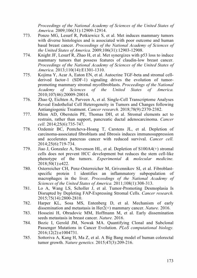
173
Proceedings of the National Academy of Sciences of the United States of America. 2009;106(31):12909-12914.
773. Ponzo MG, Lesurf R, Petkiewicz S, et al. Met induces mammary tumors with diverse histologies and is associated with poor outcome and human basal breast cancer. Proceedings of the National Academy of Sciences of the United States of America. 2009;106(31):12903-12908.
774. Knight JF, Lesurf R, Zhao H, et al. Met synergizes with p53 loss to induce mammary tumors that possess features of claudin-low breast cancer. Proceedings of the National Academy of Sciences of the United States of America. 2013;110(14):E1301-1310.
775. Kojima Y, Acar A, Eaton EN, et al. Autocrine TGF-beta and stromal cell-derived factor-1 (SDF-1) signaling drives the evolution of tumor-promoting mammary stromal myofibroblasts. Proceedings of the National Academy of Sciences of the United States of America. 2010;107(46):20009-20014.
776. Zhao Q, Eichten A, Parveen A, et al. Single-Cell Transcriptome Analyses Reveal Endothelial Cell Heterogeneity in Tumors and Changes following Antiangiogenic Treatment. Cancer research. 2018;78(9):2370-2382.
777. Rhim AD, Oberstein PE, Thomas DH, et al. Stromal elements act to restrain, rather than support, pancreatic ductal adenocarcinoma. Cancer cell. 2014;25(6):735-747.
778. Ozdemir BC, Pentcheva-Hoang T, Carstens JL, et al. Depletion of carcinoma-associated fibroblasts and fibrosis induces immunosuppression and accelerates pancreas cancer with reduced survival. Cancer cell. 2014;25(6):719-734.
779. Jiao J, Gonzalez A, Stevenson HL, et al. Depletion of S100A4(+) stromal cells does not prevent HCC development but reduces the stem cell-like phenotype of the tumors. Experimental & molecular medicine. 2018;50(1):e422.
780. Osterreicher CH, Penz-Osterreicher M, Grivennikov SI, et al. Fibroblast-specific protein 1 identifies an inflammatory subpopulation of macrophages in the liver. Proceedings of the National Academy of Sciences of the United States of America. 2011;108(1):308-313.
781. Lo A, Wang LS, Scholler J, et al. Tumor-Promoting Desmoplasia Is Disrupted by Depleting FAP-Expressing Stromal Cells. Cancer research. 2015;75(14):2800-2810.
782. Harper KL, Sosa MS, Entenberg D, et al. Mechanism of early dissemination and metastasis in Her2(+) mammary cancer. Nature. 2016.
783. Hosseini H, Obradovic MM, Hoffmann M, et al. Early dissemination seeds metastasis in breast cancer. Nature. 2016.
784. Bozic I, Gerold JM, Nowak MA. Quantifying Clonal and Subclonal Passenger Mutations in Cancer Evolution. PLoS computational biology. 2016;12(2):e1004731.
785. Sottoriva A, Kang H, Ma Z, et al. A Big Bang model of human colorectal tumor growth. Nature genetics. 2015;47(3):209-216.

174
786. Williams MJ, Werner B, Heide T, et al. Quantification of subclonalselection in cancer from bulk sequencing data. Nature genetics.2018;50(6):895-903.
787. Ulintz PJ, Greenson JK, Wu R, Fearon ER, Hardiman KM. Lymph NodeMetastases in Colon Cancer Are Polyclonal. Clinical cancer research : anofficial journal of the American Association for Cancer Research.2018;24(9):2214-2224.
788. Ullah I, Karthik GM, Alkodsi A, et al. Evolutionary history of metastaticbreast cancer reveals minimal seeding from axillary lymph nodes. TheJournal of clinical investigation. 2018;128(4):1355-1370.
789. Aceto N, Bardia A, Miyamoto DT, et al. Circulating tumor cell clustersare oligoclonal precursors of breast cancer metastasis. Cell.2014;158(5):1110-1122.
790. Cheung KJ, Padmanaban V, Silvestri V, et al. Polyclonal breast cancermetastases arise from collective dissemination of keratin 14-expressingtumor cell clusters. Proceedings of the National Academy of Sciences ofthe United States of America. 2016;113(7):E854-863.
791. Maddipati R, Stanger BZ. Pancreatic Cancer Metastases Harbor Evidenceof Polyclonality. Cancer discovery. 2015;5(10):1086-1097.
792. Kim MY, Oskarsson T, Acharyya S, et al. Tumor self-seeding bycirculating cancer cells. Cell. 2009;139(7):1315-1326.
793. Reiter JG, Makohon-Moore AP, Gerold JM, et al. Minimal functionaldriver gene heterogeneity among untreated metastases. Science.2018;361(6406):1033-1037.
794. Costa-Silva B, Aiello NM, Ocean AJ, et al. Pancreatic cancer exosomesinitiate pre-metastatic niche formation in the liver. Nature cell biology.2015;17(6):816-826.
795. Hoshino A, Costa-Silva B, Shen TL, et al. Tumour exosome integrinsdetermine organotropic metastasis. Nature. 2015;527(7578):329-335.
796. Yang D, Denny SK, Greenside PG, et al. Intertumoral Heterogeneity inSCLC Is Influenced by the Cell Type of Origin. Cancer discovery. 2018.
Copyright © 2022 FDOKUMEN