DACH1 Is a Cell Fate Determination Factor That Inhibits Cyclin D1 and Breast Tumor Growth
15
10.1128/MCB.00268-06. 2006, 26(19):7116. DOI: Mol. Cell. Biol. Ales Cvekl and Richard G. Pestell Wang, Michael P. Lisanti, Guido Sauter, Robert G. Russell, Vernon Dailey, Ying Yang, Dolores Di Vizio, Chenguang Kongming Wu, Anping Li, Mahadev Rao, Manran Liu, Growth That Inhibits Cyclin D1 and Breast Tumor DACH1 Is a Cell Fate Determination Factor http://mcb.asm.org/content/26/19/7116 Updated information and services can be found at: These include: SUPPLEMENTAL MATERIAL Supplemental material REFERENCES http://mcb.asm.org/content/26/19/7116#ref-list-1 at: This article cites 68 articles, 43 of which can be accessed free CONTENT ALERTS more» articles cite this article), Receive: RSS Feeds, eTOCs, free email alerts (when new http://journals.asm.org/site/misc/reprints.xhtml Information about commercial reprint orders: http://journals.asm.org/site/subscriptions/ To subscribe to to another ASM Journal go to: on March 12, 2014 by guest http://mcb.asm.org/ Downloaded from on March 12, 2014 by guest http://mcb.asm.org/ Downloaded from
-
Upload
independent -
Category
Documents
-
view
0 -
download
0
Transcript of DACH1 Is a Cell Fate Determination Factor That Inhibits Cyclin D1 and Breast Tumor Growth

10.1128/MCB.00268-06.
2006, 26(19):7116. DOI:Mol. Cell. Biol. Ales Cvekl and Richard G. PestellWang, Michael P. Lisanti, Guido Sauter, Robert G. Russell,Vernon Dailey, Ying Yang, Dolores Di Vizio, Chenguang Kongming Wu, Anping Li, Mahadev Rao, Manran Liu, GrowthThat Inhibits Cyclin D1 and Breast Tumor DACH1 Is a Cell Fate Determination Factor
http://mcb.asm.org/content/26/19/7116Updated information and services can be found at:
These include:
SUPPLEMENTAL MATERIAL Supplemental material
REFERENCEShttp://mcb.asm.org/content/26/19/7116#ref-list-1at:
This article cites 68 articles, 43 of which can be accessed free
CONTENT ALERTS more»articles cite this article),
Receive: RSS Feeds, eTOCs, free email alerts (when new
http://journals.asm.org/site/misc/reprints.xhtmlInformation about commercial reprint orders: http://journals.asm.org/site/subscriptions/To subscribe to to another ASM Journal go to:
on March 12, 2014 by guest
http://mcb.asm
.org/D
ownloaded from
on M
arch 12, 2014 by guesthttp://m
cb.asm.org/
Dow
nloaded from

MOLECULAR AND CELLULAR BIOLOGY, Oct. 2006, p. 7116–7129 Vol. 26, No. 190270-7306/06/$08.00�0 doi:10.1128/MCB.00268-06Copyright © 2006, American Society for Microbiology. All Rights Reserved.
DACH1 Is a Cell Fate Determination Factor That Inhibits Cyclin D1and Breast Tumor Growth†
Kongming Wu,1 Anping Li,1 Mahadev Rao,2 Manran Liu,1 Vernon Dailey,2 Ying Yang,3Dolores Di Vizio,3 Chenguang Wang,1 Michael P. Lisanti,1 Guido Sauter,4
Robert G. Russell,2 Ales Cvekl,3 and Richard G. Pestell1*Kimmel Cancer Center, Departments of Cancer Biology and Medical Oncology, 233 S. 10th Street, Bluemle Life Sciences Building,
Room 1050, Philadelphia, Pennsylvania 191071; Lombardi Comprehensive Cancer Center, Georgetown University,Washington, D.C. 200572; Department of Ophthalmology, Visual Sciences and Molecular Genetics,
Albert Einstein Cancer Center and College of Medicine, New York, New York 104613; andUniversity Medical Center Hamburg-Eppendorf, Hamburg, D-20246, Germany4
Received 13 February 2006/Returned for modification 31 March 2006/Accepted 5 July 2006
Obstacles to the expansion of cells with proliferative potential include the induction of cell death, telomere-based senescence, and the pRb and p53 tumor suppressors. Not infrequently, the molecular pathways regu-lating oncogenesis recapitulate aberrations of processes governing embryogenesis. The genetic network, con-sisting of the dachshund (dac), eyes absent (eya), eyeless, and sine oculis (so) genes, regulates cell fatedetermination in metazoans, with dac serving as a cointegrator through a So DNA-binding factor. Here,DACH1 inhibited oncogene-mediated breast oncogenesis, blocking breast cancer epithelial cell DNA synthesis,colony formation, growth in Matrigel, and tumor growth in mice. Genetic deletion studies demonstrated arequirement for cyclin D1 in DACH1-mediated inhibition of DNA synthesis. DACH1 repressed cyclin D1through a novel mechanism via a c-Jun DNA-binding partner, requiring the DACH1 �-helical DS domainwhich recruits corepressors to the local chromatin. Analysis of over 2,000 patients demonstrated increasednuclear DACH1 expression correlated inversely with cellular mitosis and predicted improved breast cancerpatient survival. The cell fate determination factor, DACH1, arrests breast tumor proliferation and growth invivo providing a new mechanistic and potential therapeutic insight into this common disease.
The Drosophila dachshund (dac) gene is the founding mem-ber of the DACH subfamily of nuclear proteins, which conductan essential role in promoting differentiation of the Drosophilaeye and limb (42). The dac gene forms part of a retinal deter-mination (RD) signaling pathway in Drosophila. The RD path-way requires eyeless (eya/Pax6), sine oculis (So, Six), eyes absent(eya/Eya), and dachshund (dac/Dach), which function togetheras genetic components of this network (24, 25, 36). In Dro-sophila, So functions as a DNA-binding factor and dac/eya aretranscription cofactors. Although So/Six binding sites havebeen identified in RD signaling target gene promoters, ge-nome-wide analysis of DACH1-regulated genes identified apreponderance of AP-1-responsive genes. Dachshund is ex-pressed prior to photoreceptor differentiation and is requiredfor retinal morphogenesis. Dachshund itself is sufficient forinducing retinal fates since targeted misexpression results inectopic eye formation from non-neuronal tissues (13, 54).
Although the RD gene network is best known for its role ineye specification, these genes, either individually or as a net-work, are expressed in postmitotic cells and contribute to di-verse developmental processes in many cell types in all meta-zoans. The So/Six family governs proliferation of progenitor
populations prior to cell type specification. Misregulated ex-pression of Six proteins occurs in human cancer (15, 21).HSIX1 is the human homologue of the Drosophila Six genethat was originally isolated through its enrichment during the Sphase of the cell cycle. The Six gene in Drosophila functions asa DNA-binding component of the transcription factor com-plex. Six1 is overexpressed in breast cancer, and forced Six1expression attenuates a G2 cell cycle check point (15, 21). Six1has been implicated as a dual-function regulator of metastasis,enhancing poorly metastatic rhabdomyosarcoma tumors (66).The molecular mechanism by which the RD pathway regulateshuman tumorigenesis is poorly understood.
Orderly cell cycle progression of nontransformed cells isorchestrated by coordinated induction of cyclin-dependent ki-nases that assemble in temporally and spatially defined com-plexes within the cell (43). Sequential phosphorylation of keysubstrates, including the retinoblastoma (pRb) protein, by cy-clin D1 and cyclin E-cdk complexes promotes the timely in-duction of cellular DNA synthesis (27). Oncogenic disruptionof the cell cycle machinery, through amplification or disruptionof the cell cycle proteins themselves, is a common finding inhuman breast cancer. The cyclin D1 gene encodes the regula-tory subunit of a holoenzyme that contributes to the phosphor-ylation and inactivation of the pRb protein and is frequentlyoverexpressed in human breast cancer epithelial cells. Specificoncogenic signals disrupt the cell cycle in a reproducible man-ner. Transformation by Ras requires the inactivation of thepRb and p53 pathways. Myc has activities compatible with thebypass of p21CIP1 (10, 14, 16, 28, 29, 51, 58) and/or activation
* Corresponding author. Mailing address: Kimmel Cancer Center,Departments of Cancer Biology and Medical Oncology, Thomas Jef-ferson University, 233 South 10th Street, Philadelphia, PA 19107.Phone: (215) 503-5649. Fax: (215) 923-9334. E-mail: [email protected].
† Supplemental material for this article may be found at http://mcb.asm.org/.
7116
on March 12, 2014 by guest
http://mcb.asm
.org/D
ownloaded from

of Arf and p53 (10), which impose a selection to the escape ofcellular apoptosis (67). Genetic studies in mice have confirmedthe fidelity of these molecular interactions in vivo. Thus, ge-netic deficiency for cyclin D1 provides resistance to Ras orErbB2 but not c-Myc-induced tumorigenesis, whereas a distinctsubset of genes antagonize Myc function including transform-ing growth factor � (TGF-�) and p21CIP1 (55).
Obstacles to the expansion of cells with proliferative poten-tial include the induction of cell death, telomere-based se-nescence, and the pRb and p53 tumor suppressors. Notinfrequently, the molecular pathways regulating oncogenesis,recapitulate aberrations of processes governing embryogenesis(1). The transcription factors encoded by the homeobox genefamily play a vital role in growth and differentiation duringnormal development. This diverse group of proteins is dividedinto groups based on similarity among the homeodomain boxand includes the MSX, Engrailed, PAX, and the SIX families.Deregulated homeobox gene expression is well known in can-cer, with genes normally expressed in undifferentiated cellsupregulated in cancer. Homeobox genes known to be deregu-lated in cancer include HOX, MSX, HSIX1, GBX2, the PAXgenes, CDX2, NKX3.1, and BARX2. Recent microarray analy-sis studies demonstrated DACH1-repressed proliferative sig-naling in cultured cells (62). We examined the expression andfunction of DACH1 in normal and tumorous breast epithe-lium. We demonstrate DACH1 blocks cellular proliferationand contact-independent growth and identify a new mecha-nism by which DACH1 regulates cellular proliferation andgrowth in vivo.
MATERIALS AND METHODS
Plasmid construction. The expression plasmids for DACH1, DACH1 DS-domain alone (DS), or DACH1 DS-domain deleted (�DS) (which includes anN-terminal Flag peptide), the reporter plasmid 3-TP lux, 3XCRE luc the pon-asterone-regulated expression system, and the full-length wild-type and mutantcyclin D1 promoter reporter constructions were previously described (5, 6, 62).The Flag-tagged DACH1 cDNA was subcloned into the MSCV-internal ribo-some entry site (IRES)-green fluorescent protein (GFP). The SKI cDNAs wassubcloned into the 3x Flag-CMV7.1 vector (Sigma, St. Louis, MO).
Cell sorting and DNA synthesis analysis. Magnetic activated cell sorting toenrich for transfected cells was conducted as previously described (7). Cell cycleparameters were determined by using laser scanning cytometry. Cells were pro-cessed by standard methods using propidium iodide staining of cell DNA. Eachsample was analyzed by flow cytometry with a FACScan flow cytometer (BectonDickinson Biosciences, Mansfield, MA) using a 488-nm laser. Histograms wereanalyzed for cell cycle compartments using ModFit version 2.0 (Verity SoftwareHouse, Topsham, ME). A minimum of 20,000 events was collected to maximizestatistical validity of the compartmental analysis.
Western blot analysis, tissue microarrays, immunohistochemistry, andpurification of DACH1 antibody. Western blot analysis with antibodies tocyclin D1, cyclin E, cdk4, phosphorylated pRB (residue amino acid Ser708),Flag, p21CIP1/WAF1, p27KIP1, c-Jun, CREB, and the loading control guaninedissociation inhibitor (GDI) and immunohistochemistry for cyclin D1 andDACH1 were performed as previously described (35, 62). Affinity-purifiedpolyclonal antibody to DACH1 was produced by Affinity BioReagentsthrough immunizing rabbits with the peptide ERTIQDGRLYLKTTVMY.Human breast tissue microarrays were from Imgenex (IMH-304) and NCICBCTR 2001 TMA#2. The human breast cancer prognostic tissue microar-ray was previously described (2). This array consists of 2,197 formalin-fixed,paraffin-embedded breast cancer tissues with clinical-pathological character-istics, clinical follow-up information, and 283 normal/precancerous tissues.The level of DACH1 protein expression in tumors and normal human breastcancer in the TMAs was categorized by a semiquantitative score of theimmunostaining intensity by light microscopy evaluation, using a standardmethodology that determined a range of staining intensities from negative tostrong with intermediate grades. The intensity of immunoperoxidase staining
was scored as 0 (negative), 1� (a minimal to low level of positive staining),2� (moderate expression), or 3� (strong staining). The evaluation and scor-ing of the cores of the tumors from the patients representing different breasttumor classifications and grades incorporated in the TMAs were conductedby a pathologist (R.G.R.). The results were viewed by two pathologists.Mitoses grade in tumor cells was determined according to the standard ofBloom, Richardson, and Elston.
Cell culture, plasmid transfection, luciferase reporter assays, and Matrigelculture. Cyclin D1�/� cells derived from cyclin D1�/� mice (64), p21CIP1�/�
fibroblast cells (provided by L. Augenlicht) and p27KIP1�/� fibroblast cells (agift from A. Koff) were cultured in Dulbecco modified Eagle medium with10% fetal bovine serum in a humidified atmosphere with 5% CO2 at 37°C.Other cells were cultured as recommended by the American Type CultureCollection and maintained in humidified atmosphere with 5% CO2 at 37°C(59). Transfections were performed by using Superfect transfection reagent(QIAGEN, Valencia, CA) according to the manufacturer’s protocol using 1.5�g of the reporter, 50 to 300 ng of expression plasmids, or equal molaramounts of control vector. The transfection efficiency was normalized bycotransfection with 0.2 �g of pRL-CMV plasmid (Promega, Madison, WI)and measured with Promega’s dual-luciferase reporter assay system accordingto the manufacturer’s protocol. Control small interfering RNA (siRNA) waspurchased from Santa Cruz. siRNAs for human DACH1 were designed andsynthesized by Ambion, Inc. (gene accession number AF 356492). The targetsequences were AAGGCCTCCTAAGAGGACTCA and AAGGACTTCGAGACCCTCTAC. DACH1-specific siRNAs or control siRNAs (100 or 200nM) were transfected according to the Oligofectamine protocol (Invitrogen).Transfection efficiency was monitored by no-silencing fluorescein siRNAfrom QIAGEN. Statistical analyses were performed by using the Student ttest. For three-dimensional culture, 1,000 cells mixed with 1:3 diluted growthfactor reduced Matrigel matrix were seeded into BD biocoated cell cultureinserts (24-well) with Matrigel basement membrane matrix from BD Bio-science (catalog no. 354447) and photographed after growth for 3 to 14 days.For confocal image analyses, MCF10A cells were mixed with 1:1-dilutedmatrigel and seeded into four-well chamber according to a previously de-scribed protocol (18).
Immunoprecipitation, immunoblotting, and Northern blot. 293T cells wereused for the detection of protein-protein interaction in vivo, and immunopre-cipitation-Western blotting was conducted as previously described using anti-Flag M2 antibody (Sigma) and immunoblotting with antibodies to CREB, c-Jun,or Flag (Santa Cruz Biotechnology). The guanine nucleotide dissociation inhib-itor antibody (i.e., GDI) (35) was used as an internal control for protein abun-dance. For Northern blot analysis, total RNA was isolated by using TRIzolreagent. Cyclin D1 cDNA was randomly labeled using NEBlot Phototope kit,and the membrane was detected by Phototope-Star detection kit (New EnglandBiolabs).
ChIP assay. Chromatin immunoprecipitation (ChIP) assays were performedas previously described (22). The human cyclin D1 promoter-specific primersused were as follows: AP-1 site, 5�-CTGCCTTCCTACCTTGACCA-3� and 5�-TGAAGGGACGTCTACACCCC-3�; and CRE site, 5�-GCCCCCCTCCCGCTCCCATT-3� and 5�-TGGGGCTCTTCCTGGGCAGC-3�. The sequences as anegative control in the cyclin D1 promoter were 5�-TTTCGGAAGCGTTTTCCC and 5�-AGCGCGTTCATTCAGGAA. 293T cells were transiently co-transfected with expression plasmids encoding wild-type or mutant DACH1, withthe cyclin D1 promoter constructions, �1745 or cyclin D1 �1745-AP-1/CREmutant reporter, and then cultured for 36 h. The cells were cross-linked withformaldehyde buffer for 10 min at 37°C, and the procedure was performed asdescribed earlier (22).
Nude mice study. Ponasterone A was purified from taxus chinensis, and 100 �gof purified ponasterone A was introduced into 21-day slow release pellets pro-duces by Innovative Research of American (Sarasota, FL) (5). Ponasterone A orplacebo pellets were implanted subcutaneously into 4- to 6-week-old athymicfemale nude mice purchased from NCI. The following day, 2 � 106 MDA-MB-231 cells, stably expressing DACH1 or vector control, were injected subcutane-ously, and the tumor growth was measured twice weekly by digital caliper.Ponasterone A or placebo pellets were implanted every 21 days.
Statistics. The Student t test and the Wilcoxon-Mann-Whitney test were usedto analyze the significance of expression of DACH1. Kaplan-Meier survivalcurves were plotted and analyzed with the Cox proportional hazards regression.The statistical significance of difference between curves was tested by using thegeneralized Wilcoxon test.
VOL. 26, 2006 INHIBITION OF DNA SYNTHESIS BY DACH1 GENE 7117
on March 12, 2014 by guest
http://mcb.asm
.org/D
ownloaded from

RESULTS
DACH1 inhibition of colony formation and tumor growth invivo. Recent studies examining the genetic signaling repressedby DACH1 identified clusters of genes promoting cellular pro-liferation, growth, and survival (62). To examine the functionof DACH1 in regulating breast epithelial cell growth in vivo,
stable cell lines inducibly expressing DACH1 under the controlof the ecdysone-regulated system were constructed in MDA-MB-231 cells. Ponasterone A induced expression of the Flag-tagged DACH1 expression vector as determined by immuno-histochemistry and by Western blotting at 12 to 96 h afterponasterone A addition (Fig. 1A and see Fig. S1A in thesupplemental material). The numbers and sizes of colonieswere reduced �70% by DACH1 (Fig. 1B). To examine theeffect of DACH1 on breast tumor growth in vivo, the ponas-terone-inducible stable lines were implanted in nude mice, anda comparison was made between the sustained release pelletsof ponasterone A and the control placebo pellets, with greaterthan 12 separate animals in each group. Ponasterone A pelletsinduced sustained expression of an ecdysone-regulated �-ga-lactosidase reporter mouse (data not shown) (5). The induc-tion of DACH1 expression reduced the growth of breast tu-mors in mice by 50% (Fig. 1C), with an inhibition of growth byDACH1 first detected at 9 weeks postimplantation (329 66mm3 versus 186 27 mm3, P 0.05). Western blot analysis oftumors isolated from mice demonstrated the induction ofDACH1 protein in mice treated with ponasterone A pelletscompared to mice treated with placebo control pellets (Veh)(Fig. 1D).
DACH1 inhibition of breast epithelial cell DNA synthesisrequires the DACH DS domain. To investigate the mechanismby which DACH1 blocked MDA-MB231 cellular growth, weexamined the effect of DACH1 on DNA synthesis and cellularapoptosis. Although serum starvation does not arrest DNAsynthesis in MDA-MB231 cells (47), the addition of serumproduces an induction of the proportion of cells in the S phaseof the cell cycle (Fig. 2A). Expression of DACH1 blockedserum-induced DNA synthesis (Fig. 2A) without affecting thefraction of apoptotic cells, inferred through analysis of the subG1 population and annexin V staining of the cells (Fig. 2B andC). MDA-MB-231 cells harbor mutation in the K-Ras gene.MCF-7 cells express amplification of c-Myc. To determinewhether DACH1 was capable of inhibiting DNA synthesis inMCF-7 cells, cells were transfected with DACH1 expressionvectors. To identify the domains of DACH1 involved in regu-lating DNA synthesis, a series of domain mutants were exam-ined (Fig. 2D). MCF-7 cells cotransfected with DACH1 or itsmutants were assessed by flow cytometry. DACH1 reduced theproportion of cells in the DNA synthetic (S) phase by 17%(Fig. 2E). One region of structural homology between DACH1and the Ski/Sno proto-oncogene family (26) is predicted toform an �-helical structure (33) (DACH DS domain) that mayconvey general or specific DNA-binding activity (31). Deletionof the conserved DS domain abrogated inhibition of DNAsynthesis. Expression of the related Ski protein did not inhibitDNA synthesis in MCF-7 cells. The inability of the �DS do-main construct to inhibit DNA synthesis compared to the DSdomain alone expression vector was not due to reduced ex-pression of the �DS domain compared to the DS domainconstruct, since the �DS domain was expressed �3-fold betterthan the DS domain alone (see Fig. S1B in the supplementalmaterial).
Phosphorylation and inactivation of the pRb protein and thecyclin D1 gene product play a key role in DNA synthesis ofMDA-MB231 cells. To investigate the candidate target cellcycle proteins regulated by DACH1, responsible for the inhi-
FIG. 1. Inducible DACH1 expression inhibits colony formationand tumor growth in vivo. (A) Immunohistochemical stain for the Flagepitope of DACH1 in MDA-MB-231 stable ponasterone-inducibleDACH1 cell lines showing nuclear colocalization with DAP1 stain.(B) Ponasterone-inducible MDA-MB-231 stable cells were seededonto six-well plates and stained with crystal violet after 14 days growth.Colony volumes (mean the standard error of the mean [SEM], inmm3) are shown on the right (*, P 0.01). (C) Tumor growth ofponasterone-inducible MDA-MB-231 stable cells in athymic femalenude mice (n � 12 separate animals in each group; *, P 0.05). Micewere treated with the ponasterone A pellets or control pellets (vehi-cle), and the sizes of the tumors were assessed weekly. (D) Represen-tative Western blot analysis of DACH1-inducible MDA-MB-231 cellsfrom animals treated with vehicle or ponasterone A pellets.
7118 WU ET AL. MOL. CELL. BIOL.
on March 12, 2014 by guest
http://mcb.asm
.org/D
ownloaded from

bition of DNA synthesis, Western blot analysis was conducted.Cells were treated with ponasterone A to induce expression ofDACH1, and serial time points were analyzed. DACH1 induc-tion was observed at 12 h, contemporaneous with a reductionin cyclin D1 and an induction of p21CIP1. The abundance of theprotein loading control GD1 was unchanged (Fig. 2F). Todetermine target genes involved in blocking serum-inducedDNA synthesis, cells were serum starved and, after inductionof DACH1 with ponasterone A, the cells were treated withserum for 18 h. After 24 h of DACH1 expression, the cyclin D1levels were reduced 60% while increasing the abundance ofp21CIP1 and p27KIP1 (Fig. 2G). A phospho-specific antibody
directed to the Ser780 site phosphorylation of the pRb proteinshowed a 60% reduction in pRb phosphorylation. Cyclin E andcdk4 levels were unchanged (Fig. 2G).
DACH1 inhibits DNA synthesis and oncogene-induced in-vasive phenotype induced by Ras or Myc through distinctpathways. The creation of genetically defined human cancercells has defined with greater precision the regulatory pathwayscontributing to the tumorigenic phenotype (19, 23). MCF10Acells are a spontaneously immortalized human luminal epithe-lial cell line (56). When grown on basement membrane, thecultures of MCF10A cells form growth-arrested three-dimen-sional structures, termed acini. These structures, comprised of
FIG. 2. DACH1 inhibits DNA synthesis through the DS domain. (A) MDA-MB-231 cells expressing inducible DACH1 were characterized forthe effects of DACH1 expression on serum-induced DNA synthesis. The effect of DACH1 expression on S phase or apoptosis assessed by Sub-G1(B) and annexin V stain (C) is shown after 18 h of serum addition. The data are means the SEM for �5 separate experiments. (D) Schematicrepresentation of DACH1 expression vectors. (E) Cell cycle analysis of MCF-7 cells transfected with DACH1 expression vectors. The percentMCF-7 cells in S phase is shown compared to the vector control. The data are mean change in S phase the SEM of DACH1-transfected MCF-7cells (n 4). (F) Dose-dependent induction of DACH1 by ponasterone A and the corresponding cyclin D1 and p21CIP1 expression inMDA-MB-231 cells. (G) Cell-cycle-related protein abundance as determined by Western blotting in MDA-MB-231 cells after 48 h of ponasteroneA treatment.
VOL. 26, 2006 INHIBITION OF DNA SYNTHESIS BY DACH1 GENE 7119
on March 12, 2014 by guest
http://mcb.asm
.org/D
ownloaded from

FIG. 3. DACH1 inhibits DNA synthesis induced by NeuT, Ras/ErbB2 or Myc. (A) The immortalized MCF10A cells were transformed with NeuT,Ras/ErbB2, or c-Myc. Cells were transduced with MSCV-IRES-GFP or MSCV-DACH1-IRES-GFP and selected through GFP FACS for analysis.Morphology of three-dimensional growth in Matrigel at 10 days was depicted by phase-contrast and fluorescence microscopy of acinar structures.(B) Confocal image of GFP and ethidium bromide staining after fixation. Transformed MCF10A cells grow as a solid ball in which the central acinarstructure is filled with proliferating cells. The expression of DACH1 reverts the oncogenic morphology with a return to the nontransformed MCF10Aphenotype with polarized epithelium and an acinar-like lumen structure. (C and D) Cell cycle distribution. (E to G) Colony formation assay results(E) and cell cycle control proteins as determined by Western blotting of transformed MCF10A cells with the indicated antibodies (F and G).
7120
on March 12, 2014 by guest
http://mcb.asm
.org/D
ownloaded from

a single layer of polarized epithelial cells surrounding a hollowlumen, resemble mammary acini and undergo morphologicalchanges in response to transforming oncogenes (50, 56). Themorphology of MCF10A cellular colony growth in three-di-mensional Matrigel culture is characteristic for specific onco-genic pathways (9, 52). To define which oncogenic signalingpathways are inhibited by DACH1, MCF10A cells were trans-duced with expression vectors encoding oncogenic ErbB2(NeuT), Ras, and ErbB2 (Ras/ErbB2) or c-Myc. These trans-formed cells were then transduced with viral expression vectors
encoding DACH1 (MSCV-IRES-GFP or MSCV-DACH1-IRES-GFP) and selected through fluorescence-activated cellsorting (FACS) of GFP-expressing cells. Analyses were con-ducted of morphology in Matrigel (Fig. 3A and B), cell cycledistribution (Fig. 3C and D), colony formation ability (Fig.3E), and cell cycle control proteins abundance determined byWestern blotting (Fig. 3F and G). MCF10A cells form acinus-like spheroids. MCF10A-NeuT cells grown in Matrigel formedcomplex acinus-like structures (39). The activation of ErbB2in MCF10A cells is known to induce this multiacinar phe-
FIG. 4. DACH1 inhibition of DNA synthesis requires cyclin D1. (A to F) DACH1 regulation of cell cycle distribution by FACS analysis (A toC and F) or DNA synthesis using [3H]thymidine uptake (D) and apoptosis using annexin V staining in 3T3 cells derived from the MEFs of micewith either the cyclin D1 (A to E) or the p21CIP1or p27KIP1 (F) gene deleted compared to sibling controls of the same strain background. Cell cycledistribution was assessed using cells transduced with MSCV-IRES-GFP or MSCV-DACH1-IRES-GFP through GFP FACS sorting. Westernblotting confirmed the expression of DACH1 with GDI as the loading control (see Fig. S2 in the supplemental material). (G) cyclin D1�/� cellswere transduced with expression vectors encoding Flag epitope-tagged cyclin D1 or DACH1. Western blot analysis demonstrated the expressionof DACH1 and cyclin D1. FACS analyses demonstrated that cyclin D1 rescues the inhibition of DNA synthesis by DACH1.
VOL. 26, 2006 INHIBITION OF DNA SYNTHESIS BY DACH1 GENE 7121
on March 12, 2014 by guest
http://mcb.asm
.org/D
ownloaded from

FIG. 5. DACH1 recruits HDAC to the AP-1/CRE site of the cyclin D1 promoter in the context of local chromatin. (A) MCF-7 cells treatedwith DACH1 or control siRNA. Endogenous cyclin D1 protein abundance and cyclin D1 mRNA level were detected; GDI was used as a loadingcontrol for Western blot analyses, and 18S RNA was used as a loading control for Northern blot analyses. (B) Cyclin D1 promoter activity assessedin MCF-7 cells transfected with DACH1 siRNA. Promoter activity is shown as the means the SEM for six separate transfections. (C) MeanS-phase analyses of DACH1 siRNA transfected MCF-7 cells. (D and E) Cyclin D1 promoter activity in cells transfected with increasing amounts
7122 WU ET AL. MOL. CELL. BIOL.
on March 12, 2014 by guest
http://mcb.asm
.org/D
ownloaded from

notype through excessive cellular proliferation and changesin apicobasal polarization (17, 44). DACH1 transduction ofoncogenic NeuT transformed MCF10A cells abolished theformation of these multicellular structures, reversing the phe-notype to the spherical morphology of parental MCF10A (Fig.3A). The MCF10A-Ras/ErbB2 colonies consisted of multicel-lular spheroids in which individual cells protruded long spikes.DACH1 transduction of these Ras/ErbB2 transformedMCF10A cells reversed the phenotype and abolished the for-mation of these spike structures (Fig. 3A). MCF10A–c-Myccells grew as multicellular acinus-like structures, and DACH1transduction resulted in the formation of well-polarized spher-oids with lumen formation (Fig. 3A and B). Collectively,these studies demonstrate that DACH1 inhibits oncogene-induced morphological abnormalities in MCF10A cells.
DACH1 inhibited NeuT, Ras/ErbB2, and c-Myc-inducedDNA synthesis as assessed by a reduction in the proportion ofcells distributed within the S phase of the cell cycle (Fig. 3Cand D). DACH1 reduced primarily the S phase but also theG2/M fraction of c-Myc-expressing MCF10A cells. MCF10Acells transformed by oncogenic NeuT or Ras/ErbB2 gain thecapacity to sustain contact-independent growth and form col-onies in soft agar. DACH1 blocked colony formation inducedby either NeuT or Ras/ErbB2 (Fig. 3E). To determine thecomponents of the cell cycle regulated by DACH1 thatmay contribute to the inhibition of cellular growth, Westernblot analyses were conducted of the oncogene-transducedMCF10A cells. We compared the effect of transducing theMCF10A cells to the effect of control vector. In randomlycycling cells, the abundance of cyclin D1 encodes a rate-limit-ing step in DNA synthesis (46, 68). In parental MCF10A, cyclinD1 abundance was reduced by DACH1, and p27KIP1 was in-duced (Fig. 3F) without a significant change in S-phase distri-bution. In NeuT or Ras/ErbB2-transformed MCF10A cells,cyclin D1 and p21CIP1 abundance was induced, a finding con-sistent with previous studies in which Raf induced cyclin D1 inMCF10A cells (50). DNA synthesis was inhibited by DACH1,associated with a reduction in cyclin D1 and induction ofp27KIP1. c-Myc repressed p21CIP1 and p27KIP1, and DACH1partially derepressed p21CIP1 and p27KIP1 abundance but didnot affect cyclin D1 expression (Fig. 3G). The analysis ofDACH1 in oncogene-transduced MCF10A cells indicates thatDACH1 consistently reduces oncogene-induced expression ofcyclin D1. Collectively, these studies show that DACH1 inhib-its oncogene-induced DNA synthesis, malignant morphologi-cal changes in Matrigel, and contact-independent growth asassessed by colony formation.
In view of the finding that DACH1 inhibited Ras and onco-genic ErbB2 induced cyclin D1 expression, we investigated therequirement for cyclin D1 in DACH1-mediated inhibition ofDNA synthesis. To examine further the role of cyclin D1,p21CIP1 and p27KIP1 in DACH1-mediated cell cycle arrest, 3T3cells were derived from mouse embryonic fibroblasts (MEFs)of mice with deletions of either the cyclin D1, the p21CIP1, orthe p27KIP1 gene, and a comparison made with MEFs fromsibling controls of the same strain background for each line.The cell cycle distribution was assessed by using cells trans-duced with MSCV-IRES-GFP or MSCV-DACH1-IRES-GFPthrough GFP FACS sorting. Western blot analysis confirmedthe expression of DACH1. GDI was used as a protein loadingcontrol (see Fig. S2 in the supplemental material). DACH1inhibited DNA synthesis in cyclin D1�/� but not cyclin D1�/�
cells assessed by either FACS analysis or tritiated thymidineuptake, and the inhibition of DNA synthesis by DACH1 re-quired the DACH1 DS domain (Fig. 4A to D). Apoptosis rateswere unaffected by DACH1 in either cyclin D1�/� or cyclinD1�/� cells (Fig. 4E). Deletion of p21CIP1 or p27KIP1 alteredthe relative distribution of cells within the cell cycle. The pro-portion of cells in S phase inhibited by DACH1 in 3T3 cellswith either the p21CIP1 or p27KIP1 gene deleted (Fig. 4F) wassimilar to that of the parental control cells. These studiesindicate that cyclin D1, but not p21CIP1 or p27KIP1, are re-quired for DACH1-mediated inhibition of DNA synthesis in3T3 fibroblasts. To further test the role of cyclin D1 inDACH1-induced growth arrest, cyclin D1�/� or cyclin D1�/�
cells were infected with Flag-tagged cyclin D1, DACH1, orboth cyclin D1 and DACH1, and Western blotting confirmedthe expression of exogenous cyclin D1 and DACH1. Cell cycleanalyses demonstrated the rescue of DACH1-mediated growthinhibition upon the expression of cyclin D1 (Fig. 4G).
DACH1 repression of the cyclin D1 promoter requires theAP-1/CRE DNA-binding site. DACH1 inhibited DNA synthe-sis in the breast cancer epithelial cell lines MCF-7 and MDA-MB-231 and in NIH 3T3 cells. We sought to determinewhether physiological levels of DACH1 regulated cyclin D1abundance. The administration of siRNA directed to endoge-nous DACH1 in MCF-7 cells reduced DACH1 abundance andinduced cyclin D1 protein and mRNA abundance (�3-fold),indicating physiological levels of DACH1 repress cyclin D1(Fig. 5A). Since DACH1 expression inhibited MCF-7 cellDNA synthesis (Fig. 2) and the abundance of cyclin D1 is ratelimiting in DNA synthesis of MCF-7 cells (68), the role ofDACH1 in regulating cyclin D1 was next assessed in thesecells. To examine the role of endogenous DACH1 in regulating
of expression vector encoding DACH1 or a DACH1 mutant with the DS domain deleted (�DS) in MCF-7 cells. The vector control value was setas 100. Cyclin D1 promoter point mutants were compared for repression by DACH1. The data (means the SEM) are shown as the percentrepression compared to the vector for eight separate experiments. (F and G) Heterologous reporters encoding the cyclin D1 AP-1 and CRE siteslinked to the luciferase reporter gene were assessed for regulation by DACH1 in 3T3 cells. (H) ChIP assay using oligonucleotides directed to DNAsequences of the endogenous human cyclin D1 promoter AP-1 or CRE sites or a control DNA sequence at �3060. MDA-MB-231 cells expressinginducible Flag-tagged DACH1 are shown treated with PonA or vehicle. (I) 293T cells cotransfected with �1745 cyclin D1 promoter andFlag-tagged wild-type or mutant DACH1 expression vectors. ChIP assays were conducted using oligonucleotides directed to DNA sequences ofthe human cyclin D1 AP-1 or CRE sites. (J) ChIP analyses of endogenous DACH1 binding to the human cyclin D1 promoter comparing the �3060region (negative) and the cyclin D1 promoter AP-1 site. (K) 293T cells were transfected with control or DACH1 or Ski expression vector andanalyzed by Western blotting to detect c-Jun and CREB. Immunoprecipitation was conducted with antibodies directed to the Flag epitope of theDACH1, �DS, and Ski expression vectors and immunoprecipitation analyzed for c-Jun or CREB.
VOL. 26, 2006 INHIBITION OF DNA SYNTHESIS BY DACH1 GENE 7123
on March 12, 2014 by guest
http://mcb.asm
.org/D
ownloaded from

the cyclin D1 promoter, DACH1 siRNA was compared tocontrol siRNA for its effect in regulating the cyclin D1 pro-moter. DACH1 siRNA induced the full-length cyclin D1 pro-moter threefold (Fig. 5B). Consistent with previous studiesdemonstrating that cyclin D1 abundance is rate limiting inMCF-7 cell DNA synthesis (40), DACH1 siRNA induced theS phase in MCF-7 cells (Fig. 5C). The cyclin D1 promoter wasrepressed in a dose-dependent manner by the expression ofDACH1, and deletion of the DACH1 DS domain abrogatedrepression of the cyclin D1 promoter (Fig. 5D). In contrastto DACH1, the related Ski protein did not inhibit cyclin D1promoter activity (not shown). Mutation of the AP-1 andCRE site of the cyclin D1 promoter reduced DACH1 re-pression by 80%, suggesting that these sequences togetherplayed a key role in repression by DACH1 (Fig. 5E).DACH1 repressed cyclin D1 promoter activity in 293T andNIH 3T3 cells and again repression required both the AP-1and the CRE sites (data not shown). The cyclin D1 pro-moter AP-1 and CRE sites serve as both basal level andinducible enhancer elements; therefore, experiments wereconducted to determine whether these elements were suffi-cient for repression by DACH1. The CRE and AP-1 siteswere sufficient for repression by DACH1, and repressionrequired the DACH1 DS domain (Fig. 5F and G).
To determine the mechanisms by which DACH1 repressedthe cyclin D1 promoter, ChIP assays were conducted to exam-ine the occupancy of DACH1 at the endogenous human cyclinD1 promoter in human breast cancer cells. Comparison wasmade between the inducible DACH1 stable cell line and thevehicle control. Using oligonucleotides directed to the endog-enous human cyclin D1 AP-1 and CRE sites, ChIP assaysconducted with the anti-Flag antibody demonstrated the pres-ence of DACH1 in the context of the local chromatin structureof the endogenous human cyclin D1 promoter upon the induc-tion of DACH1 expression (Fig. 5H). HDAC1, mSin3A, andNCoR were recruited by DACH1 to form a multimeric repres-sor complex at the AP-1 site of the endogenous human cyclinD1 promoter. Amplification was not observed using oligonu-cleotides directed toward sequences within the �3060 regionof the cyclin D1 promoter. No amplification was observedusing either the vehicle control cells or immunoglobulin G inthe ponasterone-inducible DACH1 stable cell line (Fig. 5H).To determine which region of the DACH1 protein was neces-sary for the formation of a DNA-associated complex at thecyclin D1 promoter in the context of its local chromatin struc-ture, 293T cells were transfected with the human cyclin D1luciferase promoter and wild-type or mutant DACH1 expres-sion vectors. ChIP assay using the Flag antibody demonstratedthat both wild-type DACH1 and the DS domain alone wererecruited to the AP-1 and the CRE site, whereas the DSdomain deleted mutant was defective in recruitment to thecyclin D1 promoter (Fig. 5I). Since DACH1 regulated DNAsynthesis and cyclin D1 expression and promoter activity, wesought to determine whether endogenous DACH1 boundthe cyclin D1 promoter in the context of local chromatin.Endogenous DACH1 was associated with the cyclin D1 pro-moter AP-1 site but was not identified at the �3060 region.Serum addition did not significantly alter DACH1 occu-pancy in ChIP assays (Fig. 5J).
Since DACH1 was recruited in a DNA sequence-specific
manner to the cyclin D1 AP-1/CRE site and is not thought tobind a defined DNA sequence directly, we sought to determinewhether DACH1 formed a complex with AP-1 proteins incultured cells. Immunoprecipitation was conducted with theFlag antibody using cell extracts derived from 293T cells trans-fected with DACH1 or Ski, normalized for equal amounts ofc-Jun and CREB by Western blotting (Fig. 5K). Equalamounts of DACH1 and c-Jun were coprecipitated with theFlag antibody; however, only DACH1 coprecipitated c-Jun andCREB. Deletion of the DACH1 DS domain abolished bindingto c-Jun and CREB (Fig. 5K). These findings are consistentwith a model in which DACH1 binds c-Jun and is recruited toan AP-1 site in the context of its local chromatin structure,requiring the DS domain of DACH1 to recruit the corepres-sors HDAC1, NCoR, and mSin3A to repress cyclin D1 geneexpression.
DACH1 expression is regulated during mammary gland de-velopment and reduced in metastatic breast cancer. In view ofthe finding that endogenous DACH1 inhibited cyclin D1 ex-pression and that DACH1 expression inhibited DNA synthesisin breast cancer cell lines, we investigated the distribution andphysiological regulation of DACH1 in human and murinebreast epithelium. DACH1 expression was examined in breastepithelium and breast cancer cell lines by immunohistochem-ical staining and Western blotting with a DACH1-specific an-tibody. The specificity of the antibody was confirmed throughWestern blotting of 293T cells transfected with expression vec-tors encoding the DACH1 wild-type or the DACH1 �DS do-main (Fig. 6A). Immunoreactive DACH1 was detected inMCF-7, MCF-10A, and MDA-MB-231 cells and several otherbreast cancer cell lines (Fig. 6B). As a form of positive controlfor immunohistochemical staining, DACH1 was identified inthe embryonic eye as previously described (8) (Fig. 6C). Todetermine whether Dach1 expression is regulated during nor-mal physiological changes in the breast, Dach1 immunohisto-chemistry was assessed during murine mammary gland devel-opment and lactation. Dach1 expression was detectable innormal virgin mammary gland, increased at 10 days pregnancy,and decreased in lactation (day 10) and involution (day 10)(Fig. 6D). The homeodomain protein Six1, which functions indevelopmental networks including eye development in Dro-sophila, is expressed in mammary carcinoma but is absent ordetected at low levels only in normal mammary tissues (15, 21).Six1 expression, although detectable in breast cancers, wasweakly expressed in the normal murine mammary epithelium(see Fig. S3 in the supplemental material), as recently shown(15). The expression of Six1 was dramatically increased duringpregnancy, coinciding with high expression of cyclin D1. Wetested whether Six1 was capable of inducing cyclin D1.HEK293T cells were transiently transfected with the cyclin D1promoter luciferase reporter and expression vectors encodingeither Six1 or DACH1. A dose-dependent activation of thecyclin D1 promoter by Six1 and repression by DACH1 wereobserved (see Fig. S3B in the supplemental material), a findingconsistent with recent findings that Six1 induces cyclin D1promoter in a rhabdomyosarcoma cell line (65). Collectively,These findings are consistent with a model in which the relativeabundance of the Eya/Six complex are under physiologicalcontrol and may together contribute to the relative abundanceof cyclin D1 in a given cell type.
7124 WU ET AL. MOL. CELL. BIOL.
on March 12, 2014 by guest
http://mcb.asm
.org/D
ownloaded from

FIG. 6. DACH1 expression in human breast cancer cell lines, mouse mammary gland, and breast carcinoma tissues. (A) 293T cells weretransiently transfected with control or DACH1 expression vector and analyzed by Western blotting with a DACH1 specific antibody. (B) Westernblot for DACH1 abundance of breast epithelial cell lines, breast cancer cell lines, and control extracts of 293T cells transfected with DACH1 (lane1). GDI was used as a loading control. (C) DACH1 expression in mouse embryonic eye was used as a positive control. (D) DACH1 immuno-histochemistry during the murine mammary gland development of the virgin, at pregnancy (day 10), at lactation (day 10), and at involution (day10). (E) Representative DACH1 expression in normal breast epithelium, ductal carcinoma in situ, or invasive human breast cancer samples usingDACH1 specific antibodies. (F) Analysis of DACH1 expression in subcategory of human breast tissue microarray (*, P 0.05). (G) Relationshipof DACH1 expression to mitosis index. (H) DACH1 expression and prognosis of human breast carcinoma (n 2,125 patients).
VOL. 26, 2006 INHIBITION OF DNA SYNTHESIS BY DACH1 GENE 7125
on March 12, 2014 by guest
http://mcb.asm
.org/D
ownloaded from
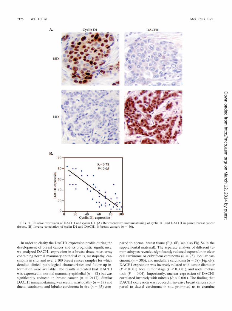
In order to clarify the DACH1 expression profile during thedevelopment of breast cancer and its prognostic significance,we analyzed DACH1 expression in a breast tissue microarraycontaining normal mammary epithelial cells, mastopathy, car-cinoma in situ, and over 2,100 breast cancer samples for whichdetailed clinical-pathological characteristics and follow-up in-formation were available. The results indicated that DACH1was expressed in normal mammary epithelial (n 81) but wassignificantly reduced in breast cancer (n 2117). SimilarDACH1 immunostaining was seen in mastopathy (n 17) andductal carcinoma and lobular carcinoma in situ (n 63) com-
pared to normal breast tissue (Fig. 6E; see also Fig. S4 in thesupplemental material). The separate analysis of different tu-mor subtypes revealed significantly reduced expression in clearcell carcinoma or cribriform carcinoma (n 75), lobular car-cinoma (n 300), and medullary carcinoma (n 58) (Fig. 6F).DACH1 expression was inversely related with tumor diameter(P 0.001), local tumor stage (P 0.0001), and nodal metas-tasis (P 0.04). Importantly, nuclear expression of DACH1correlated inversely with mitosis (P 0.001). The finding thatDACH1 expression was reduced in invasive breast cancer com-pared to ductal carcinoma in situ prompted us to examine
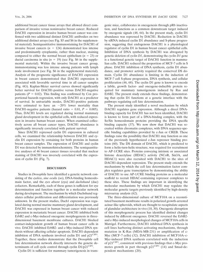
FIG. 7. Relative expression of DACH1 and cyclin D1. (A) Representative immunostaining of cyclin D1 and DACH1 in paired breast cancertissues. (B) Inverse correlation of cyclin D1 and DACH1 in breast cancers (n 46).
7126 WU ET AL. MOL. CELL. BIOL.
on March 12, 2014 by guest
http://mcb.asm
.org/D
ownloaded from
additional breast cancer tissue arrays that allowed direct com-parison of invasive versus noninvasive breast cancer. ReducedDACH1 expression in invasive human breast cancer was con-firmed with two additional distinct DACH1 antibodies on twoadditional distinct arrays (see Fig. S5 and S6 in the supplemen-tal material). Semiquantitative immunostaining for DACH1 ofinvasive breast cancers (n 124) demonstrated less intenseand predominantly cytoplasmic, rather than nuclear, stainingcompared to either the normal breast epithelium (n 23) orductal carcinoma in situ (n 19) (see Fig. S6 in the supple-mental material). Within the invasive breast cancer group,immunostaining was less intense among patients with lymphnode involvement (see Fig. S6 in the supplemental material).Analysis of the prognostic significance of DACH1 expressionin breast cancers demonstrated that DACH1 expression isassociated with favorable survival time in all cancer samples(Fig. 6G). Kaplan-Meier survival curves showed significantlybetter survival for DACH1-positive versus DACH1-negativepatients (P 0.02). This finding was confirmed by Cox pro-portional hazards regression, with only DACH1 as a predictorof survival. In univariable modes, DACH1-positive patientswere estimated to have an �20% lower mortality thanDACH1-negative patients (hazard ratio 0.81, P 0.005).Thus, DACH1 levels are regulated during normal mammarygland development in the epithelial cells, with reduced expres-sion in invasive human breast cancer. When examined collec-tively across all breast cancer types, DACH1 abundance issignificantly inversely correlated with patient survival.
Since DACH1 repressed cyclin D1 expression in culturedcells, we examined the relationship between DACH1 and cy-clin D1 expression by costaining for both proteins in humanbreast cancer samples. The expression of DACH1 and cyclinD1 was detected by immunohistochemistry. The semiquantita-tive analyses of 46 breast cancer tissues revealed that nuclearstaining of DACH1 was inversely correlated with the expres-sion of cyclin D1 (Fig. 7).
DISCUSSION
Studies in Drosophila have identified a genetic network con-sisting of the eyeless, sine oculis (so), DNA-binding homeodo-main factor, and the eyes absent (eya) and dachshund (dac)cofactors. Remarkably, each of these genes is sufficient for eyedetermination and function together in a molecular networkduring development. The mechanism by which Dachshund reg-ulates cellular proliferation and differentiation was previouslyunknown. In the present studies, Dach1 expression was regu-lated during normal murine mammary gland development, andDACH1 was expressed in human breast cancer with reducedexpression in metastatic breast cancer. DACH1 inhibited bothErbB2 and c-Myc-induced oncogenic morphogenesis in three-dimensional basement membrane cultures. DACH1 blockedcontact-independent growth of human breast cancer cells invivo. DACH1 inhibited ErbB2- and c-Myc-induced DNA syn-thesis without affecting cellular apoptosis. DACH1-dependentinhibition of DNA synthesis involved cyclin D1 and p21CIP1.Together, these studies demonstrate that the Drosophila cellfate determination network directly intersects the genetic de-terminants of cell cycle control through cyclin D1/p21CIP1.
Cyclin D1 is sufficient for mammary tumorigenesis in trans-
genic mice, collaborates in oncogenesis through pRb inactiva-tion, and functions as a common downstream target inducedby oncogenic signals (48, 64). In the present study, cyclin D1abundance was repressed by DACH1. Reduction in DACH1by siRNA induced cyclin D1 abundance and S-phase progres-sion, suggesting that endogenous DACH1 is a physiologicalregulator of cyclin D1 in human breast cancer epithelial cells.Inhibition of DNA synthesis by DACH1 was abrogated bygenetic deletion of cyclin D1, demonstrating the cyclin D1 geneis a functional genetic target of DACH1 function in mamma-lian cells. DACH1 reduced the proportion of MCF-7 cells in Sphase. DACH1 inhibition of DNA synthesis, cyclin D1 abun-dance, and promoter activity required the conserved DS do-main. Cyclin D1 abundance is limiting in the induction ofMCF-7 cell S-phase progression, DNA synthesis, and cellularproliferation (46, 68). The cyclin D1 gene is known to encodea labile, growth factor- and oncogene-inducible protein re-quired for mammary tumorigenesis induced by Ras andErbB2. The present study extends these findings, demonstrat-ing that cyclin D1 functions at a genetic interface with thepathways regulating cell fate determination.
The present study identified a novel mechanism by whichDACH1 regulates gene expression. Although a direct DNA-binding capacity for DACH1 has not been identified, DACH1is known to form part of a DNA-binding complex, with theSo/Six homeodomain proteins providing the DNA specificbinding capacity (37). We now show that DACH1 was re-cruited within chromatin complexes, with DNA sequence-spe-cific binding capabilities provided by c-Jun or CREB. Thesefindings raise the possibility that DACH1 may function as oneof the previously hypothesized c-Jun repressor binding pro-teins (60). The DS domain of DACH1, which is predicted toform a helix-turn-helix structure, was required for recruitmentto AP-1/CRE sites. Proteins conveying intrinsic or associatedhistone deacetylase (HDAC) activity (NCoR, Sin3A, andHDAC1) were also recruited with DACH1 to the sites ofDACH1-dependent repression. The present study extends themechanisms by which the cell fate determination factor com-plex regulates gene transcription by demonstrating the abilityof DACH1 to use AP-1/CRE binding proteins as a molecularscaffold to recruit HDAC-containing repressor complexes tothese sites. These findings are important in identifying themolecular mechanisms by which DACH1 may regulate themolecular genetic targets previously identified by high-densitymicroarray analysis (62).
The three-dimensional culture of MCF10A on a reconsti-tuted basement membrane results in polarized growth-arrestedacinar-like spheroids, which are thought to recapitulate aspectsof glandular architecture in vivo (18, 30). Oncogenic disruptionof this morphogenetic process has identified distinct changesinduced by different oncogenes. DACH1 reversed the ErbB2-and c-Myc-induced morphological changes of MCF10A cells inMatrigel. Furthermore, DACH1 inhibited DNA synthesis incell lines harboring distinct activating mechanisms, throughmutation in K-Ras (MDA-MB-231) or amplification of c-Myc (MCF-7 cells) (32). DACH1 inhibition of MCF10A–c-Myc-induced DNA synthesis correlated with the inductionof p21CIP1, consistent with previous findings that c-Myc pro-motes growth in part through p21CIP1 (14) and Smad-de-pendent mechanisms (20).
VOL. 26, 2006 INHIBITION OF DNA SYNTHESIS BY DACH1 GENE 7127
on March 12, 2014 by guest
http://mcb.asm
.org/D
ownloaded from

c-Jun contributes to cellular proliferation and tumorigenesis(53). c-Jun promotes G1 phase cell cycle progression, and im-munoneutralizing antibodies to c-Jun inhibit DNA synthesis(11). MEFs deficient in c-Jun exhibit a severe defect in prolif-eration due to extension of the G1 transition time (49).DACH1 repressed AP-1 activity, associating with c-Jun in thecontext of local promoter chromatin. DACH1 purified whole-cell extracts contained AP-1 binding activity, and DACH1 co-precipitated c-Jun through the DS domain. DACH1 repressionof cyclin D1 expression, DNA synthesis, and cell survival areconsistent with previous findings that AP-1 induces cyclin D1transcription (4, 12, 61). Cyclin D1 contributes to cell survivalsince cyclin D1�/� MEF cells display both reduced rates of G1
phase progression and increased cellular apoptosis (3).DACH1, but not the related Ski, blocked serum-induced DNAsynthesis in both mammary epithelial cells and in fibroblasts,demonstrating dissociable functions of these homologous pro-teins. The current model suggests DACH1, like Sno/Ski andNCoR (41, 57, 63), is recruited with DNA-binding proteins.DACH1, but not Ski, repressed the transcription of c-Jun.Recruitment to distinct DNA-binding proteins through the DSdomain may contribute to the transcriptional specificity of theDACH1 versus Ski complexes (41).
Several recent findings suggest the RD pathway may regu-late aberrant cellular growth and metastasis. Six1 expression isenhanced in metastatic rhabdomyosarcoma and contributes tocellular migration (66). Six6 functions as a tissue-specific re-pressor in association with Dach corepressors through Six6binding sites (37), and the EWS/NOR1 translocation, impli-cated in extraskeletal myxoid chondrosarcoma, is repressed bySix3 (34). It has been hypothesized that Six1, which regulatesDNA-damage-induced G2-phase arrest, may regulate migra-tion through Lbx1h (66). In view of the reduction in DACH1 inmetastatic human breast cancer and findings that cyclin D1promotes cellular migration, through altering the adhesion andphosphorylation of tyrosine-phosphorylated paxillin (38, 45), itwill be of interest to determine whether DACH1 contributes tocellular migration. The accumulating evidence that the RDpathway contributes to tumorigenesis through intersecting cellcycle control raises the possibility that this pathway may be auseful new therapeutic prospect.
ACKNOWLEDGMENTS
We thank C. Chang, S. Ishii, J. Massague, G. H. Merlino, and M. G.Rosenfeld for plasmids; L. H. Augenlicht and A. Koff for cells; G. H.Nuckolls for the Dach1 antibody; and P. A. Furth for mouse mammaryslides. We thank Dawn Scardino and Almeta Mathis for help withmanuscript preparation.
This study was supported in part by awards from the Susan KomenBreast Cancer Foundation, the Breast Cancer Alliance, Inc., and NIH(R01CA70896, R01CA75503, R01CA86072, and R01CA86071 [R.G.P.]).Work conducted at the Kimmel Cancer Center was supported by the NIHCancer Center Core grant P30CA56036 (R.G.P.). A.C. was supported byNIH grant EY12200, a Research to Prevent Blindness Career Develop-ment Award, and an American Cancer Society Junior Faculty Institu-tional Award. This project is funded, in part, under a grant with thePennsylvania Department of Health. The Department specifically dis-claims responsibility for any analyses, interpretations, or conclusions.
REFERENCES
1. Abate-Shen, C. 2002. Deregulated homeobox gene expression in cancer:cause or consequence? Nat. Rev. Cancer 2:777–785.
2. Al-Kuraya, K., P. Schraml, J. Torhorst, C. Tapia, B. Zaharieva, H. Novotny,
H. Spichtin, R. Maurer, M. Mirlacher, O. Kochli, M. Zuber, H. Dieterich, F.Mross, K. Wilber, R. Simon, and G. Sauter. 2004. Prognostic relevance ofgene amplifications and coamplifications in breast cancer. Cancer Res. 64:8534–8540.
3. Albanese, C., M. D’Amico, A. T. Reutens, M. Fu, G. Watanabe, R. J. Lee,R. N. Kitsis, B. Henglein, M. Avantaggiati, K. Somasundaram, B. Thimma-paya, and R. G. Pestell. 1999. Activation of the cyclin D1 gene by theE1A-associated protein p300 through AP-1 inhibits cellular apoptosis.J. Biol. Chem. 274:34186–34195.
4. Albanese, C., J. Johnson, G. Watanabe, N. Eklund, D. Vu, A. Arnold, andR. G. Pestell. 1995. Transforming p21ras mutants and c-Ets-2 activate thecyclin D1 promoter through distinguishable regions. J. Biol. Chem. 270:23589–23597.
5. Albanese, C., A. T. Reutens, B. Bouzahzah, M. Fu, M. D’Amico, T. Link, R.Nicholson, R. A. Depinho, and R. G. Pestell. 2000. Sustained mammarygland-directed, ponasterone A-inducible expression in transgenic mice.FASEB J. 14:877–884.
6. Albanese, C., K. Wu, M. D’Amico, C. Jarrett, D. Joyce, J. Hughes, J. Hulit,T. Sakamaki, M. Fu, A. Ben-Ze’ev, J. F. Bromberg, C. Lamberti, U. Verma,R. B. Gaynor, S. W. Byers, and R. G. Pestell. 2003. IKK� regulates mitogenicsignaling through transcriptional induction of cyclin D1 via Tcf. Mol. Biol.Cell 14:585–599.
7. Ashton, A. W., G. Watanabe, C. Albanese, E. O. Harrington, J. A. Ware, andR. G. Pestell. 1999. Protein kinase C� inhibition of S-phase transition incapillary endothelial cells involves the cyclin-dependent kinase inhibitorp27Kip1. J. Biol. Chem. 274:20805–20811.
8. Ayres, J. A., L. Shum, A. N. Akarsu, R. Dashner, K. Takahashi, T. Ikura,H. C. Slavkin, and G. H. Nuckolls. 2001. DACH: genomic characterization,evaluation as a candidate for postaxial polydactyly type A2, and develop-mental expression pattern of the mouse homologue. Genomics 77:18–26.
9. Bentires-Alj, M., G. S., R. Chan, Z. C. Wang, Y. Wang, N. Imanaka, L. N.Harris, A. Richardson, B. G. Neel, and H. Gu. 2006. A role for the scaffoldingadapter GAB2 in breast cancer. Nat. Med. 12:114–121.
10. Bouchard, C., K. Thieke, A. Maier, R. Saffrich, J. Hanley-Hyde, W. Ansorge,S. Reed, P. Sicinski, J. Bartek, and M. Eilers. 1999. Direct induction of cyclinD2 by Myc contributes to cell cycle progression and sequestration of p27.EMBO J. 18:5321–5333.
11. Bravo, R. 1990. Genes induced during the G0/G1 transition in mouse fibro-blasts. Semin. Cancer Biol. 1:337–346.
12. Brown, J. R., E. Nigh, R. J. Lee, H. Ye, M. A. Thompson, F. Saudou, R. G.Pestell, and M. E. Greenberg. 1998. Fos family members induce cell cycleentry by activating cyclin D1. Mol. Cell. Biol. 18:5609–5619.
13. Chen, R., M. Amouri, Z. Zhang, and G. Mardon. 1997. Dachshund and eyesabsent proteins form a complex and function synergistically to induce ectopiceye development in Drosophila. Cell 91:893–903.
14. Claassen, G. F., and S. R. Hann. 2000. A role for transcriptional repressionof p21CIP1 by c-Myc in overcoming transforming growth factor-inducedcell-cycle arrest. Proc. Natl. Acad. Sci. USA 97:9498–9503.
15. Coletta, R. D., K. Christensen, K. J. Reichenberger, J. Lamb, D. Micomo-naco, L. Huang, D. M. Wolf, C. Muller-Tidow, T. R. Golub, K. Kawakami,and H. L. Ford. 2004. The Six1 homeoprotein stimulates tumorigenesis byreactivation of cyclin A1. Proc. Natl. Acad. Sci. USA 101:6478–6483.
16. Coller, H. A., C. Grandori, P. Tamayo, T. Colbert, E. S. Lander, R. N.Eisenman, and T. R. Golub. 2000. Expression analysis with oligonucleotidemicroarrays reveals that MYC regulates genes involved in growth, cell cycle,signaling, and adhesion. Proc. Natl. Acad. Sci. USA 97:3260–3265.
17. Debnath, J., K. R. Mills, N. L. Collins, M. J. Reginato, S. K. Muthuswamy,and J. S. Brugge. 2002. The role of apoptosis in creating and maintainingluminal space within normal and oncogene-expressing mammary acini. Cell111:29–40.
18. Debnath, J., S. K. Muthuswamy, and J. S. Brugge. 2003. Morphogenesis andoncogenesis of MCF-10A mammary epithelial acini grown in three-dimen-sional basement membrane cultures. Methods 30:256–268.
19. Elenbaas, B., L. Spirio, F. Koerner, M. D. Fleming, D. B. Zimonjic, J. L.Donaher, N. C. Popescu, W. C. Hahn, and R. A. Weinberg. 2001. Humanbreast cancer cells generated by oncogenic transformation of primary mam-mary epithelial cells. Genes Dev. 15:50–65.
20. Feng, X. H., Y. Y. Liang, M. Liang, W. Zhai, and X. Lin. 2002. Directinteraction of c-Myc with Smad2 and Smad3 to inhibit TGF-beta-mediatedinduction of the CDK inhibitor p15 (Ink4B). Mol. Cell 9:133–143.
21. Ford, H. L., E. N. Kabingu, E. A. Bump, G. L. Mutter, and A. B. Pardee.1998. Abrogation of the G2 cell cycle checkpoint associated with overexpres-sion of HSIX1: a possible mechanism of breast carcinogenesis. Proc. Natl.Acad. Sci. USA 95:12608–12613.
22. Fu, M., M. Rao, K. Wu, C. Wang, X. Zhang, M. Hessien, Y. G. Yeung, D.Gioeli, M. J. Weber, and R. G. Pestell. 2004. The androgen receptor acety-lation site regulates cAMP and AKT but not ERK-induced activity. J. Biol.Chem. 279:29436–29449.
23. Hahn, W. C., C. M. Counter, A. S. Lundberg, R. L. Beijersbergen, M. W.Brooks, and R. A. Weinberg. 1999. Creation of human tumour cells withdefined genetic elements. Nature 400:464–468.
24. Halder, G., P. Callaerts, S. Flister, U. Walldorf, U. Kloter, and W. J. Ge-
7128 WU ET AL. MOL. CELL. BIOL.
on March 12, 2014 by guest
http://mcb.asm
.org/D
ownloaded from

hring. 1998. Eyeless initiates the expression of both sine oculis and eyesabsent during Drosophila compound eye development. Development 125:2181–2191.
25. Halder, G., P. Callaerts, and W. J. Gehring. 1995. Induction of ectopic eyesby targeted expression of the eyeless gene in Drosophila. Science 267:1788–1792.
26. Hammond, K. L., I. M. Hanson, A. G. Brown, L. A. Lettice, and R. E. Hill.1998. Mammalian and drosophila dachshund genes are related to the skiproto-oncogene and are expressed in eye and limb. Mech. Dev. 74:121–131.
27. Hanahan, D., and R. Weinberg. 2000. The hallmarks of cancer. Cell 100:57–70.
28. Hermeking, H., C. Rago, M. Schuhmacher, Q. Li, J. F. Barrett, A. J. Obaya,B. C. O’Connell, M. K. Mateyak, W. Tam, F. Kohlhuber, C. V. Dang, J. M.Sedivy, D. Eick, B. Vogelstein, and K. W. Kinzler. 2000. Identification ofCDK4 as a target of c-MYC. Proc. Natl. Acad. Sci. USA 97:2229–2234.
29. Herold, S., M. Wanzel, V. Beuger, C. Frohme, D. Beul, T. Hillukkala, J.Syvaoja, H. P. Saluz, F. Haenel, and M. Eilers. 2002. Negative regulation ofthe mammalian UV response by Myc through association with Miz-1. Mol.Cell 10:509–521.
30. Jacks, T., and R. A. Weinberg. 2002. Taking the study of cancer cell survivalto a new dimension. Cell 111:923–925.
31. Kim, S.-S., R.-G. Zhang, S. E. Braunstein, A. Joachimiak, A. Cvekl, and R. S.Hegde. 2002. Structure of the retinal determination protein dachshund re-veals a DNA binding motif. Structure 10:787–789.
32. Kozma, S. C., M. E. Bogaard, K. Buser, S. M. Saurer, J. L. Bos, B. Groner,and N. E. Hynes. 1987. The human c-Kirsten ras gene is activated by a novelmutation in codon 13 in the breast carcinoma cell line MDA-MB231. NucleicAcids Res. 15:5963–5971.
33. Kozmik, Z., P. Pfeffer, J. Kralova, J. Paces, V. Paces, A. Kalousova, and A.Cvekl. 1999. Molecular cloning and expression of the human and mousehomologues of the drosophila dachshund gene. Dev. Genes Evol. 209:537–545.
34. Laflamme, C., C. Filion, J. A. Bridge, M. Ladanyi, M. B. Goldring, and Y.Labelle. 2003. The homeotic protein Six3 is a coactivator of the nuclearreceptor NOR-1 and a corepressor of the fusion protein EWS/NOR-1 inhuman extraskeletal myxoid chondrosarcomas. Cancer Res. 63:449–454.
35. Lee, R. J., C. Albanese, M. Fu, M. D’Amico, B. Lin, G. Watanabe, G. K.Haines III, P. M. Siegel, M. C. Hung, Y. Yarden, J. M. Horowitz, W. J.Muller, and R. G. Pestell. 2000. Cyclin D1 is required for transformation byactivated Neu and is induced through an E2F-dependent signaling pathway.Mol. Cell. Biol. 20:672–683.
36. Li, X., K. A. Oghi, J. Zhang, A. Krones, K. T. Bush, C. K. Glass, S. K. Nigam,A. K. Aggarwal, R. Maas, D. W. Rose, and M. G. Rosenfeld. 2003. Eyaprotein phosphatase activity regulates Six1-Dach-Eya transcriptional effectsin mammalian organogenesis. Nature 426:247–254.
37. Li, X., V. Perrisi, F. Liu, D. W. Rose, and M. G. Rosenfeld. 2002. Tissue-specific regulation of retinal and pituitary precursor cell proliferation. Sci-ence 297:1180–1183.
38. Li, Z., C. Wang, X. Jiao, Y. Lu, M. Fu, A. A. Quong, C. Dye, J. Yang, M. Dai,X. Ju, X. Zhang, A. Li, p. Burbelo, E. R. Stanley, and R. G. Pestell. 2006.Cyclin D1 regulates cellular migration through the inhibition of throm-bospondin 1 and ROCK signaling. Mol. Biol. Cell 26:4240–4256.
39. Liu, H., D. C. Radisky, F. Wang, and M. J. Bissell. 2004. Polarity andproliferation are controlled by distinct signaling pathways downstream ofPI3-kinase in breast epithelial tumor cells. J. Cell Biol. 164:603–612.
40. Lukas, J., J. Bartkova, and J. Bartek. 1996. Convergence of mitogenicsignalling cascades from diverse classes of receptors at the cyclin D-cyclin-dependent kinase-pRb-controlled G1 checkpoint. Mol. Cell. Biol. 16:6917–6925.
41. Luo, K., S. L. Stroschein, W. Wang, D. Chen, E. Martens, S. Zhou, and Q.Zhou. 1999. The Ski oncoprotein interacts with the Smad proteins to repressTGF� signaling. Genes Dev. 13:2196–2206.
42. Mardon, G., N. M. Solomon, and G. M. Rubin. 1994. Dachshund encodes anuclear protein required for normal eye and leg development in Drosophila.Development 120:3473–3486.
43. Massague, J. 2004. G1 cell-cycle control and cancer. Nature 432:298–306.44. Muthuswamy, S. K., D. Li, S. Lelievre, M. J. Bissell, and J. S. Brugge. 2001.
ErbB2, but not ErbB1, reinitiates proliferation and induces luminal repopu-lation in epithelial acini. Nat. Cell Biol. 3:785–792.
45. Neumeister, P., F. J. Pixley, Y. Xiong, H. Xie, K. Wu, A. Ashton, M. Cammer,A. Chan, M. Symons, E. R. Stanley, and R. G. Pestell. 2003. Cyclin D1governs adhesion and motility of macrophages. Mol. Biol. Cell 14:2005–2015.
46. Pagano, M., A. M. Theodorus, S. W. Tam, and G. F. Draetta. 1994. Cyclin
D1-mediated inhibition of repair and replicative DNA synthesis in humanfibroblasts. Genes Dev. 8:1627–1639.
47. Pervin, S., R. Singh, and G. Chaudhuri. 2001. Nitric oxide-induced cytostasisand cell cycle arrest of a human breast cancer cell line (MDA-MB-231):potential role of cyclin D1. Proc. Natl. Acad. Sci. USA 98:3583–3588.
48. Pestell, R. G., C. Albanese, A. T. Reutens, R. J. Lee, J. Segall, and A. Arnold.1999. The cyclins and cyclin dependent kinase inhibitors in hormonal regu-lation of proliferation and differentiation. Endocrinol. Rev. 20:501–534.
49. Schreiber, M., A. Kolbus, F. Piu, A. Szabowski, U. Mohle-Steinlein, J. Tian,M. Karin, P. Angel, and E. F. Wagner. 1999. Control of cell cycle progressionby c-Jun is p53 dependent. Genes Dev. 13:607–619.
50. Schulze, A., K. Lehmann, H. B. Jefferies, M. McMahon, and J. Downward.2001. Analysis of the transcriptional program induced by Raf in epithelialcells. Genes Dev. 15:981–994.
51. Seoane, J., H. V. Le, and J. Massague. 2002. Myc suppression of the p21(Cip1) Cdk inhibitor influences the outcome of the p53 response to DNAdamage. Nature 419:729–734.
52. Seton-Rogers, S. E., Y. Lu, L. M. Hines, M. Koundinya, J. LaBaer, S. K.Muthuswamy, and J. S. Brugge. 2004. Cooperation of the ErbB2 receptorand transforming growth factor beta in induction of migration and invasionin mammary epithelial cells. Proc. Natl. Acad. Sci. USA 101:1257–1262.
53. Shaulian, E., and M. Karin. 2002. AP-1 as a regulator of cell life and death.Nat. Cell Biol. 4:E131–E136.
54. Shen, W., and G. Mardon. 1997. Ectopic eye development in drosophilainduced by directed dachsund expression. Development 124:45–52.
55. Sherr, C. J., and J. M. Roberts. 1995. Inhibitors of mammalian G1 cyclin-dependent kinases. Genes Dev. 9:1149–1163.
56. Soule, H. D., T. M. Maloney, S. R. Wolman, W. D. Peterson, Jr., R. Brenz,C. M. McGrath, J. Russo, R. J. Pauley, R. F. Jones, and S. C. Brooks. 1990.Isolation and characterization of a spontaneously immortalized humanbreast epithelial cell line, MCF-10. Cancer Res. 50:6075–6086.
57. Tarapore, P., C. Richmond, G. Zheng, S. B. Cohen, B. Kelder, J. Kopchick,U. Kruse, A. E. Sippel, C. Colmenares, and E. Stavnezer. 1997. DNA bindingand transcriptional activation by the Ski oncoprotein mediated by interactionwith NFI. Nucleic Acids Res. 25:3895–3903.
58. Vlach, J., S. Hennecke, K. Alevizopoulis, D. Conti, and B. Amati. 1996.Growth arrest by the cyclin-dependent kinase inhibitor p27Kip1 is abrogatedby c-Myc. EMBO J. 15:6595–6604.
59. Watanabe, G., A. Howe, R. J. Lee, C. Albanese, I. W. Shu, A. N. Karnezis, L.Zon, J. Kyriakis, K. Rundell, and R. G. Pestell. 1996. Induction of cyclin D1by simian virus 40 small tumor antigen. Proc. Natl. Acad. Sci. USA 93:12861–12866.
60. Weiss, C., S. Schneider, E. F. Wagner, X. Zhang, E. Seto, and D. Bohmann.2003. JNK phosphorylation relieves HDAC3-dependent suppression of thetranscriptional activity of c-Jun. EMBO J. 22:3686–3695.
61. Wisdom, R., R. S. Johnson, and C. Moore. 1999. c-Jun regulates cell cycleprogression and apoptosis by distinct mechanisms. EMBO J. 18:188–197.
62. Wu, K., Y. Yang, C. Wang, M. A. Davoli, M. D’Amico, A. Li, K. Cveklova, Z.Kozmik, M. P. Lisanti, R. G. Russell, A. Cvekl, and R. G. Pestell. 2003.DACH1 inhibits TGF-beta signaling through binding Smad4. J. Biol. Chem.278:51673–51684.
63. Xu, W., K. Angelis, D. Danielpour, M. M. Haddad, O. Bischof, J. Campisi,E. Stavnezer, and E. E. Medrano. 2000. Ski acts as a co-repressor with Smad2and Smad3 to regulate the response to type beta transforming growth factor.Proc. Natl. Acad. Sci. USA 97:5924–5929.
64. Yu, Q., Y. Geng, and P. Sicinski. 2001. Specific protection against breastcancers by cyclin D1 ablation. Nature 411:1017–1021.
65. Yu, Y., E. Davicioni, T. J. Triche, and G. Merlino. 2006. The homeoproteinsix1 transcriptionally activates multiple protumorigenic genes but requiresezrin to promote metastasis. Cancer Res. 66:1982–1989.
66. Yu, Y., J. Khan, C. Khanna, L. Helman, P. S. Meltzer, and G. Merlino. 2004.Expression profiling identifies the cytoskeletal organizer ezrin and the de-velopmental homeoprotein Six-1 as key metastatic regulators. Nat. Med.10:175–181.
67. Zindy, F., C. M. Eischen, D. H. Randle, T. Kamijo, J. L. Cleveland, C. J.Sherr, and M. F. Roussel. 1998. Myc signaling via the ARF tumor suppressorregulates p53-dependent apoptosis and immortalization. Genes Dev. 12:2424–2433.
68. Zwijsen, R. M. L., R. Klompmaker, E. B. H. G. M. Wientjens, P. M. P.Kristel, B. van der Burg, and R. J. A. M. Michalides. 1996. Cyclin D1 triggersautonomous growth of breast cancer cells by governing cell cycle exit. Mol.Cell. Biol. 16:2554–2560.
VOL. 26, 2006 INHIBITION OF DNA SYNTHESIS BY DACH1 GENE 7129
on March 12, 2014 by guest
http://mcb.asm
.org/D
ownloaded from