The importance of ERK activity in the regulation of cyclin D1 levels and DNA synthesis in human...
12
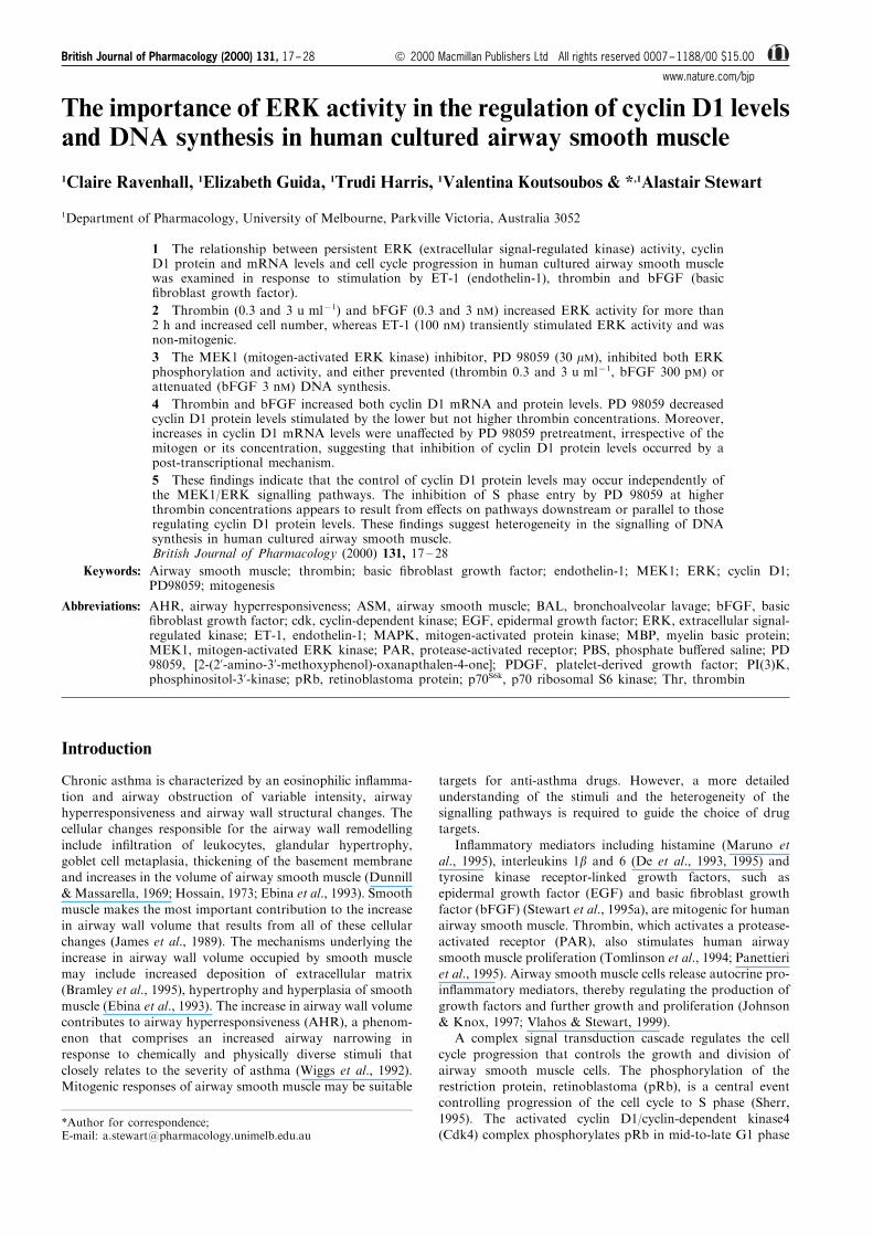
The importance of ERK activity in the regulation of cyclin D1 levels and DNA synthesis in human cultured airway smooth muscle 1 Claire Ravenhall, 1 Elizabeth Guida, 1 Trudi Harris, 1 Valentina Koutsoubos & * ,1 Alastair Stewart 1 Department of Pharmacology, University of Melbourne, Parkville Victoria, Australia 3052 1 The relationship between persistent ERK (extracellular signal-regulated kinase) activity, cyclin D1 protein and mRNA levels and cell cycle progression in human cultured airway smooth muscle was examined in response to stimulation by ET-1 (endothelin-1), thrombin and bFGF (basic fibroblast growth factor). 2 Thrombin (0.3 and 3 u ml 71 ) and bFGF (0.3 and 3 nM) increased ERK activity for more than 2 h and increased cell number, whereas ET-1 (100 nM) transiently stimulated ERK activity and was non-mitogenic. 3 The MEK1 (mitogen-activated ERK kinase) inhibitor, PD 98059 (30 mM), inhibited both ERK phosphorylation and activity, and either prevented (thrombin 0.3 and 3 u ml 71 , bFGF 300 pM) or attenuated (bFGF 3 nM) DNA synthesis. 4 Thrombin and bFGF increased both cyclin D1 mRNA and protein levels. PD 98059 decreased cyclin D1 protein levels stimulated by the lower but not higher thrombin concentrations. Moreover, increases in cyclin D1 mRNA levels were unaected by PD 98059 pretreatment, irrespective of the mitogen or its concentration, suggesting that inhibition of cyclin D1 protein levels occurred by a post-transcriptional mechanism. 5 These findings indicate that the control of cyclin D1 protein levels may occur independently of the MEK1/ERK signalling pathways. The inhibition of S phase entry by PD 98059 at higher thrombin concentrations appears to result from eects on pathways downstream or parallel to those regulating cyclin D1 protein levels. These findings suggest heterogeneity in the signalling of DNA synthesis in human cultured airway smooth muscle. British Journal of Pharmacology (2000) 131, 17 – 28 Keywords: Airway smooth muscle; thrombin; basic fibroblast growth factor; endothelin-1; MEK1; ERK; cyclin D1; PD98059; mitogenesis Abbreviations: AHR, airway hyperresponsiveness; ASM, airway smooth muscle; BAL, bronchoalveolar lavage; bFGF, basic fibroblast growth factor; cdk, cyclin-dependent kinase; EGF, epidermal growth factor; ERK, extracellular signal- regulated kinase; ET-1, endothelin-1; MAPK, mitogen-activated protein kinase; MBP, myelin basic protein; MEK1, mitogen-activated ERK kinase; PAR, protease-activated receptor; PBS, phosphate buered saline; PD 98059, [2-(2’-amino-3’-methoxyphenol)-oxanapthalen-4-one]; PDGF, platelet-derived growth factor; PI(3)K, phosphinositol-3’-kinase; pRb, retinoblastoma protein; p70 S6k , p70 ribosomal S6 kinase; Thr, thrombin Introduction Chronic asthma is characterized by an eosinophilic inflamma- tion and airway obstruction of variable intensity, airway hyperresponsiveness and airway wall structural changes. The cellular changes responsible for the airway wall remodelling include infiltration of leukocytes, glandular hypertrophy, goblet cell metaplasia, thickening of the basement membrane and increases in the volume of airway smooth muscle (Dunnill & Massarella, 1969; Hossain, 1973; Ebina et al., 1993). Smooth muscle makes the most important contribution to the increase in airway wall volume that results from all of these cellular changes (James et al., 1989). The mechanisms underlying the increase in airway wall volume occupied by smooth muscle may include increased deposition of extracellular matrix (Bramley et al., 1995), hypertrophy and hyperplasia of smooth muscle (Ebina et al., 1993). The increase in airway wall volume contributes to airway hyperresponsiveness (AHR), a phenom- enon that comprises an increased airway narrowing in response to chemically and physically diverse stimuli that closely relates to the severity of asthma (Wiggs et al., 1992). Mitogenic responses of airway smooth muscle may be suitable targets for anti-asthma drugs. However, a more detailed understanding of the stimuli and the heterogeneity of the signalling pathways is required to guide the choice of drug targets. Inflammatory mediators including histamine (Maruno et al., 1995), interleukins 1b and 6 (De et al., 1993, 1995) and tyrosine kinase receptor-linked growth factors, such as epidermal growth factor (EGF) and basic fibroblast growth factor (bFGF) (Stewart et al., 1995a), are mitogenic for human airway smooth muscle. Thrombin, which activates a protease- activated receptor (PAR), also stimulates human airway smooth muscle proliferation (Tomlinson et al., 1994; Panettieri et al., 1995). Airway smooth muscle cells release autocrine pro- inflammatory mediators, thereby regulating the production of growth factors and further growth and proliferation (Johnson & Knox, 1997; Vlahos & Stewart, 1999). A complex signal transduction cascade regulates the cell cycle progression that controls the growth and division of airway smooth muscle cells. The phosphorylation of the restriction protein, retinoblastoma (pRb), is a central event controlling progression of the cell cycle to S phase (Sherr, 1995). The activated cyclin D1/cyclin-dependent kinase4 (Cdk4) complex phosphorylates pRb in mid-to-late G1 phase *Author for correspondence; E-mail: [email protected] British Journal of Pharmacology (2000) 131, 17 – 28 ª 2000 Macmillan Publishers Ltd All rights reserved 0007 – 1188/00 $15.00 www.nature.com/bjp
Transcript of The importance of ERK activity in the regulation of cyclin D1 levels and DNA synthesis in human...
The importance of ERK activity in the regulation of cyclin D1 levelsand DNA synthesis in human cultured airway smooth muscle
1Claire Ravenhall, 1Elizabeth Guida, 1Trudi Harris, 1Valentina Koutsoubos & *,1Alastair Stewart
1Department of Pharmacology, University of Melbourne, Parkville Victoria, Australia 3052
1 The relationship between persistent ERK (extracellular signal-regulated kinase) activity, cyclinD1 protein and mRNA levels and cell cycle progression in human cultured airway smooth musclewas examined in response to stimulation by ET-1 (endothelin-1), thrombin and bFGF (basic®broblast growth factor).
2 Thrombin (0.3 and 3 u ml71) and bFGF (0.3 and 3 nM) increased ERK activity for more than2 h and increased cell number, whereas ET-1 (100 nM) transiently stimulated ERK activity and wasnon-mitogenic.
3 The MEK1 (mitogen-activated ERK kinase) inhibitor, PD 98059 (30 mM), inhibited both ERKphosphorylation and activity, and either prevented (thrombin 0.3 and 3 u ml71, bFGF 300 pM) orattenuated (bFGF 3 nM) DNA synthesis.
4 Thrombin and bFGF increased both cyclin D1 mRNA and protein levels. PD 98059 decreasedcyclin D1 protein levels stimulated by the lower but not higher thrombin concentrations. Moreover,increases in cyclin D1 mRNA levels were una�ected by PD 98059 pretreatment, irrespective of themitogen or its concentration, suggesting that inhibition of cyclin D1 protein levels occurred by apost-transcriptional mechanism.
5 These ®ndings indicate that the control of cyclin D1 protein levels may occur independently ofthe MEK1/ERK signalling pathways. The inhibition of S phase entry by PD 98059 at higherthrombin concentrations appears to result from e�ects on pathways downstream or parallel to thoseregulating cyclin D1 protein levels. These ®ndings suggest heterogeneity in the signalling of DNAsynthesis in human cultured airway smooth muscle.British Journal of Pharmacology (2000) 131, 17 ± 28
Keywords: Airway smooth muscle; thrombin; basic ®broblast growth factor; endothelin-1; MEK1; ERK; cyclin D1;PD98059; mitogenesis
Abbreviations: AHR, airway hyperresponsiveness; ASM, airway smooth muscle; BAL, bronchoalveolar lavage; bFGF, basic®broblast growth factor; cdk, cyclin-dependent kinase; EGF, epidermal growth factor; ERK, extracellular signal-regulated kinase; ET-1, endothelin-1; MAPK, mitogen-activated protein kinase; MBP, myelin basic protein;MEK1, mitogen-activated ERK kinase; PAR, protease-activated receptor; PBS, phosphate bu�ered saline; PD98059, [2-(2'-amino-3'-methoxyphenol)-oxanapthalen-4-one]; PDGF, platelet-derived growth factor; PI(3)K,phosphinositol-3'-kinase; pRb, retinoblastoma protein; p70S6k, p70 ribosomal S6 kinase; Thr, thrombin
Introduction
Chronic asthma is characterized by an eosinophilic in¯amma-
tion and airway obstruction of variable intensity, airwayhyperresponsiveness and airway wall structural changes. Thecellular changes responsible for the airway wall remodelling
include in®ltration of leukocytes, glandular hypertrophy,goblet cell metaplasia, thickening of the basement membraneand increases in the volume of airway smooth muscle (Dunnill& Massarella, 1969; Hossain, 1973; Ebina et al., 1993). Smooth
muscle makes the most important contribution to the increasein airway wall volume that results from all of these cellularchanges (James et al., 1989). The mechanisms underlying the
increase in airway wall volume occupied by smooth musclemay include increased deposition of extracellular matrix(Bramley et al., 1995), hypertrophy and hyperplasia of smooth
muscle (Ebina et al., 1993). The increase in airway wall volumecontributes to airway hyperresponsiveness (AHR), a phenom-enon that comprises an increased airway narrowing inresponse to chemically and physically diverse stimuli that
closely relates to the severity of asthma (Wiggs et al., 1992).Mitogenic responses of airway smooth muscle may be suitable
targets for anti-asthma drugs. However, a more detailed
understanding of the stimuli and the heterogeneity of thesignalling pathways is required to guide the choice of drugtargets.
In¯ammatory mediators including histamine (Maruno etal., 1995), interleukins 1b and 6 (De et al., 1993, 1995) andtyrosine kinase receptor-linked growth factors, such asepidermal growth factor (EGF) and basic ®broblast growth
factor (bFGF) (Stewart et al., 1995a), are mitogenic for humanairway smooth muscle. Thrombin, which activates a protease-activated receptor (PAR), also stimulates human airway
smooth muscle proliferation (Tomlinson et al., 1994; Panettieriet al., 1995). Airway smooth muscle cells release autocrine pro-in¯ammatory mediators, thereby regulating the production of
growth factors and further growth and proliferation (Johnson& Knox, 1997; Vlahos & Stewart, 1999).
A complex signal transduction cascade regulates the cellcycle progression that controls the growth and division of
airway smooth muscle cells. The phosphorylation of therestriction protein, retinoblastoma (pRb), is a central eventcontrolling progression of the cell cycle to S phase (Sherr,
1995). The activated cyclin D1/cyclin-dependent kinase4(Cdk4) complex phosphorylates pRb in mid-to-late G1 phase
*Author for correspondence;E-mail: [email protected]
British Journal of Pharmacology (2000) 131, 17 ± 28 ã 2000 Macmillan Publishers Ltd All rights reserved 0007 ± 1188/00 $15.00
www.nature.com/bjp
to promote the dissociation of pRb from E2F (Chellappan etal., 1991), and permit the progression from G1 to S phase ofthe cell cycle (Lundberg & Weinberg, 1998). A sustained
increase in the levels of cyclin D1, and assembly with Cdk4 aredependent on continuous mitogenic stimulation (Sherr, 1995;Matsushime et al., 1994). Withdrawal of mitogenic stimulationleads to a rapid decline in cyclin D1 levels (Sherr, 1993). The
degradation of cyclin D1 protein is mediated by ubiquitin-dependent proteolysis, which is triggered by the phosphoryla-tion of a threonine residue of cyclin D1 (Diehl et al., 1997).
Proteosomal degradation of cyclin D1 appears to be activatedby increases in cyclic AMP levels (Stewart et al., 1999).
Several studies in transformed cell lines have demonstrated
the requirement of cyclin D1 for S phase entry (Quelle et al.,1993). Recent studies by Xiong et al. (1997) demonstrated thatmitogenic stimulation of bovine airway smooth muscle
induced activation of both the cyclin D1 promoter and cyclinD1 protein expression, followed by phosphorylation of pRb(Xiong et al., 1997). Conversely, the microinjection of an anti-cyclin D1 antibody reduced DNA synthesis indicating a
requirement for cyclin D1 in S phase entry and cell cycleprogression (Xiong et al., 1997).
The p44/p42MAPK isoforms (ERK1 and 2, respectively) of
the mitogen-activated protein kinase (MAPK) family play amajor role in the signalling of cell proliferation. Thephosphorylation of ERK by MEK1 results in catalytic
activation of ERK (Posada & Cooper, 1992; Whelchel et al.,1997). Within a few minutes of mitogenic stimulation,activated ERK isoforms translocate to the nucleus in most
cell types (L'Allemain et al., 1991) thereby transmittingmitogenic signals to the cell nucleus (Chen et al., 1992;Lenormand et al., 1993) such as phosphorylation of speci®ctranscription factors that are required for cell cycle progression
(Gille et al., 1995; Janknecht et al., 1995; Kato et al., 1995).Only those agents able to induce sustained ERK activation aremitogenic for CCL39 hamster lung ®broblasts (Meloche et al.,
1992). ERK activity then rapidly declines during G1 to S phasetransition (Meloche, 1995). Persistent ERK activation has alsobeen speci®cally implicated in mitogenic responses of bovine
airway smooth muscle (Kelleher et al., 1995). Links betweenpersistent ERK activation and cyclin D1 expression have beendemonstrated in Chinese hamster lung (Lavoie et al., 1996) andembryo ®broblast cell lines (Weber et al., 1997). Catalytic
activation of ERK appears to be required for the induction ofcyclin D1 expression in primary bovine airway smooth musclecells (Ramakrishnan et al., 1998).
The relationship between ERK activation, cyclin D1transcription and translation and S phase entry in humancultured airway smooth muscle has not been fully established.
There is much species variation in the responses to di�erentmitogenic agents. For example, endothelin-1 (ET-1), which islinked to a G-protein-coupled receptor, stimulates mitogenesis
in rabbit (Noveral et al., 1992), ovine (Glassberg et al., 1994),guinea-pig (Stewart et al., 1994) and rat airway smooth muscle(Whelchel et al., 1997), but does not directly stimulateproliferation in human (Panettieri et al., 1995) or bovine
airway smooth muscle (Panettieri et al., 1996). In contrast,bradykinin, which also activates a G-protein-coupled receptor,stimulates ERK activity that leads to an increase in PGE2
levels and negatively feeds back to inhibit ERK, therebyrestricting mitogenesis in guinea-pig ASM (Pyne et al., 1997).However, thrombin, which activates another G-protein-
coupled receptor, is mitogenic for bovine (Walker et al.,1998), rat (Shapiro et al., 1996) and human (Tomlinson et al.,1994) airway smooth muscle. Growth factors, includingplatelet-derived growth factor (PDGF) and EGF, which
activate tyrosine kinase-linked receptors, also demonstratespecies dependent mitogenic potentials. Both PDGF and EGFare mitogenic for human airway smooth muscle (Tomlinson et
al., 1994; Kelleher et al., 1995), but only PDGF is active onbovine airway smooth muscle. The mechanism for the strongmitogenic response observed in guinea-pig ASM has beenpartly elucidated in that activation of the PDGF receptor
evokes a powerful stimulation of the ERK signalling pathwayby utilizing the G protein, Gi, in addition to the PDGFtyrosine kinase signalling pathway to promote mitogenesis
(Conway et al., 1999). Furthermore, the e�ect of ERKactivation can vary markedly between di�erent strains of cellsof the same species. ERK has either a stimulatory or inhibitory
e�ect on smooth muscle proliferation, depending on whetherthe arterial smooth muscle strain expresses COX-2. Expressionof COX-2 leads to the production of PGE2, which inhibits
DNA synthesis via an elevation of PKA (Bornfeldt et al.,1997). Moreover, in cytokine-stimulated ASM glucocorticoidsincrease rather than decrease growth factor-induced DNAsynthesis by repression of COX-2 induction (Vlahos &
Stewart, 1999). Species di�erences in functional (mitogenic)responses to growth factors that do not result from lack ofreceptor expression imply the existence of di�erential
activation of cell cycle progression signalling pathways. Thus,there is a clear need to establish both the functional andbiochemical mitogen responsiveness of human cultured airway
smooth muscle.The present study examines the relationship of persistent
ERK activity to cyclin D1 protein and mRNA levels and S
phase entry in human airway smooth muscle cells. Thisrelationship has been investigated using the MEK1-inhibitor,PD 98059 (Dudley et al., 1995), a selective tool that has beenwidely used to elucidate the role of the MEK1/ERK pathway
(Grammer & Blenis, 1997; Karpova et al., 1997; Weber et al.,1997; Whelchel et al., 1997). Since the primary mitogens forhuman airway wall remodelling are not known, we have
compared the signals initiated by thrombin with those inresponse to bFGF and the potent bronchoconstrictor ET-1.These putative mitogens activate di�erent classes of receptors
and have been identi®ed in in¯amed airways (Redington et al.,1995; Gabazza et al., 1998; Springall et al., 1991). In addition,as the prevailing mitogen concentrations associated withvariable intensities of in¯ammation are not known, the
ERK-dependence of near maximal and supramaximal con-centrations of bFGF and thrombin have been contrasted.
Our ®ndings suggest that persistent ERK activation makes
a substantial contribution to the signalling of mitogenesis ofhuman airway smooth muscle, but at supramaximal mitogenconcentrations other pathways may additionally be recruited
to signal mitogenesis.
Methods
Culture of human airway smooth muscle
Human airway smooth muscle was cultured from macro-scopically normal bronchi (0.5 ± 2 cm diameter) obtained fromlung resection or heart ± lung transplant specimens provided by
the Alfred Hospital (Melbourne). Cultures were prepared aspreviously described in detail (Tomlinson et al., 1995) andmaintained in Dulbecco's Modi®ed Eagle's Medium (DMEM)
(supplemented with 0.25% v v71 BSA, 2 mM L-glutamine,100 u ml71 penicillin G, 100 mg ml71 streptomycin and2 mg ml71 amphotericin B) and containing foetal calf serum(FCS) (10% v v71). Cells were maintained in Falcon culture
Heterogeneous signalling of mitogenesis18 C. Ravenhall et al
British Journal of Pharmacology, vol 131 (1)

¯asks and incubated (378C, 5% CO2) until monolayercon¯uence was reached and harvested weekly by a 10 minexposure to 0.5% (w v71) trypsin, 1 mM EDTA in PBS and
passaged at a 1 : 3 split ratio. Cells at passage numbers 4 ± 15were used for experiments, over which range of passagenumber there is no detectable relationship between passagenumber and responsiveness to growth factors or inhibitors, or
to the expression of a-actin (Stewart et al., 1997b).
Immunohistochemistry
The cellular composition of the cultures was determined usingexpression of smooth muscle speci®c a-actin and myosin as
described previously (Panettieri et al., 1989; Stewart et al.,1997a; Fernandes et al., 1999).
Measurement of cell cycle progression
DNA synthesis was measured between 24 and 28 h aftermitogen stimulation, as the human airway smooth muscle cells
enter S phase approximately 22 h after the addition ofthrombin (Stewart et al., 1995b). A longer stimulation time isrequired for the measurement of changes such as cell
proliferation or increases in cellular protein (Stewart et al.,1995b). In this study, we have measured both increases in cellnumber and the DNA pro®les of the cells 48 h after mitogen
stimulation, and have demonstrated both cell division andcycling at this time.
Measurement of DNA synthesis
Cells were subcultured into 24 well plates in a 1 : 3 split ratio ata density of approximately 1.56104 cells cm72 and grown to
monolayer con¯uence in DMEM containing 10% FCS, over a72 ± 96 h period (5% CO2 in air, 378C). Quiescence wasinduced 24 h prior to stimulation by removing the medium,
washing with PBS and replacing the medium with serum-freeDMEM (supplemented as described above). In some experi-ments, the cells were incubated with the MEK1-selective
inhibitor, PD98059 (30 mM) (Dudley et al., 1995) for 30 minbefore the addition of mitogen. Cells were stimulated by theaddition of endothelin-1 (human, 100 nM), thrombin (3 or0.3 u ml71) or bFGF (3 nM or 300 pM). Monomed A (1%
v v71), a serum-free medium supplement containing insulin(100 ng ml71), transferrin (50 ng ml71), and selenium(1.5 pg ml71) was added to all cells to provide progression
factors that are essential for mitogenic activity. Although thehigher concentrations of bFGF and thrombin were usedpredominantly in this study, one-tenth of these concentrations
were also used to contrast di�erences in the recruitment ofsignalling pathways in response to near maximal andsupramaximal mitogen concentrations. Cells were incubated
with the indicated agent for 24 h (5% CO2 in air, 378C) thenpulsed with [3H]-thymidine (1 mCi ml71) for 4 h to measure theincorporation of the radiolabel into newly synthesized DNA,according to our previous study (Stewart et al., 1997b).
Flow cytometry
Cells were seeded into six-well plates at a density of 1.56104
cells cm72 and were made quiescent as described previously,then were stimulated with the indicated mitogen for 48 h.
Monomed A was added to all cells (including control cells) atthe time of mitogen addition. During the stimulation period,the medium was removed after 24 h and replaced with warmedfresh medium and stimulants. Cells were detached from the
culture plates by incubation with trypsin for 30 min at 378C(0.5% w v71). The resulting suspension was washed in PBStwice before resuspension in 1 ml of 70% ethanol for storage
for up to 3 weeks at 7208C. Prior to staining, cells werewashed twice (2% FCS in PBS) to remove the ethanol. Fixedcells were stained with propidium iodide (50 mg ml71) inTriton X-100 (0.1% v v71) with Rnase II (180 mu ml71). The
cell suspension was passed through an 18-gauge needle tofacilitate the separation of cell clumps. Cells were stored for24 h before analysis. Cell cycle status was analysed using a
Becton-Dickinson FACScan instrument (Becton Dickinson,NJ, U.S.A.). Ten thousand events from each sample werecounted and analysed using a ModFitLT V2.0 analysis
package (Verity Software House, ME, U.S.A.).
Measurement of cell proliferation
Cells were seeded onto six-well plates at a density of 1.56104
cells cm72, made quiescent as previously described and thenstimulated for 48 h with the appropriate mitogen. During the
stimulation period, the medium was removed after 24 h andreplaced with warmed fresh medium and stimulants. Cells weredetached from the culture plate by the addition of trypsin
(0.5% w v71 in PBS containing 1 mM EDTA), washed twice(2% FCS in PBS) isolated by centrifugation (12,0006g, 5 min)and resuspended in 200 ml 2% FCS in PBS for counting in an
haemocytometer chamber.
Western blot analyses
Cells were subcultured onto six-well plates and stimulatedunder conditions identical to those used for estimation ofDNA synthesis. Thrombin, bFGF or ET-1 was added to each
well for 20 h. A stimulation period of 20 h was chosen in orderto measure ERK phosphorylation and cyclin D1 levels late inG1 before S phase entry at approximately 22 h (Stewart et al.,
1995b). The cell monolayer was washed twice with ice-coldPBS, lysed on ice and prepared for electrophoresis exactly asdescribed previously (Fernandes et al., 1999). Aliquots were
removed for protein assay (BioRad reagent, BioRad, Sydney,Australia). Identical amounts of protein (60 ± 100 mg) fromseparate samples were loaded onto lanes in the SDS-polyacrylamide gel. The samples were resolved on PAGE,
transferred to nitrocellulose membranes and Western blottedfor phospho-ERK and cyclin D1 according to the methodsdescribed previously (Fernandes et al., 1999). To visualize the
antigen, ECL reagents were added for 1 min and themembrane was then apposed to Kodak X-omat AR ®lmbefore development. Exposure levels were quantitated by laser
scanning densitometry (Molecular Dynamics Personal Densit-ometer, Molecular Dynamics, U.S.A.) and volumes normal-ized to levels of protein detected in control cells (fold increment
over baseline), or in cells incubated with thrombin.
Analysis of ERK activation
ERK activity was measured by two independent methods. TheBiotrak2 ERK (p42/p44 MAP kinase) enzyme assay kit(Amersham, Cardi�, U.K.) was used to measure the ERK
kinase activity in response to mitogenic and non-mitogenicstimulation. Cells were seeded onto 24-well plates at a densityof 1.56104 cells cm72 and made quiescent as previously
described, then incubated with either ET-1, bFGF or thrombinfor 5 min, 2 h or 20 h as indicated. Monomed A was added toall cells at the time of mitogen addition. The stimulation periodwas terminated and the cell lysates were prepared for the assay
Heterogeneous signalling of mitogenesis 19C. Ravenhall et al
British Journal of Pharmacology, vol 131 (1)
as previously described in detail (Fernandes et al., 1999). TheERK activity in the cell lysate was assayed by incubation for30 min (308C) in 15 ml bu�er containing 6 nmol ATP (1 mCi[g-32P]-ATP) and a synthetic substrate of a sequence of theepidermal growth factor receptor comprising a singlephosphorylation site which is recognized by ERK. The peptidewas separated on P81 ®lter paper and the non-incorporated
[32P]-ATP was removed by extensive washing in 1% acetic acidthen water. The paper disks were dried, Packard Microscint-40added to each disk, and then the amount of radioactivity was
counted in a Packard Topcount Scintillation Counter. Dataare presented as absolute ERK 1/2 activity (nmol 32Ptransferred min71 mg71 protein).
ERK activity was also determined by immunoprecipitationof ERK using a speci®c anti-ERK antibody (goat polyclonalIgG). Kinase activity was measured by the amount of [g-32P]-ATP incorporated into a non-speci®c protein substrate, myelinbasic protein. Cells were seeded onto six-well plates aspreviously described, grown to con¯uence, then serum starvedfor 24 h. The cells were incubated for 30 min with PD98059
(30 mM) as indicated and all cells were stimulated with bFGFand/or Monomed A for 5 min, 2 h or 20 h. The stimulationperiod was terminated and the immunoprecipitation and
kinase assay performed as previously described in detail(Fernandes et al., 1999). The phosphorylation of MBP by theimmunoprecipitated ERK was assessed by phosphorimaging
and densitometry (Molecular Dynamics Phosphorimager,Molecular Dynamics, U.S.A.).
Northern blot analyses
Cells were seeded into six-well plates at a density of 1.56104
cells cm72, grown to con¯uence, serum-starved for 24 h as
previously described and stimulated for 16 h with theappropriate mitogen to coincide with the mid to late stagesof G1 phase. Total RNA was extracted with 1 ml Trizol2
reagent according to the manufacturer's instructions. ThemRNA was isolated from 5 mg of total RNA using Dynabeadsoligo (dT)25 according to the manufacturer's instructions and
was separated on a 1.2% formaldehyde denaturing gel andtransferred to Immobilon-Ny+ nylon membranes using 206standard saline citrate (SSC). Cyclin D1 mRNA was detectedby Northern hybridization (Megaprime labelling kit, Amer-
sham, U.K.) using a 440 bp human cDNA probe (Xiong et al.,1991) labelled with a-32P-dCTP. The membranes werehybridized overnight at 658C, washed twice with
26SSC+0.1% SDS at 558C (30 min) and once with16SSC+0.1% SDS at 558C (30 min) and exposed toautoradiography ®lm. The autoradiographs were quantitated
using a Molecular Dynamics Personal Densitometer. Themembranes were also probed for GAPDH using a 1.3 kbpchicken cDNA probe (Dugaiczyk et al., 1983) and hybridized
as described above. To control for loading di�erences, cyclinD1 mRNA levels were normalized against the levels ofGAPDH mRNA.
Materials
All chemicals were of analytical grade or higher. The
compounds used and their sources were as follows: essentiallyfatty acid-free bovine serum albumin fraction V (BSA), L-glutamine, thrombin (bovine plasma), anti-smooth muscle
myosin (mouse monoclonal), HEPES bu�er, leupeptin,dithiothreitol, propidium iodide, Tris, sodium deoxycholate,phenylmethylsulfonyl¯uoride (PMSF), orthovanadate, b-mer-captoethanol (Sigma, U.S.A.); amphotericin B (fungizone),
human recombinant basic ®broblast growth factor (Promega,U.S.A.); collagenase type CLS 1, elastase (WorthingtonBiochemical, U.S.A.); Dulbecco's Modi®ed Eagle's Medium
(Flow Laboratories, U.K.); Dulbecco `A' phosphate bu�eredsaline (Oxoid, U.K.); foetal calf serum, monomed A,penicillin-G, versene, streptomycin, trypsin (CSL, Australia);Hybond2-C supernitrocellulose membranes, a-32P-dCTP(3 mCi mmol71), [6-3H]-thymidine (5 mCi mmol71), [g32P]-ATP (3 mCi mmol71), enhanced chemiluminescence reagents,Biotrak2 ERK (p42/p44 MAP kinase) enzyme assay kit,
Hyper®lm MP, (Amersham, Cardi�, U.K.); anti-smoothmuscle a-actin antibody (mouse monoclonal (M851)) (DakoCorporation, U.S.A.); sheep anti-rabbit IgG horseradish
peroxidase-conjugated antibody (Silenus Laboratories, Mel-bourne, Australia); PD 98059, phospho-speci®c p42/p44 ERKantibody (rabbit polyclonal) (New England Biolabs, U.K);
sodium hydroxide, (Biolab Scienti®c, Australia); RNaseII(Boehringer, Australia); dimethylsulphoxide (DMSO), EDTA,acetone, sodium dodecyl sulphate (SDS), glycine, methanol,Tween-20 (BDH, U.K.); endothelin-1 (Auspep, Australia);
Aprotinin, (Bayer, Germany); X-omat AR ®lm (Kodak,Australia); Triton X-100, sodium chloride, magnesiumchloride, glycerol (Ajax, Australia); anti-cyclin D1 antibody
(rabbit polyclonal IgG) (Upstate Biotechnology, NY, U.S.A.);HRP-conjugated anti-rabbit IgG secondary antibody (Silenus,Australia); P81 ®lter paper (Whatmann Int., U.K.); anti-ERK
antibody (goat polyclonal IgG, C-16) (Santa Cruz, U.S.A.); 4-(2-aminoethyl)-benzene sulphonyl ¯uoride hydrochloride (PefaBloc) (Boehringer Mannheim, Germany); protein-G Sepharose
(Pharmacia Biotech, Sweden); Trizol2 reagant, myelin basicprotein (Gibco BRL, Australia); Dynabeads, oligo (dT)25(Dynal, Oslo, Norway); Immobilon-Ny+ nylon membranes(Millipore, U.S.A).
PD 98059, initially dissolved in 100% DMSO v v71 at50 mM, was diluted 1 in 5 with DMSO to produce a solution of10 mM. The ®nal concentration of 30 mM PD 98059 in medium
resulted in a ®nal concentration of 0.3% DMSO. Cells notincubated with PD 98059 were incubated with 0.3% DMSO toaccount for the possible e�ect of DMSO on DNA synthesis.
Growth factors were prepared in BSA (0.25% w v71 in PBS).
Statistical analysis of results
All incubations for the [3H]-thymidine incorporation assayswere conducted in duplicate or quadruplicate. Experimentswere carried out in at least three cell lines each derived from
at least three di�erent individuals, as speci®ed. Results arepresented as the mean value+standard error (s.e.m.); nrepresents the number of cell lines each from a single
di�erent donor. To minimize the in¯uence of variabilitybetween tissue donors on comparisons of data, some valueshave been expressed as a fold increment of the response in
control cells, or as a percentage of the response in thepresence of thrombin, in each experiment, with eachdi�erent culture. Fold increments were calculated bydividing the response of treated cells by the response of
control cells from the same 24 well plate stimulated byMonomed A (1%) alone. Grouped data were analysed byANOVA with Dunnet's post hoc paired comparisons to
identify individual di�erences between responses in controlcells and responses in cells stimulated with thrombin, bFGFor ET-1 in the presence and absence of PD 98059.
Di�erences were considered to be statistically signi®cantwhen the 2-tailed probability was less than 0.05. Allstatistical analyses were performed using Graphpad Prismfor Windows (Version 2.01).
Heterogeneous signalling of mitogenesis20 C. Ravenhall et al
British Journal of Pharmacology, vol 131 (1)
Results
Thrombin and bFGF are mitogenic for human airwaysmooth muscle
As the concentration of mitogens in in¯amed airways islikely to be variable, we have contrasted the e�ect of
endothelin-1 (100 nM) and near-maximal or supramaximalconcentrations of bFGF (300 pM and 3 nM) and thrombin(0.3 and 3 u ml71). These concentrations were selected from
concentration-response curves for the synthesis of DNA incultured human airway smooth muscle, measured by [3H]-thymidine incorporation 24 ± 28 h after mitogen stimulation
in the presence of the insulin, transferrin and selenium-containing serum-free medium supplement (data not shown).
Basic ®broblast growth factor was the most powerful
stimulant of S phase entry with a maximum fold increase of16.8+3.1 in response to 3 nM bFGF and 16.4+3 in responseto 0.3 nM bFGF (pEC50=10.5+0.2, n=11). Thrombinstimulated maximum fold increases of 9.7+2.9 in response to
3 u ml71, and 8.6+2.7 in response to 0.3 u ml71 (pEC50
u ml71=1.7+0.5, n=6) and 100 nM ET-1 was the least activestimulating a fold increase of 3.6+0.1 (n=8) over control
levels. Each of these stimuli elicited signi®cantly greater [3H]-thymidine incorporation than that induced by the serum-freemedium supplement alone (control cells). Control responses
a b
c
Figure 1 FACS analysis and cell counts were performed to measure the extent of cell cycle progression. The media was changedand bFGF, thrombin (Thr) or ET-1 were re-added after 24 h in preparation for FACS analysis and cell counts after 48 h.Monomed A was added to all cells (including control cells) at the time of mitogen addition. (a) Representative ¯ow cytometric DNAcontent pro®les. DNA staining by propidium iodide (DNA content) is plotted on the x-axis. The cell number is plotted on the y-axis. As indicated on the histogram pro®le of the control cells for each treatment, the proportion of cells in G1 is representedbetween 50 ± 60 on the x-axis; the proportion of cells in the G2/M phases are represented between 100 ± 120 on the x-axis; and theevents in the area between the two peaks represent the proportion of cells in S phase. (b) FACS data are expressed as a percentageof the number of cells in each phase of the cell cycle. FACS values represent the mean and s.e.m. from at least four di�erent celllines. *P50.05, **P50.01, Responses are compared to the control response for each phase. Repeated measures ANOVA, followedby Dunnet's post-hoc test for multiple comparisons. (c) Cell number data represents the mean and s.e.m. of results from six di�erentcell lines and are expressed as fold increments over the baseline number of cells. *P50.05, Responses are compared to the numberof cells treated with Monomed A (1%) alone (control). Repeated measures ANOVA, followed by Dunnet's post-hoc test was usedfor multiple comparisons. The mean number of control cells was 1.8+0.26105 cells.
Heterogeneous signalling of mitogenesis 21C. Ravenhall et al
British Journal of Pharmacology, vol 131 (1)
were 23+5% of the response to thrombin (3 u ml71). ET-1was used at 100 nM in this study, based on the work ofTomlinson et al. (1994) and Panettieri et al. (1996).
Flow cytometry of propidium iodide-stained cells and cellcounts, measured after 48 h of mitogen stimulation, were usedto examine the extent of cell cycle progression after incubationwith thrombin, bFGF or ET-1. Analysis by FACS indicated
that the cell population was still cycling after 48 h incubationwith thrombin or bFGF, as indicated by the maintenance of anincrease of the proportion of cells in S phase of the cell cycle. A
small proportion of control cells are also identi®ed in S phaseprobably as a consequence of Monomed A in the media. ET-1did not stimulate detectable increases in S phase entry (Figure
1a,b). The mitogenicity of both thrombin and bFGF wascon®rmed by signi®cant increases in cell number, whereas ET-1 was inactive (Figure 1c).
Thrombin and bFGF, but not ET-1 induce persistentincreases in ERK activity
The requirement for persistent ERK activation in mitogenicresponses in human airway smooth muscle cells was examinedby contrasting the duration of ERK activation in cells
stimulated by bFGF, thrombin and ET-1. Western blottingof the phosphorylated form of ERK demonstrated increases inERK phosphorylation after 20 h in response to thrombin and
bFGF, but not ET-1 (Figure 2). Although ERK phosphoryla-tion has been equated to an increased amount of ERK activity(Posada & Cooper, 1992), ERK kinase activity was also
measured.Thrombin (3 u ml71), bFGF (3 nM) and ET-1 (100 nM)
each stimulated increases in ERK activity after 5 minincubation (Figure 3). However, this increase was prolonged
for 2 h or more only in response to thrombin and bFGF(Figures 3 and 4). An increase in the baseline level of ERKactivity in cells stimulated with Monomed A alone (control)
was also evident at 2 h, compared to the activity at 5 min and20 h (Figure 3).
MEK1 inhibition attenuates ERK activity levels andDNA synthesis
The selective MEK1 inhibitor, PD 98059 (Dudley et al., 1995),
was used to further examine the role of the ERK signallingpathway for mitogen-stimulated DNA synthesis. Inhibition ofMEK1 by PD 98059 decreased DNA synthesis in response to
ET-1 (100 nM), thrombin (0.3 and 3 u ml71) and the lowerconcentration of bFGF (300 pM). However, incubation withPD 98059 only partially inhibited DNA synthesis elicited by
3 nM bFGF (Figure 5). This partial inhibition indicated theneed to con®rm the extent of ERK inhibition in cells incubatedwith PD 98059 (30 mM).
ERK phosphorylation and activity levels were measuredafter incubation with ET-1, thrombin or bFGF in the presenceor absence of PD 98059 (30 mM, added 30 min before theaddition of mitogen). PD 98059 suppressed the phosphoryla-
tion of ERK stimulated by the higher mitogen concentrationsto levels similar to those found in cells incubated withMonomed A and DMSO alone (control) (Figure 2a,b).
However, these levels were higher than those measured in PD
a
b
c
Figure 2 Increases in ERK phosphorylation measured by Westernblotting after 20 h stimulation by thrombin or bFGF and MonomedA (1%) in the presence and absence of PD 98059 (30 mM) (0.3%DMSO control). Proteins from cell lysates were separated by SDS±PAGE and transferred to a nitrocellulose membrane. Proteins werelabelled by Western blotting and detected by enhanced chemilumi-nescence (ECL). (a) Representative blots and grouped data fromfour experiments of identical design showing levels of ERKphosphorylation following (b) stimulation with high mitogenconcentrations and (c) stimulation with low mitogen concentrations.The immunostained panels showing levels detected in response tolow and high mitogen concentrations in (a) are from separateexperiments. Each histogram represents the mean and s.e.m. ofresults from four di�erent cell lines. *P50.05, **P50.01, Mitogenresponses are compared to those of cells incubated with MonomedA (1%) alone. {P50.05, Responses in cells treated with PD 98059
are compared to those of control cells treated with PD 98059.Repeated measures ANOVA, followed by Dunnet's post-hoc testwas used for multiple comparisons.
Heterogeneous signalling of mitogenesis22 C. Ravenhall et al
British Journal of Pharmacology, vol 131 (1)
98059-treated control cells. ERK phosphorylation stimulated
by the lower concentrations of bFGF and thrombin was alsoprevented by PD 98059 (Figure 2a,c). The inhibition of ERKactivity levels in ASM incubated with PD 98059, as measuredin cell lysates (2 h) using the enzyme activity assay kit
(Biotrak2 ERK enzyme assay kit, Amersham) (Figure 4), was
consistent with the observed decrease in ERK phosphorylationlevels (Figure 2). The Biotrak2 ERK (p42/p44 MAP kinase)
enzyme assay kit uses a synthetic substrate peptide that isselective for phosphorylation by ERK. This peptide contains asingle phosphorylation site (KRELVEPT669PAGEAP-NALLR), which is recognized by ERK. However, it remains
possible that this peptide is not phosphorylated solely by ERK.Therefore, to further con®rm inhibition of ERK activity by PD98059, we performed an immunoprecipitation of ERK, and
used myelin basic protein as the ERK substrate in thesubsequent kinase assay. ERK activity detected after either5 min or 2 h incubation with bFGF (3 nM) (and Monomed A
alone (1%)), was reduced to baseline levels by PD 98059(Table 1) consistent with the results of the Biotrak2 assay kit.
Mitogen-stimulated cyclin D1 levels may be dissociatedfrom ERK activity in human airway smooth muscle
Both the near-maximal and supramaximal concentrations of
thrombin or bFGF, but not ET-1, stimulated increases incyclin D1 protein levels over 20 h, as detected by Westernblotting (Figure 6). Inhibition of ERK activity by PD 98059
completely prevented the increase in cyclin D1 protein levels incells incubated with the lower concentration of either bFGF(300 pM) or thrombin (0.3 u ml71). However, cyclin D1 levels
in cells incubated with the higher concentrations of eitherbFGF (3 nM) or thrombin (3 u ml71) remained elevated afterincubation with PD 98059 (Figure 6). As these results contrastwith those of studies conducted either in cell lines or in primary
cells from other species (Quelle et al., 1993; Baldin et al., 1993;Xiong et al., 1997; Lavoie et al., 1996; Weber et al., 1997;Ramakrishnan et al., 1998), we investigated the e�ect of
PD 98059 on bFGF- and thrombin-stimulated cyclin D1mRNA levels in human ASM cells, using Northern analysis.Cyclin D1 mRNA levels were increased over those in control
cells following incubation with either bFGF or thrombin
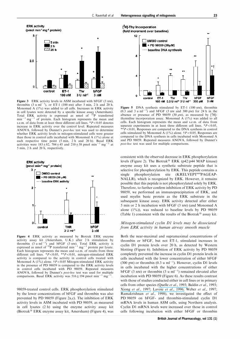
Figure 3 ERK activity levels in ASM incubated with bFGF (3 nM),thrombin (3 u ml71), or ET-1 (100 nM) after 5 min, 2 h and 20 h.Monomed A (1%) was added to all cells. Increases in ERK activityin cell lysates were detected by a speci®c kinase assay (Amersham).Total ERK activity is expressed as nmol of 32P transferredmin71 mg71 of protein. Each histogram represents the mean ands.e.m. of data from at least three di�erent cell lines. *P50.05 denotesincrease in ERK activity over the control level. Repeated measuresANOVA, followed by Dunnet's post-hoc test was used to determinewhether ERK activity levels in mitogen-stimulated cells were greaterthan those in control cells incubated with Monomed A (1%) alone ateach respective time point (5 min, 2 h and 20 h). Basal ERKactivities were 183+82, 704+45 and 210+58 pmol min71 mg71 at5 min, 2 h and 20 h, respectively.
Figure 4 ERK activity as measured by Biotrak ERK enzymeactivity assay kit (Amersham, U.K.) after 2 h stimulation bythrombin (3 u ml71) and bFGF (3 nM). Total ERK activity isexpressed as nmol of 32P transferred min71 mg71 protein per lysate.Each histogram represents the mean and s.e.m. of results from threedi�erent cell lines. *P50.05, **P50.01, mitogen-stimulated ERKactivity is compared to the activity in control cells treated withMonomed A (1%) alone. {P50.05 Mitogen-stimulated ERK activityin the presence of PD 98059 is compared to the ERK activity levelsin control cells incubated with PD 98059. Repeated measuresANOVA, followed by Dunnet's post-hoc test was used for multiplecomparisons. Basal ERK activity was 516+194 pmol min71 mg71.
Figure 5 DNA synthesis stimulated by ET-1 (100 nM), thrombin(0.3 and 3 u ml71) and bFGF (3 nM and 300 pM) for 24 h in theabsence or presence of PD 98059 (30 mM), as measured by [3H]-thymidine incorporation assay. Monomed A (1%) was added to allcells. Each histogram represents the mean and s.e.m. of data fromseparate experiments in at least three di�erent cell lines. *P50.05,**P50.01, Responses are compared to the DNA synthesis in controlcells stimulated by Monomed A (1%) alone. {P50.05, Responses arecompared to the DNA synthesis in cells incubated with Monomed Aand PD 98059. Repeated measures ANOVA, followed by Dunnet'spost-hoc test was used for multiple comparisons.
Heterogeneous signalling of mitogenesis 23C. Ravenhall et al
British Journal of Pharmacology, vol 131 (1)
(Figure 7). PD 98059 had no e�ect on the cyclin D1 mRNAlevels in cells stimulated by either of the lower or higher
concentrations of thrombin or bFGF.
Discussion and conclusion
Previous studies have demonstrated links between growthfactor receptor activation and persistent ERK activation
(Whelchel et al., 1997; Lenormand et al., 1993; Meloche etal., 1992; Kelleher et al., 1995), increased levels of cyclin D1(Baldin et al., 1993; Xiong et al., 1997; Lavoie et al., 1996;
Weber et al., 1997), retinoblastoma phosphorylation (Chellap-pan et al., 1991; Kato et al., 1993; Lundberg & Weinberg,1998) and/or DNA synthesis in various cell types (Tomlinson
et al., 1994; Panettieri et al., 1995; Karpova et al., 1997; Harriset al., 1997). We have contrasted the signalling pathwaysactivated by the mitogens thrombin and bFGF, and the non-
mitogen ET-1 in human airway smooth muscle to examine theassociation between persistent ERK activity and DNAsynthesis. Our data suggest that pathways signalling cell cycleprogression, in addition to those leading to ERK-dependent
elevation of cyclin D1 protein levels, are recruited uponexposure to powerful mitogens.
Thrombin and bFGF stimulated mitogenesis (Tomlinson et
al., 1995; Panettieri et al., 1995; Stewart et al., 1995a), whereasET-1 stimulated approximately 2 fold increases in DNAsynthesis in cultured human airway smooth muscle cells
without increasing cell number. This modest increase in DNAsynthesis was detectable by the highly sensitive [3H]-thymidineincorporation assay but not by FACS analysis. The increase inDNA synthesis without further detectable cell cycle progres-
sion could be indicative of the development of polyploidy, butno evidence for polyploidy was observed on FACS pro®les.PD 98059 prevented the ET-1 stimulated increase in DNA
synthesis suggesting that the increase was not due to celldamage. A small sub-population of the cells may be responsiveto ET-1 and the minor stimulation of DNA synthesis may
therefore re¯ect progression to S phase in that sub-population
alone. ET-1 levels are increased in the bronchoalveolar lavage(BAL) ¯uid of asthmatic airways (So®a et al., 1993). ET-1(100 nM) stimulates approximately half-maximal constriction
in isolated human airways (Hay et al., 1993; Henry et al., 1990)and therefore the concentration used in our study is expectedto activate smooth muscle ET-1 receptors. The concentrationof approximately 1 pM detected in BAL ¯uid of subjects with
in¯amed airways is ®ve orders of magnitude less than theconcentrations we used. Thus, even allowing for signi®cantdilution and metabolism in BAL, our data argues against a
direct role for ET-1 in the mitogenesis of human airwaysmooth muscle in vivo.
ERK activity and cyclin D1 protein accumulation have
been unequivocally implicated in the signalling of mitogenesisin multiple cell types. In this study, ERK activity was detectedby (a) phosphorylation of ERK detected by Western blotting(20 h), (b) ERK activity assay in cell extracts using a speci®c
substrate (5 min, 2 h and 20 h) and (c) ERK activity assay inERK immunoprecipitates using an MBP substrate (5 min and2 h). Thrombin and bFGF increased ERK activity for at least
2 h and increased both cyclin D1 mRNA and protein levels,
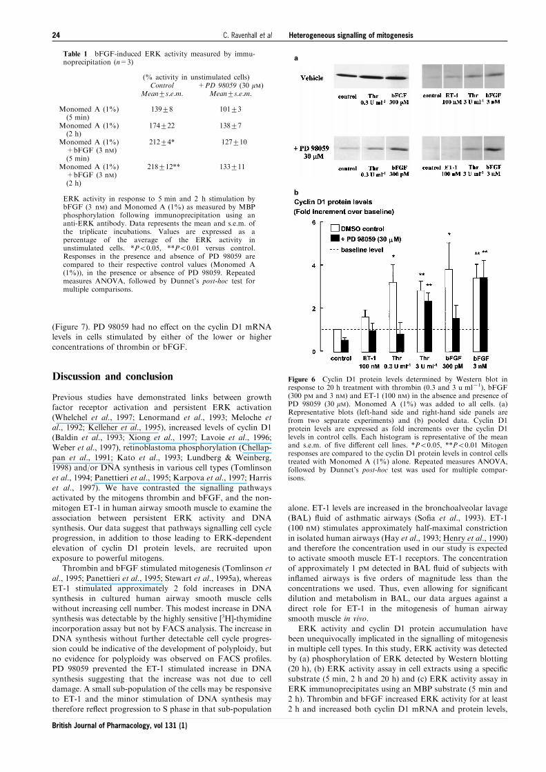
Table 1 bFGF-induced ERK activity measured by immu-noprecipitation (n=3)
(% activity in unstimulated cells)Control
Mean+s.e.m.+PD 98059 (30 mM)
Mean+s.e.m.
Monomed A (1%)(5 min)
Monomed A (1%)(2 h)
Monomed A (1%)+bFGF (3 nM)(5 min)
Monomed A (1%)+bFGF (3 nM)(2 h)
139+8
174+22
212+4*
218+12**
101+3
138+7
127+10
133+11
ERK activity in response to 5 min and 2 h stimulation bybFGF (3 nM) and Monomed A (1%) as measured by MBPphosphorylation following immunoprecipitation using ananti-ERK antibody. Data represents the mean and s.e.m. ofthe triplicate incubations. Values are expressed as apercentage of the average of the ERK activity inunstimulated cells. *P50.05, **P50.01 versus control.Responses in the presence and absence of PD 98059 arecompared to their respective control values (Monomed A(1%)), in the presence or absence of PD 98059. Repeatedmeasures ANOVA, followed by Dunnet's post-hoc test formultiple comparisons.
a
b
Figure 6 Cyclin D1 protein levels determined by Western blot inresponse to 20 h treatment with thrombin (0.3 and 3 u ml71), bFGF(300 pM and 3 nM) and ET-1 (100 nM) in the absence and presence ofPD 98059 (30 mM). Monomed A (1%) was added to all cells. (a)Representative blots (left-hand side and right-hand side panels arefrom two separate experiments) and (b) pooled data. Cyclin D1protein levels are expressed as fold increments over the cyclin D1levels in control cells. Each histogram is representative of the meanand s.e.m. of ®ve di�erent cell lines. *P50.05, **P50.01 Mitogenresponses are compared to the cyclin D1 protein levels in control cellstreated with Monomed A (1%) alone. Repeated measures ANOVA,followed by Dunnet's post-hoc test was used for multiple compar-isons.
Heterogeneous signalling of mitogenesis24 C. Ravenhall et al
British Journal of Pharmacology, vol 131 (1)
whereas ET-1 stimulated only a transient increase in ERKactivity and did not increase cyclin D1 protein or mRNA levelsor cell number (data not shown). The association between
sustained ERK activity and mitogenesis is consistent with therequirement for sustained ERK activation for mitogenesis inCCL39 cells (Meloche et al., 1992), in which ERK activity was
elevated 4 h after thrombin stimulation and in bovine trachealsmooth muscle cells (Kelleher et al., 1995) in which ERKactivity was elevated for 6 h in response to PDGF. The
addition of PD 98059 to thrombin-stimulated cells as late as6 h after thrombin addition still reduces DNA synthesis, fromwhich it is inferred that ERK activity is required for at least the
®rst 6 h of mitogen signalling (Fernandes et al., 1999).High basal levels of ERK activity, cyclin D1 protein levels
and DNA synthesis result from stimulation by the serum-freemedium growth supplement, Monomed A (comprising in part
selenium, transferrin and insulin). Insulin activates signalling
pathways, including the ERK cascade (Boulton et al., 1991;Avruch, 1998) and may therefore account for `basal' ERKactivity. Although the exposure of cultured cells to the serum-
free medium supplement decreases the apparent magnitude ofthe mitogen-induced increases in signalling through certainpathways by increasing the basal signalling, this supplement isused to provide an environment that more closely approx-
imates that encountered in vivo and is required for cellproliferation and survival. The speci®c activity of ERK,measured by the amount of 32P transferred to MBP
min71 mg71 protein is comparable to that measured inprevious studies in response to PDGF in guinea-pig ASM(Conway et al., 1999).
PD 98059, a speci®c inhibitor of MEK1 (Dudley et al.,1995), was used to characterize the role of MEK1 and ERKregulation of cyclin D1 levels in DNA synthesis. As previously
shown in PDGF-stimulated bovine ASM cells (Karpova et al.,1997), PD 98059 blocked thrombin-stimulated DNA synthesis,indicating that the activation of MEK1 is required for DNAsynthesis. The inhibitory e�ects of PD 98059 on increases in
cyclin D1 protein levels stimulated by lower concentrations ofthrombin are consistent with links between persistent ERKactivity, increased cyclin D1 protein levels and cell cycle
progression to S phase (Lavoie et al., 1996; Weber et al., 1997;Ramakrishnan et al., 1998). However, in cells incubated withthe higher concentrations of thrombin, cyclin D1 protein levels
increased in the absence of a detectable increase in ERKactivity. The PI(3)K-dependent signalling pathway representsan alternative pathway for the maintenance of elevated cyclin
D1 protein levels, since the PI(3)K pathway prolongs the halflife of cyclin D1 protein via its downstream e�ector, PKB(Diehl et al., 1997). PKB down regulates GSK-3b whichfacilitates the phosphorylation of Thr-286 in cyclin D1, the
residue known to be targeted by the ubiquitin-dependentproteasome degradation pathway (Diehl et al., 1997). Thus,the PI(3)K pathway may maintain cyclin D1 levels during
exposure to high mitogen concentrations independently ofERK activity. However, inhibition of PI(3)K activation, whichattenuates DNA synthesis, does not a�ect ERK activity levels,
indicating the independent regulation of the ERK and PI(3)Ksignalling pathways (Krymskaya et al., 1999).
Thrombin-induced increases in cyclin D1 mRNA levelswere una�ected by PD 98059 suggesting that there is an ERK-
independent signalling pathway for the regulation of cyclin D1mRNA in human ASM. In contrast, in transformed cell linesERK activity leads to stimulation of cyclin D1 promoter
activity (Lavoie et al., 1996; Weber et al., 1997; Ramakrishnanet al., 1998). An association between cyclin D1 promoteractivity and ERK activation has been demonstrated in several
di�erent cell types including a human adrenal cell line(Watanabe et al., 1996), a Chinese hamster lung ®broblast cellline (Lavoie et al., 1996), a human trophoblast cell line, a Mink
lung epithelial cell line, a Chinese ovary ®broblast cell line(Albanese et al., 1995) and in primary bovine ASM cells(Ramakrishnan et al., 1998). Mitogen-stimulated cyclin D1mRNA levels are reduced by PD 98059 in Chinese hamster
lung ®broblasts (Weber et al., 1997) and in T-47D humanbreast cancer cells (Fiddes et al., 1998). Glucocorticoids reducecyclin D1 mRNA (Fernandes et al., 1999), but b2 agonists do
not (Stewart et al., 1999). Increased cyclic AMP levels appearto accelerate cyclin D1 degradation by a pathway that issensitive to inhibition by MG132 (Stewart et al., 1999), a
potent, reversible inhibitor of proteasome function (Lee &Goldberg, 1996). Thus, lower mitogen concentrations maynegatively regulate cyclin D1 degradation by an inhibitoryaction of ERK on the proteasome degradation pathway.
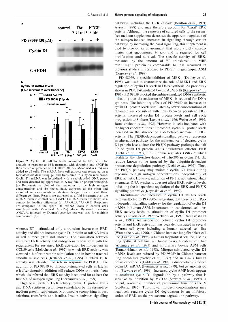
a
b
Figure 7 Cyclin D1 mRNA levels measured by Northern blotanalysis in response to 16 h treatment with thrombin and bFGF inthe absence or presence of PD 98059 (30 mM). Monomed A (1%) wasadded to all cells. The mRNA from cell extracts was separated on aformaldehyde denaturing gel and transferred to a nylon membrane.Cyclin D1 mRNA was hybridized with a radiolabelled DNA probeand then detected by apposition to X-ray ®lm or phosphorimaging.(a) Representative blot of the responses to the high mitogenconcentrations and (b) pooled data, expressed as the mean ands.e.m. of six experiments of identical design from at least threedi�erent cell lines. Results are expressed as a fold increment over themRNA levels in control cells. GAPDH mRNA levels are shown as acontrol for loading di�erences (a). *P50.05, **P50.01 Responsesare compared to the cyclin D1 mRNA levels in control cellsstimulated with Monomed A (1%) alone. Repeated measuresANOVA, followed by Dunnet's post-hoc test was used for multiplecomparisons (b).
Heterogeneous signalling of mitogenesis 25C. Ravenhall et al
British Journal of Pharmacology, vol 131 (1)

The nature of the ERK-independent regulation of cyclin D1mRNA levels in human ASM remains unclear. Cyclin D1promoter activity may be increased by c-fos and c-jun, which
make up the AP-1 complex, and c-Ets-2 (Albanese et al., 1995).The transcription factor c-Myc is also known to increase cyclinD1 mRNA and protein levels through its e�ect on eIF-4E andeIF-2a (Rosenwald et al., 1993, 1995). In addition, a change in
the synthetic rate of cyclin D1 protein synthesis, prior tochanges in cyclin D1 mRNA expression, has been demon-strated in human mammary epithelial cell lines. Regulation of
the translation of cyclin D1 independent of ERK activity ismediated via the PI(3)K and Akt kinase signalling pathways(Muise-Helmericks et al., 1998). Thus, the di�erences in
mechanisms reported to regulate cyclin D1 mRNA in di�erentcell types raise the possibility of cell-type selective pharmaco-logical regulation.
The species-dependent e�ects of EGF, a potent humanASM mitogen, but a non-mitogenic agent in bovine ASM(Tomlinson et al., 1994; Kelleher et al., 1995) and themitogenicity of ET-1 in ovine, guinea-pig, rat and rabbit, but
not human ASM (Panettieri et al., 1995; Noveral et al., 1992;Glassberg et al., 1994; Stewart et al., 1994) may be explainedby di�erential recruitment of signalling pathways in response
to the same agent, in di�erent species. Di�erential recruitmentof signalling pathways has also been demonstrated in responseto di�erent isoforms of mitogens in the same tissue type. In the
absence of serum, PDGF-BB and PDGF-AB but not PDGF-AA stimulate human ASM mitogenesis (Hirst et al., 1996).
The results of the present study indicate that the recruitmentof signalling pathways is not only dependent on mitogen typebut also on mitogen concentration. The activation of ERK in
human ASM appears to be largely, if not exclusively MEK1-dependent during both rapid (5 min) and sustained responses(2 h). Our data support an association between persistentincreases in ERK activity, cyclin D1 protein levels and DNA
synthesis stimulated by the lower concentrations of thrombin(0.3 u ml71). In the presence of high concentrations ofthrombin, MEK1/ERK-independent pathways may also be
important for the induction and maintenance of cyclin D1mRNA and protein levels. Such redundancies in the signallingpathways controlling ASM mitogenesis may limit the
therapeutic potential of agents targeting the MEK1/ERKpathway alone.
This work was supported by project grants from the NationalHealth & Medical Research Council of Australia and GlaxoWellcome (U.K.). We would like to thank Dr Darren Fernandesfor assistance with the ERK immunoprecipitation and subsequentkinase assay. We also thank Associate Professor John Wilson, DrXun Li, Mr John Bartolo and the sta� from the transplant unit atthe Alfred Hospital for the provision of human lung resectionspecimens.
References
ALBANESE, C., JOHNSON, J., WATANABE, G., EKLUND, N., VU, D.,
ARNOLD, A. & PESTELL, RG. (1995). Transforming p21rasmutants and c-Ets-2 activate the cyclin D1 promoter throughdistinguishable regions. J. Biol. Chem., 270, 23589 ± 23597.
AVRUCH, J. (1998). Insulin signal transduction through proteinkinase cascades. Mol. Cell Biochem., 182, 31 ± 48.
BALDIN, V., LUKAS, J., MARCOTE, M.J., PAGANO, M. & DRAETTA,
G. (1993). Cyclin D1 is a nuclear protein required for cell cycleprogression in G1. Genes Dev., 7, 812 ± 821.
BORNFELDT, K.E., CAMPBELL, J.S., KOYAMA, H., ARGAST, G.M.,
LESLIE, C.C., RAINES, E.W., KREBS, E.G. & ROSS, R. (1997). Themitogen-activated protein kinase pathway can mediate growthinhibition and proliferation in smooth muscle cells. Dependenceon the availability of downstream targets. Br. J. Pharmacol., 100,875 ± 885.
BOULTON, T.G., NYE, S.H., ROBBINS, D.J., IP, N.Y., RADZIEJEWS-
KA, E., MORGENBESSER, S.D., DEPINHO, R.A., PANAYOTATOS,
N., COBB, M.H. & YANCOPOULOS, G.D. (1991). ERKs: a familyof protein-serine/threonine kinases that are activated andtyrosine phosphorylated in response to insulin and NGF. Cell,65, 663 ± 675.
BRAMLEY, A.M., ROBERTS, C.R. & SCHELLENBERG, R.R. (1995).Collagenase increases shortening of human bronchial smoothmuscle in vitro. Am. J. Respir. Crit. Care Med., 152, 1513 ± 1517.
CHELLAPPAN, S.P., HIEBERT, S., MUDRYI, M., HOROWITZ, J.M. &
NEVINS, J.R. (1991). The E2F transcription factor is a cellulartarget for the RB protein. Cell, 65, 1053 ± 1061.
CHEN, R.H., SARNECKI, C. & BLENIS, J. (1992). Nuclear localizationand regulation of erk- and rsk-encoded protein kinases. Mol. CellBiol., 12, 915 ± 927.
CONWAY, A.M., RAKHIT, S., PYNE, S. & PYNE, N.J. (1999). Platelet-derived-growth-factor stimulation of the p42/p44 mitogen-activated protein kinase pathway in airway smooth muscle: roleof pertussis-toxin-sensitive G proteins, c-Src tyrosine kinases andphosphoinositide 3-kinase. Biochem. J., 337, 171 ± 177.
DE, S., ZELAZNY, E.T., SOUHRADA, J.F. & SOUHRADA, M. (1993).Interleukin-1 beta stimulates the proliferation of cultured airwaysmooth muscle cells via platelet-derived growth factor. Am. J.Respir. Cell Mol. Biol., 9, 645 ± 651.
DE, S., ZELAZNY, E.T., SOUHRADA, J.F. & SOUHRADA, M. (1995).IL-1 beta and IL-6 induce hyperplasia and hypertrophy ofcultured guinea pig airway smooth muscle cells. J. Appl. Physiol.,78, 1555 ± 1563.
DIEHL, J.A., ZINDY, F. & SHERR, C.J. (1997). Inhibition of cyclin D1phosphorylation on threonine-286 prevents its rapid degradationvia the ubiquitin-proteasome pathway. Genes Dev., 11, 957 ± 972.
DUDLEY, D.T., PANG, L., DECKER, S.J., BRIDGES, A.J. & SALTIEL,
A.R. (1995). A synthetic inhibitor of the mitogen-activatedprotein kinase cascade. Proc. Natl. Acad. Sci. U.S.A., 92,7686 ± 7689.
DUGAICZYK, A., HARON, J.A., STONE, E.M., DENNISON, O.E.,
ROTHBLUM, K.N. & SCHWARTZ, R.J. (1983). Cloning andsequencing of a deoxyribonucleic acid copy of glyceraldehyde-3-phsophate dehydrogenase messenger Ribonucleic acid isolatedfrom chicken muscle. Biochemistry, 22, 1605 ± 1613.
DUNNILL, M.S. & MASSARELLA, G.R. (1969). A comparison of thequantitative anatomy of the bronchi in normal subjects, in statusasthmaticus, in chronic bronchitis, and in emphysema. Thorax,24, 176 ± 179.
EBINA, M., TAKAHASHI, T., CHIBA, T. & MOTOMIYA, M. (1993).Cellular hypertrophy and hyperplasia of airway smooth musclesunderlying bronchial asthma. A 3-D morphometric study. Am.Rev. Respir. Dis., 148, 720 ± 726.
FERNANDES, D., GUIDA, E., KOUTSOUBOS, V., HARRIS, T.,
VADIVELOO, P., WILSON, J.W. & STEWART, A.G. (1999).Glucocorticoids inhibit proliferation, cyclin D1 expression, andretinoblastoma protein phosphorylation, but not activity of theextracellular-regulated kinases in human cultured airway smoothmuscle. Am. J. Respir. Cell Mol. Biol., 21, 77 ± 88.
FIDDES, R.J., JANES, P.W., SIVERTSEN, S.P., SUTHERLAND, R.L.,
MUSGROVE, E.A. & DALY, R.J. (1998). Inhibition of the MAPkinase cascade blocks heregulin-induced cell cycle progression inT-47D human breast cancer cells. Oncogene, 16, 2803 ± 2813.
GABAZZA, E.C., TAGUCHI, O., KOBAYASHI, T., KOBAYASHI, H.,
HATAJI, O., YASUI, H. & ADACHI, Y. (1998). Role of thrombin inairway remodeling. Am. J. Respir. Crit. Care Med., 157,A33(Abstract).
Heterogeneous signalling of mitogenesis26 C. Ravenhall et al
British Journal of Pharmacology, vol 131 (1)
GILLE, H., KORTENJANN, M., THOMAE, O., MOOMAW, C.,
SLAUGHTER, C., COBB, M.H. & SHAW, P.E. (1995). ERKphosphorylation potentiates Elk-1-mediated ternary complexformation and transactivation. EMBO J., 14, 951 ± 962.
GLASSBERG, M.K., ERGUL, A., WANNER, A. & PUETT, D. (1994).Endothelin-1 promotes mitogenesis in airway smooth musclecells. Am. J. Respir. Cell Mol. Biol., 10, 316 ± 321.
GRAMMER, T.C. & BLENIS, J. (1997). Evidence for MEK-independent pathways regulating the prolonged activation ofthe ERK-MAP kinases. Oncogene, 14, 1635 ± 1642.
HARRIS, T., RAVENHALL, C.E., SCHACHTE, L.C. & STEWART, A.G.
(1997). Relationship between airway smooth muscle mitogenesisand levels of cyclin D1 and p27Kip1. Am. J. Respir. Crit. CareMed., 155, A905.
HAY, D.W., HUBBARD, W.C. & UNDEM, B.J. (1993). Endothelin-induced contraction and mediator release in human bronchus.Br. J. Pharmacol., 110, 392 ± 398.
HENRY, P.J., RIGBY, P.J., SELF, G.J., PREUSS, J.M.H. & GOLDIE, R.G.
(1990). Relationship between endothlin-1 binding site densitiesand constrictor activities in human and animal airway smoothmuscle. Br. J. Pharmacol., 110, 1175 ± 1183.
HIRST, S.J., BARNES, P.J. & TWORT, C.H. (1996). PDGF isoform-induced proliferation and receptor expression in human culturedairway smooth muscle cells. Am. J. Physiol., 270, L415 ± L428.
HOSSAIN, S. (1973). Quantitative measurement of bronchial musclein men with asthma. Am. Rev. Respir. Dis., 107, 99 ± 109.
JAMES, A.L., PARE, P.D. & HOGG, J.C. (1989). The mechanics ofairway narrowing in asthma. Am. Rev. Respir. Dis., 139, 242 ±246.
JANKNECHT, R., ERNST, W.H., & NORDHEIM, A. (1995). SAP1a is anuclear target of signaling cascades involving ERKs. Oncogene,10, 1209 ± 1216.
JOHNSON, S.R. & KNOX, A.J. (1997). Synthetic functions of airwaysmooth muscle in asthma. Trends. Pharmacol. Sci., 18, 289 ± 292.
KARPOVA, A.Y., ABE, M.K., LI, J., LIU, P.T., RHEE, J.M., KUO, W.L. &
HERSHENSON, M.B. (1997). MEK1 is required for PDGF-induced ERK activation and DNA synthesis in trachealmyocytes. Am. J. Physiol., 272, L558 ± L565.
KATO, S., ENDOH, H., MASUHIRO, Y., KITAMOTO, T., UCHIYAMA,
S., SASAKI, H., MASUSHIGE, S., GOTOH, Y., NISHIDA, E.,
KAWASHIMA, H., METZGER, D. & CHAMBON, P. (1995).Activation of the estrogen receptor through phosphorylationby mitogen-activated protein kinase. Science, 270, 1491 ± 1494.
KATO, J., MATSUSHIME, H., HIEBERT, S.W., EWEN, M.E. & SHERR,
C.J. (1993). Direct binding of cyclin D to the retinoblastoma geneproduct (pRb) and pRb phosphorylation by the cyclin D-dependent kinase CDK4. Genes Development, 7, 331 ± 342.
KELLEHER, M.D., ABE, M.K., CHAO, T.O., JAIN, M., GREEN, J.M.,
SOLWAY, J., ROSNER, M.R. & HERSHENSON, M.B. (1995). Roleof MAP kinase activation in bovine tracheal smooth musclemitogenesis. Am. J. Physiol., 268, L894 ± L901.
KRYMSKAYA, V.P., PENN, R.B., ORSINI, M.J., SCOTT, P.H., PLEVIN,
R.J., WALKER, T.R., ESTERHAS, A.J., AMRANI, Y., CHILVERS,
E.R. & PANETTIERI, R.A. (1999). Phosphatidyl 3-Kinase mediatesmitogen-induced human airway smooth muscle cell proliferation.Am. J. Physiol., 277, L65 ± L78.
L'ALLEMAIN, G., STURGILL, T.W. & WEBER, M.J. (1991). Defectiveregulation of mitogen-activated protein kinase activity in a 3T3cell variant mitogenically nonresponsive to tetradecanoylphorbol acetate. Mol. Cell Biol., 11, 1002 ± 1008.
LAVOIE, J.N., L'ALLEMAIN, G., BRUNET, A., MULLER, R., &
POUYSSEGUR, J. (1996). Cyclin D1 expression is regulatedpositively by the p42/p44MAPK and negatively by the p38/HOGMAPK pathway. J. Biol. Chem., 271, 20608 ± 20616.
LEE, D.H. & GOLDBERG, A.L. (1996). Selective inhibitors of theproteasome-dependent and vacuolar pathways of proteindegradation in Saccharomyces cerevisiae. J. Biol. Chem., 271,27280 ± 27284.
LENORMAND, P., SARDET, C., PAGES, G., L'ALLEMAIN, G.,
BRUNET, A., & POUYSSEGUR, J. (1993). Growth factors inducenuclear translocation of MAP kinases (p42mapk and p44mapk)but not of their activator MAP kinase kinase (p45mapkk) in®broblasts. J. Cell. Biol., 122, 1079 ± 1088.
LUNDBERG, A.S. &WEINBERG, R.A. (1998). Functional inactivationof the retinoblastoma protein requires sequential modi®cation byat least two distinct cyclin-cdk complexes. Mol. Cell Biol., 18,753 ± 761.
MARUNO, K., ABSOOD, A. & SAID, S.I. (1995). VIP inhibits basal andhistamine-stimulated proliferation of human airway smoothmuscle cells. Am. J. Physiol., 268, L1047 ± L1051.
MATSUSHIME, H., QUELLE, D.E., SHURTLEFF, S.A., SHIBUYA, M.,
SHERR, C.J. & KATO, J.Y. (1994). D-type cyclin-dependent kinaseactivity in mammalian cells. Mol. Cell. Biol., 14, 2066 ± 2076.
MELOCHE, S. (1995). Cell cycle reentry of mammalian ®broblasts isaccompanied by the sustained activation of p44mapk andp42mapk isoforms in the G1 phase and their inactivation at theG1/S transition. J. Cell. Physiol., 163, 577 ± 588.
MELOCHE, S., SEUWEN, K., PAGES, G. & POUYSSEGUR, J. (1992).Biphasic and synergistic activation of p44mapk (ERK1) by growthfactors: correlation between late phase activation and mitogeni-city. Mol. Endocrinol., 6, 845 ± 854.
MULSE-HELMERICKS, R.C., GRIMES, H.L., BELLACOSA, A., MAL-
STROM, S.E., TSICHRIS, P.N. & ROSEN, N. (1998). Cyclin D1expression is controlled post-transcriptionally via a phagohati-dyl-inositol 3-kinose/Akt-dependent pathway. J. Biol. Chem.,273, 29864 ± 29872.
NOVERAL, J.P., ROSENBERG, S.M., ANBAR, R.A., PAWLOWSKI,
N.A. & GRUNSTEIN, M.M. (1992). Role of endothelin-1 inregulating proliferation of cultured rabbit airway smooth musclecells. Am. J. Physiol., 263, L317 ± L324.
PANETTIERI, R.A., GOLDIE, R.G., RIGBY, P.J., ESZTERHAS, A.J. &
HAY, D.W. (1996). Endothelin-1-induced potentiation of humanairway smooth muscle proliferation: an ETA receptor-mediatedphenomenon. Br. J. Pharmacol., 118, 191 ± 197.
PANETTIERI, R.A., HALL, I.P., MAKI, C.S. & MURRAY, R.K. (1995).alpha-Thrombin increases cytosolic calcium and induces humanairway smooth muscle cell proliferation. Am. J. Respir. Cell Mol.Biol., 13, 205 ± 216.
PANETTIERI, R.A., MURRAY, R.K., DEPALO, L.R., YADVISH, P.A. &
KOTLIKOFF, M.I. (1989). A human airway smooth muscle cellline that retains physiological responsiveness. Am. J. Physiol.,256, C329 ±C335.
POSADA, J. & COOPER, J.A. (1992). Requirements for phosphoryla-tion of MAP kinase during meiosis in Xenopus oocytes. Science,255, 212 ± 215.
PYNE, N.J., TOLAN, D. & PYNE, S. (1997). Bradykinin stimulatescAMP synthesis via mitogen-activated protein kinase-dependentregulation of cytosolic phospholipase A2 and prostaglandin E2release in airway smooth muscle. Biochem. J., 328, 689 ± 694.
QUELLE, D.E., ASHMUN, R.A., SHURTLEFF, S.A., KATO, J.Y., BAR-
SAGI, D., ROUSSEL, M.F. & SHERR, C.J. (1993). Overexpressionof mouse D-type cyclins accelerates G1 phase in rodent®broblasts. Genes Dev., 7, 1559 ± 1571.
RAMAKRISHNAN, M., MUSA, N.L., LI, J., LIU, P.T., PESTELL, R.G. &
HERSHENSON,M.B. (1998). Catalytic Activation of ExtracellularSignal-regulated Kinases Induces Cyclin D1 Expression inPrimary Tracheal Myocytes. Am. J. Respir. Cell Mol. Biol., 18,736 ± 740.
REDINGTON, A.E., MADDEN, J., FREW, A.J., DJUKANOVIC, R.,
ROCHE, W., HOLGATE, S.T. & HOWARTH, P. (1995). Basic®broblast growth factor in asthma: immunolocalization inbronchial biopsies and measurement in bronchoalveolar lavage¯uid at baseline and following allergen challenge. Am. J. Respir.Crit. Care Med., 151, A702.
ROSENWALD, I.B., KASPAR, R., ROUSSEAU, D., GEHRKE, L.,
LEBOULCH, P., CHEN, J.J., SCHMIDT, E.V., SONENBERG, N. &
LONDON, I.M. (1995). Eukaryotic translation initiation factor 4Eregulates expression of cyclin D1 at transcriptional and post-transcriptional levels. J. Biol. Chem., 270, 21176 ± 21180.
ROSENWALD, I.B., RHOADS, D.B., CALLANAN, L.D., ISSELBA-
CHER, K.J. & SCHMIDT, E.V. (1993). Increased expression ofeukaryotic translation initiation factors eIF- 4E and eIF-2 alphain response to growth induction by c-myc. Proc. Natl. Acad. Sci.U.S.A., 90, 6175 ± 6178.
SHAPIRO, P.S., EVANS, J.N., DAVIS, R.J. & POSADA, J.A. (1996). Theseven-transmembrane-spanning receptors for endothelin andthrombin cause proliferation of airway smooth muscle cells andactivation of the extracellular regulated kinase and c-Jun NH2-terminal kinase groups of mitogen-activated protein kinases. J.Biol. Chem., 271, 5750 ± 5754.
SHERR, C.J. (1993). Mammalian G1 cyclins. Cell, 73, 1059 ± 1065.SHERR, C.J. (1995). D-type cyclins. Trends. Biochem. Sci., 20, 187 ±
190.SOFIA, M., MORMILE, M., FARAONE, S., ALIFANO, M., ZOFRA, S.,
ROMANO, L., CARRATU. & L. (1993). Increased endothelin-likeimmunoreactive material on bronchoalveolar lavage ¯uid frompatients with bronchial asthma and patients with interstitial lungdisease. Respiration, 60, 89 ± 95.
Heterogeneous signalling of mitogenesis 27C. Ravenhall et al
British Journal of Pharmacology, vol 131 (1)
SPRINGALL, D.R., HOWARTH, P.H., COUNIHAN, H., DJUKANOVIC,
R., HOLGATE, S.T. & POLAK. J.M. (1991). Endothelin immunor-eactivity of airway epithelium in asthmatic patients. Lancet, 337,697 ± 701.
STEWART, A.G., FERNANDES, D. & TOMLINSON, P.R. (1995a). Thee�ect of glucocorticoids on proliferation of human culturedairway smooth muscle. Br. J. Pharmacol., 116, 3219 ± 3226.
STEWART, A.G., GRIGORIADIS, G. & HARRIS, T. (1994). Mitogenicactions of endothelin-1 and epidermal growth factor in culturedairway smooth muscle. Clin. Exp. Pharmacol. Physiol., 21, 277 ±285.
STEWART, A.G., HARRIS, T., FERNANDES, D.J., SCHACHTE, L.C.,
KOUTSOUBOS, V., GUIDA, E., RAVENHALL, C.E., VADIVELOO,
P. & WILSON, J.W. (1999). Beta2-adrenergic agonists and cAMParrest human cultured airway smooth muscle cells in G1 phase ofthe cell cycle: role of proteasome degradation of cyclin D1. Mol.Pharmacol., 56, 1079 ± 1086.
STEWART, A.G., TOMLINSON, P.R. & WILSON, J.W. (1995b).Regulation of airway wall remodelling: Prospects for thedevelopment of novel anti-asthma Drugs. In: Advances inPharmacology. ed. August, J.T., Anders, M.W., Murad, F. &Coyle, J.T. pp 209 ± 254. San Diego, Academic Press.
STEWART, A.G., TOMLINSON, P.R. & WILSON, J.W. (1997a).Regulation of airway smooth muscle proliferation by b2-adrenoceptor agonists. In: Airway wall remodelling in asthma.pp. 295 ± 330. Anonymous Boca Raton, CRC Press inc.
STEWART, A.G., TOMLINSON, P.R. & WILSON, J.W. (1997b). b2-adrenoceptor agonist-mediated inhibition of human airwaysmooth muscle cell proliferation: importance of the duration ofb2-adrenoceptor stimulation. Br. J. Pharmacol., 121, 361 ± 368.
TOMLINSON, P.R., WILSON, J.W. & STEWART, A.G. (1994).Inhibition by salbutamol of the proliferation of human airwaysmooth muscle cells grown in culture. Br. J. Pharmacol., 111,641 ± 647.
TOMLINSON, P.R., WILSON, J.W. & STEWART, A.G. (1995).Salbutamol inhibits the proliferation of human airway smoothmuscle cells grown in culture: relationship to elevated cAMPlevels. Biochem. Pharmacol., 49, 1809 ± 1819.
VLAHOS, R. & STEWART, A.G. (1999). Interleukin-1a and tumournecrosis factor-amodulate airway smooth muscle DNA synthesisby induction of cyclo-oxygenase-2: inhibition by dexamethasoneand ¯uticasone propionate. Br. J. Pharmacol., 126, 1315 ± 1324.
WALKER, T.R., MOORE, S.M., LAWSON, M.F., PANETTIERI, R.A.J. &
CHILVERS, E.R. (1998). Platelet-Derived Growth Factor-BB andThrombin Activate Phosphoinositide 3-Kinase and ProteinKinase B: Role in Mediating Airway Smooth Muscle Prolifera-tion. Mol. Pharmacol., 54, 1007 ± 1015.
WATANABE, G., LEE, R.J., ALBANESE, C., RAINEY, W.E., BATLLE,
D. & PESTELL, R.G. (1996). Angiotensin II activation of cyclinD1-dependent kinase activity. J. Biol. Chem., 271, 22570 ± 22577.
WEBER, J.D., RABEN, D.M., PHILLIPS, P.J. & BALDASSARE, J.J.
(1997). Sustained activation of extracellular-signal-regulatedkinase 1 (ERK1) is required for the continued expression ofcyclin D1 in G1 phase. Biochem. J., 326, 61 ± 68.
WHELCHEL, A., EVANS, J. & POSADA, J. (1997). Inhibition of ERKactivation attenuates endothelin-stimulated airway smoothmuscle cell proliferation. Am. J. Respir. Cell Mol. Biol., 16,589 ± 596.
WIGGS, B.R., BOSKEN, C., PARE, P.D., JAMES, A. & HOGG, J.C.
(1992). A model of airway narrowing in asthma and in chronicobstructive pulmonary disease.Am. Rev. Respir. Dis., 145, 1251 ±1258.
XIONG, Y., CONOLLY, T., FUTCHER, B. & BEACH, D. (1991). HumanD-type cyclin. Cell, 65, 691 ± 699.
XIONG, W., PESTELL, R.G., WATANABE, G., LI, J., ROSNER, M.R. &
HERSHENSON, M.B. (1997). Cyclin D1 is required for S phasetraversal in bovine tracheal myocytes. Am. J. Physiol., 272,L1205 ± L1210.
(Received February 3, 2000Revised May 4, 2000
Accepted May 4, 2000)
Heterogeneous signalling of mitogenesis28 C. Ravenhall et al
British Journal of Pharmacology, vol 131 (1)