Cyclin B/CDK1 and Cyclin A/CDK2 phosphorylate DENR to ...
14
ARTICLE Cyclin B/CDK1 and Cyclin A/CDK2 phosphorylate DENR to promote mitotic protein translation and faithful cell division Katharina Clemm von Hohenberg 1,2,3,4 , Sandra Müller 1,2 , Sibylle Schleich 1,2 , Matthias Meister 5 , Jonathan Bohlen 1,2,3,6,7 , Thomas G. Hofmann 8 & Aurelio A. Teleman 1,2,3 ✉ DENR and MCTS1 have been identified as oncogenes in several different tumor entities. The heterodimeric DENR·MCTS1 protein complex promotes translation of mRNAs containing upstream Open Reading Frames (uORFs). We show here that DENR is phosphorylated on Serine 73 by Cyclin B/CDK1 and Cyclin A/CDK2 at the onset of mitosis, and then depho- sphorylated as cells exit mitosis. Phosphorylation of Ser73 promotes mitotic stability of DENR protein and prevents its cleavage at Asp26. This leads to enhanced translation of mRNAs involved in mitosis. Indeed, we find that roughly 40% of all mRNAs with elevated translation in mitosis are DENR targets. In the absence of DENR or of Ser73 phosphorylation, cells display elevated levels of aberrant mitoses and cell death. This provides a mechanism how the cell cycle regulates translation of a subset of mitotically relevant mRNAs during mitosis. https://doi.org/10.1038/s41467-022-28265-0 OPEN 1 German Cancer Research Center (DKFZ), 69120 Heidelberg, Germany. 2 Heidelberg University, 69120 Heidelberg, Germany. 3 CellNetworks—Cluster of Excellence, Heidelberg University, Heidelberg, Germany. 4 Department of Medicine III, Universitätsmedizin Mannheim, 68167 Mannheim, Germany. 5 Division of Viral Transformation Mechanisms, German Cancer Research Center (DKFZ), Heidelberg, Germany. 6 Laboratory of Human Genetics of Infectious Diseases, Necker Branch, INSERM U1163 Paris, France. 7 University of Paris, Imagine Institute, Paris, France. 8 Institute of Toxicology, University Medical Center Mainz at the Johannes Gutenberg University of Mainz, Mainz, Germany. ✉ email: [email protected] NATURE COMMUNICATIONS | (2022)13:668 | https://doi.org/10.1038/s41467-022-28265-0 | www.nature.com/naturecommunications 1 1234567890():,;
-
Upload
khangminh22 -
Category
Documents
-
view
3 -
download
0
Transcript of Cyclin B/CDK1 and Cyclin A/CDK2 phosphorylate DENR to ...

ARTICLE
Cyclin B/CDK1 and Cyclin A/CDK2 phosphorylateDENR to promote mitotic protein translation andfaithful cell divisionKatharina Clemm von Hohenberg 1,2,3,4, Sandra Müller1,2, Sibylle Schleich1,2, Matthias Meister5,
Jonathan Bohlen 1,2,3,6,7, Thomas G. Hofmann 8 & Aurelio A. Teleman 1,2,3✉
DENR and MCTS1 have been identified as oncogenes in several different tumor entities. The
heterodimeric DENR·MCTS1 protein complex promotes translation of mRNAs containing
upstream Open Reading Frames (uORFs). We show here that DENR is phosphorylated on
Serine 73 by Cyclin B/CDK1 and Cyclin A/CDK2 at the onset of mitosis, and then depho-
sphorylated as cells exit mitosis. Phosphorylation of Ser73 promotes mitotic stability of DENR
protein and prevents its cleavage at Asp26. This leads to enhanced translation of mRNAs
involved in mitosis. Indeed, we find that roughly 40% of all mRNAs with elevated translation
in mitosis are DENR targets. In the absence of DENR or of Ser73 phosphorylation, cells
display elevated levels of aberrant mitoses and cell death. This provides a mechanism how
the cell cycle regulates translation of a subset of mitotically relevant mRNAs during mitosis.
https://doi.org/10.1038/s41467-022-28265-0 OPEN
1 German Cancer Research Center (DKFZ), 69120 Heidelberg, Germany. 2 Heidelberg University, 69120 Heidelberg, Germany. 3 CellNetworks—Cluster ofExcellence, Heidelberg University, Heidelberg, Germany. 4Department of Medicine III, Universitätsmedizin Mannheim, 68167 Mannheim, Germany.5 Division of Viral Transformation Mechanisms, German Cancer Research Center (DKFZ), Heidelberg, Germany. 6 Laboratory of Human Genetics ofInfectious Diseases, Necker Branch, INSERM U1163 Paris, France. 7 University of Paris, Imagine Institute, Paris, France. 8 Institute of Toxicology, UniversityMedical Center Mainz at the Johannes Gutenberg University of Mainz, Mainz, Germany. ✉email: [email protected]
NATURE COMMUNICATIONS | (2022) 13:668 | https://doi.org/10.1038/s41467-022-28265-0 | www.nature.com/naturecommunications 1
1234
5678
90():,;

The ability of mammalian cells to adjust their gene expres-sion to their environmental and developmental state isvital1. To this end, one of the most important layers of
regulation is mRNA translation2–5. For instance, in response tostress or quiescence, canonical translation is diminished andcellular translation becomes more dependent on noncanonicalfactors6,7. In proliferating cells, the various phases of the cell cyclelikely impose distinct gene expression requirements on the cell,for instance necessitating nucleotide biosynthesis proteins andhistones during S-phase or spindle components during mitosis8,9.The molecular mechanisms regulating protein translation at dif-ferent phases of the cell cycle, such as mitosis, are not wellunderstood.
Mitotic translation has been a topic of interest the last fewyears10–17. Several studies have investigated the mitotic regulationof translation, thereby identifying mRNAs that are selectivelytranslated during mitosis5,8,18–23. Mitosis is a key feature ofproliferative cells, such as tumor cells, hence mitotic translationmay represent an Achilles’ heel that could be targeted pharma-cologically for cancer therapy24. Specific molecular mechanismsof translation initiation in mitosis, however, have not beenidentified.
The DENR·MCTS1 heterodimeric protein complex is involvedin noncanonical translation initiation25,26 and has been linked tocell proliferation and to stress-dependent translation27–32. Bio-chemically, the DENR·MCTS1 complex promotes recycling ofpost-termination 40S ribosomes33,34, and its related proteineIF2D is able to recruit initiator tRNA in an eIF2-independentmanner on certain viral IRESs35. We previously showed that theDENR·MCTS1 complex promotes “translation re-initiation” onmRNAs containing upstream Open Reading Frames(uORFs)30,36. Translation reinitiation is the process wherebyribosomes initiate a second round of translation after translating auORF, rather than dissociating from the mRNA37,38. This processis relevant after uORFs with a strong Kozak sequence (stuORFs),which causes them to be translated rather than skipped by leakyscanning. In such cases, re-initiation likely involves stabilizationof the post-termination 40S on the mRNA, re-recruitment ofinitiation factors, resumed scanning, and a new round of initia-tion on the main downstream ORF. By doing so, DENR·MCTS1promotes translation of mRNAs involved in neurobiology and incell proliferation30–32. Phenotypically, loss-of-function mutationsin DENR are associated with impaired neurocortical migrationand brain developmental disorders39. Overexpression or copy-number gains of DENR and MCTS1, on the other hand, havebeen described in several tumor entities40–42.
In sum, DENR and MCTS1 act in a pro-proliferative and pro-tumorigenic manner and appear to do so by modulating trans-lation of mRNAs containing uORFs. One important open ques-tion is whether and how activity of the DENR·MCTS1 proteincomplex is regulated. MCTS1 can be phosphorylated by Cdc2 onSer118 and by MAPK on Thr81, the later of the two leading tostabilization of MCTS1 protein43. Whether other post-translational modifications affect activity of the complex is notknown. We study here regulation of the DENR·MCTS1 complexvia phosphorylation. We find that DENR undergoes CDK1- andCDK2-dependent phosphorylation on Ser73 in mitosis to pro-mote translation of specific mitotic target genes that enable timelyand faithful cell division and hence mitotic cell survival.
ResultsDENR is phosphorylated at Serine 73 in mitosis. To investigatethe post-translational regulation of the DENR·MCTS1 complexwe undertook three approaches. First, we mutated all sites in theDENR·MCTS1 complex that have been reported on
phosphosite.org to be phosphorylated, ubiquitylated or acetylated,and assayed the consequence on DENR·MCTS1 activity using aluciferase translation reporter. Second, we screened all serine/threonine kinases for their ability to phosphorylateDENR·MCTS1 in vitro and further delineated which residuesthey phosphorylate by mutagenizing DENR and MCTS1. Wefollowed this up by knocking down the kinases in HeLa cells andtesting if this affects DENR·MCTS1 activity with the luciferasereporter. All these data are provided to the reader for futurereference in the Supplementary Discussion, SupplementaryFigs. 8–9, and Supplementary Data 1, 2.
We focus here on the third approach: we aimed to raisephospho-specific antibodies against six phospho-sites that havebeen detected by mass spectrometry and reported at phospho-site.org (DENR Ser20, Thr69, Ser73, and Ser189 and MCTS1Thr117 and Ser118). We thereby successfully generated anantibody that specifically detects DENR when phosphorylated onSer73 (Supplementary Fig. 1a, b). In agreement with this, weanalyzed endogenous DENR·MCTS1 immuno-purified fromuntreated HeLa cells by mass spectrometry and observedphosphorylation of DENR on Ser73 and Thr69 (SupplementaryData 1), confirming that DENR is phosphorylated at Ser73in vivo. (Generation of phosphoantibodies against the other fivesites was not successful, precluding us from studying them furtherin vivo.) We screened different stresses (ER stress, apoptosis,DNA damage, amino acid and glucose starvation) and environ-mental conditions (different densities, different cell cycle phases),and discovered that DENR is phosphorylated at Ser73 in a cellcycle-dependent manner (Fig. 1): Synchronization of U2OS cellsin S-phase via a double thymidine block or in mitosis via asequential thymidine-nocodazole treatment, followed by releaseof each condition revealed that Ser73 phosphorylation is highduring mitosis (Fig. 1a, b). Phosphorylation of Ser73 is alsoelevated in mitotic cells in an unperturbed, asynchronous cellpopulation, as detected by immunostaining (mitotic cellsidentified by DNA morphology, Fig. 1d). Closer inspectionrevealed that pDENR(Ser73) is particularly high in early mitoticphases (prophase, prometaphase, metaphase) and then drops(Fig. 1d). (The pDENR staining at the cytokinetic bridge isunspecific as it does not drop upon DENR knockdown,Supplementary Fig. 1c). Also total levels of the DENR·MCTS1complex vary somewhat throughout the cell cycle, accumulatingfrom G1 to mitosis and decreasing at mitotic exit (Fig. 1a, c). Theincrease in Ser73 phosphorylation is visible, however, also whennormalized to total DENR protein levels (Fig. 1b).
CDK1/Cyclin B1 and CDK2/Cyclin A2 phosphorylate DENRon Ser73 in mitosis. We next aimed to identify the kinaseresponsible for phosphorylating DENR on Ser73 during mitosis.We noticed that Ser73 is positioned within a CDK target con-sensus motif44–46 (Supplementary Fig. 2a). We, therefore, tested apanel of CDKs for their ability to phosphorylate DENR Ser73 byin vitro kinase assay and found that both CDK1/Cyclin B1 andCDK2/Cyclin A2 phosphorylate DENR Ser73 in vitro (Fig. 2a, b,Supplementary Data 2). Since both CDK1/Cyclin B1 and CDK2/Cyclin A2 are most active in mitosis or G2/M, these data fit withDENR Ser73 phosphorylation being highest in mitosis (Fig. 1).Interestingly, the differential phosphorylation of DENR·MCTS1by CDK2 bound to Cyclin A2 (active in G2/M) versus Cyclin E1(active in G1/S) is a nice example of a cyclin providing substratespecificity to CDK2 (also shown for Cyclin D/CDK4 in47). Theseresults fit with the principle that cyclins modulate Cdk specificity,and that as cells progress through the cell cycle towards mitosisCyclin/CDK complexes become progressively more specific forthe consensus CDK phosphorylation motif45–47.
ARTICLE NATURE COMMUNICATIONS | https://doi.org/10.1038/s41467-022-28265-0
2 NATURE COMMUNICATIONS | (2022) 13:668 | https://doi.org/10.1038/s41467-022-28265-0 | www.nature.com/naturecommunications

To test if CDK1 phosphorylates Ser73 in vivo, we synchronizedHeLa cells in G2 (lane 2, Fig. 2c), released them to allow entryinto mitosis (lane 3) and then added the CDK1 inhibitor RO3306(lanes 4–6). This revealed a reduction of DENR Ser73phosphorylation upon CDK1 inhibition (Fig. 2c, d). A similareffect was observed in U2OS cells (Supplementary Fig. 2b).
We next tested if also CDK2 phosphorylates Ser73 in vivo.DENR Ser73 phosphorylation was reduced in mitotic U2OS cells ina dose-dependent manner in response to the CDK2 inhibitorsseliciclib or A-67456348,49 (Fig. 2e, f, Supplementary Fig. 2c),suggesting that CDK2 is also phosphorylating Ser73 in cells. For thisexperiment, in order to inhibit CDK2/CycA we released cells from adouble-thymidine S-phase block, waited for 9 h (Fig. 1a), and thenadded seliciclib or A-674563. However, to rule out the possibilitythat the reduction in Ser73 phosphorylation is a secondaryconsequence of impaired mitotic entry due to reduced CDK1activation by CDK2/Cyclin A50, we treated non-synchronized
U2OS cells briefly for one hour with the CDK2 inhibitor selicicliband then analyzed only the cells that were visibly in mitosis. Alsohere, DENR phosphorylation was significantly reduced in responseto CDK2 inhibition (Supplementary Fig. 2d, e). Consistent with ourprevious findings, inhibition of DENR Ser73 phosphorylation byseliciclib also reduced DENR protein levels in mitotic cells(Supplementary Fig. 2f), suggesting an effect of this phosphorylationon DENR protein stability. In comparison to U2OS cells, we foundthat inhibition of CDK2 in mitotic HeLa cells had a less strongeffect on Ser73 phosphorylation (Supplementary Fig. 2g). Therelative contribution of CDK1 and CDK2 to Ser73 phosphorylationseems to depend on the cell line, with CDK1 predominating inHeLa cells (Fig. 2c, d vs Supplementary Fig. 2g) and CDK2/CycApredominating in U2OS cells (Fig. 2e, f vs Supplementary Fig. 3b).Although seliciclib is commonly used in doses up to 100μM for 24 hand more48,51, we used only doses up to 10μM for a short time(≤4 h), to assure specificity of this drug. Since seliciclib and
CDT1
141 2 4 8
DENR
pDENR (S73)
MCTS1
130 2 6 10 12Mitotic release
pHH3 (S10)
Cyclin E1
β-Actin / CUL1
Cyclin B1
S phase release0
S phase G2 Mitosis G1 S phase
a c
d
DAPI
pDENR(S73)
Merge
Prometaphase Metaphase Anaphase TelophaseProphase
Asynchronous U2OS
0S phase G2 Mitosis G1
10
20
30
40
50
pDENR(S73)/DENR
pDENR(S73)
S phase G2 Mitosis G10.0
0.5
1.0
1.5
2.0
total DENR
totalDENR/loadingcontrol
Fig. 1 DENR protein is phosphorylated at Ser73 during mitosis. a U2OS cells were synchronized in S phase using a double thymidine block or in mitosisusing sequential thymidine and nocodazole treatment, then released, and collected at the indicated time points and subjected to immunoblotting. b–cQuantification of Western Blot experiments as described in a shows exclusive DENR (S73) phosphorylation and mild accumulation of total DENR protein inmitosis. Means of n= 1 (G2, dark-red) or n= 3 (S phase, dark-gray, mitosis, light-red, G1, light-gray) independent biological replicates are shown; unpairedt-test was performed with *p (mitosis v. G1)= 0.03, error bars indicate standard deviations. d Asynchronous U2OS cells immunostained using pDENR(S73) antibody show phosphorylation predominantly in early phases of mitosis (prophase, prometaphase, metaphase). Cells in the mitotic phases indicatedabove the respective images are encircled. Representative images from n= 3 independent biological replicates are shown. Scale bars indicate 10 µM.
NATURE COMMUNICATIONS | https://doi.org/10.1038/s41467-022-28265-0 ARTICLE
NATURE COMMUNICATIONS | (2022) 13:668 | https://doi.org/10.1038/s41467-022-28265-0 | www.nature.com/naturecommunications 3

A-674563 can inhibit ERK2 activity in vitro at an IC50 of 6- to 20-fold the CDK2 IC50
48,52, we ruled out that ERK1/2 phosphorylateDENR at Ser73 by using the specific ERK1/2 inhibitorSCH77298453 (Supplementary Fig. 2h). Together, these in vivoand in vitro data indicate that DENR Ser73 is a direct substrate ofCDK1/Cyclin B1 and CDK2/Cyclin A2 in early mitosis.
Serine 73 phosphorylation protects DENR from mitoticdegradation. To characterize the functional effects of DENR phos-phorylation at Ser73, we tested whether a non-phosphorylatablemutant, DENRS73A, is impaired in either protein stability or inter-action with its binding partner MCTS154. To this end, we expressedFLAG-tagged DENRwildtype or DENRS73A in cells and assayedMCTS1 binding by co-immunoprecipitation. In asynchronous cells,the vast majority of which are in interphase, the levels of FLAG-DENRS73A protein were similar to those of FLAG-DENRWT protein,and both co-immunoprecipitated MCTS1 equally well (Fig. 3a). Thisis consistent with the fact that Ser73 is not phosphorylated ininterphase cells, hence mutation of Ser73 to alanine has little func-tional consequence in this context. In contrast, in mitotically
synchronized cells we observed dramatically reduced levels ofDENRS73A protein compared to wildtype protein, and correspond-ingly reduced co-immunoprecipitating MCTS1 (Fig. 3b). A cyclo-heximide time course in mitotically synchronized cells revealed thatDENRS73A protein is less stable than wildtype protein (Fig. 3c). Ofnote, both DENRWT and DENRS73A proteins are stable in interphasecells (cycloheximide time course in Supplementary Fig. 3a) indicatingthat Ser73 phosphorylation protects DENR from a degradationmechanism that is most active in mitosis. Since we previously showedthat DENR and MCTS1 are interdependent on each other for proteinstability, and that DENR mutants that cannot bind MCTS1 areunstable54, this means that the S73A mutation could either directlyaffect DENR stability, or indirectly affect DENR stability by impairingMCTS1 binding. To distinguish these two options, we aimed tostabilize DENRS73A protein to test its ability to bind MCTS1. We firsttested whether proteasomal inhibition with MG132 stabilizesDENRS73A protein, but this was not the case (not shown). Sequenceanalysis revealed that DENR contains a caspase cleavage site at a.a. 26(https://web.expasy.org/peptide_cutter/). Although caspases are wellknown to be active during apoptosis, caspase activity is also involved
Cyclin A2D
MS
O
DENR
pDENR (S73)
MCTS1
CUL1
Seliciclib (µM)
0.5 1 2 5 10
a
b
DENR
pDENR (S73)
MCTS1
CDKs/Cyclins(anti-GST)
DENR/MCTS1
CDK2 / Cyclin E1CDK2 / Cyclin A2CDK1 / Cyclin B1
CDK4 / Cyclin D1CDK6 / Cyclin D1
+ - - -- -
- - - -- +- - - +- -- - + --- + - --
--
- - - -+ -
+ + ++ +
- - -- +- - +- -- + --+ - --
--
- - -+ -
c d
e f
1 2 3 4 5 6
Cyclin B1
DENR
pDENR (S73)
CUL1
DM
SO
G2/
M
Asy
nchr
onou
s
RO3306 (µM)
RO3306MitosisG2/MG2
45'30'
Mitosis
1 2 5
nokinase
CDK1/CyclinB1
CDK2/CyclinA2
CDK2/CyclinE1
CDK4/CyclinD1
CDK6/CyclinD1
0
2
4
6
pDENR/DENR
In vitro kinase assay
DMSO
RO-33065μM
0.0
0.2
0.4
0.6
0.8
1.0
pDENR/DENR
DMSO
Seliciclib10μM
0.0
0.2
0.4
0.6
0.8
1.0
pDENR/DENR
Fig. 2 Cyclin B/CDK1 and Cyclin A/CDK2 phosphorylate DENR at Ser73 in mitosis. a In vitro kinase assay using active recombinant Cyclin B1/CDK1,Cyclin A2/CDK2, Cyclin E1/CDK2, Cyclin D1/CDK4 or Cyclin D1/CDK6 (as indicated) on purified DENR·MCTS1 protein complex and analyzed by WesternBlot shows phosphorylation of DENR Ser73 by Cyclin B1/CDK1 and Cyclin A2/CDK2. b Quantification of biological replicates of the experiment shown inFig. 2a. Indicated are averages and standard deviations as error bars. n= 5 for no kinase and CDK2/CycA (orange), n= 4 for CDK2/CycE1 (yellow), n= 3for CDK1/CycB (red) and CDK6/CycD1, n= 1 for CDK4/CycD1. c Mitotic DENR phosphorylation is reduced upon CDK1 inhibition. HeLa cells weresynchronized at end of G2, followed by release and—30min later—addition of either DMSO or the CDK1 inhibitor RO3306 for 45min and DENRphosphorylation was assessed by immunoblotting. d Quantification of n= 4 independent biological replicates of the experiment performed as indicated inc with DMSO gray and RO3306 red. Error bars indicate standard deviation and one sample t-test was performed with **p= 0.009. e CDK2 inhibition usingseliciclib reduces DENR phosphorylation at Ser73 in U2OS cells that were synchronized in mitosis using a 13 h-release from double thymidine block. DMSOor the indicated doses of seliciclib were added to the cells for the last 4 h. f Quantification of n= 2 independent biological replicates performed as describedin e with DMSO gray and seliciclib orange.
ARTICLE NATURE COMMUNICATIONS | https://doi.org/10.1038/s41467-022-28265-0
4 NATURE COMMUNICATIONS | (2022) 13:668 | https://doi.org/10.1038/s41467-022-28265-0 | www.nature.com/naturecommunications
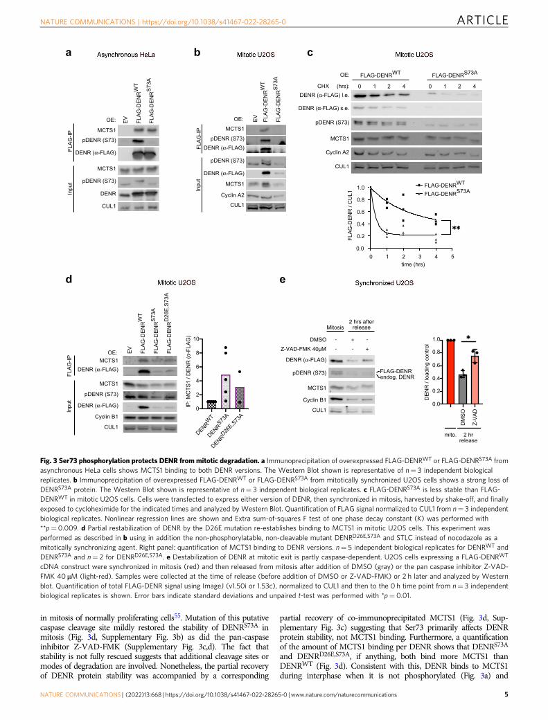
in mitosis of normally proliferating cells55. Mutation of this putativecaspase cleavage site mildly restored the stability of DENRS73A inmitosis (Fig. 3d, Supplementary Fig. 3b) as did the pan-caspaseinhibitor Z-VAD-FMK (Supplementary Fig. 3c,d). The fact thatstability is not fully rescued suggests that additional cleavage sites ormodes of degradation are involved. Nonetheless, the partial recoveryof DENR protein stability was accompanied by a corresponding
partial recovery of co-immunoprecipitated MCTS1 (Fig. 3d, Sup-plementary Fig. 3c) suggesting that Ser73 primarily affects DENRprotein stability, not MCTS1 binding. Furthermore, a quantificationof the amount of MCTS1 binding per DENR shows that DENRS73A
and DENRD26E,S73A, if anything, both bind more MCTS1 thanDENRWT (Fig. 3d). Consistent with this, DENR binds to MCTS1during interphase when it is not phosphorylated (Fig. 3a) and
a b c
DENR (α-FLAG) l.e.
pDENR (S73)
MCTS1
CUL1
FLAG-DENRWT FLAG-DENRS73A
0CHX (hrs):
OE:
1 2 4 0 1 2 4
Cyclin A2
DENR (α-FLAG) s.e.
0 1 2 3 4 50.0
0.2
0.4
0.6
0.8
1.0
time (hrs)
FLAG-DENR/CUL1
FLAG-DENRWT
FLAG-DENRS73A
e
DENR
MCTS1
CUL1
EV FLAG-DENRWT
FLAG-DENRS73A
pDENR (S73)
DENR (α-FLAG)
MCTS1
pDENR (S73)
FLAG-IP
Input
OE:
DENR (α-FLAG)
MCTS1
Cyclin A2
CUL1
EV FLAG-DENRWT
FLAG-DENRS73A
pDENR (S73)
DENR (α-FLAG)
MCTS1
pDENR (S73)
FLAG-IP
Input
OE:
EV FLAG-DENRWT
FLAG-DENRS73A
FLAG-DENRD26E,S73A
DENR (α-FLAG)
MCTS1
Cyclin B1
CUL1
pDENR (S73)
DENR (α-FLAG)MCTS1
FLAG-IP
Input
OE:
CUL1
MCTS1
DENR (α-FLAG)
pDENR (S73)
Z-VAD-FMK 40µM
DMSO - + -
- - +
Mitosis2 hrs after
release
Cyclin B1
endog. DENRFLAG-DENR
d
DENRWT
DENRS73A
DENRD26E,S73A
0
2
4
6
8
10
IP:MCTS1/DENR(α-FLAG)
DMSO
Z-VAD
0.0
0.2
0.4
0.6
0.8
1.0
DENR/loadingcontrol
mito. 2 hrrelease
Fig. 3 Ser73 phosphorylation protects DENR from mitotic degradation. a Immunoprecipitation of overexpressed FLAG-DENRWT or FLAG-DENRS73A fromasynchronous HeLa cells shows MCTS1 binding to both DENR versions. The Western Blot shown is representative of n= 3 independent biologicalreplicates. b Immunoprecipitation of overexpressed FLAG-DENRWT or FLAG-DENRS73A from mitotically synchronized U2OS cells shows a strong loss ofDENRS73A protein. The Western Blot shown is representative of n= 3 independent biological replicates. c FLAG-DENRS73A is less stable than FLAG-DENRWT in mitotic U2OS cells. Cells were transfected to express either version of DENR, then synchronized in mitosis, harvested by shake-off, and finallyexposed to cycloheximide for the indicated times and analyzed by Western Blot. Quantification of FLAG signal normalized to CUL1 from n= 3 independentbiological replicates. Nonlinear regression lines are shown and Extra sum-of-squares F test of one phase decay constant (K) was performed with**p= 0.009. d Partial restabilization of DENR by the D26E mutation re-establishes binding to MCTS1 in mitotic U2OS cells. This experiment wasperformed as described in b using in addition the non-phosphorylatable, non-cleavable mutant DENRD26E,S73A and STLC instead of nocodazole as amitotically synchronizing agent. Right panel: quantification of MCTS1 binding to DENR versions. n= 5 independent biological replicates for DENRWT andDENRS73A and n= 2 for DENRD26E,S73A. e Destabilization of DENR at mitotic exit is partly caspase-dependent. U2OS cells expressing a FLAG-DENRWT
cDNA construct were synchronized in mitosis (red) and then released from mitosis after addition of DMSO (gray) or the pan caspase inhibitor Z-VAD-FMK 40 μM (light-red). Samples were collected at the time of release (before addition of DMSO or Z-VAD-FMK) or 2 h later and analyzed by Westernblot. Quantification of total FLAG-DENR signal using ImageJ (v1.50i or 1.53c), normalized to CUL1 and then to the 0 h time point from n= 3 independentbiological replicates is shown. Error bars indicate standard deviations and unpaired t-test was performed with *p= 0.01.
NATURE COMMUNICATIONS | https://doi.org/10.1038/s41467-022-28265-0 ARTICLE
NATURE COMMUNICATIONS | (2022) 13:668 | https://doi.org/10.1038/s41467-022-28265-0 | www.nature.com/naturecommunications 5

recombinant DENR binds strongly to MCTS1 in bacteria, whereSer73 is not phosphorylated54.
Ser73 phosphorylation drops when cells exit mitosis (Fig. 1a)starting from anaphase onwards (Fig. 1d), and this drop coincideswith a drop in total DENR levels (Fig. 1a, c). We asked if thisdrop in DENR protein levels during mitotic exit is caspase-mediated. Indeed, the caspase inhibitor Z-VAD-FMK partiallyrescued the drop in DENR levels as cells exit mitosis (Fig. 3e). Insummary, phosphorylation of DENR at Ser73 prevents degrada-tion of DENR during the first stages of mitosis, from prophase tometaphase, while in later mitotic stages and mitotic exit DENRprotein is dephosphorylated and cleaved partly in a caspase-dependent manner.
pDENR (Ser73) induces mitotic translation of DENR targetgenes. Since DENR is a translation re-initiation factor, we askedwhat impact Ser73 phosphorylation has on translation. The datapresented above raise the possibility that in mitosis DENRphosphorylation on Ser73 protects it from degradation becauseDENR is required to drive translation of mitotically relevantmRNAs. We recently performed a RiboSeq analysis of DENRWT
and DENRKO HeLa cells to identify DENR target mRNAs thatrequire DENR for optimal translation32. Interestingly, amongstthe top DENR targets we found many genes with mitotic func-tions, primarily concerning the mitotic cytoskeleton (e.g., 10genes in the top 30, Supplementary Data 3). We first studiedtranslation of these target genes by measuring activity of luci-ferase reporters carrying the 5′UTRs of these mRNAs (Fig. 4a).The advantage of these luciferase reporters is that they control fortranscriptional effects (via the FLuc normalization controlreporter) as well as for protein stability effects (via the negativecontrol RLuc reporter). These luciferase assays confirmed that the5′UTRs of these mitotic genes impart DENR-dependent transla-tion, decreasing in DENR knockout cells, and returning to controllevels in DENR knockouts transfected to re-express DENR(Supplementary Fig. 4a). We tested if translation of these DENRtarget reporters increases in mitosis, by comparing their activityin asynchronous cells versus cells synchronized in mitosis. Thisrevealed that indeed translation of most reporters increased inmitotic cells by 1.5 to 2-fold, both for DENR targets with mitoticfunctions (Fig. 4b) as well as DENR targets with no known link tomitosis, which we had previously validated as bona fide DENRtargets (Fig. 4c)32. Most likely, this assay which is performed onthe bulk population of cells underestimates the magnitude of thereal mitotic effect due to the fact that available protocols onlycause a minor fraction of the cells to synchronize in mitosis. Incontrast to reporters carrying the 5’UTRs of endogenous DENRtargets, a synthetic reporter carrying a stuORF did not increasetranslation in mitosis (Fig. 4b), suggesting it lacks an elementrequired for mitotic translation. The increased translation of thesereporters in mitosis is due to elevated mitotic DENR activitybecause it is blunted in DENR knockdown cells (Fig. 4d–g, Sup-plementary Fig. 4b–d), and it is reversed by CDK2 inhibition withseliciclib (Supplementary Fig. 4e). The mitotic induction of theDUSP4 and CDKL5 reporters was abolished when the ATGs ofthe uORFs were mutated (Supplementary Fig. 4f), consistent withDENR·MCTS1 promoting translation re-initiation after uORFs32.In sum, these data indicate that DENR activity increases inmitosis compared to interphase.
We previously identified DENR targets by performing RiboSeqon asynchronous HeLa cells32. To test whether DENR promotestranslation of the same set of target mRNAs in mitotic cells as ininterphase cells, or whether phosphorylation on Ser73 mightaffect the DENR target set, we performed RiboSeq and RNA-seqon mitotic and interphase DENRWT and DENRKO cells
(Supplementary Data 4). Agents used to synchronize cells inmitosis such as nocodazole, and to a lesser extent the Eg5inhibitor STLC, induce cell stress and hence perturb thetranslatome56,57. We therefore used alternative methods to enrichor deplete mitotic cells from our populations. For the mitoticpopulation, we synchronized HeLa cells using a double thymidineblock and collected them for analysis 9 h after the second S phaserelease. For the interphase sample, we shook off and discardedmitotic cells, which anyways constitute a small minority (<5%) ofan asynchronous population. A Z-vs-Z analysis of the RNA-seqdata from wildtype cells identified 1326 transcripts correspondingto 694 genes with elevated mRNA levels in mitosis (Supplemen-tary Fig. 5a, Supplementary Data 5) and none with reducedmRNA levels, probably due to the short duration of mitosis. Acomparison of translation efficiency (ribosome footprints nor-malized to mRNA) between wildtype mitotic and interphase cellsidentified 266 transcripts (181 genes) that were translationally up-regulated in mitotic cells and 1090 transcripts (696 genes) thatwere translationally down-regulated in mitotic cells (Supplemen-tary Fig. 5b, Supplementary Data 6). A comparison of translationefficiency in DENRKO versus DENRWT cells identified 1108transcripts (653 genes) and 990 transcripts (576 genes) as DENRtargets in interphase and mitotic cells, respectively (Supplemen-tary Fig. 5c, d, Supplementary Data 7). Interestingly, acomparison of the change in translation efficiency upon DENRloss in interphase versus mitotic cells showed a good correlation(Supplementary Fig. 5e) suggesting that in general the mRNAsthat are DENR targets in mitosis are also DENR targets ininterphase, although to varying degrees. A hand-full of mRNAsappear to be strong DENR targets in interphase cells but not inmitotic cells (Supplementary Fig. 5e), but these genes are amongstthe ones with the strongest drop in translation efficiencytranscriptome-wide in wildtype mitotic cells compared tointerphase cells (e.g., IL11). This suggests that these mRNAs arenot well translated in mitosis, and hence are not sensitive toDENR loss. In sum, since the set of DENR target mRNAs doesnot change during mitosis, this suggests that phosphorylation ofDENR on Ser73 in mitosis affects its stability but not anotheraspect of its function. Interestingly, of the 266 transcripts that aretranslationally up-regulated during mitosis in wildtype cells, 114are DENR targets. This fraction (~40%) is significantly higherthan the 10% of transcripts that are DENR targets in interphasecells (p= 0.0 by binomial distribution). Hence DENR appears toplay a particularly important role in mitotic translation.
To investigate if DENR-dependent translation affects the levelof its targets during mitosis, we first performed Western Blotanalysis of asynchronous and mitotic cells and found that thelevel of DENR target proteins increases in mitosis (Supplemen-tary Fig. 6a). Interestingly, we observed that the mobility ofDENR protein on the SDS-PAGE gel shifts completely upwardsto a slower migrating form in mitotically synchronized cells(Supplementary Fig. 6a), suggesting that DENR is highlyphosphorylated in mitotic cells. We next asked whether thisincrease in protein levels is DENR dependent. To this end, weimmunostained unsynchronized DENRWT or DENRKO cells fortarget proteins and quantified protein levels specifically in mitoticcells identified by chromosome morphology. This revealed asignificant decrease in target protein levels upon loss of DENR inmitotic cells (Fig. 4h, i, Supplementary Fig. 6b, c). The same couldbe observed by western blotting lysates of mitotically synchro-nized cells (Supplementary Fig. 6d). A drop in target proteinlevels was also present in asynchronous cells, although lessdramatic than in mitotic cells (Supplementary Fig. 6e). Thesedrops in target protein levels were rescued by reconstituting theDENRKO cells with a DENRWT expression construct (Supple-mentary Fig. 6d), confirming they are on-target effects. When,
ARTICLE NATURE COMMUNICATIONS | https://doi.org/10.1038/s41467-022-28265-0
6 NATURE COMMUNICATIONS | (2022) 13:668 | https://doi.org/10.1038/s41467-022-28265-0 | www.nature.com/naturecommunications

however, reconstitution was performed with the non-phosphorylatable DENRS73A mutant, rescue of target proteinlevels in mitotic cells was impaired (compare lane 2 to lane 1 inFig. 4j, k, Supplementary Fig. 6f). Since DENR is phosphorylatedat Ser73 only in mitotic cells, this confirms there is DENR-dependent translation ongoing during mitosis. In accordance
with our previous findings showing that mutation of the caspasecleavage site partially reconstitutes DENR stability, mitotic targetprotein levels are also partially rescued when the non-phosphor-ylatable, non-cleavable version of DENR (DENRD26E,S73A) isexpressed in DENRKO cells (Fig. 4j, k). In sum, our findings showthat there is a set of mitotically relevant mRNAs, such as CDKL5
NATURE COMMUNICATIONS | https://doi.org/10.1038/s41467-022-28265-0 ARTICLE
NATURE COMMUNICATIONS | (2022) 13:668 | https://doi.org/10.1038/s41467-022-28265-0 | www.nature.com/naturecommunications 7

mRNA, that are translated in a DENR-dependent manner inmitosis.
pDENR (Ser73) prevents aberrant mitosis and promotesfaithful cell division. Since the DENR·MCTS1 complex promotestranslation of mRNAs with mitotic functions, we asked ifDENR·MCTS1 is required for proper mitosis. To this end, weperformed two-dimensional flow cytometry of unsynchronizedDENRWT or DENRKO cells and observed a four-fold accumula-tion of DENRKO cells in mitosis (Fig. 5a, Supplementary Fig. 7a),raising the possibility of a defect in progression through mitosis.Indeed, an elevated number of mitotic DENRKO cells undergoapoptosis (Fig. 5b, Supplementary Fig. 7a). This suggests that lossof DENR leads to slower mitotic progression and increasedmitotic failure, likely contributing to the reduced proliferationrate of DENRKO cells which we previously reported32. We thenexamined whether there are any mitotic defects in DENRKO cells(Fig. 5c, d). While the fraction of early mitotic phases (prophase,prometaphase, metaphase) is not significantly influenced by theabsence of DENR, there is a reduction in the number of cells inthe late mitotic phases (anaphase, telophase), and instead anaccumulation of atypical mitotic figures and mitotic blebs,representative of mitotic cell death (Fig. 5c, d). Taken togetherwith the increase in the fraction of mitotic cells, these findingsindicate that the early phases of mitosis are prolonged inDENRKO cells, and in some DENRKO cells mitotic cell deathoccurs after anaphase onset. This effect can be rescued by re-expression of DENRWT in DENRKO cells, but not by the non-phosphorylatable DENRS73A mutant, and only partially by thenon-phosphorylatable, non-cleavable DENRD26E,S73A mutant(Fig. 5c, d). We observed a similar phenotype in asynchronousDENR-knockdown U2OS cells (Supplementary Fig. 7b), rulingout a cell type-specific defect in HeLa cells. Interestingly, weobserved an increased fraction of irregular and multipolar spin-dles in DENRKO compared to DENRWT cells (SupplementaryFig. 7c), which could explain in part the mitotic failure in DENR-depleted cells. Since DENRKO cells reconstituted with DENRS73A
have mitotic defects, and since Ser73 is specifically phosphory-lated in mitosis, these data show that the translation occurringduring mitosis is important for mitosis to proceed correctly.Furthermore, the DENR/MCTS1 complex appears to be playing aparticularly important role during mitosis because 114 of the 226transcripts which are translationally upregulated during mitosisare DENR targets (i.e., 40%), which is a significantly largerfraction than the 10% of transcripts that are DENR targets ininterphase cells (p= 0 by binomial distribution).
In sum, phosphorylation of DENR at Ser73 and stabilization ofthe DENR·MCTS1 complex in the early phases of mitosis isimportant to guarantee efficient cell division and to preventaberrant mitosis. It is likely that the mitotic defects that resultfrom loss of DENR or DENR Ser73 phosphorylation (Fig. 5d)reflect a combined contribution of multiple DENR targets, sincemultiple DENR target mRNAs are insufficiently translated duringmitosis in the absence of DENR activity (Fig. 4, SupplementaryFig. 6), and multiple DENR targets have mitotic functionsincluding spindle dynamics (Supplementary Data 3).
DiscussionWe have identified a signaling pathway promoting mitotictranslation of genes that are crucial for faithful and timely celldivision (Fig. 6). At the onset of mitosis CDK1/Cyclin B andCDK2/Cyclin A phosphorylate the non-canonical translationinitiation factor DENR at serine 73 and thereby protect DENRfrom degradation (Figs. 1–3). DENR then acts to promotetranslation of a set of target genes that are known to be involvedin proper cell division (Fig. 4), thereby supporting mitotic pro-gression and preventing aberrant mitosis and mitotic cell death(Figs. 5, 6). Interestingly, almost half of all mRNAs with increasedtranslation in mitosis depend on DENR for their translation,suggesting that DENR activity is particularly important duringthis phase of the cell cycle.
Previous studies have identified mRNAs whose translation isupregulated during mitosis, however, the functional significanceof this up-regulation for mitotic progression was difficult to test,given that there was no intervention known to specifically blockmitotic translation. Since DENR phosphorylation on Ser73 isspecific for mitosis, this enables such an intervention. The factthat reconstitution of DENRKO cells with DENRS73A does notrescue their mitotic defects indicates that the translation occur-ring during mitosis is indeed important for mitosis itself.
This regulatory mechanism is initiated through DENR phos-phorylation by CDK1 and CDK2. Interestingly, we find thatCyclin A plays an important role in enabling CDK2 to phos-phorylate DENR, likely via substrate recognition, because CDK2bound to Cyclin A phosphorylates DENR in vitro more efficientlythan CDK2 bound to Cyclin E (Fig. 2a, b). This is consistent withthe fact that in vivo DENR Ser73 is phosphorylated in mitosis,when CycA/CDK2 is active, but not during S-phase, when CycE/CDK2 is active (Fig. 1a).
CDK2 and CDK1 are vital in tumors and therefore attractivetargets for cancer therapy. Different chemical CDK2 inhibitorshave been developed and the most advanced one, seliciclib, is
Fig. 4 pDENR (S73) induces mitotic translation of DENR target genes. a Reporter constructs used in this figure. A LaminB1 5’UTR-firefly luciferase (FLuc)reporter was used as a normalization control in all wells. Luminescence of target gene 5’UTR-renilla luciferase (RLuc) reporters was normalized to anequivalent negative control reporter carrying the LaminB1 5’UTR. b, c Luciferase reporters carrying the 5’UTRs of the indicated DENR target genes exhibitincreased translation in mitotic U2OS cells (sequential thymidine-nocodazole (red)) compared to those left unsynchronized (gray). One sample t-tests onn independent biological replicates: stuORF p= 0.4, SYNPO *p= 0.01, PLEC *p= 0.03, CDKL5 *p= 0.02, DUSP4 **p= 0.002, ABLIM1 **p= 0.01, CUL5**p= 0.01, SGO1 *p= 0.03, CAMSAP2 p= 0.1, MAP2K6 *p= 0.02, a-raf *p= 0.02, PIK3R2 *p = 0.04, ATF4 *p= 0.03, DROSHA *p= 0.045, c-rafp= 0.3. d–g Translation of reporter constructs carrying 5’UTRs of DENR target genes increases in mitosis in a DENR-dependent manner. GFP- (filled)or DENR- (empty) siRNA-treated U2OS cells were transfected with the indicated reporters, and synchronized in mitosis (red) or not synchronized (gray).Means and unpaired t-test from n biological replicates. d n= 3, *p= 0.03, ns p= 0.68, e n= 5, *p= 0.01 ns p= 0.08, f n= 4, *p= 0.04, ns p= 0.96,g n= 5, *p= 0.03, ns p= 0.12. Endogenous levels of DENR target proteins CDKL5 (h) or ABLIM1 (i) are reduced in mitotic DENR knockout versus mitoticcontrol cells. Unsynchronized HeLa DENRWT (gray) or DENRKO (white) cells immunostained for CDKL5 or ABLIM1. For quantifications, h n= 11 DENRWT
and n= 10 DENRKO mitotic cells. i n= 9 DENRWT and n= 17 DENRKO mitotic cells. Means of signal intensities are displayed. **** unpaired t-testp < 0.0001. Scale bars indicate 10 µM. j Reconstitution of DENRKO cells with DENRWT rescues mitotic protein levels of target genes, but not reconstitutionwith DENRS73A and only partially with DENRD26E,S73A. HeLa DENRKO cells were transfected with plasmids carrying the indicated DENR versionsand a puromycin resistance gene, selected with puromycin, synchronized (sequential thymidine-STLC) and collected for analysis by Western Blot.k Quantification of protein levels for independent biological replicates of the experiment shown in panel j with DENRWT light-gray, DENRS73A orange andDENRS73A,D26E dark-gray. In all subfigures, error bars indicate standard deviations.
ARTICLE NATURE COMMUNICATIONS | https://doi.org/10.1038/s41467-022-28265-0
8 NATURE COMMUNICATIONS | (2022) 13:668 | https://doi.org/10.1038/s41467-022-28265-0 | www.nature.com/naturecommunications

currently being tested in clinical studies. While its molecularaction has only been partially discovered, seliciclib is known toprevent faithful cell division and cause cell death in mitosis, dueto a type of uncoordinated cellular division called anaphasecatastrophe58,59. From the fact that DENR loss of function phe-nocopies this mitotic failure (Fig. 5, Supplementary Fig. 7), it ispossible that part of the effect of seliciclib might be due toinhibition of DENR phosphorylation at Ser73 and accordinglyimpaired translation of DENR-dependent mitotic target genesthat act towards coordinated cell division and cytokinesis.
In line with our observations that DENR·MCTS1 proteincomplex is essential for faithful cell division, mitotic catastropheand delayed cytokinesis have been observed in cells depleted ofMCTS160, however, the mechanism of this MCTS1 effect wasunknown. Since DENR and MCTS1 are co-dependent on eachother for protein stability and act together as one functional
heterodimeric complex, it is likely that reduced translation of themitotic target genes as we describe here is one contributing factor.
We observed that DENR is stabilized by phosphorylation atserine 73 in mitosis. When not phosphorylated, DENR isdegraded in a manner that is partly caspase-dependent (Fig. 3d, e,Supplementary Fig. 3). Although caspases are well known to beactive during apoptosis, there is also increasing evidence of non-apoptotic, cell cycle-related functions for caspases61. Caspases 2and 7 have been described to drive mitotic exit55,62–64. Consistentwith this, we observed caspase-dependent degradation of DENRduring the late phases of mitosis (anaphase, telophase). Inter-phase DENR, however, is very stable independently of its phos-phorylation status (Fig. 3a, Supplementary Fig. 3a). This suggeststhat caspase activity towards DENR increases in mitosis. Wenoticed that caspases 2 and 3 are described as being nuclear(https://www.proteinatlas.org/ENSG00000106144-CASP2/cell,
Fig. 5 pDENR (S73) prevents aberrant mitosis and promotes faithful cell division. DENR knockout cells have an elevated proportion of cells in mitosis(a) that are apoptotic (b). Unsynchronized HeLa DENRWT (gray) or DENRKO (white) cells were fixed, permeabilized, stained for cleaved caspase 3 (CC3)and pHH3 (S10) and then analyzed by flow cytometry. Shown are means, normalized to DENRWT, and standard deviations as error bars from n= 3biological replicates. One sample t-test with *p= 0.01 for both a and b. DENR knockout cells have a reduced proportion of cells in late mitotic phases andelevated aberrant mitoses and mitotic cell death, both of which are rescued by reconstitution with DENRWT (checkered) but not with DENRS73A (finelycheckered light) and only partially with DENRD26E,S73A (finely checkered dark) protein. Unsynchronized HeLa DENRWT (filled) or DENRKO (empty) cellswere transfected with empty expression vector (EV) or one of the indicated FLAG-tagged constructs for 72 h, then fixed and stained with DAPI. c Sampleimages. Mitotic cells are outlined by white circles. Examples of failed mitoses (multipolar spindles, mitotic blebs/mitotic cell death) are encircled in orange.Scale bars indicate 10 µM. d Quantification of mitotic phases and mitotic defects, assessed via DAPI stain. Displayed are means of fractions of all mitoticcells, and standard deviations as error bars from n= 3 independent biological replicates. Early mitosis comprises prophase, prometaphase and metaphase,late mitosis comprises anaphase and telophase and failed mitosis comprises aberrant mitosis or mitotic blebs. Two-way ANOVA was performed for Earlymitosis (blue) and unpaired t-tests were performed for Late mitosis (green) and Failed mitosis (orange).
NATURE COMMUNICATIONS | https://doi.org/10.1038/s41467-022-28265-0 ARTICLE
NATURE COMMUNICATIONS | (2022) 13:668 | https://doi.org/10.1038/s41467-022-28265-0 | www.nature.com/naturecommunications 9

https://www.proteinatlas.org/ENSG00000164305-CASP3/cell), soone could speculate that, breakdown of the nuclear enveloperenders DENR accessible to otherwise nuclear caspases andtherefore allows for a surge of DENR cleavage unless preventedby Ser73 phosphorylation. This interplay could be topic of futurestudy. That said, the rescue of DENR stability either uponmutating the caspase site in DENR or with the Z-VAD-FMKcaspase inhibitor is only partial (Fig. 3d, e), suggesting additionaldegradation mechanisms may be at play.
We noticed that unlike other reporter constructs carrying the5′UTRs of endogenous DENR target mRNAs, our syntheticstuORF reporter does not increase in translation during mitosis(Fig. 4b). One possible explanation we explored is that duringmitosis DENR Ser73 phosphorylation changes DENR function sothat it no longer acts on one set of interphase target mRNAs(represented by the stuORF reporter) and instead it acts on adistinct set of mRNAs. From our ribosome profiling experiment,however, this does not seem to be the case (SupplementaryFig. 5e). There is a good correlation between the mRNAs that areDENR targets in interphase and in mitosis, suggesting thereare not two distinct sets of target genes. The few target genes thatare strongly DENR dependent in interphase but less so in mitosis(e.g., IL11, ELFN2, and FBXO46) are poorly translated in mitosis.The IL11 mRNA for instance is the most down-translated mRNAtranscriptome-wide in wildtype mitotic cells compared to inter-phase cells (Supplementary Data 6). Hence if these mRNAs arenot translated in mitosis, the presence or absence of DENRcannot affect them. Therefore, although we do not know why oursynthetic stuORF reporter does not increase its translation duringmitosis, we believe the most likely explanation is that it is notbeing translated in mitosis for some technical reason.
We present here in Supplementary Materials a few unbiasedand comprehensive screens for pathways that might regulate theDENR·MCTS1 complex. We tested all serine/threonine kinasesby in vitro kinase assay, we knocked down all hits from thiskinase screen and assayed DENR·MCTS1 activity using thestuORF reporter in HeLa and MCF7 cells, and we systematicallymutated all amino acids in DENR or MCTS1 which were
reported in public databases to be post-translationally modified.While we focused here on the CDK axis, our data suggest thatthere might be additional regulatory pathways that could befurther explored. One example is the in vitro phosphorylation ofMCTS1 at threonines 81 and 179 by STK3 (SupplementaryFig. 9d). Additional examples are the effects of PRKCQ or PRKG1knockdown on DENR·MCTS1/stuORF activity (SupplementaryFig. 9a, b). Furthermore, abrogation of possible posttranslationalmodifications on MCTS1 lysine 51 showed a mild but significantreduction in stuORF activity (Supplementary Fig. 8e), suggestingacetylation or ubiquitylation might provide additional levels ofregulation to this translation complex. These might be interestingstarting points for future studies on the regulation of theDENR·MCTS1 complex.
MethodsCloning. Sequences of oligos used for cloning are provided in SupplementaryData 4. For expression of MCTS1 or DENR and their mutants (SupplementaryFig. 8b, c) the human MCTS1 or DENR coding sequences were cloned into a pRKvector backbone (pAT1063, Teleman lab collection) via restriction sites EcoRI andClaI. Then four silent mutations within exon 6 were inserted into the MCTS1coding sequence by site-directed mutagenesis to make it resistant againstMCTS1 siRNA. Likewise, all mutations of the DENR and MCTS1 coding sequencewere inserted by site-directed mutagenesis (Supplementary Fig. 8b, c, Supple-mentary Data 1). For negative controls, the empty pRK vector backbone was used.In order to clone FLAG-tagged DENR constructs (Fig. 3, Fig. 4j, k, Fig. 5c, d,Supplementary Figs. 1b, 3, 6f, 8b–e), DENR (wildtype, S73A or D26E,S73Amutant) coding sequence was amplified using primers containing a C-terminalFLAG-tag and again cloned into the above mentioned pRK vector backbone usingEcoRI and ClaI restriction sites or into a pcDNA3.1 vector containing a puromycinresistance (pGF045) using BamHI and EcoRI restriction sites.
In order to obtain mutated constructs for the in vitro kinase assays(Supplementary Fig. 9c, d) site-directed mutagenesis was performed on a plasmid(pET-DUET-1, pSS290)54 coding for both His-tagged DENRWT and His-taggedMCTS1WT, before recloning into the pET-DUET-1 vector using XbaI and NotI(DENR) or MunI/EcoRI and XhoI (MCTS1) as restriction sites.
The Lamin B1 5′UTR firefly and renilla luciferase reporters and the Lamin B1 5′UTR stuORF reporter were obtained from a previous project in our lab30. Renillaluciferase reporters with 5′UTRs of various genes (Fig. 4b–g, Supplementary Fig. 4)were cloned by amplifying the 5′UTR of the gene of interest from cDNA andcloning it into the renilla luciferase reporter plasmid at the HindIII and Bsp119lsites. Cloning of the reporter plasmids for ATF4, a-raf, c-raf, DROSHA, MAP2K6,and PIK3R2 was performed likewise32.
All sequences of primers used for cloning are detailed in Supplementary Data 8.
Expression and purification of the DENR·MCTS1 protein complex. Proteinswere expressed using E. coli BL21 (DE3) cells in 2YT media supplemented withKanamycin (30 μg/ml). Cells were grown to an OD600 of 0.8–1.0 at 37 °C, thenshifted to 18 °C. Expression was induced with the addition of 0.4 mM IPTG,and cells were grown further overnight, harvested by centrifugation, and the cellpellets either used immediately for lysis and purification or frozen with LN2 andstored at −20 °C.
All variants of the DENR·MCTS1 complex (Supplementary Fig. 9c, d,Supplementary Data 2) were purified via a C-terminal His6-tag using NiNTA andsize exclusion chromatography. Cells were resuspended in lysis buffer (30 mMHEPES, 30 mM Imidazol, 500 mM NaCl) and lysed with a Microfluidizer(Microfluidics) at 0.55MPa. The lysate was cleared by centrifugation for 35 min at35,000 × g and 4 °C, and the resulting supernatant was applied to a 2 ml NiNTAcolumn. The column was washed with 25–50 column volumes of lysis buffer andeluted with elution buffer (lysis buffer plus 400 mM Imidazol). The NiNTA-eluatewas applied to a Superdex 200 26/60 column, equilibrated with SEC-buffer I(10 mM HEPES pH 7.5, 500 mM NaCl). Peak fractions containing theDENR–MCTS1 complex were pooled, concentrated to 10–15 mg/ml, and eitherused directly or shock-frozen with LN2 and stored at −80 °C.
Cell culture. HeLa cells (American Type Culture Collection, ATCC #CCL-2),MCF7 cells (ATCC #HTB-22) and U-2 OS cells (ATCC #HTB-96) were cultured inDulbecco’s modified Eagle’s medium (DMEM), supplemented with 10% fetalbovine serum and 1% penicillin/streptomycin. HeLa DENRKO cells were generatedin our lab32. For this manuscript clone no. 3.42 was used throughout, only forRiboSeq analysis clone 2.11 was added. All cell lines were tested negative formycoplasma contamination.
The concentration of all drugs used for synchronization of cells are specified inthe “drug treatments” section below. For synchronization of U2OS cells in mitosis
CDK1
Cyclin BCDK2
Cyclin A
P
DENR
MCTS1
MCTS1
DENR
MCTS1
P
Translationof m
itotictarget mRNAs
Prophase
Pro-metaphase
MetaphaseAnaphase
Telophase
G1
Caspases
G2
Mitosis
Fig. 6 Schematic summary of DENR regulation in mitosis. During earlystages of mitosis, DENR protein is stabilized via phosphorylation of Ser73which enables it to promote translation of target genes involved in mitosis.As of anaphase, DENR phosphorylation on Ser73 decreases, concomitantwith a caspase-dependent decrease in DENR protein levels as cells exitmitosis.
ARTICLE NATURE COMMUNICATIONS | https://doi.org/10.1038/s41467-022-28265-0
10 NATURE COMMUNICATIONS | (2022) 13:668 | https://doi.org/10.1038/s41467-022-28265-0 | www.nature.com/naturecommunications

(Figs. 1a–c, 3b–h, 4b–g, Supplementary Figs. 3c, d, 4b–d, f) cells were plated inthymidine. 24–28 h later medium was removed and cells were washed twice withPBS and once for 5 min with normal medium. Then, fresh medium containingnocodazole or STLC was added for 13 h. For mitotic synchronization, HeLa cells(Fig. 4j, k, Supplementary Fig. 6a, f) were released from S phase arrest (thymidine)for 9 h (Fig. 4j, k, Supplementary Fig. 6f) or 13 h (Supplementary Fig. 6a) in thepresence of STLC, followed by Western Blot analyses or luciferase assay. Formitotic enrichment (Supplementary Fig. 6d) HeLa cells were released from a singlethymidine block into normal medium for 9 h and then analyzed as indicated.
For mitotic release (Figs. 1a–c, 3g, h) nocodazole-arrested U2OS cells wereshaken off, spun down, washed twice in PBS and once in medium and thenreplated in normal medium. The first sample (“mitotic release 0”) was collected atthe time of mitotic shake-off.
For assessment of mitotic stability (Fig. 3c, d, Supplementary Fig. 3b) U2OScells were synchronized with thymidine and nocodazole as described above, thenthey were shaken off, spun down, and collected or replated in medium containingnocodazole and cycloheximide at a concentration of 100 µg/ml.
For analysis of phosphorylation during mitotic entry U2OS (Fig. 2e, f,Supplementary Fig. 2c) or HeLa (Supplementary Fig. 2g) cells were plated inthymidine. 24–28 h later medium was removed and cells were washed twice withPBS and once for 5 min with normal medium and then fresh medium was added.In U2OS cells, 10 h later thymidine was added again. 24–28 h later cells werereleased again (washed twice in PBS, once in medium) into nocodazole-containingmedium. 9 h later DMSO or the indicated inhibitor was added and 4 h later cellswere harvested and analyzed by Western blot or luciferase assay. HeLa cells werereleased from first thymidine arrest immediately into STLC-containing medium.6 h after release DMSO or the indicated inhibitor was added and three hours latercells were harvested and analyzed by Western blot.
For synchronization in G2 phase U2OS (Supplementary Fig. 2b) or HeLa(Fig. 2c, d) were plated in thymidine-containing medium for 24–28 h, then releasedinto medium containing the CDK1 inhibitor RO3306 at a concentration of 5 μM.13 h (U2OS) or 9 h (HeLa) later – “G2” –, cells were released again using PBS andmedium for 20–30 min – “G2/M” –, and then DMSO or RO3306 in the indicatedconcentrations was added for another 45–60 min – “Mitosis” –, followed bycollection and analysis by Western Blot.
For S phase release (Fig. 1a–c) U2OS cells were subjected to a double thymidineblock as described above. After the second thymidine block cells were released,replated in nocodazole-containing medium and collected at the indicated timepoints.
Drug treatments. Where indicated, and if not otherwise specified, the followingdrugs were used: Z-VAD-FMK 40 μM (SelleckChem, #S7023), thymidine 2 mM(Sigma, #T1895-1G), nocodazole 400 ng/ml (Sigma, #SML1665-1ML), cyclohex-imide 100 µg/ml (Santa Cruz, #sc-3508), RO3306 5 μM unless otherwise specified(Sigma #SLM0569-5MG), seliciclib 10 μM unless otherwise specified (SelleckChem,#S1153), A-674563 1 μM unless otherwise specified (SelleckChem, #S2670), STLC5 μM (Sigma, #164739-5 G). Z-VAD-FMK was added at the time of FLAG-DENRplasmid transfection (Supplementary Fig. 3c, d) or of mitotic release (Fig. 3g, h).For reconstitution of DENR mutant versions, DENRKO cells were plated at 2million cells per 10 cm plate, the next day transfected using Effectene (see below).36 h after transfection cells were re-plated and puromycin (Sigma, #P9620) wasadded at a concentration of 0.5 µg/ml. 48 h later cells were re-seeded for luciferaseassay or Western Blot in puromycin and thymidine-containing medium. 24 h later,during synchronization, medium was exchanged to only thymidine (and no pur-omycin)-containing medium. Synchronization was then completed as describedabove. Cells were collected for analysis 24 h after end of puromycin selection.
Transient transfections. Transient transfections were performed using eitherEffectene Transfection Reagent (QIAGEN, Cat No./ID: 301425), Lipofectamine2000 (Thermo Fisher Scientific, Cat No. 11668030) or Lipofectamine 3000(Thermo Fisher Scientific, Cat No. L300015). The manufacturer’s instructions weremodified to reduce toxicity and allow ongoing cell cycling and division: ForLipofectamine 2000 transfection (Fig. 4b–g, Supplementary Figs. 9a, b, 4a–e)0.05 µl of transfection reagent and a total of 20 ng DNA per 96-well were applied24 h before analysis. For Lipofectamine 3000 transfection (Supplementary Fig. 4f)0.1 µl of transfection reagent (without enhancer) and a total of 50 ng DNA per 96-well were applied 24 h before analysis. For Effectene transfection (Fig. 3, Fig. 4j, k,Fig. 5c, d, Supplementary Fig. 1b, Supplementary Fig. 3, Supplementary Fig. 6d–f)all transfection reagents as well as the amount of DNA were divided in half andcells were transfected 3 days (Fig. 4, Fig. 5, Supplementary Fig. 6f) or 24 h (allothers) before analysis.
siRNAs and DENRKO. siRNAs were obtained from Dharmacon® (Horizon Dis-covery Ltd.). Catalog numbers and sequences are specified in SupplementaryData 9. For DENR knockdown, a pool of three different siRNAs (−02, −19, −20)was used. siRNA transfection was performed using Lipofectamine RNAi MAX©
Transfection Reagent (Thermo Fisher Scientific). HeLa DENRKO cells were gen-erated in our laboratory using CRISPR/Cas9-mediated knockout32. Clone 3.42 wasused throughout this paper, but effects were observed similarly with clone 2.11.
Translation/luciferase reporter assay. To produce reporter constructs, 5′UTRsequences of the indicated genes were PCR-amplified from cDNA or genomicDNA and cloned at the 5’ end of Renilla luciferase using HindIII and Bsp119I.
For the luciferase reporter assays, MCF7 or HeLaWT cells were seeded at adensity of 8.000 cells and HeLaDENR_KO cells at a density of 12.000 cells per 96-well(Supplementary Figs. 4a, 8b, c, 9a, b). For luciferase assays with synchronizedU2OS cells (Fig. 4b–g, Supplementary Fig. 4b–f) cells were seeded at a density of15.000 cells in thymidine. For knockdown experiments, U2OS or HeLa cells werereverse-transfected in 15 cm dishes containing siRNA and LipofectamineRNAiMAX transfection reagent, and replated 48–72 h thereafter at a density of10.000 cells per 96-well (all conditions), in thymidine-containing medium ifapplicable. 16–20 h after (re-)plating, cells were transfected via Lipofectamine 2000with the negative control lamin B-renilla luciferase reporter, or the respective test 5′UTR-renilla luciferase reporter, as well as a lamin B 5′UTR-firefly luciferaseplasmid for internal normalization control. 8–9 h later and 24–28 h after additionof thymidine, cells were released into nocodazole or normal medium (as applicable)as described above. Luciferase activity was analyzed 13 h thereafter, about 24 h aftertransfection, using DualGlo Luciferase Assay System (Promega) and Mithras LB940 Reader (Berthold Technologies). For asynchronous cells all medium changesand washes were performed simultaneously, without the addition of synchronizingagents.
For analysis of results, in a first step, renilla luminescence was normalized tofirefly luminescence to control for transfection/expression variabilities. In a secondstep, this ratio for the 5′UTR reporter of interest was normalized to the equivalentratio of the negative control lamin B-RLuc/lamin B-FLuc reporter from parallelly-transfected and equally-treated wells (“normalized RLuc/FLuc”), to control forvariability between conditions.
Cell lysis and immunoprecipitation. For cell lysis, cells were scraped, spun down at13.000 × g and resuspended in lysis buffer containing Tris /HCl pH 7.5 50mM, NaCl250mM, EDTA 1mM, Triton X-100 0,1%, NaF 50mM, protease inhibitors (cOm-plete Protease Inhibitor Cocktail, Sigma Cat No. 4693116001, 1 tablet in 10ml) andphosphatase inhibitors (sodium orthovanadate 1mM, β-glycerophosphate 100mM,PhosSTOP EasyPAK (Sigma, Cat No. 4906845001) 2 tablets in 10ml). After 10minincubation on ice, samples were centrifuged for 10min at 20,800 g at 4 °C. Forimmunoprecipitation, supernatant was divided into lysates and samples for immu-noprecipitation. The latter were incubated with anti-FLAGM2 affinity gel (Sigma, CatNo. A2220-1ML) for 1.5 h at 4 °C (FLAG-IP). For washing the immunoprecipitatedproteins, beads were centrifuged for 1min at 200 × g, supernatant was removed, andfresh lysis buffer was added to the beads. This step was repeated three times before thebeads were transferred to a new epi and elution was performed with 1× Laemmlibuffer. For the whole-cell lysates, their protein concentration was measured using thebiorad protein assay (biorad, Cat No. 500–0006) and adjusted before addition of5xLaemmli buffer.
Western blots. Equal protein amounts were run on SDS-PAGE gels and trans-ferred to nitrocellulose membrane with 0.2 µm pore size. After Ponceau staining,membranes were incubated in 5% skim milk PBST for 20–60 min, briefly rinsedwith PBST, and then incubated in primary antibody solution (5% BSA PBST or 5%skim milk PBST) overnight at 4 °C. Membranes were then washed three times,15 min each in PBST, incubated in secondary antibody solution (1:10,000 in 5%skim milk PBST) for 1 h at room temperature, then washed again three times for5–15 min. Finally, chemiluminescence was detected using ECL reagents anddetected using a Biorad ChemiDoc imager. For immunoblot visualization andanalysis ImageLab software version 5.2.1 was used.
Antibodies. The following antibodies were used: ABLIM1 (WB: 1:1000, IF: 1:500,rabbit, bethyl-biomol A302-237-T), β-Actin (1:5000, mouse, Sigma #A2228), ATF-4 (1:1000, rabbit, Cell Signaling #11815), γ-tubulin (1:1000, mouse, Abcam#ab27074), Caspase 3 (1:1000, rabbit, Cell Signaling #9662), CDKL5 (WB: 1:1000,IF: 1:500, rabbit, abcam ab22453), CDT1 (1:1000, rabbit, Cell Signaling #8064),cleaved Caspase 3 (WB: 1:1000, flow cytometry 1:100, rabbit, Cell Signaling #9664),CUL1 (1:500, mouse, Invitrogen #32-2400), cyclin A2 (1:1000, rabbit, Cell Sig-naling #91500), cyclin B1 (1:1000, mouse, Cell Signaling #12231), cyclin E1 (1:1000,rabbit, Cell Signaling #20808), DENR (WB and IF: 1:2000, guinea pig, in-houseproduction), pDENR_Ser73 (WB: 1:500, IF: 1:200, rabbit, custom-made by inno-vagen AB, Lund, Sweden), DUSP4 (WB: 1:1000, IF: 1:500, rabbit, abcamab216576), FLAG (1:1000, rabbit, Sigma #F7425), FLAG-M2 (1:1000, mouse,Sigma #F3165), GAPDH (1:2000, rabbit, Cell Signallling #2118), Geminin (1:1000,rabbit, Cell Signaling #52508), pHH3 (Serine 10) (for WB: 1:500, rabbit, CellSignaling #9701; for flow cytometry: 1:100, mouse, Cell Signaling #9706 S),MAP2K6 (WB: 1:1000, IF: 1:500, rabbit, Cell Signaling #8550), MCTS1 (1:1000,guinea pig, in-house production) a-raf (1:1000, rabbit, Cell Signaling #4432), c-raf(1:1000, rabbit, Cell Signaling #9422), ERK1/2 (1:1000, rabbit, Cell Signaling#4695), GST-HRP conjugate (1:5000, goat, GE Healthcare #RPN1236), pERK1/2(T202/Y204) (1:1000, rabbit, Cell Signaling #4370), p-p90RSK (T359/Y363)(1:1000, rabbit, Cell Signaling #9344),RSK1/2/3 (1:1000, rabbit, Cell Signaling#14813).
NATURE COMMUNICATIONS | https://doi.org/10.1038/s41467-022-28265-0 ARTICLE
NATURE COMMUNICATIONS | (2022) 13:668 | https://doi.org/10.1038/s41467-022-28265-0 | www.nature.com/naturecommunications 11

In vitro kinase assayNon-radioactive. 50 ng of purified His-tagged DENR·MCTS1 protein complex wasincubated with 25 ng of active CDK1-Cyclin B1, CDK2-Cyclin A2, CDK2-CyclinE1, CDK4/Cyclin D1, CDK6/Cyclin D1 proteins (Proqinase #0134-0135-1, #0050-0054-1, #0050-0055-1, #0142-0143-1, #0051-0154-2) in Protein Kinase-Buffer(50 mM HEPES-NaOH pH 7.5, 3 mM MgCl2, 3 mM MnCl2, 3 μM Na-orthova-nadate, 1 mM DTT) with 1.25 µM ATP for 30 min at 30 °C. Then Laemmli bufferwas added and half of each sample analyzed by Western blotting using phospho-specific antibodies.
Radioactive. Protein kinase assays were carried out in 20 µl kinase buffer (60 mMHEPES-NaOH (pH 7.5), 3 mM MgCl2, 3 mM MnCl2, 3 μM Na-orthovanadate,1.2 mM DTT, 50 μg/ml PEG20.000, 5 µCi of [γ-32P] ATP (Perkin Elmer(NEG502A)) and 40 µM [y-S]-ATP) either with the enzyme only, or 1 µg of therespective substrate. As positive control, 1 µg Casein was used. First, samples wereincubated for 30 min at 30 °C. The reaction was stopped by the addition of 5 µl 5×SDS-sample buffer and incubation at 95 °C for 5 min. The proteins were separatedby SDS-gel electrophoresis (12% PAA). Subsequently, SDS-gels were stained withCoomassie Staining solution (0.4% Coomassie Brilliant Blue G250, 10% citric acid,8% ammonium sulfate, 20% methanol) for 2 h and destained with several washes of20% Methanol in ddH2O until the background was clear. Finally, the SDS-gels wereplaced on thick filter paper and dried with a vacuum Gel-Drying System for 1.5 h at80 °C. Radioactive signals were visualized by exposure to X-ray films.
Immunofluorescence. For immunofluorescence microscopy, cells were plated onpoly-L-lysine (Sigma # P4707)-coated cover slips and one to 3 days later fixatedwith PFA 4% for 20 min at room temperature. After washing four times with PBT(Triton X-100 0.1% in PBS), samples were blocked with BBT (PBT with BSA 1%)for 2 h and incubated with primary antibody overnight. Anti-pDENR (Ser73)purified antibody was diluted in BBT 1:200, anti-DENR antibody was diluted inBBT 1:1000, Anti-γ-tubulin antibody (Supplementary Fig. 7c) was diluted in BBT1:1000. Next, samples were washed four times with BBT and incubated with sec-ondary antibodies (AlexaFluor488 rabbit and Alexa Flour594 goat (Life Technol-ogies)) at a dilution of 1:1000 in BBT for 2 h. Then samples were washed four timeswith BBT and during the third wash DAPI was added at a dilution of 1:1000. Afterthe fourth wash, cells were equilibrated in mounting medium for 10 min orovernight and then mounted. Images were taken using a Leica SP8 Confocal LaserScanning Microscope.
Flow cytometry. Proliferating HeLaDENR_WT or HeLaDENR_KO cells were trypsi-nized, collected, washed once with cold PBS, spun down for 5 min at 400 × g, takenup in 600 μl of cold PBS and slowly dropped into an epi containing 1.2 ml of ice-cold ethanol 70% while maintaining a constant vortex. Cells were then fixed at−20 °C overnight. Next, cells were spun down at 600 × g for 10 min and washedtwice with PBS. The cell pellet was carefully resuspended in PBS+ Triton X-1000.2% and gently rocked on ice for 15 min to allow permeabilization. Cells werewashed once more in PBS, taken up in 200 μl BSA 1% containing anti-pHH3 andanti-CC3 primary antibodies and gently rocked at 4 °C overnight. Next, the pelletwas washed once more with PBS and then incubated with fluorescent secondaryantibodies (see above, immunofluorescence) in BSA 1% gently rocking for 30 minin the dark. After a final PBS wash the pellet was taken up in PBS and analyzedusing a Guava® easyCyteTM flow cytometer running Guava Soft 3.3.
Ribosome profiling. HeLa wildtype and DENRKO (clone 3.42 or clone 2.11) cellswere seeded in 15 cm dishes at 1 million cells per dish (DENRWT) or 1.5 millioncells per dish (DENRKO) in 20 ml growth medium. Mitotically enriched cells weresynchronized as described above and asynchronous cells were left untreated andharvested 2 days later for Ribo-seq and RNA-seq. For asynchronous cells two15 cm dishes per condition were used for RiboSeq and one 10 cm dish for RNASeq.For mitotically enriched cells, four 15 cm dishes per condition were used forRiboSeq and one 15 cm dish per condition was used for RNASeq. In the asyn-chronous sample, mitotic shake-off was performed before harvest to de-enrich formitotic cells and supernatant was discarded. Then cells were washed and lysed asdescribed below. In the mitotically enriched sample, mitotic cells were shaken offand spun down at 300 × g, and remaining cells were washed and both cell pools(shaken-off and on-plate) were lysed together as described below. For RNA-seq,cells were lysed with TRIzol and total RNA was isolated following manufacturer’sprotocol. For Ribo-seq, cells were briefly rinsed with ice-cold PBS containing10 mM MgCl2, 400 μM cycloheximide (CHX), then spun down again at 300 × gand lysed with 150 µl of lysis buffer (250 mM HEPES pH 7.5, 50 mM MgCl2, 1 MKCl, 5% NP40, 1000 μM CHX) per sample. After brief vortexing, lysate was clar-ified by centrifuging for 10 min at 20.000 x g at 4 °C. Approximate RNA con-centration was measured using a Nanodrop system and 1 µl of Ambion RNAse 1was added per 120 µg of measured RNA. Lysates were incubated with RNAse for5 min on ice. Lysates were then pipetted onto 17.5–50% sucrose gradients andcentrifuged at 155.000 × g for 3.5 h in Beckmann SW40 rotor. Gradients werefractionated using a Biocomp Gradient Profiler system and 80S fractions were
collected for footprint isolation. RNA was isolated from these fractions using Acid-Phenol extraction and analyzed on an Agilent Bioanalyzer system to asses RNAintegrity.
Deep-sequencing library preparation. RNA samples were depleted of ribosomalRNA using the Illumina Ribo-Zero Gold kit. Depleted total RNA was then frag-mented using chemical cleavage in 50 mM NaHCO3 at pH 10, 95 °C for 12 min.Then total RNA was processed in parallel with the depleted RNA from 80S ribo-some fractions. For size selection, RNA was run on 15% Urea-Polyacrylamide gelsand fragments from 25–35 nt were excised using reference ssRNA nucleotides of 25and 35 basepairs run on a neighboring lane. RNA was extracted from the gel piecesand phosphorylated using T4 PNK. Deep sequencing libraries were prepared fromthese RNA fragments using the Bio-Scientific NEXTflex Small RNA-Seq Kit v3.Deep-sequencing libraries were sequenced on the Illumina Next-Seq 550 system.
Data analysis. Adapter sequences and randomized nucleotides were trimmedfrom raw reads using cutadapt. rRNA and tRNA reads were removed by alignmentto human tRNA and rRNA sequences using bowtie2 version 2.3.4.2. Then, theremaining reads were aligned to the human transcriptome using BBmap. Metageneplots, single transcript traces and grouped analyses were carried out or created withcustom software written in C available at https://github.com/aurelioteleman/Teleman-Lab and the Zenodo repository (DOI 10.5281/zenodo.5751288).
Statistics. Theoretical Z vs Observed Z analysis was carried out in excel. Thestatistical tests used are indicated in the figure panels.
Software. DNA sequence analysis was done with A Plasmid Editor (ApE)v2.0.49.10. Statistical analyses were done with GraphPad Prism version 9.0.0 (86).Analysis of flow cytometry data were done with Guava Soft 3.3.
Reporting summary. Further information on research design is available in the NatureResearch Reporting Summary linked to this article.
Data availabilityThe riboseq and RNA-seq datasets in this study have been deposited at NCBI SRA underaccession code PRJNA768478. The riboseq and RNASeq source data generated in thisstudy are provided in Supplementary Information. Source data are provided withthis paper.
Code availabilityAll custom software used in this study is available at GitHub: https://github.com/aurelioteleman/Teleman-Lab and in the Zenodo repository (https://doi.org/10.5281/zenodo.5751288)65.
Received: 27 January 2021; Accepted: 12 January 2022;
References1. Pakos-Zebrucka, K. et al. The integrated stress response. EMBO Rep. 17,
1374–1395 (2016).2. Ron, D. Translational control in the endoplasmic reticulum stress response. J.
Clin. Investig. 110, 1383–1388 (2002).3. Schwanhausser, B., Gossen, M., Dittmar, G. & Selbach, M. Global analysis of
cellular protein translation by pulsed SILAC. Proteomics 9, 205–209 (2009).4. Schwanhausser, B. et al. Global quantification of mammalian gene expression
control. Nature 473, 337–342 (2011).5. Tanenbaum M. E., Stern-Ginossar N., Weissman J. S., Vale R. D. Regulation of
mRNA translation during mitosis. eLife 4, e07957 https://doi.org/10.7554/eLife.07957 (2015).
6. Bukhari, S. I. & Vasudevan, S. FXR1a-associated microRNP: A driver ofspecialized non-canonical translation in quiescent conditions. RNA Biol. 14,137–145 (2017).
7. Sriram A., Bohlen J., Teleman A. A. Translation acrobatics: how cancer cellsexploit alternate modes of translational initiation. EMBO Rep. 19, e45947https://doi.org/10.15252/embr.201845947 (2018).
8. Stumpf, C. R., Moreno, M. V., Olshen, A. B., Taylor, B. S. & Ruggero, D. Thetranslational landscape of the mammalian cell cycle. Mol. Cell 52, 574–582(2013).
9. Sadasivam, S. & DeCaprio, J. A. The DREAM complex: master coordinator ofcell cycle-dependent gene expression. Nat. Rev. Cancer 13, 585–595 (2013).
ARTICLE NATURE COMMUNICATIONS | https://doi.org/10.1038/s41467-022-28265-0
12 NATURE COMMUNICATIONS | (2022) 13:668 | https://doi.org/10.1038/s41467-022-28265-0 | www.nature.com/naturecommunications

10. Wilker, E. W. et al. 14-3-3 sigma controls mitotic translation to facilitatecytokinesis. Nature 446, 329–332 (2007).
11. Coldwell, M. J. et al. Phosphorylation of eIF4GII and 4E-BP1 in response tonocodazole treatment: a reappraisal of translation initiation during mitosis.Cell Cycle 12, 3615–3628 (2013).
12. Shuda, M. et al. CDK1 substitutes for mTOR kinase to activate mitotic cap-dependent protein translation. Proc. Natl Acad. Sci. USA 112, 5875–5882(2015).
13. Wang, Y. et al. Mitotic MELK-eIF4B signaling controls protein synthesis andtumor cell survival. Proc. Natl Acad. Sci. USA 113, 9810–9815 (2016).
14. Stonyte V., Boye E., Grallert B. Regulation of global translation during the cellcycle. J. Cell Sci. 131, jcs220327 https://doi.org/10.1242/jcs.220327 (2018).
15. Kronja, I. & Orr-Weaver, T. L. Translational regulation of the cell cycle: when,where, how and why? Philos. Trans. R. Soc. Lond. Ser. B, Biol. Sci. 366,3638–3652 (2011).
16. An, S., Kwon, O. S., Yu, J. & Jang, S. K. A cyclin-dependent kinase, CDK11/p58, represses cap-dependent translation during mitosis. Cell Mol. Life Sci. 77,4693–4708 (2020).
17. Aramayo, R. & Polymenis, M. Ribosome profiling the cell cycle: lessons andchallenges. Curr. Genet 63, 959–964 (2017).
18. Aviner, R. et al. Proteomic analysis of polyribosomes identifies splicing factorsas potential regulators of translation during mitosis. Nucleic Acids Res. 45,5945–5957 (2017).
19. Imami, K. et al. Phosphorylation of the ribosomal protein RPL12/uL11 affectstranslation during mitosis. Mol. Cell 72, 84–98 e89 (2018).
20. Park, J. E., Yi, H., Kim, Y., Chang, H. & Kim, V. N. Regulation of poly(A) tailand translation during the somatic cell cycle. Mol. Cell 62, 462–471 (2016).
21. Miettinen T. P., Kang J. H., Yang L. F., Manalis S. R. Mammalian cell growthdynamics in mitosis. eLife 8, e44700 https://doi.org/10.7554/eLife.44700(2019).
22. Qin, X. & Sarnow, P. Preferential translation of internal ribosome entry site-containing mRNAs during the mitotic cycle in mammalian cells. J. Biol. Chem.279, 13721–13728 (2004).
23. Ryder P. V., Fang J., Lerit D. A. centrocortin RNA localization to centrosomesis regulated by FMRP and facilitates error-free mitosis. J. Cell Biol. 219,e202004101 (2020).
24. Dominguez-Brauer, C. et al. Targeting mitosis in cancer: emerging strategies.Mol. Cell 60, 524–536 (2015).
25. Skabkin, M. A. et al. Activities of ligatin and MCT-1/DENR in eukaryotictranslation initiation and ribosomal recycling. Genes Dev. 24, 1787–1801(2010).
26. Young, D. J. et al. Tma64/eIF2D, Tma20/MCT-1, and Tma22/DENR RecyclePost-termination 40S Subunits In Vivo. Mol. Cell 71, 761–774 e765 (2018).
27. Hsu, H. L., Shi, B. & Gartenhaus, R. B. The MCT-1 oncogene product impairscell cycle checkpoint control and transforms human mammary epithelial cells.Oncogene 24, 4956–4964 (2005).
28. Shih, H. J. et al. Targeting MCT-1 oncogene inhibits Shc pathway andxenograft tumorigenicity. Oncotarget 3, 1401–1415 (2012).
29. Levenson, A. S. et al. MCT-1 oncogene contributes to increased in vivotumorigenicity of MCF7 cells by promotion of angiogenesis and inhibition ofapoptosis. Cancer Res. 65, 10651–10656 (2005).
30. Schleich, S. et al. DENR-MCT-1 promotes translation re-initiationdownstream of uORFs to control tissue growth. Nature 512, 208–212 (2014).
31. Vasudevan, D. et al. Translational induction of ATF4 during integrated stressresponse requires noncanonical initiation factors eIF2D and DENR. Nat.Commun. 11, 4677 (2020).
32. Bohlen, J. et al. DENR promotes translation reinitiation via ribosome recyclingto drive expression of oncogenes including ATF4. Nat. Commun. 11, 4676(2020).
33. Makeeva, D. S. et al. Translatome and transcriptome analysis of TMA20(MCT-1) and TMA64 (eIF2D) knockout yeast strains. Data Brief. 23, 103701(2019).
34. Skabkin M. A., Skabkina O. V., Hellen C. U., Pestova T. V. Reinitiation andother unconventional post-termination events during eukaryotic translation.Mol Cell. 51, 249–264 (2013).
35. Shatsky, I. N., Dmitriev, S. E., Terenin, I. M. & Andreev, D. E. Cap- and IRES-independent scanning mechanism of translation initiation as an alternative tothe concept of cellular IRESs. Mol. Cells 30, 285–293 (2010).
36. Schleich, S., Acevedo, J. M., Clemm von Hohenberg, K. & Teleman, A. A.Identification of transcripts with short stuORFs as targets for DENR*MCTS1-dependent translation in human cells. Sci. Rep. 7, 3722 (2017).
37. Gunisova, S., Hronova, V., Mohammad, M. P., Hinnebusch, A. G. & Valasek,L. S. Please do not recycle! Translation reinitiation in microbes and highereukaryotes. FEMS Microbiol Rev. 42, 165–192 (2018).
38. Hinnebusch, A. G., Ivanov, I. P. & Sonenberg, N. Translational control by5’-untranslated regions of eukaryotic mRNAs. Science 352, 1413–1416(2016).
39. Haas, M. A. et al. De novo mutations in DENR disrupt neuronal developmentand link congenital neurological disorders to faulty mRNA translation re-initiation. Cell Rep. 15, 2251–2265 (2016).
40. Shi, B., Hsu, H. L., Evens, A. M., Gordon, L. I. & Gartenhaus, R. B. Expressionof the candidate MCT-1 oncogene in B- and T-cell lymphoid malignancies.Blood 102, 297–302 (2003).
41. Weng, Y. S. et al. MCT-1/miR-34a/IL-6/IL-6R signaling axis promotes EMTprogression, cancer stemness and M2 macrophage polarization in triple-negative breast cancer. Mol. Cancer 18, 42 (2019).
42. Wang, D. et al. High expression of density-regulated re-initiation and releasefactor drives tumourigenesis and affects clinical outcome. Oncol. Lett. 17,141–148 (2019).
43. Nandi, S. et al. Phosphorylation of MCT-1 by p44/42 MAPK is required for itsstabilization in response to DNA damage. Oncogene 26, 2283–2289 (2007).
44. Stevenson-Lindert, L. M., Fowler, P. & Lew, J. Substrate specificity of CDK2-cyclin A. What is optimal? J. Biol. Chem. 278, 50956–50960 (2003).
45. Koivomagi, M. et al. Dynamics of Cdk1 substrate specificity during the cellcycle. Mol. Cell 42, 610–623 (2011).
46. Ord, M. et al. Multisite phosphorylation code of CDK. Nat. Struct. Mol. Biol.26, 649–658 (2019).
47. Topacio, B. R. et al. Cyclin D-Cdk4,6 drives cell-cycle progression via theretinoblastoma protein’s C-terminal helix. Mol. Cell 74, 758–770 e754 (2019).
48. Meijer, L. et al. Biochemical and cellular effects of roscovitine, a potent andselective inhibitor of the cyclin-dependent kinases cdc2, cdk2 and cdk5. Eur. J.Biochem./FEBS 243, 527–536 (1997).
49. Chorner, P. M. & Moorehead, R. A. A-674563, a putative AKT1 inhibitor thatalso suppresses CDK2 activity, inhibits human NSCLC cell growth moreeffectively than the pan-AKT inhibitor, MK-2206. PloS One 13, e0193344(2018).
50. Mitra, J. & Enders, G. H. Cyclin A/Cdk2 complexes regulate activation ofCdk1 and Cdc25 phosphatases in human cells. Oncogene 23, 3361–3367(2004).
51. Raje, N. et al. Seliciclib (CYC202 or R-roscovitine), a small-molecule cyclin-dependent kinase inhibitor, mediates activity via down-regulation of Mcl-1 inmultiple myeloma. Blood 106, 1042–1047 (2005).
52. Luo, Y. et al. Potent and selective inhibitors of Akt kinases slow the progress oftumors in vivo. Mol. Cancer Ther. 4, 977–986 (2005).
53. Morris, E. J. et al. Discovery of a novel ERK inhibitor with activity in modelsof acquired resistance to BRAF and MEK inhibitors. Cancer Discov. 3,742–750 (2013).
54. Ahmed, Y. L. et al. DENR-MCTS1 heterodimerization and tRNA recruitmentare required for translation reinitiation. PLoS Biol. 16, e2005160 (2018).
55. Hashimoto, T., Kikkawa, U. & Kamada, S. Contribution of caspase(s) to thecell cycle regulation at mitotic phase. PloS One 6, e18449 (2011).
56. Wu, W. et al. S-trityl-L-cysteine, a novel Eg5 inhibitor, is a potentchemotherapeutic strategy in neuroblastoma. Oncol. Lett. 16, 1023–1030(2018).
57. Signoretto, E. et al. Nocodazole induced suicidal death of human erythrocytes.Cell Physiol. Biochem. 38, 379–392 (2016).
58. Galimberti, F. et al. Targeting the cyclin E-Cdk-2 complex represses lungcancer growth by triggering anaphase catastrophe. Clin. Cancer Res. 16,109–120 (2010).
59. Kawakami, M., Mustachio, L. M., Liu, X. & Dmitrovsky, E. Engaging anaphasecatastrophe mechanisms to eradicate aneuploid cancers. Mol. Cancer Ther. 17,724–731 (2018).
60. Shih, H. J. et al. The involvement of MCT-1 oncoprotein in inducing mitoticcatastrophe and nuclear abnormalities. Cell Cycle 11, 934–952 (2012).
61. Galluzzi, L., Kepp, O., Trojel-Hansen, C. & Kroemer, G. Non-apoptoticfunctions of apoptosis-regulatory proteins. EMBO Rep. 13, 322–330 (2012).
62. Hashimoto, T., Yamauchi, L., Hunter, T., Kikkawa, U. & Kamada, S. Possibleinvolvement of caspase-7 in cell cycle progression at mitosis. Genes Cells 13,609–621 (2008).
63. Vitale, I., Manic, G., Castedo, M. & Kroemer, G. Caspase 2 in mitoticcatastrophe: the terminator of aneuploid and tetraploid cells. Mol. Cell Oncol.4, e1299274 (2017).
64. Lim, Y. et al. Phosphorylation by Aurora B kinase regulates caspase-2 activityand function. Cell Death Differ. 28, 349–366 (2021).
65. Clemm von Hohenberg, K. et al. Cyclin B/CDK1 and Cyclin A/CDK2phosphorylate DENR to promote mitotic protein translation and faithful celldivision. Zenodo https://doi.org/10.5281/zenodo.5751288. (2021).
AcknowledgementsThis work was supported by a DFG grant (project number 316695455) to A.A.T., by theDFG SFB 1036 to A.A.T., by a DKFZ NCT3.0 Integrative Project in Cancer Research(NCT3.0_2015.54 DysregPT) grant to A.A.T., and by a Cell Networks—Cluster ofExcellence (EXC81) grant to K.C.v.H.
NATURE COMMUNICATIONS | https://doi.org/10.1038/s41467-022-28265-0 ARTICLE
NATURE COMMUNICATIONS | (2022) 13:668 | https://doi.org/10.1038/s41467-022-28265-0 | www.nature.com/naturecommunications 13

Author contributionsK.C.v.H., S.M., S.S., and M.M. performed experiments with help from J.B., K.C.v.H.,M.M., T.G.H., and A.A.T. designed the work. K.C.v.H., S.M., S.S., M.M., J.B., T.G.H., andA.A.T. analyzed and interpreted the data. K.C.v.H. and A.A.T. wrote the manuscript.
FundingOpen Access funding enabled and organized by Projekt DEAL.
Competing interestsThe authors declare no competing interests.
Additional informationSupplementary information The online version contains supplementary materialavailable at https://doi.org/10.1038/s41467-022-28265-0.
Correspondence and requests for materials should be addressed to Aurelio A. Teleman.
Peer review information Nature Communications thanks David Gatfield and the other,anonymous, reviewer(s) for their contribution to the peer review of this work. Peerreviewer reports are available.
Reprints and permission information is available at http://www.nature.com/reprints
Publisher’s note Springer Nature remains neutral with regard to jurisdictional claims inpublished maps and institutional affiliations.
Open Access This article is licensed under a Creative CommonsAttribution 4.0 International License, which permits use, sharing,
adaptation, distribution and reproduction in any medium or format, as long as you giveappropriate credit to the original author(s) and the source, provide a link to the CreativeCommons license, and indicate if changes were made. The images or other third partymaterial in this article are included in the article’s Creative Commons license, unlessindicated otherwise in a credit line to the material. If material is not included in thearticle’s Creative Commons license and your intended use is not permitted by statutoryregulation or exceeds the permitted use, you will need to obtain permission directly fromthe copyright holder. To view a copy of this license, visit http://creativecommons.org/licenses/by/4.0/.
© The Author(s) 2022
ARTICLE NATURE COMMUNICATIONS | https://doi.org/10.1038/s41467-022-28265-0
14 NATURE COMMUNICATIONS | (2022) 13:668 | https://doi.org/10.1038/s41467-022-28265-0 | www.nature.com/naturecommunications


![Pyrazolo[3,4- c ]pyridazines as Novel and Selective Inhibitors of Cyclin-Dependent Kinases](https://static.fdokumen.com/doc/165x107/632451b94d8439cb620d53b7/pyrazolo34-c-pyridazines-as-novel-and-selective-inhibitors-of-cyclin-dependent.jpg)