Lack of Cyclin-Dependent Kinase 4 Inhibits c-myc Tumorigenic Activities in Epithelial Tissues
10
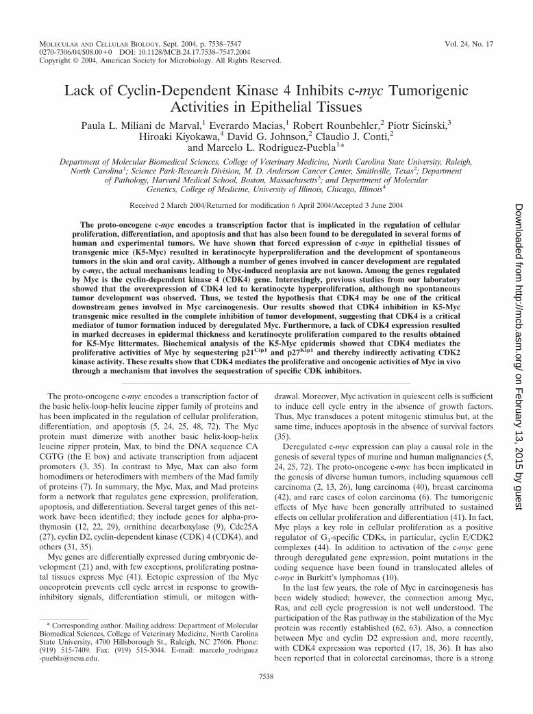
MOLECULAR AND CELLULAR BIOLOGY, Sept. 2004, p. 7538–7547 Vol. 24, No. 17 0270-7306/04/$08.000 DOI: 10.1128/MCB.24.17.7538–7547.2004 Copyright © 2004, American Society for Microbiology. All Rights Reserved. Lack of Cyclin-Dependent Kinase 4 Inhibits c-myc Tumorigenic Activities in Epithelial Tissues Paula L. Miliani de Marval, 1 Everardo Macias, 1 Robert Rounbehler, 2 Piotr Sicinski, 3 Hiroaki Kiyokawa, 4 David G. Johnson, 2 Claudio J. Conti, 2 and Marcelo L. Rodriguez-Puebla 1 * Department of Molecular Biomedical Sciences, College of Veterinary Medicine, North Carolina State University, Raleigh, North Carolina 1 ; Science Park-Research Division, M. D. Anderson Cancer Center, Smithville, Texas 2 ; Department of Pathology, Harvard Medical School, Boston, Massachusetts 3 ; and Department of Molecular Genetics, College of Medicine, University of Illinois, Chicago, Illinois 4 Received 2 March 2004/Returned for modification 6 April 2004/Accepted 3 June 2004 The proto-oncogene c-myc encodes a transcription factor that is implicated in the regulation of cellular proliferation, differentiation, and apoptosis and that has also been found to be deregulated in several forms of human and experimental tumors. We have shown that forced expression of c-myc in epithelial tissues of transgenic mice (K5-Myc) resulted in keratinocyte hyperproliferation and the development of spontaneous tumors in the skin and oral cavity. Although a number of genes involved in cancer development are regulated by c-myc, the actual mechanisms leading to Myc-induced neoplasia are not known. Among the genes regulated by Myc is the cyclin-dependent kinase 4 (CDK4) gene. Interestingly, previous studies from our laboratory showed that the overexpression of CDK4 led to keratinocyte hyperproliferation, although no spontaneous tumor development was observed. Thus, we tested the hypothesis that CDK4 may be one of the critical downstream genes involved in Myc carcinogenesis. Our results showed that CDK4 inhibition in K5-Myc transgenic mice resulted in the complete inhibition of tumor development, suggesting that CDK4 is a critical mediator of tumor formation induced by deregulated Myc. Furthermore, a lack of CDK4 expression resulted in marked decreases in epidermal thickness and keratinocyte proliferation compared to the results obtained for K5-Myc littermates. Biochemical analysis of the K5-Myc epidermis showed that CDK4 mediates the proliferative activities of Myc by sequestering p21 Cip1 and p27 Kip1 and thereby indirectly activating CDK2 kinase activity. These results show that CDK4 mediates the proliferative and oncogenic activities of Myc in vivo through a mechanism that involves the sequestration of specific CDK inhibitors. The proto-oncogene c-myc encodes a transcription factor of the basic helix-loop-helix leucine zipper family of proteins and has been implicated in the regulation of cellular proliferation, differentiation, and apoptosis (5, 24, 25, 48, 72). The Myc protein must dimerize with another basic helix-loop-helix leucine zipper protein, Max, to bind the DNA sequence CA CGTG (the E box) and activate transcription from adjacent promoters (3, 35). In contrast to Myc, Max can also form homodimers or heterodimers with members of the Mad family of proteins (7). In summary, the Myc, Max, and Mad proteins form a network that regulates gene expression, proliferation, apoptosis, and differentiation. Several target genes of this net- work have been identified; they include genes for alpha-pro- thymosin (12, 22, 29), ornithine decarboxylase (9), Cdc25A (27), cyclin D2, cyclin-dependent kinase (CDK) 4 (CDK4), and others (31, 35). Myc genes are differentially expressed during embryonic de- velopment (21) and, with few exceptions, proliferating postna- tal tissues express Myc (41). Ectopic expression of the Myc oncoprotein prevents cell cycle arrest in response to growth- inhibitory signals, differentiation stimuli, or mitogen with- drawal. Moreover, Myc activation in quiescent cells is sufficient to induce cell cycle entry in the absence of growth factors. Thus, Myc transduces a potent mitogenic stimulus but, at the same time, induces apoptosis in the absence of survival factors (35). Deregulated c-myc expression can play a causal role in the genesis of several types of murine and human malignancies (5, 24, 25, 72). The proto-oncogene c-myc has been implicated in the genesis of diverse human tumors, including squamous cell carcinoma (2, 13, 26), lung carcinoma (40), breast carcinoma (42), and rare cases of colon carcinoma (6). The tumorigenic effects of Myc have been generally attributed to sustained effects on cellular proliferation and differentiation (41). In fact, Myc plays a key role in cellular proliferation as a positive regulator of G 1 -specific CDKs, in particular, cyclin E/CDK2 complexes (44). In addition to activation of the c-myc gene through deregulated gene expression, point mutations in the coding sequence have been found in translocated alleles of c-myc in Burkitt’s lymphomas (10). In the last few years, the role of Myc in carcinogenesis has been widely studied; however, the connection among Myc, Ras, and cell cycle progression is not well understood. The participation of the Ras pathway in the stabilization of the Myc protein was recently established (62, 63). Also, a connection between Myc and cyclin D2 expression and, more recently, with CDK4 expression was reported (17, 18, 36). It has also been reported that in colorectal carcinomas, there is a strong * Corresponding author. Mailing address: Department of Molecular Biomedical Sciences, College of Veterinary Medicine, North Carolina State University, 4700 Hillsborough St., Raleigh, NC 27606. Phone: (919) 515-7409. Fax: (919) 515-3044. E-mail: marcelo_rodriguez [email protected]. 7538 on February 13, 2015 by guest http://mcb.asm.org/ Downloaded from
-
Upload
independent -
Category
Documents
-
view
0 -
download
0
Transcript of Lack of Cyclin-Dependent Kinase 4 Inhibits c-myc Tumorigenic Activities in Epithelial Tissues
MOLECULAR AND CELLULAR BIOLOGY, Sept. 2004, p. 7538–7547 Vol. 24, No. 170270-7306/04/$08.00�0 DOI: 10.1128/MCB.24.17.7538–7547.2004Copyright © 2004, American Society for Microbiology. All Rights Reserved.
Lack of Cyclin-Dependent Kinase 4 Inhibits c-myc TumorigenicActivities in Epithelial Tissues
Paula L. Miliani de Marval,1 Everardo Macias,1 Robert Rounbehler,2 Piotr Sicinski,3Hiroaki Kiyokawa,4 David G. Johnson,2 Claudio J. Conti,2
and Marcelo L. Rodriguez-Puebla1*Department of Molecular Biomedical Sciences, College of Veterinary Medicine, North Carolina State University, Raleigh,
North Carolina1; Science Park-Research Division, M. D. Anderson Cancer Center, Smithville, Texas2; Departmentof Pathology, Harvard Medical School, Boston, Massachusetts3; and Department of Molecular
Genetics, College of Medicine, University of Illinois, Chicago, Illinois4
Received 2 March 2004/Returned for modification 6 April 2004/Accepted 3 June 2004
The proto-oncogene c-myc encodes a transcription factor that is implicated in the regulation of cellularproliferation, differentiation, and apoptosis and that has also been found to be deregulated in several forms ofhuman and experimental tumors. We have shown that forced expression of c-myc in epithelial tissues oftransgenic mice (K5-Myc) resulted in keratinocyte hyperproliferation and the development of spontaneoustumors in the skin and oral cavity. Although a number of genes involved in cancer development are regulatedby c-myc, the actual mechanisms leading to Myc-induced neoplasia are not known. Among the genes regulatedby Myc is the cyclin-dependent kinase 4 (CDK4) gene. Interestingly, previous studies from our laboratoryshowed that the overexpression of CDK4 led to keratinocyte hyperproliferation, although no spontaneoustumor development was observed. Thus, we tested the hypothesis that CDK4 may be one of the criticaldownstream genes involved in Myc carcinogenesis. Our results showed that CDK4 inhibition in K5-Myctransgenic mice resulted in the complete inhibition of tumor development, suggesting that CDK4 is a criticalmediator of tumor formation induced by deregulated Myc. Furthermore, a lack of CDK4 expression resultedin marked decreases in epidermal thickness and keratinocyte proliferation compared to the results obtainedfor K5-Myc littermates. Biochemical analysis of the K5-Myc epidermis showed that CDK4 mediates theproliferative activities of Myc by sequestering p21Cip1 and p27Kip1 and thereby indirectly activating CDK2kinase activity. These results show that CDK4 mediates the proliferative and oncogenic activities of Myc in vivothrough a mechanism that involves the sequestration of specific CDK inhibitors.
The proto-oncogene c-myc encodes a transcription factor ofthe basic helix-loop-helix leucine zipper family of proteins andhas been implicated in the regulation of cellular proliferation,differentiation, and apoptosis (5, 24, 25, 48, 72). The Mycprotein must dimerize with another basic helix-loop-helixleucine zipper protein, Max, to bind the DNA sequence CACGTG (the E box) and activate transcription from adjacentpromoters (3, 35). In contrast to Myc, Max can also formhomodimers or heterodimers with members of the Mad familyof proteins (7). In summary, the Myc, Max, and Mad proteinsform a network that regulates gene expression, proliferation,apoptosis, and differentiation. Several target genes of this net-work have been identified; they include genes for alpha-pro-thymosin (12, 22, 29), ornithine decarboxylase (9), Cdc25A(27), cyclin D2, cyclin-dependent kinase (CDK) 4 (CDK4), andothers (31, 35).
Myc genes are differentially expressed during embryonic de-velopment (21) and, with few exceptions, proliferating postna-tal tissues express Myc (41). Ectopic expression of the Myconcoprotein prevents cell cycle arrest in response to growth-inhibitory signals, differentiation stimuli, or mitogen with-
drawal. Moreover, Myc activation in quiescent cells is sufficientto induce cell cycle entry in the absence of growth factors.Thus, Myc transduces a potent mitogenic stimulus but, at thesame time, induces apoptosis in the absence of survival factors(35).
Deregulated c-myc expression can play a causal role in thegenesis of several types of murine and human malignancies (5,24, 25, 72). The proto-oncogene c-myc has been implicated inthe genesis of diverse human tumors, including squamous cellcarcinoma (2, 13, 26), lung carcinoma (40), breast carcinoma(42), and rare cases of colon carcinoma (6). The tumorigeniceffects of Myc have been generally attributed to sustainedeffects on cellular proliferation and differentiation (41). In fact,Myc plays a key role in cellular proliferation as a positiveregulator of G1-specific CDKs, in particular, cyclin E/CDK2complexes (44). In addition to activation of the c-myc genethrough deregulated gene expression, point mutations in thecoding sequence have been found in translocated alleles ofc-myc in Burkitt’s lymphomas (10).
In the last few years, the role of Myc in carcinogenesis hasbeen widely studied; however, the connection among Myc,Ras, and cell cycle progression is not well understood. Theparticipation of the Ras pathway in the stabilization of the Mycprotein was recently established (62, 63). Also, a connectionbetween Myc and cyclin D2 expression and, more recently,with CDK4 expression was reported (17, 18, 36). It has alsobeen reported that in colorectal carcinomas, there is a strong
* Corresponding author. Mailing address: Department of MolecularBiomedical Sciences, College of Veterinary Medicine, North CarolinaState University, 4700 Hillsborough St., Raleigh, NC 27606. Phone:(919) 515-7409. Fax: (919) 515-3044. E-mail: [email protected].
7538
on February 13, 2015 by guest
http://mcb.asm
.org/D
ownloaded from
correlation between the overexpression of Myc and the aber-rant expression of CDK4 (6, 23, 36). It has been proposed thatthe proliferative and oncogenic roles of Myc are linked to itsability to induce the transcription of CDK4, cyclin D1, andcyclin D2 (36), which inactivates the product of the Rb tumorsuppressor gene; this hypothesis provides a link between Mycand the CDK4/cyclin D1/pRb pathway in some malignant tu-mors (32).
Several in vivo models of Myc overexpression have demon-strated that the transgenic expression of Myc in the basal andsuprabasal cell layers of stratified epithelia and in stem cellsleads to hyperplasia, increased epidermal thickness, and pro-liferation (52, 61, 69, 70). In particular, Myc transgenic micedeveloped in our laboratory with the K5 promoter (K5-Myc)showed epithelial neoplasia in the skin as well as oral mucosa(60, 61). Also, the overexpression of Myc in B lymphocytes ofE�-Myc transgenic mice resulted in the development of Bur-kitt-type lymphoma with a latency of 6 months (1).
A previous report by Miliani de Marval et al. also demon-strated that the forced expression of CDK4 in the epidermis oftransgenic mice resulted in a similar phenotype: hyperprolif-eration of the basal cell layer of the epidermis, hypertrophy,and increased epidermal thickness (46). In addition, these miceshowed a high rate of malignant progression of chemicallyinduced tumors (47). Also, the expression of a mutant form(CDK4-R24C) which lacks the capacity to bind p16Ink4a re-sulted in a wide spectrum of tumors, with the most commonbeing lymphomas, endocrine tumors, and hemangiosarcomas(55, 65). The involvement of CDK4 in the neoplastic processwas also suggested by the fact that CDK4 amplification and/oroverexpression were detected in human glioblastomas (34). Inaddition, CDK4 mutations were identified in patients withfamilial melanoma (73, 76) and, more recently, the amplifica-tion and overexpression of CDK4 were detected in sporadicbreast carcinomas (4) and sarcomas (39).
Hence, the objective of this project was to examine the roleof CDK4 in c-myc-induced tumorigenesis. Here we have dem-onstrated that the lack of CDK4 expression in Myc transgenicmice results in the complete inhibition of tumor developmentand suppression of the epidermal phenotype observed in K5-Myc mice. We have also determined that c-myc induces aber-rant expression of CDK4, which sequesters the CDK inhibitors(CKIs) p21Cip1 and p27Kip1, leading to an indirect increase inCDK2 kinase activity. These results demonstrate that CDK4can be a target for therapeutic intervention in Myc-mediatedtumorigenesis.
MATERIALS AND METHODS
Mouse experiments. K5-Myc transgenic mice were developed in an FVBbackground and backcrossed into an SSIN (Sencar) genetic background (61).CDK4- and cyclin D2-null mice were developed by targeted disruption in aC57BL/6 background and further backcrossed into an SSIN genetic backgroundfor two generations (58, 64, 67). K5-Myc transgenic mice (line MM5) werecrossed with mice heterozygous for CDK4 (CDK4�/�) to generate K5-Myctransgenic mice heterozygous for CDK4 (K5-Myc/CDK4�/�). These mice werebred with CDK4�/� mice to generate K5-Myc transgenic and nontransgenic micethat were either homozygous, heterozygous, or nullizygous for CDK4. The samestrategy was used to generate K5-Myc mice nullizygous for cyclin D2 (64).
BrdU incorporation. Epithelial cell proliferation was measured by intraperi-toneal injection of bromodeoxyuridine (BrdU) (60 mg/g of body weight) 30 minbefore the mice were sacrificed. BrdU incorporation was detected by immuno-histochemical staining of paraffin-embedded sections with mouse anti-BrdU
monoclonal antibody (Becton Dickinson Immunocytometry Systems, San Jose,Calif.). The reaction was visualized with a biotin-conjugated anti-mouse antibody(Vector Laboratories, Inc., Burlingame, Calif.) and an avidin-biotin-peroxidasekit (Vectastain Elite; Vector Laboratories, Inc.) with diaminobenzidine as thechromogen. Interfollicular basal cells were examined microscopically to deter-mine the numbers of unstained and stained cells. At least 1,000 cells werecounted per section.
Western blot analysis. The dorsal side of the mice was shaved, treated with adepilatory agent for 1 min, and then washed off. The epidermal tissue wasscraped off with a razor blade, placed in homogenization buffer (50 mM HEPES[pH 7.5], 150 mM NaCl, 2.5 mM EGTA, 1 mM EDTA, 0.1% Tween 20; 1 mMdithiothreitol [DTT], 0.1 mM phenylmethylsulfonyl fluoride, 0.2 U of aprotinin/ml, 10 mM �-glycerophosphate, 0.1 mM sodium vanadate, 1 mM NaF [pH 7.8]),and homogenized by using a manual homogenizer. The epidermal homogenatewas centrifuged at 10,000 � g in order to collect the supernatant, which was useddirectly for Western blotting analysis or stored at �80°C. The protein concen-tration was measured with a protein assay system from Bio-Rad Laboratories,Richmond, Calif. Sodium dodecyl sulfate sample buffer was added to eachsample and boiled for 5 min. Protein lysates (25 �g) were electrophoresedthrough acrylamide gels and electrophoretically transferred to nitrocellulosemembranes (Bio-Rad). After being blocked with 5% nonfat powdered milk inDulbecco’s phosphate-buffered saline (Sigma Chemical Co.), the membraneswere incubated with specific antibodies. The following antibodies were used:rabbit polyclonal antibodies against CDK4 (C22), CDK2 (M2), and CDK6 (C21)(all from Santa Cruz Biotechnology, Inc., Santa Cruz, Calif.); rabbit polyclonalantibody against cyclin D1 (Ab-3) (Lab Vision Corp./Neo Markers, Fremont,Calif.); and horseradish peroxidase-conjugated secondary antibody (AmershamCorp., Arlington Heights, Ill.). Enhanced chemiluminescence (Pierce Biotech,Inc., Rockford, Ill.) was used for immunoblotting detection. Bioimage analysiswas used to quantify the levels of expression of the proteins.
Immunoprecipitation and kinase assays. Fresh protein preparations (250 �gper sample) from epidermal tissue were immunoprecipitated for 2 h at 4°C withprotein A-agarose beads and specific antibodies and washed three times withhomogenization buffer described above. Immunoprecipitates were electropho-resed through acrylamide gels and electrophoretically transferred to nitrocellu-lose membranes. After being blocked with 5% nonfat powdered milk in Dulbec-co’s phosphate-buffered saline, the membranes were incubated with specificantibodies. Antibodies to the following were used: CDK4 (C22), CDK2 (M2),p21 (M19), and p27 (M197) (all from Santa Cruz Biotechnology); cyclin D1 (bclAb-3) (Lab Vision Corp./Neo Markers); protein A-Sepharose beads (Life Tech-nologies Inc., Grand Island, N.Y.); and horseradish peroxidase-conjugated sec-ondary antibody (Amersham). Enhanced chemiluminescence (ECL detectionkit; Amersham) was used for immunoblotting detection.
To study the kinase activities of CDK2 and CDK4, protein lysates wereobtained as described above, but the homogenate was frozen on powdered dryice, thawed in ice water, incubated on ice for 15 min, and centrifuged at 10,000� g for 30 min at 4°C. The supernatant was collected and used for a kinase assay.Protein lysates (250 �g) were immunoprecipitated with antibodies against CDK2or CDK4. Antibody-precoated beads (30 �l) were incubated with the lysates for1 h at 4°C. The beads were washed twice with NP-40 buffer (Tris [pH 7.5], 150mM NaCl, 0.5% NP-40, 50 mM NaF, 1 mM Na3VO4, 1 mM DTT, 1 mMphenylmethylsulfonyl fluoride) and twice with kinase buffer (50 mM HEPES [pH7], 10 mM MgCl2, 5 mM MbCl2). Then, 30 �l of kinase buffer, 1 �g of pRb(Santa Cruz Biotechnology) or histone H1 (Upstate Biotechnology Inc., Char-lottesville, Va.) substrate, 5 �Ci of [�-32P]ATP (6,000 Ci/mmol), 1 mM DTT, and5 �M ATP were added to the bead pellet and incubated for 30 min at 30°C.Sodium dodecyl sulfate sample buffer was added, and each sample was boiled for5 min and electrophoresed through acrylamide gels.
Statistical analysis. A one-way analysis of variance with a Tukey-Kramermultiple-comparison test was performed by using GraphPad InStat version 3.00for Windows 95 (GraphPad Software, San Diego, Calif.).
RESULTS
Lack of CDK4 expression results in complete inhibition oftumor development. Overexpression of the murine c-myc genein the basal cell layer of the stratified epithelium (K5-Myctransgenic mice) resulted in epidermal hyperplasia and hyper-trophy development (Fig. 1 and 2) (61). In addition, a highincidence of spontaneous tumors was observed in the skin andoral mucosa of transgenic mice (61). These results clearly
VOL. 24, 2004 ROLE OF CDK4 IN c-myc ONCOGENIC ACTIVITY 7539
on February 13, 2015 by guest
http://mcb.asm
.org/D
ownloaded from
showed that c-myc acts as an oncogene in the stratified epithe-lium, but the mechanisms leading to the malignant phenotypeare not fully understood. Interestingly, the forced expression ofCDK4 in the basal cell layer of stratified epithelium (K5-CDK4transgenic mice) resulted in a skin phenotype similar to that ofK5-Myc animals (46). Furthermore, chemically induced skinpapillomas from both K5-CDK4 and K5-Myc transgenic miceshowed an increase in the rate of malignant progression tosquamous cell carcinomas (47, 61).
Several other reports have demonstrated that Myc inducesthe transcription of CDK4 in cell cultures and colorectal tumorsamples (6, 23, 36). In order to investigate whether CDK4mediates the oncogenic activities of c-myc, we developed K5-Myc transgenic mice that lack the expression of CDK4 (K5-Myc/CDK4�/� mice). These mice, along with K5-Myc/CDK4�/�, K5-Myc/CDK4�/�, CDK4�/�, CDK4�/�, and wild-type siblings, were analyzed for the development ofspontaneous tumors.
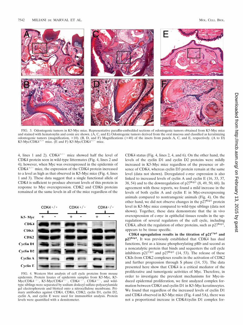
Histological analysis of mice bearing the c-myc transgenewith one or two functional CDK4 alleles revealed a wide spec-trum of tumors of the oral mucosa (Fig. 3 and Table 1) (61).The tumors were observed in mice as young as 8 weeks old withan incidence of 100%. These tumors were classified as kera-tinizing odontogenic tumors, neuroblastomas, and olfactorytumors derived from the oral mucosa, odontogenic tissues, andthe olfactory epithelium (Table 1). In sharp contrast,CDK4�/� animals carrying the K5-Myc transgene did not de-velop any malignancies during the 3-month observation period(Table 1). Thus, genetic inhibition of CDK4 renders animalsresistant to Myc-driven oncogenesis of the oral mucosa. Nofurther observation was possible because the CDK4�/� micedied at 12 to 16 weeks of age. It is likely that these micedeveloped diabetes mellitus, as previously reported (56, 67).Thus, quantification and histological analysis of oral cavitytumors were performed until 12 weeks of age for all genotypes.Nontransgenic CDK4�/� and CDK4�/� littermates did notshow any signs of spontaneous tumor development, even whenthey were 12 months old.
It has also been shown that cyclin D2 expression is regulatedby the c-myc-encoded transcription factor (14, 53), but thespecific role as a mediator of the tumorigenic effects of c-mycin vivo has yet to be defined. To test the requirement for cyclinD2 in Myc-driven oncogenesis of the epithelium, we crossedK5-Myc mice with cyclin D2�/� mice (64) and generated K5-Myc/cyclin D2�/� animals. Our analysis revealed that the cy-
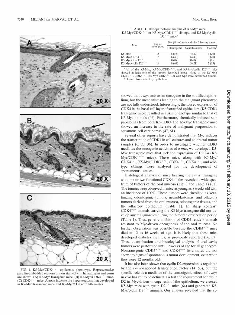
FIG. 1. K5-Myc/CDK4�/� epidermis phenotype. Representativeparaffin-embedded sections of skin stained with hematoxylin and eosinare shown. (A) K5-Myc transgenic mice. (B) K5-Myc/CDK4�/� mice.(C) CDK4�/� mice. Arrows indicate the hyperkeratosis that developedin K5-Myc transgenic mice and K5-Myc/CDK4�/� littermates.
TABLE 1. Histopathologic analysis of K5-Myc mice,K5-Myc/CDK4�/� or K5-Myc/CDK4�/� siblings, and K5-Myc/cyclin
D2�/� micea
Mice No. ofmice/group
No. (%) of mice with the following tumor:
Odontogenic Neuroblastoma Olfactoryb
K5-Myc 15 8 (53) 4 (27) 3 (20)K5-Myc/CDK4�/� 15 6 (40) 6 (40) 3 (20)K5-Myc/CDK4�/� 10 0 (0) 0 (0) 0 (0)K5-Myc/cyclin D2�/� 14 9 (64) 3 (21) 2 (15)
a All of the K5-Myc, K5-Myc/CDK4�/�, and K5-Myc/cyclin D2�/� miceshowed at least one of the tumors described above. None of the K5-Myc/CDK4�/�, CDK4�/�, K5-Myc CDK4�/�, or wild-type mice developed tumors.
b Derived from olfactory epithelium.
7540 MILIANI DE MARVAL ET AL. MOL. CELL. BIOL.
on February 13, 2015 by guest
http://mcb.asm
.org/D
ownloaded from
clin D2�/� mice remained fully susceptible to Myc-driven tu-morigenesis of the oral mucosa (Table 1). Hence, these datasuggest that cyclin D2 does not play a relevant role in thedevelopment of the spontaneous tumors observed in K5-Mycmice.
Collectively, these results indicate that CDK4, but not cyclinD2, plays a critical role in Myc-mediated tumor development.
Inactivation of CDK4 results in reduced epidermal thick-ness and proliferation. In order to investigate whether CDK4also mediates the epidermal hyperproliferative phenotype trig-gered by Myc overexpression, we analyzed the epidermis ofK5-Myc and K5-Myc/CDK4�/� mice. The skin of K5-Mycmice showed hyperplasia (increased cell number) and hyper-trophy (increased cell size), two features that contributed tothe increase in the epidermal thickness observed in these mice(Fig. 1 and 2) (61). A lack of CDK4 expression (K5-Myc/
CDK4�/� mice) resulted in reversion of the increased epider-mal thickness observed in K5-Myc mice, although the hyper-keratosis (accumulation of keratinized cells in the epidermalsurface) characteristic of Myc overexpression still persisted(Fig. 1B). Rodriguez-Puebla et al. previously demonstratedthat a lack of CDK4 expression does not affect the mouseepidermal architecture, which is similar to that in wild-typesiblings (Fig. 1C) (58). Quantification of the epidermal thick-ness revealed a significant reduction in K5-Myc/CDK4�/�
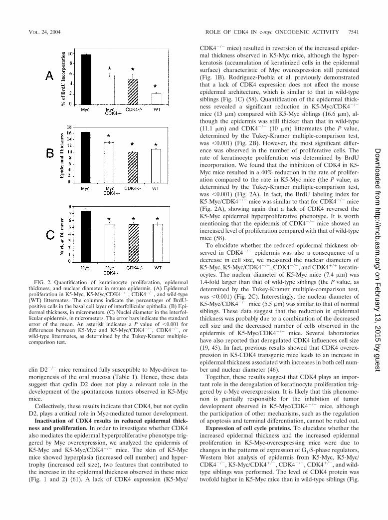
mice (13 �m) compared with K5-Myc siblings (16.6 �m), al-though the epidermis was still thicker than that in wild-type(11.1 �m) and CDK4�/� (10 �m) littermates (the P value,determined by the Tukey-Kramer multiple-comparison test,was �0.001) (Fig. 2B). However, the most significant differ-ence was observed in the number of proliferative cells. Therate of keratinocyte proliferation was determined by BrdUincorporation. We found that the inhibition of CDK4 in K5-Myc mice resulted in a 40% reduction in the rate of prolifer-ation compared to the rate in K5-Myc mice (the P value, asdetermined by the Tukey-Kramer multiple-comparison test,was �0.001) (Fig. 2A). In fact, the BrdU labeling index forK5-Myc/CDK4�/� mice was similar to that for CDK4�/� mice(Fig. 2A), showing again that a lack of CDK4 reversed theK5-Myc epidermal hyperproliferative phenotype. It is worthmentioning that the epidermis of CDK4�/� mice showed anincreased level of proliferation compared with that of wild-typemice (58).
To elucidate whether the reduced epidermal thickness ob-served in CDK4�/� epidermis was also a consequence of adecrease in cell size, we measured the nuclear diameters ofK5-Myc, K5-Myc/CDK4�/�, CDK4�/�, and CDK4�/� keratin-ocytes. The nuclear diameter of K5-Myc mice (7.4 �m) was1.4-fold larger than that of wild-type siblings (the P value, asdetermined by the Tukey-Kramer multiple-comparison test,was �0.001) (Fig. 2C). Interestingly, the nuclear diameter ofK5-Myc/CDK4�/� mice (5.5 �m) was similar to that of normalsiblings. These data suggest that the reduction in epidermalthickness was probably due to a combination of the decreasedcell size and the decreased number of cells observed in theepidermis of K5-Myc/CDK4�/� mice. Several laboratorieshave also reported that deregulated CDK4 influences cell size(19, 45). In fact, previous results showed that CDK4 overex-pression in K5-CDK4 transgenic mice leads to an increase inepidermal thickness associated with increases in both cell num-ber and nuclear diameter (46).
Together, these results suggest that CDK4 plays an impor-tant role in the deregulation of keratinocyte proliferation trig-gered by c-Myc overexpression. It is likely that this phenome-non is partially responsible for the inhibition of tumordevelopment observed in K5-Myc/CDK4�/� mice, althoughthe participation of other mechanisms, such as the regulationof apoptosis and terminal differentiation, cannot be ruled out.
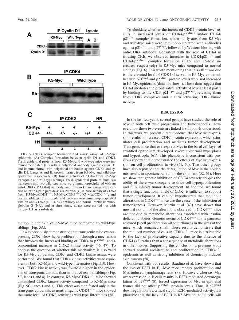
Expression of cell cycle proteins. To elucidate whether theincreased epidermal thickness and the increased epidermalproliferation in K5-Myc-overexpressing mice were due tochanges in the patterns of expression of G1/S-phase regulators,Western blot analysis of epidermis from K5-Myc, K5-Myc/CDK4�/�, K5-Myc/CDK4�/�, CDK4�/�, CDK4�/�, and wild-type siblings was performed. The level of CDK4 protein wastwofold higher in K5-Myc mice than in wild-type siblings (Fig.
FIG. 2. Quantification of keratinocyte proliferation, epidermalthickness, and nuclear diameter in mouse epidermis. (A) Epidermalproliferation in K5-Myc, K5-Myc/CDK4�/�, CDK4�/�, and wild-type(WT) littermates. The columns indicate the percentages of BrdU-positive cells in the basal cell layer of interfollicular epithelia. (B) Epi-dermal thickness, in micrometers. (C) Nuclei diameter in the interfol-licular epidermis, in micrometers. The error bars indicate the standarderror of the mean. An asterisk indicates a P value of �0.001 fordifferences between K5-Myc and K5-Myc/CDK4�/�, CDK4�/�, orwild-type littermates, as determined by the Tukey-Kramer multiple-comparison test.
VOL. 24, 2004 ROLE OF CDK4 IN c-myc ONCOGENIC ACTIVITY 7541
on February 13, 2015 by guest
http://mcb.asm
.org/D
ownloaded from
4, lines 1 and 2). CDK4�/� mice showed half the level ofCDK4 protein seen in wild-type littermates (Fig. 4, lines 2 and4); however, when Myc was overexpressed in the epidermis ofCDK4�/� mice, the expression of the CDK4 protein increasedto a level as high as that observed in K5-Myc mice (Fig. 4, lines1 and 3). These data suggest that a single functional allele ofCDK4 is sufficient to produce aberrant levels of this protein inresponse to Myc overexpression. CDK2 and CDK6 proteinsremained at the same levels in all of the mice regardless of the
CDK4 status (Fig. 4, lines 2, 4, and 6). On the other hand, thelevels of the cyclin D1 and cyclin D2 proteins were mildlyincreased in K5-Myc mice regardless of the presence or ab-sence of CDK4, whereas cyclin D3 protein remain at the samelevel (data not shown). Deregulated c-myc expression is alsolinked to increased levels of cyclin A and cyclin E (16, 33, 37,38, 54) and to the downregulation of p27Kip1 (8, 49, 50, 68). Inagreement with those reports, we found a mild increase in thelevels of both cyclin A and cyclin E in Myc-overexpressinganimals compared to nontransgenic animals (Fig. 4). On theother hand, we did not observe changes in the p27Kip1 proteinlevel in K5-Myc mice compared to wild-type siblings (data notshown). Together, these data demonstrate that the in vivooverexpression of c-myc in epithelial tissues results in the up-regulation of several regulators of the cell cycle, includingCDK4, albeit the regulation of other proteins, such as p27Kip1,appears to be tissue specific.
CDK4 upregulation results in the titration of p21Cip1 andp27Kip1. It was previously established that CDK4 has dualfunctions, first as a kinase phosphorylating pRb and second asa noncatalytic protein that binds and sequesters the cell cycleinhibitors p21Cip1 and p27Kip1 (14, 53). The release of theseCKIs from CDK2 complexes results in the activation of CDK2and further progression through S phase (14, 53). The datapresented here show that CDK4 is a critical mediator of theproliferative and tumorigenic activities of Myc. Therefore, inorder to investigate the prevalent mechanisms for Myc-in-duced epidermal proliferation, we first analyzed complex for-mation between CDK4 and cyclin D1 in K5-Myc keratinocytes.We found that regardless of the increased levels of cyclin D1and CDK4 observed in K5-Myc mice (Fig. 4 and 5A), there wasnot a proportional increase in CDK4/cyclin D1 complex for-
FIG. 3. Odontogenic tumors in K5-Myc mice. Representative paraffin-embedded sections of odontogenic tumors obtained from K5-Myc miceand stained with hematoxylin and eosin are shown. (A, C, and E) Odontogenic tumors derived from the oral mucosa and classified as keratinizingodontogenic tumors (magnification, �10). (B, D, and F) Magnifications (�40) of the insets from panels A, C, and E, respectively. (A to D)K5-Myc/CDK4�/� mice. (E and F) K5-Myc/CDK4�/� mice.
FIG. 4. Western blot analysis of cell cycle proteins from mouseepidermis. Protein lysates of epidermis samples from K5-Myc, K5-Myc/CDK4�/�, K5-Myc/CDK4�/�, CDK4�/�, CDK4�/�, and wild-type siblings were separated by sodium dodecyl sulfate-polyacrylamidegel electrophoresis and blotted onto a nitrocellulose membrane. Pri-mary antibodies against CDK4, CDK6, CDK2, cyclin D1, cyclin D2,cyclin A, and cyclin E were used for immunoblot analysis. Proteinlevels were quantified with a densitometer.
7542 MILIANI DE MARVAL ET AL. MOL. CELL. BIOL.
on February 13, 2015 by guest
http://mcb.asm
.org/D
ownloaded from
mation in the skin of K5-Myc mice compared to wild-typesiblings (Fig. 5A).
It was previously demonstrated that transgenic mice overex-pressing CDK4 show hyperproliferation through a mechanismthat involves the increased binding of CDK4 to p27Kip1 and aconcomitant increase in CDK2 kinase activity (46, 47). Toaddress the question of whether this mechanism is also validfor K5-Myc epidermis, CDK4 and CDK2 kinase assays wereperformed. We found that CDK4 kinase activities were equiv-alent in both K5-Myc and wild-type littermates (Fig. 5B). How-ever, CDK2 kinase activity was fourfold higher in the epider-mis of transgenic animals than in that of normal siblings (Fig.5C, lanes 1 and 4). In contrast, K5-Myc/CDK4�/� mice showeddiminished CDK2 kinase activity compared to K5-Myc mice(Fig. 5C, lanes 1 and 3). This effect was manifested only in thetransgenic epidermis, as nontransgenic CDK4�/� mice showedthe same level of CDK2 activity as wild-type littermates (58).
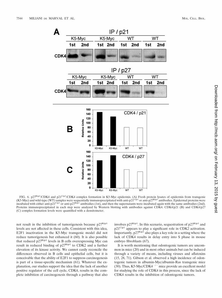
To elucidate whether the increased CDK4 protein level re-sults in increased levels of CDK4/p27Kip1 and/or CDK4/p21Cip1 complex formation, epidermal lysates from K5-Mycand wild-type mice were immunoprecipitated with antibodiesagainst p21Cip1 and p27Kip1, followed by Western blotting withanti-CDK4 antibody. Consistent with the role of CDK4 intitrating CKIs, we observed increases in CDK4/p21Cip1 andCDK4/p27Kip1 complex formation (3.12- and 1.5-fold in-creases, respectively) in K5-Myc mice compared to normalsiblings (Fig. 6). It is worth mentioning that this effect was dueto the elevated level of CDK4 observed in K5-Myc epidermisbecause p21Cip1 and p27Kip1 protein levels were not increasedin K5-Myc epidermis (data not shown). These data suggest thatCDK4 mediates the proliferative activity of Myc at least partlyby binding to the CKIs p21Cip1 and p27Kip1, releasing themfrom CDK2 complexes and in turn activating CDK2 kinaseactivity.
DISCUSSION
In the last few years, several groups have studied the role ofMyc in both cell cycle progression and tumorigenesis. How-ever, how these two events are linked is still poorly understood.In this work, we present direct evidence that Myc overexpres-sion results in increased CDK4 protein expression, which stim-ulates cell proliferation and mediates tumor development.Transgenic mice that overexpress Myc in the basal cell layer ofstratified epithelium developed severe epidermal hyperplasiaand hypertrophy (61). This phenotype is consistent with pre-vious reports that demonstrated the effects of Myc overexpres-sion on cell proliferation in vivo (69, 70). Two other groupshave also reported that the deregulation of Myc in the epider-mis results in spontaneous tumor development (52, 61). Herewe show that genetic inhibition of CDK4 severely cripples theability of the c-myc oncogene to drive cell hyperproliferationand fully inhibits tumor development. In addition, we foundthat a single functional allele of CDK4 is sufficient to supporttumor development. It can be hypothesized that metabolicalterations in CDK4�/� mice are the cause of the inhibition oftumorigenesis. However, Martin et al. (43) have shown thatmost, if not all, of the alterations observed in CDK4�/� miceare not due to metabolic alterations associated with insulin-deficient diabetes. Genetic rescue of CDK4�/� in the pancreasrestored �-cell proliferation without changes in the sizes of themice, which remained small. These results demonstrate thatthe reduced number of cells in CDK4�/� mice is attributableto the lack of proliferative capacity due to the absence ofCDK4 (43) rather than a consequence of metabolic alterationsin other tissues. Supporting this conclusion, a previous studyshowed normal architecture and proliferation in CDK4�/�
epidermis as well as strong inhibition of chemically inducedskin tumors (58).
Consistent with our results, Baudino et al. have shown thatthe loss of E2F1 in E�-Myc mice impairs proliferation andMyc-induced lymphomagenesis (8). However, whereas Mycoverexpression in B cells results in E2F1-mediated downregu-lation of p27Kip1 (8), forced expression of Myc in epithelialtissues did not affect p27Kip1 protein levels. Thus, if p27Kip1
downregulation is a critical step in E2F1-mediated activity, it isplausible that the lack of E2F1 in K5-Myc epithelial cells will
FIG. 5. CDK4 complex formation and kinase assays of K5-Mycepidermis. (A) Complex formation between cyclin D1 and CDK4.Fresh epidermal proteins from K5-Myc and wild-type mice were im-munoprecipitated (IP) with a polyclonal antibody against cyclin D1and immunoblotted with polyclonal antibodies against CDK4 and cy-clin D1. Lanes A and B, protein lysates from K5-Myc and wild-typeepidermis, respectively. (B) Kinase activity of CDK4 from K5-Myctransgenic and wild-type siblings. Fresh epidermal proteins from twotransgenic and two wild-type mice were immunoprecipitated with ananti-CDK4 (IP CDK4) antibody, and in vitro kinase assays were car-ried out with a pRb peptide as a substrate. (C) Kinase activity of CDK2from K5-Myc/CDK4�/�, K5-Myc/CDK4�/�, K5-Myc/CDK4�/�, andnormal siblings. Fresh epidermal proteins were immunoprecipitatedwith an anti-CDK2 (IP CDK2) antibody and normal rabbit immuno-globulin G (NR), and in vitro kinase assays were carried out withhistone H1 as a substrate.
VOL. 24, 2004 ROLE OF CDK4 IN c-myc ONCOGENIC ACTIVITY 7543
on February 13, 2015 by guest
http://mcb.asm
.org/D
ownloaded from
not result in the inhibition of tumorigenesis because p27Kip1
levels are not affected in these cells. Consistent with this idea,E2F1 inactivation in the K5-Myc transgenic model did notreduce tumorigenesis but enhanced it (60). It is also possiblethat reduced p27Kip1 levels in B cells overexpressing Myc canresult in reduced binding of p27Kip1 to CDK2 and a furtherelevation of its kinase activity. We cannot easily reconcile thedifferences observed in B cells and epithelial cells, but it isconceivable that the ability of E2F1 to suppress carcinogenesisis part of a tissue-specific mechanism (61). Whatever the ex-planation, our studies support the idea that the lack of anotherpositive regulator of the cell cycle, CDK4, results in the com-plete inhibition of carcinogenesis through a pathway that also
involves p27Kip1. In this scenario, sequestration of p27Kip1 andp21Cip1 appears to play a significant role in CDK2 activation.Importantly, p27Kip1 also plays a key role in a setting where thelack of CDK4 results in delay entry into S phase in mouseembryo fibroblasts (67).
It is worth mentioning that odontogenic tumors are uncom-mon in mice (20) and in most other animals but can be inducedthrough a variety of means, including viruses and aflatoxins(15, 28, 71). Gibson et al. observed a high incidence of odon-togenic tumors in albumin-Myc/albumin-Ras transgenic mice(30). Thus, K5-Myc/CDK4�/� mice provide an excellent modelfor studying the role of CDK4 in this process, since the lack ofCDK4 results in the inhibition of odontogenic tumors.
FIG. 6. p27Kip1/CDK4 and p21Cip1/CDK4 complex formation in K5-Myc epidermis. (A) Fresh protein lysates of epidermis from transgenic(K5-Myc) and wild-type (WT) samples were sequentially immunoprecipitated with anti-p21Cip1 or anti-p27Kip1 antibodies. Epidermal proteins wereincubated with either anti-p21Cip1 or anti-p27Kip1 antibodies (1st), and then the supernatants were incubated again with the same antibodies (2nd).Proteins immunoprecipitated in each step were analyzed by Western blotting with antibodies against CDK4. CDK4/p21 (B) and CDK4/p27(C) complex formation levels were quantified with a densitometer.
7544 MILIANI DE MARVAL ET AL. MOL. CELL. BIOL.
on February 13, 2015 by guest
http://mcb.asm
.org/D
ownloaded from
Two groups have reported that Myc induces cyclin D2 ex-pression, contributing to cell cycle progression due to an in-crease in the formation of cyclin D2/CDK4/p27Kip1 ternarycomplexes (14, 53). The sequestration of p27Kip1, a specificinhibitor of cyclin E/CDK2, results in the activation of cyclinE-associated kinase activity. In our in vivo model, the overex-pression of Myc in mouse epidermis also results in a mildincrease in cyclin D1 and D2 expression. However, cyclin D2appears to be dispensable for Myc-induced tumor develop-ment and hyperproliferation. The absence of cyclin D2 in K5-Myc mice did not change the outcome of tumor formation,which was as high as that in K5-Myc littermates (Table 1). Inaddition, paraffin-embedded sections of mouse skin showed nodifference in the rates of epidermal proliferation between K5-Myc and K5-Myc/cyclin D2�/� mice (unpublished data). Also,Yu et al. showed that the lack of cyclin D2 does not result inprotection against breast cancer in mouse mammary tumorvirus Myc mice (74). Thus, contrary to the in vitro evidenceimplicating cyclin D2 as a mediator of Myc proliferation, herewe show in an in vivo setting that cyclin D2 is not required forcell proliferation and does not modulate the oncogenic effectsof Myc overexpression. However, Bouchard et al. (14) andPerez-Roger et al. (54) concluded that the c-myc oncogenesignals through both cyclin D1 and cyclin D2. Thus, it is pos-sible that in the oral epithelium, Myc-driven oncogenic prolif-eration is mediated by cyclin D1/CDK4 and cyclin D2/CDK4complexes. According to this scenario, the inhibition of bothcyclin D1 and cyclin D2 would be required to block the onco-genic action of Myc in the oral mucosa.
Together, these results show that CDK4 plays a critical rolein tumor development and epidermal proliferation, althoughthe role of its regulatory subunit, cyclin D1, in K5-Myc-medi-ated tumorigenesis has not yet been investigated. Emergingevidence indicates that the wiring of the oncogenic pathways tothe core cell cycle machinery is cell type specific. For instance,mammary epithelial cells critically require cyclin D1 for Ras-driven tumorigenesis (74). In contrast, Ras can cause onco-genic transformation of fibroblasts in the absence of cyclin D1(74), while cyclin D1-deficient skin keratinocytes show reducedsusceptibility to chemical tumorigenesis (57, 59). The results ofour current study reveal that CDK4 is the critical target of thec-myc oncogene in the oral mucosa.
It is possible that in other cell types, the c-myc oncogeneimpinges on the core cell cycle machinery through other tar-gets. In addition, CDK4�/� mouse embryo fibroblasts are re-sistant to transformation in response to Ras activation withdominant-negative p53 expression or in an Ink4a/Arf�/� back-ground (75). The resistance to transformation of CDK4�/�
mouse embryo fibroblasts has been associated with the ele-vated expression of p21Cip1; however, we did not observe in-creased p21Cip1 expression in CDK4�/� keratinocytes. It is alsoworth mentioning that the lack of CDK4 in mouse epidermisresults in elevated CDK6 kinase activity (58). We have hypoth-esized that CDK6 activity can compensate for the lack ofCDK4 and is partly responsible for the mild increase in kera-tinocyte proliferation observed in CDK4�/� mice (Fig. 2A)(58); however, it is clear that it cannot compensate for the lackof CDK4 in tumor development. However, it is feasible tohypothesize that increased CDK6 activity is the result of in-creased CDK6/cyclin complex formation and further titration
of p27Kip1 and/or p21Cip1. If this is the case, then elevatedlevels of CDK6/cyclin complexes in CDK4�/� mice will resultin at least steady levels of CDK2 activity, as we observed inCDK4�/� epidermis (58). Thus, the implications of CDK6activity for the homeostasis of mouse epidermal tissue warrantfurther investigation.
Biochemical studies demonstrated that there is a significantincrease in the binding of p21Cip1 and p27Kip1 to CDK4 inK5-Myc mouse epidermis, supporting a model in which anincreased level of CDK4 indirectly activates CDK2 through itsnoncatalytic function (Fig. 6). In contrast, we did not detectchanges in the catalytic activities of CDK4, which were similarin K5-Myc and wild-type animals (Fig. 5B). Together, theseresults show that CDK4 plays an important role in Myc-in-duced proliferation by indirectly activating CDK2. The role ofCDK2 in cell proliferation was recently challenged by the factthat CDK2�/� mice developed normally with minor pheno-typic effects, mainly present during the meiotic cell cycle (11,51). It has also been shown that some tumor cell lines canproliferate independently of CDK2 (66). However, the role ofCDK2 in an in vivo tumorigenesis model has not been studiedyet. It will be interesting to determine the role of CDK2 inMyc-induced tumorigenesis through the generation of trans-genic mice overexpressing c-myc in a CDK2�/� background.
It is worth mentioning that transgenic mice which overex-press a kinase-dead mutant form of CDK4 (K5-CDK4dn) thatcan bind to CKIs also showed epidermal hyperplasia and in-creased epidermal thickness similar to those in previously re-ported K5-CDK4 mice (46; unpublished data). Thus, the hy-perproliferation observed in the epidermis of K5-CDK4dominant-negative mice is dependent not on CDK4 kinaseactivity but on CDK4 sequestration activity. These results sup-port the idea that the noncatalytic function of CDK4 is criti-cally involved in cell proliferation. Together, these data suggestthat in epithelial tissues, deregulated Myc can induce the over-expression of several G1/S-phase regulators, including cyclinD1, cyclin D2, cyclin A, cyclin E, and CDK4, resulting in thesequestration of negative regulators, such as p21Cip1 andp27Kip1, and leading to the activation of CDK2 kinase activity.The requirement for both Myc and CDK4 in malignant trans-formation was first reported by Haas et al. (32), who describedthe oncogenic action of CDK4 when coexpressed with Ha-rasand Myc. In fact, they proposed that the ability of CDK4 tobind to p16Ink4 and not its kinase activity was important for itstransforming potential (32). Rodriguez-Puebla et al. previouslydemonstrated that the lack of CDK4 expression also inhibitstumorigenesis in a setting in which mutations in the Ha-rasgene result in the initiation of keratinocytes (58). Thus, theseresults demonstrate the potential use of CDK4 as a therapeutictarget not only in Ras- but also in Myc-mediated tumorigene-sis.
ACKNOWLEDGMENTS
This work was supported by PHS grants CA 42157 and CA 90864from NCI.
We especially thank April Weiss for helping with the mouse exper-iments, the CVM-NCSU and Science Park animal facility personnel,and the Science Park histologic service for assistance with the immu-nohistochemical staining.
VOL. 24, 2004 ROLE OF CDK4 IN c-myc ONCOGENIC ACTIVITY 7545
on February 13, 2015 by guest
http://mcb.asm
.org/D
ownloaded from
REFERENCES
1. Adams, J. M., A. W. Harris, C. A. Pinkert, L. M. Corcoran, W. S. Alexander,S. Cory, R. D. Palmiter, and R. L. Brinster. 1985. The c-myc oncogene drivenby immunoglobulin enhancers induces lymphoid malignancy in transgenicmice. Nature 318:533–538.
2. Akervall, J., U. Bockmuhl, I. Petersen, K. Yang, T. E. Carey, and D. M.Kurnit. 2003. The gene ratios c-MYC:cyclin-dependent kinase (CDK)N2Aand CCND1:CDKN2A correlate with poor prognosis in squamous cell car-cinoma of the head and neck. Clin. Cancer Res. 9:1750–1755.
3. Amati, B., and H. Land. 1994. Myc-Max-Mad: a transcription factor networkcontrolling cell cycle progression, differentiation and death. Curr. Opin.Genet. Dev. 4:102–108.
4. An, H., M. W. Beckmann, G. Reifenger, H. G. Bender, and D. Niederacher.1999. Gene amplification and overexpression of CDK4 in sporadic breastcarcinomas is associated with high tumor cell proliferation. Am. J. Pathol.154:113–118.
5. Askew, D., R. Ashmun, B. Simmons, and J. Cleveland. 1991. Constitutivec-myc expression in an IL-3-dependent myeloid cell line suppresses cell cyclearrest and accelerates apoptosis. Oncogene 6:1915–1922.
6. Augenlicht, L. H., S. Wadler, G. Corner, C. Richards, L. Ryan, A. S. Multani,S. Pathak, A. Benson, D. Haller, and B. G. Heerdt. 1997. Low-level c-mycamplification in human colonic carcinoma cell lines and tumors: a frequent,p53-independent mutation associated with improved outcome in a random-ized multi-institutional trial. Cancer Res. 57:1769–1775.
7. Baudino, T. A., and J. L. Cleveland. 2001. The Max network gone mad. Mol.Cell. Biol. 21:691–702.
8. Baudino, T. A., K. H. Maclean, J. Brennan, E. Parganas, C. Yang, A.Aslanian, J. A. Lees, C. J. Sherr, M. F. Roussel, and J. L. Cleveland. 2003.Myc-mediated proliferation and lymphomagenesis, but not apoptosis, arecompromised by E2F1 loss. Mol. Cell 11:905–914.
9. Bello-Fernandez, C., G. Packham, and J. L. Cleveland. 1993. The ornithinedecarboxylase gene is a transcriptional target of c-Myc. Proc. Natl. Acad. Sci.USA 90:7804–7808.
10. Bemark, M., and M. Neuberger. 2000. The c-MYC allele that is translocatedinto the IgH locus undergoes constitutive hypermutation in a Burkitt’s lym-phoma line. Oncogene 13:3404–3410.
11. Berthet, C., E. Aleem, V. Coppola, L. Tassarollo, and P. Kaldis. 2003. Cdk2knockout mice are viable. Curr. Biol. 13:1775–1785.
12. Bishop, J. M., M. Eilers, A. L. Katzen, T. Kornberg, G. Ramsay, and S.Schirm. 1991. MYB and MYC in the cell cycle. Cold Spring Harbor Symp.Quant. Biol. 56:99–107.
13. Bitzer, M., M. Stahl, J. Arjumand, M. Rees, B. Klump, H. Heep, H. E.Gabbert, and M. Sarbia. 2003. c-Myc gene amplification in different stagesof oesophageal squamous cell carcinoma: prognostic value in relation totreatment modality. Anticancer Res. 23:1489–1493.
14. Bouchard, C., K. Thieke, A. Maier, R. Saffrich, J. Hanley-Hyde, W. Ansorge,S. Reed, P. Sicinski, J. Bartek, and M. Eilers. 1999. Direct induction of cyclinD2 by Myc contributes to cell cycle progression and sequestration of p27.EMBO J. 18:5321–5333.
15. Cullen, J. M., B. H. Ruebner, D. P. Hsieh, and E. J. Burkes, Jr. 1987.Odontogenic tumors in Fischer rats. J. Oral Pathol. 16:469–473.
16. Daksis, J. I., R. Y. Lu, L. M. Facchini, W. W. Marhin, and L. J. Penn. 1994.Myc induces cyclin D1 expression in the absence of de novo protein synthesisand links mitogen-stimulated signal transduction to the cell cycle. Oncogene9:3635–3645.
17. Dang, C. V. 1999. c-myc target genes involved in cell growth, apoptosis, andmetabolism. Mol. Cell. Biol. 19:1–11.
18. Dang, C. V., L. M. Resar, E. Emison, S. Kim, Q. Li, J. E. Prescott, D.Wonsey, and K. Zeller. 1999. Function of the c-Myc oncogenic transcriptionfactor. Exp. Cell Res. 253:63–77.
19. Datar, S. A., H. W. Jacobs, A. F. de la Cruz, C. F. Lehner, and B. A. Edgar.2000. The Drosophila cyclin D-Cdk4 complex promotes cellular growth.EMBO J. 19:4543–4554.
20. Dayan, D., T. Waner, A. Harmelin, and A. Nyska. 1984. Bilateral complexodontoma in a Swiss (CD-1) male mouse. Lab. Anim. 28:90–92.
21. Downs, K. M., G. R. Martin, and J. M. Bishop. 1989. Contrasting patterns ofmyc and N-myc expression during gastrulation of the mouse embryo. GenesDev. 3:860–869.
22. Eilers, M., S. Schirm, and J. M. Bishop. 1991. The MYC protein activatestranscription of the alpha-prothymosin gene. EMBO J. 10:133–141.
23. Erisman, M. D., P. G. Rothberg, R. E. Diehl, C. C. Morse, J. M. Spandorfer,and S. M. Astrin. 1985. Deregulation of c-myc gene expression in humancolon carcinoma is not accompanied by amplification or rearrangement ofthe gene. Mol. Cell. Biol. 5:1969–1976.
24. Evan, G., A. Wyllie, C. Gilbert, T. Littlewood, H. Land, M. Brooks, C.Waters, L. Penn, and D. Hancock. 1992. Induction of apoptosis in fibroblastsby c-myc protein. Cell 69:119–128.
25. Facchini, L. M., and L. Penn. 1998. The molecular role of Myc in growth andtransformation: recent discoveries lead to new insights. FASEB J. 12:633–651.
26. Field, J. K., D. A. Spandidos, P. M. Stell, E. D. Vaughan, G. I. Evan, and J. P.
Moore. 1989. Elevated expression of the c-myc oncoprotein correlates withpoor prognosis in head and neck squamous cell carcinoma. Oncogene4:1463–1468.
27. Galaktionov, K., X. Chen, and D. Beach. 1996. Cdc25 cell-cycle phosphataseas a target of c-myc. Nature 382:511–517.
28. Gardner, D. G. 1992. An orderly approach to the study of odontogenictumors in animals. J. Comp. Pathol. 107:427–438.
29. Gaubatz, S., A. Meichle, and M. Eilers. 1994. An E-box element localized inthe first intron mediates regulation of the prothymosin alpha gene by c-myc.Mol. Cell. Biol. 14:3853–3862.
30. Gibson, C. W., E. Lally, R. C. Herold, S. Decker, R. L. Brinster, and E. P.Sandgren. 1992. Odontogenic tumors in mice carrying albumin-myc andalbumin-rats transgenes. Calcif. Tissue Int. 51:162–167.
31. Grandori, C., and R. N. Eisenman. 1997. Myc target genes. Trends Biochem.Sci. 22:177–181.
32. Haas, K., P. Staller, C. Geisen, J. Bartek, M. Eilers, and T. Moroy. 1997.Mutual requirement of CDK4 and Myc in malignant transformation: evi-dence for cyclin D1/CDK4 and p16INK4A as upstream regulators of Myc.Oncogene 15:179–192.
33. Hanson, K. D., M. Shichiri, M. R. Follansbee, and J. M. Sedivy. 1994. Effectsof c-myc expression on cell cycle progression. Mol. Cell. Biol. 14:5748–5755.
34. He, J., J. R. Allen, V. P. Collins, M. J. Allalunis-Turner, R. Godbout, R. S.Day, and C. D. James. 1994. CDK4 amplification is an alternative mecha-nism to p16 homozygous deletion in glioma cell lines. Cancer Res. 54:5804–5807.
35. Henriksson, M., B. Luscher, and A. Pardee. 1996. Proteins of the Mycnetwork: essential regulators of cell growth and differentiation. Adv. CancerRes. 68:109–182.
36. Hermeking, H., C. Rago, M. Schuhmacher, Q. Li, J. Barret, A. Obaya, B.O’Connell, M. Mateyak, W. Tam, F. Kolhlhuber, C. Dang, J. Sedivy, D. Eick,B. Vogelstein, and K. Kinzler. 2000. Identification of CDK4 as a target ofc-Myc. Proc. Natl. Acad. Sci. USA 97:2229–2234.
37. Hoang, A. T., K. J. Cohen, J. F. Barrett, D. A. Bergstrom, and C. V. Dang.1994. Participation of cyclin A in Myc-induced apoptosis. Proc. Natl. Acad.Sci. USA 91:6875–6879.
38. Jansen-Durr, P., A. Meichle, P. Steiner, M. Pagano, K. Finke, J. Botz, J.Wessbecher, G. Draetta, and M. Eilers. 1993. Differential modulation ofcyclin gene expression by MYC. Proc. Natl. Acad. Sci. USA 90:3685–3689.
39. Kanoe, H., T. Nakayama, H. Murakami, T. Hosaka, H. Yamamoto, Y. Na-kashima, T. Tsuboyama, T. Nakamura, M. Sasaki, and J. Toguchida. 1998.Amplification of CDK4 gene in sarcomas: tumor specificity and relationshipwith the Rb mutation. Anticancer Res. 18:2317–2321.
40. Little, C. D., M. M. Nau, D. N. Carney, A. F. Gazdar, and J. D. Minna. 1983.Amplification and expression of the c-myc oncogene in human lung cancercell lines. Nature 306:194–196.
41. Marcu, K. B., S. A. Bossone, and A. J. Patel. 1992. Myc function andregulation. Annu. Rev. Biochem. 61:809–860.
42. Mariani-Costantini, R., C. Escot, C. Theillet, A. Gentile, G. Merlo, R. Lide-reau, and R. Callahan. 1988. In situ c-myc expression and genomic status ofthe c-myc locus in infiltrating ductal carcinomas of the breast. Cancer Res.48:199–205.
43. Martin, J., S. L. Hunt, P. Dubus, R. Sotillo, F. Nehme-Pelluard, M. A.Magnuson, A. F. Parlow, M. Malumbres, S. Ortega, and M. Barbacid. 2003.Genetic rescue of Cdk4 null mice restores pancreatic beta-cell proliferationbut not homeostatic cell number. Oncogene 22:5261–5269.
44. Meichle, A., A. Philipp, and M. Eilers. 1992. The functions of Myc proteins.Biochim. Biophys. Acta 1114:129–146.
45. Meyer, C. A., H. W. Jacobs, S. A. Datar, W. Du, B. A. Edgar, and C. F.Lehner. 2000. Drosophila Cdk4 is required for normal growth and is dis-pensable for cell cycle progression. EMBO J. 19:4533–4542.
46. Miliani de Marval, P., I. Gimenez-Conti, M. LaCava, L. Martinez, C. Conti,and M. Rodriguez-Puebla. 2001. Transgenic expression of CDK4 results inepidermal hyperplasia and severe dermal fibrosis. Am. J. Pathol. 159:369–379.
47. Miliani de Marval, P. L., E. Macias, C. J. Conti, and M. L. Rodriguez-Puebla. 2004. Enhanced malignant tumorigenesis in Cdk4 transgenic mice.Oncogene 23:1863–1873.
48. Moreno de Alboran, I., R. O’Hagan, F. Gartner, B. Malynn, L. Davidson, R.Rickert, K. Rajewsky, and R. DePinho. 2001. Analysis of C-MYC function innormal cells via conditional gene-targeted mutation. Immunity 14:45–55.
49. Muller, D., C. Bouchard, B. Rudolph, P. Steiner, I. Stuckmann, R. Saffrich,W. Ansorge, W. Huttner, and M. Eilers. 1997. Cdk2-dependent phosphory-lation of p27 facilitates its Myc-induced release from cyclin E/cdk2 com-plexes. Oncogene 15:2561–2576.
50. O’Hagan, R. C., M. Ohh, G. David, I. M. de Alboran, F. W. Alt, W. G. Kaelin,Jr., and R. A. DePinho. 2000. Myc-enhanced expression of Cul1 promotesubiquitin-dependent proteolysis and cell cycle progression. Genes Dev. 14:2185–2191.
51. Ortega, S., I. Prieto, J. Odajima, A. Martin, P. Dubus, R. Sotillo, J. L.Barbero, M. Malumbres, and M. Barbacid. 2003. Cyclin-dependent kinase 2is essential for meiosis but not for mitotic cell division in mice. Nat. Genet.35:25–31.
7546 MILIANI DE MARVAL ET AL. MOL. CELL. BIOL.
on February 13, 2015 by guest
http://mcb.asm
.org/D
ownloaded from
52. Pelengaris, S., T. Littlewood, M. Khan, G. Elia, and G. Evan. 1999. Revers-ible activation of c-Myc in skin: induction of a complex neoplastic phenotypeby a single oncogenic lesion. Mol. Cell 3:565–577.
53. Perez-Roger, I., S. H. Kim, B. Griffiths, A. Sewing, and H. Land. 1999.Cyclins D1 and D2 mediate myc-induced proliferation via sequestration ofp27(Kip1) and p21(Cip1). EMBO J. 18:5310–5320.
54. Perez-Roger, I., D. L. Solomon, A. Sewing, and H. Land. 1997. Myc activationof cyclin E/Cdk2 kinase involves induction of cyclin E gene transcription andinhibition of p27(Kip1) binding to newly formed complexes. Oncogene 14:2373–2381.
55. Rane, S. G., S. Cosenza, R. V. Mettus, and E. P. Reddy. 2002. Germ linetransmission of the Cdk4R24C mutation facilitates tumorigenesis and escapefrom cellular senescence. Mol. Cell. Biol. 22:644–656.
56. Rane, S. G., P. Dubus, R. V. Mettus, E. J. Galbreath, G. Boden, E. Prem-kumar Reddy, and M. Barbacid. 1999. Loss of Cdk4 expression causesinsulin-deficient diabetes and Cdk4 activation results in B-islet cell hyper-plasia. Nat. Genet. 22:44–52.
57. Robles, A., M. Rodriguez-Puebla, A. Glick, C. Trempus, L. Hansen, P.Sicinski, R. Tennant, R. Weinberg, S. Yuspa, and C. Conti. 1998. Reducedskin tumor development in cyclin D1 deficient mice highlights the oncogenicras pathway in vivo. Genes Dev. 12:2469–2474.
58. Rodriguez-Puebla, M. L., P. L. Miliani de Marval, M. LaCava, D. S. Moons,H. Kiyokawa, and C. J. Conti. 2002. Cdk4 deficiency inhibits skin tumordevelopment but does not affect keratinocyte proliferation. Am. J. Pathol.161:405–411.
59. Rodriguez-Puebla, M. L., A. I. Robles, and C. J. Conti. 1999. Ras activity andcyclin D1 expression: an essential mechanism of mouse skin tumor develop-ment. Mol. Carcinogenesis 24:1–6.
60. Rounbehler, R. J., P. M. Rogers, C. J. Conti, and D. G. Johnson. 2002.Inactivation of E2f1 enhances tumorigenesis in a Myc transgenic model.Cancer Res. 62:3276–3281.
61. Rounbehler, R. J., R. Schneider-Broussard, C. J. Conti, and D. G. Johnson.2001. Myc lacks E2F1’s ability to suppress skin carcinogenesis. Oncogene20:5341–5349.
62. Sears, R., G. Leone, J. DeGregori, and J. R. Nevins. 1999. Ras enhances Mycprotein stability. Mol. Cell 3:169–179.
63. Sears, R., F. Nuckolls, E. Haura, Y. Taya, K. Tamai, and J. R. Nevins. 2000.Multiple Ras-dependent phosphorylation pathways regulate Myc proteinstability. Genes Dev. 14:2501–2514.
64. Sicinski, P., J. Donaher, Y. geneg, S. Parker, H. Garder, M. Park, R. Robker,J. Richards, L. McGinnis, J. Biggers, J. Epping, R. Bronson, S. Elledege, andR. Weinberg. 1996. Cyclin D2 is an FSH-responsive gene involved in gonadalcell proliferation and oncogenesis. Nature 384:470–474.
65. Sotillo, R., P. Dubus, J. Martin, E. de la Cueva, S. Ortega, M. Malumbres,and M. Barbacid. 2001. Wide spectrum of tumors in knock-in mice carryinga Cdk4 protein insensitive to INK4 inhibitors. EMBO J. 20:6637–6647.
66. Tetsu, O., and F. McCormick. 2003. Proliferation of cancer cells despiteCDK2 inhibition. Cancer Cells 3:233–245.
67. Tsutsui, T., B. Hesabi, D. S. Moons, P. Pandolfi, K. Hansel, A. Koff, and H.Kiyokawa. 1999. Targeted disruption of CDK4 delays cell cycle entry withenhanced p27Kip1 activity. Mol. Cell. Biol. 19:7011–7019.
68. Vlach, J., S. Hennecke, K. Alevizopoulos, D. Conti, and B. Amati. 1996.Growth arrest by the cyclin-dependent kinase inhibitor p27Kip1 is abrogatedby c-Myc. EMBO J. 15:6595–6604.
69. Waikel, R. L., Y. Kawachi, P. A. Waikel, X. J. Wang, and D. R. Roop. 2001.Deregulated expression of c-Myc depletes epidermal stem cells. Nat. Genet.28:165–168.
70. Waikel, R. L., X. J. Wang, and D. R. Roop. 1999. Targeted expression ofc-Myc in the epidermis alters normal proliferation, differentiation and UV-Binduced apoptosis. Oncogene 18:4870–4878.
71. Walsh, K. M., L. J. Denholm, and B. J. Cooper. 1987. Epithelial odontogenictumours in domestic animals. J. Comp. Pathol. 97:503–521.
72. Waters, C., T. Littlewood, D. Hancock, J. Moore, and G. J. Evan. 1991. c-Mycprotein expression in untransformed fibroblasts. Oncogene 6:797–805.
73. Wolfel, T., M. Hauer, J. Schneider, M. Serrano, C. Wolfel, E. Klehmann-Hieb, E. De Plaen, T. Hankeln, K. H. Meyer zum Buschenfelde, and D.Beach. 1995. A p16INK4a-insensitive CDK4 mutant targeted by cytolytic Tlymphocytes in a human melanoma. Science 269:1281–1284.
74. Yu, Q., Y. Geng, and P. Sicinski. 2001. Specific protection against breastcancers by cyclin D1 ablation. Nature 411:1017–1021.
75. Zou, X., D. Ray, A. Aziyu, K. Christov, A. D. Boiko, A. V. Gudkov, and H.Kiyokawa. 2002. Cdk4 disruption renders primary mouse cells resistant tooncogenic transformation, leading to Arf/p53-independent senescence.Genes Dev. 16:2923–2934.
76. Zuo, L. 1996. Germline mutation in the p16Ink4a binding domain of cdk4 infamilial melanoma. Nat. Genet. 12:97–99.
VOL. 24, 2004 ROLE OF CDK4 IN c-myc ONCOGENIC ACTIVITY 7547
on February 13, 2015 by guest
http://mcb.asm
.org/D
ownloaded from