Molecular biology of human epidermal receptors, signaling ...
Upload
independentCategory
view
1download
0
Animal ModelCyclin D2 Overexpression in Transgenic MiceInduces Thymic and Epidermal Hyperplasia whereasCyclin D3 Expression Results Only in EpidermalHyperplasia
Marcelo L. Rodriguez-Puebla,* Margaret LaCava,*Paula L. Miliani de Marval,* Jose L. Jorcano,†Ellen R. Richie,* and Claudio J. Conti*From The University of Texas M. D. Anderson Cancer Center,*
Science Park-Research Division, Smithville, Texas; and the
Department of Cell and Molecular Biology,* Centro de
Investigaciones Energeticas, Mediambientales y Tecnologicas,
Madrid, Spain
In a previous report, we described the effects of cy-clin D1 expression in epithelial tissues of transgenicmice. To study the involvement of D-type cyclins (D1,D2, and D3) in epithelial growth and differentiationand their putative role as oncogenes in skin, trans-genic mice were developed which carry cyclin D2 orD3 genes driven by a keratin 5 promoter. As ex-pected, both transgenic lines showed expression ofthese proteins in most of the squamous tissues ana-lyzed. Epidermal proliferation increased in trans-genic animals and basal cell hyperplasia was ob-served. All of the animals also had a minor thickeningof the epidermis. The pattern of expression of keratin1 and keratin 5 indicated that epidermal differentia-tion was not affected. Transgenic K5D2 mice devel-oped mild thymic hyperplasia that reversed at 4months of age. On the other hand, high expression ofcyclin D3 in the thymus did not produce hyperplasia.This model provides in vivo evidence of the action ofcyclin D2 and cyclin D3 as mediators of proliferationin squamous epithelial cells. A direct comparisonamong the three D-type cyclin transgenic mice suggeststhat cyclin D1 and cyclin D2 have similar roles in epi-thelial thymus cells. However, overexpression of eachD-type cyclin produces a distinct phenotype in thymicepithelial cells. (Am J Pathol 2000, 157:1039–1050)
The cyclins are a family of key cell-cycle regulators thatfunction by association with and activation of cyclin-de-
pendent kinases (CDKs) at specific points of the cellcycle to phosphorylate various proteins that are importantduring cell-cycle progression.1 Three D-type cyclins (D1,D2, and D3) are expressed in the G1 phase of the cellcycle and depending on cell lineage, various combina-tions of D-type cyclins are induced by mitogens.1,2 D-type cyclins form complexes with and activate CDK4 andCDK6 during the G1 phase of the cell cycle.3 A keysubstrate for G1 cyclin/CDK complexes is the retinoblas-toma protein, pRb. Phosphorylation of pRb, a tumor sup-pressor gene product, has been attributed to cyclin/CDKcomplexes and implicated in the regulation of prolifera-tion in keratinocytes and other cell types.4,5 Thus, phos-phorylation of pRb blocks its ability to suppress the ac-tivity of S phase promoting transcription factors such asE2F.4,6 Reconstitution of D-type cyclin/kinase complexesin baculovirus showed that all possible complexes arecapable of phosphorylating pRb in vitro.7,8 These resultssuggest that the fundamental role of D-type cyclins is tointegrate extracellular signals with the cell-cycle machin-ery.2 Initially, several reports assigned redundant roles tothe three members of D-type cyclins, but in the last fewyears, it has become evident that each member plays aspecific role and is differentially expressed in varioustissues.2 Recently, CDK-independent functions of D-typecyclins were also described. For example, ligand inde-pendent activation of estrogen receptors by cyclin D1and inhibition of androgen receptors by binding of cyclinD1 or cyclin D3 was reported.9–12
Cyclin D1 and cyclin D2 seem to contribute to theneoplastic phenotypes in human and mouse tumors. In-
Supported by Department of Human Services Grants CA 42157 and CA57596; an M. D. Anderson Cancer Center institutional grant (CA 16672)for the animal facility; and a center grant (P30-E507784-01) for the histol-ogy service
Accepted for publication June 13, 2000.
Address reprint requests to Marcelo L. Rodriguez-Puebla, The Univer-sity of Texas, M. D. Anderson Cancer Center, Science Park-ResearchDivision, Smithville, TX 78957. E-mail: [email protected].
American Journal of Pathology, Vol. 157, No. 3, September 2000
Copyright © American Society for Investigative Pathology
1039

deed, the cyclin D1 gene was originally cloned as anoncogene termed PRAD1, that was activated by chromo-somal translocations present in parathyroid adenoma.13
Cyclin D2 overexpression or amplification was also de-scribed in several tumors. In fact, cyclin D2 accumulationin the cytoplasm of gastric carcinoma cells seems to playa role in cancer progression.14 Also, the overexpressionof cyclin D2 in carcinoma in situ identified it as a candi-date gene in male germ-cell malignancies.15,16 Cyclin D2is also overexpressed in chronic B-cell malignancies.17
Although, fewer reports suggest that cyclin D3 plays arole in tumorigenesis, cyclin D3 overexpression was as-sociated with increased expression of p27Kip1 in a subsetof aggressive B-cell lymphomas.18 In addition, coordi-nated elevation of cyclin D3 and cyclin D1 was observedin the breast cell line MCF-7.19 These data suggest thatnormal progression through the G1 phase requires dis-tinct sets of D-type cyclins in different tissues. For exam-ple, G1/S transition in hematopoietic cells and inhibition ofgranulocyte differentiation are regulated by cyclin D2 andD3 whereas cyclin D1 seems to be dispensable in thesecell types.20,21 In contrast cyclin D3, but not D1 or D2,was up-regulated on induction of HL-60 leukemia cells todifferentiation and has been shown to accumulate at highlevels in a wide range of quiescent cell types.22
The function of D-type cyclins has also been studied inD-type cyclin-deficient mice. Cyclin D1 knockout micepresented symptoms of neurological impairment as wellas deficient development of retina and mammaryglands.23,24 Cyclin D2-deficient mice, showed alterationsin gonadal cell proliferation.25 At present, generation ofcyclin D3-deficient mice has not been reported. Takentogether, these data suggest tissue-specific functions ofD-type cyclins.
The murine skin model is a valuable system for study-ing epidermal proliferation, precancerous changes, andtumor progression in vivo.26 The use of this model allowedus to detect expression of the three D-type cyclins innormal, hyperplastic and neoplastic epidermis invivo.27,28 Cyclin D1 and cyclin D2 are expressed at themid-G1 phase and form complexes with CDK4/6, a pro-cess that is dependent on the relative abundance ofthese cyclins. Cyclin D3 also forms complexes withCDK4/6; however, complex formation requires an addi-tional regulatory event other than the simple relativeabundance of these proteins.29 Another interesting dif-ference is that during premalignant tumor progression inchemically-induced mouse carcinogenesis, cyclin D1and cyclin D2 are overexpressed and form complexeswith CDK4/6, whereas cyclin D3 only forms complexeswith CDK4/6 in hyperproliferative epidermis (hyperplasticskin).27
We have previously reported the generation of a trans-genic mouse that expresses human cyclin D1 in squa-mous epithelial tissues, resulting in moderate epidermaland severe thymic hyperplasia.30,31 To complete thestudy of the role of D-type cyclins in squamous epithelialtissues, we generated transgenic mice that expressedeither cyclin D2 or cyclin D3, driven by the regulatorysequence of bovine keratin 5 (K5D2 and K5D3). Wedetermined that overexpression of either cyclin D2 or
cyclin D3 results in hyperproliferative epidermal hyper-plasia. However, there was a clear difference in the thy-mic phenotypes. Thymic hyperplasia in the cyclin D2transgenic mice regressed spontaneously in older mice,in contrast to the hyperplasia in the cyclin D1 transgenicmice which was progressive and fatal.30 At the other endof the spectrum are the cyclin D3 mice, which did notdevelop thymic hyperplasia. Thus, whereas overexpres-sion of cyclin D1, D2, and D3 produces a similar epider-mal phenotype, each D-type cyclin transgene induces aunique thymic phenotype.
Materials and Methods
Generation of Transgenic Mice
An EcoRV/XbaI fragment containing the mouse cyclin D2or cyclin D3 cDNA was excised from the plasmid pBlue-script II and introduced into the polylinker of the vectorpBK5 which contained the 5.2-kb bovine keratin 5 (K5)regulatory sequences, �-globin intron 2 and the 3� poly-adenylylation sequences. These constructs were desig-nated as pK5D2 and pK5D3. The transgenes were ex-cised from the plasmid vector by digestion with BssHII,separated by low-melting-point agarose electrophoresisand purified using a Geneclean II Kit (BIO101, Vista, CA).These transgenes were microinjected into the C57BL/6xSJL hybrid strains, which took place in the NationalInstitute of Child Health and Human Development Na-tional Transgenic Mouse Development Facility (NTMDF)at the University of Alabama, Birmingham. Transgenicmice were crossed for two generations with the SSINstrain to generate 75% SSIN background mice.
Transgenes DNA-Specific Polymerase ChainReaction (PCR)
Genomic DNA was extracted from mouse tail clips andused for PCR detection of the transgenes. We used anupstream primer (5�TTCAGGGTGTTGTTTAGAATGG3�)and a downstream primer (5�CAATAAGAATATTTC-CACGCCA3�) specific for the �-globin intron 2 sequence.With this process, we screened the entire transgenicmouse lines. The DNA amplification renders a 450-bpPCR product. PCR was performed by denaturation at95°C for 1 minute, followed by 32 cycles of amplificationas follows: denaturation at 95°C for 30 seconds, anneal-ing at 55°C for 40 seconds, and extension at 72°C for 45seconds, with a final extension at 72°C for 10 minutes.
Cyclin D2 and Cyclin D3 ImmunohistochemicalStaining
Immunohistochemical staining of formalin-fixed paraffin-embedded tissues was performed with polyclonal mousecyclin D2 (M20) or cyclin D3 (C16) (Santa Cruz Biotech-nology, Inc., Santa Cruz, CA). Epithelial cell proliferationwas measured by intraperitoneal injection of BrdU (60�g/g; Sigma Chemical Co., St. Louis, MO) 30 minutes
1040 Rodriguez-Puebla et alAJP September 2000, Vol. 157, No. 3

before the mice were killed. BrdU incorporation was de-tected by immunohistochemical staining of paraffin-em-bedded sections with mouse anti-BrdU monoclonal anti-body (Becton-Dickinson Immunocytometry System;Becton-Dickinson, San Jose, CA). The reaction was visu-alized with a biotin-conjugated anti-mouse antibody(Vector Laboratories, Inc., Burlingame, CA) and avidin-biotin-peroxidase kit (Vectastain Elite, Vector Laborato-ries, Inc.) with diaminobenzidine as chromogen.
Western Blotting Analysis, Immunoprecipitation,and Kinase Assay
Mouse dorsal skins were treated with a depilatory agentfor 1 minute and then washed. After mice were sacrificed,the epidermal tissue was scraped off with a razor blade,placed into homogenization buffer (50 mmol/L HEPES,pH 7.5, 150 mmol/L NaCl, 2.5 mmol/L EGTA, 1 mmol/Lethylenediaminetetraacetic acid, 0.1% Tween-20, 1mmol/L dithiothreitol, 0.1 mmol/L phenyl methyl sulfonylfluoride, 10 mmol/L �-glycerophosphate, 0.2 mmol/L so-dium vanadate, and 2 mmol/L NaF) and homogenizationwas achieved with a manual homogenizer. The epidermalhomogenate was centrifuged at 11,000 � g to collect thesupernatant which was used directly for Western blottinganalysis or stored at �70°C. Thymic proteins were ex-tracted by using the same buffer and conditions as statedabove. The protein concentration in each skin or thymuslysate was measured with the Bio-Rad protein assaysystem (Bio-Rad Laboratories, Richmond, CA). Proteinlysates (25 �g from each sample) were electrophoresedthrough 12% acrylamide gels and electrophoreticallytransferred onto nitrocellulose membranes. After beingblocked with 5% nonfat powdered milk in Dulbecco’sphosphate-buffered saline (Sigma Chemical Co.), themembranes were incubated with 1 �g/ml of specific an-tibodies. The following antibodies were used: polyclonalantibodies against cyclin D2 (M-20), cyclin D3 (C-16),pRb (C15), p107 (C18), and p130 (C20) (Santa CruzBiotechnology, Inc.). pRb (G3-245) (Pharmingen, SanDiego, CA.) was used for Western blot analysis of thymusproteins. Horseradish peroxidase-conjugated secondaryantibody (Amersham Corp., Arlington Heights, IL), fol-lowed by enhanced chemiluminescence (ECL detectionkit; Amersham Corp.) were used for immunoblotting de-tection. Bio-image analysis was used to quantitate theexpression levels of those proteins.
To study cyclin D/CDK complex formations and kinaseactivities, we used polyclonal anti-CDK4 and anti-CDK2antibodies conjugated with protein A-Sepharose beads(Life Technologies Inc., Grand Island, NY) to immunopre-cipitate fresh protein lysates for 1 hour at 4°C with con-stant rotation. After washing three times with extractionbuffer, Western blot analysis was performed as de-scribed above with polyclonal antibody described previ-ously. To study the kinase activities of CDK4 and CDK2,protein lysates were obtained as described above, butthe homogenate was frozen on powdered dry-ice,thawed in ice water, incubated on ice for 15 minutes andcentrifuged at 10,000 � g for 10 minutes at 4°C. The
supernatant was collected and used for a kinase assay.Eight hundred micrograms of protein lysate were immu-noprecipitated with antibodies against CDK4 or CDK2.Thirty �l of precoated antibody beads (Life TechnologiesInc.) was incubated with the lysate for 1 hour at 4°C. Thebeads were washed twice with homogenization bufferand twice with kinase buffer (50 mmol/L HEPES, pH 7.5,and 10 mmol/L MgCl2). Then, 30 �l of kinase buffer, 0.5�g of pRb substrate (Santa Cruz Biotechnology, Inc.), 5�Ci [�-32P]-ATP (6,000 Ci/mmol), 2.5 mmol/L EGTA, 1mmol/L dithiothreitol, 20 �mol/L ATP, 10 mmol/L �-glyc-erophosphate, 0.2 mmol/L sodium vanadate, and 2mmol/L NaF) was added to the bead pellet and incu-bated for 30 minutes at 30°C. Sodium dodecyl samplebuffer was added, and each sample was boiled for 5 min-utes and electrophoresed through a 10% acrylamide gel.
Flow Cytometry
Thymocytes were obtained by pressing thymic tissuethrough a nylon mesh. Thymocytes were stained withanti-CD4-coupled phycoerythrin and anti-CD8-coupledfluorescein isothiocyanate and analyzed by two-color im-munofluorescence with a Coulter Elite Flow cytometer aspreviously described.30
Results
Generation of Cyclin D2 and Cyclin D3Transgenic Mice
The construct used to generate transgenic mice is de-picted in Figure 1A. The expression of D-type cyclins wastargeted to stratified epithelia by the 5�-regulatory frag-ment of the bovine K5 gene. The K5-cyclin D2 and K5-cyclin D3 vectors were made by subcloning the mousecyclin D2 or cyclin D3 cDNA into a vector containing a5.2-kb fragment of the bovine K5 promoter, the rabbit�-globin intron 2, and the SV40 polyadenylation signal.As reported, this fragment drives expression of a reportergene in basal cells of squamous stratified epithelia,where K5 is normally expressed.32,33 All of the transgenicmice were generated in the C57BL/6xSJL genetic back-ground. Three mice with cyclin D3-positive and five micewith cyclin D2-positive integration were identified by PCRanalysis (Figure 1B). Based on those results, the integra-tion-positive mice were selected as founders andcrossed with SSIN inbred mice. A second screening toverify transgene expression was performed by Westernblot analysis of epidermal preparations with cyclin D2 orcyclin D3 antibodies as described.29 These results per-mitted the selection of two mice of each D-type cyclintransgene as founders of high expression lines (2101 and2102 of K5D2, 2201 and 2203 of K5D3 mice).
Expression of D-Type Cyclins and pRb FamilyProteins in Epidermis
To quantify the level of cyclin D2 and cyclin D3 proteinexpression, we isolated the epidermis of transgenic and
D-Type Cyclins Transgenic Mice 1041AJP September 2000, Vol. 157, No. 3

normal siblings for immunoblot analysis. The cyclin D3protein is expressed at high levels in both K5D3 linescompared to their normal siblings (fourfold in 2201 and5.5-fold in 2203 transgenic lines) (Figure 2). Increasedlevels of cyclin D2 protein were observed in both K5D2lines, although the level of expression was barely twofoldhigher than the level observed in their normal siblings(Figure 2). It is worth mentioning that the protein level ofcyclin D3 decreases in K5D2 mice and the cyclin D2protein level decreases in K5D3 animals. The effect wasmost notable in K5D3 animals (lines 2201 and 2203)where the level of cyclin D2 expression was half com-pared to the wild-type animals (Figure 2).
The pRb tumor suppressor is a negative regulator thatacts in the G1 phase of the cell cycle5,34 and the relatedp130 and p107 proteins may have similar functions.Therefore, we analyzed expression of pRb family proteinin epidermal lysates from transgenic and normal siblingmice (Figure 2). Both pRb and p107 protein expressionwere clearly increased in 2102-K5D2 and both K5D3transgenic lines. The transgenic line 2203-K5D3 showeda strong induction of p107 as well as changes in mobilityconsistent with phosphorylation. Similar phosphorylationwas also found in the transgenic line 2102-K5D2. Noapparent change in phosphorylation of pRb was ob-served. Increased p130 protein was detected in bothK5D2 and K5D3 transgenic lines, whereas the 2203-K5D3 line again showed a stronger induction of thisprotein (Figure 2). These data are consistent with thestronger expression of cyclin D22102 or cyclin D32203 in
Figure 1. pK5-Transgene construct and screen-ing techniques. A: Diagram of the K5D-type cy-clin construct. B: PCR amplification of DNA ex-tracted from mouse tails. �-globin sequence wasamplified resulting in a 450-bp product.
Figure 2. Western Blot analysis of cyclin D2, cyclin D3, and pRb familyexpression in epidermis of transgenic mice. Protein lysates of epidermis wereseparated by sodium dodecyl sulfate-polyacrylamide gel electrophoresis andblotted to a nitrocellulose membrane. Primary antibodies against cyclin D2,cyclin D3, pRb, p107, and p130 were used for immunoblot analysis. Thelevels of each protein were quantified with a densitometer.
1042 Rodriguez-Puebla et alAJP September 2000, Vol. 157, No. 3

these transgenic animals and with our previously re-ported data of elevated levels of p107 and pRb protein inmouse epidermal tissue after proliferative induction byTPA.29
Development of Epidermal Hyperproliferationand Hyperplasia in Transgenic Mice
The newborn cyclin D2 and cyclin D3 transgenic micedid not demonstrate any obvious developmental abnor-malities and there was no difference in size compared towild-type littermates. Cytokeratin 5 is normally expressedin the basal cell layer of the epidermis. Consistent withthis, immunohistochemical staining showed high expres-sion of cyclin D2 or cyclin D3 in the epidermis of therespective transgenic mice (Figure 3C and Figure 4C).Because the presence of cyclin D2 and D3 expressingcells is difficult to detect in wild-type epidermis the highlevel of expression detected in the K5D2 and K5D3 linesconfirms the results obtained by Western blot and PCRanalysis (Figures 1 and 2).
Epidermal thickness was similar in the K5D2 and K5D3lines, both of which contain an increased number ofnucleated cells compared to nontransgenic littermates(Figures 3A, 4A, and 5C). However, no gross phenotypewas evident in the hair follicles, which showed normaldistribution and morphology. The increased number ofnucleated cells in K5D2 and K5D3 mice is similar to thatof previously described K5D1-7111 transgenic mice30
(Figure 5B). However, direct comparison with the earlierreport was not possible because of the different geneticbackgrounds. For this reason, these parameters wherere-evaluated after crossing the K5D1 transgene into thesame genetic background as the K5D2 and K5D3 mice.Expression of each cyclin D transgene in the same ge-netic background results in a significant increase in thenumber of nucleated cells of the interfollicular epidermiscompared to normal siblings (2101, P � 0.001; 2102, P �0.008; 2201, P � 0.0003; 2203, P � 0.02; 7111, P � 0.01,Mann-Whitney) (Figures 3, 4, and 5). Consistent with theepidermal hyperplasia, hyperproliferation of the interfol-licular epithelia was observed by BrdU incorporation as amarker of cells in S phase (Figures 3E, 4E, and 5A).Compared to nontransgenic littermates, there was a 1.7-and 2.2-fold increase respectively in the number of BrdU-positive cells in K5D2 transgenic lines 2101 and 2102.The K5D3 transgenic lines, 2201 and 2203, also showeda 2.9- and 2.5-fold increase in BrdU-positive cells and theK5D1-7111 line showed a mild increase of 2.5-fold (2101,P � 0.02; 2102, P � 0.008; 2201, P � 0.001; 2203, P �0.05; 7111, P � 0.0001, Mann-Whitney) (Figure 5A). Tocomplete the description of interfollicular epidermis, wedetermined the thickness of the epidermis in transgenicand wild-type animals (Figure 5C). Each of the transgeniclines with the exception of K5D3-2201 demonstrated anincrease in epidermal thickness. A direct comparisonwith K5D1-7111 showed that, with the exception of theK5D3-2201 line, the increase of any of the three D-typecyclins produced a similar phenotype in mouse epider-mis. Table 1 summarizes the changes in the three prin-
cipal parameters (BrdU label Index, number of nucleatedcells and epidermal thickness) that define the epidermalphenotype of cyclin D1, D2, and D3 transgenic lines. Tostudy whether D-type cyclin expression affects the nor-mal pattern of epidermal differentiation, immunohisto-chemical staining was performed to detect K1, which isexpressed only in terminally differentiated cells, and K5,which is expressed in basal epithelial cells. The expres-sion pattern of these keratins was comparable to non-transgenic controls (data not shown). However, the trans-genic epithelia have a more densely packed basal andsuprabasal cell compartment (basal cell hyperplasia) re-flecting an expanded proliferative compartment.
Development of Thymic Hyperplasia inK5D2 Mice
Three of the five founders of K5D2 transgenic animalsdied at 5 months of age. Autopsy revealed a severehyperplasia of the thymus. Two of these founders diedbefore transgenic lines could be established. The re-maining founder, 2102, was crossed with SSIN inbredmice and a transgenic line was established. To study thetime frame of development of hyperplasia, K5D2-2102mice were sacrificed at intervals from 2 to 30 weeks ofage. The maximum thymus weight of the normal siblingsreached 0.11g at 8 weeks as previously reported.30 Themaximum thymus weight in the cyclin D2 transgenic micewas 0.24 g at 12 weeks of age which represents anincrease of 4.5-fold compared to wild-type animals (Fig-ure 6, A and B). After this time point, the thymic weightdeclines and at 20 weeks both the transgenic and wild-type thymi are similar in size (Figure 6A). To determinecyclin D2 expression levels, thymic extracts were ana-lyzed by Western blot. Consistent with the thymic hyper-plastic phenotype, the K5D2-2102 thymi expressed fivetimes more cyclin D2 than the wild-type animals at 12 and24 weeks of age (Figure 6C). On the other hand, theK5D2-2101 line did not develop thymic hyperplasia andthe level of protein expression was only twofold greaterthan the wild type. Notably, cyclin D2 protein expressiondid not decrease at 24 weeks of age in the 2102 linewhen the thymic involution had occurred (Figure 6C, lines2 and 3).
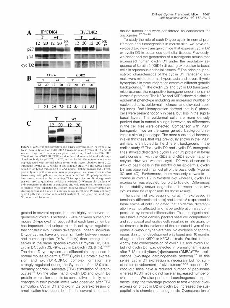
To study if cyclin D2 overexpression resulted inchanges in the composition of CDK/cyclin/CDK inhibitorcomplexes before or after thymic regression, we ana-lyzed complexes formation by immunoprecipitation withan antibody against CDK4 or CDK2 and detection ofcyclin D2 or CKIs by Western blot. Figure 7 shows asimilar level of association between CDK4 and cyclin D2at 12 and 20 weeks of age in transgenic mice. AlthoughCDK4/cyclin D2 levels were barely detectable in the wild-type thymus, these complexes were apparent when theWestern blot membranes were overexposed (data notshown). The p27Kip1 inhibitor complexed with CDK4 inboth transgenic and wild-type mice, although there was aslight decrease in the level of CDK4-associated p27Kip1
in 20-week-old wild-type thymi (Figure 7A). Interestingly,p21Cip1 was clearly associated with CDK4 in transgenic,
D-Type Cyclins Transgenic Mice 1043AJP September 2000, Vol. 157, No. 3

but not in wild-type mice (Figure 7A). As expected cyclinD2 did not co-precipitate with CDK2, whereas high levelsof p27Kip1 were associated with CDK2 in transgenic andwild-type thymi. Furthermore, there was a decrease in thelevel of p27Kip1 associated with CDK2 in 20-week wild-type thymi (Figure 7A). These results suggest that neither
p27Kip1 nor p21Cip1 are involved in the regression ofthymi hyperplasia by blocking CDK4 or CDK2 activities at20 weeks. p16Ink4a and p15Ink4b inhibitors were not de-tected in CDK4 complexes (data not shown). We alsoanalyzed the kinase activity of CDK4 and CDK2 com-plexes in vitro using pRb as a substrate. Figure 7B shows
Figure 3. Skin phenotype of K5D2 transgenic mice. Representative paraffin-sections of skin from K5D2 transgenic mice (A) and normal siblings (B) were stainedwith hematoxylin and eosin. Expression of cyclin D2 in transgenic (C) and wild-type skin (D) was determined with specific antibodies. BrdU incorporation ofparaffin sections of transgenic (E) and normal (F) skin was detected with mouse monoclonal anti-BrdU antibody. Antibody binding was detected by secondaryantibody conjugated with horseradish peroxidase, and the reaction was developed with diaminobenzidine. Original magnification, �200.
1044 Rodriguez-Puebla et alAJP September 2000, Vol. 157, No. 3

an increase in CDK4 kinase activity at 20 weeks in trans-genic and wild-type thymi. A similar pattern was ob-served for CDK2 activity in wild-type thymi. However, thekinase activity in cyclin D2 transgenic thymi was greaterat 12 than at 20 weeks. The elevated kinase activity inyoung thymi could be related to the observed hyperpla-
sia. However, we cannot rule out the possibility that thedifferent cell populations that compose the thymus couldmask the CDK activities of the epithelial component(which overexpress cyclin D2). In addition, pRb proteinlevels and phosphorylation states were determined inthymus extracts of transgenic and wild-type mice. Figure
Figure 4. Skin phenotype of K5D3 transgenic mice. Representative paraffin-sections of skin from K5D3 transgenic mice (A) and normal siblings (B) were stainedwith hematoxylin and eosin. Expression of cyclin D3 in transgenic (C) and wild-type skin (D) was determined with specific antibodies. BrdU incorporation ofparaffin sections of transgenic (E) and normal (F) skin was detected with mouse monoclonal anti-BrdU antibody. Antibody binding was detected by secondaryantibody conjugated with horseradish peroxidase, and the reaction was developed with diaminobenzidine. Original magnification, �200.
D-Type Cyclins Transgenic Mice 1045AJP September 2000, Vol. 157, No. 3

7C shows that the pRb level is elevated in transgenicmice compared with normal siblings. Also, a slower mi-grating band consistent with hyperphosphorylation is ob-served in transgenic mice at 20 weeks. Overexposure ofthe membrane showed that pRb protein levels also in-creased in wild-type mice at 20 weeks of age, althoughthe protein level was still inferior compared to transgenicmice (Figure 7C). Altogether, these results indicate thatCDK4 and CDK2 kinase activities are not reduced duringthe thymic size regression in transgenic mice and neitherp27Kip2 nor p21Cip1 seemed to be involved in this event.
Unlike K5D2, none of the K5D3 transgenic lines devel-oped thymic hyperplasia although line 2203 showed a3.5-fold increased level of cyclin D3 (Figure 6C). Consis-tent with these data none of the three K5D3 founders
developed thymus hyperplasia as was established forcyclin D130 and cyclin D2.
To determine whether expression of the cyclin D2transgene in thymic epithelium altered T cell develop-ment, we analyzed the distribution of thymocyte subsetsin normal and in transgenic mice at 12 weeks of age,when the thymus was hyperplastic. Flow cytometry de-termination showed that each major thymocyte subset,defined by CD4 and CD8 expression, is present in 2102transgenic mice at 12 weeks of age (thymic hyperplasia)(data not shown).
DiscussionD-type cyclins function as regulatory subunits of cyclin-dependent kinases and are rate-limiting factors of the G1
progression. Depending on cell lineage, various combi-nations of cyclins D1, D2, and D3 are induced by mito-gens during G1 and their continued synthesis throughoutthe cycle depends on persistent growth factor stimula-tion.2 Redundant roles of D-type cyclins have been sug-
Figure 5. Quantification of epidermal proliferation in transgenic and normalsibling mice. A: The bars indicate the labeling index or the percentage ofBrdU incorporation in basal cells from interfollicular epithelia. B: Basal cellhyperplasia. The bars indicate the number of nucleated cells in 200-�minterfollicular epithelia. C: The bars indicate the thickness of whole mouseskin in �m. Gray bars, transgenic mice; white bars, normal siblings.
Table 1. Summary of Epidermis Parameters in TransgenicMice
Transgeniclines Thickness*
Labelingindex*
Nucleatecells*
K5D2-2101 35 70 31K5D2-2102 23 123 54K5D3-2201 6 191 28K5D3-2203 23 153 46K5D1-7111 17 155 48
*Percentage of increase in transgenic mice compared to normalsibling.
Figure 6. Development of thymic hyperplasia in K5D2 mice. A: Time frameof the development of thymic hyperplasia. Thymus from K5D2 and wild-typemice were harvested and weighed from line 2102 transgenic mice andnormal siblings at different ages from 2 to 30 weeks. White bars, transgenicmice; gray bars, normal siblings. B: Thymus of K5D2-2102, K5D3-2203 andnormal sibling animals at 11 weeks of age. C: Western blot analysis of cyclinD2 and cyclin D3 expression from protein lysates of thymus of K5D2 (2101and 2102), K5D3 (2203 and 2201) and normal sibling mice (Wt) at 12 and 24weeks of age.
1046 Rodriguez-Puebla et alAJP September 2000, Vol. 157, No. 3
gested in several reports, but, the highly conserved se-quences of cyclin D proteins (�94% between human andmouse D-type cyclins) suggest that each family memberhas important and unique roles in cell-cycle regulationthat constrain evolutionary divergence. Indeed, individualD-type cyclins have a greater similarity between mouseand human species (94% identity) than among them-selves in the same species (cyclin D1/cyclin D2, 64%;cyclin D1/cyclin D3, 49%; cyclin D2/cyclin D3, 64%).35–37
The three D-type cyclins are differentially expressed innormal mouse epidermis,.27,29 Cyclin D1 protein expres-sion and cyclinD1-CDK4/6 complex formation arestrongly regulated during the G1 phase after 12-0-tetra-decanoylphorbol-13-acetate (TPA) stimulation of keratin-ocytes.29 On the other hand, cyclin D2 and cyclin D3protein expression seem to be constitutive and only minorchanges in their protein levels were observed after TPAstimulation. Cyclin D1 and cyclin D2 overexpression oramplification have been described in several human and
mouse tumors and were considered as candidates foroncogenes.27,38–40
To study the role of each D-type cyclin in normal pro-liferation and tumorigenesis in mouse skin, we have de-veloped two new transgenic mice that express cyclin D2or cyclin D3 in squamous epithelial tissues. Previously,we described the generation of a transgenic mouse thatexpressed human cyclin D1 under the regulatory se-quence of keratin 5 (K5D1) directing expression to basalcells in squamous epithelial tissues.30 The principal phe-notypic characteristics of the cyclin D1 transgenic ani-mals were mild epidermal hyperplasia and severe thymichyperplasia in three integration events of different geneticbackgrounds.30 The cyclin D2 and cyclin D3 transgenicmice express the respective transgene under the samekeratin 5 promoter. The K5D2 and K5D3 showed a similarepidermal phenotype including an increased number ofnucleated cells, epidermal thickness, and elevated label-ing index. BrdU incorporation showed that in S phase,cells were present not only in basal but also in the supra-basal layers. The epidermal cells are more denselypacked than in normal siblings, however, no differencesin the cell size were detected. Comparison with K5D1transgenic mice on the same genetic background re-veals a similar phenotype. The more substantial increasein skin thickness, that was previously shown in the K5D1animals, is attributed to the different background in theearlier study.30 The cyclin D2 and cyclin D3 transgeniclines showed detectable cyclin D2 or D3 protein in basalcells consistent with the K5D2 and K5D3 epidermal phe-notype. However, whereas cyclin D2 was observed in40% of basal cells in the interfollicular epidermis, cyclinD3 was observed in almost all of the basal cells (Figures3C and 4C). Furthermore, there was only a twofold in-crease in cyclin D2 in Western blot whereas, cyclin D3expression was elevated fourfold to fivefold. Differencesin the stability and/or degradation between these twocyclins may be responsible for those results.
The pattern of expression of keratin 1 (expressed interminally differentiated cells) and keratin 5 (expressed inbasal epithelial cells) indicated that epidermal differenti-ation was not affected and hyperproliferation was com-pensated by terminal differentiation. Thus, transgenic ani-mals have a more densely packed basal cell compartmentand suprabasal proliferative cells resulting in mild acantho-sis (increase in the thickness of the nucleated layers of theepithelia) without hyperkeratosis. No evidence of sponta-neous skin tumor development was found until 10 monthsof age in either K5D2 or K5D3 animals. We find it note-worthy that overexpression of cyclin D1 and cyclin D2,but not cyclin D3, was detected in premalignant lesionsafter 7,12-dimethylbenz[a]antracene (DMBA)/TPA appli-cations (two-stage carcinogenesis protocol).27 In thissense, cyclin D1 expression is necessary but not suffi-cient for development of skin tumors41,42 because D1knockout mice have a reduced number of papillomaswhereas K5D1 mice did not have an increased number ofskin tumors. We also performed carcinogenesis experi-ments using the two-stage protocol to test whether over-expression of cyclin D2 or cyclin D3 increased the sus-ceptibility to chemical carcinogenesis. Overexpression of
Figure 7. CDK complex formation and kinase activities in K5D2 thymus. A:Fresh protein lysates of K5D1-2102 transgenic mice thymus at 12 and 20weeks of age were immunoprecipitated with polyclonal anti-CDK4 (IP/CDK4) and anti-CDK2 (IP/CDK2) antibodies and immunoblotted with poly-clonal antibody for p27Kip1, p21Cip1, and cyclin D2. The control was immu-noprecipitated with normal rabbit serum with lysates obtained from 2102transgenic thymus at 12 weeks of age (NR/12). B: CDK4 and CDK2 kinaseactivities of K5D2 transgenic (T) and normal sibling animals (wt). Freshprotein lysates of thymus were immunoprecipitated as below in an in vitrokinase assay, with pRb as a substrate, was performed. pRb phosphorylationlevels were determined by measuring 32P incorporation and Bio-image anal-ysis was used to quantitate the kinase activities. C: Western blot analysis ofpRb expression in thymus of transgenic and wild-type mice. Protein lysatesof thymus were separated by sodium dodecyl sulfate-polyacrylamide gelelectrophoresis and blotted to a nitrocellulose membrane. Primary antibodyagainst pRb was used for immunoblot analysis. T, transgenic; wt, wild type,NR, normal rabbit serum.
D-Type Cyclins Transgenic Mice 1047AJP September 2000, Vol. 157, No. 3

cyclin D2 did not seem to increase sensitivity to spontane-ous or carcinogen-induced skin tumors (Rodriguez-PueblaM, Conti CJ, unpublished results). However, as in the caseof cyclin D1, cyclin D2 expression may be necessary topromote skin tumor development.
Another unexpected result is the down-regulation ofcyclin D2 in K5-D3 mice and down-regulation of cyclin D3in K5-D2 mice (Figure 2). These results are not specificfor these two D-type cyclins because K5-D1 mice alsoshow a reduced protein level of cyclin D3.42 We canhypothesize that overexpression of one of the D-typecyclins is compensated by reducing the level of expres-sion of another member of the family. It is possible thatthe transcription factors involved in the regulation of D-type cyclin genes are responsible for these regulatoryloops. Also noteworthy is the finding that various tran-scription factor binding sites were found in mouse cyclinD3 gene, that includes GATA, NF-�B, ATF, E2F, Aprf,TCE, GAGA, TRE/Ap1, Sp1, and Ap2.43 Because Sp1and E2F binding sites were found in the cyclin D1 gene,44
these transcription factors could participate in this regu-latory circuit.
Expression levels of the pRb family of proteins, pRband p107, were clearly elevated in K5D3 and in K5D2–2102 transgenic lines. The K5D3–2203 line showed astronger induction of p107 and changes in mobility con-sistent with phosphorylation. This is consistent with thegreater level of cyclin D3 expression in this transgenicline. Similarly, the K5D3-2203 transgenic mice showedstronger induction of p130 other cyclin D2 and D3 linesshowed a mild increase of this protein. Consistent withthese results, we previously found an increase in p107and pRb protein levels in mouse epidermal tissue afterproliferative induction by TPA.29 pRb and p107 generegulation has been suggested to be mediated by bind-ing of E2F-1 to E2F sites.45 We therefore believe thatinduction of p107 and pRb in transgenic animals may bemediated by E2F.
One of the more relevant differences between the threetransgenic mice overexpressing D-type cyclins was thedevelopment of thymic hyperplasia in cyclin D130 andcyclin D2, but not in cyclin D3 transgenic mice. The factthat cyclin D2 expression was only double in the K5D2-2101 line whereas it increased by fivefold in the K5D2-2102 line may explain why the K5D2-2101 line did notdevelop thymic hyperplasia. Mechanistic studies ofCDK4 and CDK2 complex formations in the thymus ofK5D2-2102 did not show relevant differences at 12 and20 weeks of age. We did in vitro immunoprecipitation withthymic lysates of the transgenic and wild-type mice. Todetermine whether cyclin D2 overexpression resulted inchanges in the composition of CDK/cyclin/CKI com-plexes, we analyzed CDK4 and CDK2 complex forma-tions and kinase activities. We determined that neitherp21Cip1 nor p27Kip1 are involved in the thymic regressionat 20 weeks of age in transgenic mice. The kinase activ-ities of CDK4 and CDK2 did not change during the thymicsize regression. However, elevated CDK2 kinase activityat 12 weeks of age may be responsible for the hyper-plastic phenotype in cyclin D2 transgenic thymus. Sev-eral reports have described that kinase activation of
CDK2 occurs when increased levels of CDK4/cyclin com-plexes bind to the CKIs and release CDK2. A similarmechanism could be involved in the development of thehyperplastic thymus, where an increase of cyclin D2protein levels bind some inhibitor in a binary or ternarycomplex with CDK4, although CKIs other than p21Cip1
and p27Kip1 would be responsible for this event. Interest-ingly, pRb protein levels were also increased in trans-genic mouse thymus compared with normal sibling mice,as was shown in epidermal cells. In addition, the level ofpRb phosphorylation increased at 20 weeks in the thy-mus of K5D2 animals. These data clearly show that in theolder thymus (20 weeks) the pRb protein seems to berelatively inactive rather than the active form. However,we cannot rule out the possibility that the presence of cellpopulations in the thymus other than epithelial cells masksubtle differences in complex formations and also influ-ence the pRb state observed at 12 and 20 weeks of age.
Interestingly the K5D3-2203 transgenic line expressedthe transgene at a 3.5-fold higher level than nontrans-genic, but did not develop thymic hyperplasia. This isconsistent with the fact that none of the K5D3 foundersdeveloped thymic hyperplasia whereas three K5D130
and three K5D2 founders developed this phenotype. Aninteresting possibility is that cyclin D3 has a distinct rolein thymic epithelial cells compared to the other D-typecyclins. In this regard, cyclin D3 has been reported to beinduced in differentiation in other systems. For example,cyclin D3 is strongly up-regulated on induction of HL-60leukemia cells to differentiate and it accumulates to highlevels in a wide range of quiescent cells in mouse andhuman tissues.22 In addition, myoblast induction of cyclinD3 expression is closely coupled with withdrawal fromthe cell cycle and differentiation.46 In contrast, cyclin D1and cyclin D2 have been established to play a role inproliferation and this function may be responsible for thehyperplastic thymus phenotype.
Each major subset of T cell, defined by CD4 and CD8expression, is present in K5D2-2102 transgenic line at 12weeks of age (thymic hyperplasia). These results showthat despite the altered architecture, the thymic environ-ment is able to generate mature T cells. Furthermore,these results confirm the histological diagnosis of hyper-plasia and rule out the possibility of thymic lymphomabecause, in this case, a single predominant phenotypeshould be present whereas thymus from K5D2-2102transgenic line contain each of the major T cell subsets.
A relevant difference between thymic hyperplasia ofK5D1-710830 and K5D2-2102 transgenic mice was that,in the latter, the increasing size of the thymus stops at 12weeks of age and at 20 weeks, the size was similar tonormal siblings (Figure 6A). The possibility that the trans-gene expression was turned-off after 12 weeks wasruled-out because the expression levels determined byWestern blot analysis at 24 weeks of age was similar to12-week-old mice (Figure 6C). Again the results suggestthat a different mechanism of action exists between cy-clin D1 and cyclin D2. Although both transgenic miceoverexpressing these cyclins show thymic hyperplasia,their histological characteristics are different and in one
1048 Rodriguez-Puebla et alAJP September 2000, Vol. 157, No. 3
case the hyperplasia reverts with age whereas, in theother, it progresses and causes the death of the animal.
In summary, the mammalian D-type cyclins seem tohave redundant functions in some systems, but cleardifferences have also been reported. Cyclin D1 and cy-clin D2 are considered as proto-oncogenes based ongenetic aberrations in human and animal malignancies.47
In contrast, cyclin D3 has not been firmly implicated inoncogenesis. Despite considerable similarities, uniquephenotypes were reported for cyclin D1 and cyclin D2knockout mice,23–25 whereas effects of cyclin D3 knock-outs are unknown. Our results, using two new transgenicmodels, suggest that in some tissues (epidermis) D-typecyclins may have similar functions, whereas in others(thymus) the biological roles of the individual D-type cy-clins are not fully redundant.
AcknowledgmentsWe thank April Weiss for help with the mouse experi-ments; the Science Park animal facility personnel; Dr.Irma B. Gimenez-Conti and The Science Park histologyservice for assistance with the immunohistochemicalstaining; Cassie Smith Bigbee for technical assistance;Melissa Bracher for secretarial assistance; and SharonStockman for editing the paper. We also thank Dr.Heather Poetschke for help and advice with flow cytom-etry studies.
References
1. Sherr CJ: Mammalian G1 cyclins. Cell 1993, 73:1059–10652. Sherr CJ: D-type cyclins. Trends Biochem Sci 1995, 20:187–1903. Sherr CJ: G1 phase progression: cyclin on cue. Cell 1994, 79:551–
5554. Munger K, Pieternpol JA, Pittelkow MR, Holt JT, Moses HL: Trans-
forming growth factor B1 regulation of c-myc expression, pRb phos-phorylation, and cell cycle progression in keratinocytes. Cell GrowthDiffer 1992, 3:291–298
5. Nevins J, Chellappan S, Mudryj M, Hiebert S, Devoto S, Horowitz J,Hunter T, Pines J: E2F transcription factor is a target for the RB proteinand the cyclin A protein. Cold Spring Harb Symp Quant Biol 1991,56:157–162
6. Nevins JR: E2F: a link between the Rb tumor suppressor protein andviral oncoproteins. Science 1992, 258:424–429
7. Kato J-Y, Matsuoka M, Strom DK, Sherr CJ: Regulation of cyclinD-dependent kinase 4 (cdk4) by cdk4-activating kinase. Mol Cell Biol1994, 14:2713–2721
8. Meyerson M, Harlow E: Identification of G1 kinase activity for cdk6, anovel cyclin D partner. Mol Cell Biol 1994, 14:2077–2086
9. Knudsen K, Cavenee W, Arden K: D-type cyclins complex bind withthe androgen receptor and inhibits its transcriptional transactivationability. Cancer Res 1999, 59:2297–2301
10. Zwijsen R, Wientjens E, Klompmaker R, van der Sman J, Bernards R,Michalides R: CDK-independent activation of estrogen receptor bycyclin D1. Cell 1997, 88:405–415
11. McHannon C, Suthiphongchai T, DiRenzo J, Ewen M: P/CAF associ-ates with cyclin D1 and potentiates its activation of the estrogenreceptor. Proc Natl Acad Sci USA 1999, 96:5382–5387
12. Neuman E, Ladha M, Lin N, Upton T, Miller S, DiRenzo J, Pestell R,Hinds P, Dowdy S, Brown M, Ewen M: Cyclin D1 stimulation ofestrogen receptor transcriptional activity independent of cdk4. MolCell Biol 1997, 17:5338–5347
13. Arnold A, Kim H, Gaz R, Eddy R, Fukushima Y, Byers M, Shows T,Kronenberg H: Molecular cloning and chromosomal mapping of DNA
rearranged with the parathyroid hormone gene in a parathyroid ad-enoma. J Clin Invest 1989, 83:2034–2040
14. Yasogawa Y, Takamo Y, Okayasu I, Kakita A: The 5D4 antibody(anti-cyclin D1/D2) related antigen: cytoplasmic staining is correlatedto the progression of gastric cancer. Pathol Int 1998, 48:717–722
15. Murty V, Chaganti R: A genetic perspective of male germ cell tumors.Semin Oncol 1998, 25:133–144
16. Houldsworth J, Reuter V, Bosl G, Chaganti R: Aberrant expression ofcyclin D2 in an early event in human male germ cell tumorigenesis.Cell Growth Differ 1997, 8:292–299
17. Delmer A, Ajchenbaum-Cymbalista F, Tang R, Ramond S, Faussat A,Marie J, Zittoun R: Overexpression of cyclin D2 in chronic B-cellmalignancies. Blood 1995, 85:2870–2876
18. Sanchez-Beato M, Camacho F, Martinez-Montero J, Saez A, Villuen-das R, Sanchez-Verde L, Garcia J, Piris M: Anomalous high p27/Kip1expression in a subset of aggressive B-cell lymphomas is associatedwith cyclin D3 overexpression. p27/Kip1-cyclin D3 colocalization intumor cells. Blood 1999, 94:765–772
19. Russell A, Thompson M, Hendley J, True L, Armes J, Germain D:Cyclin d1 and D3 associate with the SCF complex and are coordi-nately elevated in breast cancer. Oncogene 1999, 18:1983–1991
20. Ando K, Ajchenbaum-Cymbalista F, Griffin J: Regulation of G1/Stransition by cyclin D2 and D3 in hematopoietic cells. Proc Natl AcadSci USA 1993, 90:9571–9575
21. Kato J-Y, Sherr C: Inhibition of granulocyte differentiation by G1
cyclins D2 and D3 but not D1. Proc Natl Acad Sci USA 1993, 90:11513–11517
22. Bartkova J, Lukas J, Strauss M, Bartek J: Cyclin D3: requirement forG1/S transition anf high abundance in quiescent tissues suggest adual role in proliferation and differentiation. Oncogene 1998, 17:1027–1037
23. Sicinski P, Donaher J, Parker S, Li T, Fazeli A, Gardener H, Haslam S,Bronson R, Elledge S, Weinberg R: Cyclin D1 provides a link betweendevelopment and oncogenesis in the retina and breast. Cell 1995,82:621–630
24. Fantl V, Stamp G, Andrews A, Rosewell I, Dickson C: Mice lackingcyclin D1 are small and show defects in eye and mammary glanddevelopment. Genes Dev 1995, 9:2364–2372
25. Sicinski P, Donaher J, Geneg Y, Parker S, Garder H, Park M, RobkerR, Richards J, McGinnis L, Biggers J, Epping J, Bronson R, ElledegeS, Weinberg R: Cyclin D2 is an FSH-responsive gene involved ingonadal cell proliferation and oncogenesis. Nature 1996, 384:470–474
26. Klein-Szanto AJP: Morphological evaluation of tumor promoter effectson mammalian skin. Mechanisms of Tumor Promotion, Tumor Promo-tion and Skin Carcinogenesis. Edited by TJ Slaga. Boca Raton, CRCPress, 1984, pp 42–72
27. Rodriguez-Puebla ML, LaCava M, Gimenez-Conti IB, Jonhson DG,Conti CJ: Deregulated expression of cell-cycle proteins during pre-malignant progression in SENCAR mouse skin. Oncogene 1998,17:2251–2258
28. Zhang S-Y, Liu S-C, Goodrow T, Morris R, Klein-Szanto AJP: In-creased expression of G1 cyclins and cyclin-dependent kinases dur-ing tumor progression of chemically induced mouse skin neoplasms.Mol Carcinog 1997, 18:142–152
29. Rodriguez-Puebla ML, Robles AI, Johnson DG, LaCava M, Conti CJ:Synchronized proliferation induced by TPA treatment of mouse skin:an in vivo model for cell cycle regulation. Cell Growth Differ 1998,9:31–39
30. Robles AI, Larcher F, Whalin RB, Murillas R, Richie E, Gimenez-ContiIB, Jorcano JL, Conti CJ: Expression of cyclin D1 in epithelial tissuesof transgenic mice results in epidermal hyperproliferation and severethymic hyperplasia. PNAS 1996, 93:7634–7638
31. Klug DB, Crouch E, Carter C, Coghlan L, Conti CJ, Richie ER: Trans-genic expression of cyclin D1 in thymic epithelial precursors pro-motes epithelial and T cell development. J Immunol 2000, 164:1881–1888
32. Ramirez A, Bravo A, Jorcano J, Vidal M: Sequences 5� of the bovinekeratin 5 gene direct tissue- and cell-type-specific expression of alacZ gene in the adult and during development. Differentiation 1994,58:53–64
33. Murillas R, Larcher F, Conti C, Santos M, Ullrich A, Jorcano J: Ex-pression of a dominant negative mutant of epidermal growth factorreceptor in the epidermis of transgenic mice elicits striking alterations
D-Type Cyclins Transgenic Mice 1049AJP September 2000, Vol. 157, No. 3

in hair follicles development and skin structure. EMBO J 1995, 14:5216–5223
34. Weinberg R: Tumor suppressor genes. Science 1991, 254:1138–114635. Motokura T, Keyomarsi K, Kronenberg H, Arnold A: Cloning and
characterization of human cyclin D3, a cDNA closely related in se-quence to the PRAD1/cyclin D1 proto-oncogene. J Biol Chem 1992,267:20412–20415
36. Kiyokawa H, Busquets C, Powell C, Ngo L, Rifkind R, Marks P:Cloning of a D-type cyclin from murine erythroleukemia cells. ProcNatl Acad Sci USA 1992, 89:2444–2447
37. Matsushine H, Roussel M, Ashmun R, Sherr C: Colony-stimulatingfactor 1 regulates novel cyclins during the G1 phase of the cell cycle.Cell 1991, 65:701–713
38. Robles AI, Conti CJ: Early overexpression of cyclin D1 protein inmouse skin carcinogenesis. Carcinogenesis 1995, 16:781–786
39. Bianchi AB, Fischer SM, Robles AI, Rinchik EM, Conti CJ: Overex-pression of cyclin D1 in mouse skin carcinogenesis. Oncogene 1993,8:1127–1133
40. Sgambato A, Han E, Zhang Y, Moon R, Santella R, Weinstein I:Deregulated expression of cyclin D1 and other cell cycle-relatedgenes in carcinogen-induced rat mammary tumors. Carcinogenesis1995, 16:2193–2198
41. Robles A, Rodriguez-Puebla M, Glick A, Trempus C, Hansen L,
Sicinski P, Tennant R, Weinberg R, Yuspa S, Conti C: Reduced skintumor development in cyclin D1 deficient mice highlights the onco-genic ras pathway in vivo. Genes Dev 1998, 12:2469–2474
42. Rodriguez-Puebla ML, LaCava M, Conti C: Cyclin D1 overexpressionin mouse epidermis increases cyclin-dependent kinase activity andcell proliferation in vivo but does not affect skin tumor development.Cell Growth Differ 1999, 10:467–472
43. Wang A, Sicinski P, Weinberg R, Zhang Y, Ravid K: Characterizationof the mouse cyclin D3 gene: exon/intron organization and promoteractivity. Genomics 1996, 3:156–163
44. Motokura T, Arnold A: PRAD1/cyclin D1 proto-oncogene: genomicorganization 5� DNA sequence of a tumor-specific rearrangementbreakpoint. Genes Chromosom Cancer 1993, 7:89–95
45. Shan B, Chang C-Y, Jones D, Lee W-H: The transcription factor E2F-1mediates the autoregulation of Rb gene expression. Mol Cell Biol1994, 14:299–309
46. Kiess M, Gill R, Hamel P: Expression of the positive regulator of cellcycle progression, cyclin D3, is reduced during differentiation ofmyoblasts into quiescent myotubes. Oncogene 1995, 10:159–166
47. Hall M, Peters G: Genetic alterations of cyclins, cyclin-dependentkinases, and Cdk inhibitors in human cancer. Adv Cancer Res 1996,68:67–108
1050 Rodriguez-Puebla et alAJP September 2000, Vol. 157, No. 3
Copyright © 2022 FDOKUMEN