Screening for Epidermal Growth Factor Receptor Mutations in Lung Cancer
10
The new england journal of medicine 10.1056/nejmoa0904554 nejm.org 1 original article Screening for Epidermal Growth Factor Receptor Mutations in Lung Cancer Rafael Rosell, M.D., Teresa Moran, M.D., Cristina Queralt, B.S., Rut Porta, M.D., Felipe Cardenal, M.D., Carlos Camps, M.D., Margarita Majem, M.D., Guillermo Lopez-Vivanco, M.D., Dolores Isla, M.D., Mariano Provencio, M.D., Amelia Insa, M.D., Bartomeu Massuti, M.D., Jose Luis Gonzalez-Larriba, M.D., Luis Paz-Ares, M.D., Isabel Bover, M.D., Rosario Garcia-Campelo, M.D., Miguel Angel Moreno, M.D., Silvia Catot, M.D., Christian Rolfo, M.D., Noemi Reguart, M.D., Ramon Palmero, M.D., José Miguel Sánchez, M.D., Roman Bastus, M.D., Clara Mayo, Ph.D., Jordi Bertran-Alamillo, B.S., Miguel Angel Molina, Ph.D., Jose Javier Sanchez, M.D., and Miquel Taron, Ph.D., for the Spanish Lung Cancer Group The authors’ affiliations and the names of additional investigators are listed in the Appendix. Address reprint requests to Dr. Rosell at Catalan Institute of Oncol- ogy, Hospital Germans Trias i Pujol, Ctra Canyet, 08916 Badalona, Spain, or at [email protected]. This article (10.1056/NEJMoa0904554) was published on August 19, 2009, at NEJM.org. N Engl J Med 2009;361. Copyright © 2009 Massachusetts Medical Society. Abstract Background Activating mutations in the epidermal growth factor receptor gene ( EGFR) confer hy- persensitivity to the tyrosine kinase inhibitors gefitinib and erlotinib in patients with advanced non–small-cell lung cancer. We evaluated the feasibility of large-scale screen- ing for EGFR mutations in such patients and analyzed the association between the mutations and the outcome of erlotinib treatment. Methods From April 2005 through November 2008, lung cancers from 2105 patients in 129 in- stitutions in Spain were screened for EGFR mutations. The analysis was performed in a central laboratory. Patients with tumors carrying EGFR mutations were eligible for erlotinib treatment. Results EGFR mutations were found in 350 of 2105 patients (16.6%). Mutations were more fre- quent in women (69.7%), in patients who had never smoked (66.6%), and in those with adenocarcinomas (80.9%) (P<0.001 for all comparisons). The mutations were dele- tions in exon 19 (62.2%) and L858R (37.8%). Median progression-free survival and overall survival for 217 patients who received erlotinib were 14 months and 27 months, respectively. The adjusted hazard ratios for the duration of progression- free survival were 2.94 for men (P<0.001); 1.92 for the presence of the L858R muta- tion, as compared with a deletion in exon 19 (P = 0.02); and 1.68 for the presence of the L858R mutation in paired serum DNA, as compared with the absence of the mutation (P = 0.02). The most common adverse events were mild rashes and diar- rhea; grade 3 cutaneous toxic effects were recorded in 16 patients (7.4%) and grade 3 diarrhea in 8 patients (3.7%). Conclusions Large-scale screening of patients with lung cancer for EGFR mutations is feasible and can have a role in decisions about treatment. Copyright © 2009 Massachusetts Medical Society. All rights reserved. Downloaded from www.nejm.org on August 24, 2009 . For personal use only. No other uses without permission.
-
Upload
independent -
Category
Documents
-
view
1 -
download
0
Transcript of Screening for Epidermal Growth Factor Receptor Mutations in Lung Cancer

T h e n e w e ngl a nd j o u r na l o f m e dic i n e
10.1056/nejmoa0904554 nejm.org 1
original article
Screening for Epidermal Growth Factor Receptor Mutations in Lung Cancer
Rafael Rosell, M.D., Teresa Moran, M.D., Cristina Queralt, B.S., Rut Porta, M.D., Felipe Cardenal, M.D., Carlos Camps, M.D., Margarita Majem, M.D.,
Guillermo Lopez-Vivanco, M.D., Dolores Isla, M.D., Mariano Provencio, M.D., Amelia Insa, M.D., Bartomeu Massuti, M.D., Jose Luis Gonzalez-Larriba, M.D.,
Luis Paz-Ares, M.D., Isabel Bover, M.D., Rosario Garcia-Campelo, M.D., Miguel Angel Moreno, M.D., Silvia Catot, M.D., Christian Rolfo, M.D.,
Noemi Reguart, M.D., Ramon Palmero, M.D., José Miguel Sánchez, M.D., Roman Bastus, M.D., Clara Mayo, Ph.D., Jordi Bertran-Alamillo, B.S.,
Miguel Angel Molina, Ph.D., Jose Javier Sanchez, M.D., and Miquel Taron, Ph.D., for the Spanish Lung Cancer Group
The authors’ affiliations and the names of additional investigators are listed in the Appendix. Address reprint requests to Dr. Rosell at Catalan Institute of Oncol-ogy, Hospital Germans Trias i Pujol, Ctra Canyet, 08916 Badalona, Spain, or at [email protected].
This article (10.1056/NEJMoa0904554) was published on August 19, 2009, at NEJM.org.
N Engl J Med 2009;361.Copyright © 2009 Massachusetts Medical Society.
A bs tr ac t
Background
Activating mutations in the epidermal growth factor receptor gene (EGFR) confer hy-persensitivity to the tyrosine kinase inhibitors gefitinib and erlotinib in patients with advanced non–small-cell lung cancer. We evaluated the feasibility of large-scale screen-ing for EGFR mutations in such patients and analyzed the association between the mutations and the outcome of erlotinib treatment.
Methods
From April 2005 through November 2008, lung cancers from 2105 patients in 129 in-stitutions in Spain were screened for EGFR mutations. The analysis was performed in a central laboratory. Patients with tumors carrying EGFR mutations were eligible for erlotinib treatment.
Results
EGFR mutations were found in 350 of 2105 patients (16.6%). Mutations were more fre-quent in women (69.7%), in patients who had never smoked (66.6%), and in those with adenocarcinomas (80.9%) (P<0.001 for all comparisons). The mutations were dele-tions in exon 19 (62.2%) and L858R (37.8%). Median progression-free survival and overall survival for 217 patients who received erlotinib were 14 months and 27 months, respectively. The adjusted hazard ratios for the duration of progression-free survival were 2.94 for men (P<0.001); 1.92 for the presence of the L858R muta-tion, as compared with a deletion in exon 19 (P = 0.02); and 1.68 for the presence of the L858R mutation in paired serum DNA, as compared with the absence of the mutation (P = 0.02). The most common adverse events were mild rashes and diar-rhea; grade 3 cutaneous toxic effects were recorded in 16 patients (7.4%) and grade 3 diarrhea in 8 patients (3.7%).
Conclusions
Large-scale screening of patients with lung cancer for EGFR mutations is feasible and can have a role in decisions about treatment.
Copyright © 2009 Massachusetts Medical Society. All rights reserved. Downloaded from www.nejm.org on August 24, 2009 . For personal use only. No other uses without permission.
T h e n e w e ngl a nd j o u r na l o f m e dic i n e
10.1056/nejmoa0904554 nejm.org2
Molecular-profiling studies indi-cate that activating mutations in the epi-dermal growth factor receptor (EGFR),
PI3K, BRAF, and K-ras genes are generally nonover-lapping and identifiable in approximately 40% of non–small-cell lung cancers. These mutations, plus others that contribute to tumor progression (“driv-er” mutations), can be found in almost half of all non–small-cell lung cancers.1,2
The two proto-oncogenes that are most com-monly mutated in pulmonary adenocarcinomas are K-ras and EGFR. Nearly 90% of lung-cancer–specific EGFR mutations comprise a leucine-to-arginine substitution at position 858 (L858R) and deletion mutants in exon 19 that affect the con-served sequence LREA (delE746-A750).3-7 These mutations cause constitutive activation of the ty-rosine kinase of the EGFR by destabilizing its au-toinhibited conformation, which is normally main-tained in the absence of ligand stimulation.8 The activating mutations confer hypersensitivity to the tyrosine kinase inhibitors gefitinib and erlotinib.3-5 In transgenic mouse models, EGFR mutations in-duced adenocarcinomas that responded to sup-pression of the EGFR driving signal and to EGFR tyrosine kinase inhibitors.9,10 Furthermore, lung-cancer–specific EGFR mutants can transform fi-broblasts and Ba/F3 cells.11,12 A kinetic analysis of these mutations showed that exon 19 deletions are more sensitive to erlotinib inhibition than the L858R mutation,13 a finding that has been con-firmed in retrospective clinical studies.14-16
In retrospective studies, we17,18 and others14,19,20 found that EGFR mutations were an independent predictor of response, progression-free survival, and overall survival in patients with non–small-cell lung cancer who were treated with gefitinib; most of the patients in these studies had under-gone previous chemotherapy. Consistent with pre-vious findings,6,7 EGFR mutations were more fre-quent in women, patients with adenocarcinomas, those who had never smoked, and Asians, all of whom also had the best response to gefiti-nib.14,17,19,20 Two small, prospective, multicenter studies customized gefitinib as first-line therapy or after up to two previous chemotherapy regi-mens in patients with non–small-cell lung cancer with EGFR mutations.21,22 Response rates were as high as 75%, and 1-year survival was as high as 79%.21,22 At least five additional prospective, sin-gle-institution studies in Japan have reported simi-lar outcomes.23 However, all these studies had a
relatively short observation period and included small numbers of selected patients. Moreover, most of the studies were carried out in Japan, whereas the incidence of EGFR mutations in Eu-rope has not been defined.
We now report a prospective study of screening for EGFR mutations in patients with advanced non–small-cell lung cancer, conducted by the Spanish Lung Cancer Group. We registered patients from 129 centers from all regions of Spain and used a central laboratory and database. Patients with EGFR mutations were considered for customized erlo-tinib treatment, and we evaluated the association between EGFR mutations and outcome.
Me thods
Patients
We prospectively screened 2105 patients with non–small-cell lung cancer for EGFR mutations. Patients with mutations were then considered for erlotinib treatment at a dose of 150 mg daily until disease progression or the advent of intolerable adverse effects. The registration of patients in the database, pathological review, and EGFR mutation assessment were performed centrally at the Catalan Institute of Oncology. Eligibility criteria were the diagnosis of stage IIIB disease with pleural effusion or stage IV non–small-cell lung cancer. The smoking his-tory of the patients was obtained at baseline, and patients were categorized as those who had never smoked (<100 lifetime cigarettes), former smokers (≥1 year since cessation), or current smokers (still smoking, or <1 year since cessation).
All patients provided written informed consent. Approval was obtained from the institutional re-view board and the ethics committee at each hos-pital. Details on inclusion and exclusion criteria and on treatment and evaluation are provided in the Supplementary Appendix, available with the full text of this article at NEJM.org.
Assessment of EGFR Mutations
A total of 2105 samples of tumor tissue from pa-tients with non–small-cell lung cancer were ana-lyzed: 2060 paraffin-embedded tissues and 45 fresh specimens. All specimens were obtained from the original biopsy, before any treatment. Genomic DNA was derived from tumor tissue after laser capture microdissection (Palm Microlaser Tech-nologies). Baseline blood samples were available from 164 patients. DNA from serum, plasma, or
Copyright © 2009 Massachusetts Medical Society. All rights reserved. Downloaded from www.nejm.org on August 24, 2009 . For personal use only. No other uses without permission.
Screening for EGFR Mutations in Lung Cancer
10.1056/nejmoa0904554 nejm.org 3
both was isolated with the use of the QIAamp DNA Blood Mini Kit (Qiagen), starting from 0.4 ml of material.
For tissue samples, deletions in exon 19 (del 19) were determined by length analysis after poly-merase-chain-reaction (PCR) amplification with the use of a FAM-labeled primer in an ABI Prism 3130 DNA Analyzer (Applied Biosystems). Exon 21 point mutations in codon 858 were detected with a 5′ nuclease PCR assay (TaqMan assay) using FAM and VIC MGB-labeled probes for the wild-type and the mutant sequence, respectively. All mutants were confirmed by DNA sequencing.17,24 For blood samples, both reactions were performed in the presence of a protein nucleic acid (PNA) clamp, designed to inhibit the amplification of the wild-type allele. For L858R in exon 21, a PNA clamp was also added to the 5′ nuclease PCR reaction (TaqMan assay). (For additional details, see the Supplementary Appendix.)
Study Oversight
None of the funding agencies were involved in the study’s design or conduct, data management or analysis, manuscript preparation or review, or the decision to submit the manuscript for publication. The erlotinib that was used in the study was pur-chased from the manufacturer, which had no role in the study.
Statistical Analysis
Approximately 18,800 new cases of lung cancer are diagnosed per year in Spain.25 On the basis of our preliminary study (unpublished data), we ex-pected approximately 15% of these patients to carry EGFR mutations. With an estimated error rate of 5%, we calculated that 2105 patients would need to be enrolled during a 3-year period for a power of 80%. We randomly chose 100 of the 879 public hospitals in Spain to contact with a request for samples for inclusion in the database. However, because of extensive media coverage of this proj-ect, other centers requested permission to send samples for inclusion in the database, and these samples were also included. All centers sent sam-ples from patients who had received a diagnosis of lung cancer in a nonconsecutive manner, and there was no stratification according to sex, per-formance status, smoking history, or previous treatment. A database and a case-record form were designed and sent to all participating hospitals.
Progression-free survival was defined as sur-
vival without disease progression or death and was calculated from the start of erlotinib therapy until the first observation of disease progression. Survival was calculated from the start of erlotinib therapy until death or the last follow-up visit. The associations between EGFR status, clinical charac-teristics, and tumor response to erlotinib were analyzed with the use of the chi-square test or Fisher’s exact test. The normality of quantitative variables was analyzed with the use of the Kolm-ogorov–Smirnov test and compared by Student’s t-test analysis of variance or Mann–Whitney and Kruskal–Wallis tests. Confidence intervals were calculated with the use of binomial distribution. Progression-free survival and survival curves were constructed by the Kaplan–Meier method and compared with the use of the log-rank test and the Tarone–Ware test.
In addition to the principal analyses, we per-formed five post hoc analyses of patients’ char-acteristics and response according to sex, smoking history, age, Eastern Cooperative Oncology Group (ECOG) performance status, and treatment. The Bonferroni method was used in multiple com-parisons.
All statistical calculations were performed with the use of SPSS software (version 17.0) and S-Plus (version 6.1). Two-sided P values of less than 0.05 were considered to indicate statistical significance.
R esult s
Patients
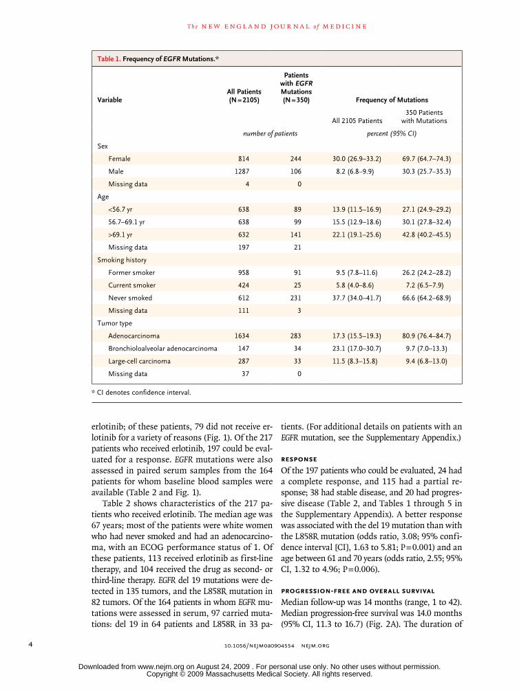
From April 2005 through November 2008, a total of 2105 patients with non–small-cell lung cancer from 129 institutions were prospectively screened for EGFR mutations. The median time required for such analysis was 7 days (range, 5 to 9) from the time the sample arrived at the laboratory until the results were reported to the investigators. Muta-tions in the EGFR gene were detected in 350 of 2105 patients (16.6%). Mutations were found more fre-quently in women (69.7%), in patients who had never smoked (66.6%), and in those with adeno-carcinomas (80.9%) (P<0.001 for all comparisons) (Table 1). Although no special call for an enriched population was made, the participating centers in-cluded more samples from women and patients who had never smoked, since physicians were aware that EGFR mutations are more frequent in these subgroups. We considered 296 patients with tu-mors carrying EGFR mutations for treatment with
Copyright © 2009 Massachusetts Medical Society. All rights reserved. Downloaded from www.nejm.org on August 24, 2009 . For personal use only. No other uses without permission.
T h e n e w e ngl a nd j o u r na l o f m e dic i n e
10.1056/nejmoa0904554 nejm.org4
erlotinib; of these patients, 79 did not receive er-lotinib for a variety of reasons (Fig. 1). Of the 217 patients who received erlotinib, 197 could be eval-uated for a response. EGFR mutations were also assessed in paired serum samples from the 164 patients for whom baseline blood samples were available (Table 2 and Fig. 1).
Table 2 shows characteristics of the 217 pa-tients who received erlotinib. The median age was 67 years; most of the patients were white women who had never smoked and had an adenocarcino-ma, with an ECOG performance status of 1. Of these patients, 113 received erlotinib as first-line therapy, and 104 received the drug as second- or third-line therapy. EGFR del 19 mutations were de-tected in 135 tumors, and the L858R mutation in 82 tumors. Of the 164 patients in whom EGFR mu-tations were assessed in serum, 97 carried muta-tions: del 19 in 64 patients and L858R in 33 pa-
tients. (For additional details on patients with an EGFR mutation, see the Supplementary Appendix.)
Response
Of the 197 patients who could be evaluated, 24 had a complete response, and 115 had a partial re-sponse; 38 had stable disease, and 20 had progres-sive disease (Table 2, and Tables 1 through 5 in the Supplementary Appendix). A better response was associated with the del 19 mutation than with the L858R mutation (odds ratio, 3.08; 95% confi-dence interval [CI], 1.63 to 5.81; P = 0.001) and an age between 61 and 70 years (odds ratio, 2.55; 95% CI, 1.32 to 4.96; P = 0.006).
Progression-free and Overall Survival
Median follow-up was 14 months (range, 1 to 42). Median progression-free survival was 14.0 months (95% CI, 11.3 to 16.7) (Fig. 2A). The duration of
Table 1. Frequency of EGFR Mutations.*
VariableAll Patients (N = 2105)
Patients with EGFR Mutations (N = 350) Frequency of Mutations
All 2105 Patients350 Patients
with Mutations
number of patients percent (95% CI)
Sex
Female 814 244 30.0 (26.9–33.2) 69.7 (64.7–74.3)
Male 1287 106 8.2 (6.8–9.9) 30.3 (25.7–35.3)
Missing data 4 0
Age
<56.7 yr 638 89 13.9 (11.5–16.9) 27.1 (24.9–29.2)
56.7–69.1 yr 638 99 15.5 (12.9–18.6) 30.1 (27.8–32.4)
>69.1 yr 632 141 22.1 (19.1–25.6) 42.8 (40.2–45.5)
Missing data 197 21
Smoking history
Former smoker 958 91 9.5 (7.8–11.6) 26.2 (24.2–28.2)
Current smoker 424 25 5.8 (4.0–8.6) 7.2 (6.5–7.9)
Never smoked 612 231 37.7 (34.0–41.7) 66.6 (64.2–68.9)
Missing data 111 3
Tumor type
Adenocarcinoma 1634 283 17.3 (15.5–19.3) 80.9 (76.4–84.7)
Bronchioloalveolar adenocarcinoma 147 34 23.1 (17.0–30.7) 9.7 (7.0–13.3)
Large-cell carcinoma 287 33 11.5 (8.3–15.8) 9.4 (6.8–13.0)
Missing data 37 0
* CI denotes confidence interval.
Copyright © 2009 Massachusetts Medical Society. All rights reserved. Downloaded from www.nejm.org on August 24, 2009 . For personal use only. No other uses without permission.

Screening for EGFR Mutations in Lung Cancer
10.1056/nejmoa0904554 nejm.org 5
response was similar for patients receiving first-line therapy (14.0 months; 95% CI, 9.7 to 18.3) and second-line therapy (13.0 months; 95% CI, 9.7 to 16.3; P = 0.62) (Fig. 2B). Median overall survival was 27.0 months (95% CI, 22.7 to 31.3) (Fig. 2C). Me-dian overall survival for patients receiving first-line therapy was 28.0 months (95% CI, 22.7 to 33), and for those receiving second-line therapy, it was 27.0 months (95% CI, 19.9 to 34.1; P = 0.67) (Fig. 2D).
Median progression-free survival was 16.0 months (95% CI, 12.7 to 19.2) in women and 9.0 months (95% CI, 6.1 to 11.9) in men (P = 0.003). Median overall survival was 29.0 months (95% CI, 24.9 to 33.1) in women and 18.0 months (95% CI, 14.5 to 21.5) in men (P = 0.05) (Fig. 1A and 1B in the Supplementary Appendix). There were no sig-
nificant differences in progression-free survival according to performance status, age, first-line therapy versus second-line or third-line therapy, smoking history, or type of mutation (data not shown). (For details regarding differences ob-served in specific subgroups and for differences according to response, see the Supplementary Ap-pendix.)
In a multivariate analysis (including sex, smok-ing status, performance status, first-line therapy vs. second-line or third-line therapy, del 19 vs. L858R, the presence or absence of brain or bone metastases, and the presence or absence of EGFR mutations in serum DNA), there was an associa-tion between poor progression-free survival and male sex (hazard ratio, 2.94; 95% CI, 1.72 to 5.03; P<0.001) and the presence of the L858R mu-
33p9
296 Were eligible for erlotinib treatment
350 Had EGFR mutations
54 Were not eligible for erlotinib treatmentowing to incomplete patient data
217 Received erlotinib
79 Did not receive erlotinib38 Began erlotinib treatment subsequent
to present analysis18 Died before starting erlotinib treatment23 Did not receive erlotinib owing
to patient or physician decision
197 Could be evaluated for response164 Had serum available for
EGFR assessment
97 Had EGFR mutation in serum
2105 Patients were screened forEGFR mutations
217 Could be evaluated for progres-sion-free and overall survival
AUTHOR:
FIGURE:
JOB: ISSUE:
4-CH/T
RETAKE
SIZE
ICM
CASE
EMail LineH/TCombo
Revised
AUTHOR, PLEASE NOTE: Figure has been redrawn and type has been reset.
Please check carefully.
REG F
Enon
1st
2nd
3rd
Rosell
1 of 2
09-03-09
ARTIST: ts
36110
Figure 1. Enrollment and Outcomes.
Copyright © 2009 Massachusetts Medical Society. All rights reserved. Downloaded from www.nejm.org on August 24, 2009 . For personal use only. No other uses without permission.

T h e n e w e ngl a nd j o u r na l o f m e dic i n e
10.1056/nejmoa0904554 nejm.org6
tation (hazard ratio, 1.92; 95% CI, 1.19 to 3.10; P = 0.02). In the multivariate analysis of overall survival, an ECOG performance status of 1, male sex, the presence of the L858R mutation, and the diagnosis of bronchioloalveolar adenocarcinoma were associated with poor prognosis (Table 3).
Therapy after Disease Progression
A total of 55 patients received additional treatment after the discontinuation of erlotinib: 49% received cisplatin-based chemotherapy; 25.5% received sin-gle-agent chemotherapy; 14.5% received erlotinib plus vorinostat, fulvestrant, or bevacizumab; and 11% received neratinib (HKI-272). The objective re-sponse rate for first-line post-erlotinib treatment was 33%, including one complete and nine partial remissions. For 11 patients receiving a second-line post-erlotinib treatment, the response rate was 40%. Median survival for all 55 patients was 29.0 months (95% CI, 20.2 to 31.4); survival for the 159 patients who did not receive post-erlotinib treatment was 27.0 months (95% CI, 22.4 to 31.6; P = 0.48).
Adverse Events
The most common adverse events were skin rash-es in 151 patients (69.6%) and diarrhea in 95 pa-tients (43.8%); most events were grade 1 or 2 in severity. Grade 3 skin toxic effects were recorded in 16 patients (7.4%) and grade 3 diarrhea in 8 pa-tients (3.7%). One 62-year-old man with del 19 had interstitial lung disease 1 month after the start of erlotinib, resulting in temporary interruption of treatment with the drug; he recovered with corti-costeroid therapy and reinitiated erlotinib therapy at a lower dose. No patient was withdrawn from the study because of adverse events (Table 7 in the Supplementary Appendix).
Discussion
We prospectively examined 2105 patients from Spain with adenocarcinoma of the lung and de-termined that 350 carried EGFR mutations (16.6%). Mutations were more frequent in women who had never smoked and in those with adenocarcinomas. In Europe, the only report of lung-cancer–specific EGFR mutations to date involved two sites in Italy, where EGFR mutations were found in 10% of 375 lung adenocarcinomas but in none of 31 large-cell carcinomas.26 In our study, EGFR mutations were also found in 33 of 287 large-cell carcinomas.
The overall rate of complete or partial response
Table 2. Characteristics and Treatment Responses of 217 Patients Receiving Erlotinib.*
Variable Value
Age — yr
Median 67
Range 22–88
Sex — no. (%)
Male 59 (27.2)
Female 158 (72.8)
Race — no. (%)†
Black 3 (1.4)
Asian 1 (0.5)
White 213 (98.2)
Smoking history — no. (%)
Former smoker 56 (25.8)
Current smoker 13 (6.0)
Never smoked 148 (68.2)
ECOG performance status — no. (%)
0 51 (23.5)
1 128 (59.0)
≥2 38 (17.5)
Tumor type — no. (%)
Adenocarcinoma 176 (81.1)
Bronchioloalveolar adenocarcinoma 22 (10.1)
Large-cell carcinoma 19 (8.8)
Tumor stage — no. (%)
IIIB 12 (5.5)
IV 205 (94.5)
Erlotinib therapy — no. (%)
First line 113 (52.1)
Second or third line 104 (47.9)
EGFR mutation — no. (%)
del 19 135 (62.2)
L858R 82 (37.8)
EGFR mutation in serum — no. (%)‡
del 19 64 (39.0)
L858R 33 (20.1)
Not detected 67 (40.9)
Response — no. (%)§
Complete response 24 (12.2)
Partial response 115 (58.4)
Complete or partial response 139 (70.6)
Stable disease 38 (19.3)
Progressive disease 20 (10.2)
Stable or progressive disease 58 (29.4)
* ECOG denotes Eastern Cooperative Oncology Group.† Race was self-reported.‡ The EGFR mutation was evaluated in the serum of 164 patients.§ The response to erlotinib therapy was evaluated in 197 patients.
Copyright © 2009 Massachusetts Medical Society. All rights reserved. Downloaded from www.nejm.org on August 24, 2009 . For personal use only. No other uses without permission.

Screening for EGFR Mutations in Lung Cancer
10.1056/nejmoa0904554 nejm.org 7
to erlotinib was 70.6%, akin to that reported for gefitinib in retrospective14,17-20 and prospective21-23 studies. A higher probability of response was as-sociated with del 19 (odds ratio, 3.08; 95% CI, 1.63 to 5.81; P = 0.001) and an age between 61 and 70 years (odds ratio, 2.55; 95% CI, 1.32 to 4.96; P = 0.006) but not with other factors. Overall, me-dian progression-free survival for the 217 patients treated with erlotinib (as first-, second-, or third-line therapy) was 14 months, and median overall
survival was 27 months, which is an improvement over findings in patients with lung cancer that have been reported previously. These results high-light the idea that EGFR-mutant lung cancer is a distinct class of non–small-cell lung cancer; in patients who do not have this mutation, chemo-therapy normally yields a 30% response, a 5-month progression-free survival, and a 12-month median survival.27 Outcomes in our study were not influ-enced by smoking status or previous chemotherapy,
36p6
1.0
Prob
abili
ty o
f Pro
gres
sion
-free
Surv
ival
0.8
0.6
0.4
0.2
0.00 12 24 36 48
Month
A Progression-free Survival in All Patients B Progression-free Survival According to Therapy
C Overall Survival in All Patients D Overall Survival According to Therapy
No. at RiskNo. of Events
2170
9086
20117
1122
1122
AUTHOR:
FIGURE:
JOB:
4-CH/T
RETAKE
SIZE
ICM
CASE
EMail LineH/TCombo
Revised
AUTHOR, PLEASE NOTE: Figure has been redrawn and type has been reset.
Please check carefully.
REG F
Enon
1st2nd
3rd
Rosell
2 of 2
09-03-09
ARTIST: ts
36110 ISSUE:
1.0
Prob
abili
ty o
f Ove
rall
Surv
ival
0.8
0.6
0.4
0.2
0.00 12 24 36 48
Month
No. at RiskNo. of Events
2170
12340
4268
380
180
1.0
Prob
abili
ty o
f Pro
gres
sion
-free
Surv
ival
0.8
0.6
0.4
0.2
0.00 12 24 36 48
Month
First LineNo. at riskNo. of events
Second LineNo. at riskNo. of events
1130
1040
4146
4841
958
1259
160
162
060
162
1.0
Prob
abili
ty o
f Ove
rall
Surv
ival
0.8
0.6
0.4
0.2
0.00 12 24 36 48
First line
Second line
First line
Second line
Month
First LineNo. at riskNo. of events
Second LineNo. at riskNo. of events
1130
1040
5821
6420
1933
2336
138
242
038
142
Figure 2. Kaplan–Meier Curves of Progression-free and Overall Survival.
Survival curves for 217 patients with EGFR mutations who received erlotinib therapy indicate the probability of progression-free survival (Panel A) and overall survival (Panel C) among all the patients and among those who received it as either first-line therapy or second-line therapy (Panels B and D). The cumulative number of events is listed for each time point.
Copyright © 2009 Massachusetts Medical Society. All rights reserved. Downloaded from www.nejm.org on August 24, 2009 . For personal use only. No other uses without permission.

T h e n e w e ngl a nd j o u r na l o f m e dic i n e
10.1056/nejmoa0904554 nejm.org8
Table 3. Multivariate Analyses of Progression-free and Overall Survival.*
Variable Progression-free Survival Overall Survival
Hazard Ratio (95% CI) P Value
Hazard Ratio (95% CI) P Value
Sex
Female 1.00 1.00
Male 2.94 (1.72–5.03) <0.001 3.48 (1.76–6.91) <0.001
EGFR mutation
del 19 1.00 1.00
L858R 1.92 (1.19–3.10 ) 0.02 2.98 (1.48–6.04) 0.002
EGFR in serum
Wild type 1.00 1.00
Mutated 1.48 (0.93–2.36) 0.09 1.50 (0.82–2.74) 0.19
ECOG performance status
0 1.00 1.00
1 1.48 (0.85–2.58) 0.16 3.50 (1.42–8.66) 0.006
≥2 1.12 (0.52–2.45) 0.76 3.04 (0.95–9.75) 0.06
Age
<60 yr 1.00 1.00
60–70 yr 0.80 (0.43–1.47) 0.48 0.50 (0.21–1.19) 0.12
>70 yr 0.88 (0.47–1.67) 0.71 0.96 (0.41–2.26) 0.93
Smoking history
Former smoker 0.72 (0.39–1.32) 0.29 0.70 (0.32–1.54) 0.38
Current smoker 1.65 (0.69–3.96) 0.26 1.37 (0.45–4.22) 0.58
Never smoked 1.00 1.00
Tumor type
Adenocarcinoma 1.00 1.00
Bronchioloalveolar adenocarcinoma 1.86 (0.90–3.85) 0.09 2.82 (1.07–7.38) 0.03
Large-cell carcinoma 1.15 (0.56–2.37) 0.70 0.81 (0.29–2.22) 0.68
Tumor stage
IIIB 1.00 1.00
IV 0.81 (0.32–2.02) 0.65 0.74 (0.21–2.65) 0.74
Erlotinib therapy
First line 1.00 1.00
Second line 0.80 (0.50–1.28) 0.36 0.92 (0.48–1.73) 0.79
Third line 1.22 (0.51–2.87) 0.66 0.44 (0.14–1.44) 0.18
Brain metastases
No 1.00 1.00
Yes 1.07 (0.53–2.18) 0.84 2.28 (1.07–4.83) 0.03
Bone metastases
No 1.00 1.00
Yes 1.37 (0.84–2.24) 0.21 1.30 (0.68–2.51) 0.42
* ECOG denotes Eastern Cooperative Oncology Group.
Copyright © 2009 Massachusetts Medical Society. All rights reserved. Downloaded from www.nejm.org on August 24, 2009 . For personal use only. No other uses without permission.

Screening for EGFR Mutations in Lung Cancer
10.1056/nejmoa0904554 nejm.org 9
which is in line with the results of a small phase 2 trial of gefitinib.21 (For details on planned ge-netic analyses, see the Supplementary Appendix.)
In conclusion, screening for EGFR mutations is warranted in women with lung cancer, in those who have never smoked, and in those with non-squamous tumors. Large-scale screening of pa-tients for EGFR mutations, with subsequent cus-tomization of erlotinib, is feasible and improves the outcome.
Supported by a grant from the Spanish Ministry of Science and Innovation (RD06/0020/0056), by the Fundación Badalona
Contra el Cáncer, and by the Bonnie J. Addario Lung Cancer Foundation.
Preliminary data were presented in part at the annual meetings of the American Society of Clinical Oncology in Atlanta, June 2–6, 2006 (abstract 7020); in Chicago, June 1–5, 2007 (abstract 7505); and in Chicago, May 30–June 3, 2008 (abstract 8038).
No potential conflict of interest relevant to this article was reported.
We thank Maria Sanchez-Ronco for her assistance in interpret-ing the statistical analyses; Maria Fernandez for coordinating the study in the Spanish Lung Cancer Group; Esther Carrasco, Mireia Tomas, and Emma Costas for data management; Ana Pradas for laboratory analyses; Maria Perez and Monica Botia for their work on laser microdissection; and Maria Luz Amador and Juan Lobera for their support throughout the study.
AppendixThe authors’ affiliations are as follows: Catalan Institute of Oncology and Autonomous University of Barcelona, Hospital Germans Trias i Pujol, Barcelona (R.R., T.M., C.Q., M.T.); Pangaea Biotech, USP Institut Universitari Dexeus, Barcelona (R.R., C.M., J.B.-A., M.A. Molina, M.T.); Catalan Institute of Oncology, Hospital Josep Trueta, Girona (R. Porta); Catalan Institute of Oncology, Hospital Duran i Reynals, Bellvitge, Barcelona (F.C., R. Palmero); Hospital General Universitario de Valencia, Valencia (C.C.); Hospital de Sant Pau, Barcelona (M.M.); Hospital de Cruces, Baracaldo, Vizcaya (G.L.-V.); Hospital Lozano Blesa, Zaragoza (D.I.); Clinica Puerta de Hierro, Madrid (M.P.); Hospital Clínico Universitario de Valencia, Valencia (A.I.); Hospital General Universitario de Alicante, Alicante (B.M.); Hospital Clínico San Carlos, Madrid (J.L.G.-L.); Hospital Universitario Virgen del Rocío, Seville (L.P.-A.); Hospital Son Llatzer, Palma de Mallorca (I.B.); Hospital Juan Canalejo, La Coruña (R.G.-C.); Complejo Hospitalario de Jaén, Jaén (M.A. Moreno); Hospital Althaia, Manresa (S.C.); Clínica Rotger, Palma de Mallorca (C.R.); Hospital Clínic, Barcelona (N.R.); Hospital 12 de Octubre, Madrid (J.M.S.); Hospital Mutua de Terrassa, Barcelona (R.B.); and Autonomous University of Madrid, Madrid (J.J.S.) — all in Spain.
The following investigators participated in the study: M. Cuello, C. Pallares, P. Lianes, J. Remon, R. Ibeas, P. Martinez del Prado, M. Angeles Sala, C. Santander-Lobera, E. Velez de Mendizabal, N. Viñolas, J. Terrasa, J. Valdivia, P. Diz, U. Jimenez-Berlana, A. Velasco-Ortiz de Taranco, E. Ales Martinez, R. Sanchez-Escribano, A. Carrato, C. Guillen-Ponce, C. Mesia, J. Antonio Macias, M. Lopez-Brea, J. Oramas, I. Barneto, P. Garrido, M.J. Mayol, A. Lopez, A. Artal, A. Saenz, S. Hernando, M. Cobo, R. Blanco, R. Bernabe, V. Guillem, M. Angel Mu-ñoz, I. Maestu, A. Salvatierra, R. De Las Peñas, J. Alfaro, V. Alberola, O. Juan, C. Martin, J. Puertas, E. Felip, J.F. González-González, L. Iglesias-Docampo, F.J. Dorta, M. Martinez-Aguillo, E. Salgado, R. Mesia, E. Lastra-Aras, J.P. Garcia-Muñoz, R. Lastra, I. Alvarez, J. Roig, J. Oruezabal, A. Poveda, J. Lavernia, D. Gutierrez, E. Filipovich, D. Aguiar, D. Rodriguez, J. Buxo, A.F. Cardona, P. Bes, A. Paredes, A.M. Tortorella, J.A. Moreno, J. Martinez-Garcia, J.L. Alonso, A. Lopez-Martin, M.J. Echarri-Gonzalez, M. Van Kooten, A. Guerrero, M. Domine, I. Diaz, L. Heras, R. Garcia, I. Anton, G. Jarchum, R. E. Bartolucci, M. Lomas, A. Rubiales, J.L. Duque, S. Escriva de Romani, E. Barbeta, J.J. Reina, J. Castro, C. Belda, J.M. Vidal, J.M. Trigo, C. Vadell, J.J. Zarba, P. Esunza, I. Garau, A. Lopez-Pousa, I. De la Gandara, J. Wida-kowich, S. Morales, M. Martinez, R. Luis, M. De la Colina, J. Calzas, I. Garcia-Castro, C. Ruiz, P. Lopez-Criado.
References
Rikova K, Guo A, Zeng Q, et al. Glob-1. al survey of phosphotyrosine signaling identifies oncogenic kinases in lung can-cer. Cell 2007;131:1190-203.
Ding L, Getz G, Wheeler DA, et al. 2. Somatic mutations affect key pathways in lung adenocarcinoma. Nature 2008;455: 1069-75.
Lynch TJ, Bell DW, Sordella R, et al. 3. Activating mutations in the epidermal growth factor receptor underlying respon-siveness of non–small-cell lung cancer to gefitinib. N Engl J Med 2004;350:2129-39.
Paez JG, Jänne PA, Lee JC, et al. EGFR 4. mutations in lung cancer: correlation with clinical response to gefitinib thera-py. Science 2004;304:1497-500.
Pao W, Miller V, Zakowski M, et al. 5. EGF receptor gene mutations are com-mon in lung cancers from “never smok-ers” and are associated with sensitivity of tumors to gefitinib and erlotinib. Proc Natl Acad Sci U S A 2004;101:13306-11.
Shigematsu H, Lin L, Takahashi T, et 6. al. Clinical and biological features associ-ated with epidermal growth factor recep-
tor gene mutations in lung cancers. J Natl Cancer Inst 2005;97:339-46.
Kosaka T, Yatabe Y, Endoh H, Kuwano 7. H, Takahashi T, Mitsudomi T. Mutations of the epidermal growth factor receptor gene in lung cancer: biological and clini-cal implications. Cancer Res 2004;64: 8919-23.
Yun CH, Boggon TJ, Li Y, et al. Struc-8. tures of lung cancer-derived EGFR mutants and inhibitor complexes: mechanism of activation and insights into differential inhibitor sensitivity. Cancer Cell 2007;11: 217-27.
Politi K, Zakowski MF, Fan PD, Schon-9. feld EA, Pao W, Varmus HE. Lung adeno-carcinomas induced in mice by mutant EGF receptors found in human lung can-cers respond to a tyrosine kinase inhibi-tor or to down-regulation of the recep-tors. Genes Dev 2006;20:1496-510.
Ji H, Li D, Chen L, et al. The impact of 10. human EGFR kinase domain mutations on lung tumorigenesis and in vivo sensi-tivity to EGFR-targeted therapies. Cancer Cell 2006;9:485-95.
Greulich H, Chen TH, Feng W, et al. 11. Oncogenic transformation by inhibitor-sensitive and -resistant EGFR mutants. PLoS Med 2005;2(11):e313.
Jiang J, Greulich H, Jänne PA, Sellers 12. WR, Meyerson M, Griffin JD. Epidermal growth factor-independent transformation of Ba/F3 cells with cancer-derived epider-mal growth factor receptor mutants in-duces gefitinib-sensitive cell cycle progres-sion. Cancer Res 2005;65:8968-74.
Carey KD, Garton AJ, Romero MS, et 13. al. Kinetic analysis of epidermal growth factor receptor somatic mutant proteins shows increased sensitivity to the epider-mal growth factor receptor tyrosine kinase inhibitor, erlotinib. Cancer Res 2006;66: 8163-71.
Mitsudomi T, Kosaka T, Endoh H, et 14. al. Mutations of the epidermal growth factor receptor gene predict prolonged survival after gefitinib treatment in pa-tients with non-small-cell lung cancer with postoperative recurrence. J Clin On-col 2005;23:2513-20.
Riely GJ, Pao W, Pham D, et al. Clini-15.
Copyright © 2009 Massachusetts Medical Society. All rights reserved. Downloaded from www.nejm.org on August 24, 2009 . For personal use only. No other uses without permission.

10.1056/nejmoa0904554 nejm.org10
Screening for EGFR Mutations in Lung Cancer
cal course of patients with non-small cell lung cancer and epidermal growth factor receptor exon 19 and exon 21 mutations treated with gefitinib or erlotinib. Clin Cancer Res 2006;12:839-44.
Jackman DM, Yeap BY, Sequist LV, et 16. al. Exon 19 deletion mutations of epider-mal growth factor receptor are associated with prolonged survival in non-small cell lung cancer patients treated with gefi-tinib or erlotinib. Clin Cancer Res 2006; 12:3908-14.
Taron M, Ichinose Y, Rosell R, et al. 17. Activating mutations in the tyrosine ki-nase domain of the epidermal growth fac-tor receptor are associated with improved survival in gefitinib-treated chemorefrac-tory lung adenocarcinomas. Clin Cancer Res 2005;11:5878-85.
Cortes-Funes H, Gomez C, Rosell R, 18. et al. Epidermal growth factor receptor activating mutations in Spanish gefitinib-treated non-small-cell lung cancer patients. Ann Oncol 2005;16:1081-6.
Han SW, Kim TY, Hwang PG, et al. 19. Predictive and prognostic impact of epi-
dermal growth factor receptor mutation in non-small-cell lung cancer patients treated with gefitinib. J Clin Oncol 2005;23:2493-501.
Takano T, Ohe Y, Sakamoto H, et al. 20. Epidermal growth factor receptor gene mutations and increased copy numbers predict gefitinib sensitivity in patients with recurrent non-small-cell lung can-cer. J Clin Oncol 2005;23:6829-37.
Tamura K, Okamoto I, Kashii T, et al. 21. Multicentre prospective phase II trial of gefitinib for advanced non-small cell lung cancer with epidermal growth fac-tor receptor mutations: results of the West Japan Thoracic Oncology Group trial (WJTOG0403). Br J Cancer 2008;98:907-14.
Sequist LV, Martins RG, Spigel D, et 22. al. First-line gefitinib in patients with ad-vanced non-small-cell lung cancer harbor-ing somatic EGFR mutations. J Clin Oncol 2008;26:2442-9. [Erratum, J Clin Oncol 2008;26:3472.]
Costa DB, Kobayashi S, Tenen DG, 23. Huberman MS. Pooled analysis of the
prospective trials of gefitinib monothera-py for EGFR-mutant non-small cell lung cancers. Lung Cancer 2007;58:95-103.
Molina-Vila MA, Bertran-Alamillo J, 24. Reguart N, et al. A sensitive method for detecting EGFR mutations in non-small cell lung cancer samples with few tumor cells. J Thorac Oncol 2008;3:1224-35.
López-Abente G, Pollán M, Aragonés 25. N, et al. Situación del cáncer en España: incidencia. An Sist Sanit Navar 2004;27: 165-73.
Marchetti A, Martella C, Felicioni L, 26. et al. EGFR mutations in non-small-cell lung cancer: analysis of a large series of cases and development of a rapid and sen-sitive method for diagnostic screening with potential implications on pharmaco-logic treatment. J Clin Oncol 2005;23:857-65.
Schiller JH, Harrington D, Belani CP, 27. et al. Comparison of four chemotherapy regimens for advanced non–small-cell lung cancer. N Engl J Med 2002;346: 92-8.Copyright © 2009 Massachusetts Medical Society.
Copyright © 2009 Massachusetts Medical Society. All rights reserved. Downloaded from www.nejm.org on August 24, 2009 . For personal use only. No other uses without permission.