Critical Role for Caspase8 in Epidermal Growth Factor Signaling
7
Research Article Critical Role for Caspase-8 in Epidermal Growth Factor Signaling Darren Finlay, Amy Howes, and Kristiina Vuori Cancer Center, Burnham Institute for Medical Research, La Jolla, California Abstract Caspase-8 has a well-defined canonical role as an apical pro- tease of the extrinsic apoptosis pathway. Evidence is growing, however, that the protein has numerous other nonapoptotic functions. We have previously shown that caspase-8 is re- quired for efficient adhesion-induced activation of the extra- cellular signal–regulated kinase (Erk)-1/2 pathway. We now show that caspase-8 is also necessary for the efficient activa- tion of downstream events associated with epidermal growth factor (EGF) signaling. This promotion of EGF-induced Erk1/2 activation is independent of the proteolytic activity of caspase-8 and can be recapitulated using only the pro- domains of the protein. In addition, we identify specific resi- dues within the caspase-8 “RXDLL motif” that are essential for Erk pathway activation. Furthermore, these residues are also involved in forming a complex with the tyrosine kinase Src. Caspase-8 null cells and cells reconstituted with caspase- 8 harboring point mutations of these critical amino acids also show defective EGF-induced migration as compared with cells reconstituted with the wild-type protein. In sum, we provide the first evidence for caspase-8 as an essential component of growth factor signaling and suggest that this may be due to its association with Src. As the EGF/Src pathway activity has been shown to promote oncogenic events, our findings that caspase-8 is necessary for these activities may help explain why it is rarely deleted or silenced in tumors. [Cancer Res 2009;69(12):5023–9] Introduction Caspase-8 is a well-characterized protease of the “extrinsic” apoptotic pathway known to be important in death receptor– mediated killing. Recruitment of caspase-8 to activated death receptors results in its dimerization, activation, subsequent autop- rocessing, and initiation of the “effector” caspase activity associat- ed with classic apoptosis (1–4). This death receptor–mediated apoptosis has been the focus of various attempts to induce cell death in tumors. Indeed, tumor necrosis factor–related apoptosis- inducing ligand has been shown to induce apoptosis in a variety of tumor, but not normal, cells (5, 6). Whereas resistance to death receptor ligands currently limits their efficacy, deletion or silencing of essential proteins of the cascade, such as caspase-8, occurs only extremely infrequently in cancers (7). Caspase-8 has in fact been shown to have increased expression in lung cancers (8), supporting the hypothesis that the protein may be involved in other nonapop- totic but potentially protumorigenic events. Mounting evidence to support alternative nonapoptotic func- tions for caspase-8 has emerged in recent years (reviewed in refs. 9–11). Several reports have shown a role for caspase-8 in hema- topoietic cell proliferation and “maturation” (e.g., refs. 12–14), whereas other laboratories have shown caspase-8 to be essential for activation of nuclear factor κB (15–17). Further data describ- ing a role for caspase-8 in adhesion and cell motility have recent- ly accumulated. Involvement of caspase-8 in cell motility has been described by several independent laboratories (18–21). We recently showed an essential role for caspase-8 in promoting cell adhesion–induced activation of the extracellular signal–regulated kinase (Erk)-1/2 pathway through association with Src (7). In- triguingly, this activation is independent of the catalytic activity of caspase-8 and can be recapitulated in caspase-8 null cells using only the NH 2 -terminal “death effector domains” (DED). Indeed, the DEDs alone are capable of forming a protein complex with Src and act indistinguishably from full-length caspase-8 in these biochemical and physiologic analyses. Here we show that caspase-8 is also critical for epidermal growth factor (EGF)–induced activation of the Erk pathway. Again the DEDs alone of caspase-8 are sufficient for this activation, and we identify residues within the so called “RXDLL motif” that are essential for the promulgation of EGF signaling. We show that caspase-8 is required for EGF-induced cell migration and that point mutants of the RXDLL motif show impaired motility similar to that of caspase-8 null cells. In sum, we provide the first evidence that caspase-8 is an essential component of growth factor signaling pathways and that its effects in this regard may be due to the ability of DEDs to associate with a protein complex containing Src. That caspase-8 is involved not only in adhesion-induced but also in growth factor–induced signal- ing may help explain why it is so seldom silenced or deleted in tu- mors. In addition, these findings suggest that potentially driving caspase-8 from nonapoptotic to more canonical proapoptotic signal- ing may be an important therapeutic intervention point in cancers. Materials and Methods Reagents. Unless otherwise specified, all reagents were from Sigma- Aldrich. AG1478, PP1, and SU6656 were from Calbiochem. Matrigel is from BD Biosciences. Zeocin and Primocin are from InvivoGen. EGF was always used at a concentration of 100 ng/mL and platelet-derived growth factor (PDGF) was used at 50 ng/mL. Cell culture, DNA transfections, and stable cell line generation. 293T and HeLa cells were cultured in standard DMEM supplemented with 10% fetal bovine serum (FBS) and penicillin/streptomycin/L-glutamine (Omega Scientific, Inc.). MDA-MB-231 cells were cultured in F12/DMEM (1:1) with 10% FBS and penicillin/streptomycin/L-glutamine, whereas SKOV3 cells were grown in McCoy's 5A medium with 10% FBS, nonessential amino acids (Hyclone), and penicillin/streptomycin/L-glutamine. SH-SY5Y cells were cultured in α-MEM plus 10% FBS and penicillin/streptomycin/L-glutamine. Caspase-8–deficient NB7 cells were a kind gift from Dr. Jill Lahti (St. Jude Children's Research Hospital, Memphis, TN) and were maintained in RPMI 1640 supplemented with 10% FBS, penicillin/streptomycin/L-glutamine, and 100 μg/mL Primocin (InvivoGen). Plasmids encoding shRNAmirs were obtained from OpenBiosystems. Caspase-8 cDNA and a c-Src expression Note: Supplementary data for this article are available at Cancer Research Online (http://cancerres.aacrjournals.org/). Requests for reprints: Kristiina Vuori, Cancer Center, Burnham Institute for Medical Research, 10901 North Torrey Pines Road, La Jolla, CA 92037. Phone: 858- 646-3129; Fax: 858-795-5272; E-mail: [email protected]. ©2009 American Association for Cancer Research. doi:10.1158/0008-5472.CAN-08-3731 5023 Cancer Res 2009; 69: (12). June 15, 2009 www.aacrjournals.org
Transcript of Critical Role for Caspase8 in Epidermal Growth Factor Signaling

Research Article
Critical Role for Caspase-8 in Epidermal Growth Factor Signaling
Darren Finlay, Amy Howes, and Kristiina Vuori
Cancer Center, Burnham Institute for Medical Research, La Jolla, California
AbstractCaspase-8 has a well-defined canonical role as an apical pro-tease of the extrinsic apoptosis pathway. Evidence is growing,however, that the protein has numerous other nonapoptoticfunctions. We have previously shown that caspase-8 is re-quired for efficient adhesion-induced activation of the extra-cellular signal–regulated kinase (Erk)-1/2 pathway. We nowshow that caspase-8 is also necessary for the efficient activa-tion of downstream events associated with epidermal growthfactor (EGF) signaling. This promotion of EGF-inducedErk1/2 activation is independent of the proteolytic activityof caspase-8 and can be recapitulated using only the pro-domains of the protein. In addition, we identify specific resi-dues within the caspase-8 “RXDLL motif” that are essentialfor Erk pathway activation. Furthermore, these residues arealso involved in forming a complex with the tyrosine kinaseSrc. Caspase-8 null cells and cells reconstituted with caspase-8 harboring point mutations of these critical amino acids alsoshow defective EGF-induced migration as compared with cellsreconstituted with the wild-type protein. In sum, we providethe first evidence for caspase-8 as an essential component ofgrowth factor signaling and suggest that this may be due toits association with Src. As the EGF/Src pathway activity hasbeen shown to promote oncogenic events, our findings thatcaspase-8 is necessary for these activities may help explainwhy it is rarely deleted or silenced in tumors. [Cancer Res2009;69(12):5023–9]
IntroductionCaspase-8 is a well-characterized protease of the “extrinsic”
apoptotic pathway known to be important in death receptor–mediated killing. Recruitment of caspase-8 to activated deathreceptors results in its dimerization, activation, subsequent autop-rocessing, and initiation of the “effector” caspase activity associat-ed with classic apoptosis (1–4). This death receptor–mediatedapoptosis has been the focus of various attempts to induce celldeath in tumors. Indeed, tumor necrosis factor–related apoptosis-inducing ligand has been shown to induce apoptosis in a variety oftumor, but not normal, cells (5, 6). Whereas resistance to deathreceptor ligands currently limits their efficacy, deletion or silencingof essential proteins of the cascade, such as caspase-8, occurs onlyextremely infrequently in cancers (7). Caspase-8 has in fact beenshown to have increased expression in lung cancers (8), supportingthe hypothesis that the protein may be involved in other nonapop-totic but potentially protumorigenic events.
Note: Supplementary data for this article are available at Cancer Research Online(http://cancerres.aacrjournals.org/).
Requests for reprints: Kristiina Vuori, Cancer Center, Burnham Institute forMedical Research, 10901 North Torrey Pines Road, La Jolla, CA 92037. Phone: 858-646-3129; Fax: 858-795-5272; E-mail: [email protected].
©2009 American Association for Cancer Research.doi:10.1158/0008-5472.CAN-08-3731
5023www.aacrjournals.org
Mounting evidence to support alternative nonapoptotic func-tions for caspase-8 has emerged in recent years (reviewed in refs.9–11). Several reports have shown a role for caspase-8 in hema-topoietic cell proliferation and “maturation” (e.g., refs. 12–14),whereas other laboratories have shown caspase-8 to be essentialfor activation of nuclear factor κB (15–17). Further data describ-ing a role for caspase-8 in adhesion and cell motility have recent-ly accumulated. Involvement of caspase-8 in cell motility hasbeen described by several independent laboratories (18–21). Werecently showed an essential role for caspase-8 in promoting celladhesion–induced activation of the extracellular signal–regulatedkinase (Erk)-1/2 pathway through association with Src (7). In-triguingly, this activation is independent of the catalytic activityof caspase-8 and can be recapitulated in caspase-8 null cellsusing only the NH2-terminal “death effector domains” (DED).Indeed, the DEDs alone are capable of forming a protein complexwith Src and act indistinguishably from full-length caspase-8 inthese biochemical and physiologic analyses.Here we show that caspase-8 is also critical for epidermal growth
factor (EGF)–induced activation of the Erk pathway. Again the DEDsalone of caspase-8 are sufficient for this activation, and we identifyresidues within the so called “RXDLLmotif” that are essential for thepromulgation of EGF signaling. We show that caspase-8 is requiredfor EGF-induced cell migration and that point mutants of theRXDLLmotif show impairedmotility similar to that of caspase-8 nullcells. In sum, we provide the first evidence that caspase-8 is anessential component of growth factor signaling pathways and thatits effects in this regardmay be due to the ability of DEDs to associatewith a protein complex containing Src. That caspase-8 is involved notonly in adhesion-induced but also in growth factor–induced signal-ing may help explain why it is so seldom silenced or deleted in tu-mors. In addition, these findings suggest that potentially drivingcaspase-8 from nonapoptotic tomore canonical proapoptotic signal-ing may be an important therapeutic intervention point in cancers.
Materials and MethodsReagents. Unless otherwise specified, all reagents were from Sigma-
Aldrich. AG1478, PP1, and SU6656 were from Calbiochem. Matrigel is fromBD Biosciences. Zeocin and Primocin are from InvivoGen. EGF was alwaysused at a concentration of 100 ng/mL and platelet-derived growth factor(PDGF) was used at 50 ng/mL.
Cell culture, DNA transfections, and stable cell line generation. 293Tand HeLa cells were cultured in standard DMEM supplemented with 10%fetal bovine serum (FBS) and penicillin/streptomycin/L-glutamine (OmegaScientific, Inc.). MDA-MB-231 cells were cultured in F12/DMEM (1:1) with10% FBS and penicillin/streptomycin/L-glutamine, whereas SKOV3 cellswere grown in McCoy's 5A medium with 10% FBS, nonessential amino acids(Hyclone), and penicillin/streptomycin/L-glutamine. SH-SY5Y cells werecultured in α-MEM plus 10% FBS and penicillin/streptomycin/L-glutamine.Caspase-8–deficient NB7 cells were a kind gift from Dr. Jill Lahti (St. JudeChildren's Research Hospital, Memphis, TN) and were maintained in RPMI1640 supplemented with 10% FBS, penicillin/streptomycin/L-glutamine, and100 μg/mL Primocin (InvivoGen). Plasmids encoding shRNAmirs wereobtained from OpenBiosystems. Caspase-8 cDNA and a c-Src expression
Cancer Res 2009; 69: (12). June 15, 2009

Cancer Research
plasmid were kind gifts from Drs. Guy Salvesen and Sara Courtneidge,respectively (both at Burnham Institute for Medical Research, La Jolla,CA). Plasmid DNA was transfected using Fugene 6 (Roche Applied Sci-ence) as per manufacturer's instructions, and point mutants were gener-ated using a QuikChange XL Mutagenesis kit (Stratagene). For stable cellline production, the cDNAs of interest were cloned into MSCV-IRES-zeoplasmids as standard. The subsequent DNA integrity was confirmed bysequencing and transfected into PheNX-A packaging cells as above. Viralsupernatants were removed (at 48 and 72 h), the debris pelleted by cen-trifugation, and polybrene added to a final concentration of 8 μg/mL be-fore being added to the cells to be infected. Cells were cultured in viralsupernatants as such for 48 h before selection with 10 μg/mL zeocin for14 d. Alternatively, cells transfected with shRNAmir-encoding DNA wereselected 48 h later in 1 μg/mL puromycin for ∼10 d until antibiotic re-sistance had been established. Cell lysates were then assayed for proteinexpression by immunoblotting.
Preparation of protein extracts and immunoblot analysis. Cellswere treated as described for experimental conditions, and protein extractsgenerated exactly as described by Finlay and Vuori (7). Protein extracts(20 μg) were resolved on SDS-polyacrylamide gels and electrophoreticallytransferred to polyvinylidene difluoride membranes (Immobilon-P, Milli-pore) by standard methods. Membranes were blocked for 1 h in TBSTw[20 mmol/L Tris-HCl (pH 7.6), 150 mmol/L NaCl, 0.05% Tween 20] contain-ing 5% nonfat dried milk (or 5% bovine serum albumin for anti-phospho-
5024Cancer Res 2009; 69: (12). June 15, 2009
tyrosine studies) and were incubated rocking overnight at 4°C with theappropriate primary antibody: anti-Erk (#9102, 1:2,000), anti–phospho-Erk(#9101, 1:2,000), anti-Akt (#9272, 1:2,000), anti–phospho-Akt-S473 (#9271,1:2,000), anti–phospho-Src-Y416 (#2101, 1:2,000), anti–EGF receptor (EGFR;C74B9 #2646, 1:2,000; all from Cell Signaling Technology, Inc.); anti-Src(mAb 327, 1:1,000; from Calbiochem); anti-HA (3F10, 1:5,000; Roche AppliedScience); anti–c-Src (EC10, 1:2,000; Upstate); or anti–caspase-8 (C15, 1:500;kind gift from Dr. Marcus Peter, University of Chicago, Chicago, IL). Afterincubation for 1 h with antirabbit IgG (111-035-003), antimouse IgG (115-035-003), or antirat IgG (712-035-150) secondary antibodies conjugated tohorseradish peroxidase (Jackson ImmunoResearch Laboratories, Inc.),bands were visualized by enhanced chemiluminescence (SuperSignal WestPico Chemiluminescent substrate, Pierce). All analyses were done at leastthrice.
Cell migration assays. Cell migration was assayed essentially as de-scribed (21). Briefly, wells of a 24-well dish were coated with 10 μg/mL fi-bronectin at 37°C overnight. Cells of interest were seeded (∼200,000 perwell) in the absence of serum and allowed to adhere for 2 h at 37°C in a5% CO2 humidified incubator. The confluent cell monolayers were thenwounded using a conditioned pipette tip as standard, and the cells treatedwith serum-free medium supplemented with 100 ng/mL EGF in the pres-ence of 1 μg/mL aphidicolin. The cells were allowed to migrate, and thedistances measured at 24 and 48 h. All experiments were carried out intriplicate at least twice.
Figure 1. Caspase-8 is essential for EGF-inducedactivation of the Erk1/2 pathway. A, immunoblotanalysis of a time course of EGF (100 ng/mL)–induced Erk and Akt pathway activation in NB7cells lacking caspase-8 (lanes 1–4) or samereconstituted with wild-type protein (lanes 5–8).B, immunoblot analysis of EGF (100 ng/mL,3 min)–induced Erk pathway activation in NB7 cellslacking caspase-8 and reconstituted with emptyvector (E.V., lanes 1 and 2), with wild-type protein(Casp8; lanes 3 and 4), or with an inactivating pointmutant of caspase-8 (Casp8C360A; lanes 5 and 6).Bottom, schematic depiction of caspase-8 showingdomains and residues of interest in this study. C,immunoblot analysis of PDGF (50 ng/mL,10 min)–induced Erk and Akt pathway activation inNB7 cells lacking caspase-8 reconstituted withempty vector, wild-type protein, or an inactivatingpoint mutant of caspase-8 (lanes 1–3, respectively).D, immunoblot analysis of EGF-induced Erk pathwayactivation in NB7 cells lacking caspase-8 andreconstituted with empty vector, Casp8C360A, or aform of caspase-8 containing DEDs alone (lanes 1–3,respectively).
www.aacrjournals.org

Caspase-8 Promotes EGF Activation of the Erk1/2 Pathway
Results and Discussion
Caspase-8 is essential for EGF-induced activation of theErk1/2 pathway. We have previously shown a role for caspase-8 in promoting cell adhesion and downstream activation of theErk1/2 pathway (7), and others have shown a role for caspase-8 in adhesion and motility (refs. 19–21 and reviewed in ref. 22).Surprisingly, while investigating EGF-induced motility, we
5025www.aacrjournals.org
discovered that caspase-8 also plays a critical role in growth factorsignaling (Fig. 1). NB7 neuroblastoma cells deficient in caspase-8(refs. 7, 23; Fig. 1A, lanes 1–4) show defective EGF-induced activa-tion of the Erk1/2 pathway as compared with cells reconstitutedwith wild-type caspase-8 (Fig. 1A, lanes 5–8) over the course of 1hour at all EGF concentrations tested (not shown). This is not acell-specific effect because SH-SY5Y neuroblastoma cells with nodetectable caspase-8 expression also show impaired activation of
Figure 2. The caspase-8 RXDLL motif iscritical for EGF-induced Erk pathwayactivation. A, alignment of the NH2-terminalprotein sequences of caspase-8 andcaspase-10. B, multispecies alignment ofcaspase-8 protein sequences of a30-amino-acid region containing theRXDLL motif (top). Cladogram of thealignment presented above (bottom).C, immunoblot analysis of EGF-inducedErk pathway activation in NB7 cells lackingcaspase-8 reconstituted with emptyvector (E.V.), with caspase-8 (W.T.), or withR71A, D73A, or L74A point mutants ofcaspase-8 (lanes 1–5, respectively).D, immunoblot analysis of EGF-induced Erkpathway activation in NB7 cells reconstitutedwith wild-type caspase-8 (Casp8; lanes1–3) or with R71A point mutant ofcaspase-8 (Casp8R71A; lanes 4–6) in thepresence and absence of AG1478(10 μmol/L; top) or PP1 (1 μmol/L) or SU6656(10 μmol/L; bottom).
Cancer Res 2009; 69: (12). June 15, 2009

Cancer Research
5026Cancer Res 2009; 69: (12). June 15, 2009
the Erk pathway in response to EGF as compared with cells recon-stituted with the protein (Supplementary Fig. S1). We note that EGFalso induces phosphorylation of Akt at serine 473 but that this ac-tivation is independent of the presence or absence of caspase-8,suggesting that the phosphatidylinositol 3-kinase pathway is not af-fected. The activation of the Erk pathway in response to EGF (100ng/mL, 3 minutes) is not dependent on the proteolytic activity ofcaspase-8 because expression of a catalytically inactive point mu-tant of caspase-8 (C360A; ref. 24) promotes activity comparableto that of the wild-type protein (Fig. 1B). A schematic diagram ofcaspase-8 indicating domains and residues of interest is also shown.We note that targeted depletion of caspase-8 (>90%) using shRNA-mirs is insufficient to inhibit EGF-induced Erk signaling, implying asignaling role that can be effected by relatively low levels of the pro-tein (Supplementary Fig. S2). As we have previously shown thatadhesion-induced activation of the Erk pathway is also dependenton caspase-8, these findings suggest that the protein may have amore generalized, nonapoptotic role as a critical factor in severalsignaling events. To investigate if other growth factors were depen-dent on caspase-8 for efficient Erk pathway signaling, caspase-8–deficient NB7 cells (NB7, Fig. 1C, lane 1) or NB7 cells reconstitutedwith wild-type caspase-8 (NB7 + Casp8, lane 2) or a catalytically in-active point mutant of caspase-8 (NB7 + Casp8C360A, lane 3) weretreated with 50 ng/mL PDGF for 10 minutes and analyzed by immu-noblotting. Again, the caspase-8 null NB7 cells showed impaired ac-tivation of the Erk, but not Akt, pathway as compared with cellsexpressing wild-type or a proteolytically inactive form of caspase-8.Caspase-8 was also found to be essential for tumor necrosis factorα–induced activation of the Erk pathway in these cells (data not shown).Our prior studies suggested that caspase-8 effected adhesion-
mediated activation of the Erk pathway by facilitating Src familykinase activity through association with its DED NH2-terminalpro-domain. In these studies, caspase-8 was further shown tomaintain Src in a detergent-soluble fraction, presumably to facili-tate appropriate cellular localization (7). Here, NB7 cells reconsti-tuted with only the DEDs of caspase-8 show EGF-induced Erkpathway activation at least comparable to that of cells expressingthe caspase-8C360A protein (Fig. 1D, lanes 3 and 2, respectively).Thus, these findings suggest that the DEDs of caspase-8 play an es-sential role in facilitating both EGF-induced (Fig. 1) and adhesion-induced (7) Erk pathway activation.The caspase-8 RXDLL motif is critical for EGF-induced Erk
pathway activation. Because the DEDs of caspase-8 are sufficientto promote both the adhesion- and EGF-induced activation of theErk1/2 pathway, it was of interest to identify specific residues thatmay be involved. Although NB7 cells do not express caspase-8, theydo express its related homologues, FADD, c-FLIP, and caspase-10
Figure 3. The caspase-8 RXDLL motif is essential both for association withSrc and for EGF-induced cell motility. A, anti-HA immunoblot of anti–c-Srcimmunoprecipitates from 293T cells transfected with c-Src and either emptyvector (E.V.; lane 1) or expression vectors for caspase-8-HA (W.T.; lane 2),caspase-8R71A-HA (lane 3), caspase-8D73A-HA (lane 4), or caspase-8L74A-HA (lane 5). Anti-HA and anti–c-Src immunoblots of the total cell lysates (TCL)used for immunoprecipitations are shown at the bottom. B, anti–endogenous Srcimmunoblot of anti-HA immunoprecipitates from NB7 cells stably infected witheither empty vector (lane 1) or expression vectors for caspase-8-HA (W.T.;lane 2), caspase-8R71A-HA (lane 3), caspase-8D73A-HA (lane 4), or caspase-8L74A-HA (lane 5). Anti–caspase-8 and anti–c-Src immunoblots of the total celllysates used for immunoprecipitations are shown at the bottom. C, migrationassay of the cells used in Fig. 2C and in B above was carried out as described.Monolayers were wounded, and the cells treated with serum-free mediumsupplemented with 100 ng/mL EGF and allowed to migrate for 24 and 48 h.D, Representative images (48 h) of the cell migration assay described in C.
www.aacrjournals.org

Caspase-8 Promotes EGF Activation of the Erk1/2 Pathway
(data not shown). The presence of caspase-10 in humans seemsto result from a recent evolutionary duplication of caspase-8 dueto its close homology (Fig. 2A) and absence from some othermammalian species such as mouse (25). Whereas caspase-10has been shown to be interchangeable with caspase-8 in someapoptotic death receptor events (26), the impaired EGF signalingin caspase-8 null cells occurs in the presence of caspase-10, suggest-ing a divergence of function between the two proteins. Further tothese findings, Muppidi and colleagues (27) identify specific resi-dues within the adaptor protein FADD that are conserved in manyDED-containing proteins and are essential for its self-association.Interestingly, this “conserved RXDLL motif” is not conserved incaspase-10 (Fig. 2A).We noticed that the RXDLLmotif of caspase-8 is widely conserved
across many divergent species from human to platypus or zebrafish(Fig. 2B), suggesting an important role. To investigate the role ofthe RXDLL motif, we generated NB7 cells reconstituted withcaspase-8 wild-type or point mutants of this region and tested theirrespective ability to transduce EGF signaling. Whereas the mutationof arginine 71 to alanine (R71A) showed Erk pathway activationcomparable to wild-type protein, the D73A and L74A mutantsshowed extremely impaired signaling similar to the caspase-8 nullcells (Fig. 2C). We used further pharmacologic analysis of the re-sponsive cell lines (NB7 + Casp8 and NB7 + Casp8R71A) to investi-gate canonical signaling. EGF-induced activation of the Erk pathwaywas blocked by the EGF receptor inhibitor AG1478 in both cell lines(Fig. 2D, top). In addition, EGF activation of the Erk pathwaywas alsoimpaired by Src kinase inhibitors PP1 or SU6656 (Fig. 2D, bottom).The caspase-8 RXDLL motif is essential both for association
with Src and for EGF-induced cell motility. The observationthat Src family members may be involved in caspase-8–mediatedsignaling events (Fig. 2D) is consistent with our previous findingsthat caspase-8 can associate with a protein complex containing Src(7). We previously showed that endogenous caspase-8 and Src areassociated in U87 glioblastoma cells and that the DEDs alone ofcaspase-8 were sufficient yet critical for this protein complex for-mation. Furthermore, others have shown that Src can phosphory-late caspase-8 (on Y380), suggesting that at least a temporalassociation indeed takes place (28). Using transient expressionstudies, we show that c-Src coimmunoprecipitates with wild-typeand the R71A mutant of caspase-8. Consistent with the impaired
5027www.aacrjournals.org
Erk signaling, however, both the D73A and L74A mutants fail toassociate with c-Src under the same experimental conditions(Fig. 3A, left). We also confirm that the catalytic activity ofcaspase-8 is not required for this association as the C360A mutantis also coimmunoprecipitated with c-Src. Interestingly, we showthat this association is not affected by a dual C360A/Y380F muta-tion of caspase-8 (Fig. 3A, right). These coimmunoprecipitationswere also confirmed in the reverse direction. We show that thewild-type caspase-8 or the R71A mutant coimmunoprecipitateswith endogenous Src in NB7 cells, whereas the D73A and L74Apoint mutants are defective in this respect (Fig. 3B, left). Consistentwith our previous finding (Fig. 3A), nonproteolytic (C360A) and theC360A/Y380F dual mutant of caspase-8 also coprecipitate with en-dogenous Src (Fig. 3B, right).To investigate if this ability to associate with Src and promote Erk
pathway signaling is involved in EGF-induced cell motility, we usedmigration assays as described by Barbero and colleagues (21).Caspase-8 null NB7 cells and NB7 cells reconstituted with the wild-type protein or with the R71A, D73A, or L74A mutants (Fig. 2) wereanalyzed for cell motility as described. NB7 cells reconstituted withwild-type or R71A caspase-8 showed enhanced EGF-induced cell mo-tility compared with caspase-8 null NB7 cells or cells expressing theD73A or L74A mutants (Fig. 3C). Representative images of the cellmigration in wound healing assays used to generate quantitative dataare depicted in Fig. 3D. Furthermore, the caspase-8 null NB7 cellsseeded in Matrigel failed to migrate and/or invade the matrix andremained instead as individual cells. Cells reexpressing caspase-8,however, showed enhanced invasive properties by migrating throughthe Matrigel and showing an elongated phenotype (SupplementaryFig. S3).Caspase-8 seems to modulate EGF signaling through Src.
Because caspase-8 associates with a protein complex containingSrc, it was of interest to further elucidate mechanistically if it couldmodulate Src signaling. We show that EGF promotes the associa-tion of caspase-8 and Src (Fig. 4A) and, furthermore, that the Srccoupled with caspase-8 in response to EGF is catalytically active(Fig. 4B). More striking evidence is provided by our observationthat cells lacking caspase-8 show impaired EGF-induced Src acti-vation as compared with cells reconstituted with caspase-8C360A(Fig. 4C). Also we present data showing that the presence orabsence of caspase-8 has no effect on the phosphorylation of the
Figure 4. Caspase-8 seems to modulate EGF signaling through Src. A, anti-Src immunoblot of anti-HA immunoprecipitates from NB7 + Casp8C360A-HA cells serumstarved for 24 h and then treated with either vehicle or EGF (100 ng/mL) for 3 min. Anti-Src and anti–caspase-8 immunoblots of the total cell lysates (TCL) usedfor immunoprecipitations are shown at the bottom. B, anti–P-Y416Src immunoblot of anti-HA immunoprecipitates from NB7 + Casp8C360A-HA cells serum starvedfor 24 h and then treated with either vehicle or EGF (100 ng/mL) for 3 min. Anti-Src and anti–caspase-8 immunoblots of the total cell lysates used forimmunoprecipitations are shown at the bottom. C, anti–P-Y416Src immunoblot of anti-Src immunoprecipitates from NB7 + E.V. or NB7 + Casp8C360A-HA cellsserum starved for 24 h and then treated with EGF (100 ng/mL) for 3 min. Anti-Src and anti–caspase-8 immunoblots of the total cell lysates used for immunoprecipitationsare shown at the bottom. D, immunoblot analysis of vehicle (lanes 1 and 3) or EGF (100 ng/mL, 3 min)–induced EGFR activation in NB7 cells lacking caspase-8(lanes 1 and 2) or same reconstituted with wild-type protein (lanes 3 and 4).
Cancer Res 2009; 69: (12). June 15, 2009
Cancer Research
EGFR in response to EGF (Fig. 4D). Taken together, these data sug-gest that caspase-8 modulates the observed effects in growth factorsignaling, not at the level of the growth factor receptor itself butthrough Src activation. Presently, it remains to be determined ifcaspase-8 can modulate EGF signaling in a Src-independent man-ner, or whether the role of caspase-8 in EGF-induced Erk activationis limited to cells in which EGF-induced Erk activation is Srcdependent.
5028Cancer Res 2009; 69: (12). June 15, 2009
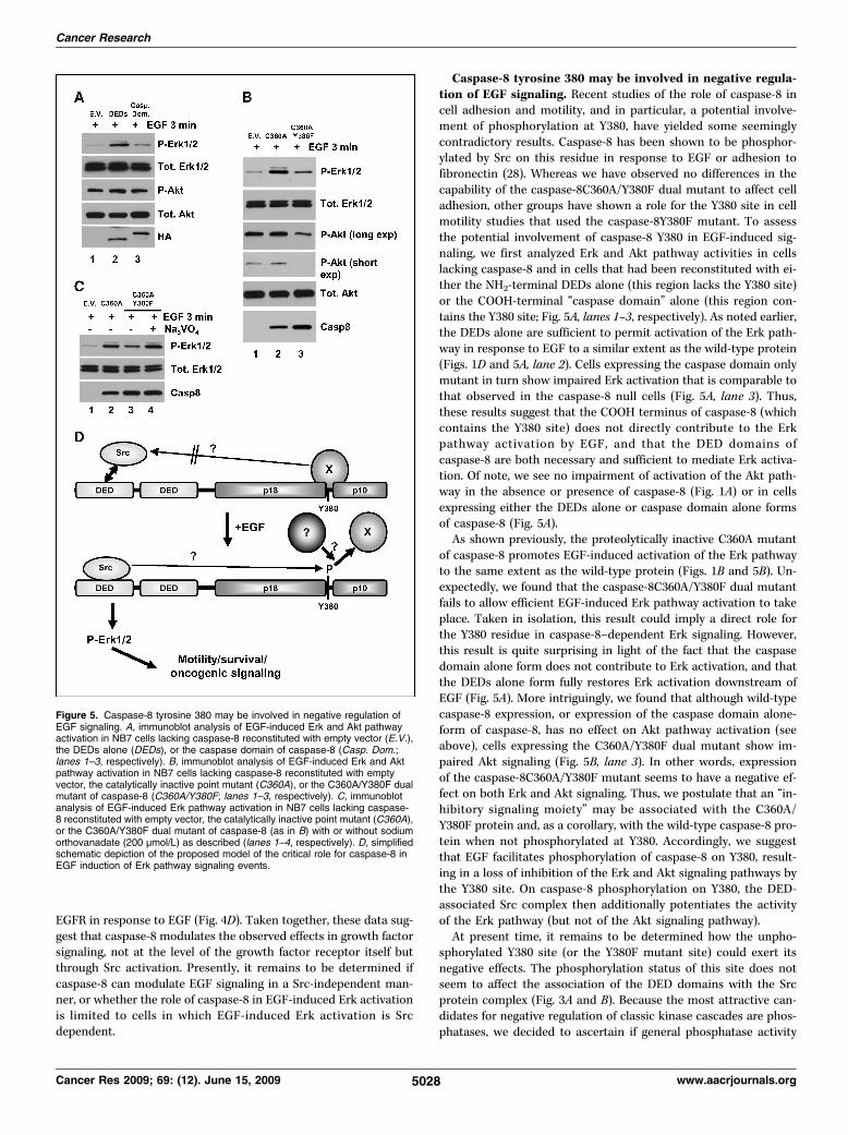
Caspase-8 tyrosine 380 may be involved in negative regula-tion of EGF signaling. Recent studies of the role of caspase-8 incell adhesion and motility, and in particular, a potential involve-ment of phosphorylation at Y380, have yielded some seeminglycontradictory results. Caspase-8 has been shown to be phosphor-ylated by Src on this residue in response to EGF or adhesion tofibronectin (28). Whereas we have observed no differences in thecapability of the caspase-8C360A/Y380F dual mutant to affect celladhesion, other groups have shown a role for the Y380 site in cellmotility studies that used the caspase-8Y380F mutant. To assessthe potential involvement of caspase-8 Y380 in EGF-induced sig-naling, we first analyzed Erk and Akt pathway activities in cellslacking caspase-8 and in cells that had been reconstituted with ei-ther the NH2-terminal DEDs alone (this region lacks the Y380 site)or the COOH-terminal “caspase domain” alone (this region con-tains the Y380 site; Fig. 5A, lanes 1–3, respectively). As noted earlier,the DEDs alone are sufficient to permit activation of the Erk path-way in response to EGF to a similar extent as the wild-type protein(Figs. 1D and 5A, lane 2). Cells expressing the caspase domain onlymutant in turn show impaired Erk activation that is comparable tothat observed in the caspase-8 null cells (Fig. 5A, lane 3). Thus,these results suggest that the COOH terminus of caspase-8 (whichcontains the Y380 site) does not directly contribute to the Erkpathway activation by EGF, and that the DED domains ofcaspase-8 are both necessary and sufficient to mediate Erk activa-tion. Of note, we see no impairment of activation of the Akt path-way in the absence or presence of caspase-8 (Fig. 1A) or in cellsexpressing either the DEDs alone or caspase domain alone formsof caspase-8 (Fig. 5A).As shown previously, the proteolytically inactive C360A mutant
of caspase-8 promotes EGF-induced activation of the Erk pathwayto the same extent as the wild-type protein (Figs. 1B and 5B). Un-expectedly, we found that the caspase-8C360A/Y380F dual mutantfails to allow efficient EGF-induced Erk pathway activation to takeplace. Taken in isolation, this result could imply a direct role forthe Y380 residue in caspase-8–dependent Erk signaling. However,this result is quite surprising in light of the fact that the caspasedomain alone form does not contribute to Erk activation, and thatthe DEDs alone form fully restores Erk activation downstream ofEGF (Fig. 5A). More intriguingly, we found that although wild-typecaspase-8 expression, or expression of the caspase domain alone-form of caspase-8, has no effect on Akt pathway activation (seeabove), cells expressing the C360A/Y380F dual mutant show im-paired Akt signaling (Fig. 5B, lane 3). In other words, expressionof the caspase-8C360A/Y380F mutant seems to have a negative ef-fect on both Erk and Akt signaling. Thus, we postulate that an “in-hibitory signaling moiety” may be associated with the C360A/Y380F protein and, as a corollary, with the wild-type caspase-8 pro-tein when not phosphorylated at Y380. Accordingly, we suggestthat EGF facilitates phosphorylation of caspase-8 on Y380, result-ing in a loss of inhibition of the Erk and Akt signaling pathways bythe Y380 site. On caspase-8 phosphorylation on Y380, the DED-associated Src complex then additionally potentiates the activityof the Erk pathway (but not of the Akt signaling pathway).At present time, it remains to be determined how the unpho-
sphorylated Y380 site (or the Y380F mutant site) could exert itsnegative effects. The phosphorylation status of this site does notseem to affect the association of the DED domains with the Srcprotein complex (Fig. 3A and B). Because the most attractive can-didates for negative regulation of classic kinase cascades are phos-phatases, we decided to ascertain if general phosphatase activity
Figure 5. Caspase-8 tyrosine 380 may be involved in negative regulation ofEGF signaling. A, immunoblot analysis of EGF-induced Erk and Akt pathwayactivation in NB7 cells lacking caspase-8 reconstituted with empty vector (E.V.),the DEDs alone (DEDs), or the caspase domain of caspase-8 (Casp. Dom.;lanes 1–3, respectively). B, immunoblot analysis of EGF-induced Erk and Aktpathway activation in NB7 cells lacking caspase-8 reconstituted with emptyvector, the catalytically inactive point mutant (C360A), or the C360A/Y380F dualmutant of caspase-8 (C360A/Y380F; lanes 1–3, respectively). C, immunoblotanalysis of EGF-induced Erk pathway activation in NB7 cells lacking caspase-8 reconstituted with empty vector, the catalytically inactive point mutant (C360A),or the C360A/Y380F dual mutant of caspase-8 (as in B) with or without sodiumorthovanadate (200 μmol/L) as described (lanes 1–4, respectively). D, simplifiedschematic depiction of the proposed model of the critical role for caspase-8 inEGF induction of Erk pathway signaling events.
www.aacrjournals.org
Caspase-8 Promotes EGF Activation of the Erk1/2 Pathway
was involved in our model of negative signaling by the caspase-8C360A/Y380F protein. To test this hypothesis, we repeated the pre-vious experiment but assayed Erk pathway activity in the cells ex-pressing the C360A/Y380F dual mutant in the presence or absenceof the phosphatase inhibitor sodium orthovanadate. We show thatthe ablation of phosphatase activity restores the Erk signaling ofthe C360A/Y380F dual mutant to levels comparable to, but not ex-ceeding, those of the C360A mutant (Fig. 5C). We also show thatthis is a true caspase-8 response because treatment of NB7 cellslacking caspase-8 with sodium orthovanadate either alone or incombination with EGF has no effect on Erk pathway activation(Supplementary Fig. S4). Thus, we not only provide preliminaryevidence of an inhibitory moiety potentially associated withcaspase-8 Y380 but also suggest that phosphatase activity may beinvolved. Future proteomic analyses of the components of thecaspase-8 signaling complex should allow for identification of thephosphatase potentially involved, thus facilitating more robust con-firmation. A very basic diagram displaying the proposed mechanismof our model system as discussed is shown in Fig. 5D. We note,however, that the system is likely to be more complicated andmay vary based on temporal, dynamic, and/or contextual factors.Indeed, others have shown that phosphorylation of caspase-8 mayallow binding of the p85 subunit of phosphatidylinositol 3-kinase,potentially displacing an inhibitory molecule or complex (20).In sum, we provide further evidence to suggest that caspase-8 is
involved in nonapoptotic and indeed potentially antiapoptoticroles. We show that caspase-8 is essential for EGF (and PDGF)-induced activation of the Erk, but not Akt, pathway. We further
5029www.aacrjournals.org
show that the protein effects these actions via its DED pro-domainand identify residues within the RXDLL motif that are critical. Wepostulate that this is presumably through an ability to associatewith a protein complex containing Src because point mutants un-able to do so show impaired EGF-induced signaling and cell migra-tion. We further show that cells lacking caspase-8 show impairedactivation of Src in response to EGF. We also provide the first pre-liminary evidence that phosphorylation of caspase-8 at Y380 mightrelieve inhibitory signaling rather than positively potentiating theEGF response directly per se. Because the EGF-Src axis has beenstrongly implicated in oncogenic progression (e.g., reviewed ref.29), further research should confirm this unexpected layer of com-plexity and potentially identify novel therapeutic interventionpoints. Indeed, EGFR and Src kinase activities have been the focusof the development of novel antitumor treatments (e.g., reviewedin refs. 30, 31). Thus, these findings and the recent observationsfrom other laboratories (7, 19–21) may explain why caspase-8 israrely absent in cancers.
Disclosure of Potential Conflicts of InterestNo potential conflicts of interest were disclosed.
AcknowledgmentsReceived 10/1/08; revised 3/23/09; accepted 4/7/09; published OnlineFirst 5/26/09.
Grant support: Grants from the NIH (K. Vuori).The costs of publication of this article were defrayed in part by the payment of page
charges. This article must therefore be herebymarked advertisement in accordance with18 U.S.C. Section 1734 solely to indicate this fact.
References1. Muzio M, Chinnaiyan AM, Kischkel FC, et al. FLICE, anovel FADD-homologous ICE/CED-3-like protease, isrecruited to the CD95 (Fas/APO-1) death-inducing sig-naling complex. Cell 1996;85:817–27.
2. Srinivasula SM, Ahmad M, Fernandes-Alnemri T,Litwack G, Alhemri ES. Molecular ordering of the Fas-apoptotic pathway: the Fas/APO-1 protease Mch5 is aCrmA-inhibitable protease that activates multipleCed-3/ICE-like cysteine proteases. Proc Natl Acad SciU S A 1996;93:14486–91.
3. Medema JP, Scaffidi C, Kischkel FC, et al. FLICE is ac-tivated by association with the CD95 death-inducingsignaling complex (DISC). EMBO J 1997;16:2794–804.
4. Vincenz C, Dixit VM. Fas-associated death domainprotein interleukin-1β-converting enzyme 2 (FLICE2),an ICE/Ced-3 homologue, is proximally involved inCD95- and p55-mediated death signaling. J Biol Chem1997;272:6578–83.
5. Wiley SR, Schooley K, Smolak PJ, et al. Identificationand characterization of a new member of the TNF fam-ily that induces apoptosis. Immunity 1995;3:673–82.
6. Walczak H, Miller RE, Ariail K, et al. Tumoricidal activ-ity of tumor necrosis factor-related apoptosis-inducingligand in vivo. Nat Med 1999;5:157–63.
7. Finlay D, Vuori K. Novel noncatalytic role for caspase-8 in promoting SRC-mediated adhesion and Erk signal-ing in neuroblastoma cells. Cancer Res 2007;67:11704–11.
8. Shen J, Behrens C, Wistuba II, et al. Identification andvalidation of differences in protein levels in normal, pre-malignant, and malignant lung cells and tissues usinghigh-throughput Western Array and immunohisto-chemistry. Cancer Res 2006;66:11194–206.
9. Algeciras-Schimnich A, Barnhart BC, Peter ME.Apoptosis-independent functions of killer caspases.Curr Opin Cell Biol 2002;14:721–6.
10. Schwerk C, Schulze-Osthoff K. Non-apoptotic func-tions of caspases in cellular proliferation and differenti-ation. Biochem Pharmacol 2003;66:1453–8.
11. Park SM, Schickel R, Peter ME. Nonapoptotic func-tions of FADD-binding death receptors and their signal-ing molecules. Curr Opin Cell Biol 2005;17:610–6.
12. Olson NE, Graves JD, Shu GL, Ryan EJ, Clark EA. Cas-pase activity is required for stimulated B lymphocytesto enter the cell cycle. J Immunol 2003;170:6065–72.
13. Salmena L, Lemmers B, Hakem A, et al. Essential rolefor caspase 8 in T-cell homeostasis and T-cell-mediatedimmunity. Genes Dev 2003;17:883–95.
14. O'Reilly LA, Divisekera U, Newton K, et al. Modifica-tions and intracellular trafficking of FADD/MORT1 andcaspase-8 after stimulation of T lymphocytes. CellDeath Differ 2004;11:724–36.
15. Chaudhary PM, Eby MT, Jasmin A, Kumar A, Liu L,Hood L. Activation of the NF-κB pathway by caspase8 and its homologs. Oncogene 2000;19:4451–60.
16. Hu WH, Johnson H, Shu HB. Activation of NF-κB byFADD, Casper, and caspase-8. J Biol Chem 2000;275:10838–44.
17. Su H, Bidere N, Zheng L, et al. Requirement forcaspase-8 in NF-κB activation by antigen receptor.Science 2005;307:1465–8.
18. Barnhart BC, Legembre P, Pietras E, Bubici C,Franzoso G, Peter ME. CD95 ligand induces motilityand invasiveness of apoptosis-resistant tumor cells.EMBO J 2004;23:3175–85.
19. Helfer B, Boswell BC, Finlay D, et al. Caspase-8 pro-motes cell motility and calpain activity under nonapop-totic conditions. Cancer Res 2006;66:4273–8.
20. Senft J, Helfer B, Frisch SM. Caspase-8 interacts withthe p85 subunit of phosphatidylinositol 3-kinase to reg-ulate cell adhesion and motility. Cancer Res 2007;67:11505–9.
21. Barbero S, Barila D, Mielgo A, Stagni V, Clair K,Stupack D. Identification of a critical tyrosine residuein caspase 8 that promotes cell migration. J Biol Chem2008;283:13031–4.
22. Frisch SM. Caspase-8: fly or die. Cancer Res 2008;68:4491–3.
23. Teitz T, Wei T, Valentine MB, et al. Caspase 8 is de-leted or silenced preferentially in childhood neuroblas-tomas with amplification of MYCN. Nat Med 2000;6:529–35.
24. Boldin MP, Goncharov TM, Goltsev YV, Wallach D. In-volvement of MACH, a novel MORT1/FADD- interactingprotease, in Fas/APO-1- and TNF receptor-induced celldeath. Cell 1996;85:803–15.
25. Eckhart L, Ballaun C, Hermann M, et al. Identifica-tion of novel mammalian caspases reveals an importantrole of gene loss in shaping the human caspase reper-toire. Mol Biol Evol 2008;25:831–41.
26. Kischkel FC, Lawrence DA, Tinel A, et al. Death re-ceptor recruitment of endogenous caspase-10 and apo-ptosis initiation in the absence of caspase-8. J BiolChem 2001;276:46639–46.
27. Muppidi JR, Lobito AA, Ramaswamy M, et al. Homo-typic FADD interactions through a conserved RXDLLmotif are required for death receptor-induced apopto-sis. Cell Death Differ 2006;13:1641–50.
28. Cursi S, Rufini A, Stagni V, et al. Src kinase phosphor-ylates Caspase-8 on Tyr380: a novel mechanism of apo-ptosis suppression. EMBO J 2006;25:1895–905.
29. Ishizawar R, Parsons SJ. c-Src and cooperating part-ners in human cancer. Cancer Cell 2004;6:209–14.
30. Wakeling AE. Inhibitors of growth factor signalling.Endocr Relat Cancer 2005;12 Suppl 1:S183–7.
31. Hatake K, Tokudome N, Ito Y. Next generation mo-lecular targeted agents for breast cancer: focus on EGFRand VEGFR pathways. Breast Cancer 2007;14:132–49.
Cancer Res 2009; 69: (12). June 15, 2009