The UV (Ribotoxic) Stress Response of Human Keratinocytes Involves the Unexpected Uncoupling of the...
16
10.1128/MCB.22.15.5380-5394.2002. 2002, 22(15):5380. DOI: Mol. Cell. Biol. Thanh-Hoai Dinh, Robert K. Bright and Bruce E. Magun Mihail S. Iordanov, Remy J. Choi, Olga P. Ryabinina, Epidermal Growth Factor Receptor Signaling Cascade from the Activated Ras-Extracellular Signal-Regulated Kinase Unexpected Uncoupling of the Human Keratinocytes Involves the The UV (Ribotoxic) Stress Response of http://mcb.asm.org/content/22/15/5380 Updated information and services can be found at: These include: REFERENCES http://mcb.asm.org/content/22/15/5380#ref-list-1 at: This article cites 77 articles, 31 of which can be accessed free CONTENT ALERTS more» articles cite this article), Receive: RSS Feeds, eTOCs, free email alerts (when new http://journals.asm.org/site/misc/reprints.xhtml Information about commercial reprint orders: http://journals.asm.org/site/subscriptions/ To subscribe to to another ASM Journal go to: on March 17, 2014 by guest http://mcb.asm.org/ Downloaded from on March 17, 2014 by guest http://mcb.asm.org/ Downloaded from
-
Upload
independent -
Category
Documents
-
view
4 -
download
0
Transcript of The UV (Ribotoxic) Stress Response of Human Keratinocytes Involves the Unexpected Uncoupling of the...

10.1128/MCB.22.15.5380-5394.2002.
2002, 22(15):5380. DOI:Mol. Cell. Biol. Thanh-Hoai Dinh, Robert K. Bright and Bruce E. MagunMihail S. Iordanov, Remy J. Choi, Olga P. Ryabinina, Epidermal Growth Factor ReceptorSignaling Cascade from the ActivatedRas-Extracellular Signal-Regulated Kinase Unexpected Uncoupling of theHuman Keratinocytes Involves the The UV (Ribotoxic) Stress Response of
http://mcb.asm.org/content/22/15/5380Updated information and services can be found at:
These include:
REFERENCEShttp://mcb.asm.org/content/22/15/5380#ref-list-1at:
This article cites 77 articles, 31 of which can be accessed free
CONTENT ALERTS more»articles cite this article),
Receive: RSS Feeds, eTOCs, free email alerts (when new
http://journals.asm.org/site/misc/reprints.xhtmlInformation about commercial reprint orders: http://journals.asm.org/site/subscriptions/To subscribe to to another ASM Journal go to:
on March 17, 2014 by guest
http://mcb.asm
.org/D
ownloaded from
on M
arch 17, 2014 by guesthttp://m
cb.asm.org/
Dow
nloaded from
MOLECULAR AND CELLULAR BIOLOGY, Aug. 2002, p. 5380–5394 Vol. 22, No. 150270-7306/02/$04.00�0 DOI: 10.1128/MCB.22.15.5380–5394.2002Copyright © 2002, American Society for Microbiology. All Rights Reserved.
The UV (Ribotoxic) Stress Response of Human Keratinocytes Involvesthe Unexpected Uncoupling of the Ras–Extracellular Signal-Regulated
Kinase Signaling Cascade from the Activated Epidermal GrowthFactor Receptor
Mihail S. Iordanov,1 Remy J. Choi,1 Olga P. Ryabinina,1 Thanh-Hoai Dinh,1 Robert K. Bright,2and Bruce E. Magun1*
Department of Cell and Developmental Biology, Oregon Health Sciences University, Portland, Oregon 97201,1
and Department of Microbiology and Immunology, Southwest Cancer Center, Texas TechUniversity Health Sciences Center, Lubbock, Texas 794302
Received 24 January 2002/Returned for modification 21 February 2002/Accepted 29 April 2002
In mammals, UVB radiation is of biological relevance primarily for the cells of the epidermis. We report herethe existence of a UVB response that is specific for proliferating human epidermal keratinocytes. Unlike othercell types that also display a UVB response, keratinocytes respond to UVB irradiation with a transient butpotent downregulation of the Ras–extracellular signal-regulated kinase (ERK) signaling cascade. The down-regulation of ERK precedes a profound decrease in the steady-state levels of cyclin D1, a mediator of theproliferative action of ERK. Keratinocytes exhibit high constitutive activity of the Ras-ERK signaling cascadeeven in culture medium lacking supplemental growth factors. The increased activity of Ras and phosphory-lation of ERK in these cells are maintained by the autocrine production of secreted molecules that activate theepidermal growth factor receptor (EGFR). Irradiation of keratinocytes increases the phosphorylation of EGFRon tyrosine residues Y845, Y992, Y1045, Y1068, Y1086, Y1148, and Y1173 above the basal levels and leads tothe increased recruitment of the adaptor proteins Grb2 and ShcA and of a p55 form of the regulatory subunitof the phosphatidylinositide 3-kinase to the UVB-activated EGFR. Paradoxically, however, UVB causes, at thesame time, the inactivation of Ras and a subsequent dephosphorylation of ERK. By contrast, the signalingpathway leading from the activated EGFR to the phosphorylation of PKB/Akt1 is potentiated by UVB. The UVBresponse of keratinocytes appeared to be a manifestation of the more general ribotoxic stress responseinasmuch as the transduction of the UVB-generated inhibitory signal to Ras and ERK required the presenceof active ribosomes at the time of irradiation.
Exposure to UVB radiation is the major cause of cutaneousmalignancies in the United States (45). UVB is a completecarcinogen that is known to act both as a tumor initiator and atumor promoter (43, 59). Exposure of the skin to UVB inducesthe stereotypic early responses of genes such as c-fos and c-jun(6, 26, 27, 63) and delayed responses, including hyperplasiaand skin cancer (21, 26, 48, 72). The induction of the earlyresponse genes results from a cascade of signaling events thateventually modulates the activity of specific transcription fac-tors. The signaling pathways that transduce UVB-initiated sig-nals to modulate gene expression involve the superfamily of theproline-directed mitogen-activated protein (MAP) kinases (6,55). The stress-activated protein kinase (SAPK) family ofMAP kinases includes the c-Jun-NH2-terminal kinases (JNK;SAPK1) and the p38 MAP kinases (SAPK2) (15, 46, 73). Theactivation of SAPK in mammalian cells leads to decision mak-ing via the activation of competing prosurvival and proapop-totic pathways (15). Activation of SAPK has also been shownto be a major pathway leading to the synthesis and release ofproinflammatory cytokines, which function to communicate
the presence of cellular damage to other cells of the organism(16, 57). The extracellular signal-regulated kinases, p44 ERK1and p42 ERK2 (ERKs), represent the other family of MAPkinases. Although ERKs may play specific roles in specific celltypes, it is generally accepted that mitogen-induced sustainedactivation of ERKs mediates cell cycle progression through G1-and S-phase entry by regulating the expression of cyclin D1 (2,3, 12, 60, 66, 76).
The epidermal growth factor receptor (EGFR) plays a cru-cial role in mediating both the proliferative and prosurvivalprograms of keratinocytes, the main cell type of the epidermis(reference 68 and references therein). At least five mitogenicgrowth factors bind to and activate EGFR in the skin. Inaddition to epidermal growth factor (EGF), these factors in-clude transforming growth factor � (TGF-�) (11), amphiregu-lin (13), heparin-binding EGF (HB-EGF) (25), and epiregulin(67). The classical signal-transduction cascade that translatesEGFR activation into the increased activity of ERK has beeninvestigated at length (10, 40, 64). The binding of a cognateligand stabilizes receptor dimerization and stimulates the ty-rosine kinase activity of the intracellular portion of the recep-tor, resulting in the increased auto(trans-)phosphorylation ofseveral tyrosine residues within the receptor dimer (7, 65). Thephosphorylated tyrosines serve as recruitment (docking) sitesfor several adaptor proteins, among which the adaptor proteins
* Corresponding author. Mailing address: Department of Cell andDevelopmental Biology, Oregon Health Sciences University, Portland,OR 97201. Phone: (503) 494-7811. Fax: (503) 494-4253. E-mail: [email protected].
5380
on March 17, 2014 by guest
http://mcb.asm
.org/D
ownloaded from

Grb2 (19, 42) and Shc (44, 53, 62) are of particular importancefor the activation of ERK (60). Grb2 contains one Src-homol-ogy 2 (SH2) domain flanked by two Src-homology 3 (SH3)domains (42). The SH2 domain of Grb2 binds to tyrosine-phosphorylated residues in growth factor receptors, such asY1068 and Y1086 of EGFR (5, 50, 74), whereas the SH3domains bind to proline-rich motifs found in many signalingmolecules, such as the Son of Sevenless (Sos) protein (9, 19,52). Sos is a guanine nucleotide exchange factor that facilitates,in response to activation of growth factor receptors, the con-version of the inactive (GDP-bound) form of the cell mem-brane-localized small GTPase Ras into the activated (GTP-bound) form of Ras (9). Activated Ras recruits to the cellmembrane the serine/threonine protein kinase c-Raf, an eventthat leads to the activation of c-Raf itself (37). c-Raf, in turn,phosphorylates and activates the MAP/ERK kinases MEK1and MEK2, eventually leading to the activation of ERK (10,37, 40, 64). Grb2 also binds EGFR indirectly via EGFR-boundShcA (4). ShcA, however, binds to different phosphotyrosineresidues of EGFR, namely Y1148, Y1173, and Y992 (4, 5, 49,74).
The study of the molecular basis of the UV response (en-compassing the cellular responses to both UVC and UVB) ina tissue culture system has traditionally been performed inrodent and human fibroblasts and in HeLa cells (6, 30, 31, 34,36, 55, 63) under the assumption that the UV response isconserved in all cell types. By use of NIH 3T3 fibroblasts andHeLa cells, ERK was identified as the first signal transductionkinase activated early after exposure to UVC radiation (55).The identification of the ability of UVC and UVB to triggerthe activation of growth factor receptors (36, 63) favored theconclusion that an operational growth factor receptor-ERKcascade is an important mediator of UV-induced gene expres-sion (6, 26).
In the search for novel and relevant UV-induced signal-transduction pathways, we recently discovered that the activa-tion of SAPK by UVC and UVB is initiated in, or in closeproximity to, the functional center of actively translating eu-karyotic ribosomes (31, 34). This center contains the 3� end of28S rRNA and its proteinaceous environment and is respon-sible for aminoacyl-tRNA binding, peptidyl transfer, and ribo-some translocation. This region of the 28S rRNA is the targetof the antibiotics anisomycin and blasticidin S and of the en-zymatic ribotoxins ricin A chain and �-sarcin, all of whichstrongly activate SAPK (reference 33 and references therein).The activation of SAPK by the foregoing agents was termedthe ribotoxic stress response and is characterized by the abso-lute requirement for the presence of actively translating ribo-somes at the moment of cellular encounter with the antibioticor ribotoxin acting on 28S rRNA (33). Cells whose ribosomesare not engaged in translational elongation fail to activateSAPK in response to these agents. In contrast, the activation ofSAPK by nonribotoxic stressors, such as inflammatory cyto-kines, osmotic stress, and some DNA-damaging drugs, is intactin cells containing nontranslating ribosomes (31–33). Interest-ingly, UVB requires the presence of active ribosomes to acti-vate SAPK. Furthermore, nucleotide- and position-specificdamage to the 3� end of 28S rRNA was detected in UVB-irradiated cells, and the time of appearance and degree of thisdamage correlated with the activation of SAPK (34). It was
concluded therefore that UVB triggers the ribotoxic stressresponse that leads to the activation of SAPK. In cells thatrespond to UVB with the activation of ERK (e.g., fibroblasts orHeLa cells), however, the activation of ERK was not depen-dent on ribotoxic stress inasmuch as it did not require activeribosomes (34).
In the present study, we employed normal human keratino-cytes to investigate whether these cells (that are frequentlyexposed to solar radiation) possess a specific UVB response.We demonstrate for the first time that the UVB response ofhuman keratinocytes is characterized by both stimulatory (ac-tivation of SAPK) and inhibitory (inactivation of ERK) signaltransduction components. The elevated basal levels of ERKactivity in these cells resulted from the autocrine and paracrineactions of extracellular molecules that bind to and activate theEGFR. Despite the activation of EGFR resulting from UVBirradiation, UVB interfered with signals that lead to ERKactivation at the level of a signal-transduction step(s) locateddownstream of the EGFR-bound Grb2 and ShcA but up-stream of Ras. At the same time, the activation of EGFR byUVB triggered the phosphorylation of the protein kinase B(PKB/Akt1) that is involved in mediating cell survival (14).Finally, we show that both the stimulatory and the inhibitoryUVB responses of MAP kinases are downstream of a ribo-some-initiated signaling event.
MATERIALS AND METHODS
Cell culture. Human epidermal keratinocytes, neonatal (HEKn) were main-tained in EpiLife basal keratinocyte medium (BKM) supplemented with asemidefined human keratinocyte growth supplement (HKGS) (the final concen-trations of the components in the supplemented medium were as follows: 0.2%[vol/vol] bovine pituitary extract, 5 mg of bovine insulin per ml, 0.18 mg ofhydrocortisone per ml, 5 mg of bovine transferrin per ml, and 0.2 ng of humanepidermal growth factor per ml). The BKM supplemented with HKGS is re-ferred to as BKM�exoGF (BKM plus exogenous growth factors), and BKMlacking HKGS is referred to as BKM–exoGF (BKM minus exogenous growthfactors). The cells and all cell culture reagents were obtained from CascadeBiologics, Inc. Immortalized HEKn (HEKn-E6/E7) were established throughinfection with an amphitropic recombinant retroviral vector encoding the E6 andE7 transforming proteins of human papillomavirus serotype 16 as previouslydescribed (35). HEKn-E6/E7 were maintained in culture in a way identical towhat was done with HEKn. The HaCaT cell line (a gift from Tim Bowden) andHeLa cells were maintained in Dulbecco’s modified Eagle’s medium (DMEM)supplemented with 10% fetal calf serum and antibiotics. For experiments, Ha-CaT and HeLa cells were plated in BKM�exoGF.
Irradiation of cells and other treatments. The spectral characterization of theUVB source and the mode of the irradiation of cells with UVB have beendescribed previously (31, 34). Human recombinant EGF (Sigma) was kept as a100-�g/ml stock solution in 5 � 10�3 N HCl at �70°C. AG1478 and UO126(both from Calbiochem), anisomycin, emetine, actinomycin D (all from Sigma),and pactamycin (a gift from Pharmacia and Upjohn) were dissolved in dimethylsulfoxide (DMSO) and kept at �70°C. Sodium arsenite (Sigma) was freshlyprepared before use as a 40 mM stock solution in double-distilled deionizedwater. To prepare conditioned media, the cells (originally plated inBKM�exoGF) were washed extensively with BKM–exoGF and then placed for20 to24 h in BKM–exoGF (2 ml for each 60-mm-diameter cell culture plate).Conditioned media were never frozen and were used only once. The EGFR-neutralizing monoclonal antibody LA1 was obtained from Upstate Biotechnol-ogy.
Immunocomplex kinase assay. HEKn from 6-cm-diameter tissue culturedishes were harvested by lysis on ice in an ice-cold solution containing 20 mMHEPES-KOH (pH 7.4), 2 mM EGTA, 50 mM �-glycerophosphate, 1 mM di-thiothreitol (DTT), 10% glycerol, 1% Triton X-100, 1 mM sodium vanadate, and1� Complete protease inhibitors (Roche Molecular Biochemicals). ERK1/ERK2 was immunoprecipitated for 3 h at 4°C with an anti-ERK1/ERK2 antibody(C-16; Santa Cruz Biotechnology) precoupled to protein A-agarose (Santa Cruz
VOL. 22, 2002 KERATINOCYTE-SPECIFIC UVB RESPONSE 5381
on March 17, 2014 by guest
http://mcb.asm
.org/D
ownloaded from

Biotechnology). The immunoprecipitates were washed once with lysis buffer;once with a solution consisting of 100 mM Tris-HCl (pH 7.6), 500 mM LiCl, 1mM DTT, and 0.1% Triton X-100; and once with a buffer containing 20 mMmorpholinepropanesulfonic acid (MOPS; pH 7.2), 10 mM MgCl2, 2 mM EGTA,1 mM DTT, and 0.1% Triton X-100. For the kinase reaction, the immunopre-cipitates were incubated with 1 mg of glutathione S-transferase (GST)–Elk1fusion proteins in the presence of 10 mM MOPS (pH 7.2), 20 mM MgCl2, 1 mMEGTA, 0.5 mM DTT, 0.05% Triton X-100 and 1 mCi of [�-32P]ATP for 20 minat 30°C. After the reactions were stopped by adding 10 �l of 4� sodium dodecylsulfate-polyacrylamide gel electrophoresis (SDS-PAGE) loading buffer, the sam-ples were resolved by SDS–13% PAGE. The phosphorylated GST-Elk1 wasquantified from dried gels with a Molecular Dynamics PhosphorImager and IPLab Gel software.
Immunoblot analysis of proteins. For immunoblot analyses, the cells werelysed directly in 2� SDS-PAGE loading buffer and lysates corresponding to 1 �105 to 1.5 � 105 cells were resolved in SDS-PAGE with the appropriate per-centage of acrylamide (7.5 to 15%). The antibodies against ERK1 (C-16), EGFR(EGFR-1005), the phosphorylated form of EGFR (pY1173), Sos1 (C-23 andD-21), ShcA (PG-797), c-Myc (N-262), p63 (4A4-HRP), p53 (DO-1-HRP), cy-clin D1 (M-20), MEK2 (N-20), JNK1 (C-17), p38 MAP kinase (C-20), p85 PI-3K(Z-8), and PKB/Akt1/2 (H-136) were obtained from Santa Cruz Biotechnology.The antibodies against the phosphorylated forms of EGFR (pY1148 andpY1086) were obtained from BioSource International. The antibodies againstthe phosphorylated forms of EGFR (pY845, pY992, pY1045, and pY1068) wereobtained from Cell Signaling Technology. The pan-Ras antibody (31–43) wasobtained from Calbiochem. The antibodies against the phosphorylated forms ofERK, p38 MAP kinase, JNK, MEK, PKB/Akt1 (S473), and FKHR were ob-tained from Cell Signaling Technology. The antibody against Grb2 was obtainedfrom BD-Transduction Laboratories. The separation of proteins in SDS-PAGEand the electrotransfer onto polyvinylidene difluoride membranes (Millipore)were performed by standard procedures. Immunodetections with phospho-epitope-specific antibodies were performed according to the instructions of therespective manufacturers.
Coimmunoprecipitations. The coimmunoprecipitation of EGFR with associ-ated proteins was performed as described by Dulin et al. (17), and the coimmu-noprecipitation of Sos1 with associated proteins was performed as described byGross et al. (23).
Ras activity assay. Recombinant protein composed of the Ras-binding domainof c-Raf fused to GST (GST/Raf-RBD; a gift from Rudolf Juliano) was bacte-rially expressed in advance, affinity bound to glutathione-coupled Sepharose 4Bbeads (Amersham Pharmacia Biotech AB) shortly before use, and kept on ice.All subsequent procedures were performed with the protein on ice or at 4°C.Cells were lysed by scraping and vigorous mixing in 1 ml (on a 100-mm-diametercell culture plate) of lysis buffer (50 mM Tris [pH 8.0], 150 mM NaCl, 1%IGEPAL [Sigma; equivalent to Nonidet P-40], 10% glycerol, 5 mM MgCl2, 10mM NaF, 1 mM NaVO4, 1� Complete protease inhibitors [Roche MolecularBiochemicals], and 1 �M DTT). The lysates were precleared for 10 min at 14,000rpm in an Eppendorf table-top centrifuge. One hundred microliters of GST/Raf-RBD-bound slurry was mixed with cell lysate (typically corresponding to 3 � 106
to 4 � 106 cells per experimental point) and incubated, with continuous mixing,for 30 min. After the beads were washed three times with lysis buffer, GTP-bound Ras proteins were eluted by boiling in 100 �l of 2� SDS-PAGE loadingbuffer. Ras proteins were resolved with SDS-15% PAGE and detected by im-munoblot analysis.
RESULTS
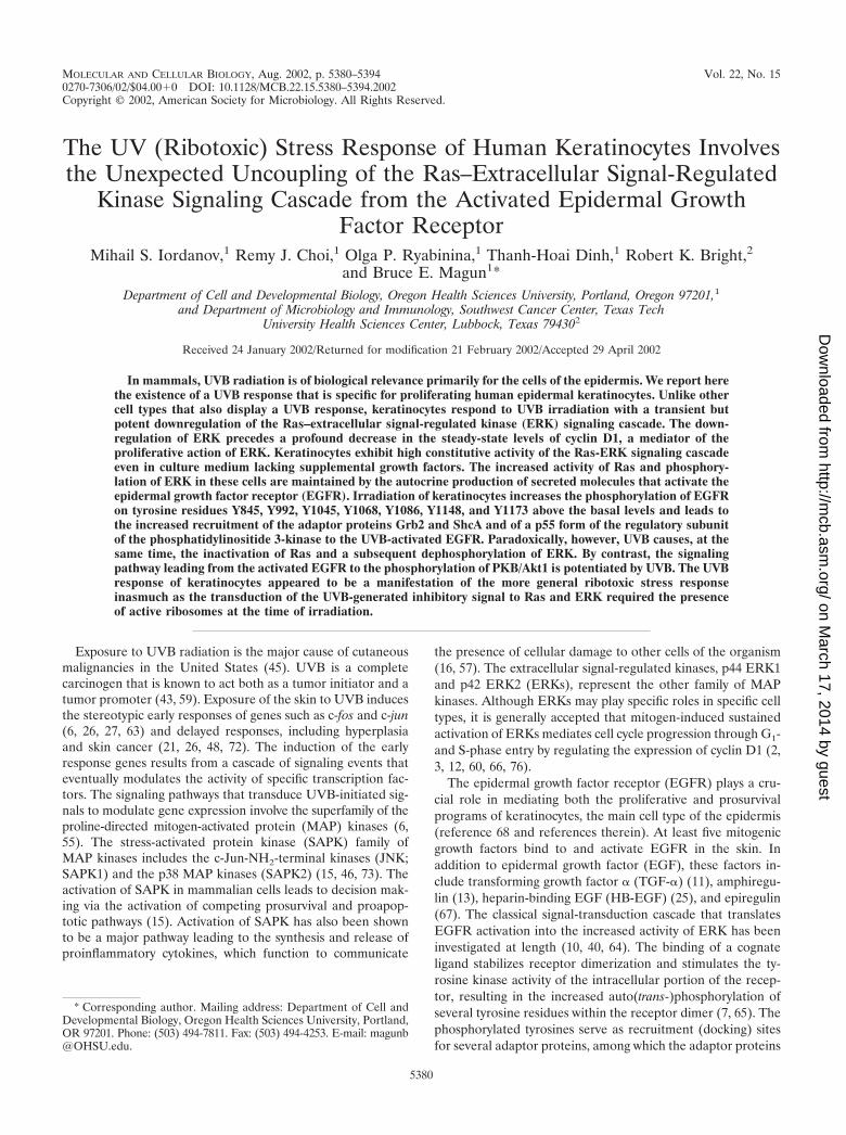
Inactivation of ERK by UV in human epidermal keratino-cytes. In order to investigate the regulation of MAP kinases byUVB in a cell type that naturally encounters UV radiation, weemployed freshly explanted HEKn. We also employed un-cloned HEKn-E6/E7 (see Materials and Methods). BothHEKn and HEKn-E6/E7 display the expression of basal ker-atinocyte markers (Np63�) (54, 77) (Fig. 2A, lanes 1 and 6)and integrin � 1 (75) (data not shown). When cultured inserum-free BKM�exoGF (see Materials and Methods), bothHEKn and HEKn-E6/E7 displayed detectable levels of double-phosphorylated p42 ERK2 and, to a much lesser extent, p44ERK1 (Fig. 1A, lane 1, and B, lane 1), as detected in immu-noblot assays with phosphorylation-specific antibodies. To in-
vestigate the regulation of ERK activity by UV in keratino-cytes, we subjected HEKn and HEKn-E6/E7 to UVB.Irradiation of HEKn and HEKn-E6/E7 induced a dose-depen-dent increase in the phosphorylation of SAPK (JNK and p38�MAP kinase) at 30 min postirradiation (Fig. 1A and B, lanes 1to 5 of each). Surprisingly, we found that, instead of activation,UVB-irradiated HEKn and HEKn-E6/E7 displayed a dose-dependent downregulation of the phosphorylation state ofERK at 30 min postirradiation (Fig. 1A and B, lanes 1 to 5 ofeach). The dephosphorylation of ERK in HEKn (Fig. 1A)occurred after doses of UVB (600 to 1,200 J/m2) that causedboth cell cycle withdrawal (evident by an inhibition of DNAsynthesis [data not shown] and a decrease in the steady-statelevels of c-Myc [Fig. 2B]) and apoptosis (“sunburn cells” [38,47]) [evident by the cleavage of the caspase substrate poly-(ADP)ribose polymerase (Fig. 2A) and the decrease in thesteady-state levels of Np63� (41) (Fig. 2A)]. The UVB-in-
FIG. 1. Dose-dependent regulation of the phosphorylation of MAPkinases by UVB. HEKn and HeLa cells (A) and HEKn-E6/E7 andHaCaT cells (B) (grown in BKM�exoGF) were treated with the in-dicated doses of UVB and were harvested 30 min later. Shown are theresults of immunoblot analyses using phosphorylation-specific antibod-ies against the indicated proteins.
5382 IORDANOV ET AL. MOL. CELL. BIOL.
on March 17, 2014 by guest
http://mcb.asm
.org/D
ownloaded from
duced dephosphorylation of ERK resulted in a substantialinhibition of ERK activity (93% 2% inhibition) as deter-mined by immunocomplex kinase activity assays 30 min postir-radiation (Fig. 2B). The dephosphorylation and inactivation ofERK following exposure to UVB became detectable 20 minafter the irradiation (Fig. 3A, lane 3) and persisted, with somevariation from experiment to experiment, for typically 2 to 6 hafter irradiation (Fig. 3A, lanes 3 to 10). The UV-inducedinactivation of ERK has been observed invariably in all sixindependent preparations of HEKn (each derived from a dif-ferent individual) that we have examined. For instance, Fig.1A, 3A, and 9 depict the UVB responses of three independentHEKn preparations (i.e., three different individuals). ERK1and ERK2 proteins were expressed at similar steady-state lev-els in HEKn and HEKn-E6/E7, and these levels were notaffected by exposure to UVB (Fig. 1, 3, 5, 6A, 7, 8, 10, 11, and12A). Therefore, the decrease in ERK phosphorylation and
activity after exposure to UV was due to a bona fide dephos-phorylation event and not to reduced ERK levels. In summary,these results demonstrated that, in contrast to what occurs inother cell types that have been investigated, the activity ofERK in keratinocytes was transiently, but potently, suppressedin response to UVB.
Human epidermal keratinocytes maintain high steady-statelevels of ERK activity via the autocrine production of EGFRligand(s). To determine whether the phosphorylated state ofERK in keratinocytes was due to the presence of supplementalgrowth factors in BKM�exoGF, we assessed the phosphory-lation status of ERK after placing HEKn or HEKn-E6/E7 inBKM–exoGF for 20 h. The absence of exogenously supplied
FIG. 2. (A) Effects of UVB irradiation on cell cycle and apoptosismarkers in HEKn and HEKn-E6/E7. Cells grown in BKM�exoGFwere irradiated with the indicated doses of UVB, and the expression ofthe indicated proteins was determined in immunoblot analyses 21 hpostirradiation. PARP, poly(ADP)ribose polymerase. (B) Inhibition ofERK activity by UVB and anisomycin. HEKn (grown inBKM�exoGF) were treated with UVB (1,200 J/m2) or anisomycin (10�g/ml). ERK activity was assessed 30 min later in immunocomplexkinase assays (see Materials and Methods). Lane 4 of the boxed insertshows the control immunoprecipitation (IP) with an irrelevant anti-body. Error bars represent the standard deviations from experimentalpoints in triplicates.
FIG. 3. Time-dependent inhibition of ERK phosphorylation byUVB. Shown are the results of immunoblot analyses using phosphor-ylation-specific antibodies against the indicated proteins. (A) HEKnwere treated with UVB (1,200 J/m2) UVB and later harvested at theindicated times. Co, control. (B) HEKn and HEKn-E6/E7, placed 20 hearlier in either BKM�exoGF or BKM–exoGF, were irradiated withUVB (1,200 J/m2) and harvested at the indicated times after theirradiation. –, control cells. Note that a prior hybridization of a mem-brane with antibodies against the phosphorylated forms of ERK orEGFR interferes with a subsequent hybridization of the same mem-brane with the antibodies against nonphosphorylated ERK or EGFRat any location in the membrane where the phospho-specific signal wasstrong. This explains the apparent weaker signal of the nonphospho-rylated p42 ERK2 seen in the bottom panel of lane 6. The samephenomenon can also be seen in Fig. 4A, lanes 3 and 10 (ERK2); Fig.5A, lane 2 (EGFR and ERK2); Fig. 6A, lanes 1 to 3 and 7 (ERK2);Fig. 7A, lanes 5 to 7 (ERK2); Fig 9, panel l, lanes 7 and 12 (ERK2);and Fig. 12A, lanes 2 to 4 (ERK2).
VOL. 22, 2002 KERATINOCYTE-SPECIFIC UVB RESPONSE 5383
on March 17, 2014 by guest
http://mcb.asm
.org/D
ownloaded from

growth factors failed to decrease the degree of phosphoryla-tion of ERK (Fig. 3B, compare lanes 1 and 6), suggesting thatthe activity of ERK is regulated by autocrine mechanisms inthese cells. To test this possibility, we transferred HEKn-E6/E7-conditioned BKM–exoGF onto serum-deprived HeLacells, whose levels of ERK phosphorylation are extremely low.Keratinocyte-conditioned, but not HeLa-cell-conditioned(data not shown), or unconditioned BKM–exoGF stimulatedthe phosphorylation of ERK in the HeLa cells (Fig. 4A, lane3). Furthermore, only the keratinocyte-conditioned BKM–exoGF was capable of causing the phosphorylation of EGFR attyrosine-1173 (Y1173) (Fig. 4A, lane 3). The keratinocyte-conditioned BKM–exoGF was unable to induce the phosphor-
ylation of ERK (Fig. 4A, lane 7) when the conditioned mediumwas added to HeLa cells that had been pretreated withAG1478, a specific inhibitor of the EGFR tyrosine kinase ac-tivity (20, 51). Following the exposure of HeLa cells to kera-tinocyte-conditioned BKM–exoGF (Fig. 4A, lane 7), the phos-phorylation of EGFR at Y1173 was also inhibited by AG1478,suggesting that the receptor phosphorylation was caused by thetyrosine kinase activity of the receptor itself and not by adifferent tyrosine kinase. These results suggest that keratino-cytes produce a soluble ligand(s) that is capable of activatingEGFR and, subsequently, ERK. These results were not exclu-sively specific for the immortalized HEKn-E6/E7, since themortal HEKn also displayed a potent production of a soluble
FIG. 4. Production of a soluble ligand(s) of EGFR by HEKn-E6/E7 but not by HaCaT cells. Shown are the results of immunoblot analyses usingantibodies specific for the phosphorylated states of EGFR (Y1173) and ERK. (A) The phosphorylation state of EGFR (Y1173) and ERK in HeLa(lanes 1 to 8) or HaCaT (lanes 9 to 12) cells that have been subjected to unconditioned, HEKn-E6/E7-conditioned, or HaCaT-conditionedBKM–exoGF according to the method described in the figure body. The cells were pretreated for 30 min with 10 �M AG1478 (�) or the vehiclesolvent DMSO(�). (B) HEKn-E6/E7, placed 20 h earlier in either BKM�exoGF (� GF) or BKM–exoGF (– GF), were treated with 10 �MAG1478 or the vehicle solvent DMSO (lanes 1 to 6) for the indicated times.
5384 IORDANOV ET AL. MOL. CELL. BIOL.
on March 17, 2014 by guest
http://mcb.asm
.org/D
ownloaded from

ligand(s) of EGFR (Fig. 5B). To determine whether this li-gand(s) provides the main extracellular signal for the highERK activity in keratinocytes, we incubated HEKn-E6/E7 withAG1478 and assessed the phosphorylation status of ERK. Im-mediately (less than 10 s) after the addition of AG1478, thekeratinocytes displayed an immediate decrease in the basalphosphorylation of EGFR (Fig. 4B, lane 7) without a concom-itant decrease in the phosphorylation of ERK. Five minutesafter the addition of AG1478, the basal phosphorylation ofERK dramatically decreased, and the low levels of phosphor-ylated ERK persisted for the duration of the experiment (Fig.4B, lanes 7 to 12). The effect of AG1478 was independent ofthe presence or the absence in BKM of supplemental growthfactors (Fig. 4B). To verify the specificity of AG1478, we com-pared its abilities to inhibit the activation of ERK by EGF andby fibroblast growth factor 2 (FGF-2). FGF-2 also signals
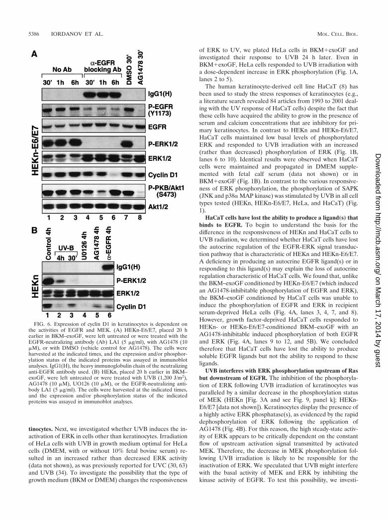
through a cognate receptor with intrinsic tyrosine kinase activ-ity but not through EGFR (22). Addition of EGF to cellspretreated with AG1478 failed to induce the phosphorylationof either EGFR or ERK (Fig. 5A, lane 5). However, AG1478had no effect on the ability of FGF-2 to induce the phosphor-ylation of ERK (Fig. 5A, lane 6) (FGF-2 did display an abilityto trigger the phosphorylation of ERK because the basal phos-phorylation of ERK had been reduced to very low levels byAG1478). These results (Fig. 4 and 5) suggested that the ele-vated activity of ERK in keratinocytes is sustained predomi-nantly through the autocrine production of a soluble ligand(s)that activates EGFR. To verify by a different approach that theERK activity in keratinocytes is exclusively dependent on theautocrine-maintained activity of EGFR, we employed theEGFR-neutralizing monoclonal antibody LA1. Thirty minutesafter the addition of LA1 to the culture medium (BKM–ex-oGF) of HEKn-E6/E7, the phosphorylation of Y1173 ofEGFR was substantially (albeit not completely) decreased andthe decrease remained unchanged for as long as 6 h (Fig. 6A,lanes 4 to 6). Importantly, the EGFR-neutralizing antibodycompletely blocked the phosphorylation of ERK (Fig. 6A,lanes 4 to 6) in a manner similar to the effect of AG1478 (Fig.6A, lane 8).
To investigate whether the supplemental growth factorspresent in BKM�exoGF influence the dephosphorylation ofERK by UVB, we irradiated HEKn or HEKn-E6/E7 20 h afterplacing the cells in BKM–exoGF. The dephosphorylation ofERK by UVB was more pronounced in the cells maintained inBKM–exoGF in terms of both amplitude and duration (Fig.3B, compare lanes 2 to 5 with lanes 7 to 10). We concluded thatthe supplemental growth factors present in BKM�exoGF fa-cilitate the recovery of ERK phosphorylation levels followingexposure to UVB.
The expression of cyclin D1 in keratinocytes is regulated bythe EGFR-MEK-ERK cascade. To investigate the potentialphysiological consequences of the UVB-induced inactivationof ERK in keratinocytes, we studied the steady-state levels ofcyclin D1. Proliferating HEKn (Fig. 3A, lane 1, and 6B, lane 1)or HEKn-E6/E7 (Fig. 6A, lane 1) displayed detectable levels ofcyclin D1. Following UVB irradiation, the steady-state levels ofcyclin D1 declined gradually within the first 2 h after irradia-tion and remained low for as long as 6 h (Fig. 3A, lanes 2 to 10,and 6B, lanes 2 and 3). This was consistent with the withdrawalof the cells from the cell cycle described above (see the legendto Fig. 2A). To investigate whether cyclin D1 expression isdetermined in keratinocytes by the activity of the EGFR-MEK-ERK cascade, we studied the effects of AG1478, theLA1 EGFR-neutralizing antibody, and UO126, a specific in-hibitor of MEK, on cyclin D1 levels. The levels of cyclin D1were not affected by short (30 to 60 min) treatments with eitherAG1478 or LA1 (Fig. 6A, lanes 4, 5, and 8). However, 6 h afterthe application of LA1 (Fig. 6A, lane 6) and 4 h after theapplication of AG1478 (Fig. 6B, lane 5) a substantial decreasein the cyclin D1 levels was evident. Similarly, 4 h after theapplication of UO126, we detected a marked decrease in theexpression of cyclin D1 (Fig. 6B, lane 4). Taken together, thesedata are strongly consistent with the conclusion that the linearEGFR-ERK signal transduction cascade is the major determi-nant of cyclin D1 expression in proliferating keratinocytes.
The UVB-induced inactivation of ERK is specific for kera-
FIG. 5. Human epidermal keratinocytes maintain high steady-statelevels of ERK activity via the autocrine production of EGFR ligand(s).(A) Results of immunoblot analyses showing that AG1478 inhibitsspecifically the EGF-induced signaling but not the FGF-2-inducedsignaling to ERK. HEKn-E6/E7 were pretreated for 30 min with 10�M AG1478 (lanes 4 to 6) or the vehicle solvent DMSO (lanes 1 to 3)and were then treated for 15 min with either EGF (100 ng/ml) orFGF-2 (20 ng/ml). (B) Immunoblot analyses of the phosphorylationstate of EGFR (Y1173), ERK, and PKB/Akt1 (Ser473) in HaCaT cellsthat have been subjected to either HaCaT-conditioned or HEKn-E6/E7-conditioned BKM–exoGF according to the method described inthe figure body.
VOL. 22, 2002 KERATINOCYTE-SPECIFIC UVB RESPONSE 5385
on March 17, 2014 by guest
http://mcb.asm
.org/D
ownloaded from
tinocytes. Next, we investigated whether UVB induces the in-activation of ERK in cells other than keratinocytes. Irradiationof HeLa cells with UVB in growth medium optimal for HeLacells (DMEM, with or without 10% fetal bovine serum) re-sulted in an increased rather than decreased ERK activity(data not shown), as was previously reported for UVC (30, 63)and UVB (34). To investigate the possibility that the type ofgrowth medium (BKM or DMEM) changes the responsiveness
of ERK to UV, we plated HeLa cells in BKM�exoGF andinvestigated their response to UVB 24 h later. Even inBKM�exoGF, HeLa cells responded to UVB irradiation witha dose-dependent increase in ERK phosphorylation (Fig. 1A,lanes 2 to 5).
The human keratinocyte-derived cell line HaCaT (8) hasbeen used to study the stress responses of keratinocytes (e.g.,a literature search revealed 84 articles from 1993 to 2001 deal-ing with the UV response of HaCaT cells) despite the fact thatthese cells have acquired the ability to grow in the presence ofserum and calcium concentrations that are inhibitory for pri-mary keratinocytes. In contrast to HEKn and HEKn-E6/E7,HaCaT cells maintained low basal levels of phosphorylatedERK and responded to UVB irradiation with an increased(rather than decreased) phosphorylation of ERK (Fig. 1B,lanes 6 to 10). Identical results were observed when HaCaTcells were maintained and propagated in DMEM supple-mented with fetal calf serum (data not shown) or inBKM�exoGF (Fig. 1B). In contrast to the various responsive-ness of ERK phosphorylation, the phosphorylation of SAPK(JNK and p38� MAP kinase) was stimulated by UVB in all celltypes tested (HEKn, HEKn-E6/E7, HeLa, and HaCaT) (Fig.1).
HaCaT cells have lost the ability to produce a ligand(s) thatbinds to EGFR. To begin to understand the basis for thedifference in the responsiveness of HEKn and HaCaT cells toUVB radiation, we determined whether HaCaT cells have lostthe autocrine regulation of the EGFR-ERK signal transduc-tion pathway that is characteristic of HEKn and HEKn-E6/E7.A deficiency in producing an autocrine EGFR ligand(s) or inresponding to this ligand(s) may explain the loss of autocrineregulation characteristic of HaCaT cells. We found that, unlikethe BKM–exoGF conditioned by HEKn-E6/E7 (which inducedan AG1478-inhibitable phosphorylation of EGFR and ERK),the BKM–exoGF conditioned by HaCaT cells was unable toinduce the phosphorylation of EGFR and ERK in recipientserum-deprived HeLa cells (Fig. 4A, lanes 3, 4, 7, and 8).However, growth factor-deprived HaCaT cells responded toHEKn- or HEKn-E6/E7-conditioned BKM–exoGF with anAG1478-inhibitable induced phosphorylation of both EGFRand ERK (Fig. 4A, lanes 9 to 12, and 5B). We concludedtherefore that HaCaT cells have lost the ability to producesoluble EGFR ligands but not the ability to respond to theseligands.
UVB interferes with ERK phosphorylation upstream of Rasbut downstream of EGFR. The inhibition of the phosphoryla-tion of ERK following UVB irradiation of keratinocytes wasparalleled by a similar decrease in the phosphorylation statusof MEK (HEKn [Fig. 3A and see Fig. 9, panel k]; HEKn-E6/E7 [data not shown]). Keratinocytes display the presence ofa highly active ERK phosphatase(s), as evidenced by the rapiddephosphorylation of ERK following the application ofAG1478 (Fig. 4B). For this reason, the high steady-state activ-ity of ERK appears to be critically dependent on the constantflow of upstream activation signal transmitted by activatedMEK. Therefore, the decrease in MEK phosphorylation fol-lowing UVB irradiation is likely to be responsible for theinactivation of ERK. We speculated that UVB might interferewith the basal activity of MEK and ERK by inhibiting thekinase activity of EGFR. To test this possibility, we investi-
FIG. 6. Expression of cyclin D1 in keratinocytes is dependent onthe activities of EGFR and MEK. (A) HEKn-E6/E7, placed 20 hearlier in BKM–exoGF, were left untreated or were treated with theEGFR-neutralizing antibody (Ab) LA1 (5 �g/ml), with AG1478 (10�M), or with DMSO (vehicle control for AG1478). The cells wereharvested at the indicated times, and the expression and/or phosphor-ylation status of the indicated proteins was assayed in immunoblotanalyses. IgG1(H), the heavy immunoglobulin chain of the neutralizinganti-EGFR antibody used. (B) HEKn, placed 20 h earlier in BKM–exoGF, were left untreated or were treated with UVB (1,200 J/m2),AG1478 (10 �M), UO126 (10 �M), or the EGFR-neutralizing anti-body LA1 (5 �g/ml). The cells were harvested at the indicated times,and the expression and/or phosphorylation status of the indicatedproteins was assayed in immunoblot analyses.
5386 IORDANOV ET AL. MOL. CELL. BIOL.
on March 17, 2014 by guest
http://mcb.asm
.org/D
ownloaded from

gated the effect of UVB on the basal and EGF-induced ty-rosine phosphorylation of EGFR in HEKn-E6/E7. Previously,we had determined that a 0.1-ng/ml dose of EGF is sufficient totrigger reproducibly a detectable, but submaximal, tyrosinephosphorylation of EGFR in HEKn-E6/E7 (Fig. 7A, lanes 5 to7); this dose was therefore selected to test the ability of UVB
to modulate the receptor’s kinase activity. Treatment ofHEKn-E6/E7 with EGF resulted in the expected coordinatedupregulation of the phosphorylation of EGFR and ERK butnot of JNK (Fig. 7A, lanes 5 to 7). By contrast, irradiation ofthe cells with UVB resulted in the upregulation of JNK phos-phorylation and the inhibition of ERK phosphorylation (Fig.7A, lanes 2 to 4). However, the inactivation of ERK by UVBwas paralleled by a substantial increase in the phosphorylationof EGFR on Y1173 (Fig. 7A, lanes 2 to 4). The functionalpotentiation of EGFR’s kinase activity by UVB was manifestedby the increase in Y1173 phosphorylation and was accompa-nied by a detectable shift in the electrophoretic mobility of thereceptor in SDS-PAGE (Fig. 7A, lanes 2 to 4), suggesting thatother tyrosine residues may be phosphorylated in UVB-irradi-ated keratinocytes as well (Fig. 7B). The UVB-induced inhib-itory signal to ERK was apparently dominant over the EGF-induced stimulating signal to ERK (as evidenced by theinability of EGF to trigger the phosphorylation of ERK in theUVB-pretreated HEKn-E6/E7 [Fig. 7A, lanes 8 to 10]). Weconcluded therefore that UVB interfered with the signalingpathway that leads to the activation of ERK at a point that islocated upstream of MEK but downstream of EGFR.
To investigate whether UVB causes a functional activationof EGFR, we employed phospho-specific antibodies and per-formed analyses of the phosphorylation states of seven of thenine known tyrosine phosphorylation sites within EGFR. It hasbeen shown that these phosphotyrosines serve as specific dock-ing sites for a variety of signal-transducing adaptors and en-zymes, such as Grb2 and Gab1 (pY1068 and pY1086 [5, 50, 58,74]), ShcA (pY992, pY1148, and pY1173 [4, 5, 49, 74]), c-Cbl(pY1045 [39]), PLC-� (pY992 [61]), and the AP-2 clathrinadaptor complex (pY974 [69]). Figure 7B shows that UVB(1,200 J/m2) caused the substantial phosphorylation of all ty-rosine residues, with the exception of Y845, tested at 30 minpostirradiation. The levels of tyrosine phosphorylationsachieved by UVB were above those achieved by a “low” doseof EGF (0.1 ng/ml) that is fully capable of triggering increasedERK phosphorylation in HEKn-E6/E7 (Fig. 7B, boxed insert).The less pronounced effect of UVB on the phosphorylationstate of Y845 (Fig. 7B) is likely to result from the fact that thistyrosine displays a substantial degree of basal phosphorylationin untreated cells. We concluded that UVB triggers a func-tional activation of EGFR in keratinocytes and that thereforethe interference of UVB with the EGFR-dependent ERK ac-tivation must occur downstream of EGFR.
Since Ras proteins are involved in coupling the signal gen-erated by the ligated EGFR to the kinase cascade composed ofc-Raf, MEK, and ERK, we investigated whether UVB is ca-pable of modulating Ras activity in keratinocytes. To this end,we employed Raf-RBD fused to GST. Ras proteins bind Raf-RBD only in their activated state (i.e., GTP loaded) and areincapable of binding Raf-RBD in their inactive state (i.e., GDPloaded) (71). Using agarose beads coupled to GST/Raf-RBD,we pulled down Ras.GTP from lysates prepared from controlor UVB-irradiated keratinocytes (HEKn and HEKn-E6/E7).Previous analysis using discriminating antibodies demon-strated the presence in these cells of p21 H-Ras, p21 K-Ras,and p23 R-Ras (data not shown). In agreement with the resultsof a previous report (28), p23 R-Ras did not display a detect-able Raf-RBD binding activity (data not shown). Therefore,
FIG. 7. UVB activates EGFR but triggers the functional uncou-pling of ERK (but not of PKB/Akt1) from EGFR. (A) Immunoblotanalyses with HEKn-E6/E7 placed in BKM–exoGF and treated 2 hlater with UVB (1,200 J/m2), with a dose of EGF that is submaximalwith respect to EGFR phosphorylation (0.1 ng/ml), or with EGF aftera pretreatment with UVB. The phosphorylation statuses of EGFR(Y1173), ERK, JNK, and PKB/Akt1 (Ser473) were assessed at theindicated times after the treatments. (B) Immunoblot analyses withHEKn-E6/E7 handled as described for panel A, except that two dosesof EGF (0.1 ng/ml for experiment 1 and 100 ng/ml for experiment 2)were applied. The cells were harvested 10 min after the EGF treat-ments and 30 min after the UVB (1,200 J/m2) treatments (identicalUVB treatments were done in experiments 1 and 2). Lanes 1 to 6, eachpanel represents the analysis of the EGFR phosphorylation with phos-phorylation-specific antibodies against the tyrosine residues indicatedat left. N/A, antibody not available. The boxed insert shows the EGF-induced phosphorylation of ERK in experiment 1 or 2.
VOL. 22, 2002 KERATINOCYTE-SPECIFIC UVB RESPONSE 5387
on March 17, 2014 by guest
http://mcb.asm
.org/D
ownloaded from

the combined Raf-RBD-binding activity in keratinocytes (Fig.8A) was composed predominantly of p21 H-Ras and p21 K-Ras (Ras activity, for short). Both HEKn and HEKn-E6/E7displayed a detectable basal activity of Ras (Fig. 8A, lane 1).Importantly, UVB caused a dose- and time-dependent de-crease in Ras activity (GTP loading) in both HEKn andHEKn-E6/E7 (Fig. 8A, lanes 2 to 4). These results demon-strated that UVB irradiation induced a transient but potentinhibition of Ras activity in keratinocytes.
Curiously, HaCaT cells also displayed easily detectable lev-els of basal Ras activity, despite the almost undetectable levels
of ERK phosphorylation in these cells (Fig. 8A, lane 1). Fur-thermore, following UVB irradiation, HaCaT cells failed todisplay an inhibition of their Ras activity (Fig. 8A, lanes 2 to 4).Consistent with the results reported above (Fig. 1B, lanes 6 to10), the lack of Ras inactivation was paralleled by a transientactivation of ERK (Fig. 8A, lane 2). These results furtherdemonstrate that the regulations of the Ras-ERK cascade byUVB in keratinocytes and HaCaT cells are markedly different.
Next, we investigated the possibility that in HEKn UVBeffected a functional uncoupling of the pathway that joins theactivated EGFR to Ras. The intermediary proteins that con-stitute this pathway are Grb2, Shc, and Sos. Using an antibodyagainst Sos1, we were able to coimmunoprecipitate from un-treated HEKn a multiprotein complex containing Sos1, Grb2,and all three forms of ShcA (p46, p52, and p66) (Fig. 8B).Furthermore, under conditions of strong UVB-induced inhibi-tion of ERK, we failed to detect any disruption of this preex-isting complex (Fig. 8B), suggesting that UVB did not causethe inhibition of Ras by disrupting the interactions of Sos withthe adaptor protein Grb2.
We next investigated whether UVB would interfere with therecruitment of the adaptor proteins Grb2 and ShcA to theactivated EGFR. As expected, the treatment of HEKn withEGF (100 ng/ml) for 10 min led to the increased association ofShcA and Grb2 with the phosphorylated EGFR, as demon-strated by the immunoprecipitation of lysates with an antibodydirected against EGFR and immunodetected, subsequently,with antibodies directed against ShcA and Grb2 (Fig. 9, lanes1 and 6 of panels c, d, e, and f). Interestingly, exposure of cellsto UVB (600 to 2,400 J/M2) also led to a dose-dependentincrease in the association of ShcA and Grb2 with EGFR (Fig.9, lanes 1 to 4 of panels c, d, e, and f), despite the fact that inthe same cells the phosphorylation of MEK and ERK wasstrongly inhibited by UVB (Fig. 9, lanes 8 to 10 of panels i andk).
Inactivation of ERK by ribotoxic stressors in keratinocytes.We next investigated whether the inactivation of ERK by UVin keratinocytes is a manifestation of the more general UV-ribotoxic stress response (31, 33, 34). The ribotoxic stressoranisomycin (33) triggered a profound dephosphorylation ofERK in HEKn that was contemporaneous with the inducedphosphorylation of JNK (Fig. 10A, lanes 1 to 9). This aniso-mycin-triggered dephosphorylation of ERK reflected a strong(�95%) inhibition in the basal catalytic activity of ERK (Fig.2B). The decrease in the levels of phosphorylated ERK wasdetectable between 10 and 20 min after the addition of aniso-mycin, and it persisted for at least 4 h after treatment (Fig.10A, lanes 1 to 9). Emetine and pactamycin, two inhibitors ofthe UV-ribotoxic stress response (31, 33, 34), completely pre-vented both the activation of JNK and the inactivation of ERKby UVB in HEKn (Fig. 10B, lanes 4 to 6). In contrast, actino-mycin D, an inhibitor of mRNA transcription that does notaffect the ribotoxic stress response (31, 33, 34), failed to pre-vent UVB from causing both the activation of JNK and theinactivation of ERK in HEKn (Fig. 10A, lanes 10 to 12). Wehave reported previously that emetine specifically inhibits theribotoxic, but not the oxidative, stress response in Rat-1 cells(31, 33, 34). To test whether emetine is a specific inhibitor ofthe ribotoxic, but not of the oxidative, stress response in ker-atinocytes as well, we investigated the regulation of ERK in
FIG. 8. (A) Inhibition of Ras activity by UVB in HEKn and HEKn-E6/E7 but not in HaCaT cells. Cells (plated and maintained inBKM�exoGF) were irradiated with the indicated doses of UVB, andthe levels of GTP loading of Ras (and ERK phosphorylation in thecase of HaCaT cells) were determined by immunoblot analysis at theindicated times after the irradiation as described in the text. (B) Ex-istence of a multiprotein complex containing Sos1, Grb2, and ShcA inuntreated or UVB-treated HEKn. HEKn (grown in BKM�exoGF)were treated with UVB (1,200 J/m2). Sos1 was immunoprecipitated 30min later, and the presence of Sos1, Grb2, and the three forms of ShcA(p46, p52, and p66) in the immunoprecipitate was detected in immu-noblot analyses.
5388 IORDANOV ET AL. MOL. CELL. BIOL.
on March 17, 2014 by guest
http://mcb.asm
.org/D
ownloaded from

HEKn by sodium arsenite, an oxidative stressor. Two hoursafter addition, arsenite triggered the activation of the SAPKsJNK1, JNK2, and p38 MAPK and, simultaneously, the inacti-vation of ERK (Fig. 11A, lane 6). However, emetine was un-able to inhibit any of the effects of arsenite in HEKn (Fig. 11A,compare lanes 6 and 7).
Finally, we determined whether emetine would be able toblock the UVB-induced inactivation of Ras. As demonstratedin Fig. 11B, emetine completely prevented the inactivation ofRas triggered by UVB (Fig. 11B, compare lanes 1 to 4 withlanes 5 to 9). Emetine also prevented the UVB-induced inac-tivation of ERK and the activation of JNK (Fig. 11B, comparelanes 1 to 4 with lanes 5 to 9). The results presented in Fig. 10and 11 prompted us to conclude that in keratinocytes the UVBresponse of MAP kinases is indeed a manifestation of the moregeneral ribotoxic stress response.
The phosphorylation of PKB/Akt1 in keratinocytes is main-tained by autocrine mechanisms via EGFR and is potentiated,not downregulated, by UVB. Keratinocyte-conditioned me-dium (BKM–exoGF) contains a secreted factor(s) that trig-
gered a sustained (�2 h) phosphorylation of PKB/Akt1 in therecipient HaCaT cell line (Fig. 5B, lanes 5 to 8), which displaysvery low levels of basal phosphorylation of PKB/Akt1 (Fig. 5B,lanes 1 to 4). Addition of AG1478 to keratinocytes in BKM–exoGF triggered a complete dephosphorylation of PKB/Akt1(Fig. 12B, compare lanes 1 and 5), suggesting that the activityof PKB/Akt1 in keratinocytes is maintained via the autocrinestimulation of EGFR. In support of this conclusion, treatmentof HEKn-E6/E7 with EGF potently increased the levels ofphosphorylated PKB/Akt1. Interestingly, the lowest dose (0.1ng/ml) of EGF employed (that was able to cause a detectablephosphorylation of ERK [Fig. 12A, lanes 5 to 7, and 7A, lanes5 to 7]) was unable to cause a similar phosphorylation ofPKB/Akt1 (Fig. 12B, lanes 5 to 7, and 7A, lanes 5 to 7). Incontrast to the inhibitory effect of UVB on the Ras-ERKcascade, UVB irradiation induced the phosphorylation ofPKB/Akt1 (Fig. 7A, lanes 1 to 4, and 12B, lanes 1 to 4). Boththe EGF- and the UVB-induced phosphorylations of PKB/Akt1 led to the functional activation of the kinase, since bothtreatments caused the phosphorylation (on serine 256) of the
FIG. 9. UVB-induced association of the activated EGFR with ShcA, Grb2, and a p55 PI3K. HEKn were maintained in BKM�exoGF and weretreated for 30 min with the indicated doses of UVB or for 10 min with the indicated doses of EGF. Lanes 1 to 6, coimmunoprecipitation of theindicated proteins with EGFR; lanes 7 to 12, cell lysates before the immunoprecipitation. Shown are the results of immunoblot analyses usingantibodies as indicated in each panel. For the detection of ShcA (panels c and d) and Grb2 (panels e and f), the presentation of short and longfilm exposures was necessary. IgG, the heavy immunoglobulin chain of the anti-EGFR antibody used for precipitation.
VOL. 22, 2002 KERATINOCYTE-SPECIFIC UVB RESPONSE 5389
on March 17, 2014 by guest
http://mcb.asm
.org/D
ownloaded from

transcription factor forkhead in rhabdomyosarcoma (FKHR),a known downstream effector of PKB/Akt1 (56) (Fig. 12A,lanes 1 to 4, and 12B, lane 1 to 4). Pretreatment of cells withAG1478 abolished the ability of UVB to cause the phosphor-ylation of PKB/Akt1 (Fig. 12B, lanes 5 to 8), suggesting thatUVB requires the kinase activity of EGFR to trigger PKB/Akt1 phosphorylation. The EGFR-neutralizing antibody LA1inhibited the phosphorylation of PKB/Akt1 within 1 h afterapplication (Fig. 6A, lane 5). Intriguingly, however, the phos-phorylation of PKB/Akt1 was restored to the original basallevels 6 h after the application of LA1 (Fig. 6A, compare lanes5 and 6), suggesting that a compensatory, EGFR-independent,mechanism of maintaining the phosphorylated status of PKB/Akt1 is activated in keratinocytes in response to a prolongedinhibition of the activity of EGFR.
UVB-specific recruitment of a 55-kDa form of the p85 phos-phatidylinositide 3-kinase regulatory subunit (PI3K p85 RS)to the activated EGFR. To investigate the molecular basis forthe UVB-induced activation of PKB/Akt1 in keratinocytes andthe role of EGFR in this process, we studied the recruitment ofPI3K p85 RS to EGFR after UVB and EGF treatments ofHEKn. Mammalian PI3K p85 RS is a family of five differentpolypeptides encoded by three different genes: (i) the p85 �protein; (ii) the p85 � protein, a product of a different gene;(iii) the p50 � protein, a splice variant of p85 �; (iv) the p55 �protein, another splice variant of p85 �; and (v) the p55 �protein, a product of the p55pik gene (18). All PI3K p85 RSproteins share a highly homologous region consisting of twoSH2 domains and an inter-SH2 domain (18). Using a pan-p85
RS antibody that recognizes all five polypeptides, we observedthat a single form of PI3K p85 RS with an apparent molecularmass of 55 kDa associated with EGFR specifically after theUVB irradiation but, surprisingly, not after the treatment withEGF (Fig. 9, lanes 1 to 6 of panels g and h [the binding of the55 kDa form of PI3K p85 RS is obvious in lanes 3 and 4 abovethe interfering immunoglobulin heavy-chain band]). Whetherthe p55 protein represents p55 � or p55 � remains to beelucidated. Neither the p85 �/� nor the p50 � forms associatedwith EGFR under the experimental conditions employed forimmunoprecipitation (Fig. 9, lanes 1 to 6 of panels g and h).
DISCUSSION
The most important findings of the research presented herecan be summarized as follows: (i) normal human keratinocytesdisplay high levels of ERK activity that is sustained via theautocrine production of an EGFR ligand(s); (ii) keratinocytesrespond to UVB irradiation with a stimulation of EGFR ac-tivity above the levels normally maintained by autocrine acti-vation; (iii) the UVB-induced activation of EGFR is followed,
FIG. 10. The ribotoxic stress response of human keratinocytes.(A) Immunoblot analyses with HEKn (plated and maintained inBKM�exoGF) treated with anisomycin (10 �g/ml) (lanes 1 to 9) orwith UVB (1,200 J/m2) (lanes 11 and 12) in the absence or in thepresence of a pretreatment (30 min) with actinomycin D (20 �M). Thephosphorylation statuses of JNK and ERK were determined at theindicated times after the treatments. (B) Immunoblot analyses withHEKn treated with UVB (1,200 J/m2) in the absence or in the presenceof a pretreatment (5 min) with either emetine (Em; 100 �g/ml) orpactamycin (Pa; 0.2 �g/ml). The phosphorylation statuses of JNK andERK were determined 30 min after the irradiation.
FIG. 11. The ribotoxic stress response of human keratinocytes.(A) Immunoblot analyses with HEKn (plated and maintained inBKM�exoGF) treated with UVB (1,200 J/m2) or sodium arsenite (200�g/ml) in the absence or in the presence of a pretreatment (5 min) withemetine (Em, 100 �g/ml). The phosphorylation statuses of p38� MAPkinase, JNK, and ERK were determined at the indicated times afterthe treatments. –, cells pretreated with the vehicle solvent DMSO.(B) Immunoblot analyses with HEKn-E6/E7 (plated and maintained inBKM�exoGF) treated with UVB (1,200 J/m2) in the absence or in thepresence of a pretreatment (5 min) with emetine (100 �g/ml). RasGTPlevels were determined at the indicated times after the treatments asshown in Fig. 8A. The phosphorylation statuses of JNK and ERK wereassessed from the same cell lysates before the precipitation of Ras withGST/Raf-RBD.
5390 IORDANOV ET AL. MOL. CELL. BIOL.
on March 17, 2014 by guest
http://mcb.asm
.org/D
ownloaded from

unexpectedly, by a rapid and potent inactivation of ERK; (iv)the inactivation of ERK correlates with a UVB-induced (andyet-to-be-identified) signal transduction event that uncouplesthe EGFR-recruited ShcA and Grb2 from Ras; and (v) theUVB-induced inactivation of the Ras-ERK cascade in keratin-ocytes belongs to the more general ribotoxic stress response.To our knowledge, this is the first report showing that thefunctional activation of EGFR need not invariably result in anelevated ERK activity and that it may coincide even with aninhibition of ERK activity. We discuss here both the mecha-nisms of this inhibition and its potential physiological implica-tions.
We have found that both HEKn and HEKn-E6/E7 appearedto maintain high basal activity of ERK even in the absence ofexogenous growth factors (Fig. 3B and 4B). The most likelyreason for the high basal activity of ERK in proliferating ker-atinocytes is the autocrine production of soluble growth fac-tor(s) (Fig. 4A and 5B). Furthermore, based on the use ofAG1478 and of the EGFR-neutralizing antibody, we con-cluded that these factors signal through EGFR. On the other
hand, HaCaT cells, a human keratinocyte-derived cell line, (i)have lost the ability to produce an autocrine EGFR ligand(s)(Fig. 4A and 5B), (ii) proliferate poorly even in completekeratinocyte medium (BKM�exoGF) (data not shown), and(iii) proliferate significantly better in the presence of growthfactors supplied by fetal calf serum (data not shown). Takentogether, these observations make a convincing argument thatthe autocrine production of an EGFR ligand(s) is criticallyimportant for the proliferation and homeostasis of human ker-atinocytes. Evidence from mice with manipulated egfr alleles(hypomorphic, dominant-negative, or null) suggests thatEGFR provides both proliferative and survival signals for thebasal keratinocytes in mouse skin (reference 68 and referencestherein). It has been reported that keratinocytes produce fourEGFR ligands, namely TGF-� (11), amphiregulin (13), HB-EGF (25), and epiregulin (67). We are currently investigatingthe identity of the factors that maintain the high basal levels ofERK activity in HEKn and HEKn-E6/E7.
Using NIH 3T3 mouse fibroblasts and HeLa cells, ERK wasidentified as the first MAP kinase whose activation was able tobe detected early after irradiation with UVC radiation (55).The identification of the ability of UVC to trigger the activa-tion of EGFR (36, 63) gave further credence to the concept ofan operational EGFR-ERK cascade as an important mediatorof the UV-induced gene expression (6, 26). Evidence has beenpresented that the ability of UVC and UVB to activate EGFRresulted from the UV-induced incapacitation of protein phos-phatases (36). We demonstrate here for the first time thatUVB causes the inactivation, rather than activation, of ERK innormal human keratinocytes. The wavelength of the radiationused (UVC versus UVB) cannot account for the differences inERK behavior, since we have observed the same potent inac-tivation of ERK in keratinocytes with UVC and with UVB(data not shown) and since both UVC and UVB activateEGFR (36). Therefore, we conclude that it is the cell type andnot the type of UV radiation that determines the direction ofthe response of ERK.
A striking finding in our experiments was that the activationof EGFR in keratinocytes by UVB (Fig. 7B) did not result inthe subsequent activation of Ras and ERK but rather in theirinactivation. We propose that an important role in this processis played by a UVB-triggered event that uncouples the func-tionally EGFR-recruited adaptor proteins ShcA and Grb2(Fig. 9) from the activation of Ras (Fig. 8A). Grb2 containsone SH2 domain flanked by two SH3 domains (42). The SH2domain of Grb2 binds to tyrosine-phosphorylated residues ingrowth factor receptors, such as Y1068 and Y1086 of EGFR(5, 50, 74), whereas the SH3 domains bind to proline-richmotifs found in many signaling molecules, such as Sos (9, 52).Sos is a guanine nucleotide exchange factor that greatly in-creases the activation of Ras in response to activation ofgrowth factor receptors (9). Since we failed to detect a UVB-induced disruption of the interaction of Sos with Grb2 andShcA (Fig. 8B), it is likely that the Sos-Grb2 association is notthe relevant target of the UVB-triggered inhibitory signal. It ispossible that UVB interferes with the enzymatic activities ofSos and/or Ras GAP proteins (for a review, see reference 71).
Since the UVB response of keratinocytes has both stimula-tory (activation of SAPK) and inhibitory (inactivation of ERK)signal transduction components, it is reasonable to ask whether
FIG. 12. Activation of PKB/Akt1 in response to EGF and UVB inkeratinocytes. (A) Immunoblot analyses with HEKn-E6/E7 placed inBKM–exoGF overnight and treated with low (0.1 ng/ml) or high (10ng/ml) doses of EGF. The phosphorylation statuses of ERK, PKB/Akt1 (Ser473), and FKHR (Ser256) were assessed at the indicatedtimes after the treatments. (B) Immunoblot analyses with HEKn-E6/E7 placed in BKM–exoGF and treated 2 h later with UVB (1,200J/M2) in the absence (–) or presence (�) of a 30-min pretreatment withAG1478 (10 �M). The phosphorylation statuses of ERK, PKB/Akt1(Ser473), and FKHR (Ser256) were assessed at the indicated timesafter the treatments.
VOL. 22, 2002 KERATINOCYTE-SPECIFIC UVB RESPONSE 5391
on March 17, 2014 by guest
http://mcb.asm
.org/D
ownloaded from

the two components originate from the same primary event(e.g., the interaction of UVB with the same sensory chro-mophore). Recent studies using nonepidermal cells have sug-gested that ribosomes actively engaged in translational elon-gation may be the cellular structures where the UV-inducedsignal leading to the activation of SAPK is initiated (31, 34).The results presented in Fig. 10 and 11 strongly suggest thatboth the stimulatory (activation of SAPK) and the inhibitory(inactivation of ERK) signal transduction components of theUVB response in keratinocytes require active ribosomes tooccur. Interestingly, while the activation of SAPK by UVB isinvariant in nonepidermal cells as well as in keratinocytes (31,34), the UVB response of ERK is markedly different. In non-epidermal cells, UVC and UVB stimulated the activity of ERKin a way that required growth factor receptors but did notrequire active ribosomes (31, 34; unpublished data). In con-trast, keratinocytes require EGFR to maintain the basal activ-ity of ERK but require active ribosomes for the UVB-inducedinhibition of ERK activity.
Although the UVB-induced inhibition of ERK is invariant inits occurrence in all experiments that we have performed inHEKn or HEKn-E6/E7, the duration of this inhibition wassubject to some variation from experiment to experiment. Fur-thermore, while we have observed a precise temporal correla-tion between the onsets of inhibitions of Ras and of ERKwhenever these analyses have been performed in parallel, wehave observed that the basal activity of Ras tends to be re-stored faster than that of ERK. For instance, 2 h after irradi-ation of keratinocytes with 1,200 J/m2 of UVB, the activity ofRas was typically similar to the basal Ras activity (Fig. 8A). Atthe same time, the phosphorylation of ERK in most experi-ments did not reach basal levels until at least 4 to 6 h after theirradiation (Fig. 3A). These results suggest that the activity ofERK in keratinocytes may be regulated by UVB at a morecomplex level than the activity of Ras alone.
Observations with mice with a genetic inactivation of the egfrgene have demonstrated the involvement of this receptor inthe development of normal mouse skin (reference 68 and ref-erences therein). However, similar genetic evidence for thepossible roles of ERK in the development of the mouse skin isstill lacking. Particularly insufficient is our knowledge of theinvolvement of the EGFR-Ras-ERK cascade in the mainte-nance of skin homeostasis in long-lived mammals such as hu-mans. However, indirect evidence strongly suggests that theproliferative state of basal keratinocytes in human skin cru-cially depends on the activity of the EGFR-Ras-ERK cascade.For instance, phosphorylated ERK has been detected, via im-munohistochemical methods, almost exclusively in the stratumbasale, the proliferative compartment of the skin harboring thekeratinocyte stem cells (see below) (1, 70). Furthermore, ab-normally increased phosphorylation of ERK in the stratumspinosum has been observed in cases of psoriasis, a disorderthat is characterized by a keratinocyte hyperproliferation in thesuprabasal layers (24). Finally, patients enrolled in clinicaltrials of blocking anti-EGFR antibodies for the treatment ofhead-and-neck cancer displayed a marked decrease of ERKphosphorylation in the stratum basale. This decrease in ERKphosphorylation correlated with a dramatic decrease in theproliferation of basal keratinocytes (1). In view of the above-mentioned observations, we propose that the inactivation of
the Ras-ERK pathway in proliferating basal keratinocytes(particularly in epidermal stem cells; see below) may providean important initial signal arresting the cell cycle progressionof cells that have not yet entered S phase (Fig. 13). The modelshown in Fig. 13 is supported by the rapid (�50% within 2 h)decrease in the proportion of keratinocytes engaged in DNAsynthesis after irradiation with UVB (unpublished data). It isthought that the major mediator of cell cycle arrest in UV-irradiated cells is the p53 tumor suppressor (29). Surprisingly,we found that UVB irradiation of HEKn (p53-proficient cells[Fig. 2A, lanes 1 to 5]) and HEKn-E6/E7 (p53-deficient cells[Fig. 2A, lanes 6 to 10]) resulted in a similar rapid decrease inthe [3H]thymidine incorporation in both cell types (unpub-lished data). These results suggest that a p53-independentmechanism of cell cycle arrest is operational in UVB-irradi-ated keratinocytes. In support of the hypothesis that this p53-independent signal triggering a cell cycle arrest in irradiatedkeratinocytes may indeed be provided by the inhibition ofERK activity, we have found that the steady-state levels ofcyclin D1 decline substantially in UVB-irradiated keratino-cytes. It has been found that the expression of cyclin D1 re-quires the sustained activity of ERK (3, 76). Therefore, it isintriguing to speculate that even a transient inhibition of ERKactivity in response to UVB may trigger a prolonged decline inthe cyclin D1 levels (Fig. 3A).
Epidermal stem cells (i.e., cells that remain in stratum basaleafter each cell division and retain their full proliferative po-
FIG. 13. A model for the selective interference of UVB radiationwith the EGFR-dependent signal transduction in keratinocytes. (A) Inthe absence of UVB, the autocrine stimulation of EGFR maintains anactive Ras-ERK cascade that is involved in cell proliferation (seeDiscussion). (B) EGFR also generates an antiapoptotic signal that ismediated via the PI3K-PKB/Akt1 cascade (reference 68 and refer-ences therein). UVB irradiation causes the selective inactivation of theRas-ERK cascade, whereas the signal transduction pathway leading tothe activation of PKB/Akt1 remains intact (or is even stimulated [Fig.12]).
5392 IORDANOV ET AL. MOL. CELL. BIOL.
on March 17, 2014 by guest
http://mcb.asm
.org/D
ownloaded from

tential) in the skin of long-lived mammalian organisms arelikely to have developed unique ways to deal with repeatedepisodes of acute encounter with solar radiation. Suprabasalcells, such as differentiating or fully differentiated keratino-cytes, can be eliminated by apoptosis (sunburn cells) withoutlasting consequences for the organism. However, shunting stra-tum basale stem cells into apoptosis after repeated episodes ofacute irradiation may lead to a gradual depletion of the epi-dermis of stem cells, thereby severely hampering the continu-ous renewal of the skin. At the other extreme, tipping thebalance toward increased survival after an encounter with apotent mutagen such as UVB carries the risk of a malignanttransformation of the epidermal stem cell (thus leading toepidermal carcinomas). It seems likely that one sound strategyfor fostering the survival of UVB-irradiated epidermal stemcells without the accumulation of mutations would be to arresttheir cell cycle progression before they enter S phase (whileactively suppressing apoptosis) until the DNA damage hasbeen repaired. Upon completion of DNA repair, the epider-mal stem cells (having been spared apoptotic cell death) canresume their important role of providing a continuous supplyof differentiation-committed suprabasal keratinocytes. Thenewly described phenomenon of the UVB-induced uncouplingof the signaling pathway leading from EGFR to the activationof ERK presented here may suggest one possible mechanismexecuting exactly such a complex response to UVB. This hy-pothesis is outlined in Fig. 13. In proliferating basal keratino-cytes, the low-level but persistent activation of EGFR (due toautocrine EGFR ligands) provides survival (antiapoptotic) andproliferative signals. The antiapoptotic program may be medi-ated through the sustained activity of the EGFR-PI3K-PKB/Akt1 cascade, whereas the mitogenic program may be medi-ated through the EGFR-Ras-ERK cascade (reference 68 andreferences therein) (Fig. 13A). By uncoupling EGFR from theRas-ERK cascade, UVB may trigger the growth arrest pro-gram of the cell (Fig. 13B). At the same time, by stimulatingthe intrinsic tyrosine kinase activity of EGFR, UVB may po-tentiate the necessary survival signals, mediated by an EGFR-driven activation of PKB/Akt1, to prevent apoptosis (Fig. 13B).The experimental testing of this hypothesis is under way.
Finally, it should be noted that with regard to the respon-siveness of ERK signaling to UVB, the HaCaT cell line be-haves like fibroblasts and HeLa cells but unlike HEKn andHEKn-E6/E7. Cell lines selected by atypical culture conditions(e.g., serum-dependent growth in the case of HaCaT cells) thusmay not provide useful (under some circumstances) models forthe study of tissue-specific responses to toxic metabolites, car-cinogens, and environmental stresses. The results presentedhere underscore the need for the development of suitablehuman tissue explants for studying these responses.
ACKNOWLEDGMENTS
We thank Paul Spitz for the excellent technical assistance, GailClinton for the FGF-2, and Tim Bowden for the HaCaT cell line.
This work was supported by U.S. Public Health Service grants CA-39360 and ES-08456 to B.E.M. and CA-93718 to M.S.I.
REFERENCES
1. Albanell, J., J. Codony-Servat, F. Rojo, J. M. Del Campo, S. Sauleda, J.Anido, G. Raspall, J. Giralt, J. Rosello, R. I. Nicholson, J. Mendelsohn, andJ. Baselga. 2001. Activated extracellular signal-regulated kinases: association
with epidermal growth factor receptor/transforming growth factor alphaexpression in head and neck squamous carcinoma and inhibition by anti-epidermal growth factor receptor treatments. Cancer Res. 61:6500–6510.
2. Assoian, R. K., and M. A. Schwartz. 2001. Coordinate signaling by integrinsand receptor tyrosine kinases in the regulation of G1 phase cell-cycle pro-gression. Curr. Opin. Genet. Dev. 11:48–53.
3. Balmanno, K., and S. J. Cook. 1999. Sustained MAP kinase activation isrequired for the expression of cyclin D1, p21Cip1 and a subset of AP-1proteins in CCL39 cells. Oncogene 18:3085–3097.
4. Batzer, A. G., P. Blaikie, K. Nelson, J. Schlessinger, and B. Margolis. 1995.The phosphotyrosine interaction domain of Shc binds an LXNPXY motif onthe epidermal growth factor receptor. Mol. Cell. Biol. 15:4403–4409.
5. Batzer, A. G., D. Rotin, J. M. Urena, E. Y. Skolnik, and J. Schlessinger. 1994.Hierarchy of binding sites for Grb2 and Shc on the epidermal growth factorreceptor. Mol. Cell. Biol. 14:5192–5201.
6. Bender, K., C. Blattner, A. Knebel, M. Iordanov, P. Herrlich, and H. J.Rahmsdorf. 1997. UV-induced signal transduction. J. Photochem. Photobiol.B 37:1–17.
7. Bogdan, S., and C. Klambt. 2001. Epidermal growth factor receptor signal-ing. Curr. Biol. 11:R292–R295.
8. Boukamp, P., R. T. Petrussevska, D. Breitkreutz, J. Hornung, A. Markham,and N. E. Fusenig. 1988. Normal keratinization in a spontaneously immor-talized aneuploid human keratinocyte cell line. J. Cell Biol. 106:761–771.
9. Chardin, P., J. H. Camonis, N. W. Gale, L. van Aelst, J. Schlessinger, M. H.Wigler, and D. Bar-Sagi. 1993. Human Sos1: a guanine nucleotide exchangefactor for Ras that binds to GRB2. Science 260:1338–1343.
10. Cobb, M. H. 1999. MAP kinase pathways. Prog. Biophys. Mol. Biol. 71:479–500.
11. Coffey, R. J., Jr., R. Derynck, J. N. Wilcox, T. S. Bringman, A. S. Goustin,H. L. Moses, and M. R. Pittelkow. 1987. Production and auto-induction oftransforming growth factor-alpha in human keratinocytes. Nature 328:817–820.
12. Coleman, M. L., and C. J. Marshall. 2001. A family outing: small GTPasescyclin’ through G1. Nat. Cell Biol. 3:E250–E251.
13. Cook, P. W., P. A. Mattox, W. W. Keeble, M. R. Pittelkow, G. D. Plowman,M. Shoyab, J. P. Adelman, and G. D. Shipley. 1991. A heparin sulfate-regulated human keratinocyte autocrine factor is similar or identical toamphiregulin. Mol. Cell. Biol. 11:2547–2557.
14. Datta, S. R., A. Brunet, and M. E. Greenberg. 1999. Cellular survival: a playin three Akts. Genes Dev. 13:2905–2927.
15. Davis, R. J. 2000. Signal transduction by the JNK group of MAP kinases.Cell 103:239–252.
16. Dong, C., R. J. Davis, and R. A. Flavell. 2001. Signaling by the JNK group ofMAP kinases. c-jun N-terminal kinase. J. Clin. Immunol. 21:253–257.
17. Dulin, N. O., A. Sorokin, and J. G. Douglas. 1998. Arachidonate-inducedtyrosine phosphorylation of epidermal growth factor receptor and Shc-Grb2-Sos association. Hypertension 32:1089–1093.
18. Funaki, M., H. Katagiri, K. Inukai, M. Kikuchi, and T. Asano. 2000. Struc-ture and function of phosphatidylinositol-3,4 kinase. Cell. Signal. 12:135–142.
19. Gale, N. W., S. Kaplan, E. J. Lowenstein, J. Schlessinger, and D. Bar-Sagi.1993. Grb2 mediates the EGF-dependent activation of guanine nucleotideexchange on Ras. Nature 363:88–92.
20. Gazit, A., P. Yaish, C. Gilon, and A. Levitzki. 1989. Tyrphostins I: synthesisand biological activity of protein tyrosine kinase inhibitors. J. Med. Chem.32:2344–2352.
21. Gilchrest, B. A., H. Y. Park, M. S. Eller, and M. Yaar. 1996. Mechanisms ofultraviolet light-induced pigmentation. Photochem. Photobiol. 63:1–10.
22. Goldfarb, M. 2001. Signaling by fibroblast growth factors: the inside story.Science’s STKE 2001:PE37.
23. Gross, I., B. Bassit, M. Benezra, and J. D. Licht. 2001. Mammalian sproutyproteins inhibit cell growth and differentiation by preventing ras activation.J. Biol. Chem. 276:46460–46468.
24. Haase, I., R. M. Hobbs, M. R. Romero, S. Broad, and F. M. Watt. 2001. Arole for mitogen-activated protein kinase activation by integrins in the patho-genesis of psoriasis. J. Clin. Investig. 108:527–536.
25. Hashimoto, K., S. Higashiyama, H. Asada, E. Hashimura, T. Kobayashi, K.Sudo, T. Nakagawa, D. Damm, K. Yoshikawa, and N. Taniguchi. 1994.Heparin-binding epidermal growth factor-like growth factor is an autocrinegrowth factor for human keratinocytes. J. Biol. Chem. 269:20060–20066.
26. Herrlich, P., K. Bender, A. Knebel, F. D. Bohmer, S. Gross, C. Blattner, H. J.Rahmsdorf, and M. Gottlicher. 1999. Radiation-induced signal transduction.Mechanisms and consequences. C. R. Acad. Sci. Ser. III 322:121–125.
27. Herrlich, P., C. Sachsenmaier, A. Radler-Pohl, S. Gebel, C. Blattner, andH. J. Rahmsdorf. 1994. The mammalian UV response: mechanism of DNAdamage induced gene expression. Adv. Enzyme Regul. 34:381–395.
28. Herrmann, C., G. Horn, M. Spaargaren, and A. Wittinghofer. 1996. Differ-ential interaction of the ras family GTP-binding proteins H-Ras, Rap1A, andR-Ras with the putative effector molecules Raf kinase and Ral-guaninenucleotide exchange factor. J. Biol. Chem. 271:6794–6800.
29. Inohara, S., K. Kitagawa, and Y. Kitano. 1996. Coexpression of p21Waf1/
VOL. 22, 2002 KERATINOCYTE-SPECIFIC UVB RESPONSE 5393
on March 17, 2014 by guest
http://mcb.asm
.org/D
ownloaded from

Cip1 and p53 in sun-exposed normal epidermis, but not in neoplastic epi-dermis. Br. J. Dermatol. 135:717–720.
30. Iordanov, M., K. Bender, T. Ade, W. Schmid, C. Sachsenmaier, K. Engel, M.Gaestel, H. J. Rahmsdorf, and P. Herrlich. 1997. CREB is activated by UVCthrough a p38/HOG-1-dependent protein kinase. EMBO J. 16:1009–1022.
31. Iordanov, M. S., and B. E. Magun. 1999. Different mechanisms of c-JunNH(2)-terminal kinase-1 (JNK1) activation by ultraviolet-B radiation and byoxidative stressors. J. Biol. Chem. 274:25801–25806.
32. Iordanov, M. S., and B. E. Magun. 1998. Loss of cellular K� mimics ribotoxicstress. Inhibition of protein synthesis and activation of the stress kinasesSEK1/MKK4, stress-activated protein kinase/c-Jun NH2-terminal kinase 1,and p38/HOG1 by palytoxin. J. Biol. Chem. 273:3528–3534.
33. Iordanov, M. S., D. Pribnow, J. L. Magun, T. H. Dinh, J. A. Pearson, S. L.Chen, and B. E. Magun. 1997. Ribotoxic stress response: activation of thestress-activated protein kinase JNK1 by inhibitors of the peptidyl transferasereaction and by sequence-specific RNA damage to the alpha-sarcin/ricinloop in the 28S rRNA. Mol. Cell. Biol. 17:3373–3381.
34. Iordanov, M. S., D. Pribnow, J. L. Magun, T. H. Dinh, J. A. Pearson, andB. E. Magun. 1998. Ultraviolet radiation triggers the ribotoxic stress re-sponse in mammalian cells. J. Biol. Chem. 273:15794–15803.
35. Iordanov, M. S., J. Wong, D. L. Newton, S. M. Rybak, R. K. Bright, R. A.Flavell, R. J. Davis, and B. E. Magun. 2000. Differential requirement for thestress-activated protein kinase/c-Jun NH(2)-terminal kinase in RNA dam-age-induced apoptosis in primary and in immortalized fibroblasts. Mol. CellBiol. Res. Commun. 4:122–128.
36. Knebel, A., H. J. Rahmsdorf, A. Ullrich, and P. Herrlich. 1996. Dephos-phorylation of receptor tyrosine kinases as target of regulation by radiation,oxidants or alkylating agents. EMBO J. 15:5314–5325.
37. Kolch, W. 2000. Meaningful relationships: the regulation of the Ras/Raf/MEK/ERK pathway by protein interactions. Biochem. J. 351:289–305.
38. Kulms, D., and T. Schwarz. 2000. Molecular mechanisms of UV-inducedapoptosis. Photodermatol. Photoimmunol. Photomed. 16:195–201.
39. Levkowitz, G., H. Waterman, S. A. Ettenberg, M. Katz, A. Y. Tsygankov, I.Alroy, S. Lavi, K. Iwai, Y. Reiss, A. Ciechanover, S. Lipkowitz, and Y.Yarden. 1999. Ubiquitin ligase activity and tyrosine phosphorylation underliesuppression of growth factor signaling by c-Cbl/Sli-1. Mol. Cell 4:1029–1040.
40. Lewis, T. S., P. S. Shapiro, and N. G. Ahn. 1998. Signal transduction throughMAP kinase cascades. Adv. Cancer Res. 74:49–139.
41. Liefer, K. M., M. I. Koster, X. J. Wang, A. Yang, F. McKeon, and D. R. Roop.2000. Down-regulation of p63 is required for epidermal UVB-induced apo-ptosis. Cancer Res. 60:4016–4020.
42. Lowenstein, E. J., R. J. Daly, A. G. Batzer, W. Li, B. Margolis, R. Lammers,A. Ullrich, E. Y. Skolnik, D. Bar-Sagi, and J. Schlessinger. 1992. The SH2and SH3 domain-containing protein GRB2 links receptor tyrosine kinases toras signaling. Cell 70:431–442.
43. Matsui, M. S., and V. A. DeLeo. 1991. Longwave ultraviolet radiation andpromotion of skin cancer. Cancer Cells 3:8–12.
44. Migliaccio, E., S. Mele, A. E. Salcini, G. Pelicci, K. M. Lai, G. Superti-Furga,T. Pawson, P. P. Di Fiore, L. Lanfrancone, and P. G. Pelicci. 1997. Oppositeeffects of the p52shc/p46shc and p66shc splicing isoforms on the EGF re-ceptor-MAP kinase-fos signalling pathway. EMBO J. 16:706–716.
45. Miller, D. L., and M. A. Weinstock. 1994. Nonmelanoma skin cancer in theUnited States: incidence. J. Am. Acad. Dermatol. 30:774–778.
46. Minden, A., and M. Karin. 1997. Regulation and function of the JNKsubgroup of MAP kinases. Biochim. Biophys. Acta 1333:F85–F104.
47. Murphy, G., A. R. Young, H. C. Wulf, D. Kulms, and T. Schwarz. 2001. Themolecular determinants of sunburn cell formation. Exp. Dermatol. 10:155–160.
48. Norris, D. A., K. Whang, K. David-Bajar, and S. D. Bennion. 1997. Theinfluence of ultraviolet light on immunological cytotoxicity in the skin. Pho-tochem. Photobiol. 65:636–646.
49. Okabayashi, Y., Y. Kido, T. Okutani, Y. Sugimoto, K. Sakaguchi, and M.Kasuga. 1994. Tyrosines 1148 and 1173 of activated human epidermalgrowth factor receptors are binding sites of Shc in intact cells. J. Biol. Chem.269:18674–18678.
50. Okutani, T., Y. Okabayashi, Y. Kido, Y. Sugimoto, K. Sakaguchi, K. Mat-uoka, T. Takenawa, and M. Kasuga. 1994. Grb2/Ash binds directly to ty-rosines 1068 and 1086 and indirectly to tyrosine 1148 of activated humanepidermal growth factor receptors in intact cells. J. Biol. Chem. 269:31310–31314.
51. Osherov, N., and A. Levitzki. 1994. Epidermal-growth-factor-dependent ac-tivation of the src-family kinases. Eur. J. Biochem. 225:1047–1053.
52. Pawson, T., and J. D. Scott. 1997. Signaling through scaffold, anchoring, andadaptor proteins. Science 278:2075–2080.
53. Pelicci, G., L. Lanfrancone, F. Grignani, J. McGlade, F. Cavallo, G. Forni,I. Nicoletti, T. Pawson, and P. G. Pelicci. 1992. A novel transforming protein(SHC) with an SH2 domain is implicated in mitogenic signal transduction.Cell 70:93–104.
54. Pellegrini, G., E. Dellambra, O. Golisano, E. Martinelli, I. Fantozzi, S.
Bondanza, D. Ponzin, F. McKeon, and M. De Luca. 2001. p63 identifieskeratinocyte stem cells. Proc. Natl. Acad. Sci. USA 98:3156–3161.
55. Radler-Pohl, A., C. Sachsenmaier, S. Gebel, H. P. Auer, J. T. Bruder, U.Rapp, P. Angel, H. J. Rahmsdorf, and P. Herrlich. 1993. UV-induced acti-vation of AP-1 involves obligatory extranuclear steps including Raf-1 kinase.EMBO J. 12:1005–1012.
56. Rena, G., S. Guo, S. C. Cichy, T. G. Unterman, and P. Cohen. 1999. Phos-phorylation of the transcription factor forkhead family member FKHR byprotein kinase B. J. Biol. Chem. 274:17179–17183.
57. Rincon, M., R. A. Flavell, and R. J. Davis. 2001. Signal transduction by MAPkinases in T lymphocytes. Oncogene 20:2490–2497.
58. Rodrigues, G. A., M. Falasca, Z. Zhang, S. H. Ong, and J. Schlessinger.2000. A novel positive feedback loop mediated by the docking protein Gab1and phosphatidylinositol 3-kinase in epidermal growth factor receptor sig-naling. Mol. Cell. Biol. 20:1448–1459.
59. Romerdahl, C. A., L. C. Stephens, C. Bucana, and M. L. Kripke. 1989. Therole of ultraviolet radiation in the induction of melanocytic skin tumors ininbred mice. Cancer Commun. 1:209–216.
60. Roovers, K., and R. K. Assoian. 2000. Integrating the MAP kinase signal intothe G1 phase cell cycle machinery. Bioessays 22:818–826.
61. Rotin, D., B. Margolis, M. Mohammadi, R. J. Daly, G. Daum, N. Li, E. H.Fischer, W. H. Burgess, A. Ullrich, and J. Schlessinger. 1992. SH2 domainsprevent tyrosine dephosphorylation of the EGF receptor: identification ofTyr992 as the high-affinity binding site for SH2 domains of phospholipase Cgamma. EMBO J. 11:559–567.
62. Rozakis-Adcock, M., J. McGlade, G. Mbamalu, G. Pelicci, R. Daly, W. Li, A.Batzer, S. Thomas, J. Brugge, P. G. Pelicci, et al. 1992. Association of theShc and Grb2/Sem5 SH2-containing proteins is implicated in activation ofthe Ras pathway by tyrosine kinases. Nature 360:689–692.
63. Sachsenmaier, C., A. Radler-Pohl, R. Zinck, A. Nordheim, P. Herrlich, andH. J. Rahmsdorf. 1994. Involvement of growth factor receptors in the mam-malian UVC response. Cell 78:963–972.
64. Schaeffer, H. J., and M. J. Weber. 1999. Mitogen-activated protein kinases:specific messages from ubiquitous messengers. Mol. Cell. Biol. 19:2435–2444.
65. Schlessinger, J. 2000. Cell signaling by receptor tyrosine kinases. Cell 103:211–225.
66. Schwartz, M. A., and R. K. Assoian. 2001. Integrins and cell proliferation:regulation of cyclin-dependent kinases via cytoplasmic signaling pathways.J. Cell Sci. 114:2553–2560.
67. Shirakata, Y., T. Komurasaki, H. Toyoda, Y. Hanakawa, K. Yamasaki, S.Tokumaru, K. Sayama, and K. Hashimoto. 2000. Epiregulin, a novel mem-ber of the epidermal growth factor family, is an autocrine growth factor innormal human keratinocytes. J. Biol. Chem. 275:5748–5753.
68. Sibilia, M., A. Fleischmann, A. Behrens, L. Stingl, J. Carroll, F. M. Watt, J.Schlessinger, and E. F. Wagner. 2000. The EGF receptor provides an es-sential survival signal for SOS-dependent skin tumor development. Cell102:211–220.
69. Sorkin, A., M. Mazzotti, T. Sorkina, L. Scotto, and L. Beguinot. 1996.Epidermal growth factor receptor interaction with clathrin adaptors is me-diated by the Tyr974-containing internalization motif. J. Biol. Chem. 271:13377–13384.
70. Takahashi, H., M. Honma, A. Ishida-Yamamoto, K. Namikawa, H. Kiyama,and H. Iizuka. 2001. Expression of human cystatin A by keratinocytes ispositively regulated via the Ras/MEKK1/MKK7/JNK signal transductionpathway but negatively regulated via the Ras/Raf-1/MEK1/ERK pathway.J. Biol. Chem. 276:36632–36638.
71. Takai, Y., T. Sasaki, and T. Matozaki. 2001. Small GTP-binding proteins.Physiol. Rev. 81:153–208.
72. Taylor, C. R., R. S. Stern, J. J. Leyden, and B. A. Gilchrest. 1990. Photoag-ing/photodamage and photoprotection. J. Am. Acad. Dermatol. 22:1–15.
73. Tibbles, L. A., and J. R. Woodgett. 1999. The stress-activated protein kinasepathways. Cell. Mol. Life Sci. 55:1230–1254.
74. Ward, C. W., K. H. Gough, M. Rashke, S. S. Wan, G. Tribbick, and J. Wang.1996. Systematic mapping of potential binding sites for Shc and Grb2 SH2domains on insulin receptor substrate-1 and the receptors for insulin, epi-dermal growth factor, platelet-derived growth factor, and fibroblast growthfactor. J. Biol. Chem. 271:5603–5609.
75. Watt, F. M. 2001. Stem cell fate and patterning in mammalian epidermis.Curr. Opin. Genet. Dev. 11:410–417.
76. Weber, J. D., D. M. Raben, P. J. Phillips, and J. J. Baldassare. 1997.Sustained activation of extracellular-signal-regulated kinase 1 (ERK1) isrequired for the continued expression of cyclin D1 in G1 phase. Biochem. J.326:61–68.
77. Yang, A., M. Kaghad, Y. Wang, E. Gillett, M. D. Fleming, V. Dotsch, N. C.Andrews, D. Caput, and F. McKeon. 1998. p63, a p53 homolog at 3q27–29,encodes multiple products with transactivating, death-inducing, and domi-nant-negative activities. Mol. Cell 2:305–316.
5394 IORDANOV ET AL. MOL. CELL. BIOL.
on March 17, 2014 by guest
http://mcb.asm
.org/D
ownloaded from