Developmental and Tissue-Specific Involvement of Peroxisome Proliferator-Activated Receptor in the...
10
Developmental and Tissue-Specific Involvement of Peroxisome Proliferator-Activated Receptor- in the Control of Mouse Uncoupling Protein-3 Gene Expression Neus Pedraza,* Meritxell Rosell,* Joan Villarroya, Roser Iglesias, Frank J. Gonzalez, Gemma Solanes, and Francesc Villarroya Departament de Bioquı ´mica i Biologia Molecular (N.P., M.R., J.V., R.I., G.S., F.V.), Universitat de Barcelona, E-08028 Barcelona, Spain; and Laboratory of Metabolism (F.J.G.), National Cancer Institute, Bethesda, Maryland 20892 Uncoupling protein-3 (UCP3) is a member of the mitochon- drial carrier family expressed preferentially in skeletal mus- cle and heart. It appears to be involved in metabolic handling of fatty acids in a way that minimizes excessive production of reactive oxygen species. Fatty acids are powerful regulators of UCP3 gene transcription. We have found that the role of peroxisome proliferator-activated receptor- (PPAR) on the control of UCP3 gene expression depends on the tissue and developmental stage. In adults, UCP3 mRNA expression is unaltered in skeletal muscle from PPAR-null mice both in basal conditions and under the stimulus of starvation. In con- trast, UCP3 mRNA is down-regulated in adult heart both in fed and fasted PPAR-null mice. This occurs despite the in- creased levels of free fatty acids caused by fasting in PPAR- null mice. In neonates, PPAR-null mice show impaired UCP3 mRNA expression in skeletal muscle in response to milk in- take, and this is not a result of reduced free fatty acid levels. The murine UCP3 promoter is activated by fatty acids through either PPAR or PPAR but not by PPAR or retinoid X re- ceptor alone. PPAR-dependent activation could be a poten- tial compensatory mechanism to ensure appropriate expres- sion of UCP3 gene in adult skeletal muscle in the absence of PPAR. However, among transcripts from other PPAR and PPAR target genes, only those acutely induced by milk in- take in wild-type neonates were altered in muscle or heart from PPAR-null neonates. Thus, PPAR-dependent regula- tion is required for appropriate gene regulation of UCP3 as part of the subset of fatty-acid-responsive genes in neonatal muscle and heart. (Endocrinology 147: 4695– 4704, 2006) U NCOUPLING PROTEIN-3 (UCP3) is a mitochondrial protein similar to the thermogenic UCP1 protein present in brown adipose tissue. UCP3 is expressed prefer- entially in skeletal muscle and brown fat and to a minor extent in heart and white adipose tissue (1). Although this protein appears to be capable of lowering mitochondrial membrane potential, its precise biological function is a mat- ter of debate. Various reports suggest that it can play a role in mitochondrial fatty acid metabolism (2) as well as in the control of mitochondrial reactive oxygen species production (3). In any case, UCP3 appears to be involved in the regu- lation of biological processes associated with mitochondrial energy metabolism, and accordingly, UCP3 gene expression is tightly regulated under physiological conditions associ- ated with major changes in fuel metabolism, such as the early neonatal period (4). Birth leads to a major change in nutrient use for energy metabolism. In rodents, there is a sudden change after de- livery from a glucose-based fetal nutrition to a lipid-based diet coming from milk intake. Thus, the metabolic adaptation to birth requires the induction of hepatic gluconeogenesis to maintain glycemia and the activation of lipid oxidation in most tissues to use this fuel for energy metabolism. In neo- nates, extrahepatic tissues such as skeletal muscle or heart switch from the use of glucose to the use of fatty acids and ketone bodies as the major fuel source instead of glucose (5). These metabolic events, which resemble those elicited by fasting in adults, are induced in neonates by the fed state, because they result from the sudden imbalance between glucose and lipid availability in the transition from the fetal to the neonatal period. UCP3 gene expression is very low in skeletal muscle dur- ing the fetal period, and it is dramatically induced after birth under the stimulus of the rise of circulating free fatty acids (FFA) because of the initiation of milk intake (6). In fact, FFA levels determine UCP3 gene expression in skeletal muscle regardless of their origin and the feeding status of mice. This is exemplified in adult rodents, in which it is fasting that up-regulates UCP3 mRNA expression in response to the rise in circulating FFA, in this case derived from lipolysis of stored triacylglycerols in white adipose tissue (7). Acute treatment of adults or neonates with fibrates, drugs capable of activating peroxisome proliferator-activated re- ceptor- (PPAR) or PPAR plus PPAR (also known as PPAR) were shown to mimic the effects of fatty acids on UCP3 gene expression in skeletal muscle (6, 8). Studies on the transcriptional control of the human UCP3 gene established that both PPAR and PPAR can activate UCP3 gene tran- First Published Online July 20, 2006 * N.P. and M.R. contributed equally to this work. Abbreviations: ChiP, Chromatin immunoprecipitation; CPT, carni- tine palmitoyl transferase; FFA, free fatty acids; MCAD, medium-chain acyl-CoA dehydrogenase; PDK4, pyruvate dehydrogenase kinase-4; PGC1, peroxisome proliferator-activated receptor-coactivator 1; PPAR, peroxisome proliferator-activated receptor-; RXR, retinoid X receptor; UCP3, uncoupling protein-3. Endocrinology is published monthly by The Endocrine Society (http:// www.endo-society.org), the foremost professional society serving the endocrine community. 0013-7227/06/$15.00/0 Endocrinology 147(10):4695– 4704 Printed in U.S.A. Copyright © 2006 by The Endocrine Society doi: 10.1210/en.2006-0226 4695 at Univeristat de Barcelona. Biblioteca on March 3, 2009 endo.endojournals.org Downloaded from
-
Upload
independent -
Category
Documents
-
view
2 -
download
0
Transcript of Developmental and Tissue-Specific Involvement of Peroxisome Proliferator-Activated Receptor in the...
Developmental and Tissue-Specific Involvement ofPeroxisome Proliferator-Activated Receptor-� in theControl of Mouse Uncoupling Protein-3 Gene Expression
Neus Pedraza,* Meritxell Rosell,* Joan Villarroya, Roser Iglesias, Frank J. Gonzalez, Gemma Solanes,and Francesc Villarroya
Departament de Bioquımica i Biologia Molecular (N.P., M.R., J.V., R.I., G.S., F.V.), Universitat de Barcelona, E-08028Barcelona, Spain; and Laboratory of Metabolism (F.J.G.), National Cancer Institute, Bethesda, Maryland 20892
Uncoupling protein-3 (UCP3) is a member of the mitochon-drial carrier family expressed preferentially in skeletal mus-cle and heart. It appears to be involved in metabolic handlingof fatty acids in a way that minimizes excessive production ofreactive oxygen species. Fatty acids are powerful regulatorsof UCP3 gene transcription. We have found that the role ofperoxisome proliferator-activated receptor-� (PPAR�) on thecontrol of UCP3 gene expression depends on the tissue anddevelopmental stage. In adults, UCP3 mRNA expression isunaltered in skeletal muscle from PPAR�-null mice both inbasal conditions and under the stimulus of starvation. In con-trast, UCP3 mRNA is down-regulated in adult heart both in fedand fasted PPAR�-null mice. This occurs despite the in-creased levels of free fatty acids caused by fasting in PPAR�-null mice. In neonates, PPAR�-null mice show impaired UCP3
mRNA expression in skeletal muscle in response to milk in-take, and this is not a result of reduced free fatty acid levels.The murine UCP3 promoter is activated by fatty acids througheither PPAR� or PPAR� but not by PPAR� or retinoid X re-ceptor alone. PPAR�-dependent activation could be a poten-tial compensatory mechanism to ensure appropriate expres-sion of UCP3 gene in adult skeletal muscle in the absence ofPPAR�. However, among transcripts from other PPAR� andPPAR� target genes, only those acutely induced by milk in-take in wild-type neonates were altered in muscle or heartfrom PPAR�-null neonates. Thus, PPAR�-dependent regula-tion is required for appropriate gene regulation of UCP3 aspart of the subset of fatty-acid-responsive genes in neonatalmuscle and heart. (Endocrinology 147: 4695–4704, 2006)
UNCOUPLING PROTEIN-3 (UCP3) is a mitochondrialprotein similar to the thermogenic UCP1 protein
present in brown adipose tissue. UCP3 is expressed prefer-entially in skeletal muscle and brown fat and to a minorextent in heart and white adipose tissue (1). Although thisprotein appears to be capable of lowering mitochondrialmembrane potential, its precise biological function is a mat-ter of debate. Various reports suggest that it can play a rolein mitochondrial fatty acid metabolism (2) as well as in thecontrol of mitochondrial reactive oxygen species production(3). In any case, UCP3 appears to be involved in the regu-lation of biological processes associated with mitochondrialenergy metabolism, and accordingly, UCP3 gene expressionis tightly regulated under physiological conditions associ-ated with major changes in fuel metabolism, such as the earlyneonatal period (4).
Birth leads to a major change in nutrient use for energymetabolism. In rodents, there is a sudden change after de-livery from a glucose-based fetal nutrition to a lipid-based
diet coming from milk intake. Thus, the metabolic adaptationto birth requires the induction of hepatic gluconeogenesis tomaintain glycemia and the activation of lipid oxidation inmost tissues to use this fuel for energy metabolism. In neo-nates, extrahepatic tissues such as skeletal muscle or heartswitch from the use of glucose to the use of fatty acids andketone bodies as the major fuel source instead of glucose (5).These metabolic events, which resemble those elicited byfasting in adults, are induced in neonates by the fed state,because they result from the sudden imbalance betweenglucose and lipid availability in the transition from the fetalto the neonatal period.
UCP3 gene expression is very low in skeletal muscle dur-ing the fetal period, and it is dramatically induced after birthunder the stimulus of the rise of circulating free fatty acids(FFA) because of the initiation of milk intake (6). In fact, FFAlevels determine UCP3 gene expression in skeletal muscleregardless of their origin and the feeding status of mice. Thisis exemplified in adult rodents, in which it is fasting thatup-regulates UCP3 mRNA expression in response to the risein circulating FFA, in this case derived from lipolysis ofstored triacylglycerols in white adipose tissue (7).
Acute treatment of adults or neonates with fibrates, drugscapable of activating peroxisome proliferator-activated re-ceptor-� (PPAR�) or PPAR� plus PPAR� (also known asPPAR�) were shown to mimic the effects of fatty acids onUCP3 gene expression in skeletal muscle (6, 8). Studies on thetranscriptional control of the human UCP3 gene establishedthat both PPAR� and PPAR� can activate UCP3 gene tran-
First Published Online July 20, 2006* N.P. and M.R. contributed equally to this work.Abbreviations: ChiP, Chromatin immunoprecipitation; CPT, carni-
tine palmitoyl transferase; FFA, free fatty acids; MCAD, medium-chainacyl-CoA dehydrogenase; PDK4, pyruvate dehydrogenase kinase-4;PGC1�, peroxisome proliferator-activated receptor-coactivator 1�;PPAR�, peroxisome proliferator-activated receptor-�; RXR, retinoid Xreceptor; UCP3, uncoupling protein-3.Endocrinology is published monthly by The Endocrine Society (http://www.endo-society.org), the foremost professional society serving theendocrine community.
0013-7227/06/$15.00/0 Endocrinology 147(10):4695–4704Printed in U.S.A. Copyright © 2006 by The Endocrine Society
doi: 10.1210/en.2006-0226
4695
at Univeristat de Barcelona. Biblioteca on March 3, 2009 endo.endojournals.orgDownloaded from
scription and mediate fatty acid effects through a PPAR-responsive element in the proximal region of the promoter(9). However, PPAR� is more potent than PPAR� in trans-activating the human UCP3 gene promoter, and in rodents,Wy14,643, a PPAR�-specific activator, was also capable ofmaximal induction of UCP3 mRNA expression (10, 11). Thisled to the proposal that PPAR� could be the preferentialmediator of UCP3 gene activation in response to fatty acids.In contrast, studies using myogenic cell lines differentiatedin culture, such as C2C12 or L6 cells, indicated that the UCP3gene was particularly sensitive to PPAR� agonists. However,the relevance of these observations in relation to UCP3 generegulation in vivo is unclear considering the abnormal re-pression in PPAR� expression in these cell models withrespect to skeletal muscle (10).
Mice with a targeted disruption of the PPAR� gene(PPAR�-null) provide a unique tool to establish the involve-ment of this nuclear receptor in the control of UCP3 geneexpression in vivo in response to fatty acids. Previous reportshave shown that basal expression and responsiveness of theUCP3 gene to induction by fasting in adults is unaltered inskeletal muscle of PPAR�-null mice, but it is down-regulatedin heart (12–14). In the present study, we report that PPAR�is required for the induction of UCP3 gene in response tomilk intake in both skeletal muscle and heart from neonatalmice. This indicates that the compensatory mechanisms tomediate fatty-acid-dependent responsiveness of the UCP3are activated during skeletal muscle development, whereasthey are never able to compensate the absence of PPAR� inheart or in neonatal muscle.
Materials and MethodsMaterials
Oleic acid, Wy14,643 (pirinixic acid), phytanic acid, GW501516, andbezafibrate were obtained from Sigma Chemical Co. (St. Louis, MO).Rosiglitazone was from Cayman (Ann Arbor, MI). AGN 194204 was akind gift from Dr. R. Chandraratna (Allergan, Irvine, CA).
Animals
The care and use of mice were in accordance with the EuropeanCommunity Council Directive 86/609/EEC and approved by the Ex-perimental Animal Ethics Committee of the University of Barcelona. Forstudies in PPAR�-null mice, heterozygous females carrying the corre-sponding targeted deletion (15) were mated with heterozygous males,and the day of gestation was determined by the presence of vaginalplugs. For studies in neonates, pups were studied at birth (consideredto be the time at which pups had been born but had not yet startedsuckling) and 8 and 16 h after birth. In adult mice, the effects of fastingwere determined by food withdrawal for 30 h. Mice were killed bydecapitation, and blood was collected into heparinized tubes and cen-trifuged to obtain plasma. Hearts and whole muscle from the leg inneonates and gastrocnemius muscle from adult mice were extracted andfrozen in liquid nitrogen. Fed and fasted animals, as well as wild-typecompared with homozygous gene-disrupted mice, were taken from thesame litter in each experiment, and at least three different litters perexperiment were analyzed.
Quantitative real-time RT-PCR
Total RNA was extracted from heart and skeletal muscle using Tri-pure (Roche, Indianapolis, IN). The quality and concentration of theRNA was determined by measuring the absorbance at 260 and 280 nm,and the RNA integrity was confirmed by electrophoresis in agarose/MOPS gel. The RNA was treated with DNase I to remove genomic DNA
(DNA-free; Ambion, Austin, TX). RT was performed in 20 �l, usingrandom hexamer primers (Applied Biosystems, Foster City, CA) and 0.5�g RNA. PCR were conducted in duplicate for increased accuracy. Each25 �l of reaction mixture contained 1 �l cDNA, 12.5 �l TaqMan UniversalPCR Master Mix (Applied Biosystems), 250 nm probes, and 900 nmprimers from Assays-on-Demand Gene Expression Assay Mix or As-says-by-Design Gene Expression Assay Mix (Applied Biosystems). TheTaqMan Gene Expression Assays used were as follows: UCP3,Mm00494074_m1; PPAR�, Mm 00440939_m1; carnitine palmitoyl trans-ferase Ib (CPT-Ib), Mm00487200; CPT-II, Mm00487202; medium-chainacyl-CoA dehydrogenase (MCAD), Mm00431611; peroxisome prolif-erator-activated receptor-coactivator 1� (PGC-1�), Mm00447183;UCP2, Mm00495907; pyruvate dehydrogenase kinase-4 (PDK4),Mm00443325_m1; and 18S rRNA, Hs99999901_s1. Primers and probe forthe detection of PPAR� were designed (Custom TaqMan Gene Expres-sion Assays; Applied Biosystems), and the sequences were GCCCCG-GAGCTCAATGG (forward) and TGGTCCAGCAGGGAGGAA (re-verse) and the FAM-labeled probe was CTGTGCAGACCTCTCC. Theamplification was performed as follows: 2 min at 50 C, 10 min at 95 C,and then 40 cycles each at 95 C for 15 sec and 60 C for 60 sec in theABI/Prism 7700 Sequence Detection System. Controls with no RNA,primers, or reverse transcriptase were included in each set of experi-ments. Each sample was run in duplicate, and the mean value of theduplicate was used to calculate the mRNA expression of the gene ofinterest and the housekeeping reference gene (18S rRNA). The amountof the gene of interest in each sample was normalized to that of thereference control using the comparative (2��CT) method following themanufacturer’s instructions.
Immunoblot assay of UCP3 protein levels
Mitochondrial preparations were obtained from neonatal muscle asalready reported (8). Samples of mitochondrial protein were mixed withequal volumes of 2� SDS loading buffer, incubated at 90 C for 5 min,and subjected to SDS-PAGE (12% wt/vol gel). Protein (40 �g) wastransferred to polyvinylidene difluoride membranes, and immunode-tection was performed using a rabbit affinity-pure UCP3 antiserum(Chemicon AB3046; Chemicon, Temecula, CA). It was used at a 1:1000dilution, and detection was achieved by the use of a horseradish-per-oxidase-coupled antirabbit secondary antibody (sc-2004; Santa CruzBiotechnology, Santa Cruz, CA) and an enhanced chemiluminescence(ECL) detection kit (Amersham, Piscataway, NJ). Blots were strippedand probed with an antibody for subunit IV of cytochrome c oxidase(A-6409; Molecular Probes, Eugene, OR). The sizes of the proteins wereestimated using protein molecular mass standards (Bio-Rad, Richmond,CA).
Plasma metabolites
Plasma FFA levels were quantified using a colorimetric acyl-CoAsynthase and acyl-CoA oxidase-based method (NEFA C; Wako Chem-icals, Neuss, Germany). Plasma glucose levels were determined with aglucose-oxidase-based test (Accutrend; Roche). Plasma �-hydroxybu-tyrate levels were quantified using a spectrophotometric �-hydroxybu-tyrate dehydrogenase-based assay (Sigma).
Cell culture and transient transfection assays
Myoblastic L6 cells were obtained from American Type Culture Col-lection (Rockville, MD) and were grown in DMEM containing 10% fetalbovine serum. Transfection experiments were carried out in L6 cells at50% confluence by using FuGene6 Transfection Reagent (Roche) andwere performed according to the manufacturer’s instructions. For L6transfection, each point was assayed in triplicate in a six-well plate. Eachtransfection contained 1.5 �g of the plasmid 2mUCP3-Luc in which theregion from �1946 to � 60 of the mouse UCP3 gene drives the expressionof the cDNA for firefly (Photinus pyralis) luciferase, as reported elsewhere(16). The plasmid 2mUCP3-PPREmut-Luc, containing point mutationsat the putative peroxisome proliferator response element (CC instead ofAG at sites �51 and �50, G instead of C at site �45, and TA instead ofGG at sites �43 and �42), was generated using the QuikChange site-directed mutagenesis kit (Stratagene, La Jolla, CA) and checked by directDNA sequencing. When indicated, transfection assays contained also 0.3
4696 Endocrinology, October 2006, 147(10):4695–4704 Pedraza et al. • PPAR� Control of UCP3 Gene
at Univeristat de Barcelona. Biblioteca on March 3, 2009 endo.endojournals.orgDownloaded from
�g of the mammalian expression vectors pCMV-MyoD (17), pRSV-RXR� (18), pSG5-PPAR� (19), pSG5-PPAR�, and pSG5-PPAR� (20) and3 ng pRL-CMV (Promega, Madison, WI), an expression vector for the seapansy (Renilla reniformis) luciferase used as an internal transfection con-trol. Cells were incubated for 48 h after transfection, and when indicated,cells were treated for 24 h before harvest with or without 10 �mWy14,643, 100 �m bezafibrate, 1 �m GW501516, 1 �m AGN194204, 30�m phytanic acid, 10 �m rosiglitazone, and 500 �m oleic acid.
Firefly luciferase and Renilla luciferase activities were measured in aTurner Designs Luminometer (model TD20/20) using the Dual Lucif-erase Reporter assay system kit (Promega). Homogenates from cellswere prepared with 500 �l passive lysis buffer (Promega). Cells werelysed in agitation for 15 min, and 20 �l of homogenate was used formeasurement. Luciferase activity elicited by UCP3 promoter constructswas normalized for variation in transfection efficiency using Renillaluciferase as an internal standard.
EMSA
The cDNAs for mouse PPAR� and PPAR� and human retinoid Xreceptor-� (RXR�) were transcribed and translated in vitro using theTNT Quick-Coupled Transcription/Translation Systems (Promega).The sequence of the double-stranded oligonucleotide used in EMSAcorresponds to positions �58 to �25 of the mouse UCP3 gene (see Fig.4B for the sequence). The 32P-labeled oligonucleotide (10,000–20,000cpm) was incubated for 30 min at 25 C with 5 �l of in vitro-transcribed/translated proteins. Reactions were carried out in a volume of 20 �lcontaining 10 mm Tris-HCl (pH 8.0), 0.05% Nonidet P-40, 1 mm dithio-threitol, 40 mm KCl, 6% glycerol, and 2 �g of poly(dI)�(dC). Sampleswere analyzed by electrophoresis at 4 C in nondenaturing 5% poly-acrylamide gels in 0.5� TBE (44.5 mm Tris, 44.5 mm borate, 1 mm EDTA).The specificity of the binding was determined by including 100-foldmolar excess of unlabeled oligonucleotide as competitor.
Chromatin immunoprecipitation (ChiP) assay
L6 cells were transfected with 2mUCP3-Luc (containing 1946 bp of thepromoter) or 2mUCP3-PPREmut-Luc in the presence of PPAR� expres-sion vector and treated with oleic acid. ChiP was performed basically asdescribed elsewhere (16). The precleared chromatin solutions were in-cubated overnight at 4 C with 2 �g anti-PPAR� antibody (Santa Cruzsc-1983) or an equal amount of an unrelated Ig (Santa Cruz sc-9314).DNA obtained after phenol-chloroform extraction was used for PCRanalysis. The primers used for the amplification of the promoter regioncontaining the DR-1 element are CCGTCTTCTCCTCTCCCCTC (for-ward) and GTTGTCTCTGCTGTCCCTGG (reverse).
Statistical analysis
Where appropriate, statistical analysis was performed by Student’s ttest; significance is indicated in the text.
ResultsDifferential regulation of UCP3 mRNA expressionin heart and skeletal muscle in adult wild-type andPPAR�-null mice
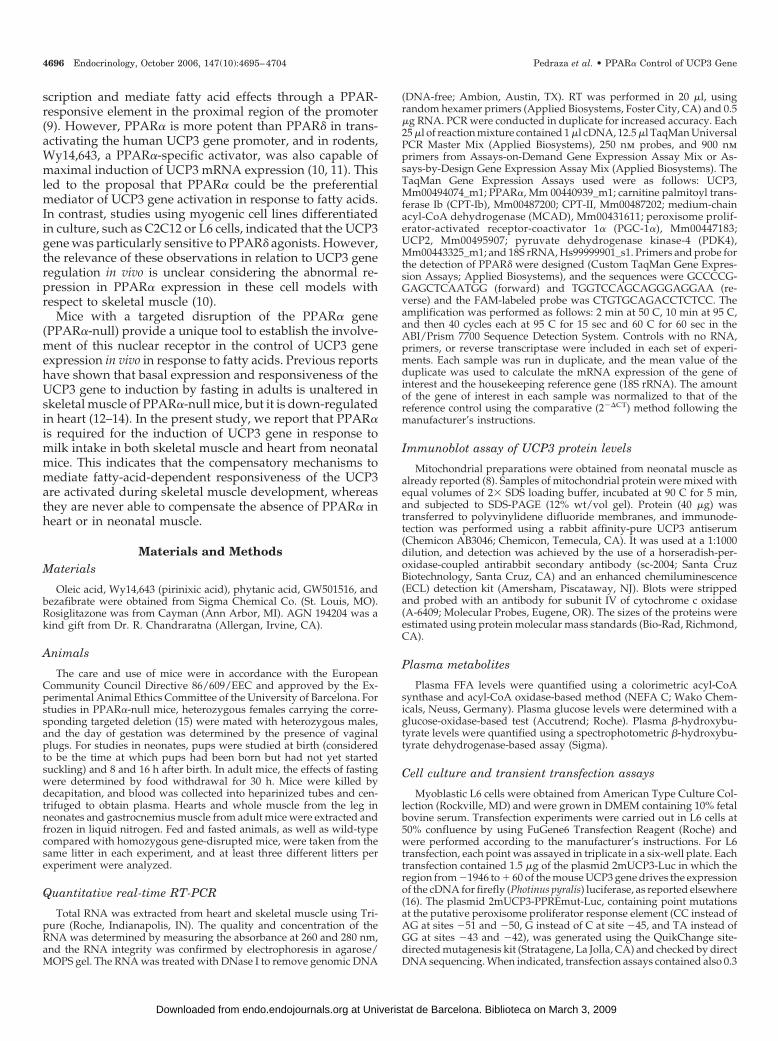
To analyze the effects of the lack of expression of PPAR�in skeletal muscle on the regulation of UCP3 gene, UCP3mRNA levels were measured in gastrocnemius skeletal mus-cle in adult PPAR�-null mice in comparison with their con-trol littermates (Fig. 1A). The results showed that under fedconditions, UCP3 mRNA levels were similar between bothgenotypes. The increase in UCP3 mRNA in response to 30 hof starvation was also similar in control and PPAR�-nullmice (around 5-fold). The increase of UCP3 mRNA levelsresulting from starvation was also not impaired in tibialisanterior or soleus muscles from PPAR�-null mice (data notshown).
UCP3 mRNA levels were analyzed in heart under the
same experimental conditions. UCP3 mRNA abundance wassignificantly reduced (around 40%) in fed PPAR�-null micewith respect to fed controls (Fig. 1B). When mice werestarved for 30 h, the levels of UCP3 mRNA in heart increasedmore than 7-fold in controls, whereas in PPAR�-null mice,the induction was significantly lower with respect to fedPPAR�-null mice (around 5-fold), and the total amount ofUCP3 mRNA was just 30% of the levels reached in the heartof the starved control littermates. These findings are similarto other reports (12, 13) and had been interpreted either asindicating a minor role for PPAR� in the control of skeletalmuscle UCP3 gene expression in vivo or as evidence of theexistence of compensatory mechanisms acting preferentiallyin muscle to maintain normal regulation of UCP3 geneexpression.
To analyze the alterations in circulating metabolites re-lated to energy metabolism, glucose, FFA, and ketone bodies(�-hydroxybutyrate) were determined in plasma (Table 1).Although glucose levels were similar in wild-type andPPAR�-null mice in fed conditions and the levels of glucosedecrease in both genotypes because of starvation, in PPAR�-null mice, the levels of glucose were statistically lower thanin wild-type mice. FFA levels were similar under fed con-
FIG. 1. UCP3 mRNA levels in skeletal muscle and heart from wild-type and PPAR�-null adult mice. Eight-week-old mice were used inbasal conditions (fed) or under 30 h of starvation (fasted). Gastroc-nemius skeletal muscle or heart was isolated, and UCP3 mRNAexpression was analyzed by quantitative RT-PCR as described inMaterials and Methods. Bars indicate the mean � SEM of six to 10 miceper group coming from at least three different litters. Data are shownas fold induction with respect to values in wild-type fed mice. Sig-nificant differences between fed and fasted mice are shown: *, P �0.05; **, P � 0.01; comparison between wild-type and PPAR�-nullmice: #, P � 0.05.
Pedraza et al. • PPAR� Control of UCP3 Gene Endocrinology, October 2006, 147(10):4695–4704 4697
at Univeristat de Barcelona. Biblioteca on March 3, 2009 endo.endojournals.orgDownloaded from
ditions in both genotypes, but in response to starvation, FFAfrom PPAR�-null were significantly higher than in wild-typemice. �-Hydroxybutyrate levels showed no differences in fedconditions between wild-type and PPAR�-null mice. As ex-pected, �-hydroxybutyrate levels increased in response tostarvation, but this response was strongly impaired inPPAR�-null mice, being 60% less than that in their wild-typelittermates. The pattern of alterations in the metabolic re-sponse to starvation in PPAR�-null mice is in agreement withprevious reports on the characterization of the phenotype ofthese mice (21, 22).
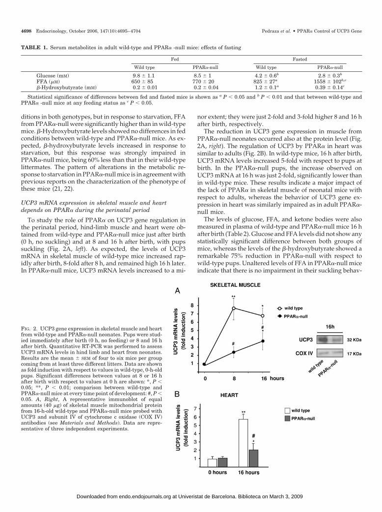
UCP3 mRNA expression in skeletal muscle and heartdepends on PPAR� during the perinatal period
To study the role of PPAR� on UCP3 gene regulation inthe perinatal period, hind-limb muscle and heart were ob-tained from wild-type and PPAR�-null mice just after birth(0 h, no suckling) and at 8 and 16 h after birth, with pupssuckling (Fig. 2A, left). As expected, the levels of UCP3mRNA in skeletal muscle of wild-type mice increased rap-idly after birth, 8-fold after 8 h, and remained high 16 h later.In PPAR�-null mice, UCP3 mRNA levels increased to a mi-
nor extent; they were just 2-fold and 3-fold higher 8 and 16 hafter birth, respectively.
The reduction in UCP3 gene expression in muscle fromPPAR�-null neonates occurred also at the protein level (Fig.2A, right). The regulation of UCP3 by PPAR� in heart wassimilar to adults (Fig. 2B). In wild-type mice, 16 h after birth,UCP3 mRNA levels increased 5-fold with respect to pups atbirth. In the PPAR�-null pups, the increase observed onUCP3 mRNA at 16 h was just 2-fold, significantly lower thanin wild-type mice. These results indicate a major impact ofthe lack of PPAR� in skeletal muscle of neonatal mice withrespect to adults, whereas the behavior of UCP3 gene ex-pression in heart was similarly impaired as in adult PPAR�-null mice.
The levels of glucose, FFA, and ketone bodies were alsomeasured in plasma of wild-type and PPAR�-null mice 16 hafter birth (Table 2). Glucose and FFA levels did not show anystatistically significant difference between both groups ofmice, whereas the levels of the �-hydroxybutyrate showed aremarkable 75% reduction in PPAR�-null with respect towild-type pups. Unaltered levels of FFA in PPAR�-null miceindicate that there is no impairment in their suckling behav-
TABLE 1. Serum metabolites in adult wild-type and PPAR� -null mice: effects of fasting
Fed Fasted
Wild type PPAR�-null Wild type PPAR�-null
Glucose (mM) 9.8 � 1.1 8.5 � 1 4.2 � 0.6b 2.8 � 0.3b
FFA (�M) 650 � 85 770 � 20 825 � 27a 1558 � 102b,c
�-Hydroxybutyrate (mM) 0.2 � 0.01 0.2 � 0.04 1.2 � 0.1a 0.39 � 0.14c
Statistical significance of differences between fed and fasted mice is shown as a P � 0.05 and b P � 0.01 and that between wild-type andPPAR� -null mice at any feeding status as c P � 0.05.
FIG. 2. UCP3 gene expression in skeletal muscle and heartfrom wild-type and PPAR�-null neonates. Pups were stud-ied immediately after birth (0 h, no feeding) or 8 and 16 hafter birth. Quantitative RT-PCR was performed to assessUCP3 mRNA levels in hind limb and heart from neonates.Results are the mean � SEM of four to six mice per groupcoming from at least three different litters. Data are shownas fold induction with respect to values in wild-type, 0-h-oldpups. Significant differences between values at 8 or 16 hafter birth with respect to values at 0 h are shown: *, P �0.05; **, P � 0.01; comparison between wild-type andPPAR�-null mice at every time point of development: #, P �0.05. A, Right, A representative immunoblot of equalamounts (40 �g) of skeletal muscle mitochondrial proteinfrom 16-h-old wild-type and PPAR�-null mice probed withUCP3 and subunit IV of cytochrome c oxidase (COX IV)antibodies (see Materials and Methods). Data are repre-sentative of three independent experiments.
4698 Endocrinology, October 2006, 147(10):4695–4704 Pedraza et al. • PPAR� Control of UCP3 Gene
at Univeristat de Barcelona. Biblioteca on March 3, 2009 endo.endojournals.orgDownloaded from
ior, because FFA in neonatal blood are highly dependent onthe extent of milk intake. Thus, lowered UCP3 mRNA inPPAR�-null pups could not be caused by the exposure ofskeletal muscle and heart to lower levels of circulating FFAand should be caused by a lack of responsiveness of thetissues. Similarly to adults, PPAR�-null pups showed low-ered circulating ketone bodies, which is indicative of a re-duced hepatic oxidation of fatty acids, in this case comingfrom milk intake. This is in agreement with the reportedimpairment in gene expression for ketogenic enzymes inperinatal liver of PPAR�-null mice (23).
PPAR� and PPAR� can mediate fatty-acid-dependentactivation of mouse UCP3 gene transcription
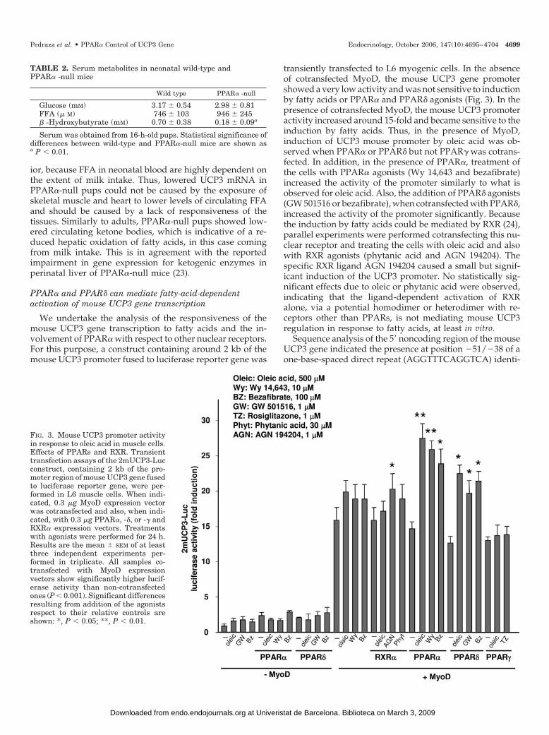
We undertake the analysis of the responsiveness of themouse UCP3 gene transcription to fatty acids and the in-volvement of PPAR� with respect to other nuclear receptors.For this purpose, a construct containing around 2 kb of themouse UCP3 promoter fused to luciferase reporter gene was
transiently transfected to L6 myogenic cells. In the absenceof cotransfected MyoD, the mouse UCP3 gene promotershowed a very low activity and was not sensitive to inductionby fatty acids or PPAR� and PPAR� agonists (Fig. 3). In thepresence of cotransfected MyoD, the mouse UCP3 promoteractivity increased around 15-fold and became sensitive to theinduction by fatty acids. Thus, in the presence of MyoD,induction of UCP3 mouse promoter by oleic acid was ob-served when PPAR� or PPAR� but not PPAR� was cotrans-fected. In addition, in the presence of PPAR�, treatment ofthe cells with PPAR� agonists (Wy 14,643 and bezafibrate)increased the activity of the promoter similarly to what isobserved for oleic acid. Also, the addition of PPAR� agonists(GW 501516 or bezafibrate), when cotransfected with PPAR�,increased the activity of the promoter significantly. Becausethe induction by fatty acids could be mediated by RXR (24),parallel experiments were performed cotransfecting this nu-clear receptor and treating the cells with oleic acid and alsowith RXR agonists (phytanic acid and AGN 194204). Thespecific RXR ligand AGN 194204 caused a small but signif-icant induction of the UCP3 promoter. No statistically sig-nificant effects due to oleic or phytanic acid were observed,indicating that the ligand-dependent activation of RXRalone, via a potential homodimer or heterodimer with re-ceptors other than PPARs, is not mediating mouse UCP3regulation in response to fatty acids, at least in vitro.
Sequence analysis of the 5� noncoding region of the mouseUCP3 gene indicated the presence at position �51/�38 of aone-base-spaced direct repeat (AGGTTTCAGGTCA) identi-
TABLE 2. Serum metabolites in neonatal wild-type andPPAR� -null mice
Wild type PPAR� -null
Glucose (mM) 3.17 � 0.54 2.98 � 0.81FFA (� M) 746 � 103 946 � 245� -Hydroxybutyrate (mM) 0.70 � 0.38 0.18 � 0.09a
Serum was obtained from 16-h-old pups. Statistical significance ofdifferences between wild-type and PPAR�-null mice are shown asa P � 0.01.
FIG. 3. Mouse UCP3 promoter activityin response to oleic acid in muscle cells.Effects of PPARs and RXR. Transienttransfection assays of the 2mUCP3-Lucconstruct, containing 2 kb of the pro-moter region of mouse UCP3 gene fusedto luciferase reporter gene, were per-formed in L6 muscle cells. When indi-cated, 0.3 �g MyoD expression vectorwas cotransfected and also, when indi-cated, with 0.3 �g PPAR�, -�, or -� andRXR� expression vectors. Treatmentswith agonists were performed for 24 h.Results are the mean � SEM of at leastthree independent experiments per-formed in triplicate. All samples co-transfected with MyoD expressionvectors show significantly higher lucif-erase activity than non-cotransfectedones (P � 0.001). Significant differencesresulting from addition of the agonistsrespect to their relative controls areshown: *, P � 0.05; **, P � 0.01.
Pedraza et al. • PPAR� Control of UCP3 Gene Endocrinology, October 2006, 147(10):4695–4704 4699
at Univeristat de Barcelona. Biblioteca on March 3, 2009 endo.endojournals.orgDownloaded from
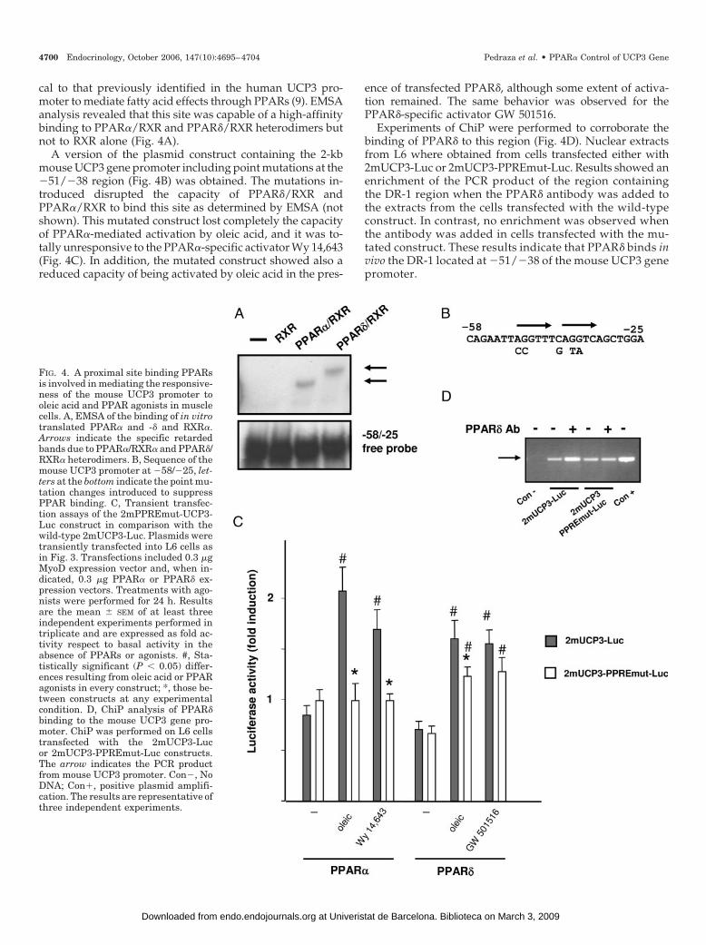
cal to that previously identified in the human UCP3 pro-moter to mediate fatty acid effects through PPARs (9). EMSAanalysis revealed that this site was capable of a high-affinitybinding to PPAR�/RXR and PPAR�/RXR heterodimers butnot to RXR alone (Fig. 4A).
A version of the plasmid construct containing the 2-kbmouse UCP3 gene promoter including point mutations at the�51/�38 region (Fig. 4B) was obtained. The mutations in-troduced disrupted the capacity of PPAR�/RXR andPPAR�/RXR to bind this site as determined by EMSA (notshown). This mutated construct lost completely the capacityof PPAR�-mediated activation by oleic acid, and it was to-tally unresponsive to the PPAR�-specific activator Wy 14,643(Fig. 4C). In addition, the mutated construct showed also areduced capacity of being activated by oleic acid in the pres-
ence of transfected PPAR�, although some extent of activa-tion remained. The same behavior was observed for thePPAR�-specific activator GW 501516.
Experiments of ChiP were performed to corroborate thebinding of PPAR� to this region (Fig. 4D). Nuclear extractsfrom L6 where obtained from cells transfected either with2mUCP3-Luc or 2mUCP3-PPREmut-Luc. Results showed anenrichment of the PCR product of the region containingthe DR-1 region when the PPAR� antibody was added tothe extracts from the cells transfected with the wild-typeconstruct. In contrast, no enrichment was observed whenthe antibody was added in cells transfected with the mu-tated construct. These results indicate that PPAR� binds invivo the DR-1 located at �51/�38 of the mouse UCP3 genepromoter.
FIG. 4. A proximal site binding PPARsis involved in mediating the responsive-ness of the mouse UCP3 promoter tooleic acid and PPAR agonists in musclecells. A, EMSA of the binding of in vitrotranslated PPAR� and -� and RXR�.Arrows indicate the specific retardedbands due to PPAR�/RXR� and PPAR�/RXR� heterodimers. B, Sequence of themouse UCP3 promoter at �58/�25, let-ters at the bottom indicate the point mu-tation changes introduced to suppressPPAR binding. C, Transient transfec-tion assays of the 2mPPREmut-UCP3-Luc construct in comparison with thewild-type 2mUCP3-Luc. Plasmids weretransiently transfected into L6 cells asin Fig. 3. Transfections included 0.3 �gMyoD expression vector and, when in-dicated, 0.3 �g PPAR� or PPAR� ex-pression vectors. Treatments with ago-nists were performed for 24 h. Resultsare the mean � SEM of at least threeindependent experiments performed intriplicate and are expressed as fold ac-tivity respect to basal activity in theabsence of PPARs or agonists. #, Sta-tistically significant (P � 0.05) differ-ences resulting from oleic acid or PPARagonists in every construct; *, those be-tween constructs at any experimentalcondition. D, ChiP analysis of PPAR�binding to the mouse UCP3 gene pro-moter. ChiP was performed on L6 cellstransfected with the 2mUCP3-Lucor 2mUCP3-PPREmut-Luc constructs.The arrow indicates the PCR productfrom mouse UCP3 promoter. Con�, NoDNA; Con�, positive plasmid amplifi-cation. The results are representative ofthree independent experiments.
4700 Endocrinology, October 2006, 147(10):4695–4704 Pedraza et al. • PPAR� Control of UCP3 Gene
at Univeristat de Barcelona. Biblioteca on March 3, 2009 endo.endojournals.orgDownloaded from
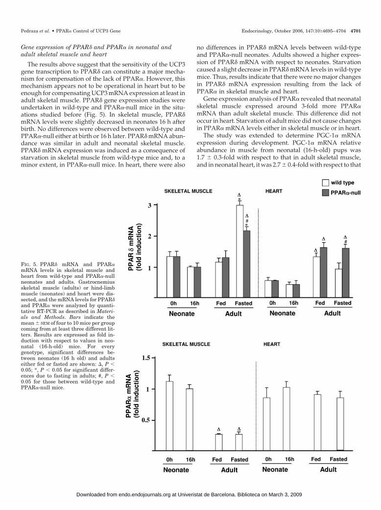
Gene expression of PPAR� and PPAR� in neonatal andadult skeletal muscle and heart
The results above suggest that the sensitivity of the UCP3gene transcription to PPAR� can constitute a major mecha-nism for compensation of the lack of PPAR�. However, thismechanism appears not to be operational in heart but to beenough for compensating UCP3 mRNA expression at least inadult skeletal muscle. PPAR� gene expression studies wereundertaken in wild-type and PPAR�-null mice in the situ-ations studied before (Fig. 5). In skeletal muscle, PPAR�mRNA levels were slightly decreased in neonates 16 h afterbirth. No differences were observed between wild-type andPPAR�-null either at birth or 16 h later. PPAR� mRNA abun-dance was similar in adult and neonatal skeletal muscle.PPAR� mRNA expression was induced as a consequence ofstarvation in skeletal muscle from wild-type mice and, to aminor extent, in PPAR�-null mice. In heart, there were also
no differences in PPAR� mRNA levels between wild-typeand PPAR�-null neonates. Adults showed a higher expres-sion of PPAR� mRNA with respect to neonates. Starvationcaused a slight decrease in PPAR� mRNA levels in wild-typemice. Thus, results indicate that there were no major changesin PPAR� mRNA expression resulting from the lack ofPPAR� in skeletal muscle and heart.
Gene expression analysis of PPAR� revealed that neonatalskeletal muscle expressed around 3-fold more PPAR�mRNA than adult skeletal muscle. This difference did notoccur in heart. Starvation of adult mice did not cause changesin PPAR� mRNA levels either in skeletal muscle or in heart.
The study was extended to determine PGC-1� mRNAexpression during development. PGC-1� mRNA relativeabundance in muscle from neonatal (16-h-old) pups was1.7 � 0.3-fold with respect to that in adult skeletal muscle,and in neonatal heart, it was 2.7 � 0.4-fold with respect to that
FIG. 5. PPAR� mRNA and PPAR�mRNA levels in skeletal muscle andheart from wild-type and PPAR�-nullneonates and adults. Gastrocnemiusskeletal muscle (adults) or hind-limbmuscle (neonates) and heart were dis-sected, and the mRNA levels for PPAR�and PPAR� were analyzed by quanti-tative RT-PCR as described in Materi-als and Methods. Bars indicate themean � SEM of four to 10 mice per groupcoming from at least three different lit-ters. Results are expressed as fold in-duction with respect to values in neo-natal (16-h-old) mice. For everygenotype, significant differences be-tween neonates (16 h old) and adultseither fed or fasted are shown: �, P �0.05; *, P � 0.05 for significant differ-ences due to fasting in adults; #, P �0.05 for those between wild-type andPPAR�-null mice.
Pedraza et al. • PPAR� Control of UCP3 Gene Endocrinology, October 2006, 147(10):4695–4704 4701
at Univeristat de Barcelona. Biblioteca on March 3, 2009 endo.endojournals.orgDownloaded from

in adult heart. Similar findings have been reported in cardiacmouse development (25). Levels of PGC-1� transcripts werehigher in heart than in skeletal muscle, in both neonates andadults. This is in agreement with previous reports in adultmice (26). PGC-1� transcript levels were not significantlymodified in muscle or heart from PPAR�-null mice, either inneonates or in adults (not shown).
Differential effects of PPAR� invalidation on the expressionof PPAR� and PPAR� target genes
To determine whether the reduction in UCP3 mRNA ex-pression in neonatal muscle and heart from PPAR�-null micecorresponds to a general impairment in the expression ofPPAR� target genes, additional gene expression analysis wasperformed on neonates. Genes involved in fatty acid catab-olism, especially those whose transcripts were up-regulatedin transgenic mouse models of overexpression of PPAR� inskeletal muscle, were analyzed (27). In wild-type mice, tran-script levels for UCP2, CPT-II, and PDK4 were dramaticallyup-regulated in heart from 16-h-old, suckling pups, withrespect to the those before initiation of suckling (0 h),whereas no postnatal induction was observed for CPT-I� orMCAD mRNA. In skeletal muscle, only UCP2 mRNA levelsshowed postnatal induction. In skeletal muscle and heartfrom PPAR�-null neonates, the postnatal induction of UCP2mRNA was impaired (Fig. 6). CPT-II mRNA and PDK4mRNA levels were down-regulated only in postnatal heartand unaffected in muscle. No effects were observed inCPT-I� or MCAD mRNA levels in skeletal muscle or heartfrom PPAR�-null mice, in accordance with the low sensi-tivity of these genes to PPAR�-dependent regulation in vivo(27). UCP2 mRNA, the only transcript lowered in postnatalmuscle, was not significantly altered in skeletal muscle fromadult PPAR�-null mice either in fed (1.2 � 0.4 1-fold change)or in fasted conditions (1.5 � 0.7-fold change). However, inheart, UCP2 mRNA levels were significantly reduced infasted (0.23 � 0.1-fold change with respect to wild-type, P �0.05) but not in fed (0.8 � 0.2-fold change with respect towild-type) PPAR�-null mice, similarly to UCP3 mRNA.
Discussion
In the present study, we describe a differential impact ofthe lack of PPAR� on UCP3 gene expression depending onthe tissue and the stage of development. Thus, UCP3 geneexpression in heart is systematically impaired both in neo-nates and adults, either fed or fasted, when mice lack PPAR�.In contrast, skeletal muscle from PPAR�-null adult miceshows unaltered UCP3 gene expression in basal conditionsand in response to starvation. However, because the increasein FFA concentration is considered the signal inducer forUCP3 mRNA expression in response to fasting in muscle, itis noteworthy that the higher increase in FFA levels observedin fasted PPAR�-null mice results just in an equal inductionof UCP3 mRNA with respect to wild-type mice. Previousdata in rodents under different physiological and experi-mental situations indicate a very close association betweenthe levels of circulating FFA and skeletal muscle UCP3mRNA levels (4) The differential relationship between FFAand UCP3 mRNA levels in PPAR�-null mice may indicate
that despite UCP3 mRNA levels being preserved, there issome extent of impairment or resistance in the responsive-ness of UCP3 gene expression to fatty acids because of thelack of PPAR�.
In contrast with adults, the UCP3 mRNA induction inneonates as a consequence of initiation of suckling is severelyimpaired not only in heart but also in skeletal muscle fromPPAR�-null mice. Lowered UCP3 mRNA in PPAR�-nullpups is not caused by lower levels of circulating FFA, whichwere unaltered. Thus, it appears that PPAR� is particularlyrequired for UCP3 regulation in response to fatty acids in theneonatal period.
FIG. 6. Gene expression analysis of genes involved in fatty acid me-tabolism in skeletal muscle and heart from wild-type and PPAR�-nullneonates. mRNA was isolated from hind limb and heart from neo-nates just after birth (0 h) or after 16 h being with their mothers (16h), and quantitative RT-PCR was performed as described in Materialsand Methods to analyze the levels of expression of UCP2, PDK4, CPTI�, CPT II, and MCAD. Results are shown as fold induction withrespect to values in wild-type mice at 0 h, and bars indicate themean � SEM of four to six mice per group. For every genotype, sig-nificant differences between neonates at 0 and 16 h are shown: *, P �0.05; #, significant differences between wild-type and PPAR�-nullmice at P � 0.05.
4702 Endocrinology, October 2006, 147(10):4695–4704 Pedraza et al. • PPAR� Control of UCP3 Gene
at Univeristat de Barcelona. Biblioteca on March 3, 2009 endo.endojournals.orgDownloaded from
The present findings on the transcriptional regulation ofthe mouse UCP3 gene promoter indicate a similar behaviorwith respect to the human promoter (9); it is dependent onMyoD, and responsiveness to fatty acids can be mediated byPPAR� or PPAR�. This is in contrast with the responsivenessof the UCP3 gene to thyroid hormones, which is remarkablydifferent between humans and mice (16). In fact, despite thelarge divergence in overall promoter sequence and func-tional structure between humans and mice (28), the one-base-spaced direct repeat previously identified in the humanUCP3 promoter to mediate fatty acid effects through PPARsis completely conserved in the mouse proximal promoterregion at position �51/�38. We found that, like in humans,it binds PPAR�/RXR and PPAR�/RXR heterodimers as wellas being involved in mediating the responsiveness to fattyacids via PPARs. This identical sequence is also present in theproximal region of the UCP3 genes from all the other mam-malian species in which the 5� noncoding region sequence isavailable in databases (accession number AF168989 for Rat-tus norvegicus, AY523564 for Phodopus sungorus, Nw876273for Canis familiaris, and Nw92833 for Bos taurus). However,it should be noted that in contrast with PPAR�, some re-sponsiveness to PPAR� remains in mutated constructs at thissite, thus indicating the possibility of additional mechanismsof PPAR�-dependent activation of the mouse UCP3 gene.
Overall, the present findings indicate that the requirementof PPAR� for the regulation of UCP3 gene expression istissue and development specific, and PPAR� plays an espe-cially critical role in the perinatal period. Compensatorymechanisms other than PPAR� activation for fatty-acid-de-pendent regulation of UCP3 gene transcription are present inadults to ensure normal UCP3 mRNA expression when mus-cle is exposed to high levels of circulating FFA, but they arenot active in adult heart or in the heart and skeletal muscleduring the neonatal period. Although other mechanisms offatty-acid-dependent regulation of transcription cannot beexcluded, PPAR� is a likely candidate to mediate this alter-ative pathway of regulation of UCP3 gene expression, con-sidering its activity on UCP3 gene promoter regulation inresponse to fatty acids. However, PPAR�-mediated compen-satory mechanisms of control of UCP3 gene expressionwould not require up-regulation of PPAR� mRNA levels,which are unaltered in different tissues and developmentalstages of mice lacking PPAR�. However, the high expressionof PPAR� mRNA in neonatal skeletal muscle with respect toadults may be consistent with a more critical requirement ofPPAR� to mediate gene expression responsiveness to fattyacids in muscle during the perinatal period.
The fact that a gene such as UCP2, sharing dual PPAR�-and PPAR�-dependent regulation like UCP3 (27, 29–31), alsoshows altered gene expression in heart and in neonatal mus-cle but not in adult muscle supports the notion of a lack ofcompensatory effects of PPAR�-dependent pathways inmuscle from neonates. However, the unaltered expression ofthe transcripts for PDK4 and CPT-II, which are also targetsof both PPAR� and PPAR� regulation in adult muscle (27, 29,31), suggests other potential mechanisms to explain the tis-sue- and development-dependent impact of PPAR� defi-ciency in the UCP3 and UCP2 genes. Only those genes in-volved in lipid catabolism whose expression is significantly
induced in muscle or heart when milk intake begins wereaffected in neonates by the lack of PPAR�. It cannot beexcluded that the differential origin and composition of fattyacids stimulating gene expression of UCP3 and UCP2 geneswhen they come from milk (neonates) or from lipolysis inwhite fat (starved adults) act differently on the PPAR�- andPPAR�-dependent mechanisms of regulation of gene expres-sion in muscle and heart. Another aspect to be considered isthe potential differential requirement of coactivators in mus-cle and heart from neonates with respect to adults to mediatePPAR-dependent regulation of UCP3 gene and other genesinvolved in lipid catabolism. Our present data do not indi-cate lower levels of PGC-1�, at least at the transcript level, inneonates with respect to adults, but the opposite. P300, an-other PPAR� coactivator involved in UCP3 promoter regu-lation in humans, has been reported to be more abundant inneonates than in adults (32). PGC-1� and p300 expression arehigher in heart than in skeletal muscle at any time of devel-opment. Thus, these data do not suggest that limitingamounts of these coactivators could explain the specific im-pairment of gene expression in skeletal muscle from PPAR�-null neonates or in heart with respect to muscle. However,an extensive analysis of developmental regulation in muscleand heart of the multiple coactivators of PPARs reported todate should be undertaken to settle this issue.
On the other hand, although the precise physiologicalfunction is still a matter of debate, UCP3 appears to be in-volved in metabolic handling of fatty acids in muscle andheart in a way that minimizes lipotoxicity and excessiveproduction of reactive oxygen species (33, 34). In fact, itspattern of regulation reported here in relation to neonataldevelopment and PPAR�-dependent pathways is shared inmuscle only by the other gene involved in similar functions,UCP2. Genes involved in lipid catabolism but without rela-tion to mitochondrial bioenergetics or reactive oxygen spe-cies regulation, such as PDK4 or CPT-II, are altered in parallelwith UCP genes only in heart. In this sense, modulation ofUCP3 gene expression through drugs acting on PPARs couldinfluence muscle oxidative metabolism and would be ex-pected to act in parallel upon UCP2 gene expression. UCP3gene promoter studies described above as well as studies invivo using transgenic mice overexpressing PPAR� or PPAR�in skeletal muscle indicate that both types of PPAR receptorscan induce UCP3 gene expression in adults (27, 29). Presentfindings indicate that in other physiological situations suchas early development, UCP3 is much more dependent ofPPAR� activation. Moreover, despite that UCP3 mRNA lev-els are lower in heart than in skeletal muscle, recent data areindicative of a relevant role of UCP3 in human heart inrelation to the partitioning of metabolic fuels (35), andpresent results point to PPAR� as the receptor unequivocallymediating UCP3 gene regulation in response to fatty acids orfibrates in rodent heart.
Acknowledgments
We thank Drs. S. Green, P. Grimaldi, R. Evans, and L. Fajas for kindlysupplying expression vectors and Dr. Chandraratna for AGN 194204.
Received February 21, 2006. Accepted July 11, 2006.
Pedraza et al. • PPAR� Control of UCP3 Gene Endocrinology, October 2006, 147(10):4695–4704 4703
at Univeristat de Barcelona. Biblioteca on March 3, 2009 endo.endojournals.orgDownloaded from
Address all correspondence and requests for reprints to: Dr. G.Solanes, Departament de Bioquımica i Biologia Molecular, Universitatde Barcelona, Avda Diagonal 645, E-08028 Barcelona, Spain. E mail:[email protected].
This work was supported by Grants SAF2002-03648 and SAF2005-01722 from Ministerio de Educacion y Ciencia and Grant C03/08 fromInstituto de Salud Carlos III, Ministerio de Sanidad y Consumo, Spain.
Disclosure of potential conflicts of interest: N.P., M.R., J.V., R.I., F.J.G.,G.S., and F.V. have nothing to declare.
References
1. Vidal-Puig A, Solanes G, Grujic D, Flier JS, Lowell BB 1997 UCP3: anuncoupling protein homologue expressed preferentially and abundantly inskeletal muscle and brown adipose tissue. Biochem Biophys Res Commun235:79–82
2. Garcia-Martinez C, Sibille B, Solanes G, Darimont C, Mace K, Villarroya F,Gomez-Foix AM 2001 Overexpression of UCP3 in cultured human musclelowers mitochondrial membrane potential, raises ATP/ADP ratio, and favorsfatty acid vs. glucose oxidation. FASEB J 15:2033–2035
3. Vidal-Puig AJ, Grujic D, Zhang CY, Hagen T, Boss O, Ido Y, Szczepanik A,Wade J, Mootha V, Cortright R, Muoio DM, Lowell BB 2000 Energy metab-olism in uncoupling protein 3 gene knockout mice. J Biol Chem 275:16258–16266
4. Brun S, Carmona MC, Mampel T, Vinas O, Giralt M, Iglesias R, VillarroyaF 1999 Uncoupling protein-3 gene expression in skeletal muscle during de-velopment is regulated by nutritional factors that alter circulating non-ester-ified fatty acids. FEBS Lett 453:205–209
5. Girard J, Ferre P, Pegorier JP, Duee PH 1992 Adaptations of glucose and fattyacid metabolism during perinatal period and suckling-weaning transition.Physiol Rev 72:507–562
6. Brun S, Carmona MC, Mampel T, Vinas O, Giralt M, Iglesias R, VillarroyaF 1999 Activators of peroxisome proliferator-activated receptor-� induce theexpression of the uncoupling protein-3 gene in skeletal muscle: a potentialmechanism for the lipid intake-dependent activation of uncoupling protein-3gene expression at birth. Diabetes 48:1217–1222
7. Gong DW, He Y, Karas M, Reitman M 1997 Uncoupling protein-3 is a me-diator of thermogenesis regulated by thyroid hormone, �3-adrenergic ago-nists, and leptin. J Biol Chem 272:24129–24132
8. Pedraza N, Solanes G, Carmona MC, Iglesias R, Vinas O, Mampel T,Vazquez M, Giralt M, Villarroya F 2000 Impaired expression of the uncou-pling protein-3 gene in skeletal muscle during lactation: fibrates and trogli-tazone reverse lactation-induced downregulation of the uncoupling protein-3gene. Diabetes 49:1224–1230
9. Solanes G, Pedraza N, Iglesias R, Giralt M, Villarroya F 2003 Functionalrelationship between MyoD and peroxisome proliferator-activated receptor-dependent regulatory pathways in the control of the human uncoupling pro-tein-3 gene transcription. Mol Endocrinol 17:1944–1958
10. Son C, Hosoda K, Matsuda J, Fujikura J, Yonemitsu S, Iwakura H, MasuzakiH, Ogawa Y, Hayashi T, Itoh H, Nishimura H, Inoue G, Yoshimasa Y, YamoriY, Nakao K 2001 Up-regulation of uncoupling protein 3 gene expression byfatty acids and agonists for PPARs in L6 myotubes. Endocrinology 142:4189–4194
11. Cabrero A, Alegret M, Sanchez R, Adzet T, Laguna JC, Vazquez M 2001Uncoupling protein-3 mRNA up-regulation in C2C12 myotubes after etomoxirtreatment. Biochim Biophys Acta 1532:195–202
12. Young ME, Patil S, Ying J, Depre C, Ahuja HS, Shipley GL, Stepkowski SM,Davies PJ, Taegtmeyer H 2001 Uncoupling protein 3 transcription is regulatedby peroxisome proliferator-activated receptor � in the adult rodent heart.FASEB J 15:833–845
13. Muoio DM, MacLean PS, Lang DB, Li S, Houmard JA, Way JM, Winegar DA,Corton JC, Dohm GL, Kraus WE 2002 Fatty acid homeostasis and inductionof lipid regulatory genes in skeletal muscles of peroxisome proliferator-acti-vated receptor (PPAR)� knock-out mice. Evidence for compensatory regula-tion by PPAR�. J Biol Chem 277:26089–26097
14. Murray AJ, Panagia M, Hauton D, Gibbons GF, Clarke K 2005 Plasma freefatty acids and peroxisome proliferator-activated receptor � in the control ofmyocardial uncoupling protein levels. Diabetes 54:3496–3502
15. Lee SS, Pineau T, Drago J, Lee EJ, Owens JW, Kroetz DL, Fernandez-Sal-guero PM, Westphal H, Gonzalez FJ 1995 Targeted disruption of the � isoform
of the peroxisome proliferator-activated receptor gene in mice results in abol-ishment of the pleiotropic effects of peroxisome proliferators. Mol Cell Biol15:3012–3022
16. Solanes G, Pedraza N, Calvo V, Vidal-Puig A, Lowell BB, Villarroya F 2005Thyroid hormones directly activate the expression of the human and mouseuncoupling protein-3 genes through a thyroid response element in the prox-imal promoter region. Biochem J 386:505–513
17. Crescenzi M, Fleming TP, Lassar AB, Weintraub H, Aaronson SA 1990 MyoDinduces growth arrest independent of differentiation in normal and trans-formed cells. Proc Natl Acad Sci USA 87:8442–8446
18. Kliewer SA, Forman BM, Blumberg B, Ong ES, Borgmeyer U, MangelsdorfDJ, Umesono K, Evans RM 1994 Differential expression and activation of afamily of murine peroxisome proliferator-activated receptors. Proc Natl AcadSci USA 91:7355–7359
19. Issemann I, Green S 1990 Activation of a member of the steroid hormonereceptor superfamily by peroxisome proliferators. Nature 347:645–650
20. Amri EZ, Bonino F, Ailhaud G, Abumrad NA, Grimaldi PA 1995 Cloning ofa protein that mediates transcriptional effects of fatty acids in preadipocytes.Homology to peroxisome proliferator-activated receptors. J Biol Chem 270:2367–2371
21. Kersten S, Seydoux J, Peters JM, Gonzalez FJ, Desvergne B, Wahli W 1999Peroxisome proliferator-activated receptor � mediates the adaptive responseto fasting. J Clin Invest 103:1489–1498
22. Leone TC, Weinheimer CJ, Kelly DP 1999 A critical role for the peroxisomeproliferator-activated receptor � (PPAR�) in the cellular fasting response: thePPAR�-null mouse as a model of fatty acid oxidation disorders. Proc Natl AcadSci USA 96:7473–7478
23. Yubero P, Hondares E, Carmona MC, Rossell M, Gonzalez FJ, Iglesias R,Giralt M, Villarroya F 2004 The developmental regulation of peroxisomeproliferator-activated receptor-� coactivator-1� expression in the liver is par-tially dissociated from the control of gluconeogenesis and lipid catabolism.Endocrinology 145:4268–4277
24. Goldstein JT, Dobrzyn A, Clagett-Dame M, Pike JW, DeLuca HF 2003 Iso-lation and characterization of unsaturated fatty acids as natural ligands for theretinoid-X receptor. Arch Biochem Biophys 420:185–193
25. Lehman JJ, Barger PM, Kovacs A, Saffitz JE, Medeiros DM, Kelly DP Per-oxisome proliferator-activated receptor � coactivator-1 promotes cardiac mi-tochondrial biogenesis. J Clin Invest 106:847–856
26. Puigserver P, Wu Z, Park CW, Graves R, Wright M, Spiegelman BM 1998 Acold-inducible coactivator of nuclear receptors linked to adaptive thermogen-esis. Cell 92:829–839
27. Finck BN, Bernal-Mizrachi C, Han DH, Coleman T, Sambandam N,LaRiviere LL, Holloszy JO, Semenkovich CF, Kelly DP 2005 A potential linkbetween muscle peroxisome proliferator-activated receptor-� signaling andobesity-related diabetes. Cell Metab 1:133–144
28. Esterbauer H, Oberkofler H, Krempler F, Strosberg AD, Patsch W 2000 Theuncoupling protein-3 gene is transcribed from tissue-specific promoters inhumans but not in rodents. J Biol Chem 275:36394–36399
29. Wang YX, Zhang CL, Yu RT, Cho HK, Nelson MC, Bayuga-Ocampo CR, HamJ, Kang H, Evans RM 2004 Regulation of muscle fiber type and runningendurance by PPAR�. PLoS Biol 2:e294
30. Luquet S, Lopez-Soriano J, Holst D, Fredenrich A, Melki J, RassoulzadeganM, Grimaldi PA 2003 Peroxisome proliferator-activated receptor � controlsmuscle development and oxidative capability. FASEB J 17:2299–2301
31. Tanaka T, Yamamoto J, Iwasaki S, Asaba H, Hamura H, Ikeda Y, WatanabeM, Magoori K, Ioka RX, Tachibana K, Watanabe Y, Uchiyama Y, Sumi K,Iguchi H, Ito S, Doi T, Hamakubo T, Naito M, Auwerx J, Yanagisawa M,Kodama T, Sakai J 2003 Activation of peroxisome proliferator-activated re-ceptor � induces fatty acid �-oxidation in skeletal muscle and attenuatesmetabolic syndrome. Proc Natl Acad Sci USA 100:15924–15929
32. Li Q, Xiao H, Isobe K 2002 Histone acetyltransferase activities of cAMP-regulated enhancer-binding protein and p300 in tissues of fetal, young, and oldmice. J Gerontol A Biol Sci Med Sci 57:B93–B98
33. Hesselink MK, Schrauwen P 2005 Towards comprehension of the physio-logical role of UCP3. Horm Metab Res 37:550–554
34. MacLellan JD, Gerrits MF, Gowing A, Smith PJ, Wheeler MB, Harper ME2005 Physiological increases in uncoupling protein 3 augment fatty acid ox-idation and decrease reactive oxygen species production without uncouplingrespiration in muscle cells. Diabetes 54:2343–2350
35. Murray AJ, Anderson RE, Watson GC, Radda GK, Clarke K2004 Uncouplingproteins in human heart. Lancet 364:1786–1788
Endocrinology is published monthly by The Endocrine Society (http://www.endo-society.org), the foremost professional society serving theendocrine community.
4704 Endocrinology, October 2006, 147(10):4695–4704 Pedraza et al. • PPAR� Control of UCP3 Gene
at Univeristat de Barcelona. Biblioteca on March 3, 2009 endo.endojournals.orgDownloaded from