Large-Scale Spatial Distribution Patterns of Echinoderms in Nearshore Rocky Habitats

RESEARCH ARTICLE Open Access
Uncoupling of complex regulatory patterningduring evolution of larval development inechinodermsKristen A Yankura, Megan L Martik, Charlotte K Jennings, Veronica F Hinman*
Abstract
Background: Conservation of orthologous regulatory gene expression domains, especially along the neuroectodermalanterior-posterior axis, in animals as disparate as flies and vertebrates suggests that common patterning mechanismshave been conserved since the base of Bilateria. The homology of axial patterning is far less clear for the many marineanimals that undergo a radical transformation in body plan during metamorphosis. The embryos of these animals aremicroscopic, feeding within the plankton until they metamorphose into their adult forms.
Results: We describe here the localization of 14 transcription factors within the ectoderm during earlyembryogenesis in Patiria miniata, a sea star with an indirectly developing planktonic bipinnaria larva. We find thatthe animal-vegetal axis of this very simple embryo is surprisingly well patterned. Furthermore, the patterning thatwe observe throughout the ectoderm generally corresponds to that of “head/anterior brain” patterning known forhemichordates and vertebrates, which share a common ancestor with the sea star. While we suggest here thataspects of head/anterior brain patterning are generally conserved, we show that another suite of genes involved inretinal determination is absent from the ectoderm of these echinoderms and instead operates within themesoderm.
Conclusions: Our findings therefore extend, for the first time, evidence of a conserved axial pattering toechinoderm embryos exhibiting maximal indirect development. The dissociation of head/anterior brain patterningfrom “retinal specification” in echinoderm blastulae might reflect modular changes to a developmental generegulatory network within the ectoderm that facilitates the evolution of these microscopic larvae.
BackgroundThe astonishing diversity of animal forms, coupled withthe complex life histories typical of many marine inverte-brates, presents numerous challenges in inferring theancestral character of members of the closely relatedphyla collectively known as the deuterostomes. Modernmolecular phylogenies place four phyla within the mono-phyletic deuterostomes: Echinodermata and Hemichor-data comprise a distinct clade called the Ambulacraria[1-3] that is a sister group to Chordata [4]. Xenoturbellais a recent out-group addition to the Ambulacraria [5].Within the Ambulacraria, the free-swimming, bilater-
ally symmetric larvae of echinoderms, especially thebipinnaria larva of sea stars and the auricularia larva of
sea cucumbers, share many similarities with the tornarialarva of indirectly developing hemichordates. Thesemicroscopic larvae have an apical concentration of sero-tonergic neurons [6] and one or two concentrations, orbands, of cilia used to feed and swim in the plankton[7,8]. Neurons lie beneath this ciliated epithelium andinnervate the bands [9]. Similarities in larval form initi-ally provided the basis for many of the hypotheses sur-rounding the evolutionary origins of the chordates and,in particular, the centralized nervous system. Thesehypotheses, in which a microscopic larval stage isassumed ancestral to the entire deuterostome clade, pro-pose that a centralized nervous system evolved from aninfolding of the larval ciliary bands [10-12].Not all Ambulacrarians develop through a larval stage,
however, and recent comparisons of regulatory geneexpression have revealed that orthologs of many genes
* Correspondence: [email protected] of Biological Sciences, Carnegie Mellon University, Pittsburgh,PA 15213, USA
Yankura et al. BMC Biology 2010, 8:143http://www.biomedcentral.com/1741-7007/8/143
© 2010 Yankura et al; licensee BioMed Central Ltd. This is an Open Access article distributed under the terms of the Creative CommonsAttribution License (http://creativecommons.org/licenses/by/2.0), which permits unrestricted use, distribution, and reproduction inany medium, provided the original work is properly cited.

and signaling molecules involved in vertebrate neuralpatterning are expressed in spatially restricted domainsalong the anterior-posterior (AP) axis of the directdeveloping vermiform hemichordate juvenile Saccoglos-sus kowalevskii [13,14], a species that does not developvia a tornaria larva. This general correspondence in APposition of orthologs between the vertebrate and hemi-chordate nervous systems implies some homology inaxial patterning. In addition, the broad ectodermalexpression of these genes in hemichordates suggeststhat a diffuse panectodermal neural domain was theancestral state of the deuterostome nervous system andthat the centralization event occurred later within thelineage leading to the chordates. These findings andthose of other researchers (reviewed in [15,16]) thereforenegate the need to invoke a ciliated ancestor for deuter-ostomes as suggested by Garstang [10]. In addition, gen-eral homologies in axial expression patterns betweenvertebrates and direct developing protostomes havebeen observed as well [17], suggesting that common pat-terning mechanisms have been employed since theradiation of Bilaterians. Indirect developing larval formshave less obviously distinguished body axes and nostrong homology of axial patterning and, as a result,appear derived and possibly secondarily simplified incomparison.Here we examine the expression of regulatory gene
orthologs that have known or suspected roles in pat-terning the axial neuroectoderm of many protostomeand deuterostome embryos within the indirectly devel-oping sea star, Patiria miniata (previously Asterina min-iata), which forms a typical bipinnaria larva. We showthat these genes are expressed in diffuse concentricectodermal domains that pattern the early embryonicaxis. Furthermore, we observe in the sea star a generalcorrespondence of domains of orthologous gene expres-sion to those found along the AP and dorsal-ventral(DV) axis of direct developing deuterostomes. In addi-tion, we detect expression of retinal determining geneorthologs in the mesoderm of echinoderm larvae, butnot within the ectoderm of gastrulating embryos. Wediscuss the role of this implied modularity of regulatorypatterning during evolution.
Results and discussionIsolation of sea star transcription factorsP. miniata orthologs of regulatory genes that haveknown or suspected roles in patterning the axial neu-roectoderm of many protostome and deuterostomeembryos were isolated using a candidate gene approach.Recombinants for the following seven genes that encodehomeodomain proteins were obtained: retinal homeobox(rx), optix-like homeobox 3 (six3), gastrulation andbrain-specific homeobox (gbx), lim domain homeobox 2
(lhx2) and paired box homeobox 6 (pax6), as well asmembers of the Nkx gene family, nk2.1 and nk1. Wealso identified partial sequences of eyes absent (eya), theets family gene pea3, and two C2H2 zinc-finger genes,zic and krupple-like factor 13 (klf13). The following fourwinged-helix forkhead box genes were isolated: foxq2,foxj1, foxd and foxg. A complete list of orthologs,sequence lengths, and orthology of gene sequences isprovided in Additional file 1.
Animal-vegetal patterning of the sea star blastulaectodermSea star late blastulae have a morphologically distinctanimal-vegetal (AV) axis that is first readily observedwhen elongation of cells at the vegetal pole results in anoticeable thickening of epithelium termed the vegetalplate [18]. During gastrulation, the vegetal plate invagi-nates to produce the mesoderm and endoderm of thelarva, leaving the remaining animal epithelium asciliated ectoderm [18]. At this stage, no obvious mor-phological differences in ectodermal cell type have beenobserved, but we nonetheless reveal here a remarkablecomplexity of regulatory states within the ectoderm(summarized in Figure 1).Transcripts of sea star regulatory genes are localized
throughout the animal ectoderm in overlapping con-centric domains along the AV axis. Some of these tran-scripts, such as those of zic, foxq2, rx and nk2.1, arefound only in the animal-most ectoderm (Figures 2A-2D). Of these genes, zic appears to be most closely loca-lized to the animal pole, while expression of foxq2 and rxoverlaps with zic, yet extends further. Transcripts of six3and klf13 (Figures 2E and 2F) also are detected in the ani-mal-most ectoderm of blastulae; however, they show astill broader distribution. Although there is no clear cellmorphology that demarcates the boundary between theanimal ectoderm and the vegetal plate endomesoderm,we observe a ring of nk1 expression above the vegetalplate (Figure 2G) that partially overlaps with endoder-mally localized gatae transcripts (Figure 2H). The nk1expression domain therefore likely marks the vegetal-most ectoderm of the blastula. foxj1 and pea3 (Figures 2Iand 2J) are expressed throughout the ectoderm.Taken together, the spatial expression of these regula-
tory genes demonstrates that the ectoderm of the sea starblastula is patterned along the AV axis in at least fivenested concentric domains (summarized in Figure 2K).
Regulatory gene expression within the ectoderm of theciliary bands and animal pole domainDuring gastrulation, the ectoderm appears to undergovery little morphological change other than the coales-cence of cilia within two bands: a preoral ciliary bandthat loops above the opening of the mouth and a postoral
Yankura et al. BMC Biology 2010, 8:143http://www.biomedcentral.com/1741-7007/8/143
Page 2 of 10

ciliary band that loops below it and around the aboralsurface at the “back” of the embryo (Figure 3A) [7,18].Transcripts of several genes that were distributed broadlythroughout the ectoderm prior to gastrulation are laterexpressed within the ectoderm of the ciliary bands of the
larva following gastrulation (for example, foxj1 and klf13in Figures 3B-3E, pea3 as summarized in Figure 1 and aspreviously reported for otx and hnf-6/onecut expression[18,19]). At present, it is unclear if these patterns ofexpression reflect a migration of ectodermal cells to the
otxpax6
gbxhox1
hox3,4hox7/8
hox10/13
six3“foxg”
rx“nk2.1”
foxj1otx
hnf-6foxg
nk1, gbx
zic, foxq2, rx, six3, pea3,nk2.1, klf13, foxj1,
foxd, lhx2
six3klf13
foxq2rx
nk2.1
nk1
foxj1pea3otx*
hnf-6*
zic
(a)
zicrxfoxj1
pea3otx*
hnf-6*klf13pax6 foxg
nk1gbx
foxgfoxdnk2.1
foxq2
six3
lhx2(c)(b)
(f)(d)
(g)
Mouth
zic, foxq2, rx, six3, pea3, pax6
foxj1pea3otx*
hnf-6*klf13foxglhx2
nk1gbx
foxdnk2.1lhx2
zic, foxq2, rx,nk2.1, klf13, foxj1
nk1
hnf-6
six3
foxglhx2
lhx2
otx
zic, foxq2,“foxg”, lhx2, “tbr”
rx, six3, pax6,“nk2.1”,
“nk1”hox1hox3hox4hox8
hox10-13
“pea3”“hnf-6”
MR1R2R3R4R5R6R7R8
F
gbx
foxj1
(e)
Figure 1 Comparison of orthologous neuroectodermal gene expression domains among the deuterostomes. (A-E) Indirectly developingechinoderms. (F) Directly developing hemichordate. (G) Generalized vertebrate. Sea stars (Figures 1A-1C) and sea urchins (Figures 1D and 1E) areviewed laterally; animal pole is up and oral side is right. Figures 1F and 1G are dorsal views; anterior is up. Genes are listed beside their cognateexpression domains. Vertical bars in Figures 1A, 1B, 1F and 1G approximate domain boundaries. The orange to yellow gradient in Figures 1A, 1Fand 1G reflects a general conservation of anterior (animal)-most axial patterning among the three phyla. (A) Nested, concentric expressiondomains pattern the animal-vegetal (AV) axis of blastulae; asterisks denote previously reported expression [18,19]. (B) Concentric domains of zic,foxq2, rx and six3 persist in gastrulae (orange to peach gradient); additional oral (for example, foxg, foxd and gbx; light orange) and aboral (forexample, lhx2; purple) domains are evident. Genes (left) are broadly expressed. (C) Expression in larval animal pole domain (orange to peach)and/or ciliary bands (gold). (D) Sea urchin animal pole (orange and light orange), ciliary band (gold), aboral ectoderm (turquoise) and oralectoderm (foxg; gray) are molecularly distinct territories in blastulae. (E) Expression is maintained in gastrulae animal pole (orange) and ciliaryband (gold). (D and E) Pink circles represent skeletogenic mesoderm. See references [20,22,23,41-49]. (F) Orthologs expressed in hemichordateanterior, middle and posterior body segments show corresponding expression in the vertebrate forebrain, midbrain and hindbrain, respectively;data are summarized from Lowe et al. [13]. (G) Expression in generalized vertebrate centralized nervous system. F, forebrain; M, midbrain; R1-R8,rhombomeres of hindbrain. zic [50]; pea3 [51]; hnf-6/onecut [52]; and tbr [53]; foxj1 [54]; hox genes [55]. See references [24] and [26-32].Echinoderm gene names (quotations) are substituted for simplicity in Figures 1F and 1G.
Yankura et al. BMC Biology 2010, 8:143http://www.biomedcentral.com/1741-7007/8/143
Page 3 of 10

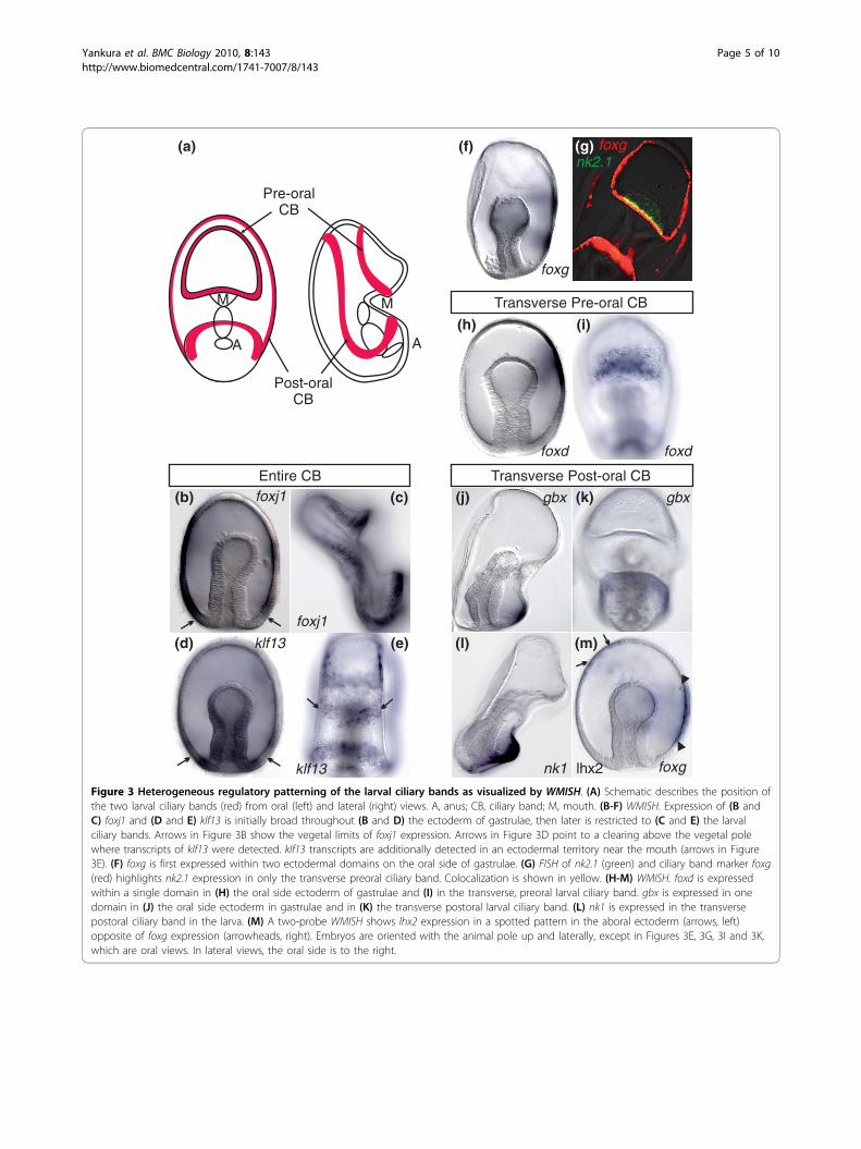
sites of the future ciliary bands or if there is another pat-terning mechanism that restricts the earlier broad expres-sion. Other transcripts are first detected at this stagewithin the ectoderm on the oral side of the gastrula andthen later within the ciliary bands (for example, tran-scripts of foxg, foxd and gbx; Figures 3F and 3G, 3H and3I, and 3J and 3K, respectively). A two-probe wholemount in situ hybridization (WMISH) of foxg and lhx2, agene localized to the aboral ectoderm, further highlightsthe oral side restriction of foxg transcripts in the gastrula(Figure 3).The expression patterns at this later stage also show
that the regulatory state of the early larval ciliary bandsis heterogeneous, for example, nk2.1 and foxd areexpressed in part of the preoral ciliary band directlyabove the mouth (Figures 3G and 3I, respectively), whilegbx and nk1 are localized to part of the postoral ciliaryband below the mouth (Figures 3K and 3L, respectively).Therefore, while the regulatory state of the ciliary bandectoderm can be defined by a suite of transcription fac-tors (that is, klf13, foxj1, pea3, foxg, otx and hnf-6/one-cut), they are further subdivided into pre- and postoralregions on the basis of the localization of foxd, nk2.1,nk1 and gbx.
Other transcripts that we detected within the ecto-derm of the blastula remained within the animal ecto-derm as gastrulation proceeded in what we define hereas the animal pole domain. Unlike sea urchins, the seastar, P. miniata, does not appear to have a morphologi-cally distinct animal pole domain at this stage. Tran-scripts of foxq2, pax6 and pea3 (Figures 4A-4C) tightlylocalize to the animal pole ectoderm, although theirvegetal boundaries do not exactly coincide. Transcriptsof zic, rx and six3 are expressed within the animal poledomain as well, but even more vegetally throughout theanimal ectoderm (Figures 4D-4F). The vegetal boundaryof the animal pole domain therefore is not clearlydefined by regulatory gene expression. The preoral andpostoral ciliary bands run through the sea star animalpole domain as demonstrated by a two-probe fluores-cence in situ hybridization (FISH) using the ciliary bandmarker, foxg, and the animal pole domain gene, pax6(Figure 4G). Thus, despite its lack of morphologicalregionalization, the animal pole has a distinct regulatorystate, as defined by foxq2, pax6, pea3, zic, rx and six3expression, suggesting that it is a unique territory withinthe sea star. It is not yet clear whether these genesremain expressed in all cells of the sea star animal pole
Figure 2 Nested concentric expression domains pattern the axial ectoderm of sea star, P. miniata, blastulae. Embryos are oriented withthe animal pole up. (A-G) Whole mount in situ hybridization (WMISH). (A) zic, (B) foxq2, (C) rx, and (D) nk2.1 expression is restricted to theanimal-most ectoderm. Transcripts of (E) six3 and (F) klf13 are detected in the ectoderm and in the vegetal plate endomesoderm. Arrows in (E)and (F) point to a clearing above the vegetal pole where no or few transcripts are detected. (G) nk1 transcripts are localized to a ring above thevegetal pole. (H) The boundary between the vegetal-most ectoderm (nk1, red) and the endoderm (gatae, green) as visualized by fluorescence insitu hybridization (FISH). Colocalization is in yellow. (I and J) WMISH. Transcripts of (I) foxj1 and (J) pea3 are detected throughout the entireectoderm. pea3 is weakly detected in the vegetal plate endomesoderm. Arrows in Figure 1J point to the limits of foxj1 expression. (K) Schematicshows the patterns described above as five nested domains of expression along the AV axis. For simplicity, nk2.1 is grouped here with theconcentric domains of foxq2 and rx expression. Gene names are listed next to their cognate expression domains. Vertical bars approximate theexpression boundaries of associated genes. The color gradient spans the animal (orange) to vegetal (yellow) limits of the ectoderm.
Yankura et al. BMC Biology 2010, 8:143http://www.biomedcentral.com/1741-7007/8/143
Page 4 of 10
Pre-oralCB
Post-oralCB
M
A
M
A
(a)
foxj1
*foxj1
klf13
(b)
(d)
Entire CB
(c)
nk2.1foxg
Transverse Pre-oral CB
Transverse Post-oral CB
gbx(j)
nk1
(g)
foxj1
(h)
foxd
(f)
foxg
(l) (m)
lhx2 foxgklf13
(e)
foxd
(i)
gbx(k)
Figure 3 Heterogeneous regulatory patterning of the larval ciliary bands as visualized by WMISH. (A) Schematic describes the position ofthe two larval ciliary bands (red) from oral (left) and lateral (right) views. A, anus; CB, ciliary band; M, mouth. (B-F) WMISH. Expression of (B andC) foxj1 and (D and E) klf13 is initially broad throughout (B and D) the ectoderm of gastrulae, then later is restricted to (C and E) the larvalciliary bands. Arrows in Figure 3B show the vegetal limits of foxj1 expression. Arrows in Figure 3D point to a clearing above the vegetal polewhere transcripts of klf13 were detected. klf13 transcripts are additionally detected in an ectodermal territory near the mouth (arrows in Figure3E). (F) foxg is first expressed within two ectodermal domains on the oral side of gastrulae. (G) FISH of nk2.1 (green) and ciliary band marker foxg(red) highlights nk2.1 expression in only the transverse preoral ciliary band. Colocalization is shown in yellow. (H-M) WMISH. foxd is expressedwithin a single domain in (H) the oral side ectoderm of gastrulae and (I) in the transverse, preoral larval ciliary band. gbx is expressed in onedomain in (J) the oral side ectoderm in gastrulae and in (K) the transverse postoral larval ciliary band. (L) nk1 is expressed in the transversepostoral ciliary band in the larva. (M) A two-probe WMISH shows lhx2 expression in a spotted pattern in the aboral ectoderm (arrows, left)opposite of foxg expression (arrowheads, right). Embryos are oriented with the animal pole up and laterally, except in Figures 3E, 3G, 3I and 3K,which are oral views. In lateral views, the oral side is to the right.
Yankura et al. BMC Biology 2010, 8:143http://www.biomedcentral.com/1741-7007/8/143
Page 5 of 10

domain during later stages of larval development or ifexpression becomes refined to only subsets of cellswithin this domain.
Comparisons of ectodermal patterning between seaurchin and sea star embryosAt first inspection, the expression patterns of manygenes appear markedly different in the earlier blastulastages of sea urchin and sea star embryos. The laterrestrictions within the animal pole or ciliary bands are,with some exceptions, more similar (Figure 1). We sug-gest that the sea urchin embryo may simply undergo arelatively more rapid specification of these territories,with an associated loss of intermediate domains that weobserve in the sea star. Indeed, a careful examination ofexpression patterns in sea urchin has recently shownthat the apical plate in sea urchin consists of at leasttwo regulatory domains: an inner animal pole domain
flanked by a ring of six3 expression [20]. These twodomains in the sea urchin hatched blastula may there-fore represent a more apically compressed version of thenested, concentric regulatory domains found in the seastar blastula.Some of the patterning differences between sea urchin
and sea star ectoderm also seem to account for the dif-ferences observed in the localization of the pan-neuronalmarker, synaptotagmin-B [21]. For example, similar tothe patterns of gene expression that we describe here,synaptotagmin-B is detected broadly throughout theectoderm of the sea star gastrula, but in the larva it isfound primarily in neurons associated with the ciliarybands and animal pole [9]. In the sea urchin, however,synaptotagmin-B is already localized to the animal poledomain and the presumptive ciliary band by the gastrulastage [9].Although expression of many of the genes within the
ciliary bands of the sea star appears conserved in the seaurchin, nk2.1 and foxd show clear differences in expres-sion that may be associated with the evolutionary transi-tion from a double looping of the ciliary band aroundthe body of the sea star bipinnaria and hemichordatetornaria to a single looping of the ciliary band observedin sea urchins. This single ciliary band in the sea urchindevelops at the junction between oral and aboral ecto-derm. nk2.1 and foxd are expressed in part of the pre-oral ciliary band of the oral hood of the sea star, whilesea urchin orthologs of these are found in the animalplate ectoderm (compare Figures 1C-1E). Interestingly,both of these genes in sea urchin appear enriched onthe oral side of the embryos [22,23]. Thus, we speculatethat the preoral ciliary band may have been compressedinto the oral-side animal plate territory in sea urchinsand that this region within the sea urchin may thereforeconstitute a different territory than the remaining ani-mal plate.
Conservation of anterior (animal)-most regulatorypatterning with other deuterostomesComparisons of the regulatory gene expression patternsthat we observed in these indirectly developing sea starembryos with those known in directly developing bilater-ians illuminate additional surprising patterns of conserva-tion. We observe a general mapping of gene expressionpatterns along body axes (compare Figures 1A-1C withFigures 1F-1G). For instance, in the sea star, foxq2, rx,pax6 and six3 orthologs are apically expressed within theectoderm. foxq2 expression in the amphioxus, a basalchordate, is restricted to the anterior-most end of theembryo [24]. Orthologs of rx and pax6 are expressed inthe anterior-most neuroectoderm in the hemichordateSaccoglossus [13], and they also pattern the anteriorlylocalized eye primordium in vertebrates [25,26]. The
foxgpax6
zic rx
six3
pax6foxq2
(d) (e)
(g)
(b)(a)
pea3
(f)
(c)
Figure 4 Gene expression molecularly defines the animal poledomain in the sea star. Embryos are shown laterally, with theanimal pole up and oral side to the right. (A-F) WMISH. Expressionof (A) foxq2, (B) pax6, (C) pea3, (D) zic, (E) rx and (F) six3 within theapical-most ectoderm defines the animal pole domain within lategastrulae (Figures 4A, 4D and 4F) and early larvae (Figures 4B, 4Cand 4E). The vegetal limits of this domain are variable (see dottedlines in Figures 4E and 4F). Transcripts of pea3 additionally localizewithin the ectoderm of the larval ciliary bands (arrows in Figure 4C).pax6 expression in mesodermally derived coelom (arrow in Figure4B). (G) FISH demonstrates that the ciliary bands, as marked by foxg(red), run through the ectoderm of the animal pole domain, asmarked by pax6 (green). Colocalization is shown in yellow.
Yankura et al. BMC Biology 2010, 8:143http://www.biomedcentral.com/1741-7007/8/143
Page 6 of 10

Drosophila rx ortholog is required for brain development[27]. Orthologs of six3 and otx are expressed in anteriorneuroectoderm in members of all three deuterostomephyla [13,28,29]. The most vegetal ectoderm in sea starsis characterized by the presence of nk1 and gbx tran-scripts. In vertebrates, a gbx ortholog establishes the mid-brain-hindbrain boundary [30]. The zebrafish ortholog ofnk1, sax2, is expressed within the midbrain-hindbrainboundary as well, although its expression is not exclusiveto this territory [31]. Expression of nk1 and gbx in seastars, and possibly sea urchins, marks the vegetal (poster-ior)-most ectoderm.There is some evidence of additional conservation
between the DV and oral-aboral axes as well. The mouseortholog of nk2.1 (nkx2.1) is involved in the formation ofmotor neurons in the ventral telencephalon [32]. Sacco-glossus nk2.1 orthologs also show a ventral bias in expres-sion [13]. Furthermore, foxg plays a role in ventralforebrain development, while lhx2 specifies dorsal telence-phalic fates [32]. We similarly show that expression of seastar orthologs of foxg and nk2.1 is restricted to the oral(ventral) ectoderm, while lhx2 orthologs are expressedwithin the aboral (dorsal) ectoderm (Figure 1B).While in these comparisons we do not intend to con-
vey a tight homology in gene expression patterns acrossdeuterostome phyla, we predict that similarities in theoverall patterning are an ancestral innovation and per-haps evidence of maintenance of some elements of adevelopmental gene regulatory network (GRN) inheritedfrom a common ancestor. Conservation, however, is notmaintained for orthologs of genes expressed withinregions posterior to the midbrain-hindbrain boundary inchordates and hemichordates as nk1 marks the vegetal-most ectoderm. Also, the overlapping expression of hoxgene orthologs needed to pattern the posterior of manyembryos are found only later in echinoderm develop-ment within the mesoderm of the rudiment [33].
Separation of “retinal” from “anterior neural” regulatorypatterningVertebrate orthologs of transcription factors such aspax6, six3 and rx play known roles in pattering and spe-cifying anterior vertebrate sensory systems, most notablythe eyes [25,26,34]. Furthermore, orthologs of pax6, thesix gene family members and eya operate in a similargene network for retinal determination in both verte-brates and Drosophila (as reviewed in [35]).Having established that orthologs of many regulatory
genes involved in anterior neural specification are alsoexpressed within the anterior ectoderm of sea starembryos, we sought to determine if orthologs of tran-scription factors involved within the retinal determina-tion network are also expressed within echinodermembryos.
We have already shown that the sea star pax6 orthologis expressed within the animal pole domain (Figure 4B),although it is not expressed within the ectoderm of thesea urchin embryo (Figure 5). Transcripts of both pax6and eya in both sea urchins and sea stars, however, aredetected in the mesoderm of midgastrulae and thenmore prominently in one mesodermally derived coelomin late gastrulae (Figures 5A-5D and Figures 5G-5L).While we were unable to obtain a six1.2 ortholog fromthe sea star, this gene is expressed also within the meso-dermal coelom in the sea urchin (Figures 5E and 5F).In the sea urchin, expression of two members of the
light-sensing rhodopsin family of G-coupled proteinreceptors, opsin1 and opsin4, has been shown as early as1 week [36]. We were unable to obtain sea star opsinsequences; however, we confirmed the expression ofopsin1 and opsin4 1-week-old sea urchin larvae (Figures5M and 5N). The morphology of the late larval seaurchin embryos makes it difficult to decipher the preciselocation of these transcripts within the embryo. Wetherefore sought to determine if opsins collocalize witheya, which we show is expressed likely within one orboth coeloms (depending on developmental timing) in1-week-old larvae (Figure 5O). Using a two-probe FISH,we observe that transcripts of opsin1 colocalized withthose of eya in 1-week-old sea urchin larvae (Figures 5P-5R). Expression of retinal determination orthologs withinthe mesoderm of gastrulae and larvae allow for the possi-bility that these genes operate within a common GRN.The tightly coupled GRNs for anterior neural and
visual sensory structures that are found in vertebratesand also in invertebrates, such as Drosophila, thereforeare spatially separated in echinoderms. The presence ofgene transcripts of pax6, rx and six3, but not, for exam-ple, eya, within the animal ectoderm of sea star bipin-naria larva may indicate a partial retention of anancestral retinal determination network that once oper-ated within this embryonic territory. This might alsoexplain the absence of apically localized rhabdomericeyespots, which are characteristic of the indirectly devel-oping tornaria larvae of some hemichordates but werelikely lost in the echinoderm lineage [16].
ConclusionsInferring ancestral statesThe detailed expression analyses reported here supportthe hypothesis that indirectly developing planktonicechinoderm embryos likely utilize ancient regulatorymechanisms for various anterior neuroectodermal and/or sensory developmental processes that are potentiallyconserved throughout the Bilateria. Compared to verte-brates and well-studied protostome model organismssuch as Drosophila, however, echinoderm embryos sepa-rate the deployment of these subcircuits in space and/or
Yankura et al. BMC Biology 2010, 8:143http://www.biomedcentral.com/1741-7007/8/143
Page 7 of 10

time. Thus, echinoderm embryos may have conservedsets of genetic regulatory relationships for “head/anteriorbrain” within the ectoderm of the early blastula andothers for “retinal determination” within the mesoder-mal coelom of the gastrula.Much of the difficulty in inferring the ancestral state
of the deuterostomes and the mysteries of the origin ofthe phylum to which we belong arises from the com-plex life histories found within extant lineages [37].Given the conservation of complex sensory and APpatterning between protostomes such as Drosophila
and vertebrates, the parsimonious explanation is thatancestral developmental regulatory interactions, per-haps even entire GRN subcircuits, have beenuncoupled along the lineage of echinoderms, possiblycoincident with a simplification in early development.However, until a greater breath of taxa have resolvedGRNs, we cannot know the flexibility with which mod-ular subcircuits can be deployed during evolution ofalternative body plans or if intercalation of GRN sub-circuits occurs independently or coincident with anincrease in complexity.
Pm
Eya
SpS
ix1.
2S
pPax
6S
pEya
(a) (b)
(c) (d)
(e) (f)
(g) (h) (i)
(j) (k) (l)
(q)
SpEyaSpOpsin1SpEya
SpOpsin1
(p) (r)SpOpsin1
(m) (n)
SpOpsin4
Pm
Pax
6
(o)
SpSm50SpEya
Figure 5 Retinal determination orthologs are expressed within sea urchin, (Strongylocentrotus purpuratus, Sp), and sea star mesoderm.(A-N) WMISH. (A and B) SpEya, (C and D) SpPax6 and (E and F) SpSix1.2 are expressed at the tip of the archenteron in gastrulae (Figures 5A, 5Cand 5E) and in a mesodermally derived coelom in early pluteus larvae (Figures 5B, 5D and 5F). PmEya (Figures 5G-5I) and PmPax6 (Figures 5J-5L)expression is first detected in the mesoderm of the archenteron bulb in midgastrulae (Figures 5H and 5K; arrows) and then more prominently ina mesodermally derived coelom in late gastrulae (Figures 5I and 5L; arrow). PmPax6 transcripts are also found broadly throughout the ectoderm(Figure 5K), with more pronounced expression in the apical ectoderm (Figure 5L). SpOpsin1 (Figure 5M) and SpOpsin4 (Figure 5N) are expressedin 1-week-old larvae. (O-R) FISH in 1-week-old larvae. SpEya (Figure 5O) is shown relative to the skeletal marker SpSm50, which was used toorient the embryo. Figures 5P-5R show transcripts of SpOpsin1 (green) colocalizing with those of SpEya (red). Colocalization is shown in yellow.Arrows in Figures 5P, 5Q and 5R point to expression. Additional areas of expression within the 1-week-old larvae may be a result of nonspecificstaining.
Yankura et al. BMC Biology 2010, 8:143http://www.biomedcentral.com/1741-7007/8/143
Page 8 of 10

MethodsSea star and sea urchin embryo culture andcharacterization of gene expressionP. miniata embryos, previously named Asterina miniata,and Strongylocentrotus purpuratus embryos were cul-tured as described previously [18,38]. Partial genesequences were obtained via screening a 3-day-old (late-gastrula stage) P. miniata arrayed cDNA library usingS. purpuratus sequence-specific probes and low strin-gency conditions as previously described [39]. Wholemount in situ hybridization (WMISH) was performed asdescribed previously [18].
Two-color FISHWMISH was performed essentially as described by Hin-man et al. [18], with modifications to detect riboprobesusing fluorescence as described by Denkers et al. [40].In brief, both digoxygenin (DIG) and 2,4-dinitrophenol(DNP) labeled riboprobes were used. Hybridized probeswere detected using anti-DIG antibody (1:2,000; Roche:Indianapolis, IN, USA) and anti-DNP antibody (1:1,000;PerkinElmer: Chicago, IL, USA), both conjugated tohorseradish peroxidase, and the Tyramide Signal Ampli-fication (TSA) Plus Fluorescence Systems Kit (PerkinEl-mer). A CyIII- or fluorescein-labeled tyramide wasdeposited near the in situ riboprobe in a reaction cata-lyzed by horseradish peroxidase, allowing for fluores-cence detection of DIG- and DNP-labeled riboprobes.
Additional material
Additional file 1: Table 1. List of sea star, P. miniata, orthologs andorthology of gene sequences.
AcknowledgementsThe authors thank Dr Daniel Brown, Brenna McCauley, Alys Cheatle andthree anonymous reviewers for helpful comments on the manuscript. Thiswork was partially supported by National Science Foundation grant 0844948to VFH, and CKJ and MLM were funded by a Howard Hughes MedicalInstitute Undergraduate Education Research Grant.
Authors’ contributionsVFH and KAY conceived of the study, participated in its design and draftedthe manuscript. KAY cloned sea star orthologs and performed WMISH andFISH. CKJ cloned sea urchin orthologs and performed WMISH. MLM clonedsea star orthologs and performed WMISH and FISH. All authors read andapproved the final manuscript.
Competing interestsThe authors declare that they have no competing interests.
Received: 5 August 2010 Accepted: 30 November 2010Published: 30 November 2010
References1. Bromham LD, Degnan BM: Hemichordates and deuterostome evolution:
robust molecular phylogenetic support for a hemichordate +echinoderm clade. Evol Dev 1999, 1:166-171.
2. Castresana J, Feldmaier-Fuchs G, Yokobori S, Satoh N, Paabo S: Themitochondrial genome of the hemichordate Balanoglossus carnosus andthe evolution of deuterostome mitochondria. Genetics 1998,150:1115-1123.
3. Cameron CB, Garey JR, Swalla BJ: Evolution of the chordate body plan:new insights from phylogenetic analyses of deuterostome phyla. ProcNatl Acad Sci USA 2000, 97:4469-4474.
4. Turbeville JM, Schulz JR, Raff RA: Deuterostome phylogeny and the sistergroup of the chordates: evidence from molecules and morphology. MolBiol Evol 1994, 11:648-655.
5. Bourlat SJ, Juliusdottir T, Lowe CJ, Freeman R, Aronowicz J, Kirschner M,Lander ES, Thorndyke M, Nakano H, Kohn AB, Heyland A, Moroz LL,Copley RR, Telford MJ: Deuterostome phylogeny reveals monophyleticchordates and the new phylum Xenoturbellida. Nature 2006, 444:85-88.
6. Byrne M, Nakajima Y, Chee FC, Burke RD: Apical organs in echinodermlarvae: insights into larval evolution in the Ambulacraria. Evol Dev 2007,9:432-445.
7. Hyman LH: The Invertebrates: Echinodermata. New York: McGraw-Hill;1955.
8. Hyman LH: The Invertebrates: Smaller Coelomate Groups. New York:McGraw-Hill; 1959.
9. Nakajima Y, Kaneko H, Murray G, Burke RD: Divergent patterns of neuraldevelopment in larval echinoids and asteroids. Evol Dev 2004, 6:95-104.
10. Garstang W: Preliminary note on a new theory of the phylogeny of theChordata. Zool Anz 1894, 17:122-125.
11. Lacalli TC: Apical organs, epithelial domains, and the origin of thechordate central nervous system. Am Zool 1994, 34:533-541.
12. Nielsen C: Origin of the chordate central nervous system - and the originof chordates. Dev Genes Evol 1999, 209:198-205.
13. Lowe CJ, Wu M, Salic A, Evans L, Lander E, Stange-Thomann N, Gruber CE,Gerhart J, Kirschner M: Anteroposterior patterning in hemichordates andthe origins of the chordate nervous system. Cell 2003, 113:853-865.
14. Lowe CJ, Terasaki M, Wu M, Freeman RM Jr, Runft L, Kwan K, Haigo S,Aronowicz J, Lander E, Gruber C, Smith M, Kirschner M, Gerhart J:Dorsoventral patterning in hemichordates: insights into early chordateevolution. PLoS Biol 2006, 4:e291.
15. Lacalli TC: Protochordate body plan and the evolutionary role of larvae:old controversies resolved? Can J Zool 2005, 83:216-224.
16. Brown FD, Prendergast A, Swalla BJ: Man is but a worm: chordate origins.Genesis 2008, 46:605-613.
17. Arendt D, Denes AS, Jekely G, Tessmar-Raible K: The evolution of nervoussystem centralization. Philos Trans R Soc Lond B Biol Sci 2008,363:1523-1528.
18. Hinman VF, Nguyen AT, Davidson EH: Expression and function of astarfish Otx ortholog, AmOtx: a conserved role for Otx proteins inendoderm development that predates divergence of the eleutherozoa.Mech Dev 2003, 120:1165-1176.
19. Otim O, Hinman VF, Davidson EH: Expression of AmHNF6, a sea starorthologue of a transcription factor with multiple distinct roles in seaurchin development. Gene Expr Patterns 2005, 5:381-386.
20. Wei Z, Yaguchi J, Yaguchi S, Angerer RC, Angerer LM: The sea urchinanimal pole domain is a Six3-dependent neurogenic patterning center.Development 2009, 136:1179-1189.
21. Burke RD, Osborne L, Wang D, Murabe N, Yaguchi S, Nakajima Y: Neuron-specific expression of a synaptatogmin gene in the sea urchinStrongylocentrotus purpuratus. J Comp Neurol 2006, 496:244-251.
22. Tu Q, Brown CT, Davidson EH, Oliveri P: Sea urchin Forkhead gene family:phylogeny and embryonic expression. Dev Biol 2006, 300:49-62.
23. Takacs CM, Amore G, Oliveri P, Poustka AJ, Wang D, Burke RD, Peterson KJ:Expression of an NK2 homeodomain gene in the apical ectodermdefines a new territory in the early sea urchin embryo. Dev Biol 2004,269:152-164.
24. Yu JK, Holland ND, Holland LZ: AmphiFoxQ2, a novel winged helix/forkhead gene, exclusively marks the anterior end of the amphioxusembryo. Dev Genes Evol 2003, 213:102-105.
25. Bailey TJ, El-Hodiri H, Zhang L, Shah R, Mathers PH, Jamrich M: Regulationof vertebrate eye development by Rx genes. Int J Dev Biol 2004,48:761-770.
26. Pichaud F, Desplan C: Pax genes and eye organogenesis. Curr Opin GenetDev 2002, 12:430-434.
Yankura et al. BMC Biology 2010, 8:143http://www.biomedcentral.com/1741-7007/8/143
Page 9 of 10

27. Davis RJ, Tavsanli BC, Dittrich C, Walldorf U, Mardon G: Drosophila retinalhomeobox (drx) is not required for establishment of the visual system,but is required for brain and clypeus development. Dev Biol 2003,259:272-287.
28. Simeone A, Acampora D, Mallamaci A, Stornaiuolo A, D’Apice MR, Nigro V,Boncinelli E: A vertebrate gene related to orthodenticle contains ahomeodomain of the bicoid class and demarcates anteriorneuroectoderm in the gastrulating mouse embryo. EMBO J 1993,12:2735-2747.
29. Oliver G, Mailhos A, Wehr R, Copeland NG, Jenkins NA, Gruss P: Six3, amurine homologue of the sine oculis gene, demarcates the mostanterior border of the developing neural plate and is expressed duringeye development. Development 1995, 121:4045-4055.
30. Rhinn M, Brand M: The midbrain-hindbrain boundary organizer. Curr OpinNeurobiol 2001, 11:34-42.
31. Bae YK, Shimizu T, Muraoka O, Yabe T, Hirata T, Nojima H, Hirano T, Hibi M:Expression of sax1/nkx1.2 and sax2/nkx1.1 in zebrafish. Gene Expr Patterns2004, 4:481-486.
32. Hebert JM, Fishell G: The genetics of early telencephalon patterning:some assembly required. Nat Rev Neurosci 2008, 9:678-685.
33. Arenas-Mena C, Cameron AR, Davidson EH: Spatial expression of Hoxcluster genes in the ontogeny of a sea urchin. Development 2000,127:4631-4643.
34. Carl M, Loosli F, Wittbrodt J: Six3 inactivation reveals its essential role forthe formation and patterning of the vertebrate eye. Development 2002,129:4057-4063.
35. Wawersik S, Maas RL: Vertebrate eye development as modeled inDrosophila. Hum Mol Genet 2000, 9:917-925.
36. Raible F, Tessmar-Raible K, Arboleda E, Kaller T, Bork P, Arendt D, Arnone MI:Opsins and clusters of sensory G-protein-coupled receptors in the seaurchin genome. Dev Biol 2006, 300:461-475.
37. Swalla BJ: Building divergent body plans with similar genetic pathways.Heredity 2006, 97:235-243.
38. Ettensohn CA, Wessel GM, Wray GA: Development of Sea Urchins,Ascidians, and Other Invertebrate Deuterostomes: ExperimentalApproaches. San Diego: Elsevier Academic Press; 2004.
39. Hinman VF, Nguyen AT, Cameron RA, Davidson EH: Developmental generegulatory network architecture across 500 million years of echinodermevolution. Proc Natl Acad Sci USA 2003, 100:13356-13361.
40. Denkers N, Garcia-Villalba P, Rodesch CK, Nielson KR, Mauch TJ: FISHing forchick genes: triple-label whole-mount fluorescence in situ hybridizationdetects simultaneous and overlapping gene expression in avianembryos. Dev Dyn 2004, 229:651-657.
41. Materna SC, Howard-Ashby M, Gray RF, Davidson EH: The C2H2 zinc fingergenes of Strongylocentrotus purpuratus and their expression inembryonic development. Dev Biol 2006, 300:108-120.
42. Burke RD, Angerer LM, Elphick MR, Humphrey GW, Yaguchi S, Kiyama T,Liang S, Mu X, Agca C, Klein WH, Brandhorst BP, Rowe M, Wilson K,Churcher AM, Taylor JS, Chen N, Murray G, Wang D, Mellott D, Olinski R,Hallbook F, Thorndyke MC: A genomic view of the sea urchin nervoussystem. Dev Biol 2006, 300:434-460.
43. Rizzo F, Fernandez-Serra M, Squarzoni P, Archimandritis A, Arnone MI:Identification and developmental expression of the ets gene family inthe sea urchin (Strongylocentrotus purpuratus). Dev Biol 2006, 300:35-48.
44. Howard-Ashby M, Materna SC, Brown CT, Chen L, Cameron RA,Davidson EH: Identification and characterization of homeoboxtranscription factors genes in Strongylocentrotus purpuratus, and theirexpression in embryonic development. Dev Biol 2006, 300:74-89.
45. Su YH, Li E, Geiss GK, Longabaugh WJ, Kramer A, Davidson EH: Aperturbation model of the gene regulatory network for oral and aboralectoderm specification in the sea urchin embryo. Dev Biol 2010,329:410-21.
46. Otim O, Amore G, Minokawa T, McClay DR, Davidson EH: SpHnf6, atranscription factor that executes multiple functions in sea urchinembryogenesis. Dev Biol 2004, 273:226-243.
47. Poustka AJ, Kuhn A, Radosavljevic V, Wellenreuther R, Lehrach H,Panopoulou G: On the origin of the chordate central nervous system:expression of onecut in the sea urchin embryo. Evol Dev 2004, 6:227-236.
48. Minokawa T, Rast JP, Arenas-Mena C, Franco CB, Davidson EH: Expressionpatterns of four different regulatory genes that function during seaurchin development. Gene Expr Patterns 2004, 4:449-456.
49. Yuh CH, Brown CT, Livi CB, Rowen L, Clarke PJC, Davidson EH: Patchyinterspecific sequence similarities efficiently identify positive cis-regulatory elements in the sea urchin. Dev Biol 2002, 246:148-161.
50. Aruga J: The role of Zic genes in neural development. Mol Cell Neurosci2004, 26:205-221.
51. Munchberg SR, Ober EA, Steinbeisser H: Expression of the Ets transcriptionfactors erm and pea3 in early zebrafish development. Mech Dev 1999,88:233-236.
52. Hong SK, Kim CH, Yoo KW, Kim HS, Kudoh T, Dawid IB, Huh TL: Isolationand expression of a novel neuron-specific onecut homeobox gene inzebrafish. Mech Dev 2002, 112:199-202.
53. Bulfone A, Smiga SM, Shimamura K, Peterson A, Puelles L, Rubenstein JL: T-brain-1: a homolog of Brachyury whose expression defines molecularlydistinct domains within the cerebral cortex. Neuron 1995, 15:63-78.
54. Aamar E, Dawid IB: Isolation and expression analysis of foxj1 and foxj1.2in zebrafish embryos. Int J Dev Biol 2008, 52:985-991.
55. Tümpel S, Wiedemann LM, Krumlauf R: Hox genes and segmentation ofthe vertebrate hindbrain. Curr Top Dev Biol 2009, 88:103-137.
doi:10.1186/1741-7007-8-143Cite this article as: Yankura et al.: Uncoupling of complex regulatorypatterning during evolution of larval development in echinoderms. BMCBiology 2010 8:143.
Submit your next manuscript to BioMed Centraland take full advantage of:
• Convenient online submission
• Thorough peer review
• No space constraints or color figure charges
• Immediate publication on acceptance
• Inclusion in PubMed, CAS, Scopus and Google Scholar
• Research which is freely available for redistribution
Submit your manuscript at www.biomedcentral.com/submit
Yankura et al. BMC Biology 2010, 8:143http://www.biomedcentral.com/1741-7007/8/143
Page 10 of 10
Copyright © 2022 FDOKUMEN