BLADE-ON-PETIOLE���dependent signaling controls leaf and floral patterning in Arabidopsis
16
BLADE-ON-PETIOLE–Dependent Signaling Controls Leaf and Floral Patterning in Arabidopsis Shelley R. Hepworth, a,1 Yuelin Zhang, a,b,1 Sarah McKim, a Xin Li, a,b and George W. Haughn a,2 a Department of Botany, University of British Columbia, Vancouver, British Columbia, Canada V6T 1Z4 b Michael Smith Laboratories, University of British Columbia, Vancouver, British Columbia, Canada V6T 1Z4 NONEXPRESSOR OF PR GENES1 (NPR1) is a key regulator of the plant defense response known as systemic acquired resistance. Accumulation of the signal molecule salicylic acid (SA) leads to a change in intracellular redox potential, enabling NPR1 to enter the nucleus and interact with TGACG sequence–specific binding protein (TGA) transcription factors, which in turn bind to SA-responsive elements in the promoters of defense genes. Here, we show that two NPR1-like genes, BLADE-ON-PETIOLE1 (BOP1) and BOP2, function redundantly to control growth asymmetry, an important aspect of patterning in leaves and flowers. Phenotypes in the double mutant include leafy petioles, loss of floral organ abscission, and asymmetric flowers subtended by a bract. We demonstrate that BOP2 is localized to both the nucleus and the cytoplasm, but unlike NPR1, it is highly expressed in young floral meristems and in yeast interacts preferentially with the TGA tran- scription factor encoded by PERIANTHIA (PAN). In support of a biological relevance for this interaction, we show that bop1 bop2 and pan mutants share a pentamerous arrangement of first whorl floral organs, a patterning defect that is retained in bop1 bop2 pan triple mutants. Our data provide evidence that BOP proteins control patterning via direct interactions with TGA transcription factors and demonstrate that a signaling mechanism similar to that formally associated with plant defense is likely used for the control of developmental patterning. INTRODUCTION The shoot apical meristem comprises a group of undifferentiated cells that give rise to all aerial tissues of a plant. Lateral organs (e.g., leaves) and shoot structures (e.g., flowers) arise on the flanks of the meristem. Initially, these structures appear as undifferentiated primordia, but they rapidly attain the distinct morphological features associated with specific organ and shoot types. A crucial component of patterning is the development of asymmetry. This process has three distinct phases. First, an axis must be specified, often achieved by long-range signals or unequal partitioning of a determinant within a cell before division. Second, specification of different identities occurs in domains along the axis, often the result of activation of different transcrip- tion factors that interact to reinforce distinctions between do- mains. Finally, the pattern is elaborated, a process that is often coupled to growth (reviewed in Hudson, 2000). During vegetative development, leaf primordia are first initi- ated at specific sites on the periphery of the meristem according to a phyllotaxic pattern. Regional patterning then occurs along abaxial-adaxial, proximal-distal, and central-lateral axes to cause cells to adopt distinct developmental identities. Finally, organ growth occurs through regulated cell division and cell expansion, causing a leaf to acquire its final size and shape. Establishment of the abaxial-adaxial axis is of primary impor- tance in leaf patterning because loss of either abaxial or adaxial identity affects blade outgrowth and symmetry, thus altering the patterning of the other two axes. This interrelationship was first revealed through analysis of extreme phenotypes of phantastica mutants in Antirrhinum majus. These mutants possess radially symmetrical leaves in which adaxial leaf tissues are absent or misexpressed (Waites and Hudson, 1995; Waites et al., 1998). Radially symmetrical leaves also result when abaxial leaf tissues are missing or misexpressed, as in phabulosa-1d mutants of Arabidopsis thaliana (McConnell and Barton, 1998). These and other observations indicate that juxtaposition of expression domains of abaxial and adaxial genes is required for the alteration of growth direction that results in blade formation in wild-type leaves (Waites and Hudson, 1995; reviewed in Bowman et al., 2002). Although patterning of the abaxial-adaxial axis in leaves is well studied, comparatively little is known about the mechanisms involved in patterning the proximal-distal axis of leaves. Leaves are bilaterally symmetrical along this axis and are divided into two domains, consisting of a distal region, where the blade develops, and a proximal region, which is the petiole. The formation of this axis appears to correlate with the polar transport of auxin to the growing tip of young leaf primordia (discussed in Benkova ´ et al., 2003). Several genes that perturb proximal-distal leaf patterning have been identified by virtue of their ability to enhance blade development in the petiole region of leaves when mutated. These include ASYMMETRIC LEAVES1 (AS1), which encodes a MYB transcription factor (Byrne et al., 2000; Ori et al., 1 These authors contributed equally to this work. 2 To whom correspondence should be addressed. E-mail haughn@ interchange.ubc.ca; fax 604-822-6089. The author responsible for distribution of materials integral to the findings presented in this article in accordance with the policy described in the Instructions for Authors (www.plantcell.org) is: George W. Haughn ([email protected]). Article, publication date, and citation information can be found at www.plantcell.org/cgi/doi/10.1105/tpc.104.030536. The Plant Cell, Vol. 17, 1434–1448, May 2005, www.plantcell.org ª 2005 American Society of Plant Biologists
-
Upload
independent -
Category
Documents
-
view
0 -
download
0
Transcript of BLADE-ON-PETIOLE���dependent signaling controls leaf and floral patterning in Arabidopsis
BLADE-ON-PETIOLE–Dependent Signaling Controls Leaf andFloral Patterning in Arabidopsis
Shelley R. Hepworth,a,1 Yuelin Zhang,a,b,1 Sarah McKim,a Xin Li,a,b and George W. Haughna,2
a Department of Botany, University of British Columbia, Vancouver, British Columbia, Canada V6T 1Z4bMichael Smith Laboratories, University of British Columbia, Vancouver, British Columbia, Canada V6T 1Z4
NONEXPRESSOR OF PR GENES1 (NPR1) is a key regulator of the plant defense response known as systemic acquired
resistance. Accumulation of the signal molecule salicylic acid (SA) leads to a change in intracellular redox potential,
enabling NPR1 to enter the nucleus and interact with TGACG sequence–specific binding protein (TGA) transcription factors,
which in turn bind to SA-responsive elements in the promoters of defense genes. Here, we show that two NPR1-like genes,
BLADE-ON-PETIOLE1 (BOP1) and BOP2, function redundantly to control growth asymmetry, an important aspect of
patterning in leaves and flowers. Phenotypes in the double mutant include leafy petioles, loss of floral organ abscission, and
asymmetric flowers subtended by a bract. We demonstrate that BOP2 is localized to both the nucleus and the cytoplasm,
but unlike NPR1, it is highly expressed in young floral meristems and in yeast interacts preferentially with the TGA tran-
scription factor encoded by PERIANTHIA (PAN). In support of a biological relevance for this interaction, we show that bop1
bop2 and pan mutants share a pentamerous arrangement of first whorl floral organs, a patterning defect that is retained in
bop1 bop2 pan triple mutants. Our data provide evidence that BOP proteins control patterning via direct interactions with
TGA transcription factors and demonstrate that a signaling mechanism similar to that formally associated with plant
defense is likely used for the control of developmental patterning.
INTRODUCTION
The shoot apical meristem comprises a group of undifferentiated
cells that give rise to all aerial tissues of a plant. Lateral organs
(e.g., leaves) and shoot structures (e.g., flowers) arise on the
flanks of the meristem. Initially, these structures appear as
undifferentiated primordia, but they rapidly attain the distinct
morphological features associated with specific organ and shoot
types. A crucial component of patterning is the development of
asymmetry. This process has three distinct phases. First, an axis
must be specified, often achieved by long-range signals or
unequal partitioning of a determinant within a cell before division.
Second, specification of different identities occurs in domains
along the axis, often the result of activation of different transcrip-
tion factors that interact to reinforce distinctions between do-
mains. Finally, the pattern is elaborated, a process that is often
coupled to growth (reviewed in Hudson, 2000).
During vegetative development, leaf primordia are first initi-
ated at specific sites on the periphery of the meristem according
to a phyllotaxic pattern. Regional patterning then occurs along
abaxial-adaxial, proximal-distal, and central-lateral axes to
cause cells to adopt distinct developmental identities. Finally,
organ growth occurs through regulated cell division and cell
expansion, causing a leaf to acquire its final size and shape.
Establishment of the abaxial-adaxial axis is of primary impor-
tance in leaf patterning because loss of either abaxial or adaxial
identity affects blade outgrowth and symmetry, thus altering the
patterning of the other two axes. This interrelationship was first
revealed through analysis of extreme phenotypes of phantastica
mutants in Antirrhinum majus. These mutants possess radially
symmetrical leaves in which adaxial leaf tissues are absent or
misexpressed (Waites and Hudson, 1995; Waites et al., 1998).
Radially symmetrical leaves also result when abaxial leaf tissues
are missing or misexpressed, as in phabulosa-1d mutants of
Arabidopsis thaliana (McConnell and Barton, 1998). These and
other observations indicate that juxtaposition of expression
domains of abaxial and adaxial genes is required for the
alteration of growth direction that results in blade formation
in wild-type leaves (Waites and Hudson, 1995; reviewed in
Bowman et al., 2002).
Although patterning of the abaxial-adaxial axis in leaves is well
studied, comparatively little is known about the mechanisms
involved in patterning the proximal-distal axis of leaves. Leaves
are bilaterally symmetrical along this axis and are divided into
two domains, consisting of a distal region, where the blade
develops, and a proximal region, which is the petiole. The
formation of this axis appears to correlatewith the polar transport
of auxin to the growing tip of young leaf primordia (discussed in
Benkova et al., 2003). Several genes that perturb proximal-distal
leaf patterning have been identified by virtue of their ability to
enhance blade development in the petiole region of leaves when
mutated. These include ASYMMETRIC LEAVES1 (AS1), which
encodes a MYB transcription factor (Byrne et al., 2000; Ori et al.,
1 These authors contributed equally to this work.2 To whom correspondence should be addressed. E-mail [email protected]; fax 604-822-6089.The author responsible for distribution of materials integral to thefindings presented in this article in accordance with the policy describedin the Instructions for Authors (www.plantcell.org) is: George W. Haughn([email protected]).Article, publication date, and citation information can be found atwww.plantcell.org/cgi/doi/10.1105/tpc.104.030536.
The Plant Cell, Vol. 17, 1434–1448, May 2005, www.plantcell.orgª 2005 American Society of Plant Biologists
2000; Semiarti et al., 2001; Sun et al., 2002),AS2, which encodes
a LOB domain/Leu zipper transcription factor (Iwakawa et al.,
2002; Lin et al., 2003), and BLADE-ON-PETIOLE1 (BOP1), re-
cently shown to encode a NONEXPRESSOR OF PR GENES1
(NPR1)-like transcription factor (Ha et al., 2003, 2004). AS1 and
AS2 function in the same genetic pathway and bind to each other
in yeast, suggesting that they are members of a complex (Byrne
et al., 2002; Iwakawa et al., 2002; Lin et al., 2003; Xu et al., 2003).
Analysis of double mutants shows that BOP1 has a synergistic
genetic relationshipwithAS1 andAS2 (Ha et al., 2003), indicating
that AS1/AS2 and BOP1 might function in separate genetic
pathways controlling proximal-distal patterning. A common fea-
ture of each mutant, however, is the misexpression of meristem-
promoting class 1 knotted1-like homeobox (knox) genes in leaf
tissue, indicating that exclusion of knox gene expression and/or
repression of meristematic activity might be an important de-
terminant of proximal-distal patterning (Byrne et al., 2000; Ori
et al., 2000; Semiarti et al., 2001; Ha et al., 2003).
In flowers, bilateral symmetry occurs along the plane of the
abaxial-adaxial axis. However, morphological asymmetry along
this axis isminor in Arabidopsis, and its flowers are considered to
have radial symmetry. Nevertheless, the abaxial sepal is always
larger, arising before the adaxial and lateral sepals, and grows to
overlie the adaxial sepal during development, indicating that
there are spatial differences along this axis (Kunst et al., 1989;
Smyth et al., 1990). How this asymmetry is controlled remains
unclear. It has long been understood that flowers occur in two
basic designs: those with radial symmetry (two or more planes of
symmetry) and those with bilateral symmetry (one plane of
symmetry along the abaxial-adaxial axis). Evolutionary analyses
indicate that bilateral forms of flowers are likely derived from
radial forms and that bilateral symmetry is a feature that has
arisen independently in many lineages, because of its associa-
tion with the need to influence the behavior of pollinators
(reviewed in Endress, 2001; Smyth, 2005). In species with strong
bilateral symmetry, mutants with radial patterning sometimes
arise, revealing that the switch between radial and bilateral
symmetry is genetically controlled. The cloning of two related
genes in the bilateral model species A. majus has revealed that
CYCLOIDIA (CYC) andDICHOTOMA (DICH), which encode TB1,
CYC, PCF basic-helix-loop-helix (TCP) domain transcription fac-
tors, act to suppress growth in the upper (adaxial) part of de-
veloping flowers and create polarity along the abaxial-adaxial
axis of flowers (Luo et al., 1995, 1999). Another highly conserved
feature of each floral species is the arrangement and number of
each type of floral organ. Most eudicots are either tetramerous or
pentamerous in form (reviewed in Endress, 2001; Smyth, 2005).
Arabidopsis flowers are tetramerous in form (with four sepals,
four petals, and six stamens), but mutation of the basic domain/
Leu zipper (bZIP) transcription factor encoded by PERIANTHIA
(PAN) is sufficient to cause most flowers to become pentamer-
ous (with five sepals, petals, and stamens). How spatial in-
formation is generated to set up this pattern and how pan
mutations perturb this process are unclear (Chuang et al., 1999).
Here, we describe an important role for two NPR1-like signal-
ing proteins in controlling growth asymmetry, an important
aspect of pattern formation during morphogenesis. The NPR1
signaling protein is a positive regulator of the plant defense
response known as systemic acquired resistance (SAR), and its
mechanism of action is well characterized. Upon pathogen
attack, accumulation of the signal molecule salicylic acid (SA)
causes a change in the redox potential of cells, leading to the
conversion of NPR1 into an active monomer that enters the
nucleus and interacts with members of the TGACG sequence–
specific binding protein (TGA) family of transcription factors. This
interaction, in turn, stimulates the binding of TGA factors to SA-
responsive elements in the promoters of pathogenesis-related
genes, launching the onset of SAR (Eckardt, 2003; reviewed in
Dong, 2004).
The Arabidopsis genome contains five NPR1-like genes, in-
cluding BOP1. BOP1 has a close homolog that we have
designated BOP2. We show here that simultaneous disruption
of both BOP1 and BOP2 causes numerous developmental
defects, including loss of floral organ abscission and asymmetric
changes in growth, that have striking effects on the morpholog-
ical symmetry of both leaves and flowers. We also provide
several lines of evidence that indicate similarities in the way that
BOP and NPR1 proteins function. Together, these data suggest
that a regulatory mechanism previously associated only with the
area of plant–pathogen interactions is used in plant morphogen-
esis and development.
RESULTS
NPR1 Is the Founding Member of a Small Gene Family
in Arabidopsis
Phylogenetic analysis shows that members of the NPR1 family of
proteins are closely related but form three distinct pairs (Figure
1A). This branching pattern suggests that theremay be functional
redundancy between members in each pair. Two protein in-
teraction domains are conserved in all members of the NPR1
protein family (Figure 1B). The firstmotif is a BTB/POZ (for Broad-
Complex, Tramtrack, and Bric-a-Brac/POX virus and Zinc finger)
domain (Figure 1C), which in animals mediates dimerization and
has been shown to interact with Cullin-3 proteins (reviewed in
Collins et al., 2001; Pintard et al., 2004; van den Heuvel, 2004).
Biochemical and genetic analyses indicate that BTB/POZ-
containing proteins provide substrate specificity for Cullin-3–
basedE3ubiquitin ligases and thus target proteins for degradation
via the proteosome (reviewed in Pintard et al., 2004; van den
Heuvel, 2004). Although such a role for NPR1 has not been
established, a mutation in this region (npr1-2 allele, C150Y)
results in the loss of NPR1 function (Cao et al., 1997). The second
conserved domain is a series of four ankyrin repeats (Figure 1D),
which in NPR1 have been shown to interact with members of the
TGA family of transcription factors (Zhang et al., 1999; Despres
et al., 2000; Zhou et al., 2000). Finally, several Cys residues that
control the oligomerization state and nuclear localization of
NPR1 are conserved in members of the NPR1 protein family.
Nuclear localization of NPR1 is regulated by the intercellular
redox potential of cells (Mou et al., 2003). In noninduced cells,
intermolecular disulfide bonds hold NPR1 polypeptides together
as a complex in the cytoplasm. During SAR, a biphasic change
in redox potential occurs, resulting in the reduction of NPR1 to
an active monomer that accumulates in the nucleus. Two Cys
NPR1-Like Signaling in Leaf and Floral Patterning 1435
residues (Cys-82 and Cys-261) crucial for this form of regulation
(Mou et al., 2003) are conserved in each of the NPR1 homologs.
One of these Cys residues is in the BTB/POZ domain and is
marked by an asterisk in Figure 1C. The other occurs in a
conserved region just downstream of the BTB/POZ domain
(data not shown). With respect to their positions in the phyloge-
netic tree, the genes that are the most distantly related to NPR1
are At3g57130 and At2g41370. They have shorter C termini than
does NPR1 and lack a recognizable nuclear localization signal
(data not shown). Based on their overall homology with NPR1
and their clustering in the phylogenetic tree, we reasoned that
these two genesmight have a redundant or shared function in an
NPR1-like signaling pathway. As we were preparing to submit
this article describing our analysis of these two genes, Ha et al.
(2004) published an article identifying At3g57130 as BOP1.
Therefore, we designate At2g41370 as BOP2.
BOP1 and BOP2 Function Redundantly
To identify a function for BOP1 and BOP2, we first obtained
T-DNA insertionmutants from the Salk sequence-indexed library
of insertion mutations in the Arabidopsis genome (Alonso et al.,
2003). The insertion points for these T-DNAs were located at
nucleotide �437 within the putative promoter region of BOP1
and at nucleotide�154within the 59 untranslated region ofBOP2
(numbers relative to the ATG start site for each gene). In lines that
were homozygous for T-DNA insertions at these loci, RT-PCR
analysis indicated that few or no BOP2 transcripts were present,
but the expression of BOP1 was not abolished completely
(Figure 2A). As monitored by RT-PCR, transcripts for both genes
were detected in leaves, stems, apices, flowers, and roots but
not in siliques (Figure 2B).
Plants that were homozygous for single T-DNA insertions had
no obvious mutant phenotype (data not shown). To test whether
these genes function redundantly, we constructed plant lines
that were homozygous for both mutations by crossing. Among
72 F2 progeny, 4 were genotyped to be bop1 bop2 double
homozygotes. These four plants and no others had phenotypes
that affected multiple aspects of development (described be-
low). In addition, introduction of a construct harboring a fragment
of genomic DNA containing the BOP2 coding sequence plus
2.2 kb of 59 flanking sequence into bop1 bop2 double mutants
byAgrobacteriumtumefaciens–mediatedtransformationpartially
complemented phenotype conferred by bop1 bop2 (Figures 3F
and 3G). Together, these results confirmed that the phenotype
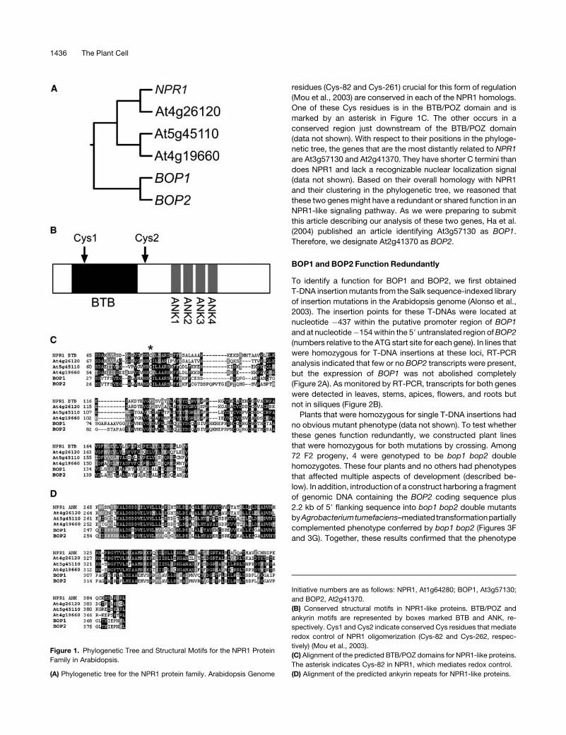
Figure 1. Phylogenetic Tree and Structural Motifs for the NPR1 Protein
Family in Arabidopsis.
(A) Phylogenetic tree for the NPR1 protein family. Arabidopsis Genome
Initiative numbers are as follows: NPR1, At1g64280; BOP1, At3g57130;
and BOP2, At2g41370.
(B) Conserved structural motifs in NPR1-like proteins. BTB/POZ and
ankyrin motifs are represented by boxes marked BTB and ANK, re-
spectively. Cys1 and Cys2 indicate conserved Cys residues that mediate
redox control of NPR1 oligomerization (Cys-82 and Cys-262, respec-
tively) (Mou et al., 2003).
(C) Alignment of the predicted BTB/POZ domains for NPR1-like proteins.
The asterisk indicates Cys-82 in NPR1, which mediates redox control.
(D) Alignment of the predicted ankyrin repeats for NPR1-like proteins.
1436 The Plant Cell

conferred by bop1 bop2 was attributable to the loss of BOP
activity.
To check the disease resistance status of bop1 bop2mutants,
leaves of 4-week-old plants were infiltrated with the virulent
bacteria Pseudomonas syringae maculicola ES4326 at a dose of
OD600¼ 0.0001. The bacteria grew to a similar titer in bop1 bop2
mutants as in wild-type control plants, indicating that bop1 bop2
plants were neither more susceptible nor more resistant than
were wild-type plants to challenge with the pathogen (data not
shown). Thus, we concluded that these two NPR1 homologs are
probably not involved in disease resistance signaling.
BOP1 and BOP2 Control Leaf Patterning and Floral
Organ Abscission
The earliest notable defect in the bop1 bop2 double mutant was
in leaf morphology. Wild-type Arabidopsis rosette leaves are
divided into distinct proximal (petiole) and distal (blade) zones. In
Figure 2. Expression of BOP1 and BOP2 in the Wild Type and bop1 bop2 Mutants.
(A) Analysis of BOP1 and BOP2 expression in wild-type (WT) and bop1 bop2 seedlings as monitored by RT-PCR. The gene amplified in each set of
reactions is indicated at left of each gel. BOP1, 40 cycles; BOP2, 30 cycles; glyceraldehyde-3-phosphate dehydrogenase (GAPC) control, 30 cycles. A
small amount of BOP1 transcript was detected in bop1 bop2 double mutants.
(B) RT-PCR analysis of tissue-specific expression of BOP1 and BOP2 in wild-type plants. The indicated tissues (top) were harvested from 6-week-old
soil-grown plants, and the gene amplified in each set of reactions is indicated at left of each gel. RT-PCR conditions and cycle numbers were as in (A).
(C) to (H) In situ localization of CER6 (control) or BOP2mRNA in the inflorescence apex and flowers of wild-type plants. Hybridization is indicated by the
presence of a purple precipitate.
(C) Central section of an inflorescence meristem (im) and young floral buds hybridized with an L1-specific CER6 control probe.
(D) to (F) Central longitudinal sections showing inflorescence meristems and young flower buds hybridized with a BOP2 probe. BOP2 mRNA is first
detected in the floral anlagen (P0) on the flanks of the inflorescence meristem. Arrows in (D) and (F) indicate a strong band of expression at the base of
stage 1 (P1) and stage 2 (P2) floral primordia in the abaxial region of these primordia corresponding to the cryptic bract.
(G) Cross section of an inflorescence meristem and young floral buds hybridized with a BOP2 probe. Arrows indicate expression in the developing sepal
primordia of a stage 3 floral primordium (P3).
(H) Cross section of a mature flower showing strong bands of expression at the bases of anthers and petals (arrows). The inset shows detail at the base
of the anther.
Bars ¼ 100 mm, except 50 mm in (H).
NPR1-Like Signaling in Leaf and Floral Patterning 1437

contrast with wild-type leaves, leaf growth in bop1 bop2 was
indeterminate along the proximal-distal leaf axis, resulting in
abnormally long leaves (cf. Figures 3A and 3B). In addition, blade
development was derepressed along the petiole, resulting in the
loss of distinct proximal and distal zones in the leaf. Leaflets were
initiated along the petiole repeatedly throughout development
(Figure 3C). This one aspect of the leaf phenotype conferred by
bop1 bop2 is similar to that described previously for bop1-1
single mutants (Ha et al., 2003, 2004) (see Discussion). The
inflorescences of bop1 bop2 double mutants contain several
notable defects as well. First, cauline leaves in the doublemutant
were serrated at their bases, as opposed to smooth in the wild
type (Figure 3D); second, floral organs failed to abscise (Figure
3E). Abscission normally occurs after pollination and is a form of
developmental programmed cell death that is promoted by
ethylene and inhibited by auxin (reviewed in Bleecker and
Patterson, 1997; Roberts et al., 2002). It is not yet knownwhether
the abscission defect in bop1 bop2 reflects a structural defect in
the abscission zone or an altered response to the hormonal
regulation of this process.
BOP1 and BOP2 Control Floral Patterning
The most dramatic aspect of the phenotype conferred by bop1
bop2 was observed in flowers, and it is here that we focus most
of our analysis. Wild-type Arabidopsis flowers develop in the
absence of a subtending bract and possess a high degree of
radial symmetry. Four types of floral organs are formed on the
flanks of the floral meristem, and these are arranged in concen-
tric whorls composed of four sepals in the first (outer) whorl, four
petals in the secondwhorl, six stamens in the thirdwhorl, and two
carpels in the fourth whorl, as diagrammed in Figure 4C (left). In
contrast with the wild type, bop1 bop2 flowers were often
subtended by a small floral bract that curled around the pedicel
toward the adaxial side of the flower (Figures 4A [compare panels
a to c with d and e] and 4B, panel f). Development of this bract
was sometimes incomplete (Figure 4A, panel i), and sepals or
filaments often were produced in the axil of this bract, causing it
to appear inflorescence-like (Figure 4A, panel k). For reasons that
are unclear, the floral bract was not always located at the base of
the pedicel but at variable locations on the floral pedicel.
Inbop1 bop2 flowers, therewas also an altered pattern of floral
organ formation. Mature flowers had an asymmetric appearance
and typically possessed five organs in the sepal whorl, four
organs in the petal whorl, six or seven organs in the stamen
whorl, and two organs in the carpel whorl, as diagrammed in
Figure 4C (right; see also Table 1). The extra floral organs in bop1
bop2 mutants were always formed on the abaxial side of the
flowers. In the sepal whorl, the adaxial sepal was always
positioned as in the wild type, with the other organs placed
equidistantly in the whorl. The two organs straddling the abaxial
position in the sepal whorl grew more slowly compared with
sepals in the lateral and adaxial positions and were usually
petaloid in appearance. Sometimes the abaxial organs fused
together (Figure 4A, panel h). The first whorl abaxial organs also
had a unique growth trajectory compared with the other organs
in the whorl, growing outward like wings toward the adaxial
side of the flower (Figure 4A, panels d and e). Thus, mutation of
the BOP genes altered growth specifically on the abaxial side of
the flower, causing the loss of radial symmetry. Such bilateral
symmetry is not seen in flowers of the Brassicaciae family but
is common in many species of flowering plants, including the
model plant species A. majus (reviewed in Endress, 2001).
We further investigated these floral defects by examining the
inflorescence apices of wild-type and bop1 bop2 flowers using
scanning electronmicroscopy.Wild-type and bop1 bop2mutant
plants produced a similar number of stage 1 floral primordia of
similar size on the flanks of the inflorescence meristem (Figure
4B, compare panels a and b). A small buttress corresponding to
the floral bract was visible on the flanks of stage 1 and stage 2
floral primordia in the bop1 bop2 double mutant but not in the
wild type (Figure 4B, panels b and c, arrows). We next compared
the development of sepal primordia in wild-type and bop1 bop2
inflorescence meristems. Sepal primordia are first visible in the
wild type as four ridges that appear high on the flanks of the floral
meristem; their formation marks the beginning of stage 3 of floral
development (Kunst et al., 1989; Smyth et al., 1990). Although
Arabidopsis flowers are described as radially symmetrical, the
abaxial sepal arises first, is slightly larger than the others, and
Figure 3. Leaf and Inflorescence Phenotypes for Wild-Type, bop1,
bop2, and bop1 bop2 Plants and Complementation of bop1 bop2 with
BOP2.
(A) and (B) Twenty-eight-day-old plants are shown for the wild type (A)
and bop1 bop2 (B). Bar ¼ 2 cm for (A) and (B).
(C) Leaf series from 3-week-old wild-type (top row) and bop1 bop2
(bottom row) plants.
(D) Cauline leaves from wild-type (left) and bop1 bop2 (middle and right)
plants.
(E) Inflorescences from wild-type (left) and bop1 bop2 (right) plants.
Arrows indicate lack of floral organ abscission. The inset shows wild-type
(left) and bop1 bop2 (right) siliques.
(F) Leaf series showing representative rosette leaves from a wild-type
plant (left), a bop1 bop2 plant (middle), and a bop1 bop2 plant
complemented by a fragment of genomic DNA containing the BOP2
gene (right).
(G) Inflorescence of a bop1 bop2 plant partially complemented for floral
organ abscission by a fragment of genomic DNA containing the BOP2
gene.
1438 The Plant Cell
grows to overlie the lateral and adaxial sepals (Kunst et al., 1989;
Smyth et al., 1990) (Figure 4B, panel a, asterisk). In the wild type,
the sepals grow rapidly to enclose the interior of the floral
meristem such that development of the stamens, petals, and
carpels is obscured. In the floral meristem of bop1 bop2, the
appearance of the sepal primordia was slightly delayed relative
to the plastochron compared with the wild-type. In addition, two
first whorl primordia were formed on the abaxial side of the
flower, slightly to the interior of where a wild-type abaxial sepal
primordium would form, resulting in an apparent whorl of five
evenly spaced organs (Figure 4B, panel d, asterisks). The two
abaxial primordia in the first whorl were smaller and developed
more slowly than the primordia in the adaxial and lateral positions
(Figure 4B, panel e). However, in the double mutant, the growth
of all first whorl organs was retarded compared with that in the
wild type, allowing us to see the stamen and carpel primordia
without having to dissect the sepals away. At later stages in floral
development, the two abaxial organs in the first whorl of bop1
bop2 flowers differentiated as petals or sepal-petals. We spec-
ulate that ectopic development of the bract and/or changes in
growth pattern on the abaxial side of the floral meristem may
cause displacement of the abaxial organs in the sepal whorl,
causing them to be lodged partially or entirely within the anlagen
specifying petal identity. Evidence of this comes from the
observation that some of these abaxial organs develop as half-
sepal and half-petal (Figures 4A, panels g and h, and 4B, panel f).
BOP2 Is Expressed in Young Floral Primordia and Encodes
a Protein Localized to Both the Cytoplasm and the Nucleus
The developmental pattern of BOP2 mRNA localization was
determined in flowers by in situ hybridization. This analysis
showed that BOP2 transcript is not expressed above back-
ground in the central part of the inflorescence meristem but is
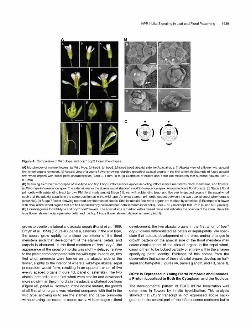
Figure 4. Comparison of Wild-Type and bop1 bop2 Floral Phenotypes.
(A) Morphology of mature flowers. (a) Wild type. (b) bop1. (c) bop2. (d) bop1 bop2 abaxial side. (e) Adaxial side. (f) Abaxial view of a flower with abaxial
first whorl organs removed. (g) Abaxial view of a young flower showing retarded growth of abaxial organs in the first whorl. (h) Example of fused abaxial
first whorl organs with sepal-petal characteristics. Bars ¼ 1 mm. (i) to (k) Examples of bracts and bract-like structures that subtend flowers. Bar ¼0.5 mm.
(B) Scanning electron micrographs of wild-type and bop1 bop2 inflorescence apices depicting inflorescence meristems, floral meristems, and flowers.
(a) Wild-type inflorescence apex. The asterisk marks the abaxial sepal. (b) bop1 bop2 inflorescence apex. Arrows indicate floral bracts. (c) Stage 2 floral
primordia with subtending bract (arrow). FM, floral meristem. (d) Stage 5 flower with subtending bract and five evenly spaced organs in the sepal whorl
such that the adaxial sepal is in the same position as in the wild type. An extra stamen primordia occurs between the two abaxial sepal whorl organs
(asterisks). (e) Stage 7 flower showing retarded development of sepals. Smaller abaxial first whorl organs are marked by asterisks. (f) Example of a flower
with abaxial first whorl organs that are half-sepal (bumpy cells) and half-petal (smooth inner cells). Bars ¼ 50 mm except 100 mm in (e) and 500 mm in (f).
(C) Floral diagrams for wild-type and bop1 bop2 flowers. The adaxial side is marked with a closed circle and indicates the position of the stem. The wild-
type flower shows radial symmetry (left), and the bop1 bop2 flower shows bilateral symmetry (right).
NPR1-Like Signaling in Leaf and Floral Patterning 1439
expressed strongly in floral anlagen situated on the flanks of the
inflorescence meristem (Figure 2D, P0). Expression persisted
throughout stage 1 and stage 2 floral primordia, with the
strongest expression often seen near the base of these struc-
tures in the region that gives rise to the floral bract (Figures 2D to
2G, see arrows in 2D and 2F). At stage 3 of flower development,
expression of BOP2 subsided from the central part of the floral
meristem but was detected throughout very young organ pri-
mordia in all four whorls as these structures developed (Figures
2E to 2G and data not shown). As flowersmatured, a strong band
of BOP2 transcript was detected at the base of floral organs in
the region corresponding to the abscission zone (e.g., as seen for
the mature flower shown in Figure 2H). All features of the BOP2
expression pattern were distinct from that of CER6, a control
probe (Figure 2C).
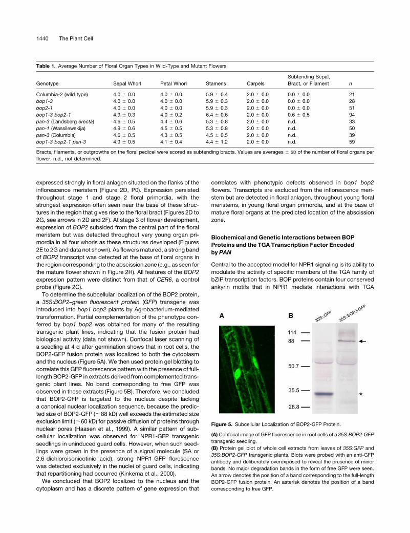
To determine the subcellular localization of the BOP2 protein,
a 35S:BOP2–green fluorescent protein (GFP) transgene was
introduced into bop1 bop2 plants by Agrobacterium-mediated
transformation. Partial complementation of the phenotype con-
ferred by bop1 bop2 was obtained for many of the resulting
transgenic plant lines, indicating that the fusion protein had
biological activity (data not shown). Confocal laser scanning of
a seedling at 4 d after germination shows that in root cells, the
BOP2-GFP fusion protein was localized to both the cytoplasm
and the nucleus (Figure 5A). We then used protein gel blotting to
correlate this GFP fluorescence pattern with the presence of full-
length BOP2-GFP in extracts derived from complemented trans-
genic plant lines. No band corresponding to free GFP was
observed in these extracts (Figure 5B). Therefore, we concluded
that BOP2-GFP is targeted to the nucleus despite lacking
a canonical nuclear localization sequence, because the predic-
ted size of BOP2-GFP (;88 kD) well exceeds the estimated size
exclusion limit (;60 kD) for passive diffusion of proteins through
nuclear pores (Haasen et al., 1999). A similar pattern of sub-
cellular localization was observed for NPR1-GFP transgenic
seedlings in uninduced guard cells. However, when such seed-
lings were grown in the presence of a signal molecule (SA or
2,6-dichloroisonicotinic acid), strong NPR1-GFP florescence
was detected exclusively in the nuclei of guard cells, indicating
that repartitioning had occurred (Kinkema et al., 2000).
We concluded that BOP2 localized to the nucleus and the
cytoplasm and has a discrete pattern of gene expression that
correlates with phenotypic defects observed in bop1 bop2
flowers. Transcripts are excluded from the inflorescence meri-
stem but are detected in floral anlagen, throughout young floral
meristems, in young floral organ primordia, and at the base of
mature floral organs at the predicted location of the abscission
zone.
Biochemical and Genetic Interactions between BOP
Proteins and the TGA Transcription Factor Encoded
by PAN
Central to the accepted model for NPR1 signaling is its ability to
modulate the activity of specific members of the TGA family of
bZIP transcription factors. BOP proteins contain four conserved
ankyrin motifs that in NPR1 mediate interactions with TGA
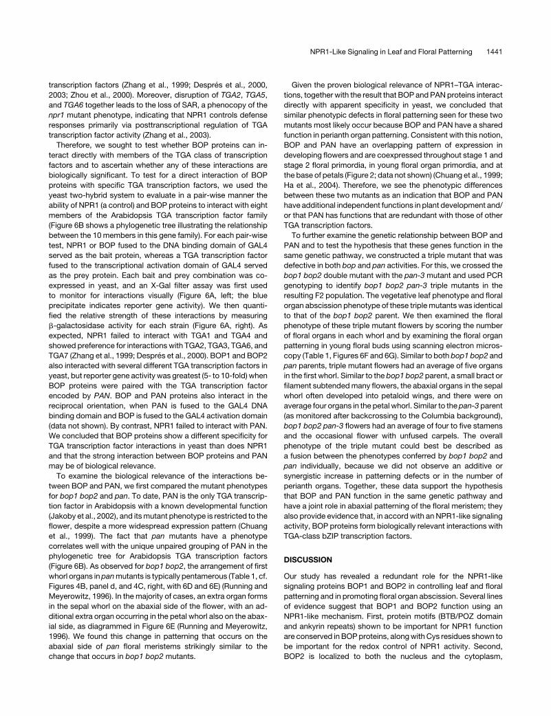
Table 1. Average Number of Floral Organ Types in Wild-Type and Mutant Flowers
Genotype Sepal Whorl Petal Whorl Stamens Carpels
Subtending Sepal,
Bract, or Filament n
Columbia-2 (wild type) 4.0 6 0.0 4.0 6 0.0 5.9 6 0.4 2.0 6 0.0 0.0 6 0.0 21
bop1-3 4.0 6 0.0 4.0 6 0.0 5.9 6 0.3 2.0 6 0.0 0.0 6 0.0 28
bop2-1 4.0 6 0.0 4.0 6 0.0 5.9 6 0.3 2.0 6 0.0 0.0 6 0.0 51
bop1-3 bop2-1 4.9 6 0.3 4.0 6 0.2 6.4 6 0.6 2.0 6 0.0 0.6 6 0.5 94
pan-3 (Landsberg erecta) 4.6 6 0.5 4.4 6 0.6 5.3 6 0.8 2.0 6 0.0 n.d. 33
pan-1 (Wassilewskija) 4.9 6 0.6 4.5 6 0.5 5.3 6 0.8 2.0 6 0.0 n.d. 50
pan-3 (Columbia) 4.6 6 0.5 4.3 6 0.5 4.5 6 0.5 2.0 6 0.0 n.d. 39
bop1-3 bop2-1 pan-3 4.9 6 0.5 4.1 6 0.4 4.4 6 1.2 2.0 6 0.0 n.d. 59
Bracts, filaments, or outgrowths on the floral pedicel were scored as subtending bracts. Values are averages 6 SD of the number of floral organs per
flower. n.d., not determined.
Figure 5. Subcellular Localization of BOP2-GFP Protein.
(A) Confocal image of GFP fluorescence in root cells of a 35S:BOP2-GFP
transgenic seedling.
(B) Protein gel blot of whole cell extracts from leaves of 35S:GFP and
35S:BOP2-GFP transgenic plants. Blots were probed with an anti-GFP
antibody and deliberately overexposed to reveal the presence of minor
bands. No major degradation bands in the form of free GFP were seen.
An arrow denotes the position of a band corresponding to the full-length
BOP2-GFP fusion protein. An asterisk denotes the position of a band
corresponding to free GFP.
1440 The Plant Cell
transcription factors (Zhang et al., 1999; Despres et al., 2000,
2003; Zhou et al., 2000). Moreover, disruption of TGA2, TGA5,
and TGA6 together leads to the loss of SAR, a phenocopy of the
npr1 mutant phenotype, indicating that NPR1 controls defense
responses primarily via posttranscriptional regulation of TGA
transcription factor activity (Zhang et al., 2003).
Therefore, we sought to test whether BOP proteins can in-
teract directly with members of the TGA class of transcription
factors and to ascertain whether any of these interactions are
biologically significant. To test for a direct interaction of BOP
proteins with specific TGA transcription factors, we used the
yeast two-hybrid system to evaluate in a pair-wise manner the
ability of NPR1 (a control) and BOP proteins to interact with eight
members of the Arabidopsis TGA transcription factor family
(Figure 6B shows a phylogenetic tree illustrating the relationship
between the 10 members in this gene family). For each pair-wise
test, NPR1 or BOP fused to the DNA binding domain of GAL4
served as the bait protein, whereas a TGA transcription factor
fused to the transcriptional activation domain of GAL4 served
as the prey protein. Each bait and prey combination was co-
expressed in yeast, and an X-Gal filter assay was first used
to monitor for interactions visually (Figure 6A, left; the blue
precipitate indicates reporter gene activity). We then quanti-
fied the relative strength of these interactions by measuring
b-galactosidase activity for each strain (Figure 6A, right). As
expected, NPR1 failed to interact with TGA1 and TGA4 and
showed preference for interactions with TGA2, TGA3, TGA6, and
TGA7 (Zhang et al., 1999; Despres et al., 2000). BOP1 and BOP2
also interacted with several different TGA transcription factors in
yeast, but reporter gene activity was greatest (5- to 10-fold) when
BOP proteins were paired with the TGA transcription factor
encoded by PAN. BOP and PAN proteins also interact in the
reciprocal orientation, when PAN is fused to the GAL4 DNA
binding domain and BOP is fused to the GAL4 activation domain
(data not shown). By contrast, NPR1 failed to interact with PAN.
We concluded that BOP proteins show a different specificity for
TGA transcription factor interactions in yeast than does NPR1
and that the strong interaction between BOP proteins and PAN
may be of biological relevance.
To examine the biological relevance of the interactions be-
tween BOP and PAN, we first compared the mutant phenotypes
for bop1 bop2 and pan. To date, PAN is the only TGA transcrip-
tion factor in Arabidopsis with a known developmental function
(Jakoby et al., 2002), and itsmutant phenotype is restricted to the
flower, despite a more widespread expression pattern (Chuang
et al., 1999). The fact that pan mutants have a phenotype
correlates well with the unique unpaired grouping of PAN in the
phylogenetic tree for Arabidopsis TGA transcription factors
(Figure 6B). As observed for bop1 bop2, the arrangement of first
whorl organs inpanmutants is typically pentamerous (Table 1, cf.
Figures 4B, panel d, and 4C, right, with 6D and 6E) (Running and
Meyerowitz, 1996). In the majority of cases, an extra organ forms
in the sepal whorl on the abaxial side of the flower, with an ad-
ditional extra organ occurring in the petal whorl also on the abax-
ial side, as diagrammed in Figure 6E (Running and Meyerowitz,
1996). We found this change in patterning that occurs on the
abaxial side of pan floral meristems strikingly similar to the
change that occurs in bop1 bop2 mutants.
Given the proven biological relevance of NPR1–TGA interac-
tions, together with the result that BOP and PANproteins interact
directly with apparent specificity in yeast, we concluded that
similar phenotypic defects in floral patterning seen for these two
mutants most likely occur because BOP and PAN have a shared
function in perianth organ patterning. Consistent with this notion,
BOP and PAN have an overlapping pattern of expression in
developing flowers and are coexpressed throughout stage 1 and
stage 2 floral primordia, in young floral organ primordia, and at
the base of petals (Figure 2; data not shown) (Chuang et al., 1999;
Ha et al., 2004). Therefore, we see the phenotypic differences
between these two mutants as an indication that BOP and PAN
have additional independent functions in plant development and/
or that PAN has functions that are redundant with those of other
TGA transcription factors.
To further examine the genetic relationship between BOP and
PAN and to test the hypothesis that these genes function in the
same genetic pathway, we constructed a triple mutant that was
defective in both bop and pan activities. For this, we crossed the
bop1 bop2 double mutant with the pan-3 mutant and used PCR
genotyping to identify bop1 bop2 pan-3 triple mutants in the
resulting F2 population. The vegetative leaf phenotype and floral
organ abscission phenotype of these triplemutants was identical
to that of the bop1 bop2 parent. We then examined the floral
phenotype of these triple mutant flowers by scoring the number
of floral organs in each whorl and by examining the floral organ
patterning in young floral buds using scanning electron micros-
copy (Table 1, Figures 6F and 6G). Similar to both bop1 bop2 and
pan parents, triple mutant flowers had an average of five organs
in the first whorl. Similar to the bop1 bop2 parent, a small bract or
filament subtendedmany flowers, the abaxial organs in the sepal
whorl often developed into petaloid wings, and there were on
average four organs in the petal whorl. Similar to thepan-3parent
(as monitored after backcrossing to the Columbia background),
bop1 bop2 pan-3 flowers had an average of four to five stamens
and the occasional flower with unfused carpels. The overall
phenotype of the triple mutant could best be described as
a fusion between the phenotypes conferred by bop1 bop2 and
pan individually, because we did not observe an additive or
synergistic increase in patterning defects or in the number of
perianth organs. Together, these data support the hypothesis
that BOP and PAN function in the same genetic pathway and
have a joint role in abaxial patterning of the floral meristem; they
also provide evidence that, in accord with an NPR1-like signaling
activity, BOP proteins form biologically relevant interactions with
TGA-class bZIP transcription factors.
DISCUSSION
Our study has revealed a redundant role for the NPR1-like
signaling proteins BOP1 and BOP2 in controlling leaf and floral
patterning and in promoting floral organ abscission. Several lines
of evidence suggest that BOP1 and BOP2 function using an
NPR1-like mechanism. First, protein motifs (BTB/POZ domain
and ankyrin repeats) shown to be important for NPR1 function
are conserved in BOPproteins, alongwith Cys residues shown to
be important for the redox control of NPR1 activity. Second,
BOP2 is localized to both the nucleus and the cytoplasm,
NPR1-Like Signaling in Leaf and Floral Patterning 1441
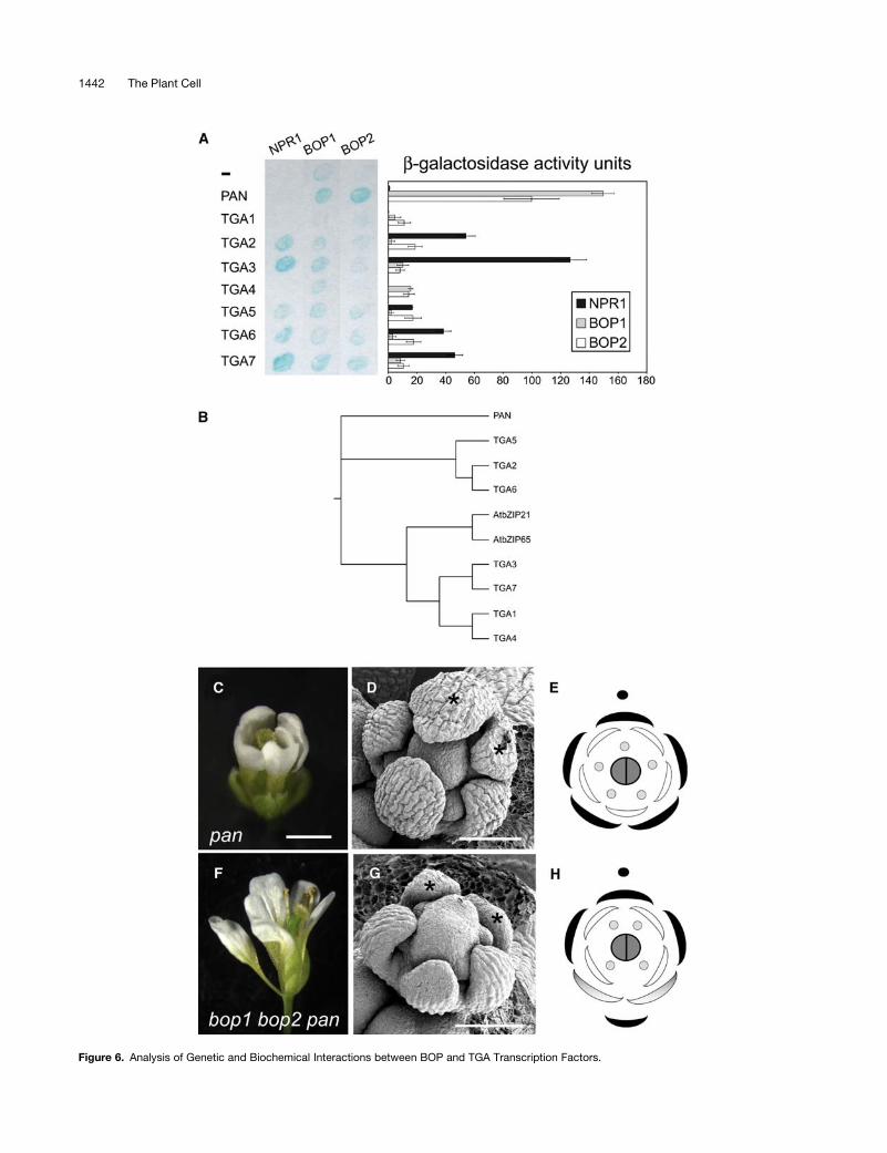
Figure 6. Analysis of Genetic and Biochemical Interactions between BOP and TGA Transcription Factors.
1442 The Plant Cell
as shown previously for NPR1 (Despres et al., 2000; Kinkema
et al., 2000). Third, BOP proteins interact directly with TGA
transcription factors in yeast and show specificity for PAN. This
specificity appears to have biological significance because bop1
bop2 and pan mutants have an overlapping defect in perianth
patterning and because this patterning defect does not change in
an additive or synergistic manner in bop1 bop2 pan triple
mutants, consistent with these two proteins functioning in the
same genetic pathway. Collectively, these results provide evi-
dence that BOP1 andBOP2 function in amanner similar to that of
NPR1, and we speculate that in response to an appropriate
signal, BOP proteins may accumulate in the nucleus and/or
become activated so that they may complex with TGA transcrip-
tion factors such as PAN to regulate the transcription of target
genes involved in the control of leaf and floral patterning and
floral organ abscission.
BOP and Proximal-Distal Leaf Patterning
Themechanisms involved in establishing pattern formation along
the proximal-distal leaf axis are still not well defined. Leaves are
bilaterally symmetric along this axis and are divided into two
distinct domains consisting of a distal blade region and a prox-
imal petiole region. Differentiation in leaves tends to occur
basipetally, with distal blade forming first, followed by proximal
petiole. This pattern has an inverse relationship to cell division
rates, which are highest in the proximal region of leaves, as
monitored through genetic clonal analysis (Poethig and Sussex,
1985) and through observation of a cyclin AT:b-glucuronidase
cell cycle reporter gene (Donnelly et al., 1999).
The leafy petiole defect found in bop1 bop2mutants is similar
to that reported for the bop1-1 single mutant. This mutation
causes leaflet-like structures to form on leaf petioles and the
proximal parts of leaf blades but does not affect growth de-
terminacy along the proximal-distal leaf axis (Ha et al., 2003). In
our analysis of plants homozygous for single alleles of bop1 or
bop2, we failed to detect a leaf phenotype. Although bop1-3 is
not a complete loss-of-function allele (we detected a small
amount of transcript), the lack of a prominent leaf defect is also
seen for bop1-4, which is a transcript null mutation (Ha et al.,
2004). Such phenotypic differences between alleles may be the
result of sensitivity to growth conditions or different genetic
backgrounds (bop1-1 is a Landsberg erecta allele, whereas the
other alleles are in the Columbia background), but another
possibility is that bop1-1 is an atypical allele. Ha et al. (2004)
point out that bop1-1 has a weak semidominant phenotype, is
expressed at slightly higher levels than is wild-type transcript,
and contains an unusual mutation in which the stop codon
is altered, adding an additional four amino acids onto the
C terminus of the protein. These details suggest that the protein
encoded by bop1-1 may exert a weak dominant negative effect
by interfering with the function of an interacting partner, such as
BOP2. BOP1 and BOP2 can form self-dimers and heterodimers
in yeast (S.R. Hepworth and G.W. Haughn, unpublished results)
and have the same pattern of gene expression in flowers (Ha
et al., 2004; this study), an indication that these two proteins may
form a complex together and function interchangeably in some
cases. These data are also consistent with redundant functions
for these two genes. Nevertheless, the fact that bop1-1 primarily
affects the petiole suggests that BOP1 may make a greater
contribution to the control of leaf patterning than does BOP2 and
that there may be some specialization of function for these two
proteins.
Leafy petioles are also caused by the ectopic expression of
LEAFY PETIOLE (LEP), which encodes an EREBP/AP2-type
transcription factor (van der Graaff et al., 2000), and JAGGED
(JAG), which encodes a C2H2 zinc finger transcription factor
(Dinneny et al., 2004; Ohno et al., 2004). One possibility is that
BOP activity regulates the spatial expression of leaf-promoting
transcription factors such as JAG or LEP in the proximal parts
of leaves and in floral bracts. Consistent with this notion, we
detected upregulation of JAG transcript in bop1 bop2 leaves
(S.R. Hepworth and G.W. Haughn, unpublished results). Plants
misexpressing JAG also develop floral bracts (Dinneny et al.,
2004; Ohno et al., 2004), another aspect of thebop1bop2mutant
phenotype. Previous analyses of bop1-1 plants have shown that
knox genes are also misexpressed in the leaves, predominantly
in the petiole region (Ha et al., 2003). Thus, an additional function
Figure 6. (continued).
(A) Yeast two-hybrid interactions between NPR1, BOP1 and BOP2, and TGA transcription factors. Left, X-Gal filter-lift assay. Bait proteins are indicated
at top and prey proteins are indicated at left. Accumulation of blue precipitate indicates b-galactosidase activity. Right, quantitative determination of
reporter gene expression for each corresponding interaction shown at left, presented in chart format as b-galactosidase activity units. All data shown
were corrected for self-activation of reporter gene expression by bait protein.
(B) Phylogenetic tree for the Arabidopsis TGA transcription factor family (Jakoby et al., 2002). Arabidopsis Genome Initiative numbers are as follows:
PAN, At1g68640; TGA5, At5g06960; TGA2, At5g06950; TGA6, At3g12250; AtbZIP21, At1g08320; AtbZIP65, At5g06839; TGA3, At1g22070; TGA7,
At1g77920; TGA1, At5g65210; TGA4, At5g10030.
(C) to (H) Comparison of pan-3 and bop1 bop2 pan-3 mutant floral phenotypes.
(C) Typical pan-3 mutant flower with pentamerous arrangement of floral organs: five sepals, five petals, and five stamens (abaxial view).
(D) Scanning electron micrograph of a stage 5 pan-3 flower with pentamerous arrangement of sepals. Abaxial sepals are marked with asterisks.
(E) Floral diagram of an average pan-3 flower. The adaxial side is marked with a closed circle.
(F) Typical bop1 bop2 pan-3 flower with pentamerous arrangement of first whorl organs (lateral view).
(G) Scanning electron micrograph of a stage 5 bop1 bop2 pan-3 flower that retains a pentamerous arrangement of first whorl organs. Abaxial organs are
marked with asterisks.
(H) Floral diagram of an average bop1 bop2 pan-3 triple mutant flower. The adaxial side is marked with a closed circle.
Bars ¼ 1 mm for (C) and (F) and 100 mm for (D) and (G).
NPR1-Like Signaling in Leaf and Floral Patterning 1443
of BOPmaybe to exclude knox gene expression from leaf tissues
and maintain a developmentally determinate state in cells. knox
genemisexpression is often correlatedwith changes in proximal-
distal leaf patterning (reviewed in Byrne et al., 2001; Hake et al.,
2004).
BOP and Floral Symmetry
Wild-type Arabidopsis flowers are nearly radially symmetrical but
show some asymmetry along the abaxial-adaxial axis. Mutation
of the BOP genes appears to exaggerate this asymmetry, as
indicated by the formation of an ectopic floral bract, extra abaxial
floral organs, and the transformation of the abaxial sepal whorl
organs into petaloid structures that grow outward like wings
toward the adaxial side of the flower. Information on how
asymmetries develop along the abaxial-adaxial axis is best
understood in the model species A. majus, which possesses
flowers with strong bilateral asymmetry. In this species, flowers
are subtended by floral bracts and have a pentamerous arrange-
ment of sepals, petals, and stamens, similar to that observed in
bop1 bop2 pan triple mutants; additionally, the abaxial petals of
each flower are of a different shape than the adaxial petals.
These asymmetries depend on the actions of CYC and DICH,
which are members of the TCP family of DNA binding proteins.
These transcription factors are thought to respond to an abaxial-
adaxial prepattern in floral meristems (Luo et al., 1995, 1999). In
Arabidopsis flowers, althoughmorphological asymmetries along
the abaxial-adaxial axis are slight, there is retained adaxial-
specific expression of TCP1 (an Arabidopsis ortholog of CYC) in
floral meristems (Cubas et al., 2001). The abaxial-specific floral
defects that we see in bop1 bop2mutants are further evidence of
asymmetries along this axis, and it is possible that BOP activity
limits abaxial growth as part of a mechanism to promote radial
symmetry in Arabidopsis flowers.
We find it intriguing that bop1 bop2 mutations both promote
bilateral symmetry and cause flowers to switch to a pan-like
pentamerous arrangement of sepal whorl organs. Interaction of
BOP with PAN suggests that BOP may modulate PAN activity,
possibly to control growth on the abaxial side of the floral
meristem. However, no measurable increase in floral meristem
size is detected in bop1 bop2 mutants (this study) or in pan
mutants (Running and Meyerowitz, 1996). Double mutant anal-
yses, however, have revealed that PAN functions redundantly
with ULTRAPETALA and ETTIN/AUXIN RESPONSE FACTOR3
to control the size of the floral meristem and the initiation of floral
organs, respectively (Sessions et al., 1997; Fletcher, 2001). Thus,
a clearer picture of the role of BOP in controlling the size of the
floral meristem and/or the initiation of floral organs may be
obtained from further genetic analyses.
BOP and Floral Organ Abscission
In addition to its role in leaf and floral patterning, BOP activity is
required for floral organ abscission. Abscission is a developmen-
tally controlled process commencing soon after pollination in
which floral organs senesce and are shed through separation at
a specialized zone of cells located at the base of stamens and
perianth organs. This late role for BOP activity in floral develop-
ment correlates with the expression of BOP1 and BOP2 at the
base of floral organs in mature flowers (Ha et al., 2004; this
study). It is possible that this defect represents a structural defect
within the abscission zone, but alternatively, it may represent
failure to respond to hormonal signals controlling this process.
The predominant view is that auxin delays abscission and
ethylene promotes abscission. The roles of other plant hormones
(e.g., abscisic acid, gibberellic acid, and cytokinins) involved in
this process have been postulated primarily to involve altering
the levels of or sensitivity to auxin and ethylene in abscising
tissues (Sexton and Roberts, 1982). Although a link between
growth asymmetry and floral organ abscission is not immediately
obvious, it is possible that these two processes are controlled by
a common signal to which BOP responds.
Spatial Regulation of BOP Activity
Aunique feature of the phenotype conferred by bop1 bop2 is that
ectopic growth and development is restricted to specific do-
mains along the axes of bilateral symmetry within leaves and
flowers. In leaves, excess growth occurs along the proximal-
distal axis and ectopic blade development occurs in the proximal
region. In flowers, ectopic bract development and floral defects
are centered on the abaxial side. The asymmetric nature of these
defects suggests that BOP activity links spatial information with
pattern elaboration. In part, this is accomplished through the
spatial regulation of BOP expression. BOP1 and/or BOP2 are
expressed in the proximal part of young leaves, in young floral
meristems, and at the base of mature floral organs (Ha et al.,
2004; this study). Each of these locations closely correlates with
the phenotypes in bop1 bop2 mutants affecting leaf petioles,
floral patterning, and floral organ abscission. However, although
BOP2 transcript is present throughout stage 1 and stage 2 floral
primordia, defects are centered on the abaxial side of the flower,
raising the possibility that BOP activity is also controlled at the
posttranscriptional level by a developmental signal.
Characterization of the NPR1 protein has shown that its
activity is redox-sensitive and that the signal molecule SA
controls activity indirectly by causing changes in the intracellular
redox potential of cells (reviewed in Eckardt, 2003; Dong, 2004).
In the absence of signal, intermolecular disulfide bonds hold
NPR1 proteins together in an inactive complex. The signal
molecule SA causes a rapid oxidative burst, with the cellular
redox state recovering to a more reduced state, so that the
intermolecular disulfide bonds holding NPR1 proteins together
are reduced and the majority of NPR1 is present in an active
monomeric form that accumulates in the nucleus (Mou et al.,
2003). TGA1 also contains redox-sensitive Cys residues that
modulate its ability to interact with NPR1 (Despres et al., 2003).
Because the Cys residues required for the redox control of NPR1
are conserved in the BOPproteins, it is possible that BOP activity
is controlled by a signal that alters the redox state of the cell, but
this remains to be tested. Although crucial for the activation of
plant defenses, SA does not appear to play amajor role in growth
or development. One report suggests that SA may regulate the
balance between cell growth and cell death, but the significance
of this finding with respect to development is unclear (Vanacker
et al., 2001). Another possibility is that signals other than SA are
1444 The Plant Cell
perceived in the cell as a change in redox potential. Consistent
with this notion, a role for reactive oxygen species as a signal for
the hormonal regulation of various aspects of plant development
has been proposed in recent studies (Sagi et al., 2004, and
references therein). Also, TGA binding elements (also known as
as1 or ocs elements) appear to mediate activation by hydrogen
peroxide and by plant phytohormones such as auxin, SA, and
jasmonic acid (Xiang et al., 1996, and references therein). Auxin
is particularly attractive as a signal molecule for the control of
BOP activity because of its roles in regulating cell growth and cell
expansion, patterning of lateral organs, and floral organ abscis-
sion, combined with its polarized transport throughout the plant
(Rogg and Bartel, 2001; Friml, 2003; reviewed in Roberts et al.,
2002). Nevertheless, other signals cannot be excluded, and
further experiments are needed to resolve this issue.
METHODS
Plant Materials and Growth Conditions
Wild type was the Columbia-0 ecotype of Arabidopsis thaliana. All plants
were grown in continuous light as described (Bellaoui et al., 2001) on GM
agar plates or on prepared soil mix (SunshineMix 5; SunGro, SebaBeach,
Alberta, Canada). The bop1-3 and bop2-1 T-DNA insertion mutants used
in this study were obtained from the ABRC (Ohio State University,
Columbus, OH; stock numbers SALK_012994 and SALK_075879, re-
spectively); the pan-3 mutant (Landsberg erecta background) was a gift
from Elliot Meyerowitz and the pan-1mutant (Wassilewskija background)
was a gift from Mark Running (Running and Meyerowitz, 1996). bop1
bop2 double mutants and bop1 bop2 pan-3 triple mutants were con-
structed by crossing, and their genotypes were confirmed by PCR-based
genotyping. Floral stages were determined according to Smyth et al.
(1990). For the definition of floral bract, see Dinneny et al. (2004).
Sequence Analysis
The complete protein sequences for members of the NPR1 gene family
were aligned using ClustalW (Thompson et al., 1994), and phylogenetic
trees (Neighbor Joining method with 1000 Bootstrap output) were pro-
duced using the Web-based Phylodendron program (http://iubio.bio.
indiana.edu/soft/molbio/java/apps/trees). The Web-based SMART pro-
gram was used to predict the BTB/POZ domains and to confirm the
locations of ankyrin repeats for each protein (Schultz et al., 2002). The
individual domains were then realigned and shaded using ClustalW and
BOXSHADE.
Complementation of bop1 bop2 Double Mutants
To create plasmid pGreen0229/BOP2, a 6.1-kb EagI-PstI fragment was
excised from Arabidopsis BAC F13H10 (AC005662) and cloned into the
corresponding sites of thebinary vector pGreen0229 (Hellens et al., 2000).
This fragment contained the BOP2 gene plus;2.2 kb of sequence 59 to
the ATG start codon. The construct was introduced into bop1 bop2 plants
by floral dipping (Clough and Bent, 1998). Basta-resistant transformants
were selected on soil by treatment of seedlings with the herbicide Final ev
150 (AgrEvo, Paris, France). The Agrobacterium tumefaciens strain used
was C58C1 pGV3101 pMP90 (Koncz and Schell, 1986).
Subcellular Localization of BOP2 Protein
To create the 35S:BOP2-GFP transgene, full-length BOP2 cDNA was
amplified by PCR using pCR2-BOP2 as the template (see below) andwith
primers that incorporated EcoRI and BamHI sites at the 59 and 39 ends of
the gene, respectively. The resulting fragment was digested with EcoRI
and BamHI and cloned into corresponding sites of pBS-GFP5 (Haseloff
et al., 1997). The resulting plasmid was sequenced to confirm that the
fusion was in-frame and without PCR-induced mistakes. The fragment
containing the BOP2-GFP fusion was then excised from pBS-GFP by
digestion with EcoRI and SacI and cloned into the corresponding sites of
pBI1.4T to yield pBI-BOP2-GFP (Mindrinos et al., 1994). This construct
was introduced into bop1 bop2 plants by Agrobacterium-mediated
transformation as described above. Transgenic plants were selected on
MS medium containing 50 mg/mL kanamycin sulfate. Roots of the
transgenic seedlingswere examined for GFP fluorescence using confocal
microscopy as described (Haseloff et al., 1997) using a Bio-RadRadiance
2000 multiphoton microscope (Bio-Rad, Hercules, CA).
Immunoblot Analysis of GFP and GFP Fusion Proteins
Ten microliters of a crude protein extract from leaves of 35S:BOP2-GFP
or 35S:GFP transgenic plants was resolved on a 10% SDS–polyacryl-
amide gel by electrophoresis and blotted onto a nitrocellulosemembrane.
The anti-GFP antibody (A6455; Molecular Probes, Eugene, OR) was used
as recommended by the supplier. The blots were developed using an
alkaline phosphatase detection system according to the manufacturer’s
instructions (Roche, Indianapolis, IN).
Scanning Electron Microscopy
Scanning electron microscopy samples were prepared as described by
Modrusan et al. (1994). Flower buds and inflorescences weremounted on
stubs. If necessary, the organs surrounding the inflorescence meristem
were dissected away. The stubs were coated with gold-palladium in
a SEMPrep2 sputter coater (Nanotech, Manchester, UK) and observed
using a Hitachi S4700 scanning electron microscope (Hitachi, Tokyo,
Japan) with an accelerating voltage of 0.8 or 2 kV.
In Situ Hybridization Analysis
Tissue fixation, sectioning, hybridization, signal detection, and strategy
for probe synthesis were as described previously (Hooker et al., 2002). To
synthesize a BOP2 antisense probe, a DNA template was amplified by
PCR using pCR2-BOP2 as the template and using 59-CTCATATGAAT-
GAGGAGCAC-39 and 59-GATAATACGACTCACTATAGGGACTCATAC-
CTTCCCTCTGA-39 (which incorporates a binding site for T7 polymerase)
as primers. CER6 probe was as described previously (Hooker et al.,
2002). Sections were photographed using bright-field optics.
Photography and Light Microscopy
Photographs were taken with a digital camera (Coolpix; Nikon, Tokyo,
Japan). A dissecting microscope fitted with a digital camera (SPOT
Diagnostics) was used to take close-up pictures of flowers, cauline
leaves, and siliques. Digital photographs and micrographs were manip-
ulated using Photoshop 5.0 (Adobe Systems, San Jose, CA).
Construction of Yeast Two-Hybrid Plasmids
Full-length cDNAs corresponding to BOP1 and BOP2 were amplified by
RT-PCR using the Expand Hi-Fidelity PCR system (Roche). The template
used for these reactions was cDNA derived from total RNA isolated from
20-d-old seedlings grown on agar plates. The BOP1 and BOP2 cDNAs
were subcloned into pCR2 using the TA cloning system (Invitrogen,
Carlsbad, CA) to yield pCR2-BOP1 and pCR2-BOP2, respectively. Full-
length cDNAs corresponding to PAN (stock No. U50929) and NPR1
(stock number U13446) were obtained from the ABRC. The sequences
NPR1-Like Signaling in Leaf and Floral Patterning 1445
of all four cDNAs were verified by sequencing. To create the constructs
used for the yeast two-hybrid assay, the full-length coding sequences of
BOP1,BOP2,PAN, andNPR1were amplified by PCR using pCR2-BOP1,
pCR2-BOP2, pU1539, and pU13446, respectively, as the templates for
Pwo polymerase (Roche). Recognition sites for restriction enzymes were
incorporated at the 59 ends of the primers used in these PCR procedures
to facilitate the directional cloning of these cDNA fragments into the bait
(GAL4-DB) plasmid pBI-880 or the prey (GAL4-TA) plasmid pBI-881
(Kohalmi et al., 1998). The BOP1, BOP2, and NPR1 cDNA inserts were
ligated as SalI-NotI fragments into the corresponding sites of pBI-880 to
create pBI-880/BOP1, pBI-880/BOP2, and pBI-880/NPR1, respectively.
The PAN cDNA insert was ligated as a BamHI-NotI fragment into the
corresponding sites of pBI-881 to create pBI-881/PAN. The sequences of
all primers are available upon request. pBI-881 prey plasmids containing
TGA1-7 were described previously (Despres et al., 2000).
Two-Hybrid Assays
We used a GAL4-based yeast two-hybrid system that has been de-
scribed previously (Kohalmi et al., 1998). Briefly, the appropriate bait
(GAL4-DB) and prey (GAL4-TA) constructs were introduced into yeast
two-hybrid strain YPB2 (Fields and Song, 1989). Transformants were
selected on SD plates lacking Leu and Trp and then spread in patches
onto fresh plates of the same composition. Interactions were detected
visually using an X-Gal filter assay performed as described previously
(Kohalmi et al., 1998). Quantitative assays for b-galactosidase were
performed using the liquid culture assays with 2-nitrophenyl-b-D-galac-
topyranoside as the substrate as described in the Yeast Protocol
Handbook (Clontech Laboratories, Palo Alto, CA).
RT-PCR
RT was performed using 1 mg of total RNA isolated from seedlings or
individual tissues of wild-type or bop1 bop2 plants and Superscript II RT
(Invitrogen). To amplify transcripts, PCR was performed using 1 mL of
cDNA as the template with 1 unit of Taq polymerase. Primers used to
amplify BOP1 were 59-TCTGTGAATCTGATCCTTCGCAACC-39 and
59-CCTGATCTTCTGGTCTTCGAGGTCT-39 or 59-ACTGCAGCCGTCGA-
TCTC-39 and 59-ATCTCTTGTGGAAGCCCGGA-39. Primers used to
amplify BOP2 were 59-ACAGACTCACCACAACCTGTCACAG-39 and
59-GATCTTCTAGGTCTTGAGCCACGCT-39. Amplification of the cyto-
solic glyceraldehyde-3-phosphate dehydrogenase cDNA from the same
cDNA pools was performed under the same conditions and served as
a control (Western et al., 2004).
Sequence data from this article have been deposited with the EMBL/
GenBank data libraries under accession number AY928830 (BOP2
cDNA).
ACKNOWLEDGMENTS
We thank Elliot Meyerowitz for providing the pan-3 mutant, Mark
Running for providing the pan-1 mutant, Tanya Hooker for providing
the CER6 in situ probe, Owen Rowland for providing tissue-specific total
cDNAs, and Pierre Fobert for providing TGA-containing two-hybrid
constructs. We are grateful to the Biological Sciences Microscopy and
Imaging Centre at the University of British Columbia for providing
microscopy support and to Hugo Zheng for confocal imaging. We
also thank Owen Rowland, Ravi Kumar, and Gillian Dean for critical
reading of the manuscript. S.M. was a recipient of a postgraduate
scholarship B award from the Natural Science and Engineering Re-
search Council (NSERC) of Canada. Work in G.W.H.’s laboratory is
supported by an NSERC Discovery Grant. Work in X.L.’s laboratory is
supported by grants from the NSERC and the Canadian Foundation for
Innovation.
Received December 22, 2004; accepted March 6, 2005.
REFERENCES
Alonso, J.M., et al. (2003). Genome-wide insertional mutagenesis of
Arabidopsis thaliana. Science 301, 653–657.
Bellaoui, M., Pidkowich, M.S., Samach, A., Kushalappa, K., Kohalmi,
S.E., Modrusan, Z., Crosby, W.L., and Haughn, G.W. (2001). The
Arabidopsis BELL1 and KNOX TALE homeodomain proteins interact
through a domain conserved between plants and animals. Plant Cell
13, 2455–2470.
Benkova, E., Michneiwicz, M., Suaer, M., Teichmann, T., Seifertova,
D., Jurgens, G., and Friml, J. (2003). Local, efflux-dependent auxin
gradients as a common module for plant organ formation. Cell 115,
591–602.
Bleecker, A.B., and Patterson, S.E. (1997). Last exit: Senescence,
abscission, and meristem arrest in Arabidopsis. Plant Cell 9,
1169–1179.
Bowman, J.L., Eshed, Y., and Baum, S.F. (2002). Establishment of
polarity in angiosperm lateral organs. Trends Genet. 18, 134–141.
Byrne, M., Timmermans, J., Kidner, C., and Martienssen, R. (2001).
Development of leaf shape. Curr. Opin. Plant Biol. 4, 38–43.
Byrne, M.E., Barley, R., Curtis, M., Arroyo, J.M., Dunham, M.,
Hudson, A., and Martienssen, R.A. (2000). Asymmetric leaves1
mediates leaf patterning and stem cell function in Arabidopsis. Nature
408, 967–971.
Byrne, M.E., Simorowski, J., and Martienssen, R.A. (2002).
ASYMMETRIC LEAVES1 reveals knox gene redundancy in Arabi-
dopsis. Development 129, 1957–1965.
Cao, H., Glazebrook, J., Clarke, J.D., Volko, S., and Dong, X. (1997).
The Arabidopsis NPR1 gene that controls systemic acquired resis-
tance encodes a novel protein containing ankyrin repeats. Cell 88,
57–63.
Chuang, C.-F., Running, M.P., Williams, R.W., and Meyerowitz, E.M.
(1999). The PERIANTHIA gene encodes a bZIP protein involved in the
determination of floral organ number in Arabidopsis thaliana. Genes
Dev. 13, 334–344.
Clough, S.J., and Bent, A.F. (1998). Floral dip: A simplified method for
Agrobacterium-mediated transformation of Arabidopsis thaliana. Plant
J. 16, 735–743.
Collins, T., Stone, J.R., and Williams, A.J. (2001). All in the family: The
BTB/POZ, KRAB, and SCAN domains. Mol. Cell. Biol. 21, 3609–3615.
Cubas, P., Coen, E., and Martınez-Zapater, J.M. (2001). Ancient
asymmetries in the evolution of flowers. Curr. Biol. 11, 1050–1052.
Despres, C., Chubak, C., Rochon, A., Clark, R., Bethune, T.,
Desveaux, D., and Fobert, P.R. (2003). The Arabidopsis NPR1
disease resistance protein is a novel cofactor that confers redox
regulation of DNA binding activity to the basic domain/leucine zipper
transcription factor TGA1. Plant Cell 15, 2181–2191.
Despres, C., DeLong, C., Glaze, S., Liu, E., and Fobert, P.R. (2000).
The Arabidopsis NPR1/NIM1 protein enhances the DNA binding
activity of a subgroup of the TGA family of bZIP transcription factors.
Plant Cell 12, 279–290.
Dinneny, J.R., Yadegari, R., Fischer, R.L., Yanofsky, M.F., and
Weigel, D. (2004). The role of JAGGED in shaping lateral organs.
Development 131, 1101–1110.
1446 The Plant Cell
Dong, X. (2004). NPR1, all things considered. Curr. Opin. Plant Biol. 7,
547–552.
Donnelly, P.M., Bonetta, D., Tsukaya, H., Dengler, R.E., and Dengler,
N.G. (1999). Cell cycling and cell enlargement in developing leaves of
Arabidopsis. Dev. Biol. 215, 407–419.
Eckardt, N. (2003). A new twist on systemic acquired resistance: Redox
control of the NPR1–TGA1 interaction by salicylic acid. Plant Cell 15,
1947–1949.
Endress, P.K. (2001). Evolution of floral symmetry. Curr. Opin. Plant
Biol. 4, 86–91.
Fields, S., and Song, O. (1989). A novel genetic system to detect
protein-protein interactions. Nature 340, 245–246.
Fletcher, J.C. (2001). The ULTRAPETALA gene controls shoot and floral
meristem size in Arabidopsis. Development 128, 1323–1333.
Friml, J. (2003). Auxin transport: Shaping the plant. Curr. Opin. Plant
Biol. 6, 7–12.
Ha, C.M., Jun, J.H., Nam, H.G., and Fletcher, J.C. (2004). BLADE-
ON-PETIOLE1 encodes a BTB/POZ domain protein required for
leaf morphogenesis in Arabidopsis thaliana. Plant Cell Physiol. 45,
1361–1370.
Ha, C.M., Kim, G.-T., Kim, B.C., Jun, J.H., Soh, M.S., Ueno, Y.,
Mahida, Y., Tsukaya, H., and Nam, H.G. (2003). The BLADE-
ON-PETIOLE1 gene controls leaf pattern formation through the
modulation of meristematic activity in Arabidopsis. Development
130, 161–172.
Haasen, D., Kohler, C., Neuhaus, G., and Merkle, T. (1999). Nuclear
export of proteins in plants: AtXPO1 is the export receptor for
leucine-rich nuclear export signals in Arabidopsis thaliana. Plant J.
20, 695–705.
Hake, S., Smith, H.M., Holtan, H., Magnani, E., Mele, G., and
Ramirez, J. (2004). The role of KNOX genes in plant development.
Annu. Rev. Cell Dev. Biol. 20, 125–151.
Haseloff, J., Siemering, K.R., Prasher, D.C., and Hodge, S. (1997).
Removal of a cryptic intron and subcellular localization of green
fluorescent protein are required to mark transgenic Arabidopsis plants
brightly. Proc. Natl. Acad. Sci. USA 94, 2122–2127.
Hellens, R.P., Edwards, E.A., Leyland, N.R., Bean, S., and
Mullineaux, P.M. (2000). pGreen: A versatile and flexible binary Ti
vector for Agrobacterium-mediated plant transformation. Plant Mol.
Biol. 42, 819–832.
Hooker, T.S., Millar, A.A., and Kunst, L. (2002). Significance of the
expression of the CER6 condensing enzyme for cuticular wax pro-
duction in Arabidopsis. Plant Physiol. 129, 1568–1580.
Hudson, A. (2000). Development of symmetry in plants. Annu. Rev.
Plant Physiol. Plant Mol. Biol. 51, 349–370.
Iwakawa, H., Ueno, Y., Semiarti, E., Onouchi, H., Kojima, S.,
Tsukaya, H., Hasebe, M., Soma, T., Ikezaki, M., Machida, C.,
and Machida, Y. (2002). The ASYMMETRIC LEAVES2 gene of
Arabidopsis thaliana, required for formation of a symmetric flat leaf
lamina, encodes a member of a novel family of proteins characterized
by cysteine repeats and a leucine zipper. Plant Cell Physiol. 43,
467–478.
Jakoby, M., Weisshaar, B., Droge-Laser, W., Vincente-Carbajosa,
J., Tiedemann, J., Kroj, T., and Parcy, F. (2002). bZIP transcription
factors in Arabidopsis. Trends Plant Sci. 7, 106–111.
Kinkema, M., Fan, W., and Dong, X. (2000). Nuclear localization of
NPR1 is required for activation of PR gene expression. Plant Cell 12,
2339–2350.
Kohalmi, S.E., Reader, L.J.W., Samach, A., Nowak, J., Haughn,
G.W., and Crosby, W.L. (1998). Identification and characterization of
protein interactions using the yeast 2-hybrid system. In Plant Molec-
ular Biology Manual M1, S.B. Gelvin and R. A. Schilperoort, eds
(Dordrecht, The Netherlands: Kluwer Academic Publishers), pp. 1–30.
Koncz, C., and Schell, J. (1986). The promoter of T1-DNA gene 5
controls the tissue-specific expression of chimeric genes carried by
a novel type of Agrobacterium binary vector. Mol. Gen. Genet. 204,
383–396.
Kunst, L., Klenz, J.E., Martinez-Zapater, J., and Haughn, G.W.
(1989). AP2 gene determines the identity of perianth organs in flowers
of Arabidopsis thaliana. Plant Cell 1, 1195–1208.
Lin, W., Shuai, B., and Springer, P. (2003). The Arabidopsis LATERAL
ORGAN BOUNDARIES–domain gene ASYMMETRIC LEAVES2 func-
tions in the repression of KNOX gene expression and in adaxial-
abaxial patterning. Plant Cell 15, 2241–2252.
Luo, D., Carpenter, R., Copsey, L., Vincent, C., Clark, J., and Coen,
E. (1999). Control of organ asymmetry in flowers of Antirrhinum. Cell
99, 367–376.
Luo, D., Carpenter, R., Vincent, C., Copsey, L., and Coen, E. (1995).
Origin of floral asymmetry in Antirrhinum. Nature 383, 794–799.
McConnell, J.R., and Barton, M.K. (1998). Leaf polarity and meristem
formation in Arabidopsis. Development 121, 2935–2942.
Mindrinos, M., Katagiri, F., Yu, G.-L., and Ausubel, F.M. (1994). The
A. thaliana disease resistance gene RPS2 encodes a protein contain-
ing a nucleotide-binding site and leucine-rich repeats. Cell 78,
1089–1099.
Modrusan, Z., Reiser, L., Feldmann, K.A., Fischer, R.L., and Haughn,
G.W. (1994). Homeotic transformation of ovules into carpel-like
structures in Arabidopsis. Plant Cell 6, 333–349.
Mou, Z., Fan, W., and Dong, X. (2003). Inducers of plant systemic
acquired resistance regulate NPR1 function through redox changes.
Cell 113, 935–944.
Ohno, C.K., Reddy, G.V., Heisler, M.G.B., and Meyerowitz,
E.M. (2004). The Arabidopsis JAGGED gene encodes a zinc finger
protein that promotes leaf tissue development. Development 131,
1111–1122.
Ori, N., Eshed, Y., Chuck, G., Bowman, J.L., and Hake, S. (2000).
Mechanisms that control knox gene expression in the Arabidopsis
shoot. Development 127, 5523–5532.
Poethig, R.S., and Sussex, I.M. (1985). The cellular parameters of leaf
development in tobacco: A clonal analysis. Planta 165, 170–184.
Pintard, L., Willems, A., and Peter, M. (2004). Cullin-based
ubiquitin ligases: Cul3-BTB complexes join the family. EMBO J. 23,
1681–1687.
Roberts, J.A., Elliot, K.A., and Gonzalez-Carranza, Z.H. (2002).
Abscission, dehiscence, and other cell separation processes. Annu.
Rev. Plant Biol. 53, 131–158.
Rogg, L.E., and Bartel, B. (2001). Auxin signaling: Derepression
through regulated proteolysis. Dev. Cell 1, 595–604.
Running, M.P., and Meyerowitz, E.M. (1996). Mutations in the
PERIANTHIA gene of Arabidopsis specifically alter floral organ num-
ber and initiation pattern. Development 122, 1261–1269.
Sagi, M., Davydov, O., Orazova, S., Yesbergenova, Z., Ophir, R.,
Stratmann, J.W., and Fluhr, R. (2004). Plant respiratory burst
oxidase homologs impinge on wound responsiveness and develop-
ment in Lycopersicon esculentum. Plant Cell 16, 616–628.
Schultz, J., Copley, R.R., Doerks, T., Ponting, C.P., and Bork, P.
(2002). SMART: A web-based tool for the study of genetically mobile
domains. Nucleic Acids Res. 28, 231–234.
Semiarti, E., Ueno, Y., Tsukaya, H., Iwakaka, H., Machida, C., and
Machida, Y. (2001). The ASYMMETRIC LEAVES2 gene of Arabidop-
sis thaliana regulates formation of a symmetric lamina, establishment
of venation and repression of meristem-related homeobox genes in
leaves. Development 128, 1771–1783.
Sessions, A., Nemhauser, J.L., McCall, A., Roe, J.L., Feldmann,
K.A., and Zambryski, P.C. (1997). ETTIN patterns the Arabidopsis
NPR1-Like Signaling in Leaf and Floral Patterning 1447
floral meristem and reproductive organs. Development 124,
4481–4491.
Sexton, R., and Roberts, J.A. (1982). Cell biology of abscission. Annu.
Rev. Plant Physiol. 33, 133–162.
Smyth, D.R. (2005). Morphogenesis of flowers: Our evolving view. Plant
Cell 17, 330–341.
Smyth, D.R., Bowman, J.L., and Meyerowitz, E.M. (1990). Early flower
development in Arabidopsis. Plant Cell 2, 755–767.
Sun, Y., Zhou, Q., Zhang, W., Fu, Y., and Huang, H. (2002).
ASYMMETRIC LEAVES1, an Arabidopsis gene that is involved in the
control of cell differentiation in leaves. Planta 214, 694–702.
Thompson, J.D., Higgins, D.G., and Gibson, T.J. (1994). Clustal W:
Improving the sensitivity of progressive multiple sequence alignments
through sequence weighting, position-specific gap penalties and
weight matrix choices. Nucleic Acids Res. 22, 4673–4680.
Vanacker, H., Lu, H., Rate, D.N., and Greenberg, J.T. (2001). A role for
salicylic acid and NPR1 in regulating cell growth in Arabidopsis. Plant
J. 28, 209–216.
van den Heuvel, S. (2004). Protein degradation: CUL-3 and BTB—
partners in proteolysis. Curr. Biol. 14, R59–R61.
van der Graaff, E., Dulk-Ras, A.D., Hooykaas, P.J.J., and Keller, B.
(2000). Activation tagging of the LEAFY PETIOLE gene affects leaf
petiole development in Arabidopsis thaliana. Development 127, 4971–
4980.
Waites, R., and Hudson, A. (1995). phantastica: A gene required for
dorsoventrality of leaves in Antirrhinum majus. Development 121,
2143–2154.
Waites, R., Slevadurai, H.R.N., Oliver, I.R., and Hudson, A. (1998).
The PHANTASTICA gene encodes a MYB transcription factor in-
volved in growth and dorsoventrality of lateral organs in Antirrhinum.
Cell 93, 779–789.
Western, T.L., Young, D.S., Dean, G.H., Tan, W.L., Samuels, A.L.,
and Haughn, G.W. (2004). MUCILAGE-MODIFIED4 encodes a puta-
tive pectin biosynthetic enzyme developmentally regulated by
APETALA2, TRANSPARENT TESTA GLABRA1, and GLABRA2 in
the Arabidopsis seed coat. Plant Physiol. 134, 296–306.
Xiang, C., Miao, Z.H., and Lam, E. (1996). Coordinated activation of
as-1-type elements and a tobacco glutathione S-transferase gene
by auxins, salicylic acid, methyl jasmonate and hydrogen peroxide.
Plant Mol. Biol. 32, 415–426.
Xu, L., Xu, Y., Dong, A., Sun, Y., Pi, L., Xu, Y., and Huang, H. (2003).
Novel as1 and as2 defects in leaf adaxial-abaxial polarity reveal the
requirement for ASYMMETRIC LEAVES1 and 2 and ERECTA func-
tions in specifying leaf adaxial polarity. Development 130, 4097–4107.
Zhang, Y., Fan, W., Kinkema, M., Li, X., and Dong, X. (1999).
Interaction of NPR1 with basic leucine zipper protein transcription
factors that bind sequences required for salicylic acid induction of the
PR-1 gene. Proc. Natl. Acad. Sci. USA 96, 6523–6528.
Zhang, Y., Tessaro, M.J., Lassner, M., and Li, X. (2003). Knockout
analysis of Arabidopsis transcription factors TGA2, TGA5, and TGA6
reveals their redundant and essential roles in systemic acquired
resistance. Plant Cell 15, 2647–2653.
Zhou, J.M., Trifa, Y., Silva, H., Pontier, D., Lam, E., Shah, J., and
Klessig, D.F. (2000). NPR1 differentially interacts with members of the
TGA/OBF family of transcription factors that bind an element of the
PR-1 gene required for induction by salicylic acid. Mol. Plant-Microbe
Interact. 13, 191–202.
1448 The Plant Cell
DOI 10.1105/tpc.104.030536; originally published online April 1, 2005; 2005;17;1434-1448Plant Cell
Shelley R. Hepworth, Yuelin Zhang, Sarah McKim, Xin Li and George W. HaughnDependent Signaling Controls Leaf and Floral Patterning in Arabidopsis−BLADE-ON-PETIOLE
This information is current as of June 4, 2016
References http://www.plantcell.org/content/17/5/1434.full.html#ref-list-1
This article cites 70 articles, 35 of which can be accessed free at:
Permissions https://www.copyright.com/ccc/openurl.do?sid=pd_hw1532298X&issn=1532298X&WT.mc_id=pd_hw1532298X
eTOCs http://www.plantcell.org/cgi/alerts/ctmain
Sign up for eTOCs at:
CiteTrack Alerts http://www.plantcell.org/cgi/alerts/ctmain
Sign up for CiteTrack Alerts at:
Subscription Information http://www.aspb.org/publications/subscriptions.cfm
is available at:Plant Physiology and The Plant CellSubscription Information for
ADVANCING THE SCIENCE OF PLANT BIOLOGY © American Society of Plant Biologists