Uncoupling proteins: their roles in adaptive thermogenesis ...
Upload
khangminh22Category
view
1download
0
�����������������
Citation: Križancic Bombek, L.; Cater,
M. Skeletal Muscle Uncoupling
Proteins in Mice Models of Obesity.
Metabolites 2022, 12, 259. https://
doi.org/10.3390/metabo12030259
Academic Editors: Muthu Periasamy
and Naresh Chandra Bal
Received: 20 January 2022
Accepted: 15 March 2022
Published: 17 March 2022
Publisher’s Note: MDPI stays neutral
with regard to jurisdictional claims in
published maps and institutional affil-
iations.
Copyright: © 2022 by the authors.
Licensee MDPI, Basel, Switzerland.
This article is an open access article
distributed under the terms and
conditions of the Creative Commons
Attribution (CC BY) license (https://
creativecommons.org/licenses/by/
4.0/).
metabolites
H
OH
OH
Review
Skeletal Muscle Uncoupling Proteins in Mice Modelsof ObesityLidija Križancic Bombek * and Maša Cater
Institute of Physiology, Faculty of Medicine, University of Maribor, 2000 Maribor, Slovenia; [email protected]* Correspondence: [email protected]; Tel.: +386-2-2345-848
Abstract: Obesity and accompanying type 2 diabetes are among major and increasing world-wide problems that occur fundamentally due to excessive energy intake during its expenditure.Endotherms continuously consume a certain amount of energy to maintain core body temperaturevia thermogenic processes, mainly in brown adipose tissue and skeletal muscle. Skeletal muscle glu-cose utilization and heat production are significant and directly linked to body glucose homeostasisat rest, and especially during physical activity. However, this glucose balance is impaired in diabeticand obese states in humans and mice, and manifests as glucose resistance and altered muscle cellmetabolism. Uncoupling proteins have a significant role in converting electrochemical energy intothermal energy without ATP generation. Different homologs of uncoupling proteins were identified,and their roles were linked to antioxidative activity and boosting glucose and lipid metabolism.From this perspective, uncoupling proteins were studied in correlation to the pathogenesis of dia-betes and obesity and their possible treatments. Mice were extensively used as model organisms tostudy the physiology and pathophysiology of energy homeostasis. However, we should be aware ofinterstrain differences in mice models of obesity regarding thermogenesis and insulin resistance inskeletal muscles. Therefore, in this review, we gathered up-to-date knowledge on skeletal muscleuncoupling proteins and their effect on insulin sensitivity in mouse models of obesity and diabetes.
Keywords: uncoupling protein; skeletal muscle; insulin; diabetes; obesity
1. Introduction
Thermogenesis is of utmost importance for maintaining a stable body temperatureof around 36–37 ◦C in humans [1] and around 36–38 ◦C in different strains of mice [2,3].This temperature homeostasis is ensured by shivering and nonshivering thermogenesis,mainly in the brown adipose tissue (BAT) and skeletal muscle. In healthy adult humans,BAT is scarce and becomes dysfunctional or even proinflammatory in obese individualsfavoring the development of type 2 diabetes [4]. In contrast, skeletal muscle shiveringand nonshivering thermogenesis remain active throughout adult life. However, BAT andskeletal muscle thermogenesis remain substantial throughout their lives in mice.
Parallel to its role in heat production, the skeletal muscle, being the largest glucose sinkin the body, largely contributes to glucose homeostasis. Glucose enters skeletal muscle cellsthrough type 4 glucose transporters (GLUT4), which translocate to the plasma membranein response to increased blood glucose and insulin levels, thus allowing massive entry ofglucose into cells. These processes are enhanced under increased energy demands suchas physical activity or cold exposure. Since glucose is also a fuel for heat production,it is of paramount importance that its transport into cells is adequate, and that skeletalmuscle cells respond properly to insulin signaling. In diabetes, however, skeletal musclecells are resistant to insulin signaling, and glucose entry into the cytoplasm is impaired.Consequently, less glucose is available for cellular metabolism, thus affecting thermogenicprocesses [5]. Furthermore, the derailed glucose metabolism also results in impaired lipidand protein metabolism and their regulation.
Metabolites 2022, 12, 259. https://doi.org/10.3390/metabo12030259 https://www.mdpi.com/journal/metabolites
Metabolites 2022, 12, 259 2 of 22
This review first briefly describes the metabolic and physiological mechanisms of shiv-ering and nonshivering thermogenesis in skeletal muscle. Then, we focus on mitochondrialcoupling and uncoupling processes and their interaction. We emphasize tissue specificityin expressing different isoforms of uncoupling proteins (UCPs) and their roles. In the nextsection, we focus on insulin secretion from pancreatic beta cells, insulin resistance, and thefunctioning of skeletal muscle UCPs in mouse models of obesity and diabetes. We alsodiscuss gender and age differences in UCP expression and their correlation with adulthoodweight gain. The second part of the article reviews studies of obesity and diabetes mousemodels, as well as transgenic and knockout genetic alterations. Lastly, we give a briefoverview of diet-induced obesity and diabetic mouse models, including the effects ofcaloric restriction diets that are very promising in diabetes management.
2. Metabolic and Physiological Mechanisms of Shivering and NonshiveringThermogenesis in Skeletal Muscle
In cold exposure, total heat production in the body can increase by up to five timesthat of the resting metabolic rate at room temperature. The major source of metabolic heatproduction used for conserving body temperature is skeletal muscle tissue. It dissipatesheat in shivering and nonshivering thermogenic processes, which together can contributearound 40% of heat production in cold exposure [6,7].
2.1. Shivering Thermogenesis
Shivering thermogenesis in skeletal muscle is driven by neural mechanisms involvedin recruiting the muscles into the shivering response and regulating the substrates used tofuel the metabolic processes. The research shows that the preoptic area of the hypothalamusis the main thermoregulatory center [8]. It receives sensory input from thermoreceptorsin the skin, thermoreceptors in the vicinity of internal organs [9], and somatosensoryfibers in the dorsal spinal horn [10], as well as from the brain and spinal cord neuronssensitive to thermal stimuli [7,8]. The central neural network responsible for the recruitmentof shivering integrates the input information. It sends feedback to thermoregulatoryeffectors such as skin vasculature, muscle spindles, and BAT to initiate the shiveringresponse [11,12]. In humans, muscle-shivering thermogenesis was estimated to account forup to 40% of whole-body energy expenditure during a mild cold exposure [6]. To produceheat by oxidation, the shivering muscle uses different combinations of carbohydrates, lipids,and proteins [13]. For sustained shivering for hours, carbohydrate reserves in the form ofglycogen and selective recruitment of type II muscle fibers are vital [14,15]. Haman et al.reported that in the case of low carbohydrate stores in human muscles, lipids were thepredominant fuel for shivering thermogenesis, with 53% of total heat produced. Of theremaining total heat production, 28% originated from carbohydrates and 19% from proteins.Conversely, in the case of high carbohydrate stores, cold-induced muscle shivering used23% lipids, 65% carbohydrates, and 12% proteins for heat production. Regardless ofglycogen stores, plasma glucose oxidation remained a minor fuel source, accounting for7–13% of the total heat production [15].
2.2. Nonshivering Thermogenesis
The two greatest nonshivering contributors to heat production in skeletal muscle areunequivocally the uncoupling of the mitochondrial oxidative phosphorylation by UCPs,also called the proton leak [7,16], and uncoupling of the sarco-endoplasmic reticulumCa2+-ATPase (SERCA) pump, also called futile calcium cycling [17,18]. In addition, energy-releasing cellular processes involving enzymes such as myosin ATPase and creatine kinasealso dissipate some heat, thus contributing to thermogenesis. Since mitochondrial un-coupling is extensively described in the following sections, we only briefly present othernonshivering processes in this section.
Metabolites 2022, 12, 259 3 of 22
2.2.1. Sarco-Endoplasmic Reticulum Ca2+-ATPase (SERCA) Pump
The proper functioning of muscle cells depends on maintaining ionic gradients acrosscell membranes. It is achieved by ATPases pumping ions against their concentrationgradients using energy from the hydrolysis of adenosine triphosphate (ATP). Muscle con-traction is triggered by an increase in sarcoplasmic Ca2+ concentration due to the openingof dihydropyridine receptors on the sarcoplasmic reticulum (SR), releasing Ca2+ from theSR lumen into the sarcoplasm. Calcium ions bind to myofilaments setting into motionthe myosin–actin cross-bridge cycling and mitochondrial oxidative metabolism, whichsubsequently activates SERCA and ensures that in optimally coupled conditions, two Ca2+
ions are pumped back into the SR lumen at the expense of hydrolyzing one molecule ofATP [19,20]. However, in a nonideal live setting, the byproduct of the imperfectly coupledCa2+ transport-to-ATP hydrolysis is heat [21], accounting for up to 40–50% of the murinefast- and slow-twitch muscles’ resting metabolic rate, which is equivalent to 8–10% of thetotal body metabolic rate [22]. In skeletal muscle, isoform SERCA 1 is expressed in fast-twitch muscle fibers. In contrast, slow-twitch muscle fibers express SERCA 1 and SERCA2a [23], and only SERCA 1 can modulate the amount of heat produced during the ATPhydrolysis in the range of 7–32 Kcal/mol, depending on the established transmembraneCa2+ gradient across the SR membrane [24]. An important regulator of SERCA’s activityis sarcolipin (SLN) [25]. In Sln −/− knockout mice, hypothermia ensued after exposureto acute cold (4 ◦C), since these animals could not maintain body temperature. However,overexpression of Sln in the Sln −/− mice restored muscle thermogenesis, supportingthe idea that Sln is involved in SERCA-based heat production, and that its absence mayresult in diet-induced obesity during increased caloric intake [17]. However, SLN is notjust a SERCA uncoupler, but in mice, also regulates temperature homeostasis by affectingheat production, whole-body metabolism, and weight gain. Its expression is upregulatedin cases of increased metabolic demand such as different muscle diseases, exercise, coldexposure, and diet-induced obesity [26].
2.2.2. Myosin ATPase
In the resting muscle, a low metabolic rate is a consequence of the transition of themyosin ATPase into a super-relaxed state, thereby slowing down its metabolism anddecreasing energy consumption. At lower temperatures, the exchange of GTP for ATP onmyosin ATPase and the increase in myosin phosphorylation both decrease the fraction ofmyosin ATPases in the super-relaxed state, thus increasing heat production and muscleenergy consumption [27]. In addition, the fraction of super-relaxed myosin ATPases at restis lower in type IIa (fast-twitch) muscle fibers, making them greater glucose and energyconsumers compared to type I (slow-twitch) muscle fibers [28]. The transition of only20% of myosin ATPases from the super-relaxed into a normal-relaxed state approximatelydoubles muscle thermogenesis [29]. Targeting the super-relaxed state of myosin ATPasesmay provide new approaches to treat obesity, high blood sugar, or type II diabetes byincreasing muscle glucose utilization [30].
2.2.3. Creatine Kinase
Creatine kinase catalyzes the reversible reaction of creatine phosphorylation and existsin at least four isoforms, two of which are cytosolic and two mitochondrial. Their expressionis tissue-specific and compartmentalized within cells [31,32]. Despite substantial creatinemetabolism in skeletal muscle, its turnover is still higher in adipose tissue [33]; therefore,it was studied mainly in this context in the past decades. In thermogenic fat (BAT andbeige adipose tissue), creatine enhanced mitochondrial respiration and energy dissipation.In mice, cold exposure stimulated creatine kinase activity and induced the expression ofgenes linked to creatine metabolism. This induction was further enhanced in the case ofabsent UCP1-dependent thermogenesis, linking a futile cycle of creatine metabolism toenergy expenditure and thermoregulation [34–36]. Upon ablation of creatine kinase B,mice showed reduced blood glucose levels, triglycerides, and leptin, as well as disrupted
Metabolites 2022, 12, 259 4 of 22
thermogenic capacity and glucose homeostasis [37,38]. Recently, a mechanism combiningcreatine thermogenesis with futile Ca2+ cycling in the ER has been proposed. Still, this areaneeds further research [35].
2.3. Cold Acclimation
The effects of environmental temperature on mitochondrial efficiency and ATP pro-duction can influence thermal tolerance and performance of the body. Thus, organismsdeveloped the ability to alter mitochondrial processes through acclimation to mitigate theseeffects [39]. Nonshivering thermogenesis, which is used to generate heat and warm up thebody, is a plastic process and is affected by environmental factors such as chronic cold [40].The capacity for heat production increases with cold acclimation, resulting in successfulcoping with chronic cold stress in endothermic mammals. Interestingly, small mammalsacclimate to cold differently than larger mammals.
In small mammals, sustained cold exposure causes an increased nonshivering thermo-genesis [41–45] based on the activity of UCP1, primarily taking place in the BAT. Nonshiv-ering BAT thermogenesis is based on norepinephrine acting on BAT adrenergic receptors.Sympathetic activation of BAT stimulates intracellular lipolysis and the production of UCP1.A release of free fatty acids (FFAs) fuels the respiratory chain in which UCP1 dissipates themitochondrial proton gradient as heat [41,42]. UCP1 uncouples oxidative phosphorylationfrom ATP production and permits protons to leak back into the mitochondrial matrix fromthe inner-membrane space, resulting in a high rate of substrate oxidation, liberating heatin the absence of ATP synthesis. Adaptive BAT thermogenesis is sufficient to compensatefor heat loss and maintaining body temperature of mammals below 10 kg. Uncoupledrespiration in BAT, driven by UCP1 in the majority as well as UCP3 [46], is supported byadditional heat production from ATP turnover in BAT and other tissues. Moreover, coldacclimation has been observed to enlarge BAT mass and increase BAT citrate synthaseactivity in mice [43]. BAT mass can be increased by hypertrophy and hyperplasia [47,48],and it counts as one of the mechanisms to increase the capacity of nonshivering thermogen-esis in cold-acclimated rodents. In contrast, shivering thermogenesis does not increase incold-acclimated small mammals [44].
In larger mammals such as humans, BAT tissue is present in much smaller volumes,therefore nonshivering BAT thermogenesis is not sufficient for maintaining body tempera-ture on its own. Skeletal muscle plays an important role as a tissue primarily responsible forthermogenesis in mammals heavier than 10 kg. UCPs, other than UCP1, have been foundto be important for their activity in skeletal muscle. UCP3 localizes in skeletal muscle andalso in BAT, where its abundance is highly correlated with that of UCP1 in BAT, and playsa major role in FFA oxidation [46]. UCP4 and UCP5 reside in skeletal muscle and are alsoinvolved in FFA metabolism [49]. Acclimation to cold causes an increased basal metabolicrate to survive in cold climates. Basal metabolism changes (increase of up to 35%) andan elevated total energy expenditure have been found in arctic human populations [42].Adaptive changes in muscle properties in response to thermogenesis occur in cooperationwith BAT activity to successfully maintain metabolic homeostasis. Metabolic changes inskeletal muscles, such as increased aerobic performance due to sustained cold exposure,resemble those observed following endurance exercise training [50,51].
3. Coupling, Uncoupling, and Their Interplay in Skeletal Muscle Cells
In 1961, Peter Mitchell proposed the mechanism of ATP production in mitochondriaknown as the chemiosmotic mechanism of ATP synthesis [52–54], for which he receivedthe Nobel Prize for Chemistry in 1978 [55]. According to Mitchell’s chemiosmotic theory,ATP synthesis exploits the electrochemical gradient across the inner mitochondrial mem-brane. This gradient arises from passing electrons from NADH and FADH2 formed in theKrebs cycle during the mitochondrial metabolism of energy-rich molecules through a seriesof membrane-bound protein complexes I–IV. At the same time, hydrogen ions (H+) arepumped from the mitochondrial matrix to the interspace between the mitochondrial inner

Metabolites 2022, 12, 259 5 of 22
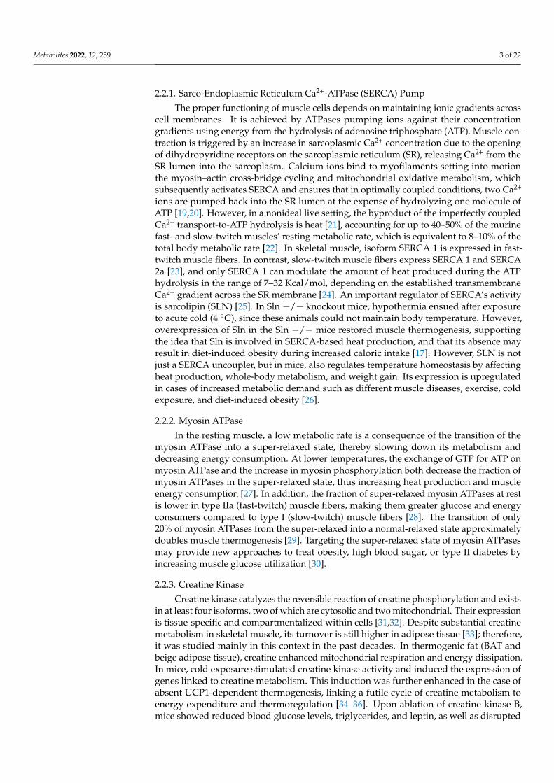
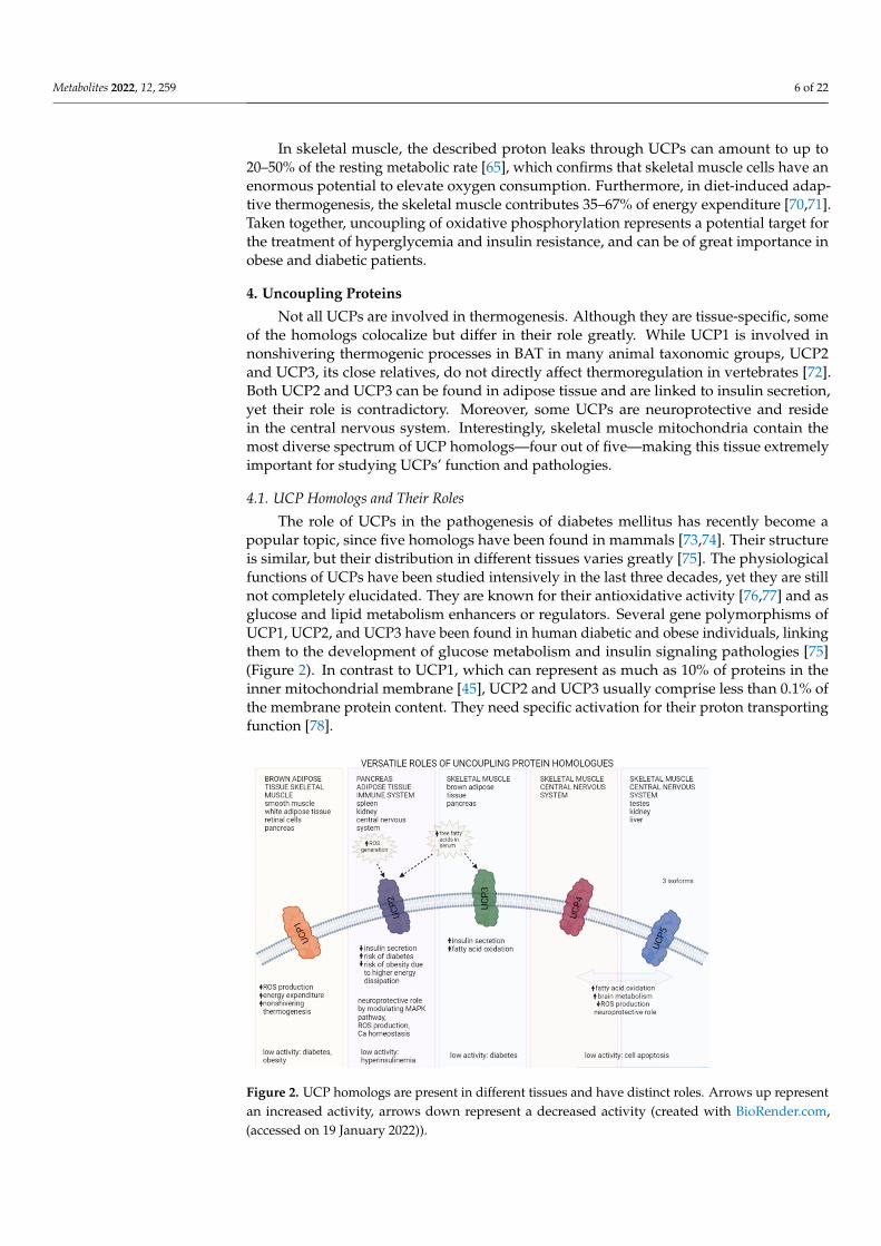
and outer membrane through transmembrane complexes I, III, and IV [56,57]. Accumula-tion of H+ ions in the intermembrane space results in a powerful proton gradient acrossthe membrane, driving their diffusion back into the matrix through F1F0 ATP synthase(Figure 1).
Metabolites 2022, 12, x FOR PEER REVIEW 5 of 24
3. Coupling, Uncoupling, and Their Interplay in Skeletal Muscle Cells In 1961, Peter Mitchell proposed the mechanism of ATP production in
mitochondria known as the chemiosmotic mechanism of ATP synthesis [52–54], for which he received the Nobel Prize for Chemistry in 1978 [55]. According to Mitchell’s chemiosmotic theory, ATP synthesis exploits the electrochemical gradient across the inner mitochondrial membrane. This gradient arises from passing electrons from NADH and FADH2 formed in the Krebs cycle during the mitochondrial metabolism of energy-rich molecules through a series of membrane-bound protein complexes I–IV. At the same time, hydrogen ions (H+) are pumped from the mitochondrial matrix to the interspace between the mitochondrial inner and outer membrane through transmembrane complexes I, III, and IV [56,57]. Accumulation of H+ ions in the intermembrane space results in a powerful proton gradient across the membrane, driving their diffusion back into the matrix through F1F0 ATP synthase (Figure 1).
Figure 1. A schematic overview of UCP function and localization in a mitochondrion (created with BioRender.com, (accessed on 19 January 2022)).
During this diffusion, the released energy subsequently drives phosphorylation of ADP to ATP [58,59]. Recently, Nath showed that the molecular mechanism of uncoupling the proton transport from ATP synthesis by dinitrophenols cannot be explained by simple proton conduction through uncouplers, as postulated by Mitchell’s theory. Instead, it requires a two-ion theory of energy coupling/uncoupling in ATP synthase [60]. The enzymatic activity of ATP synthase also catalyzes the reverse reaction; namely, the ATP hydrolysis, thereby influencing the forward reaction equilibrium. The reverse-forward activity of ATP-synthase is strictly controlled to prevent ATP hydrolysis at the site. This control is achieved by inhibiting the reversal of ATP synthase. An inhibitor protein named IF1 has been identified to play a role in this task, but it is insufficient by itself, and other potential regulating mechanisms are under investigation [61,62]. Due to many unresolved questions, researchers recently proposed an update of the chemiosmotic theory questioning the form of proton-motive force across the membrane based on many discoveries with novel and improved research methods [57,63].
To complicate things further, the coupling of respiration to ATP synthesis is imperfect, and mitochondrial energy consumption persists even when ATP synthesis is
Figure 1. A schematic overview of UCP function and localization in a mitochondrion (created withBioRender.com, (accessed on 19 January 2022)).
During this diffusion, the released energy subsequently drives phosphorylation ofADP to ATP [58,59]. Recently, Nath showed that the molecular mechanism of uncouplingthe proton transport from ATP synthesis by dinitrophenols cannot be explained by sim-ple proton conduction through uncouplers, as postulated by Mitchell’s theory. Instead,it requires a two-ion theory of energy coupling/uncoupling in ATP synthase [60]. The en-zymatic activity of ATP synthase also catalyzes the reverse reaction; namely, the ATPhydrolysis, thereby influencing the forward reaction equilibrium. The reverse-forwardactivity of ATP-synthase is strictly controlled to prevent ATP hydrolysis at the site. This con-trol is achieved by inhibiting the reversal of ATP synthase. An inhibitor protein namedIF1 has been identified to play a role in this task, but it is insufficient by itself, and otherpotential regulating mechanisms are under investigation [61,62]. Due to many unresolvedquestions, researchers recently proposed an update of the chemiosmotic theory questioningthe form of proton-motive force across the membrane based on many discoveries withnovel and improved research methods [57,63].
To complicate things further, the coupling of respiration to ATP synthesis is imperfect,and mitochondrial energy consumption persists even when ATP synthesis is inhibited, con-firming the presence of uncoupling or leak mechanisms [64–66]. In the light of strict controlover ATP production, specific cellular mechanisms dissociate mitochondrial membranepotential generation from its usage to generate ATP. These processes might have evolved tocontrol ATP production and match it to cellular consumption, or to convert electrochemicalenergy into thermal energy to regulate body temperature. Such bypassing of the ATPsynthase through specific proteins called UCPs produces thermal energy without ATPsynthesis. One of the first UCPs to be discovered and described was thermogenin, found inBAT [67,68]. Many different homologs followed in human and other taxonomic species,which we will briefly summarize in the next section [69].
Metabolites 2022, 12, 259 6 of 22
In skeletal muscle, the described proton leaks through UCPs can amount to up to20–50% of the resting metabolic rate [65], which confirms that skeletal muscle cells have anenormous potential to elevate oxygen consumption. Furthermore, in diet-induced adap-tive thermogenesis, the skeletal muscle contributes 35–67% of energy expenditure [70,71].Taken together, uncoupling of oxidative phosphorylation represents a potential target forthe treatment of hyperglycemia and insulin resistance, and can be of great importance inobese and diabetic patients.
4. Uncoupling Proteins
Not all UCPs are involved in thermogenesis. Although they are tissue-specific, someof the homologs colocalize but differ in their role greatly. While UCP1 is involved innonshivering thermogenic processes in BAT in many animal taxonomic groups, UCP2and UCP3, its close relatives, do not directly affect thermoregulation in vertebrates [72].Both UCP2 and UCP3 can be found in adipose tissue and are linked to insulin secretion,yet their role is contradictory. Moreover, some UCPs are neuroprotective and residein the central nervous system. Interestingly, skeletal muscle mitochondria contain themost diverse spectrum of UCP homologs—four out of five—making this tissue extremelyimportant for studying UCPs’ function and pathologies.
4.1. UCP Homologs and Their Roles
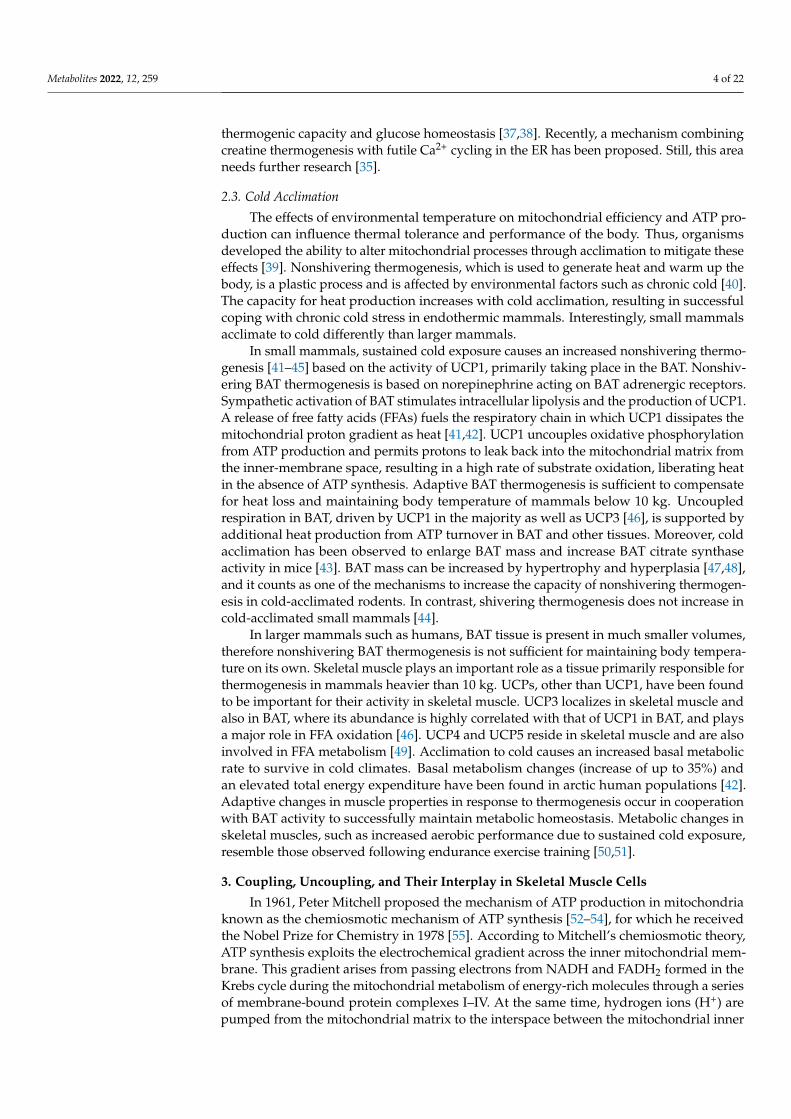
The role of UCPs in the pathogenesis of diabetes mellitus has recently become apopular topic, since five homologs have been found in mammals [73,74]. Their structureis similar, but their distribution in different tissues varies greatly [75]. The physiologicalfunctions of UCPs have been studied intensively in the last three decades, yet they are stillnot completely elucidated. They are known for their antioxidative activity [76,77] and asglucose and lipid metabolism enhancers or regulators. Several gene polymorphisms ofUCP1, UCP2, and UCP3 have been found in human diabetic and obese individuals, linkingthem to the development of glucose metabolism and insulin signaling pathologies [75](Figure 2). In contrast to UCP1, which can represent as much as 10% of proteins in theinner mitochondrial membrane [45], UCP2 and UCP3 usually comprise less than 0.1% ofthe membrane protein content. They need specific activation for their proton transportingfunction [78].
Metabolites 2022, 12, x FOR PEER REVIEW 7 of 24
Figure 2. UCP homologs are present in different tissues and have distinct roles. Arrows up represent an increased activity, arrows down represent a decreased activity (created with BioRender.com, (accessed on 19 January 2022)).
4.1.1. UCP1 Studies in rodents have shown that BAT starts to develop in the interscapular
region during embryonic days E15–16, and that UCP1 mRNA expression increases around days E18–19 just before birth. The BAT continues to develop postnatally until between postnatal days P15–21 and remains present throughout adult life [79,80]. Recent research revealed the existence of two subpopulations of brown adipocytes in mice. One subpopulation has high thermogenic activity and high UCP1 expression, and the other has low thermogenic activity and low UCP1 expression [81]. At birth, all adipocytes express high levels of UCP1 and have high thermogenic activity to meet newborns’ thermal requirements. Postnatally, some adipocytes begin to convert to the subpopulation with low UCP1 expression so that both subpopulations coexist in adult mice and might switch between each other during normal thermogenesis at room temperature. When exposed to cold, the transcription of genes in the subpopulation with the low UCP1 expression increases, thereby increasing the total thermogenic capacity of BAT [82]. During long-term cold exposure, de novo adipogenesis was observed in BAT [83,84]. In senescence, the capacity of adipocytes to increase UCP1 expression after cold exposure becomes impaired [82].
UCP1 mainly localizes to the inner mitochondrial membrane of BAT. Its proton conductance increases in elevated concentrations of long-chain free fatty acids (FFAs) [41] and is controlled by insulin [85,86]. Apart from BAT, recent studies also reported UCP1 expression in white adipose tissue, skeletal muscle, longitudinal smooth muscle layers, retinal cells, and Langerhans islet cells [75,87].
In skeletal muscle mitochondria, the expression of UCP1 reaches only 13% of the expression in BAT and increases the GDP-sensitive proton leak [88]. The roles of UCP1 are decreasing membrane potential, reducing reactive oxygen species (ROS) generation, increasing energy expenditure, and increasing nonshivering thermogenesis [89–91]. Compared to BAT, the ability of UCP1 in skeletal muscle to increase glutathione levels and reduce ROS production is far greater, suggesting different specific roles and possibly distinct mechanisms of UCP1 in both tissues [88]. Some research shows that
Figure 2. UCP homologs are present in different tissues and have distinct roles. Arrows up representan increased activity, arrows down represent a decreased activity (created with BioRender.com,(accessed on 19 January 2022)).
Metabolites 2022, 12, 259 7 of 22
4.1.1. UCP1
Studies in rodents have shown that BAT starts to develop in the interscapular regionduring embryonic days E15–16, and that UCP1 mRNA expression increases around daysE18–19 just before birth. The BAT continues to develop postnatally until between postnataldays P15–21 and remains present throughout adult life [79,80]. Recent research revealedthe existence of two subpopulations of brown adipocytes in mice. One subpopulation hashigh thermogenic activity and high UCP1 expression, and the other has low thermogenicactivity and low UCP1 expression [81]. At birth, all adipocytes express high levels of UCP1and have high thermogenic activity to meet newborns’ thermal requirements. Postnatally,some adipocytes begin to convert to the subpopulation with low UCP1 expression so thatboth subpopulations coexist in adult mice and might switch between each other duringnormal thermogenesis at room temperature. When exposed to cold, the transcription ofgenes in the subpopulation with the low UCP1 expression increases, thereby increasingthe total thermogenic capacity of BAT [82]. During long-term cold exposure, de novoadipogenesis was observed in BAT [83,84]. In senescence, the capacity of adipocytes toincrease UCP1 expression after cold exposure becomes impaired [82].
UCP1 mainly localizes to the inner mitochondrial membrane of BAT. Its proton con-ductance increases in elevated concentrations of long-chain free fatty acids (FFAs) [41]and is controlled by insulin [85,86]. Apart from BAT, recent studies also reported UCP1expression in white adipose tissue, skeletal muscle, longitudinal smooth muscle layers,retinal cells, and Langerhans islet cells [75,87].
In skeletal muscle mitochondria, the expression of UCP1 reaches only 13% of theexpression in BAT and increases the GDP-sensitive proton leak [88]. The roles of UCP1are decreasing membrane potential, reducing reactive oxygen species (ROS) generation,increasing energy expenditure, and increasing nonshivering thermogenesis [89–91]. Com-pared to BAT, the ability of UCP1 in skeletal muscle to increase glutathione levels andreduce ROS production is far greater, suggesting different specific roles and possibly dis-tinct mechanisms of UCP1 in both tissues [88]. Some research shows that diabetes andobesity development involves specific polymorphisms of the Ucp1 gene [92]. Mutations inUcp1 affect the activity or expression of the UCP1 protein and reduce regulated or basalenergy expenditure, resulting in altered pancreatic function and insulin secretion [93,94].
4.1.2. UCP2
UCP2 mRNA is expressed in many tissues, such as muscle, spleen, pancreas, kidney,central nervous system, and immune system. The UCP2 gene is already expressed duringfetal life in murine skeletal muscle. Its expression increases immediately after birth, reachinga maximum on day 2, and steadily declines after that regardless of the lactating mother’sdiet [95].
UCP2 is most widely present and highly expressed among UCPs in diabetic pancreaticbeta-cells [96]; therefore, its involvement in diabetes development has been proposed.Its role in the pancreas as a negative regulator of insulin secretion has been studied inten-sively in ob/ob mice. The activation of UCP2 by ROS causes mitochondrial membraneproton leak, which reduces ATP synthesis in pancreatic β-cells and downregulates glucose-stimulated insulin secretion [97–99]. The ob/ob mice lacking UCP2 have increased ATP syn-thesis and glucose-stimulated insulin secretion from beta-cells in Langerhans islets [96,100].DeSouza et al. (2007) used an antisense oligonucleotide to Ucp2 in ob/ob mice and Swissmice with hyperlipidemic diet-induced obesity and diabetes to inhibit UCP2 expression,resulting in metabolic improvement [99]. Finally, results from a human study on ethnicitydifferences in UCP2 polymorphisms demonstrated that in Asians, the UCP2-866G/A poly-morphism is protective against, while the UCP2 Ala55Val polymorphism is susceptibleto, type 2 diabetes [101]. Similar traits might also exist in mice, but these have not beenthoroughly researched yet.
One of the reported other roles of UCP2 is controlling immune cell activation bymodulating MAPK pathways and mitochondrial ROS production [102,103]. Additionally,

Metabolites 2022, 12, 259 8 of 22
a neuroprotective role has been proposed. By regulating mitochondrial membrane potential,production of ROS, and calcium homeostasis, UCP2 modulates neuronal activity andinhibits cellular damage [104].
4.1.3. UCP3
UCP3 is expressed in skeletal muscle and BAT [105–108]. In BAT, UCP3 is almostone order of magnitude more abundant than in skeletal muscle or heart, and is directlycorrelated with the abundance of UCP1 [46]. The predominant isoform in skeletal muscleis UCP3, and its expression is highly skeletal-muscle-specific [109]. In mice, UCP3 mRNAlevels were highest in skeletal muscle, followed by heart, white adipose tissue, and spleen,which was somewhat different than in rats, where the expression in tissues other thanskeletal muscle was negligible [110].
UCP3 expression was almost undetectable in murine muscle tissue during fetal life.In contrast, its expression became noticeable soon after birth in response to sucklingand lipid intake, and steadily increased for 15 days. Interestingly, after 15 days of life,the UCP3 mRNA levels became dependent on dietary interventions. If lactating mice werefed regular high-carbohydrate chow, UCP3 expression levels in pups started to decrease,whereas if mothers were fed a high-fat diet, the levels of UCP3 expression in pups remainedhigh [95]. Research shows that nutritional factors regulate UCP3 expression. Specifically,its expression is induced by elevated circulating FFAs, which is typical for fasting orstarvation [95,111]. Pedraza et al. reported that the UCP3 expression in skeletal muscle isdramatically downregulated in lactating mice, and this effect is reversed with weaning.These changes come hand-in-hand with changes in circulating FFAs, which are reducedduring lactation and return to normal after weaning [112].
Pancreatic beta cells also express UCP3 [113], linking its role to energy expenditure,glucose metabolism, diabetes, and obesity [114,115]. Pancreatic UCP3 also affects insulinsecretion, but acts differently than UCP2 [113]. In humans, the expression of the Ucp3gene in skeletal muscle and pancreas of diabetic patients is decreased [116], suggestingUcp3 involvement in the development of type 2 diabetes. Muscle UCP3 is also importantin FFA metabolism. It protects mitochondria from oxidative stress induced by lipidsand modulates insulin sensitivity [117], making it a potential player in type 2 diabetesdevelopment. UCP3 protein levels are upregulated when FFAs’ supply to the mitochondriaexceeds their oxidative capacity, and downregulated when oxidative capacity is improved.
The degradation of both UCP2 and UCP3 is very rapid [118], making their half-lives only approximately 30 min [119]. In comparison, the half-life of UCP1 is around30 h [120]. The short half-lives of UCP2 and UCP3 enable rapid adjustments of their proteinlevels, which are needed when facing the rapidly changing metabolic needs and differentrates of ROS production during mitochondrial oxidative processes. Because of this rapiddegradation, the UCP2 protein level can decrease before the level of its mRNA drops [121].It is crucial to consider this when evaluating data and drawing conclusions solely onmRNA expression.
4.1.4. Other UCPs
UCP4 and UCP5 are mainly expressed in the central nervous system, where they playroles in brain metabolism and thermoregulatory heat production and are therefore oftennamed neuronal UCPs [122,123]. However, their expression has also been determined inskeletal muscle, controlling energy expenditure and lipid oxidation. UCP5 is expressed inhuman skeletal muscle in three different isoforms, with UCP5L being the most abundantisoform, followed by UCP5S and UCP5SI [49]. UCP4 and UCP5 have a similar role in theprotection against oxidative stress and mitochondrial dysfunction as other homologs [124].High levels of UCP5 mRNA have been detected in testes, and lower levels in the kidneysand liver [49].

Metabolites 2022, 12, 259 9 of 22
4.2. Sex Differences in UCP Expression
Studies with rodents of both genders have shown significant sex-associated differencesin the regulation of UCPs, which occur due to sex hormones and other distinct gender-based biological functions [125]. Sex hormone receptors are localized in the mitochondriaof specific cells and can affect mitochondrial physiology [126]. In rodents, sex hormonesinfluence different features of skeletal muscle, such as fiber diameter and myosin heavy-chain expression [127]. They also regulate UCP1 expression in brown adipocytes [128,129].
Age plays a vital role in the sex dimorphism of UCP expression. In prepubertal agein mice, UCPs are expressed at similar levels in both sexes, with significant differences,especially in UCP1 and UCP3 expression, being observed only later in adulthood. Expres-sion of these proteins decreased with time in adult males, while in females, UCP1 andUCP3 expression decreased during young adulthood and increased later [130]. This age-dependent UCPs expression pattern correlates with weight gain. In several studies, weightgain with aging was more significant in males than in female mice, which showed only aslight increase in body weight with senescence. This finding suggests that upregulationof UCP1 and UCP3 in BAT helps female mice avoid triglyceride accumulation in skeletalmuscle and prevents obesity development [114,130,131].
Caloric diet feeding causes different overweight-induced expression of UCP3 in muscleand UCP1 in BAT in males than in females. Females tend to have a higher capacity to storefat when food is in excess than males, resulting in weight gain [125]. On the other hand,experiments with fasting showed interesting sex-dependent differences in UCP expression.Bazhan et al. (2019) studied sex asymmetry in the fasting effects on the transcription of theUcp3 gene in muscle. A significant upregulation of muscle Ucp3 occurred in females afterfasting for 24 h, while these changes were much less evident in males [132].
5. UCP2 and Insulin Secretion in Pancreatic Beta-Cells
The mitochondria were estimated to produce 98% of the cell’s ATP in oxidativemetabolism [133]. Glucose influx into pancreatic beta-cells followed by an increase inglucose metabolism elevates cytosolic adenosine triphosphate (ATP) concentration, clos-ing ATP-dependent K+ (KATP) channels and decreasing K+ efflux. Consequent cell mem-brane depolarization opens voltage-dependent Ca2+ (VDCC) channels and Ca2+ ionsinflux, which triggers insulin secretion [134–136]. The predominant UCP in beta-cells isUCP2, which regulates cellular oxidative stress, ROS production, and energy metabolism,and protects mice from aging [137,138].
Recently, a new role of UCP2 was revealed, linking its expression to the regulationof embryonic development of the pancreas. Broche et al. showed that UCP2 regulatesembryonic development of the pancreas through the ROS-AKT signaling pathway bydecreasing ROS production. Their study confirmed an increased pancreas size with ahigher number of α- and β-cells in Ucp2−/− fetuses, a faster perinatal proliferation ofendocrine cells, and increased ROS production [137].
Increased UCP2 expression can cause a lack of glucose effect on insulin secretion intype 2 diabetes [139]. UCP2 allows H+ ions to bypass ATP synthase, reducing cellular ATPcontent [140]. Subsequently, beta-cell membrane depolarization and glucose-stimulated in-sulin secretion (GSIS) decrease. A high-fat diet (HFD) or long-term exposure of beta-cells toelevated concentrations of FFAs creates glucolipotoxic conditions, which upregulate UCP2mRNA levels and protein content up to twofold [141]. Consistent with the proposed mecha-nism of UCPs action, the decreased cellular ATP-to-ADP ratio suppressed glucose-induceddepolarization of the plasma membrane. Furthermore, the GSIS decrease was inverselyproportional to UCP2 expression [142,143]. UCP2 mRNA content increased followingexposure to palmitate or its nonoxidizable derivative bromopalmitate. This observationsuggests that FFAs can directly upregulate UCP2 gene expression without their precedingmetabolism [141]. In contrast, UCP2 knockout (UCP2−/−) mice on a control diet havesimilar fasting blood glucose, fed plasma-free FFAs, and triglyceride (TG) levels as theirwild-type (WT) littermates. On the other hand, their fasting blood insulin concentration

Metabolites 2022, 12, 259 10 of 22
was significantly higher, responding differently to the HFD [144]. In UCP2−/− mice,fasting FFA concentration following the HFD was not elevated. In addition, the fed plasmaTG increase was nearly twofold less than WT control, suggesting that faster mitochondrialFFA oxidation in UCP2−/− islets prevents lipotoxicity-associated TG accumulation. Fur-thermore, fasting and fed plasma insulin levels following the HFD rose significantly inUCP−/− and control mice, but the increase was much higher in the UCP2−/− group.Pancreatic insulin content increased around fourfold in UCP2−/−mice, while it slightlydecreased in the WT control. Interestingly, in UCP2−/−mice, the fasting blood glucoseconcentration was not affected by the HFD, and the fed blood glucose level increase wassignificantly lower than in the WT control. Further research on islets from UCP2−/−mice on a control diet showed increased insulin content per islet, relative beta-cell area perpancreas, and average islet size, but a comparable number of islets per mm2 of pancreascompared to the WT islets [144]. Following the HFD, the islet size and beta-cell area increasein UCP−/−mice were significantly larger than in WT islets. In addition, the number ofislets per mm2 of the pancreas increased significantly, which was not evident in controlislets [144].
When researching mitochondrial metabolism, Joseph et al. showed that basal insulinsecretion in WT and UCP−/−mice on the HFD was elevated; however, GSIS was attenu-ated in WT mice, while UCP−/− mice showed significantly increased GSIS. The beta-cellsfrom UCP−/− islets had no alterations in mitochondrial membrane potential, ATP/ADPratio, and cytosolic Ca2+ responses to high glucose concentration after palmitate treatmentcompared to the dysfunctional responses of WT cells. The HFD neither resulted in glucosesensitivity loss nor elevated TG concentration as it did in the WT control, because thepalmitate oxidation was faster in the UCP2−/− islets [145]. These data suggest thatUCP2−/− beta-cells can resist the toxic effects of a high-lipid environment by preventingTG accumulation due to faster FFA oxidation rates and maintaining highly functionalglucose-dependent metabolism–secretion coupling [145].
The most studied obesity mouse models, the ob/ob mice, have elevated UCP2 expres-sion and impaired insulin secretion [96,99,144]. While in lean mice, GSIS improved after ashort-term knockdown of islet UCP2, it remained unaffected in ob/ob mice, confirming thedysfunctionality of glucose homeostasis in the ob/ob diabetes model. In addition, this lossof glucose homeostasis and insulin secretion impairment were preceded by an increase inUCP2 expression [146].
6. UCPs in Mouse Models of Diabetes and Obesity6.1. Obesity and Diabetic Models
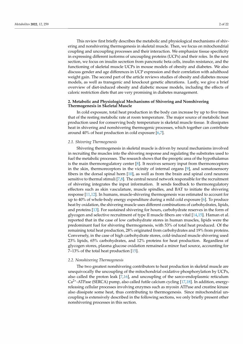
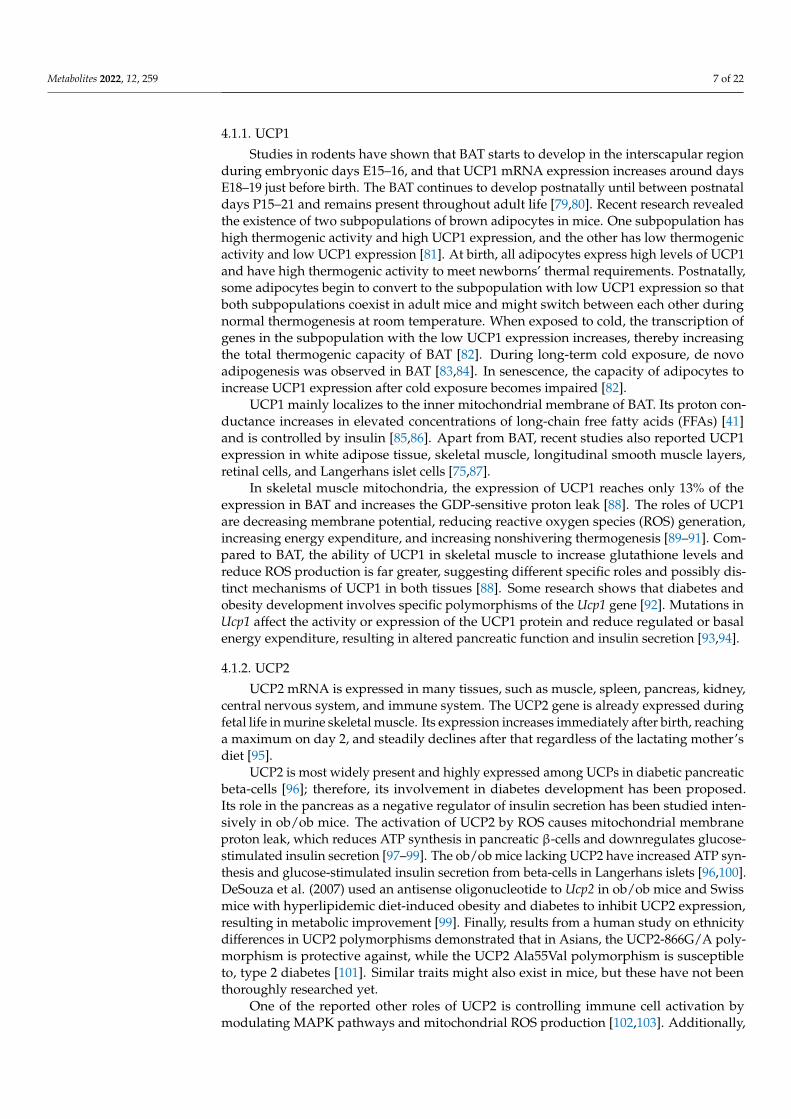
Obesity is an important factor affecting UCP expression in skeletal muscle. For thisreason, there are many UCP expression studies using mouse models of obesity and diabetes(Figure 3). Masaki et al. studied the thermogenic roles of UCPs in different tissues in acold-exposed db/db mouse model [147]. Cold is known to increase energy expenditure andthermogenesis [148]. Db/db mice resulted in impaired body temperature maintenance thatpresented as decreased thermogenic capacity. Compared to lean littermates, db/db micewere incapable of cold acclimation and had a diminished increase in UCP expression inskeletal muscle and brown and white adipose tissue. While cold exposure downregulatedlipoprotein lipase mRNA and increased serum levels of FFAs in lean mice, these effectswere not seen in db/db mice. These results suggest that in db/db mice, the reducedlipolysis may impair UCPs’ function [147].

Metabolites 2022, 12, 259 11 of 22
Metabolites 2022, 12, x FOR PEER REVIEW 11 of 24
palmitate treatment compared to the dysfunctional responses of WT cells. The HFD neither resulted in glucose sensitivity loss nor elevated TG concentration as it did in the WT control, because the palmitate oxidation was faster in the UCP2−/− islets [145]. These data suggest that UCP2−/− beta-cells can resist the toxic effects of a high-lipid environment by preventing TG accumulation due to faster FFA oxidation rates and maintaining highly functional glucose-dependent metabolism–secretion coupling [145].
The most studied obesity mouse models, the ob/ob mice, have elevated UCP2 expression and impaired insulin secretion [96,99,144]. While in lean mice, GSIS improved after a short-term knockdown of islet UCP2, it remained unaffected in ob/ob mice, confirming the dysfunctionality of glucose homeostasis in the ob/ob diabetes model. In addition, this loss of glucose homeostasis and insulin secretion impairment were preceded by an increase in UCP2 expression [146].
5. UCPs in Mouse Models of Diabetes and Obesity 5.1. Obesity and Diabetic Models
Obesity is an important factor affecting UCP expression in skeletal muscle. For this reason, there are many UCP expression studies using mouse models of obesity and diabetes (Figure 3). Masaki et al. studied the thermogenic roles of UCPs in different tissues in a cold-exposed db/db mouse model [147]. Cold is known to increase energy expenditure and thermogenesis [148]. Db/db mice resulted in impaired body temperature maintenance that presented as decreased thermogenic capacity. Compared to lean littermates, db/db mice were incapable of cold acclimation and had a diminished increase in UCP expression in skeletal muscle and brown and white adipose tissue. While cold exposure downregulated lipoprotein lipase mRNA and increased serum levels of FFAs in lean mice, these effects were not seen in db/db mice. These results suggest that in db/db mice, the reduced lipolysis may impair UCPs’ function [147].
Figure 3. Different mouse models enable research on UCP’s roles in insulin sensitivity, diabetes, and obesity.
In skeletal muscle, exposure to cold caused a reduction in UCP3 mRNA expression. The cause for the disruption of temperature maintenance in db/db mice is most likely a leptin-signaling malfunction, as db/db mice are leptin-receptor-mutated [149], thus confirming the vital role of leptin in regulating UCP expression. Further studies
Figure 3. Different mouse models enable research on UCP’s roles in insulin sensitivity, diabetes,and obesity.
In skeletal muscle, exposure to cold caused a reduction in UCP3 mRNA expression.The cause for the disruption of temperature maintenance in db/db mice is most likely aleptin-signaling malfunction, as db/db mice are leptin-receptor-mutated [149], thus con-firming the vital role of leptin in regulating UCP expression. Further studies supported thisidea by confirming increased UCP2 and UCP3 mRNA expression in pancreatic islets andmuscle, respectively, in ob/ob mice with adenoviral-mediated leptin expression [150]. Lep-tin induces skeletal muscle UCP expression and sympathetic innervation [151], significantlycontributing to thermogenesis.
Apart from impaired leptin action in db/db mice, poor responsiveness of UCPs toregulation with serum FFAs has been suggested as a possible cause of thermogenesisimpairment. FFAs are modulators of UCP regulation [152], and during cold exposure,their concentration in serum increases. Typically, this would trigger an increase in mi-tochondrial UCP3 expression in skeletal muscle as a mechanism to dispose of excessFFAs [153]. However, in db/db mice, acute elevation of FFAs in the serum after coldexposure failed to upregulate UCP3 mRNA expression in skeletal muscle [147].
In the last few decades, many studies searched for possible treatments to increasethe amount of UCPs in skeletal muscle to achieve an antiobesity effect. Sympatheticnerves directly control skeletal muscle and adipose tissue thermogenesis through theβ-adrenergic action of norepinephrine [154]. Nagase et al. (1996) used a β3-adrenergicagonist to increase UCP expression in muscles and BAT, which resulted in increased energyexpenditure, enhanced oxygen consumption, improved glucose tolerance, and reducedbody fat in obese yellow KK mice [155,156].
6.2. Transgenic and Knockout Mice
Besides the obesity and diabetic mouse models, knockout and transgenic mousemodels enabled targeted studies of UCP roles [157] (Figure 3). UCP1 knockout micecan maintain their energy balance despite the absence of UCP1-mediated thermogenesisvia higher proton-leak-dependent oxygen consumption in muscles [158]. In the adiposetissue–UCP1 knockout mice, the ectopic expression of UCP1 in skeletal muscle revealed anessential role of skeletal muscle respiratory uncoupling in preventing diet-induced obesity,insulin resistance, and cholesterolemia [159]. Accelerated metabolism in skeletal muscle byuncoupling activity due to UCP1 expression may also delay age-related diseases such asdiabetes, hypertension, atherosclerosis, and cancer [160].

Metabolites 2022, 12, 259 12 of 22
Furthermore, the roles of UCP1 in skeletal muscle and BAT are different. The impact ofUCP1 on glutathione and ROS levels has been studied using a transgenic mouse model withselective expression of UCP1 in skeletal muscle. Even though UCP1 expression in skeletalmuscle reached only up to 13% of levels compared to BAT, the increased GDP-sensitiveproton leak through UCP1 in muscle cells increased total mitochondrial glutathione levelsmore than sevenfold compared to BAT [88]. However, unlike in BAT, the leak throughUCP1 affected the mitochondrial ROS emission. Upon subsequent inhibition of UCP1 withGDP, ROS production in transgenic mice increased 2.8-fold relative to WT littermates [88],confirming the involvement of UCP1 in the mitochondrial oxidative state.
UCP2 affects glucose homeostasis by controlling insulin secretion, food intake behav-ior, and adiponectin secretion in the pancreas, brain, and adipose tissue [161]. The role ofUCP2 in β-cell glucose sensing was revealed in a study using ob/ob UCP2 knockout mice,as they expressed higher ATP levels and increased insulin secretion than the control groupof ob/ob mice with an active UCP2 gene [96]. Like UCP1 knockout mice, UCP2 knockoutmice do not become obese when fed a HFD. Moreover, they have increased ROS productionand a normal response to cold exposure [162]. Therefore, the negative regulation of insulinsecretion by UCP2 represents a strong link between obesity, β-cell dysfunction, and thedevelopment of type 2 diabetes. On the other hand, overexpression of UCP2 proteins inmice decreases obesity and improves insulin sensitivity [163].
Furthermore, UCP3 promotes FFA oxidation in muscle, thereby indirectly influencingglucose metabolism [161]. Knockout mice lacking UCP3 have been created for physiologicalstudies, and their phenotype was also confirmed to be nonobese. An increased ROS pro-duction and no significant thermoregulatory function of UCP3 were determined [108,164].Transgenic mice expressing human ortholog of UCP3 at high levels in skeletal muscle wereused in several studies and resulted in leanness. Their mitochondrial thioesterase-1 (MTE-1)and lipoprotein lipase expression were increased [153]. Adipose tissue mass decreased,and glucose clearance rate increased, making them resistant to obesity and diabetes de-velopment [163,165]. Within the mitochondria, MTE-1 cleaves acyl-CoA long-chain FFAsto FFA anions and CoASH. Further on, UCP3′s role is to export these anions from themitochondrial matrix under conditions of increased β-oxidation [166,167]. Thus, UCP3 andMTE-1 have been implicated in FFA metabolism, and correlate with situations and tissuesin which FFA β-oxidation is increased, such as skeletal muscle and BAT.
6.3. Diet-Induced Obesity and Diabetic Models
Studies in various mouse models revealed expression and function changes of differentUCPs in various tissues in response to specific diets (Figure 3). The Western diet, whichis high in sugar, protein, and fat, and the HFD are often used to trigger the developmentof systemic insulin resistance, type 2 diabetes, and obesity. A study in UCP1 knockoutmice showed that loss of UCP1 increased susceptibility to Western-diet-induced insulinresistance and glucose intolerance [168].
As the loci of UCP2 and UCP3 genes on mouse chromosome 7 and human chromosome11 are close [107], they represent good candidate genes for the quantitative trait locus ofdiet-induced obesity and diabetes [169,170]. Research shows that diet can regulate UCP2and UCP3 expression, and that their levels can increase due to fat consumption or refeedingafter starvation [152,170]. In addition, cold stimulation and the sympathetic nervous systemalso regulate UCP3 content.
Feeding C57BL/6 mice with the HFD causes insulin resistance and reduced proteinexpression of GLUT4 and phosphorylation of AMPK in skeletal muscle and adiposetissue [171]. Interestingly, the HFD increases UCP2 expression in adipose tissue in obesity-resistant A/J mice, but not in obesity- and diabetes-prone mouse strains [170]. Similarresults were obtained by Surwit et al. (1998). They also studied the correlation between theexpression of different UCPs in adipose tissue and skeletal muscle, and the consumption ofthe HFD in obesity-prone C57BL/6J mice and obesity-resistant A/J and C57BL/KsJ mice.Their study indicated that the HFD increases UCP2 expression in white adipose tissue
Metabolites 2022, 12, 259 13 of 22
in obesity- and diabetes-resistant mouse strains. However, the HFD did not affect UCP2expression in any tissue in obesity-prone mice. Moreover, the HFD did not affect UCP3or UCP2 mRNA expression in skeletal muscle in any studied strain. Thus, the inductionof diabetes and obesity by the HFD seems related mainly to UCP1 and UCP2 expressionin adipose tissue, but not to the expression of UCP2 and UCP3 in skeletal muscle [172].Therefore, uncoupling activity in adipocytes seems to have greater importance in controllingthe effects of fat feeding on the development of obesity and diabetes than uncouplingactivity in skeletal muscles.
In mice with overexpression of a UCP, a more potent uncoupler than UCP2 or UCP3in skeletal muscle, an enhanced insulin action and resistance to weight gain and insulinresistance induced by the HFD were determined [159]. Thus, skeletal muscle respirationuncoupling could contribute to treating obesity and pathologies linked to it.
Many studies of UCP function used food restriction and fasting. A study in WTmice confirmed that fasting increases UCP2 and UCP3 expression in skeletal muscle whileleaving proton leak unchanged [173]. Differences in respiratory quotients between wild-type and UCP3 knockout mice were found, and the absence of UCP3 resulted in impairedFFA oxidation. Altogether, these results suggest that UCP3 and UCP2 are highly linked toFFA oxidation and are therefore physiologically essential for FFA metabolism.
Moreover, mitochondrial dysfunction and spontaneous skeletal muscle apoptosisoccur in muscle pathology mouse models such as collagen VI-knockout mice, which resem-ble human myopathy. Starvation and a low-protein diet proved beneficial for dystrophicmuscles, as they protected them from atrophy and mitochondrial defects via inducedautophagy [174] Not long ago, a low-protein/high-carbohydrate diet (LPCD) was shownto induce UCP1 expression, enhance mitochondrial oxidative metabolism, and recruitdifferent energy-dissipating routes in murine beige adipocytes in subcutaneous adiposetissue. The induction of AMPK-dependent thermogenesis by the LPCD thus shows greatpotential as a valuable strategy for preventing metabolic diseases [175] The beneficial effectsof caloric restriction have also been confirmed regarding muscle stem cells. The enhancedmyogenic activity of satellite cells’ oxidative metabolism due to increased mitochondrialmass and function has been observed in mice that underwent short-term and long-termcaloric restriction [176]. Therefore, caloric restriction has tremendous therapeutic potentialin accelerating endogenous repair and improving the capacity of muscle stem cells.
Apart from caloric restriction and fasting, short-term HFD has also been shown tocounteract metabolic alterations in muscle fibers of dystrophic muscles. Fibro/adipogenicprogenitors, which reside in muscle and represent an interstitial stem cell population ofmesenchymal origin, have an important role in muscle regeneration [177]. Dystrophicprogenitors have an impaired mitochondrial metabolism, which reduces their ability toproliferate and differentiate into adipocytes. Using dystrophic mice, HFD has been shownto metabolically reprogram these progenitors and modulate their adipogenic potentialby restoring their mitochondrial functionality and UCP activity [178]. Several other stud-ies confirmed the involvement of UCPs in muscle regeneration. It has been discoveredthat muscle fibro/adipogenic progenitors can differentiate into UCP1-expressing beigeadipocytes, resulting in induction of muscle regeneration [179,180]. Moreover, transplan-tation of BAT into injured skeletal muscle has been shown to increase muscle mass andcontractile force [181], pointing to a significant role of UCP1 in muscle regeneration and agreat potential for therapy development.
The prevention of HFD consequences has been a hot topic in recent mice research.Polyphenols such as resveratrol, anthocyanin, curcumin, and epigallocatechin gallate havethe potential to alleviate hyperglycemia and insulin sensitivity caused by the HFD. The mostplausible mechanism of action involves stimulation of GLUT4 translocation on the plasmamembrane of skeletal and cardiac muscle cells and adipocytes via an activated AMPK-dependent signaling pathway [171,182,183]. Besides GLUT4 translocation, the activationof AMPK regulates the expression of UCP3 in skeletal muscle [184,185]. Furthermore,polyphenol-rich cacao liquor extract has been successful as a supplement in preventing
Metabolites 2022, 12, 259 14 of 22
the development of hyperglycemia in the db/db obesity mouse model [186]. In addition,in C57BL/6 mice, supplementation of the HFD with cacao liquor extract prevented micefrom diet-induced obesity. Polyphenols in cacao increased UCP3 expression in skeletalmuscle and other UCP expressions in different tissues. This triggered the activation ofAMPK, increased energy expenditure and thermogenesis, and prevented diet-inducedhyperglycemia, insulin resistance, and obesity [171]. Metformin, notably an anti-diabeticdrug, also has been extensively studied for its role in improving skeletal muscle metabolicand regenerative function. Metformin has several beneficial effects on stem cells, as it hasbeen recently discovered and reported in many papers [187–189]. It is known to reduceROS levels and protect mitochondria from oxidative damage, thus enabling an efficientactivity of UCPs. Metformin is therefore developing as a valuable therapeutic for treatingmuscle atrophy and dystrophies, as studies by Pavlidou et al. demonstrated that metformindelayed satellite cell activation and differentiation by favoring a quiescent, low metabolicstate, resulting in alleviated depletion of the stem cell pool and the functional loss of satellitecells [190].
6.4. Translational Precautions
Lately, the knowledge of mouse thermal physiology has raised questions about the rel-evance and translation of the results from preclinical studies on mice housed at a standardtemperature to a clinical level due to the environmental temperature effects on energy home-ostasis and metabolic rates [191]. As the body mass-to-surface ratio in mice is different fromhumans, the mouse body uses distinct thermo-biological processes to maintain temperaturehomeostasis [192]. In mice, an ambient temperature of about 23 ◦C triggers cold-inducedthermogenesis devoted to maintaining core body temperature, mainly in BAT, representingmore than one-third of the total energy expenditure. The basal metabolic rate, physicalactivity, and the thermogenic effect of food account for the remaining energy expenditure.To circumvent this energy-consuming process, mice can enter regulated hypothermia [193],which does not even closely resemble human sedentary processes at this temperature [194].This fact has led scientists to conclude that thermoneutral points for humans and mice arefar from similar and must be considered when conducting studies of obesity and diabetesmechanisms and treatment in mouse models. In further studies, researchers determinedthat the thermoneutral point for mice is around 30 ◦C, with energy expenditure consistingof approximately 70% basal metabolic rate, 20% physical activity energy expenditure, and10% thermic effect of food [195]. Therefore, this environmental temperature should be usedto better model human energy homeostasis in mouse models [196].
7. Conclusions
Maintaining temperature homeostasis is mainly achieved through thermogenic pro-cesses involving UCPs in BAT and skeletal muscle. The same proteins are involved in glu-cose homeostasis, thus linking high-energy dissipation and body weight control, a promis-ing research topic for treating obesity and type 2 diabetes. Research in obesity and diabeticmouse models has demonstrated that leptin and serum levels of FFAs play an essentialrole in regulating UCP3 expression in skeletal muscle, and that muscle thermogenesis inthese models is impaired. To elucidate the specific roles of different UCPs, knockout andtransgenic mouse models have been created. Experimental data revealed that overexpress-ing UCP1 in skeletal muscle can accelerate metabolic energy consumption and preventdiet-induced obesity and insulin resistance. On the other hand, UCP2 overexpressiondecreases insulin secretion in beta-cells, leading to obesity, β-cell dysfunction, and type2 diabetes. Changes in UCP expression in different tissues can also result from high-fatand/or high-carbohydrate diets such as the Western diet. However, such diets mainly affectUCP expression in adipose tissue, but have an insignificant impact on the skeletal muscle.
Harnessing the processes of thermogenic systems based on the UCPs offers a greatpotential to reduce obesity and diabetes. As many UCPs homologs are expressed in a broadrange of tissues, many targets exist to induce mitochondrial uncoupling to stimulate energy
Metabolites 2022, 12, 259 15 of 22
expenditure. In future research, attention should be paid to the thermoneutral point andenvironmental temperature when conducting studies in mice for a better translation offindings from mouse models to humans.
Author Contributions: Conceptualization, L.K.B.; writing—original draft preparation, M.C. andL.K.B.; writing—review and editing, M.C. and L.K.B.; visualization, M.C.; supervision, L.K.B. All au-thors have read and agreed to the published version of the manuscript.
Funding: This article was funded by Slovenian Research Agency, grant numbers: J3-9289 (LKB),N3-0133 (LKB), P3-0396 (LKB).
Conflicts of Interest: The authors declare no conflict of interest.
References1. Refinetti, R. The circadian rhythm of body temperature. Front. Biosci. 2010, 15, 564–594. [CrossRef] [PubMed]2. Talan, I.M.; Ingram, D.K. Age comparisons of body temperature and cold tolerance among different strains of Mus musculus.
Mech. Ageing Dev. 1986, 33, 247–256. [CrossRef]3. Gonzales, P.; Rikke, B.A. Thermoregulation in mice exhibits genetic variability early in senescence. AGE 2010, 32, 31–37.
[CrossRef] [PubMed]4. Levy, S.B. Brown adipose tissue and type 2 diabetes. Evol. Med. Public Health 2020, 2020, 70–71. [CrossRef]5. Vidal, H.; Langin, D.; Andreelli, F.; Millet, L.; Larrouy, D.; Laville, M. Lack of skeletal muscle uncoupling protein 2 and 3 mRNA
induction during fasting in type-2 diabetic subjects. Am. J. Physiol. Content 1999, 277, E830–E837. [CrossRef] [PubMed]6. Din, M.U.; Raiko, J.; Saari, T.; Kudomi, N.; Tolvanen, T.; Oikonen, V.; Teuho, J.; Sipilä, H.T.; Savisto, N.; Parkkola, R.; et al. Human
brown adipose tissue [15O]O2 PET imaging in the presence and absence of cold stimulus. Eur. J. Pediatr. 2016, 43, 1878–1886.[CrossRef] [PubMed]
7. Blondin, P.D.; Haman, F. Shivering and nonshivering thermogenesis in skeletal muscles. Handb. Clin. Neurol. 2018, 156,153–173. [PubMed]
8. Morrison, S.F. Central neural control of thermoregulation and brown adipose tissue. Auton. Neurosci. 2016, 196, 14–24.[CrossRef] [PubMed]
9. Ma, S.; Yu, H.; Zhao, Z.; Luo, Z.; Chen, J.; Ni, Y.; Jin, R.; Ma, L.; Wang, P.; Zhu, Z.; et al. Activation of the cold-sensing TRPM8channel triggers UCP1-dependent thermogenesis and prevents obesity. J. Mol. Cell Biol. 2012, 4, 88–96. [CrossRef] [PubMed]
10. Bautista, D.M.; Siemens, J.; Glazer, J.M.; Tsuruda, P.R.; Basbaum, A.I.; Stucky, C.L.; Jordt, S.-E.; Julius, D. The menthol receptorTRPM8 is the principal detector of environmental cold. Nature 2007, 448, 204–208. [CrossRef]
11. Romanovsky, A.A. Thermoregulation: Some concepts have changed. Functional architecture of the thermoregulatory system. Am.J. Physiol. Integr. Comp. Physiol. 2007, 292, R37–R46. [CrossRef] [PubMed]
12. Nakamura, K.; Morrison, S.F. Central efferent pathways for cold-defensive and febrile shivering. J. Physiol. 2011, 589, 3641–3658.[CrossRef] [PubMed]
13. Weber, M.J.; Haman, F. Fuel selection in shivering humans. Acta Physiol. Scand. 2005, 184, 319–329. [CrossRef] [PubMed]14. Haman, F. Shivering in the cold: From mechanisms of fuel selection to survival. J. Appl. Physiol. 2006, 100, 1702–1708. [CrossRef]15. Haman, F.; Péronnet, F.; Kenny, G.P.; Doucet, E.; Massicotte, D.; Lavoie, C.; Weber, J.-M. Effects of carbohydrate availability on
sustained shivering I. Oxidation of plasma glucose, muscle glycogen, and proteins. J. Appl. Physiol. 2004, 96, 32–40. [CrossRef]16. Wijers, S.L.J.; Schrauwen, P.; Saris, W.H.M.; Lichtenbelt, W.D.V.M. Human Skeletal Muscle Mitochondrial Uncoupling Is
Associated with Cold Induced Adaptive Thermogenesis. PLoS ONE 2008, 3, e1777. [CrossRef] [PubMed]17. Bal, N.C.; Maurya, S.K.; Sopariwala, D.H.; Sahoo, S.K.; Gupta, S.C.; Shaikh, S.A.; Pant, M.; Rowland, L.A.; Bombardier, E.;
Goonasekera, S.A.; et al. Sarcolipin is a newly identified regulator of muscle-based thermogenesis in mammals. Nat. Med. 2012,18, 1575–1579. [CrossRef] [PubMed]
18. Gamu, D.; Juracic, E.S.; Hall, K.J.; Tupling, A.R. The sarcoplasmic reticulum and SERCA: A nexus for muscular adaptivethermogenesis. Appl. Physiol. Nutr. Metab. 2020, 45, 1–10. [CrossRef]
19. Shishmarev, D. Excitation-contraction coupling in skeletal muscle: Recent progress and unanswered questions. Biophys. Rev. 2020,12, 143–153. [CrossRef] [PubMed]
20. Toyoshima, C.; Nakasako, M.; Nomura, H.; Ogawa, H. Crystal structure of the calcium pump of sarcoplasmic reticulum at 2.6 Åresolution. Nature 2000, 405, 647–655. [CrossRef]
21. De Meis, L.; Arruda, A.P.; Carvalho, D.P. Role of Sarco/Endoplasmic Reticulum Ca2+-ATPase in Thermogenesis. Biosci. Rep. 2005,25, 181–190. [CrossRef] [PubMed]
22. Smith, I.C.; Bombardier, E.; Vigna, C.; Tupling, A.R. ATP Consumption by Sarcoplasmic Reticulum Ca2+ Pumps Accounts for 40–50% of Resting Metabolic Rate in Mouse Fast and Slow Twitch Skeletal Muscle. PLoS ONE 2013, 8, e68924. [CrossRef] [PubMed]
23. Lytton, J.; Westlin, M.; Burk, S.E.; Shull, G.E.; MacLennan, D.H. Functional comparisons between isoforms of the sarcoplasmic orendoplasmic reticulum family of calcium pumps. J. Biol. Chem. 1992, 267, 14483–14489. [CrossRef]
24. De Meis, L. Energy interconversion by the sarcoplasmic reticulum Ca2+-ATPase: ATP hydrolysis, Ca2+ transport, ATP synthesisand heat production. An. Acad. Bras. Ciências 2000, 72, 365–379. [CrossRef] [PubMed]

Metabolites 2022, 12, 259 16 of 22
25. Bal, N.C.; Periasamy, M. Uncoupling of sarcoendoplasmic reticulum calcium ATPase pump activity by sarcolipin as the basis formuscle non-shivering thermogenesis. Philos. Trans. R. Soc. B Biol. Sci. 2020, 375, 20190135. [CrossRef]
26. Pant, M.; Bal, N.C.; Periasamy, M. Sarcolipin: A Key Thermogenic and Metabolic Regulator in Skeletal Muscle. Trends Endocrinol.Metab. 2016, 27, 881–892. [CrossRef]
27. Stewart, M.A.; Franks-Skiba, K.; Chen, S.; Cooke, R. Myosin ATP turnover rate is a mechanism involved in thermogenesis inresting skeletal muscle fibers. Proc. Natl. Acad. Sci. USA 2009, 107, 430–435. [CrossRef]
28. Phung, L.A.; Foster, A.D.; Miller, M.S.; Lowe, D.A.; Thomas, D.D. Super-relaxed state of myosin in human skeletal muscle isfiber-type dependent. Am. J. Physiol. Physiol. 2020, 319, C1158–C1162. [CrossRef] [PubMed]
29. Cooke, R. The role of the myosin ATPase activity in adaptive thermogenesis by skeletal muscle. Biophys. Rev. 2011, 3,33–45. [CrossRef]
30. Wilson, C.; Naber, N.; Cooke, R. The role of the super-relaxed state of myosin in human metabolism. Metab. Open 2021, 9,100068. [CrossRef]
31. Wallimann, T.; Wyss, M.; Brdiczka, D.; Nicolay, K.; Eppenberger, H.M. Intracellular compartmentation, structure and functionof creatine kinase isoenzymes in tissues with high and fluctuating energy demands: The ‘phosphocreatine circuit’ for cellularenergy homeostasis. Biochem. J. 1992, 281, 21–40. [CrossRef]
32. Kazak, L.; Cohen, P. Creatine metabolism: Energy homeostasis, immunity and cancer biology. Nat. Rev. Endocrinol. 2020, 16,421–436. [CrossRef]
33. Berlet, H.; Bonsmann, I.; Birringer, H. Occurrence of free creatine, phosphocreatine and creatine phosphokinase in adipose tissue.Biochim. Biophys. Acta (BBA)-Gen. Subj. 1976, 437, 166–174. [CrossRef]
34. Kazak, L.; Chouchani, E.T.; Jedrychowski, M.P.; Erickson, B.; Shinoda, K.; Cohen, P.; Vetrivelan, R.; Lu, G.Z.; Laznik-Bogoslavski,D.; Hasenfuss, S.C.; et al. A Creatine-Driven Substrate Cycle Enhances Energy Expenditure and Thermogenesis in Beige Fat. Cell2015, 163, 643–655. [CrossRef]
35. Wallimann, T.; Tokarska-Schlattner, M.; Kay, L.; Schlattner, U. Role of creatine and creatine kinase in UCP1-independent adipocytethermogenesis. Am. J. Physiol. Metab. 2020, 319, E944–E946. [CrossRef] [PubMed]
36. Rahbani, J.F.; Chouchani, E.T.; Spiegelman, B.M.; Kazak, L. Measurement of Futile Creatine Cycling Using Respirometry. In BrownAdipose Tissue; Humana: New York, NY, USA, 2022; pp. 141–153.
37. Streijger, F.; Pluk, H.; Oerlemans, F.; Beckers, G.; Bianco, A.C.; Ribeiro, M.O.; Wieringa, B.; Van der Zee, C.E. Mice lackingbrain-type creatine kinase activity show defective thermoregulation. Physiol. Behav. 2009, 97, 76–86. [CrossRef] [PubMed]
38. Rahbani, J.F.; Roesler, A.; Hussain, M.F.; Samborska, B.; Dykstra, C.B.; Tsai, L.; Jedrychowski, M.P.; Vergnes, L.; Reue, K.;Spiegelman, B.M.; et al. Creatine kinase B controls futile creatine cycling in thermogenic fat. Nature 2021, 590, 480–485. [CrossRef]
39. Bryant, H.J.; Chung, D.J.; Schulte, P.M. Subspecies differences in thermal acclimation of mitochondrial function and the role ofuncoupling proteins in killifish. J. Exp. Biol. 2018, 221, jeb.186320. [CrossRef]
40. Coulson, S.Z.; Robertson, C.E.; Mahalingam, S.; McClelland, G.B. Plasticity of non-shivering thermogenesis and brown adiposetissue in high-altitude deer mice. J. Exp. Biol. 2021, 224, 242279. [CrossRef] [PubMed]
41. Fedorenko, A.; Lishko, P.V.; Kirichok, Y. Mechanism of Fatty-Acid-Dependent UCP1 Uncoupling in Brown Fat Mitochondria. Cell2012, 151, 400–413. [CrossRef] [PubMed]
42. Levy, S.B.; Leonard, W.R. The evolutionary significance of human brown adipose tissue: Integrating the timescales of adaptation.Evol. Anthr. Issues News Rev. 2021, 1–17. [CrossRef]
43. Mineo, P.; Cassell, E.; Roberts, M.; Schaeffer, P. Chronic cold acclimation increases thermogenic capacity, non-shivering thermoge-nesis and muscle citrate synthase activity in both wild-type and brown adipose tissue deficient mice. Comp. Biochem. Physiol. PartA Mol. Integr. Physiol. 2011, 161, 395–400. [CrossRef]
44. Heldmaier, G.; Klaus, S.; Wiesinger, H.; Friedrichs, U.; Wenzel, M. Cold acclimation and thermogenesis. In Living in the Cold; JohnLibbey: Momtrouge, France, 1989; pp. 347–358.
45. Cannon, B.; Nedergaard, J. Brown Adipose Tissue: Function and Physiological Significance. Physiol. Rev. 2004, 84,277–359. [CrossRef]
46. Hilse, K.E.; Kalinovich, A.V.; Rupprecht, A.; Smorodchenko, A.; Zeitz, U.; Staniek, K.; Erben, R.G.; Pohl, E.E. The expression ofUCP3 directly correlates to UCP1 abundance in brown adipose tissue. Biochim. Biophys. Acta 2016, 1857, 72–78. [CrossRef]
47. Klingenspor, M.; Ebbinghaus, C.; Hülshorst, G.; Stöhr, S.; Spiegelhalter, F.; Haas, K.; Heldmaier, G. Multiple regulatory steps areinvolved in the control of lipoprotein lipase activity in brown adipose tissue. J. Lipid Res. 1996, 37, 1685–1695. [CrossRef]
48. Bukowiecki, L.; Collet, A.J.; Follea, N.; Guay, G.; Jahjah, L. Brown adipose tissue hyperplasia: A fundamental mechanism ofadaptation to cold and hyperphagia. Am. J. Physiol. Metab. 1982, 242, E353–E359. [CrossRef]
49. Yang, X.; Pratley, R.E.; Tokraks, S.; Tataranni, P.A.; Permana, P.A. UCP5/BMCP1 transcript isoforms in human skeletal mus-cle: Relationship of the short-insert isoform with lipid oxidation and resting metabolic rates. Mol. Genet. Metab. 2002, 75,369–373. [CrossRef]
50. Hong, S.K.; Rennie, D.W.; Park, Y.S. Humans Can Acclimatize to Cold: A Lesson from Korean Women Divers. Physiology 1987, 2,79–82. [CrossRef]
51. Schaeffer, P.J.; Villarin, J.J.; Lindstedt, S.L. Chronic Cold Exposure Increases Skeletal Muscle Oxidative Structure and Function inMonodelphis domestica, a Marsupial Lacking Brown Adipose Tissue. Physiol. Biochem. Zool. 2003, 76, 877–887. [CrossRef]

Metabolites 2022, 12, 259 17 of 22
52. Mitchell, P. Coupling of Phosphorylation to Electron and Hydrogen Transfer by a Chemi-Osmotic type of Mechanism. Nature1961, 191, 144–148. [CrossRef]
53. Mitchell, P.; Moyle, J. Chemiosmotic Hypothesis of Oxidative Phosphorylation. Nature 1967, 213, 137–139. [CrossRef] [PubMed]54. Mitchell, P. Possible molecular mechanisms of the protonmotive function of cytochrome systems. J. Theor. Biol. 1976, 62,
327–367. [CrossRef]55. Mitchell, P. Nobel Lecture, Reimpression de les Prix Nobel en 1978; Nobel Foundation: Stockholm, Sweden, 1979.56. Sazanov, L. A giant molecular proton pump: Structure and mechanism of respiratory complex I. Nat. Rev. Mol. Cell Biol. 2015, 16,
375–388. [CrossRef] [PubMed]57. Papa, S.; Capitanio, G.; Papa, F. The mechanism of coupling between oxido-reduction and proton translocation in respiratory
chain enzymes. Biol. Rev. 2017, 93, 322–349. [CrossRef] [PubMed]58. von Ballmoos, C.; Wiedenmann, A.; Dimroth, P. Essentials for ATP Synthesis by F1F0 ATP Synthases. Annu. Rev. Biochem. 2009,
78, 649–672. [CrossRef]59. Junge, W. Protons, proteins and ATP. In Discoveries in Photosynthesis; Springer: Berlin/Heidelberg, Germany, 2005; pp. 573–595.60. Nath, S. Molecular mechanistic insights into uncoupling of ion transport from ATP synthesis. Biophys. Chem. 2018, 242,
15–21. [CrossRef]61. Ravera, S.; Panfoli, I.; Aluigi, M.G.; Calzia, D.; Morelli, A. Characterization of myelin sheath F o F 1-ATP synthase and its
regulation by IF 1. Cell Biochem. Biophys. 2011, 59, 63–70. [CrossRef] [PubMed]62. Fujikawa, M.; Imamura, H.; Nakamura, J.; Yoshida, M. Assessing Actual Contribution of IF1, Inhibitor of Mitochondrial
FoF1, to ATP Homeostasis, Cell Growth, Mitochondrial Morphology, and Cell Viability. J. Biol. Chem. 2012, 287, 18781–18787. [CrossRef] [PubMed]
63. Morelli, A.M.; Ravera, S.; Calzia, D.; Panfoli, I. An update of the chemiosmotic theory as suggested by possible proton currentsinside the coupling membrane. Open Biol. 2019, 9, 180221. [CrossRef] [PubMed]
64. Rolfe, D.F.; Brand, M.D. Contribution of mitochondrial proton leak to skeletal muscle respiration and to standard metabolic rate.Am. J. Physiol. Content 1996, 271, 1380–1389. [CrossRef]
65. Rolfe, D.F.; Brown, G.C. Cellular energy utilization and molecular origin of standard metabolic rate in mammals. Physiol. Rev.1997, 77, 731–758. [CrossRef] [PubMed]
66. Rolfe, D.F.S.; Brand, M. The Physiological Significance of Mitochondrial Proton Leak in Animal Cells and Tissues. Biosci. Rep.1997, 17, 9–16. [CrossRef] [PubMed]
67. Ricquier, D. UCP1, the mitochondrial uncoupling protein of brown adipocyte: A personal contribution and a historical perspective.Biochimie 2017, 134, 3–8. [CrossRef] [PubMed]
68. Nicholls, D.G.; Bernson, V.S.; Heaton, G.M. The identification of the component in the inner membrane of brown adipose tissuemitochondria responsible for regulating energy dissipation. In Effectors of Thermogenesis; Springer: Berlin/Heidelberg, Germany,1978; pp. 89–93.
69. Ricquier, D.; Bouillaud, F. The uncoupling protein homologues: UCP1, UCP2, UCP3, StUCP and AtUCP. Biochem. J. 2000, 345,161–179. [CrossRef] [PubMed]
70. Van Baak, M.A. Meal-induced activation of the sympathetic nervous system and its cardiovascular and thermogenic effects inman. Physiol. Behav. 2008, 94, 178–186. [CrossRef] [PubMed]
71. van den Berg, S.A.A.; Lichtenbelt, W.V.M.; van Dijk, K.W.; Schrauwen, P. Skeletal muscle mitochondrial uncoupling, adaptivethermogenesis and energy expenditure. Curr. Opin. Clin. Nutr. Metab. Care 2011, 14, 243–249. [CrossRef] [PubMed]
72. Gaudry, M.J.; Jastroch, M. Molecular evolution of uncoupling proteins and implications for brain function. Neurosci. Lett. 2019,696, 140–145. [CrossRef] [PubMed]
73. Géloën, A.; Trayhurn, P. Regulation of the level of uncoupling protein in brown adipose tissue by insulin. Am. J. Physiol. Integr.Comp. Physiol. 1990, 258, R418–R424. [CrossRef] [PubMed]
74. Krauss, S.; Zhang, C.-Y.; Lowell, B.B. The mitochondrial uncoupling-protein homologues. Nat. Rev. Mol. Cell Biol. 2005, 6, 248–261.[CrossRef] [PubMed]
75. Liu, J.; Li, J.; Li, W.-J.; Wang, C.-M. The Role of Uncoupling Proteins in Diabetes Mellitus. J. Diabetes Res. 2013, 2013, 585897.[CrossRef] [PubMed]
76. Mailloux, R.J.; Harper, M.-E. Uncoupling proteins and the control of mitochondrial reactive oxygen species production. FreeRadic. Biol. Med. 2011, 51, 1106–1115. [CrossRef] [PubMed]
77. Ježek, P.; Holendova, B.; Garlid, K.D.; Jaburek, M. Mitochondrial Uncoupling Proteins: Subtle Regulators of Cellular RedoxSignaling. Antioxid. Redox Signal. 2018, 29, 667–714. [CrossRef] [PubMed]
78. Esteves, T.C.; Brand, M.D. The reactions catalysed by the mitochondrial uncoupling proteins UCP2 and UCP3. Biochim. Biophys.Acta 2005, 1709, 35–44. [CrossRef] [PubMed]
79. Giralt, M.; Martin, I.; Iglesias, R.; Viñas, O.; Villarroya, F.; Mampel, T. Ontogeny and perinatal modulation of gene expressionin rat brown adipose tissue: Unaltered iodothyronine 5′-deiodinase activity is necessary for the response to environmentaltemperature at birth. Eur. J. Biochem. 1990, 193, 297–302. [CrossRef] [PubMed]
80. Houštek, J.; Kopecky, J.; Rychter, Z.; Soukup, T. Uncoupling protein in embryonic brown adipose tissue—Existence of nonthermo-genic and thermogenic mitochondria. Biochim. Biophys. Acta 1988, 935, 19–25. [CrossRef]
Metabolites 2022, 12, 259 18 of 22
81. Shinde, A.B.; Song, A.; Wang, Q.A. Brown Adipose Tissue Heterogeneity, Energy Metabolism, and Beyond. Front. Endocrinol.2021, 12, 651763. [CrossRef]
82. Song, A.; Dai, W.; Jang, M.J.; Medrano, L.; Li, Z.; Zhao, H.; Shao, M.; Tan, J.; Li, A.; Ning, T.; et al. Low- and high-thermogenicbrown adipocyte subpopulations coexist in murine adipose tissue. J. Clin. Investig. 2019, 130, 247–257. [CrossRef]
83. Bukowiecki, L.J.; Géloën, A.; Collet, A.J. Proliferation and differentiation of brown adipocytes from interstitial cells during coldacclimation. Am. J. Physiol. Physiol. 1986, 250, C880–C887. [CrossRef]
84. Lee, Y.; Petkova, A.P.; Konkar, A.A.; Granneman, J.G. Cellular origins of cold-induced brown adipocytes in adult mice. FASEB J.2015, 29, 286–299. [CrossRef]
85. Burcelin, R.; Kande, J.; Ricquier, D.; Girard, J. Changes in uncoupling protein and GLUT4 glucose transporter expressions ininterscapular brown adipose tissue of diabetic rats: Relative roles of hyperglycaemia and hypoinsulinaemia. Biochem. J. 1993, 291,109–113. [CrossRef]
86. Betz, M.J.; Enerbäck, S. Targeting thermogenesis in brown fat and muscle to treat obesity and metabolic disease. Nat. Rev.Endocrinol. 2018, 14, 77–87. [CrossRef] [PubMed]
87. Sale, M.M.; Hsu, F.-C.; Palmer, N.D.; Gordon, C.J.; Keene, K.L.; Borgerink, H.M.; Sharma, A.J.; Bergman, R.N.; Taylor, K.D.; Saad,M.F.; et al. The uncoupling protein 1 gene, UCP1, is expressed in mammalian islet cells and associated with acute insulin responseto glucose in African American families from the IRAS Family Study. BMC Endocr. Disord. 2007, 7, 1. [CrossRef] [PubMed]
88. Adjeitey, C.N.-K.; Mailloux, R.J.; Dekemp, R.A.; Harper, M.-E. Mitochondrial uncoupling in skeletal muscle by UCP1 augmentsenergy expenditure and glutathione content while mitigating ROS production. Am. J. Physiol. Metab. 2013, 305, E405–E415.[CrossRef] [PubMed]
89. Orava, J.; Nuutila, P.; Lidell, M.E.; Oikonen, V.; Noponen, T.; Viljanen, T.; Scheinin, M.; Taittonen, M.; Niemi, T.; Enerbäck, S.; et al.Different Metabolic Responses of Human Brown Adipose Tissue to Activation by Cold and Insulin. Cell Metab. 2011, 14, 272–279.[CrossRef] [PubMed]
90. Gaudry, M.J.; Campbell, K.L.; Jastroch, M. Evolution of UCP1. In Brown Adipose Tissue; Springer: Berlin/Heidelberg, Germany,2018; pp. 127–141.
91. Chouchani, E.T.; Kazak, L.; Spiegelman, B.M. New Advances in Adaptive Thermogenesis: UCP1 and Beyond. Cell Metab. 2019,29, 27–37. [CrossRef] [PubMed]
92. Jia, J.-J.; Tian, Y.-B.; Cao, Z.-H.; Tao, L.-L.; Zhang, X.; Gao, S.-Z.; Ge, C.-R.; Lin, Q.-Y.; Jois, M. The polymorphisms of UCP1 genesassociated with fat metabolism, obesity and diabetes. Mol. Biol. Rep. 2010, 37, 1513–1522. [CrossRef] [PubMed]
93. Hamann, A.; Tafel, J.; Büsing, B.; Münzberg, H.; Hinney, A.; Mayer, H.; Siegfried, W.; Ricquier, D.; Greten, H.; Hebebrand, J.; et al.Analysis of the uncoupling protein-1 (UCP1) gene in obese and lean subjects: Identification of four amino acid variants. Int. J.Obes. 1998, 22, 939–941. [CrossRef]
94. Kim, K.S.; Cho, D.-Y.; Kim, Y.J.; Choi, S.M.; Kim, J.Y.; Shin, S.U.; Yoon, Y.S. The finding of new genetic polymorphism of UCP-1A-1766G and its effects on body fat accumulation. Biochim. Biophys. Acta (BBA)-Mol. Basis Dis. 2005, 1741, 149–155. [CrossRef]
95. Brun, S.; Carmona, M.C.; Mampel, T.; Viñas, O.; Giralt, M.; Iglesias, R.; Villarroya, F. Uncoupling protein-3 gene expression inskeletal muscle during development is regulated by nutritional factors that alter circulating non-esterified fatty acids. FEBS Lett.1999, 453, 205–209. [CrossRef]
96. Zhang, C.-Y.; Baffy, G.; Perret, P.; Krauss, S.; Peroni, O.; Grujic, D.; Hagen, T.; Vidal-Puig, A.J.; Boss, O.; Kim, Y.-B.; et al.Uncoupling Protein-2 Negatively Regulates Insulin Secretion and Is a Major Link between Obesity, β Cell Dysfunction, and Type2 Diabetes. Cell 2001, 105, 745–755. [CrossRef]
97. Krauss, S.; Zhang, C.Y.; Scorrano, L.; Dalgaard, L.T.; St-Pierre, J.; Grey, S.T.; Lowell, B.B. Superoxide-mediated activation ofuncoupling protein 2 causes pancreatic β cell dysfunction. J. Clin. Investig. 2003, 112, 1831–1842. [CrossRef] [PubMed]
98. Ma, Z.A.; Zhao, Z.; Turk, J. Mitochondrial dysfunction and β-cell failure in type 2 diabetes mellitus. Exp. Diabetes Res. 2012, 2012,703538. [CrossRef]
99. De Souza, C.T.; Araújo, E.P.; Stoppiglia, L.F.; Pauli, J.R.; Ropelle, E.; Rocco, S.A.; Marin, R.M.; Franchini, K.G.; Carvalheira, J.B.;Saad, M.J.; et al. Inhibition of UCP2 expression reverses diet-induced diabetes mellitus by effects on both insulin secretion andaction. FASEB J. 2007, 21, 1153–1163. [CrossRef] [PubMed]
100. Robson-Doucette, C.A.; Sultan, S.; Allister, E.M.; Wikstrom, J.D.; Koshkin, V.; Bhattacharjee, A.; Prentice, K.J.; Sereda, S.B.; Shirihai,O.S.; Wheeler, M.B. β-Cell Uncoupling Protein 2 Regulates Reactive Oxygen Species Production, Which Influences Both Insulinand Glucagon Secretion. Diabetes 2011, 60, 2710–2719. [CrossRef]
101. Huang, R.; Cai, T.; Zhou, Y.; Wang, Y.; Wang, H.; Shen, Z.; Xia, W.; Liu, X.; Ding, B.; Luo, Y.; et al. Ethnicity Differences in theAssociation of UCP1-3826A/G, UCP2-866G/A and Ala55Val, and UCP3-55C/T Polymorphisms with Type 2 Diabetes MellitusSusceptibility: An Updated Meta-Analysis. BioMed Res. Int. 2021, 2021, 3482879. [CrossRef]
102. Emre, Y.; Nübel, T. Uncoupling protein UCP2: When mitochondrial activity meets immunity. FEBS Lett. 2010, 584,1437–1442. [CrossRef]
103. Diano, S.; Horvath, T.L. Mitochondrial uncoupling protein 2 (UCP2) in glucose and lipid metabolism. Trends Mol. Med. 2012, 18,52–58. [CrossRef]
104. Mehta, S.L.; Li, P.A. Neuroprotective role of mitochondrial uncoupling protein 2 in cerebral stroke. J. Cereb. Blood Flow Metab.2009, 29, 1069–1078. [CrossRef]
Metabolites 2022, 12, 259 19 of 22
105. Boss, O.; Samec, S.; Paoloni-Giacobino, A.; Rossier, C.; Dulloo, A.; Seydoux, J.; Muzzin, P.; Giacobino, J.-P. Uncoupling protein-3:A new member of the mitochondrial carrier family with tissue-specific expression. FEBS Lett. 1997, 408, 39–42. [CrossRef]
106. Vidal-Puig, A.; Solanes, G.; Grujic, D.; Flier, J.S.; Lowell, B.B. UCP3: An Uncoupling Protein Homologue Expressed Prefer-entially and Abundantly in Skeletal Muscle and Brown Adipose Tissue. Biochem. Biophys. Res. Commun. 1997, 235, 79–82.[CrossRef] [PubMed]
107. Gong, D.-W.; He, Y.; Karas, M.; Reitman, M. Uncoupling Protein-3 Is a Mediator of Thermogenesis Regulated by ThyroidHormone, β3-Adrenergic Agonists, and Leptin. J. Biol. Chem. 1997, 272, 24129–24132. [CrossRef] [PubMed]
108. Vidal-Puig, A.; Grujic, D.; Zhang, C.-Y.; Hagen, T.; Boss, O.; Ido, Y.; Szczepanik, A.; Wade, J.; Mootha, V.; Cortright, R.; et al.Energy Metabolism in Uncoupling Protein 3 Gene Knockout Mice. J. Biol. Chem. 2000, 275, 16258–16266. [CrossRef] [PubMed]
109. Azzu, V.; Jastroch, M.; Divakaruni, A.S.; Brand, M.D. The regulation and turnover of mitochondrial uncoupling proteins. Biochim.Biophys. Acta 2010, 1797, 785–791. [CrossRef]
110. Alán, L.; Smolková, K.; Kronusová, E.; Šantorová, J.; Ježek, P. Absolute levels of transcripts for mitochondrial uncoupling proteinsUCP2, UCP3, UCP4, and UCP5 show different patterns in rat and mice tissues. J. Bioenerg. Biomembr. 2009, 41, 71–78. [CrossRef]
111. Weigle, D.S.; Selfridge, L.E.; Schwartz, M.W.; Seeley, R.J.; Cummings, D.E.; Havel, P.J.; Kuijper, J.L.; BeltrandelRio, H. Elevatedfree fatty acids induce uncoupling protein 3 expression in muscle: A potential explanation for the effect of fasting. Diabetes 1998,47, 298–302. [CrossRef] [PubMed]
112. Pedraza, N.; Solanes, G.; Carmona, M.C.; Iglesias, R.; Viñas, O.; Mampel, T.; Vazquez, M.; Giralt, M.; Villarroya, F. Impairedexpression of the uncoupling protein-3 gene in skeletal muscle during lactation: Fibrates and troglitazone reverse lactation-induced downregulation of the uncoupling protein-3 gene. Diabetes 2000, 49, 1224–1230. [CrossRef]
113. Li, Y.; Maedler, K.; Shu, L.; Haataja, L. UCP-2 and UCP-3 Proteins Are Differentially Regulated in Pancreatic Beta-Cells. PLoSONE 2008, 3, e1397. [CrossRef]
114. Costford, S.R.; Chaudhry, S.N.; Crawford, S.A.; Salkhordeh, M.; Harper, M.-E. Long-term high-fat feeding induces greater fatstorage in mice lacking UCP3. Am. J. Physiol. Metab. 2008, 295, E1018–E1024. [CrossRef]
115. Holloway, G.P.; Jain, S.S.; Bezaire, V.; Han, X.X.; Glatz, J.F.C.; Luiken, J.J.F.P.; Harper, M.-E.; Bonen, A. FAT/CD36-null mice revealthat mitochondrial FAT/CD36 is required to upregulate mitochondrial fatty acid oxidation in contracting muscle. Am. J. Physiol.Integr. Comp. Physiol. 2009, 297, R960–R967. [CrossRef]
116. Schrauwen, P.; Mensink, M.; Schaart, G.; Moonen-Kornips, E.; Sels, J.-P.; Blaak, E.E.; Russell, A.P.; Hesselink, M.K.C. ReducedSkeletal Muscle Uncoupling Protein-3 Content in Prediabetic Subjects and Type 2 Diabetic Patients: Restoration by RosiglitazoneTreatment. J. Clin. Endocrinol. Metab. 2006, 91, 1520–1525. [CrossRef]
117. Nabben, M.; Hoeks, J. Mitochondrial uncoupling protein 3 and its role in cardiac- and skeletal muscle metabolism. Physiol. Behav.2008, 94, 259–269. [CrossRef] [PubMed]
118. Azzu, V.; Brand, M.D. The on-off switches of the mitochondrial uncoupling proteins. Trends Biochem. Sci. 2010, 35, 298–307.[CrossRef] [PubMed]
119. Rousset, S.; Mozo, J.; Dujardin, G.; Emre, Y.; Masscheleyn, S.; Ricquier, D.; Cassard-Doulcier, A.-M. UCP2 is a mitochondrialtransporter with an unusual very short half-life. FEBS Lett. 2007, 581, 479–482. [CrossRef] [PubMed]
120. Puigserver, P.; Herron, D.; Gianotti, M.; Palou, A.; Cannon, B.; Nedergaard, J. Induction and degradation of the uncouplingprotein thermogenin in brown adipocytes in vitro and in vivo. Evidence for a rapidly degradable pool. Biochem. J. 1992, 284,393–398. [CrossRef]
121. Pecqueur, C.; Alves-Guerra, M.-C.; Gelly, C.; Lévi-Meyrueis, C.; Couplan, E.; Collins, S.; Ricquier, D.; Bouillaud, F.; Miroux, B.Uncoupling Protein 2, in Vivo Distribution, Induction upon Oxidative Stress, and Evidence for Translational Regulation. J. Biol.Chem. 2001, 276, 8705–8712. [CrossRef]
122. Sanchis, D.; Fleury, C.; Chomiki, N.; Goubern, M.; Huang, Q.; Neverova, M.; Grégoire, F.; Easlick, J.; Raimbault, S.; Lévi-Meyrueis,C.; et al. BMCP1, a Novel Mitochondrial Carrier with High Expression in the Central Nervous System of Humans and Rodents,and Respiration Uncoupling Activity in Recombinant Yeast. J. Biol. Chem. 1998, 273, 34611–34615. [CrossRef]
123. Mao, W.; Yu, X.X.; Zhong, A.; Li, W.; Brush, J.; Sherwood, S.W.; Adams, S.H.; Pan, G. UCP4, a novel brain-specific mitochondrialprotein that reduces membrane potential in mammalian cells. FEBS Lett. 1999, 443, 326–330. [CrossRef]
124. Ramsden, D.B.; Ho, P.W.; Ho, J.W.; Liu, H.; So, D.H.; Tse, H.; Chan, K.; Ho, S. Human neuronal uncoupling proteins 4 and 5 (UCP4and UCP5): Structural properties, regulation, and physiological role in protection against oxidative stress and mitochondrialdysfunction. Brain Behav. 2012, 2, 468–478. [CrossRef]
125. Rodríguez, A.M.; Palou, A. Uncoupling proteins: Gender dependence and their relation to body weight control. Int. J. Obes. 2004,28, 500–502. [CrossRef]
126. Yang, S.-H.; Liu, R.; Perez, E.J.; Wen, Y.; Stevens, S.M.; Valencia, T.; Brun-Zinkernagel, A.-M.; Prokai, L.; Will, Y.; Dykens, J.; et al.Mitochondrial localization of estrogen receptor β. Proc. Natl. Acad. Sci. USA 2004, 101, 4130–4135. [CrossRef]
127. Eason, J.M.; Schwartz, G.A.; Pavlath, G.K.; English, A.W. Sexually dimorphic expression of myosin heavy chains in the adultmouse masseter. J. Appl. Physiol. 2000, 89, 251–258. [CrossRef] [PubMed]
128. Rodríguez, A.M.; Monjo, M.; Roca, P.; Palou, A. Opposite actions of testosterone and progesterone on UCP1 mRNA expression incultured brown adipocytes. Cell. Mol. Life Sci. CMLS 2002, 59, 1714–1723. [CrossRef]
129. Valle, A.; García-Palmer, F.; Oliver, J.; Roca, P. Sex Differences in Brown Adipose Tissue Thermogenic Features During CaloricRestriction. Cell. Physiol. Biochem. 2007, 19, 195–204. [CrossRef] [PubMed]
Metabolites 2022, 12, 259 20 of 22
130. Moschinger, M.; Hilse, K.E.; Rupprecht, A.; Zeitz, U.; Erben, R.G.; Rülicke, T.; Pohl, E.E. Age-related sex differences in theexpression of important disease-linked mitochondrial proteins in mice. Biol. Sex Differ. 2019, 10, 56. [CrossRef] [PubMed]
131. Aguer, C.; Fiehn, O.; Seifert, E.L.; Bézaire, V.; Meissen, J.K.; Daniels, A.; Scott, K.; Renaud, J.; Padilla, M.; Bickel, D.R.; et al. Muscleuncoupling protein 3 overexpression mimics endurance training and reduces circulating biomarkers of incomplete β-oxidation.FASEB J. 2013, 27, 4213–4225. [CrossRef]
132. Bazhan, N.; Jakovleva, T.; Feofanova, N.; Denisova, E.; Dubinina, A.; Sitnikova, N.; Makarova, E. Sex Differences in Liver, AdiposeTissue, and Muscle Transcriptional Response to Fasting and Refeeding in Mice. Cells 2019, 8, 1529. [CrossRef]
133. Erecinska, M.; Bryła, J.; Michalik, M.; Meglasson, M.D.; Nelson, D. Energy metabolism in islets of Langerhans. Biochim. Biophys.Acta (BBA)-Bioenerg. 1992, 1101, 273–295. [CrossRef]
134. Henquin, J.-C. The dual control of insulin secretion by glucose involves triggering and amplifying pathways in β-cells. DiabetesRes. Clin. Pract. 2011, 93, S27–S31. [CrossRef]
135. Klemen, M.S.; Dolenšek, J.; Rupnik, M.S.; Stožer, A. The triggering pathway to insulin secretion: Functional similarities anddifferences between the human and the mouse β cells and their translational relevance. Islets 2017, 9, 109–139. [CrossRef]
136. Stožer, A.; Leitgeb, E.P.; Pohorec, V.; Dolenšek, J.; Bombek, L.K.; Gosak, M.; Klemen, M.S. The Role of cAMP in Beta CellStimulus-Secretion and Inter-cellular Coupling. Cells 2021, 10, 1658. [CrossRef]
137. Broche, B.; Ben Fradj, S.; Aguilar, E.; Sancerni, T.; Bénard, M.; Makaci, F.; Berthault, C.; Scharfmann, R.; Alves-Guerra, M.-C.;Duvillié, B. Mitochondrial Protein UCP2 Controls Pancreas Development. Diabetes 2017, 67, 78–84. [CrossRef] [PubMed]
138. Hirose, M.; Schilf, P.; Lange, F.; Mayer, J.; Reichart, G.; Maity, P.; Jöhren, O.; Schwaninger, M.; Scharffetter-Kochanek, K.;Sina, C.; et al. Uncoupling protein 2 protects mice from aging. Mitochondrion 2016, 30, 42–50. [CrossRef]
139. Chan, C.B.; MacDonald, P.E.; Saleh, M.C.; Johns, D.C.; Marbàn, E.; Wheeler, M.B. Overexpression of uncoupling protein 2 inhibitsglucose-stimulated insulin secretion from rat islets. Diabetes 1999, 48, 1482–1486. [CrossRef]
140. Krauss, S.; Zhang, C.-Y.; Lowell, B.B. A significant portion of mitochondrial proton leak in intact thymocytes depends onexpression of UCP2. Proc. Natl. Acad. Sci. USA 2001, 99, 118–122. [CrossRef] [PubMed]
141. Lameloise, N.; Muzzin, P.; Prentki, M.; Assimacopoulos-Jeannet, F. Uncoupling Protein 2: A Possible Link Between Fatty AcidExcess and Impaired Glucose-Induced Insulin Secretion? Diabetes 2001, 50, 803–809. [CrossRef] [PubMed]
142. Chan, C.B.; de Leo, D.; Joseph, J.W.; McQuaid, T.S.; Ha, X.F.; Xu, F.; Tsushima, R.G.; Pennefather, P.S.; Salapatek, A.M.F.;Wheeler, M.B. Increased uncoupling protein-2 levels in β-cells are associated with impaired glucose-stimulated insulin secretion:Mechanism of action. Diabetes 2001, 50, 1302–1310. [CrossRef]
143. Chan, C.B.; Saleh, M.C.; Koshkin, V.; Wheeler, M.B. Uncoupling Protein 2 and Islet Function. Diabetes 2004, 53 (Suppl. 1),S136–S142. [CrossRef]
144. Joseph, J.W.; Koshkin, V.; Zhang, C.-Y.; Wang, J.; Lowell, B.B.; Chan, C.B.; Wheeler, M.B. Uncoupling Protein 2 Knockout MiceHave Enhanced Insulin Secretory Capacity After a High-Fat Diet. Diabetes 2002, 51, 3211–3219. [CrossRef] [PubMed]
145. Joseph, J.W.; Koshkin, V.; Saleh, M.C.; Sivitz, W.I.; Zhang, C.-Y.; Lowell, B.B.; Chan, C.B.; Wheeler, M.B. Free Fatty Acid-inducedβ-Cell Defects Are Dependent on Uncoupling Protein 2 Expression. J. Biol. Chem. 2004, 279, 51049–51056. [CrossRef] [PubMed]
146. Saleh, M.C.; Wheeler, M.B.; Chan, C.B. Endogenous islet uncoupling protein-2 expression and loss of glucose homeostasis inob/ob mice. J. Endocrinol. 2006, 190, 659–667. [CrossRef] [PubMed]
147. Masaki, T.; Yoshimatsu, H.; Chiba, S.; Sakata, T. Impaired response of UCP family to cold exposure in diabetic (db/db) mice. Am.J. Physiol. Integr. Comp. Physiol. 2000, 279, R1305–R1309. [CrossRef] [PubMed]
148. Enerbäck, S.; Jacobsson, A.; Simpson, E.; Guerra, C.; Yamashita, H.; Harper, M.-E.; Kozak, L.P. Mice lacking mitochondrialuncoupling protein are cold-sensitive but not obese. Nat. 1997, 387, 90–94. [CrossRef] [PubMed]
149. Chen, H.; Charlat, O.; Tartaglia, L.A.; Woolf, E.A.; Weng, X.; Ellis, S.J.; Lakey, N.D.; Culpepper, J.; More, K.J.; Breitbart, R.E.; et al.Evidence That the Diabetes Gene Encodes the Leptin Receptor: Identification of a Mutation in the Leptin Receptor Gene in db/dbMice. Cell 1996, 84, 491–495. [CrossRef]
150. Liu, Q.; Bai, C.; Chen, F.; Wang, R.; MacDonald, T.; Gu, M.; Zhang, Q.; Morsy, M.A.; Caskey, C. Uncoupling protein-3: Amuscle-specific gene upregulated by leptin in ob/ob mice. Gene 1998, 207, 1–7. [CrossRef]
151. Scarpace, P.J.; Matheny, M. Leptin induction of UCP1 gene expression is dependent on sympathetic innervation. Am. J. Physiol.Endocrinol. Metab. 1998, 275, E259–E264. [CrossRef]
152. Samec, S.; Seydoux, J.; Dulloo, A.G. Post-starvation gene expression of skeletal muscle uncoupling protein 2 and uncou-pling protein 3 in response to dietary fat levels and fatty acid composition: A link with insulin resistance. Diabetes 1999, 48,436–441. [CrossRef]
153. Moore, G.B.; Himms-Hagen, J.; Harper, M.-E.; Clapham, J.C. Overexpression of UCP-3 in Skeletal Muscle of Mice Re-sults in Increased Expression of Mitochondrial Thioesterase mRNA. Biochem. Biophys. Res. Commun. 2001, 283, 785–790.[CrossRef] [PubMed]
154. Lafontan, M.; Berlan, M. Fat cell adrenergic receptors and the control of white and brown fat cell function. J. Lipid Res. 1993, 34,1057–1091. [CrossRef]
155. Yoshida, T.; Sakane, N.; Wakabayashi, Y.; Umekawa, T.; Kondo, M. Anti-obesity and anti-diabetic effects of CL 316, 243, a highlyspecific β3-adrenoceptor agonist, in yellow KK mice. Life Sci. 1994, 54, 491–498. [CrossRef]
Metabolites 2022, 12, 259 21 of 22
156. Nagase, I.; Yoshida, T.; Kumamoto, K.; Umekawa, T.; Sakane, N.; Nikami, H.; Kawada, T.; Saito, M. Expression of uncouplingprotein in skeletal muscle and white fat of obese mice treated with thermogenic beta 3-adrenergic agonist. J. Clin. Investig. 1996,97, 2898–2904. [CrossRef]
157. Argyropoulos, G.; Harper, M.-E. Invited Review: Uncoupling proteins and thermoregulation. J. Appl. Physiol. 2002, 92,2187–2198. [CrossRef]
158. Monemdjou, S.; Kozak, L.P.; Harper, M.-E. Mitochondrial proton leak in brown adipose tissue mitochondria of Ucp1-deficientmice is GDP insensitive. Am. J. Physiol. Content 1999, 276, E1073–E1082. [CrossRef] [PubMed]
159. Li, B.; Nolte, L.A.; Ju, J.-S.; Han, D.H.; Coleman, T.; Holloszy, J.O.; Semenkovich, C.F. Skeletal muscle respiratory uncouplingprevents diet-induced obesity and insulin resistance in mice. Nat. Med. 2000, 6, 1115–1120. [CrossRef] [PubMed]
160. Gates, A.C.; Bernal-Mizrachi, C.; Chinault, S.L.; Feng, C.; Schneider, J.G.; Coleman, T.; Malone, J.P.; Townsend, R.R.; Chakravarthy,M.V.; Semenkovich, C.F. Respiratory Uncoupling in Skeletal Muscle Delays Death and Diminishes Age-Related Disease. CellMetab. 2007, 6, 497–505. [CrossRef] [PubMed]
161. Giralt, M.; Villarroya, F. Mitochondrial uncoupling and the regulation of glucose homeostasis. Curr. Diabetes Rev. 2017, 13,386–394. [CrossRef]
162. Arsenijevic, D.; Onuma, H.; Pecqueur, C.; Raimbault, S.; Manning, B.S.; Miroux, B.; Couplan, E.; Alves-Guerra, M.-C.; Goubern,M.; Surwit, R.; et al. Disruption of the uncoupling protein-2 gene in mice reveals a role in immunity and reactive oxygen speciesproduction. Nat. Genet. 2000, 26, 435–439. [CrossRef] [PubMed]
163. Horvath, T.L.; Diano, S.; Miyamoto, S.; Barry, S.; Gatti, S.; Alberati, D.; Livak, F.; Lombardi, A.; Moreno, M.; Goglia, F.; et al. Uncou-pling proteins-2 and 3 influence obesity and inflammation in transgenic mice. Int. J. Obes. 2003, 27, 433–442. [CrossRef] [PubMed]
164. Gong, D.-W.; Monemdjou, S.; Gavrilova, O.; Leon, L.R.; Marcus-Samuels, B.; Chou, C.J.; Everett, C.; Kozak, L.P.; Li, C.;Deng, C.; et al. Lack of Obesity and Normal Response to Fasting and Thyroid Hormone in Mice Lacking Uncoupling Protein-3.J. Biol. Chem. 2000, 275, 16251–16257. [CrossRef]
165. Clapham, J.C.; Arch, J.R.S.; Chapman, H.; Haynes, A.; Lister, C.; Moore, G.B.T.; Piercy, V.; Carter, S.A.; Lehner, I.;Smith, S.A.; et al. Mice overexpressing human uncoupling protein-3 in skeletal muscle are hyperphagic and lean. Nature 2000,406, 415–418. [CrossRef]
166. Clapham, J.C.; Coulthard, V.H.; Moore, G.B. Concordant mRNA Expression of UCP-3, but Not UCP-2, with MitochondrialThioesterase-1 in Brown Adipose Tissue and Skeletal Muscle in db/db Diabetic Mice. Biochem. Biophys. Res. Commun. 2001, 287,1058–1062. [CrossRef] [PubMed]
167. Bezaire, V.; Spriet, L.L.; Campbell, S.; Sabet, N.; Gerrits, M.; Bonen, A.; Harper, M. Constitutive UCP3 overexpression atphysiological levels increases mouse skeletal muscle capacity for fatty acid transport and oxidation. FASEB J. 2005, 19, 977–979.[CrossRef] [PubMed]
168. Winn, N.; Vieira-Potter, V.J.; Gastecki, M.L.; Welly, R.J.; Scroggins, R.J.; Zidon, T.M.; Gaines, T.L.; Woodford, M.L.; Karasseva, N.G.;Kanaley, J.A.; et al. Loss of UCP1 exacerbates Western diet-induced glycemic dysregulation independent of changes in bodyweight in female mice. Am. J. Physiol. Integr. Comp. Physiol. 2017, 312, R74–R84. [CrossRef] [PubMed]
169. Warden, C.H.; Fisler, J.S.; Shoemaker, S.M.; Wen, P.Z.; Svenson, K.L.; Pace, M.J.; Lusis, A.J. Identification of four chromosomal locidetermining obesity in a multifactorial mouse model. J. Clin. Investig. 1995, 95, 1545–1552. [CrossRef]
170. Fleury, C.; Neverova, M.; Collins, S.; Raimbault, S.; Champigny, O.; Levi-Meyrueis, C.; Bouillaud, F.; Seldin, M.F.; Surwit, R.S.;Ricquier, D.; et al. Uncoupling protein-2: A novel gene linked to obesity and hyperinsulinemia. Nat. Genet. 1997, 15, 269–272.[CrossRef] [PubMed]
171. Yamashita, Y.; Okabe, M.; Natsume, M.; Ashida, H. Prevention mechanisms of glucose intolerance and obesity by cacao liquorprocyanidin extract in high-fat diet-fed C57BL/6 mice. Arch. Biochem. Biophys. 2012, 527, 95–104. [CrossRef] [PubMed]
172. Surwit, R.S.; Wang, S.; Petro, A.E.; Sanchis, D.; Raimbault, S.; Ricquier, D.; Collins, S. Diet-induced changes in uncoupling proteinsin obesity-prone and obesity-resistant strains of mice. Proc. Natl. Acad. Sci. USA 1998, 95, 4061–4065. [CrossRef] [PubMed]
173. Bézaire, V.; Hofmann, W.; Kramer, J.K.G.; Kozak, L.P.; Harper, M.-E. Effects of fasting on muscle mitochondrial energetics andfatty acid metabolism in Ucp3(−/−) and wild-type mice. Am. J. Physiol. Metab. 2001, 281, E975–E982. [CrossRef]
174. Grumati, P.; Coletto, L.; Sabatelli, P.; Cescon, M.; Angelin, A.; Bertaggia, E.; Blaauw, B.; Urciuolo, A.; Tiepolo, T.; Merlini, L.; et al.Autophagy is defective in collagen VI muscular dystrophies, and its reactivation rescues myofiber degeneration. Nat. Med. 2010,16, 1313–1320. [CrossRef]
175. Aquilano, K.; Sciarretta, F.; Turchi, R.; Li, B.-H.; Rosina, M.; Ceci, V.; Guidobaldi, G.; Arena, S.; D’Ambrosio, C.; Audano, M.; et al.Low-protein/high-carbohydrate diet induces AMPK-dependent canonical and non-canonical thermogenesis in subcutaneousadipose tissue. Redox Biol. 2020, 36, 101633. [CrossRef]
176. Cerletti, M.; Jang, Y.C.; Finley, L.W.; Haigis, M.C.; Wagers, A.J. Short-Term Calorie Restriction Enhances Skeletal Muscle Stem CellFunction. Cell Stem Cell 2012, 10, 515–519. [CrossRef]
177. Murphy, M.M.; Lawson, J.A.; Mathew, S.; Hutcheson, D.A.; Kardon, G. Satellite cells, connective tissue fibroblasts and theirinteractions are crucial for muscle regeneration. Development 2011, 138, 3625–3637. [CrossRef] [PubMed]
178. Reggio, A.; Rosina, M.; Krahmer, N.; Palma, A.; Petrilli, L.L.; Maiolatesi, G.; Massacci, G.; Salvatori, I.; Valle, C.; Testa, S.; et al.Metabolic reprogramming of fibro/adipogenic progenitors facilitates muscle regeneration. Life Sci. Alliance 2020, 3,e202000646. [CrossRef]
Metabolites 2022, 12, 259 22 of 22
179. Lee, C.; Liu, M.; Agha, O.; Kim, H.T.; Liu, X.; Feeley, B.T. Beige fibro-adipogenic progenitor transplantation reduces muscledegeneration and improves function in a mouse model of delayed repair of rotator cuff tears. J. Shoulder Elb. Surg. 2020, 29,719–727. [CrossRef] [PubMed]
180. Liu, M.; Wang, Z.; Lee, C.; Nguyen, C.; Lee, L.; Kim, H.; Feeley, B.; Liu, X. Brown/Beige Fat Activation after Skeletal MuscleIschemia-Reperfusion Injury. Muscles Ligaments Tendons J. (MLTJ) 2020, 10, 579–588. [CrossRef]
181. Bryniarski, A.R.; Meyer, G.A. Brown Fat Promotes Muscle Growth During Regeneration. J. Orthop. Res. 2019, 37, 1817–1826.[CrossRef] [PubMed]
182. Shabrova, E.V.; Tarnopolsky, O.; Singh, A.P.; Plutzky, J.; Vorsa, N.; Quadro, L. Insights into the Molecular Mechanisms of theAnti-Atherogenic Actions of Flavonoids in Normal and Obese Mice. PLoS ONE 2011, 6, e24634. [CrossRef]
183. Minakawa, M.; Miura, Y.; Yagasaki, K. Piceatannol, a resveratrol derivative, promotes glucose uptake through glucose transporter4 translocation to plasma membrane in L6 myocytes and suppresses blood glucose levels in type 2 diabetic model db/db mice.Biochem. Biophys. Res. Commun. 2012, 422, 469–475. [CrossRef]
184. Putman, C.T.; Kiricsi, M.; Pearcey, J.; MacLean, I.M.; Bamford, J.A.; Murdoch, G.K.; Dixon, W.T.; Pette, D. AMPK activationincreases uncoupling protein-3 expression and mitochondrial enzyme activities in rat muscle without fibre type transitions.J. Physiol. 2003, 551, 169–178. [CrossRef] [PubMed]
185. Do, G.-M.; Jung, U.J.; Park, H.-J.; Kwon, E.-Y.; Jeon, S.-M.; McGregor, R.A.; Choi, M.-S. Resveratrol ameliorates diabetes-relatedmetabolic changes via activation of AMP-activated protein kinase and its downstream targets in db/db mice. Mol. Nutr. Food Res.2012, 56, 1282–1291. [CrossRef]
186. Tomaru, M.; Takano, H.; Osakabe, N.; Yasuda, A.; Inoue, K.-I.; Yanagisawa, R.; Ohwatari, T.; Uematsu, H. Dietary supplementationwith cacao liquor proanthocyanidins prevents elevation of blood glucose levels in diabetic obese mice. Nutrition 2007, 23, 351–355.[CrossRef] [PubMed]
187. Jiang, L.-L.; Liu, L. Effect of metformin on stem cells: Molecular mechanism and clinical prospect. World J. Stem Cells 2020, 12,1455–1473. [CrossRef] [PubMed]
188. Ljubicic, V.; Jasmin, B.J. Metformin increases peroxisome proliferator–activated receptor γ Co-activator-1α and utrophin aexpression in dystrophic skeletal muscle. Muscle Nerve 2015, 52, 139–142. [CrossRef]
189. Langone, F.; Cannata, S.; Fuoco, C.; Barbato, D.L.; Testa, S.; Nardozza, A.P.; Ciriolo, M.R.; Castagnoli, L.; Gargioli, C.; Cesareni, G.Metformin Protects Skeletal Muscle from Cardiotoxin Induced Degeneration. PLoS ONE 2014, 9, e114018. [CrossRef] [PubMed]
190. Pavlidou, T.; Marinkovic, M.; Rosina, M.; Fuoco, C.; Vumbaca, S.; Gargioli, C.; Castagnoli, L.; Cesareni, G. Metformin DelaysSatellite Cell Activation and Maintains Quiescence. Stem Cells Int. 2019, 2019, 5980465. [CrossRef] [PubMed]
191. Reitman, M.L. Of mice and men—Environmental temperature, body temperature, and treatment of obesity. FEBS Lett. 2018, 592,2098–2107. [CrossRef] [PubMed]
192. Gordon, C. Thermal physiology of laboratory mice: Defining thermoneutrality. J. Therm. Biol. 2012, 37, 654–685. [CrossRef]193. Abreu-Vieira, G.; Xiao, C.; Gavrilova, O.; Reitman, M.L. Integration of body temperature into the analysis of energy expenditure
in the mouse. Mol. Metab. 2015, 4, 461–470. [CrossRef] [PubMed]194. Brychta, R.J.; Chen, K.Y. Cold-induced thermogenesis in humans. Eur. J. Clin. Nutr. 2016, 71, 345–352. [CrossRef] [PubMed]195. Karp, C. Unstressing intemperate models: How cold stress undermines mouse modeling. J. Exp. Med. 2012, 209, 1069–1074.
[CrossRef] [PubMed]196. Fischer, A.; Cannon, B.; Nedergaard, J. Optimal housing temperatures for mice to mimic the thermal environment of humans: An
experimental study. Mol. Metab. 2018, 7, 161–170. [CrossRef]
Copyright © 2022 FDOKUMEN