Checkpoint-apoptosis uncoupling in human and mouse embryonic stem cells: a source of karyotpic...
11
doi:10.1182/blood-2006-10-054247 Prepublished online February 8, 2007; 2007 109: 4518-4527 Seiji Fukuda, Mervin C. Yoder, Louis M. Pelus, Kye-Seong Kim and Hal E. Broxmeyer Charlie Mantel, Ying Guo, Man Ryul Lee, Min-Kyoung Kim, Myung-Kwan Han, Hirohiko Shibayama, cells: a source of karyotpic instability Checkpoint-apoptosis uncoupling in human and mouse embryonic stem http://bloodjournal.hematologylibrary.org/content/109/10/4518.full.html Updated information and services can be found at: (166 articles) Stem Cells in Hematology (3132 articles) Hematopoiesis and Stem Cells (746 articles) Apoptosis Articles on similar topics can be found in the following Blood collections http://bloodjournal.hematologylibrary.org/site/misc/rights.xhtml#repub_requests Information about reproducing this article in parts or in its entirety may be found online at: http://bloodjournal.hematologylibrary.org/site/misc/rights.xhtml#reprints Information about ordering reprints may be found online at: http://bloodjournal.hematologylibrary.org/site/subscriptions/index.xhtml Information about subscriptions and ASH membership may be found online at: Copyright 2011 by The American Society of Hematology; all rights reserved. Washington DC 20036. by the American Society of Hematology, 2021 L St, NW, Suite 900, Blood (print ISSN 0006-4971, online ISSN 1528-0020), is published weekly For personal use only. by guest on June 8, 2013. bloodjournal.hematologylibrary.org From
-
Upload
independent -
Category
Documents
-
view
1 -
download
0
Transcript of Checkpoint-apoptosis uncoupling in human and mouse embryonic stem cells: a source of karyotpic...
doi:10.1182/blood-2006-10-054247Prepublished online February 8, 2007;2007 109: 4518-4527
Seiji Fukuda, Mervin C. Yoder, Louis M. Pelus, Kye-Seong Kim and Hal E. BroxmeyerCharlie Mantel, Ying Guo, Man Ryul Lee, Min-Kyoung Kim, Myung-Kwan Han, Hirohiko Shibayama, cells: a source of karyotpic instabilityCheckpoint-apoptosis uncoupling in human and mouse embryonic stem
http://bloodjournal.hematologylibrary.org/content/109/10/4518.full.htmlUpdated information and services can be found at:
(166 articles)Stem Cells in Hematology � (3132 articles)Hematopoiesis and Stem Cells �
(746 articles)Apoptosis �Articles on similar topics can be found in the following Blood collections
http://bloodjournal.hematologylibrary.org/site/misc/rights.xhtml#repub_requestsInformation about reproducing this article in parts or in its entirety may be found online at:
http://bloodjournal.hematologylibrary.org/site/misc/rights.xhtml#reprintsInformation about ordering reprints may be found online at:
http://bloodjournal.hematologylibrary.org/site/subscriptions/index.xhtmlInformation about subscriptions and ASH membership may be found online at:
Copyright 2011 by The American Society of Hematology; all rights reserved.Washington DC 20036.by the American Society of Hematology, 2021 L St, NW, Suite 900, Blood (print ISSN 0006-4971, online ISSN 1528-0020), is published weekly
For personal use only. by guest on June 8, 2013. bloodjournal.hematologylibrary.orgFrom

STEM CELLS IN HEMATOLOGY
Checkpoint-apoptosis uncoupling in human and mouse embryonic stem cells:a source of karyotpic instabilityCharlie Mantel,1 Ying Guo,1 Man Ryul Lee,4 Min-Kyoung Kim,2 Myung-Kwan Han,1 Hirohiko Shibayama,3 Seiji Fukuda,1
Mervin C. Yoder,1 Louis M. Pelus,1 Kye-Seong Kim,4 and Hal E. Broxmeyer1
1Department of Microbiology & Immunology and the Walther Oncology Center, Indiana University School of Medicine, and the Walther Cancer Institute,Indianapolis, IN; 2Department of Medical Genetics, Hanyang University College of Medicine, Seoul, South Korea; 3Department of Hematology and Oncology,Osaka University Graduate School of Medicine, Japan; 4Department of Anatomy and Cell Biology, Hanyang University College of Medicine, Seoul, South Korea
Karyotypic abnormalities in cultured em-bryonic stem cells (ESCs), especiallynear-diploid aneuploidy, are potential ob-stacles to ESC use in regenerative medi-cine. Events causing chromosomal abnor-malities in ESCs may be related to eventsin tumor cells causing chromosomalinstability (CIN) in human disease. How-ever, the underlying mechanisms are un-known. Using multiparametric permeabil-ized-cell flow cytometric analysis, wefound that the mitotic-spindle check-point, which helps maintain chromo-
somal integrity during all cell divisions,functions in human and mouse ESCs, butdoes not initiate apoptosis as it does insomatic cells. This allows an unusualtolerance to polyploidy resulting fromfailed mitosis, which is common in rap-idly proliferating cell populations andwhich is reduced to near-diploid aneu-ploidy, which is also common in humanneoplastic disease. Checkpoint activa-tion in ESC-derived early-differentiatedcells results in robust apoptosis withoutpolyploidy/aneuploidy similar to that in
somatic cells. Thus, the spindle check-point is “uncoupled” from apoptosis inESCs and is a likely source of karyotypicabnormalities. This natural behavior ofESCs to tolerate/survive varying degreesof ploidy change could complicate ge-nome-reprogramming studies and stem-cell plasticity studies, but could also re-veal clues about the mechanisms of CINin human tumors. (Blood. 2007;109:4518-4527)
© 2007 by The American Society of Hematology
Introduction
An important task facing living organisms from birth to death ismaintenance of the genome and its transfer to offspring. Elaboratemechanisms have developed to detect, repair, and prevent transferof genome damage.1,2 Mechanisms such as DNA repair or apopto-tic culling of damaged cells have been evolutionarily conservedfrom the simplest multicellular organisms. Genome maintenance isespecially important in cells of developing mammalian embryosderiving from a single zygotic cell and in adult stem cells, such ashematopoietic stem cells. A particularly vulnerable time in the lifeof eutherian mammals is the time from fertilization throughcleavage and blastocyst formation, prior to uterine implantation,where developing embryos must survive almost independent frommaternal nurturing. A highly specialized program of cellularregulation operates during this time, especially in pluripotentembryonic stem cells (ESCs) derived from the blastocyst that giverise to all adult somatic tissues.3-11 ESCs from several mammalianspecies, including humans, isolated and cultured in vitro asimmortalized cell lines,12,13 provide the potential for therapeuticuse in humans. Understanding these specialized embryonic strate-gies of genome maintenance is necessary to ensure their safe andeffective use and may also reveal clues for studies of potentiallysimilar behavior in adult stem cells.
Immortalized mouse (m) and human (h) ESCs are subject togenetic and epigenetic instability, primarily chromosomal aberra-tions such as loss of heterozygosity, uniparental disomy, andaneuploidy.14-21 This increases the risk of tumorigenic potential and
other complications if hESCs are to be used therapeutically. Suchbehavior is likely related to their specialized strategies for genomemaintenance, such as truncated cell cycles with very short or absentgap phases and differences in certain cell-cycle checkpointscompared with somatic cells.2-5 A problem with analyzing proteinbiochemistry of ESCs using conventional techniques such as gelelectrophoresis/immunoblotting is that changes in protein contentin small but distinct populations such as those cells in M phase ofthe cell cycle, or in subpopulations of heterogeneous ESC colonies,might be masked when large numbers of cells are used for proteinextraction. We have overcome this problem by using permeabilized-cell flow cytometry techniques that can quantitate proteins inindividual cells where their precise cell-cycle states or developmen-tal marker statuses can be simultaneously determined. This also hasan advantage over immunocytochemical techniques because largenumbers of cells can be analyzed quickly. Using this approach, wenow report in mESCs, and for the first time in hESCs, that themitotic spindle assembly checkpoint (SAC) is functional, but failsto prevent rereplication and polyploidy after drug-induced spindlemicrotubule disruption and SAC activation or after DNA double-strand breaks. We demonstrate that h/mESCs, which do have themolecular machinery for apoptosis, have a remarkable tolerance formitotic failure-induced polyploidy, a condition rarely observed inmost mammalian somatic cells. Polyploid ESC mitotic cell divi-sions (4C-8C-4C) also occur for brief periods in culture, but upon
Submitted October 25, 2006; accepted January 23, 2007. Prepublished onlineas Blood First Edition Paper, February 8, 2007; DOI 10.1182/blood-2006-10-054247.
The online version of this manuscript contains a data supplement.
The publication costs of this article were defrayed in part by page chargepayment. Therefore, and solely to indicate this fact, this article is herebymarked ‘‘advertisement’’ in accordance with 18 USC section 1734.
© 2007 by The American Society of Hematology
4518 BLOOD, 15 MAY 2007 � VOLUME 109, NUMBER 10
For personal use only. by guest on June 8, 2013. bloodjournal.hematologylibrary.orgFrom

induced differentiation, preformed and isolated polyploid/aneu-ploid ESCs initiate caspase-dependent apoptosis. This indicatesthat switching from pluripotency to lineage specification activatessilenced cell-death checkpoint-coupling programs. We suggest thatESCs display intrinsic absence of checkpoint-apoptosis coupling.Because the SAC is crucial during every cell division and becausemitotic errors often occur in rapidly proliferating cell populations,this coupling is important for genome maintenance. Therefore,uncoupling can contribute to karyotypic abnormalities seen inESCs cultured in vitro, which is an obstacle that must be overcomefor their safe use in therapeutic applications in humans.
Materials and methods
Cells, cell lines, and culture methods
mESC lines E14, R1, CCE, and JSR were cultured as described22,23 onprimary mouse embryonic fibroblast (MEF) feeder layers after MEFinactivation by � irradiation, and transferred to gelatin-coated dishes forexperiments. Initial passage number for all mESC lines was between 6 and10, and new cultures were started from frozen stocks after the 20th passage.The hESC line MI01 (MIZ-hES1) was obtained from MizMedi Women’sHospital (Seoul, Korea) at passage number 56, and new cultures werestarted after passage 80. The MI01 cell line has a karyotype of 46, XY, andits characterization can be found online at the National Institutes of Health(NIH) Human Stem Cell Registry.24 MI01 was cultured as described25,26 onmitomycin-C–inactivated MEF feeder layers using manual colony microdi-section.26 This method was also used for harvesting cells for experiments,and single-cell suspensions were prepared using 0.05 M EDTA.25 Shortlyafter these studies were done, it was revealed by the Korean governmentthat MI01 (MIZ-hES1) was, in fact, the MIZ-hES5 cell line. This hESC lineis 46X,Y, and its further characterization can be obtained from MizMediWomen’s Hospital. The mouse growth-factor–dependent pro–B-lympho-cyte parental cell line Ba/F3 was maintained as reported.27-29 The anamorsin-overexpressing Ba/F3 cell line and empty vector control cells weremaintained as we reported.28 Human growth-factor–dependent MO7e cellswere maintained in culture as we reported.27,30 Ba/F3 cells overexpressingsurvivin were maintained as reported.29
Antibodies, cytokines, and drugs
FITC-labeled antibody to active (cleaved) caspase-3, and isotype-matchedcontrol antibodies, FITC-labeled annexin-V, and propidium iodide wereobtained from BD Biosciences Pharmingen (San Diego, CA). Otherantibodies and their isotype controls were from Cell Signaling Technology(Beverly, MA). LIF, interleukin-3, and other cytokines were obtained fromR&D Systems (Minneapolis, MN). Nocodazole, paclitaxel, retinoic acid,and Wright-Giemsa were from Sigma Chemical (St Louis, MO). Etoposidewas from Bristol-Meyers Squibb Oncology (Princeton, NJ). Drugs weredissolved and diluted either in ethanol or DMSO. Treatments withnocodazole, paclitaxel, or etoposide were done as we reported.27,30 Fornocodazole treatment of hESC colonies, a dose-response experiment wasperformed (Figure S5, available on the Blood website; see the SupplementalMaterials link at the top of the online article). A concentration of 0.05�g/mL was selected for all other experiments.
Multivariate permeabilized-cell flow cytometryand cell-cycle analysis
After harvest and washing with PBS, single-cell suspensions were perme-abilized and fixed using Cytofix/Cytoperm (BD Biosciences Pharmingen),stained with various labeled antibodies, and washed and counterstainedwith propidium iodide.27,30 Flow cytometric data were acquired withFACScan, FACSCaliber II, or FACSVantage flow cytometers usingCellQuest software for initial compensation (BD Biosciences Pharmingen).Relative fluorescence intensity histograms and dot-plots of data were madeand analyzed with WinList 5.0 and ModFit 3.0 programs from Verity
Software House (Topsham, MA). Postacquisition compensation and hyper-log transformation31 were applied with the WinList 5.0 program. Cellpermeabilization, fixation, staining, and data acquisition for all sampleswere done on the same day, using the same instrument for each individualexperiment for consistency. Tests for statistical significance among indepen-dent experiments were done with the Student t test.
Chromosome counting and photomicroscopy
After harvest and washing, cells fixed with a methanol–acetic acid mixture,and metaphase chromosome spreads were prepared as per Henegariu etal32,33 and online (http://info.med.yale.edu/genetics/ward/tavi/FISH.html).Chromosomes were stained with Wright-Giemsa (Sigma Chemical). Forcellular morphology, slides were prepared using a Hematech-1000 cytocen-trifuge from Miles Diagnostics (Elkhart, IN), and cells were stained withWright-Giemsa. Photomicroscopy for hESC colony morphology was donewith an Olympus Imaging America (Center Valley, PA) SZ51 invertedmicroscope system, or Nikon Diaphot, or Nikon Labophot-2 compoundmicroscope (Tokyo, Japan) was used for mESC photos. Immersion oilwas from Cargille (Cedar Grove, NJ). Photomicrographs using NikonDiaphot and Labophot-2 were taken with Nikon Coolpix-SI with SI v10software. Photos were organized and cropped with MS Office PictureManager software.
Results
mESCs exit mitosis and become polyploid after prolonged spindlecheckpoint activation
To begin to understand the sources of karyotypic abnormalitiesduring culture of ESCs, we investigated the function of the SAC.mESCs accumulate in the M phase of the cell cycle aftermicrotubule disruption with nocodazole or paclitaxel, but also exitmitotic delay and reenter polyploid mitosis (Figure 1A-C). A totalof 2 populations of mESCs with high phosphohistone H3, 1 with4C and 1 with 8C DNA content, demonstrate transiency of theSAC. Mitotic exit with 4C DNA content does not activateprocesses known in normal somatic cells to initiate apoptosis orsenescence.34-36 This finding in 4 different mESC lines (Figures1,S1) suggests it is a generalized response in mESCs. Metaphasechromosome counts confirmed polyploidy/aneuploidy (Figures1D-E,S2C). Figures 1F and S2A demonstrate the chromosomes arelocated in a single nucleus. We refer to these as mononuclearpolyploid/aneuploid (MNP). It is important to understand thatnormal somatic cells and cell lines with normal p53 responses donot respond to prolonged SAC activation in this way. They initiateapoptosis after exiting the cell cycle in a G0/G1-like state with 4CDNA content, often in 2 nuclei, or enter senescence.34,35 MNP cellsexpressed the pluripotent marker SSEA-1 (Figure S2B), indicatingthey are undifferentiated.37 We conclude that the SAC, which isessential for correct chromosome segregation in somatic cells,2 istransiently functional in mESCs but fails to prevent rereplicationand MNP formation. This suggests that some aspects of SACand/or other related checkpoints (like the G1 MTA/tetraploidycheckpoint30,38), are absent or silenced in ESCs compared withsomatic cells.
mESCs resist initiation of apoptosis after SAC activationand MNP cell formation
To better understand mechanisms of MNP cell formation andsurvival after aberrant mitotic exit, we investigated apoptoticresponses of mESCs after SAC activation. mESCs are resistant tocaspase-3 activation after SAC activation and MNP cell formation(Figure 2A-B). Identical experiments were also performed on
KARYOTYPIC INSTABILITY IN STEM CELLS 4519BLOOD, 15 MAY 2007 � VOLUME 109, NUMBER 10
For personal use only. by guest on June 8, 2013. bloodjournal.hematologylibrary.orgFrom
differentiated mouse embryoid body (mEB) cells, which efficientlyactivated robust caspase-3–dependent apoptosis. Interestingly, theDNA-damaging agent etoposide (a topoisomerase II inhibitor thatcauses DNA double-strand breaks) induced robust caspase-3activation in mEB cells but not in mESCs. Etoposide treatmentalso caused polyploidy and MNP cell formation similar to SACactivation (Figure S3). Etoposide typically induces cell-cyclearrest in somatic cells in the S phase of the cell cycle and notmitosis.27 This suggests that ESCs are also resistant to apoptosisinduced by double-strand breaks. Thus, several pathways ofapoptosis induction are different in ESCs compared with somaticcells, suggesting this is a generalized response to different kinds ofstresses in vitro.
It has been noted that late apoptotic cells can be activecaspase-3–negative.39 It is therefore likely that apoptosis may beunderestimated by measurement of active caspase-3 alone. Anothermarker of apoptosis is the “sub-G1” population (cells with less than2C DNA content).40 Figure 2C shows that nearly all treated mEBcells are sub-G1, further demonstrating the marked differencebetween mESC and mEB apoptotic responses. We also investigatedcaspase-3–independent apoptosis by annexin-V binding (Figure2D-E), which recognizes early apoptosis.41 These results substanti-ate the caspase-3–dependent apoptosis results. Thus, mESCs do notinitiate apoptosis (caspase-3–dependent or –independent) after
SAC activation and MNP cell formation. However, after LIFremoval–induced differentiation, SAC activation or DNA damageresults in robust apoptosis consistent with that seen in MEFs34,35
and other somatic cells.36
Mouse MNP cells are tolerant to polyploidy but activateapoptosis upon differentiation
Because mESCs become polyploid after transient SAC activationwhile mEBs do not, and because mEBs activate apoptosis afterSAC activation, we hypothesized there is a transition wherepolyploidy tolerance gives way to intolerance and subsequentapoptosis during transition from pluripotency to lineage specifica-tion. We investigated the stability of MNP cells in culture andeffects of LIF removal–induced differentiation of MNP cells thatwere already formed. After nocodazole treatment and MNPformation, cells were washed free of nocodazole and reculturedin LIF. After subculture and expansion for 4 to 6 passages,phosphohistone H3/cell-cycle analysis demonstrates that MNPcells underwent polyploid mitosis (4C3 8C3 4C). Polyploidyincreased from 31% (after 24 hours of treatment) to 60% in 4passages after treatment, wash, and reculture (4-pass; Figure 3A),and there was little apoptosis (Figure 3B; 4-pass). Control
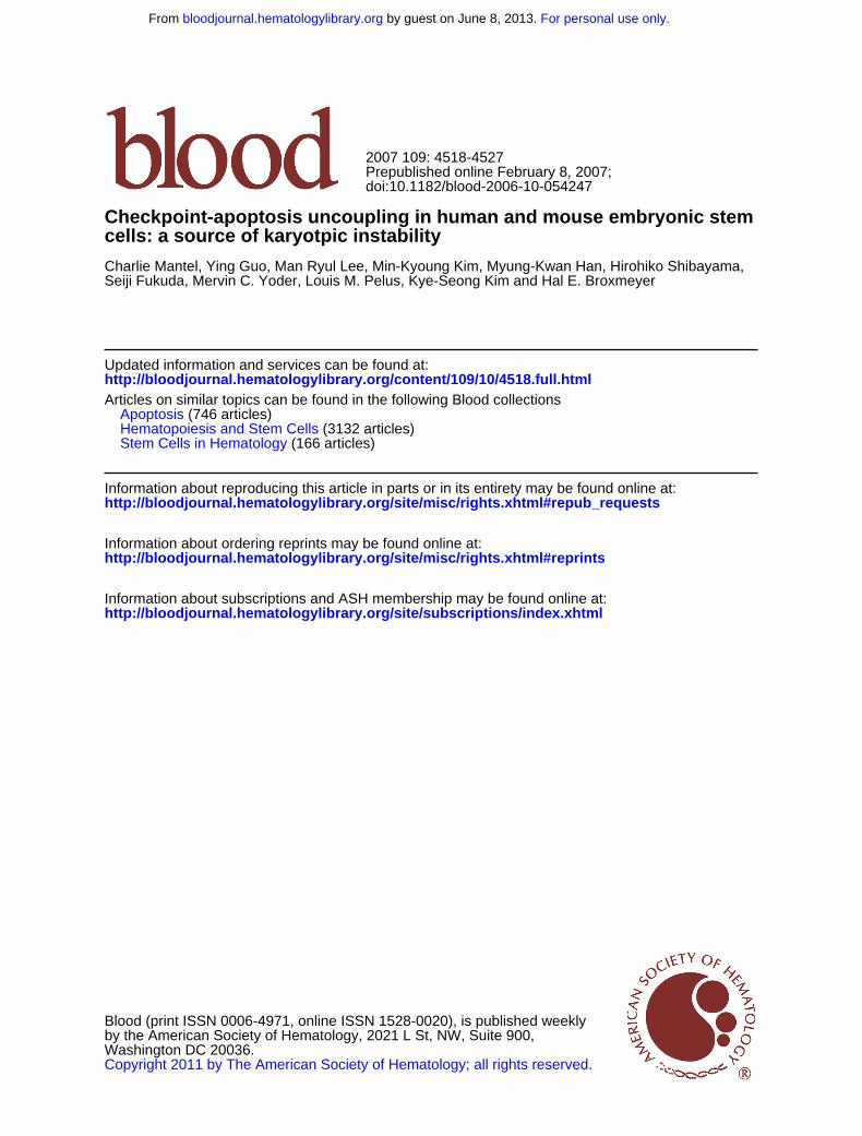
Figure 1. Microtubule disruption-induced mitotic arrest and polyploidy in mESCs. mESC lines E14 and R1 were treated with nocodazole (for microtubuledepolymerization), paclitaxel (for microtubule overstabilization), or control solvent for 24 hours in complete culture medium containing LIF as described in “Materials andmethods.” (A) Cells were harvested and assayed by multivariate permeabilized-cell cell-cycle analysis for simultaneous phospho(ser10)histone-H3 and DNA content. Regions1 and 2 indicate E14 cells that are in M phase as indicated by increased phosphohistone-H3 content at 4C and 8C DNA content. (B) Results of polyploidy (cells with � 4C DNAcontent) analysis in E14 and R1 cells from 6 independent experiments is shown as the mean � 1SD. (C) Percentage of M-phase cells (regions 1 and 2) are shown. (D) Relativefrequency histograms of chromosome number in metaphase E14 cells treated with nocodazole for the indicated times showing an average of 40 chromosomes per cell(euploid) at 0 time and showing the increase in cells with 80 chromosomes (tetraploid) at 24 hours. Chromosome number indicates BIN number times 5. The experiment wasrepeated once with the E14 cell line and once with the R1 cell line with similar results. Chromosome counts in normal MEF cells are shown for comparison in Figure S2C. MEFcells had 40 chromosomes per cell. (E) Typical metaphase chromosome appearance in E14 cells before and after nocodazole treatment; the number of chromosomes isindicated (Nikon Labophot-2; 10 � 100; oil). (F) Wright-Giemsa stain of E14 cells harvested 24 hours after nocodazole treatment displaying a single nucleus. No E14 cells withmore than 1 nucleus were observed (Nikon Labophot-2; 10 � 40).
4520 MANTEL et al BLOOD, 15 MAY 2007 � VOLUME 109, NUMBER 10
For personal use only. by guest on June 8, 2013. bloodjournal.hematologylibrary.orgFrom

(untreated) cells remained 2% polyploid after wash and recul-ture and 1% active caspase-3–positive. The polyploidy in MNPcells decayed to near-diploid aneuploidy (indicated by DNAcontent), with increased caspase-3 activation when culturedbeyond 4 expansion passages (Figure 3C). It is also noteworthythat the active caspase-3–negative MNP cells were primarily notsub-G1, while the active caspase-3–positive populations con-tained large numbers of sub-G1 cells, further supporting theapoptosis analysis of MNP cells similar to that described formESCs and mEBs (Figure 2C).
We next questioned whether pre-existing MNP cells wereLIF independent or if they initiate apoptosis or differentiationupon LIF removal. Normal mESCs were placed into EB mediumwithout LIF for 3 days. EBs formed and had about 12%apoptotic cells, which is typical for mEB formation (Figure 3D).In contrast, 4-passage MNP cells (washed free of drugs)initiated apoptosis 2 days after LIF removal and polyploidydecayed to near-diploid aneuploidy. This continued on day 3 anddisplayed robust caspase-3 activation without EB formation.Remarkably, active caspase-3–negative (nonapoptotic) cellsremaining in the culture on day 3 were near-diploid (indicatedby DNA content). We conclude that mESCs are unusually tolerantto the polyploid condition and can undergo tetraploid cell divisionsin culture. Upon removal of LIF, MNP cells initiate apoptosis anddo not form EBs. Also, the ploidy status of MNP cells decays afterseveral passages, even with LIF, to become aneuploid and near-diploid within 10 to 12 passages.
Ectopic suppression of apoptosis in 2 models of highlydifferentiated somatic cells results in polyploidyafter SAC activation
There are 2 possible interpretations of these results of LIFremoval–induced (differentiation-induced) apoptosis of MNP cells.One is intrinsic silencing of processes linking SAC to apoptosis inESCs. We refer to this as “uncoupling.” Upon differentiation, theseprocesses are unsilenced and activated (coupled), thus preventingMNP cell formation and survival. Another interpretation is that LIF,a known antiapoptotic cytokine,8 suppresses apoptosis, allowingsurvival of MNP cells. This would suggest that culture conditionsallow MNP cells to survive and is not due to intrinsic uncoupling.To evaluate these possibilities, we hypothesized that if MNP cellsurvival is due to intrinsic suppression of apoptosis, then ectopicsuppression of apoptosis in a highly differentiated model somatic(coupled) cell line would also result in polyploidy after SACactivation. To test this hypothesis, we used the mouse growth-factor–dependent pro–B-lymphocyte cell line Ba/F3 and its derivative,which overexpresses the antiapoptotic protein anamorsin.28 SACactivation or DNA damage–induced caspase-3–dependent apopto-sis in Ba/F3 cells and anamorsin overexpression suppressed thisresponse (Figure S4). Anamorsin-induced apoptosis suppressionalone caused Ba/F3 cells to contain more than 4C DNA content.Nocodazole treatment for 48 hours caused increased polyploid(8C) cells that were not observed in treated parental Ba/F3 cultures
Figure 2. Analysis of apoptosis of E14 cells before and after treatment with microtubule-disrupting agents or after DNA damage. (A) E14 mESCs were treated with theindicated agent for 24 hours and harvested as in Figure 1. Cells were analyzed by permeabilized-cell flow cytometry as in Figure 1 except an antibody to activated caspase-3was used. Cells above the bar are positive and those below the bar are negative for caspase-3 activation. (B) Caspase-3 activation in day-3 mEB cells after 24 hours oftreatment. Percentage apoptosis (mean � 1SD for 3 independent experiments) as indicated by caspase-3 activation. (C) Percentage apoptosis (mean � 1SD for 3independent experiments) as indicated by sub-G1 cells. *Statistically significant difference from control; P � .05. (D-E) Apoptosis measurement in treated and untreatedmESCs or mEB cells as indicated by Annexin-V binding. Cells were simultaneously stained with propidium iodide to indicate cellular membrane integrity. Early apoptosis(Annexin-V� and PI�) and total apoptosis (Annexin-V� and PI�/�) for E14 or mEB cells before and after treatment as in panels A and B. Results are mean percentages � 1SDfor 3 independent experiments.
KARYOTYPIC INSTABILITY IN STEM CELLS 4521BLOOD, 15 MAY 2007 � VOLUME 109, NUMBER 10
For personal use only. by guest on June 8, 2013. bloodjournal.hematologylibrary.orgFrom

(Figure 4A). We also investigated a Ba/F3 derivative overexpress-ing the antiapoptotic protein survivin (Figure 4B).29 Paclitaxel-activated SAC caused polyploidy in these cells, but not inempty-vector control cells. Thus, ectopic suppression of apoptosisin 2 model somatic cell lines caused polyploidy after SACactivation, analogous to mESCs. Therefore, polyploidy can resultafter checkpoint activation in normally coupled somatic cells if theapoptotic machinery is deregulated, supporting the interpretation ofintrinsic uncoupling in ESCs.
Differentiation in the presence of LIF does not prevent SACapoptosis coupling
To further evaluate the role of LIF in MNP cell formation, we usedretinoic acid (RA) to induce differentiation in mESCs while in thecontinued presence of LIF. Figure 4C-D demonstrates that RAtreatment for 3 days causes decreased SSEA-1 expression associ-ated with a pronounced morphology change (not shown) as alreadyreported,37 indicating they were differentiated cells (SSEA-1-Lo).After treatment with nocodazole, SSEA-1-Lo (RA-differentiated)cells failed to become polyploid, while SSEA-1-Hi (remainingundifferentiated) cells accumulated with more than 4C DNA. Thissuggests that even in the continued presence of LIF, differentiatedmESCs will induce apoptosis (as indicated by hypodiploidy andcaspase-3 activation) without tolerance for the tetraploid/polyploidcondition. This also supports the hypothesis that checkpoint-apoptosis uncoupling is an intrinsic property of mESCs and not duedirectly to the survival effects of LIF or other culture conditions.
hESC colonies contain 2 distinct cell types that differin response to SAC activation, polyploidy,and pluripotent marker expression
We next sought to extend our observations to hESCs. This alsoafforded us the opportunity to investigate polyploidy in the absenceof exogenous LIF. hESCs do not require LIF for proliferation invitro and were cultured on MEF feeder layers. An unexpected
Figure 3. Apoptosis and cell-cycle analysis in preformed mouse polyploidyESCs and their EB formation after expansion culture. Phosphohistone H3 (A) andcaspase-3 (B) is shown in control (solvent-treated) E14 cells or in preformedpolyploid (MNP) cells after cells were washed free of nocodazole or control solvent,recultured in complete medium containing LIF, and expanded by subculture for 4passages (4-pass). They were then harvested, and multivariate cell-cycle analysisperformed as in Figure 1. DNA content, percentage of polyploidy, and percentage ofapoptosis are numerically indicated. (C) Apoptosis and polyploidy after 6 and 10expansion passages of MNP is numerically shown. (D) Untreated control and MNPcells from panel A were then washed free of LIF and placed into EB medium for theindicated time, then harvested and apoptosis analysis done. DNA content andnumerical percentages of polyploidy and apoptosis for untreated control cell–derivedmEB cells and MNP-derived mEB cells are indicated. This experiment was repeatedonce with similar results.
Figure 4. Intrinsic apoptosis-suppression uncouplessomatic cells, while differentiation of mESCs in thepresence of LIF does not prevent coupling. Ba/F3cells and their derivatives containing an expressionvector for overexpressing anamorsin were treated withcontrol solvent or nocodazole or etoposide for 48 hours,then harvested and cell cycle/apoptosis analysis per-formed (A) as in Figure 2A. Percentages of polyploidyand apoptosis (mean � 1SD) from 3 experiments isshown. (B) MO7e cells expressing empty vector or avector containing survivin. Cells were treated with pacli-taxel or control solvent for 48 hours then harvested, andcell-cycle analysis was performed. This experiment wasrepeated once with similar results. (C) E14 mESCs weretreated with RA for 3 days, than treated for 1 additionalday with nocodazole added. SSEA-1 expression is shownin the top panel, and cell-cycle analysis of SSEA-1-Hi andSSEA-1-Lo gated cells is shown in the bottom panel. (D)Caspase-3 activation in cultures from panel C.
4522 MANTEL et al BLOOD, 15 MAY 2007 � VOLUME 109, NUMBER 10
For personal use only. by guest on June 8, 2013. bloodjournal.hematologylibrary.orgFrom
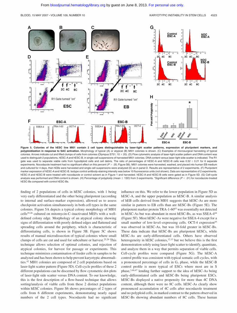
finding of 2 populations of cells in hESC colonies, with 1 beingvery early differentiated and the other being pluripotent (accordingto internal and surface-marker expression), allowed us to assesscheckpoint activation simultaneously in both cell types in the samecolonies. Figure 5A depicts a typical colony morphology of MI01cells25,26 cultured on mitomycin-C–inactivated MEFs with a well-defined colony edge. Morphology of an atypical colony showingsigns of differentiation with poorly defined edges and flattened andspreading cells around the periphery, which is characteristic ofdifferentiating cells, is shown in Figure 5B. Figure 5C showsresults of manual microdissection of typical colonies where smallclumps of cells are cut and used for subculture or harvest.25,26 Thistechnique allows selection of optimal colonies, and rejection ofatypical colonies, for harvest for passage or experiments. Thistechnique minimizes contamination of feeder cells in samples to beanalyzed and has been shown to help prevent karyotypic abnormali-ties.42 MI01 colonies are composed of 2 cell populations based onlaser-light scatter pattern (Figure 5D). Cell-cycle profiles of these 2different populations can be discerned by flow cytometric dot-plotsof laser-light side scatter versus DNA content. To our knowledge,this is the first description of a flow-based technique that allowssorting/analysis of viable cells from these 2 distinct populationswithin hESC colonies. Figure S6 shows percentages of 2 types ofcells from 6 different experiments, demonstrating nearly equalnumbers of the 2 cell types. Nocodazole had no significant
influence on this. We refer to the lower population in Figure 5D ashESC-A, and the upper population as hESC-B. A similar analysisof hEB cells derived from MI01 suggests that hESC-As are moresimilar in pattern to EB cells than are hESC-Bs (Figure 5E). Thepluripotent marker protein TRA-1-6043 was essentially not detectedin hESC-As but was abundant in most hESC-Bs, as was SSEA-444
(Figure 5F). Most hESC-As were negative for SSEA-4 except for asmall number of low-level–expressing cells. OCT-4 expression45
was observed in hESC-As, but was 10-fold greater in hESC-Bs.These data indicate that hESC-Bs are pluripotent hESCs, whilehESC-As are early-differentiated cells. Others have observedheterogeneity in hESC colonies,12,13 but we believe this is the firstdemonstration solely using laser-light scatter to identify, quantitate,and analyze them in a way that permits separation of viable cells.Cell-cycle profiles were compared (Figure 5G). The hESC-Acontrol profile was consistent with typical somatic cell cycles, witha pronounced percentage of cells in G1 phase, while the hESC-Bcontrol profile is more typical of ESCs where most are in Sphase,3,46,47 lending further support to the idea of hESC-As beingearly-differentiated cells and hESC-Bs being pluripotent ESCs.hESC-Bs displayed a native propensity for more than 4C DNAcontent, although there were no 8C cells. hESC-As clearly showpronounced accumulation of 4C cells after nocodazole treatmentand no polyploid cells, in marked contrast to the pattern observed inhESC-Bs showing abundant numbers of 8C cells. These human
Figure 5. Colonies of the hESC line MI01 contain 2 cell types distinguishable by laser-light scatter patterns, expression of pluripotent markers, andpolyploidization in response to SAC activation. Morphology of typical (A) or atypical (B) MI01 colonies is shown. (C) Examples of microsurgical harvesting of typicalcolonies. Arrows indicate cut and lifted clumps of cells from colonies (Olympus S751; 10 � 20). (D) Flow cytometric analysis of laser-light scatter pattern and DNA content wasused to distinguish 2 populations, hESC-A and hESC-B, in single-cell suspensions of harvested MI01 colonies. DNA content versus laser-light side-scatter is indicated. The R1gate was used to separate viable cells from hypodiploid cells and cell debris. The ratio of percentages of hESC-A and hESC-B cells was 0.52 � 0.21 for 6 separateexperiments. Nocodazole treatment had no significant effect on this percent (P � .05; Figure S6). MI01 colonies were harvested, washed, and placed into human EB mediumand cultured for 4 days, then hEBs were harvested and single cell suspensions were analyzed (E) as in panel D. Results are representative of 2 experiments. (F) Pluripotentmarker expression of hESC-A and hESC-B. Isotype control antibody-staining intensity was below 10 fluorescence units (not shown). Data are representative of 2 experiments.hESC-A and hESC-B were treated with nocodazole or control solvent as in Figure 1 and harvested. hESC-A and hESC-B cells were gated as in Figure 5D. (G) Cell-cycleanalysis was performed and DNA content is shown. (H) Percentage of polyploidy (mean � 1SD) from 3 experiments. *Significant difference (P � .01) for nocodazole-treatedhESC-Bs compared with control hESC-Bs.
KARYOTYPIC INSTABILITY IN STEM CELLS 4523BLOOD, 15 MAY 2007 � VOLUME 109, NUMBER 10
For personal use only. by guest on June 8, 2013. bloodjournal.hematologylibrary.orgFrom
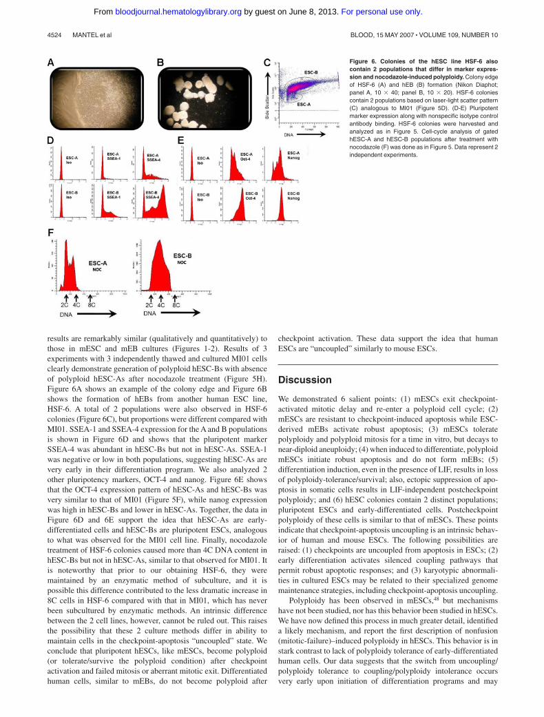
results are remarkably similar (qualitatively and quantitatively) tothose in mESC and mEB cultures (Figures 1-2). Results of 3experiments with 3 independently thawed and cultured MI01 cellsclearly demonstrate generation of polyploid hESC-Bs with absenceof polyploid hESC-As after nocodazole treatment (Figure 5H).Figure 6A shows an example of the colony edge and Figure 6Bshows the formation of hEBs from another human ESC line,HSF-6. A total of 2 populations were also observed in HSF-6colonies (Figure 6C), but proportions were different compared withMI01. SSEA-1 and SSEA-4 expression for the A and B populationsis shown in Figure 6D and shows that the pluripotent markerSSEA-4 was abundant in hESC-Bs but not in hESC-As. SSEA-1was negative or low in both populations, suggesting hESC-As arevery early in their differentiation program. We also analyzed 2other pluripotency markers, OCT-4 and nanog. Figure 6E showsthat the OCT-4 expression pattern of hESC-As and hESC-Bs wasvery similar to that of MI01 (Figure 5F), while nanog expressionwas high in hESC-Bs and lower in hESC-As. Together, the data inFigure 6D and 6E support the idea that hESC-As are early-differentiated cells and hESC-Bs are pluripotent ESCs, analogousto what was observed for the MI01 cell line. Finally, nocodazoletreatment of HSF-6 colonies caused more than 4C DNA content inhESC-Bs but not in hESC-As, similar to that observed for MI01. Itis noteworthy that prior to our obtaining HSF-6, they weremaintained by an enzymatic method of subculture, and it ispossible this difference contributed to the less dramatic increase in8C cells in HSF-6 compared with that in MI01, which has neverbeen subcultured by enzymatic methods. An intrinsic differencebetween the 2 cell lines, however, cannot be ruled out. This raisesthe possibility that these 2 culture methods differ in ability tomaintain cells in the checkpoint-apoptosis “uncoupled” state. Weconclude that pluripotent hESCs, like mESCs, become polyploid(or tolerate/survive the polyploid condition) after checkpointactivation and failed mitosis or aberrant mitotic exit. Differentiatedhuman cells, similar to mEBs, do not become polyploid after
checkpoint activation. These data support the idea that humanESCs are “uncoupled” similarly to mouse ESCs.
Discussion
We demonstrated 6 salient points: (1) mESCs exit checkpoint-activated mitotic delay and re-enter a polyploid cell cycle; (2)mESCs are resistant to checkpoint-induced apoptosis while ESC-derived mEBs activate robust apoptosis; (3) mESCs toleratepolyploidy and polyploid mitosis for a time in vitro, but decays tonear-diploid aneuploidy; (4) when induced to differentiate, polyploidmESCs initiate robust apoptosis and do not form mEBs; (5)differentiation induction, even in the presence of LIF, results in lossof polyploidy-tolerance/survival; also, ectopic suppression of apo-ptosis in somatic cells results in LIF-independent postcheckpointpolyploidy; and (6) hESC colonies contain 2 distinct populations;pluripotent ESCs and early-differentiated cells. Postcheckpointpolyploidy of these cells is similar to that of mESCs. These pointsindicate that checkpoint-apoptosis uncoupling is an intrinsic behav-ior of human and mouse ESCs. The following possibilities areraised: (1) checkpoints are uncoupled from apoptosis in ESCs; (2)early differentiation activates silenced coupling pathways thatpermit robust apoptotic responses; and (3) karyotypic abnormali-ties in cultured ESCs may be related to their specialized genomemaintenance strategies, including checkpoint-apoptosis uncoupling.
Polyploidy has been observed in mESCs,48 but mechanismshave not been studied, nor has this behavior been studied in hESCs.We have now defined this process in much greater detail, identifieda likely mechanism, and report the first description of nonfusion(mitotic-failure)–induced polyploidy in hESCs. This behavior is instark contrast to lack of polyploidy tolerance of early-differentiatedhuman cells. Our data suggests that the switch from uncoupling/polyploidy tolerance to coupling/polyploidy intolerance occursvery early upon initiation of differentiation programs and may
Figure 6. Colonies of the hESC line HSF-6 alsocontain 2 populations that differ in marker expres-sion and nocodazole-induced polyploidy. Colony edgeof HSF-6 (A) and hEB (B) formation (Nikon Diaphot;panel A, 10 � 40; panel B, 10 � 20). HSF-6 coloniescontain 2 populations based on laser-light scatter pattern(C) analogous to MI01 (Figure 5D). (D-E) Pluripotentmarker expression along with nonspecific isotype controlantibody binding. HSF-6 colonies were harvested andanalyzed as in Figure 5. Cell-cycle analysis of gatedhESC-A and hESC-B populations after treatment withnocodazole (F) was done as in Figure 5. Data represent 2independent experiments.
4524 MANTEL et al BLOOD, 15 MAY 2007 � VOLUME 109, NUMBER 10
For personal use only. by guest on June 8, 2013. bloodjournal.hematologylibrary.orgFrom

coincide with lineage specification and loss of “stem-like” self-renewal. The novel concept of checkpoint-apoptosis uncoupling inpluripotent stem cells, as opposed to the concept that the check-point itself is nonfunctional, may also be applicable to othercheckpoints that activate apoptosis, such as the DNA damagecheckpoint.4,49 Others50-53 have shown mouse and human ESCstolerate chemical fusion–induced tetraploidy, a technique used togenerate ESC-specific cloned animals (tetraploid-embryo comple-mentation). This technique (chemical fusion) has also been used todemonstrate reprogramming of somatic genomes into embryonic-like genomes;54 cultures containing tetraploid (somatic/ESC) hy-brid cells formed teratomas composed of cells representing 3 germlayers after injection into mice, indicating the hybrids werepluripotent. This is an important step toward full use of ESCtechnology for human benefit. However, our data indicates thatpolyploid ESCs can reduce their ploidy to near-diploid aneuploidyafter short times in culture, thus raising the possibility that teratomaformed from hybrid cells may be derived from near-diploid/aneuploid ESCs instead of complete tetraploids, especially sinceevidence demonstrating tetraploidy in the hybrid-derived teratomacells is lacking. It is reported that chemical fusion–inducedtetraploid ESCs display a decay/reduction in chromosome numberduring culture, similar to our findings. Unequal segregation ofchromosomes and even a degree of chromosome selectivity in thisprocess favoring the embryonic chromosomes (ie, loss of somaticchromosomes) has been reported.51 It is therefore possible that thecomplete somatic genome contingent of somatic/ESC hybrid cellsmay not be represented in the teratoma cells, and this might lead tofalse impressions of genome reprogramming.
Our findings are consistent with Stewart et al,55 who reportedcellular heterogeneity in hESC colonies. They observed differencesin cell-cycle behavior between SSEA3� and SSEA3� cells in thesame colonies, similar to types A and B cell populations weobserved in hESC colonies (Figures 5-6) using markers SSEA1,SSEA4, TRA-1-60, Oct-4, and nanog. Our hESC-A and -B cellsmay be similar, if not identical, to SSEA3�/� cells.55 It is likely thatthe SSEA1-Hi/Lo mESC populations (Figure 4C) are murineanalogs to the 2 human cell types. Our data and those of Stewart etal are consistent with a very narrowly defined phenotypic switchfrom very primitive ESC-like behavior to less primitive progenitorcell–like behavior. It is interesting that this apparently abrupt shiftin phenotype in both studies is characterized by a marked andspecific shift in cell-cycle behavior. This is reminiscent of theabrupt shift in cell-cycle behavior at the midblastula transition inamphibian and starfish embryos, where the G1 phase is greatlylengthened asynchronously.56 Our data suggest that checkpoint-apoptosis coupling may be another determinant characterizing 1 ofthe earliest changes from pluripotency/self-renewal (ESC-like) todifferentiation/lineage commitment (progenitor cell–like).
Another issue highlighted by our data is the interpretations ofcheckpoint deficiency or absence in ESCs.57 Many techniques tomeasure and quantify checkpoint integrity often rely on properfunction of other checkpoints like the SAC. Because our datasuggest the SAC is functional in ESCs but does not conform toconventional concepts (ie, uncoupling), it is possible to be misledby data where the SAC is assumed to behave as in somatic cells.The decatenation checkpoint is reported to be deficient in mESCsand neural and hematopoietic progenitor cells.57 Because thetechnique used to quantitate function of this checkpoint (pseudomi-totic index) relies on accumulation of cells in metaphase bytreatment with spindle inhibitors like colchicine or nocodazole, andresults are expressed as a ratio of frequency of pseudomitotic cells
(cells that have entangled but condensed chromosomes) versusfrequency of normal mitotic cells (normally condensed chromo-somes), results can be influenced not only by changes in pseudomi-totic cell numbers, but also by changes in normal mitotic cellnumbers. Our data suggest that the frequency of normal mitoticcells observed after treatment with spindle poisons, like colchicine,may not accurately reflect the numbers of cells that have enteredand exited mitosis in mESCs compared with differentiated cells.Thus, an alternative interpretation of the reported pseudomitoticindex data57 is that the SAC, and not necessarily the decatenationcheckpoint, is deficient. We observed low numbers of apparentpseudomitotic cells in mESCs after short treatment times (6-10hours), but they were not quantitated (data not shown). Our data arethus consistent with previous reports of an apparent deficientdecatenation checkpoint in mESCs compared with mEBs.
Based on findings of increased pseudomitotic index in neuraland hematopoietic progenitor cells57 and our data, we hypothesizethat tissue-specific stem/progenitor cells may also be characterizedby lack of SAC-apoptosis coupling. This may have implications fortechniques to culture and expand, ex vivo, adult stem cells andprimary progenitor cells. Rigorous examination of SAC function inpopulations of primary mouse sca1�/lin�/c-kit� and human CD34�/CD38� cells, highly enriched in primitive hematopoietic progeni-tors, could be used to test this hypothesis. The physiologicrelevance of checkpoint-apoptosis uncoupling in tissue-specificprogenitors is unknown, but is likely important for strategies tomaintain genomic integrity and tumor suppression in progenitors asit appears to be for ESCs.
Our studies lead to the question why ESCs would maintaincorrupted/genome-damaged cells, and what advantage this behav-ior might have during early embryogenesis. One possibility is thatin early developing embryos, a cell, even though it may begenetically corrupt, can still maintain a supportive role for initialpatterning and asymmetry generation via lateral- inhibition andmorphogen-diffusion/gradient formation,58 and might be importantfor early embryo survival when cell numbers are low. Only later,after initial patterning and sufficient “cell mass” is achieved suchthat loss of some cells via apoptotic culling of damaged cells can betolerated without disruption of overall patterning, would surveil-lance and culling become safely activated. This could be whycheckpoint-apoptosis uncoupling might exist in very early develop-ing embryos, especially for caspase-dependent apoptosis, which iscontext dependent in mammals and related to stress/damagingagent or tissue type and is highly dependent on developmentalstatus.58 If this notion is correct, then when ESCs are removed fromthe context of early embryonic development in the preimplantationblastocyst and immortalized in culture, they could remain un-coupled and subject to increased genome damage. If this is so, itshould lead to new strategies to help maintain genomic fidelity inESC cultures.
Finally, our studies suggest that links between karyotypicinstability in stem cells and notions of a “cancer stem cell”59 maybe partly founded in the propensity of ESCs, and potentially adultstem cells,57 to suffer from intrinsic checkpoint-apoptosis uncou-pling. A Lats2-Mdm2-p53 axis checkpoint has recently beendescribed in somatic cells,60 which is critical for maintenance ofproper chromosomal segregation after mitotic slippage due todysfunction of the mitotic spindle apparatus. Better understandingof this process is lacking and this has not yet been evaluated inmESCs. p53 down-regulates nanog expression,61 which maintainsself-renewal/pluripotency in ESCs.
KARYOTYPIC INSTABILITY IN STEM CELLS 4525BLOOD, 15 MAY 2007 � VOLUME 109, NUMBER 10
For personal use only. by guest on June 8, 2013. bloodjournal.hematologylibrary.orgFrom
We recently found that the histone-deacetylase SIRT1 (whichhas p53 as one of its substrates), is required for proper p53 nucleartranslocation and nanog down-regulation61 (M.K.H., Y.G., X.O.,C.M., and H.E.B., unpublished observations, November 2006). p53and SIRT1 are highly expressed in mESCs, but p53 is believed tobe in a nonfunctional form in mESCs (M.K.H., Y.G., X.O., C.M.,and H.E.B., unpublished observations, November 2006), whichcould be explained by SIRT1 maintaining p53 in a deacetylated(cytoplasmic) form, which is unable to activate apoptosis-relatedgenes or down-regulate nanog. In somatic cells, SIRT1 is very low,permitting access of p53 into the nucleus to activate its proapop-totic function, down-regulate nanog, and initiate differentiation,where p53 would behave as it is known to do in somatic cells.These ideas are highly speculative at this time.
Understanding molecular mechanisms of checkpoint-apoptosiscoupling/uncoupling in ESCs and adult stem cells and theirdifferentiated derivatives could provide important clues aboutchromosomal instability (CIN) in human tumors,62 as well asprovide new ways to exploit this behavior for therapeutic benefit.
Acknowledgments
We thank P. Mantel for editing the manuscript; B. Graham,Mee-Young Cho, and Kyu-Yong Han for helpful discussions; andS. Rhorabough and C. Kaufman for superb technical support.
This work was supported by US Public Health Service GrantsR01 HL67384, a supplement to HL67384, R01 HL56416, a Projectin P01 HL053586 (H.E.B.), and R01 HL79654 and R01 HL69669(L.M.P.), and a grant (SC2060) from the Stem Cell Research Centerof the 21st Century Frontier Research Program and the Ministry ofScience and Technology, Republic of Korea (K.-S.K.).
Authorship
Contribution: C.M. designed and performed research, collected andanalyzed data, and cowrote the paper. Y.G. designed research;M.R.L., M.-K.K., and M.-K.H. performed difficult but vital cellline culture and designed research; H.S. contributed vital reagentsand cell lines; S.F., M.C.Y., and L.M.P. contributed vital cell lines;K.-S.K. supported the study, contributed vital cell lines andreagents, and analyzed the data; and H.E.B. supported the study,analyzed data, and cowrote the paper.
Conflict-of-interest statement: All authors declare no competingfinancial interests.
Correspondence: Hal E. Broxmeyer or Charlie R. Mantel,Walther Oncology Center, Indiana University School of Medicine,Room 302A, 950 West Walnut St, Indianapolis, IN 46202; e-mail:[email protected] or [email protected]; or Kye-Seong Kim, De-partment of Anatomy and Cell Biology, Hanyang University College ofMedicine, Seoul, Korea; e-mail: [email protected].
References
1. Huppertz B, Herrler A. Regulation of proliferationand apoptosis during development of the preim-plantation embryo and the placenta. Birth DefectsRes C Embryo Today. 2005;75:249-261.
2. Jefford CE, Irminger-Finger I. Mechanisms ofchromosome instability in cancers. Crit Rev On-col Hematol. 2006;59:1-14.
3. Burdon T, Smith A, Savatier P. Signalling, cellcycle and pluripotency in embryonic stem cells.Trends Cell Biol. 2002;12:432-438.
4. Holway AH, Kim SH, La Volpe A, Michael WM.Checkpoint silencing during the DNA damageresponse in Caenorhabditis elegans embryos.J Cell Biol. 2006;172:999-1008.
5. Fluckiger AC, Marcy G, Marchand M, et al. Cellcycle features of primate embryonic stem cells.Stem Cells. 2006;24:547-556.
6. Pampfer S, Donnay I. Apoptosis at the time ofembryo implantation in mouse and rat. Cell DeathDiffer. 1999;6:533-545.
7. Spanos S, Rice S, Karagiannis P, et al. Caspaseactivity and expression of cell death genes duringdevelopment of human preimplantation embryos.Reproduction. 2002;124:353-363.
8. Duval D, Trouillas M, Thibault C, et al. Apoptosisand differentiation commitment: novel insightsrevealed by gene profiling studies in mouse em-bryonic stem cells. Cell Death Differ. 2006;13:564-575.
9. Wells D, Bermudez MG, Steuerwald N, et al. Ex-pression of genes regulating chromosome segre-gation, the cell cycle and apoptosis during humanpreimplantation development. Hum Reprod.2005;20:1339-1348.
10. Greenwood J, Costanzo V, Robertson K, HenseyC, Gautier J. Responses to DNA damage in Xe-nopus: cell death or cell cycle arrest. NovartisFound Symp. 2001;237:221-230.
11. Greenwood J, Gautier J. From oogenesis throughgastrulation: developmental regulation of apopto-sis. Semin Cell Dev Biol. 2005;16:215-224.
12. Semb H. Human embryonic stem cells: origin,properties and applications. APMIS. 2005;113:743-750.
13. Edwards RG. Stem cells today, A: origin and po-tential of embryo stem cells. Reprod Biomed On-line. 2004;8:275-306.
14. Longo L, Bygrave A, Grosveld FG, Pandolfi PP.The chromosome make-up of mouse embryonicstem cells is predictive of somatic and germ cellchimaerism. Transgenic Res. 1997;6:321-328.
15. Humpherys D, Eggan K, Akutsu H, et al. Epige-netic instability in ES cells and cloned mice. Sci-ence. 2001;293:95-97.
16. Humpherys D, Eggan K, Akutsu H, et al. Abnor-mal gene expression in cloned mice derived fromembryonic stem cell and cumulus cell nuclei. ProcNatl Acad Sci U S A. 2002;99:12889-12894.
17. Cervantes RB, Stringer JR, Shao C, TischfieldJA, Stambrook PJ. Embryonic stem cells and so-matic cells differ in mutation frequency and type.Proc Natl Acad Sci U S A. 2002;99:3586-3590.
18. Pera MF. Unnatural selection of cultured humanES cells? Nat Biotechnol. 2004;22:42-43.
19. Draper JS, Moore HD, Ruban LN, Gokhale PJ,Andrews PW. Culture and characterization of hu-man embryonic stem cells. Stem Cells Dev. 2004;13:325-336.
20. Draper JS, Smith K, Gokhale P, et al. Recurrentgain of chromosomes 17q and 12 in cultured hu-man embryonic stem cells. Nat Biotechnol. 2004;22:53-54.
21. Eggan K, Rode A, Jentsch I, et al. Male and fe-male mice derived from the same embryonicstem cell clone by tetraploid embryo complemen-tation. Nat Biotechnol. 2002;20:455-459.
22. Guo Y, Costa R, Ramsey H, et al. The embryonicstem cell transcription factors Oct-4 and FoxD3interact to regulate endodermal-specific promoterexpression. Proc Natl Acad Sci U S A. 2002;99:3663-3667.
23. Guo Y, Graham-Evans B, Broxmeyer HE. Murineembryonic stem cells secrete cytokines/growthmodulators that enhance cell survival/anti-apo-ptosis and stimulate colony formation of murinehematopoietic progenitor cells. Stem Cells. 2006;24:850-856.
24. National Institutes of Health Human Stem CellRegistry. http://stemcells.nih.gov/research/regis-try.html. Accessed December 12, 2005.
25. Suh MR, Lee Y, Kim JY, et al. Human embryonicstem cells express a unique set of microRNAs.Dev Biol. 2004;270:488-498.
26. Lee JB, Lee JE, Park JH, et al. Establishmentand maintenance of human embryonic stem celllines on human feeder cells derived from uterineendometrium under serum-free condition. BiolReprod. 2005;72:42-49.
27. Mantel C, Braun SE, Reid S, et al. p21(cip-1/waf-1) deficiency causes deformed nuclear archi-tecture, centriole overduplication, polyploidy, andrelaxed microtubule damage checkpoints in hu-man hematopoietic cells. Blood. 1999;93:1390-1398.
28. Shibayama H, Takai E, Matsumura I, et al. Identi-fication of a cytokine-induced antiapoptotic mole-cule anamorsin essential for definitive hematopoi-esis. J Exp Med. 2004;199:581-592.
29. Fukuda S, Mantel CR, Pelus LM. Survivin regu-lates hematopoietic progenitor cell proliferationthrough p21WAF1/Cip1-dependent and -indepen-dent pathways. Blood. 2004;103:120-127.
30. Mantel CR, Braun SE, Lee Y, Kim YJ, BroxmeyerHE. The interphase microtubule damage check-point defines an S-phase commitment point anddoes not require p21(waf-1). Blood. 2001;97:1505-1507.
31. Bagwell CB. Hyperlog-a flexible log-like transformfor negative, zero, and positive valued data. Cy-tometry A. 2005;64:34-42.
32. Henegariu O, Heerema NA, Bray-Ward P, WardDC. Colour-changing karyotyping: an alternativeto M-FISH/SKY. Nat Genet. 1999;23:263-264.
33. Henegariu O, Heerema NA, Lowe Wright L, Bray-Ward P, Ward DC, Vance GH. Improvements incytogenetic slide preparation: controlled chromo-some spreading, chemical aging and gradual de-naturing. Cytometry. 2001;43:101-109.
34. Lanni JS, Jacks T. Characterization of the p53-dependent postmitotic checkpoint following
4526 MANTEL et al BLOOD, 15 MAY 2007 � VOLUME 109, NUMBER 10
For personal use only. by guest on June 8, 2013. bloodjournal.hematologylibrary.orgFrom
spindle disruption. Mol Cell Biol. 1998;18:1055-1064.
35. Lanni JS, Lowe SW, Licitra EJ, Liu JO, Jacks T.p53-independent apoptosis induced by paclitaxelthrough an indirect mechanism. Proc Natl AcadSci U S A. 1997;94:9679-9683.
36. Sanchez-Aguilera A, Montalban C, de la Cueva P,et al. Tumor microenvironment and mitotic check-point are key factors in the outcome of classicalHodgkin lymphoma. Blood. 2006;108:662-668.
37. Andrews PW, Goodfellow PN. Antigen expressionby somatic cell hybrids of a murine embryonalcarcinoma cell with thymocytes and L cells. So-matic Cell Genet. 1980;6:271-284.
38. Borel F, Lohez OD, Lacroix FB, Margolis RL. Mul-tiple centrosomes arise from tetraploidy check-point failure and mitotic centrosome clusters inp53 and RB pocket protein-compromised cells.Proc Natl Acad Sci U S A. 2002;99:9819-9824.
39. Pozarowski P, Huang X, Halicka DH, Lee B,Johnson G, Darzynkiewicz Z. Interactions of fluo-rochrome-labeled caspase inhibitors with apopto-tic cells: a caution in data interpretation. Cytom-etry A. 2003;55:50-60.
40. Darzynkiewicz Z, Bedner E. Analysis of apoptoticcells by flow and laser scanning cytometry. Meth-ods Enzymol. 2000;322:18-39.
41. van Engeland M, Nieland LJ, Ramaekers FC,Schutte B, Reutelingsperger CP. Annexin V-affin-ity assay: a review on an apoptosis detection sys-tem based on phosphatidylserine exposure. Cy-tometry. 1998;31:1-9.
42. Mitalipova MM, Rao RR, Hoyer DM, et al. Pre-serving the genetic integrity of human embryonicstem cells. Nat Biotechnol. 2005;23:19-20.
43. Andrews PW, Banting G, Damjanov I, Arnaud D,Avner P. Three monoclonal antibodies definingdistinct differentiation antigens associated withdifferent high molecular weight polypeptides on
the surface of human embryonal carcinoma cells.Hybridoma. 1984;3:347-361.
44. Kannagi R, Cochran NA, Ishigami F, et al. Stage-specific embryonic antigens (SSEA-3 and -4) areepitopes of a unique globo-series ganglioside iso-lated from human teratocarcinoma cells. EMBOJ. 1983;2:2355-2361.
45. Brehm A, Ovitt CE, Scholer HR. Oct-4: more thanjust a POUerful marker of the mammalian germ-line? APMIS. 1998;106:114-124.
46. Artus J, Babinet C, Cohen-Tannoudji M. The cellcycle of early mammalian embryos: lessons fromgenetic mouse models. Cell Cycle. 2006;5:499-502.
47. Fujii-Yamamoto H, Kim JM, Arai K, Masai H. Cellcycle and developmental regulations of replica-tion factors in mouse embryonic stem cells. J BiolChem. 2005;280:12976-12987.
48. Malashicheva AB, Kisliakova TV, Savatier P,Pospelov VA. [Embryonal stem cells do not un-dergo cell cycle arrest upon exposure to damag-ing factors]. Tsitologiia. 2002;44:643-648.
49. Aladjem MI, Spike BT, Rodewald LW, et al. EScells do not activate p53-dependent stress re-sponses and undergo p53-independent apoptosisin response to DNA damage. Curr Biol. 1998;8:145-155.
50. Nagy A, Rossant J, Nagy R, Abramow-NewerlyW, Roder JC. Derivation of completely cell cul-ture-derived mice from early-passage embryonicstem cells. Proc Natl Acad Sci U S A. 1993;90:8424-8428.
51. Matveeva NM, Pristyazhnyuk IE, Temirova SA, etal. Unequal segregation of parental chromo-somes in embryonic stem cell hybrids. Mol Re-prod Dev. 2005;71:305-314.
52. Li X, Wei W, Yong J, Jia Q, Yu Y, Di K. The ge-netic heterozygosity and fitness of tetraploid em-bryos and embryonic stem cells are crucial pa-
rameters influencing survival of mice derived fromembryonic stem cells by tetraploid embryo aggre-gation. Reproduction. 2005;130:53-59.
53. Li X, Wei W, Yong J, Jia Q, Yu Y, Di K. The ge-netic heterozygosity and fitness of tetraploid em-bryos and embryonic stem cells are crucial pa-rameters influencing survival of mice derived fromembryonic stem cells by tetraploid embryo aggre-gation. Reproduction. 2005;130:53-59.
54. Cowan CA, Atienza J, Melton DA, Eggan K.Nuclear reprogramming of somatic cells after fu-sion with human embryonic stem cells. Science.2005;309:1369-1373.
55. Stewart MH, Bosse M, Chadwick K, Menendez P,Bendall SC, Bhatia M. Clonal isolation of hESCsreveals heterogeneity within the pluripotent stemcell compartment. Nat Methods. 2006;3:807-815.
56. Etkin LD. Regulation of the mid-blastula transitionin amphibians. Dev Biol (N Y 1985). 1988;5:209-225.
57. Damelin M, Sun YE, Sodja VB, Bestor TH. Decat-enation checkpoint deficiency in stem and pro-genitor cells. Cancer Cell. 2005;8:479-484.
58. Gurdon JB, Bourillot PY. Morphogen gradient in-terpretation. Nature. 2001;413:797-803.
59. Zhang M, Rosen JM. Stem cells in the etiologyand treatment of cancer. Curr Opin Genet Dev.2006;16:60-64.
60. Aylon Y, Michael D, Shmueli A, Yabuta N, NojimaH, Oren M. A positive feedback loop between thep53 and Lats2 tumor suppressors prevents tet-raploidization. Genes Dev. 2006;20:2687-2700.
61. Lin T, Chao C, Saito S, et al. p53 induces differen-tiation of mouse embryonic stem cells by sup-pressing Nanog expression. Nat Cell Biol. 2005;7:165-171.
62. Lengauer C, Kinzler KW, Vogelstein B. Geneticinstabilities in human cancers. Nature. 1998;396:643-649.
KARYOTYPIC INSTABILITY IN STEM CELLS 4527BLOOD, 15 MAY 2007 � VOLUME 109, NUMBER 10
For personal use only. by guest on June 8, 2013. bloodjournal.hematologylibrary.orgFrom