Peroxisome proliferator-activated receptor Γ(PPAΓ) modulates hepatic stellate cells activation
18
Peroxisome proliferator-activated receptor g in human breast carcinoma: a modulator of estrogenic actions T Suzuki, S Hayashi 1 , Y Miki, Y Nakamura, T Moriya, A Sugawara 2 , T Ishida 3 , N Ohuchi 3 and H Sasano Department of Pathology, Tohoku University School of Medicine, 2-1 Seiryo-machi, Aoba-ku, Sendai, 980-8575, Japan 1 Department of Molecular Medical Technology, Tohoku University School of Medicine, Sendai, Japan 2 Department of Medicine, Tohoku University School of Medicine, Sendai, Japan 3 Department of Surgery, Tohoku University School of Medicine, Sendai, Japan (Requests for offprints should be addressed to T Suzuki; Email: [email protected]) Abstract It has been reported that agonists of peroxisome proliferator-activated receptor g (PPARg ) inhibit proliferation of breast carcinoma cells, but the biological significance of PPARg remains undetermined in human breast carcinomas. Therefore, we immunolocalized PPARg in 238 human breast carcinoma tissues. PPARg immunoreactivity was detected in 42% of carcinomas, and was significantly associated with the status of estrogen receptor (ER) a, ERb, progesterone receptor, retinoic X receptors, p21 or p27, and negatively correlated with histological grade or cyclooxygenase-2 status. PPARg immunoreactivity was significantly associated with an improved clinical outcome of breast carcinoma patients by univariate analysis, and multivariate analysis demonstrated that PPARg immunoreactivity was an independent prognostic factor for overall survival in ERa-positive patients. We then examined possible mechanisms of modulation by PPARg on estrogenic actions in MCF-7 breast carcinoma cells. A PPARg activator, 15-deoxy-D 12,14 - prostaglandin J 2 (15d-PGJ 2 ), significantly inhibited estrogen-responsive element-dependent transactivation by estradiol in MCF-7 cells, which was blocked by addition of a PPARg antagonist GW9662. Subsequent study, employing a custom-made microarray focused on estrogen- responsive genes, revealed that mRNA expression was significantly regulated by estradiol in 49 genes, but this significance vanished on addition of 15d-PGJ 2 in 16 out of 49 (33%) genes. These findings were confirmed by real-time PCR in 11 genes. 15d-PGJ 2 significantly inhibited estrogen- mediated proliferation of MCF-7 cells, and caused accumulation of p21 and p27 protein. These results suggest that PPARg is mainly expressed in well-differentiated and ER-positive breast carcinomas, and modulates estrogenic actions. Endocrine-Related Cancer (2006) 13 233–250 Introduction Peroxisome proliferator-activated receptor g (PPARg ) is a member of the nuclear hormone receptor super- family, and has also been designated NR1C3 (Lemberger et al. 1996, Schoonjans et al. 1996). PPARg functions as a transactivation factor following heterodimerization with retinoic X receptors (RXRs), and binds to its specific response elements termed peroxisome proliferating responsive elements (PPREs) of various target genes (Mangelsdorf & Evans 1995). PPARg is one of the ligand-activated transcription factors, and 15-deoxy-D 12,14 -prostaglandin J 2 (15d- PGJ 2 ) is currently considered a naturally occurring PPARg ligand, which activates PPARg at mM con- centrations in the human (Koeffler 2003). It is well known that PPARg plays essential roles in adipogenesis (Tontonoz et al. 1994), insulin resistance (Celi & Shuldiner 2002), and development of various organs (Barak et al. 1999). In addition, various in vitro studies have demonstrated that PPARg ligands have a potent antiproliferative activity against a wide variety Endocrine-Related Cancer (2006) 13 233–250 Endocrine-Related Cancer (2006) 13 233–250 DOI:10.1677/erc.1.01075 1351-0088/06/013–233 g 2006 Society for Endocrinology Printed in Great Britain Online version via http://www.endocrinology-journals.org
-
Upload
independent -
Category
Documents
-
view
3 -
download
0
Transcript of Peroxisome proliferator-activated receptor Γ(PPAΓ) modulates hepatic stellate cells activation

Peroxisome proliferator-activated receptorg in human breast carcinoma: a modulatorof estrogenic actions
T Suzuki, S Hayashi1, Y Miki, Y Nakamura, T Moriya, A Sugawara2, T Ishida3,N Ohuchi3 and H Sasano
Department of Pathology, Tohoku University School of Medicine, 2-1 Seiryo-machi, Aoba-ku, Sendai, 980-8575, Japan1Department of Molecular Medical Technology, Tohoku University School of Medicine, Sendai, Japan2Department of Medicine, Tohoku University School of Medicine, Sendai, Japan3Department of Surgery, Tohoku University School of Medicine, Sendai, Japan
(Requests for offprints should be addressed to T Suzuki; Email: [email protected])
Abstract
It has been reported that agonists of peroxisome proliferator-activated receptor g (PPARg)inhibit proliferation of breast carcinoma cells, but the biological significance of PPARg remainsundetermined in human breast carcinomas. Therefore, we immunolocalized PPARg in 238 humanbreast carcinoma tissues. PPARg immunoreactivity was detected in 42% of carcinomas, andwas significantly associated with the status of estrogen receptor (ER) a, ERb, progesteronereceptor, retinoic X receptors, p21 or p27, and negatively correlated with histological grade orcyclooxygenase-2 status. PPARg immunoreactivity was significantly associated with an improvedclinical outcome of breast carcinoma patients by univariate analysis, and multivariate analysisdemonstrated that PPARg immunoreactivity was an independent prognostic factor for overallsurvival in ERa-positive patients. We then examined possible mechanisms of modulation by PPARgon estrogenic actions in MCF-7 breast carcinoma cells. A PPARg activator, 15-deoxy-D12,14-prostaglandin J2 (15d-PGJ2), significantly inhibited estrogen-responsive element-dependenttransactivation by estradiol in MCF-7 cells, which was blocked by addition of a PPARg antagonistGW9662. Subsequent study, employing a custom-made microarray focused on estrogen-responsive genes, revealed that mRNA expression was significantly regulated by estradiol in 49genes, but this significance vanished on addition of 15d-PGJ2 in 16 out of 49 (33%) genes. Thesefindings were confirmed by real-time PCR in 11 genes. 15d-PGJ2 significantly inhibited estrogen-mediated proliferation of MCF-7 cells, and caused accumulation of p21 and p27 protein. Theseresults suggest that PPARg is mainly expressed in well-differentiated and ER-positive breastcarcinomas, and modulates estrogenic actions.
Endocrine-Related Cancer (2006) 13 233–250
Introduction
Peroxisome proliferator-activated receptor g (PPARg)is a member of the nuclear hormone receptor super-
family, and has also been designated NR1C3
(Lemberger et al. 1996, Schoonjans et al. 1996).
PPARg functions as a transactivation factor following
heterodimerization with retinoic X receptors (RXRs),
and binds to its specific response elements termed
peroxisome proliferating responsive elements (PPREs)
of various target genes (Mangelsdorf & Evans 1995).
PPARg is one of the ligand-activated transcription
factors, and 15-deoxy-D12,14-prostaglandin J2 (15d-
PGJ2) is currently considered a naturally occurring
PPARg ligand, which activates PPARg at mM con-
centrations in the human (Koeffler 2003).
It is well known that PPARg plays essential roles in
adipogenesis (Tontonoz et al. 1994), insulin resistance
(Celi & Shuldiner 2002), and development of various
organs (Barak et al. 1999). In addition, various in vitro
studies have demonstrated that PPARg ligands have a
potent antiproliferative activity against a wide variety
Endocrine-Related Cancer (2006) 13 233–250
Endocrine-Related Cancer (2006) 13 233–250 DOI:10.1677/erc.1.010751351-0088/06/013–233 g 2006 Society for Endocrinology Printed in Great Britain Online version via http://www.endocrinology-journals.org

of neoplastic cells (Koeffler 2003). For instance,
PPARg agonist inhibits the proliferation of human
breast cancers (Elstner et al. 1998, Mueller et al. 1998,
Yee et al. 1999), and a phase II clinical trial using
PPARg ligands has been recently performed as a novel
therapy for advanced breast cancer patients (Burstein
et al. 2003).
It then becomes very important to obtain a better
understanding of the clinical and/or biological roles of
PPARg in breast cancer tissues in order to improve the
potential clinical efficiency of PPARg ligand therapy
for breast cancer patients. Mueller et al. (1998)
previously demonstrated the expression of PPARgin human primary and metastatic cancers, and Wang
et al. (2004) reported higher amounts of PPARgexpression in breast carcinoma cells than in normal
human mammary epithelial cells. However, it has been
also demonstrated that PPARg expression is signifi-
cantly lower in breast cancer tissues at both mRNA
(Jiang et al. 2003) and protein (Watkins et al. 2004)
levels than that in normal tissues. Expression of
PPARg has been examined in human breast carcino-
mas by several groups, but little information is
available on the clinicopathological features of
PPARg-positive breast cancers. Therefore, the bio-
logical significance of PPARg remains largely undeter-
mined in human breast carcinoma. In this study, we
examined immunolocalization of PPARg in 238 cases
of human breast carcinoma patients, and correlated
these findings with various clinicopathological param-
eters. As the results of immunohistochemistry demon-
strated a strong association between PPARg and
estrogen receptor (ER) a in breast carcinomas, we
also examined a possible modulation by PPARgon estrogenic actions in breast cancer cells for
further characterization of PPARg in human breast
carcinoma.
Materials and methods
Patients and tissues
Two hundred and thirty-eight surgical pathology
specimens of invasive ductal carcinoma of the breast
were retrieved from pathology archives of the Depart-
ment of Surgery, Tohoku University Hospital, Sendai,
Japan. Breast tissue specimens were obtained from
female patients who underwent mastectomy from 1982
to 1992 with a mean age of 54.1 years (range 22–82).
The patients did not receive chemotherapy or irradia-
tion prior to surgery. Review of the charts of patients
revealed that 194 patients received adjuvant chemo-
therapy, and 43 patients received tamoxifen therapy
after surgery. The mean follow-up time was 102
months (range 2–157 months). The histological grade
of each specimen was evaluated based on the method
of Elston & Ellis (1991). All specimens were fixed with
10% formalin and embedded in paraffin wax. Research
protocols for this study were approved by the Ethics
Committee at Tohoku University School of Medicine
(approved number: 2000-142).
Antibodies
Rabbit polyclonal antibody for PPARg was raised
against a synthetic peptide corresponding to amino
acids 60–79 of mouse PPARg1 (accession number;
AAA62110), which also corresponds to amino acids
62–81 of human PPARg1 (CAA62152) or 90–109 of
human PPARg2 (AAB04028). This antibody therefore
recognizes both human PPARg1 and g2. The character-ization of this antibody has been previously confirmed
by both immunoblotting and immunohistochemistry
(Sato et al. 2004). The characteristics of polyclonal
antibodies for RXRa, RXRb and RXRg have been
previously reported by the authors (Sugawara et al.
1995, Suzuki et al. 2001). Monoclonal antibodies for
ERa (ER1D5), progesterone receptor (PR; MAB429),
Ki-67 (MIB1), p21 (6B6), p27 (1B4), c-Myc (1-6E10),
pS2 (M7184), and cyclin D1 (P2D11F11) were
purchased from Immunotech (Marseille, France),
Chemicon (Temecula, CA, USA), DAKO (Carpin-
teria, CA, USA), Pharmingen (San Diego, CA, USA),
Novocastra Laboratories (Newcastle, UK), Cambridge
Research Biochemical (Cambridge, UK), DAKO and
Novocastra Laboratories respectively. Rabbit poly-
clonal antibodies for ERb (06-629), HER2 (A0485)
and cathepsin D (A0561) were obtained from Upstate
Biotechnology (Lake Placid, NY, USA), DAKO and
DAKO respectively. Goat polyclonal antibody for
cyclooxygenase-2 (COX2) (C-20) was purchased from
Santa Cruz Biotechnology (Santa Cruz, CA, USA).
Immunohistochemistry
A Histofine Kit (Nichirei, Tokyo, Japan), which
employs the streptavidin-biotin amplification method
was used in this study. Antigen retrieval for PPARg ,ERa,b, PR, RXRa,b,g, HER2, Ki-67, p21, p27 and
cyclin D1 immunostaining was performed by heating
the slides in an autoclave at 120 �C for 5min in citric
acid buffer (2mM citric acid and 9mM trisodium
citrate dehydrate, pH6.0), and antigen retrieval for
COX2 and pS2 immunostaining was done by heating
the slides in a microwave oven for 15min in the citric
acid buffer. Dilutions of primary antibodies used in
this study were as follows: PPARg; 1/1500, ERa; 1/50,
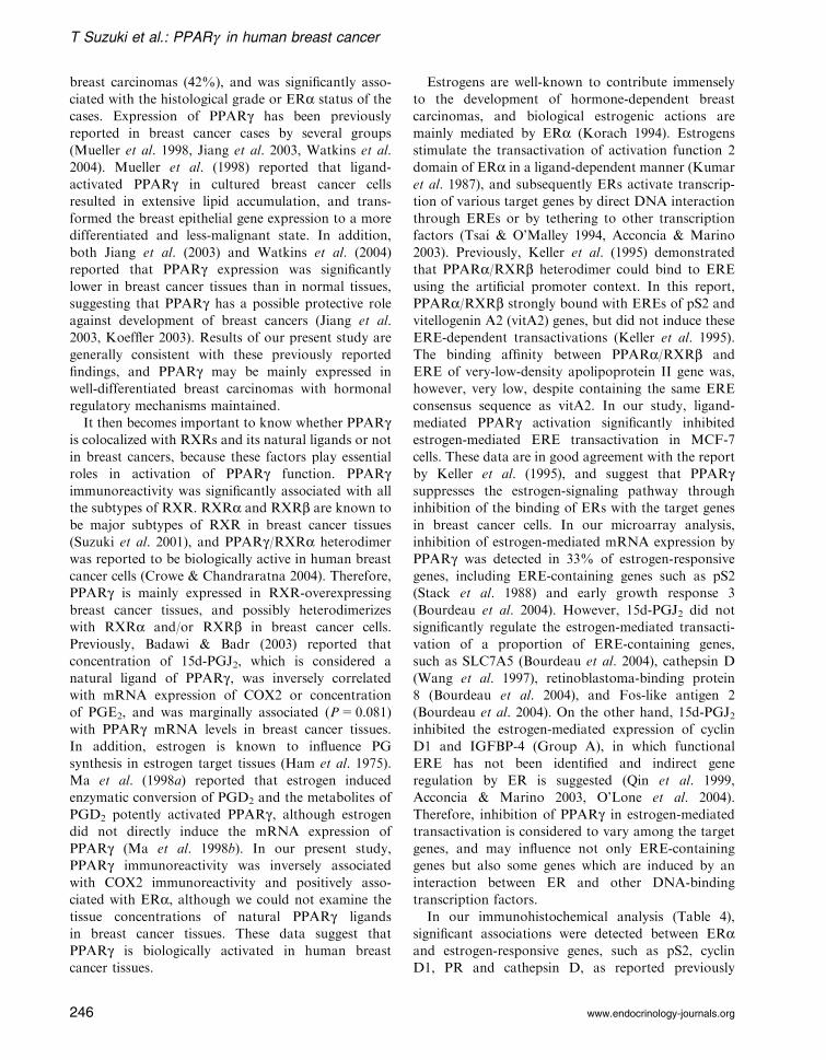
T Suzuki et al.: PPARg in human breast cancer
234 www.endocrinology-journals.org

ERb; 1/50, PR; 1/30; RXRa; 1/4000, RXRb; 1/4000,RXRg ; 1/2000, COX2; 1/500, HER2; 1/200, Ki-67;
1/50, p21; 1/250, p27; 1/150, c-Myc 1/600, pS2; 1/30,
cyclin D1; 1/40 and cathepsin D; 1/300. The antigen–
antibody complex was visualized with 3,30-diamino-
benzidine solution (1mM, in 50mM Tris–HCl buffer
(pH7.6) and 0.006% H2O2), and counterstained with
hematoxylin. As a negative control, normal rabbit,
mouse or goat IgG was used instead of the primary
antibodies. For PPARg immunohistochemistry, a pre-
absorption test was also performed as a negative
control.
Scoring of immunoreactivity and statisticalanalysis
PPARg , ERa,b, PR, RXRa,b,g , Ki-67, p21, p27,
c-Myc and cyclin D1 immunoreactivity was detected in
the nucleus, and the immunoreactivity was evaluated
in more than 1000 carcinoma cells for each case, and
subsequently the percentage of immunoreactivity, i.e.
labeling index (LI), was determined. Inter-observer
differences were less than 5%, and the mean of the
three values was obtained. Cases with PPARg , ERa or
c-Myc LIs of more than 10% were considered PPARg-,ERa- or c-Myc-positive breast carcinomas in this
study, according to a report for ERa by Allred et al.
(1998). For p21 and p27 immunohistochemistry, the
cut-off values used were 5 and 50% respectively,
according to previous reports (Barbareschi et al.
2000, Pellikainen et al. 2003). Immunoreactivity for
COX2 and cathepsin D was detected in the cyto-
plasm, and cases that had more than 10% of positive
carcinoma cells were considered positive.
An association between immunoreactivity for
PPARg and clinicopathological factors was evaluated
using a one-way ANOVA and a Bonferroni test or a
cross-table using the chi-square test. Overall and
disease-free survival curves were generated according
to the Kaplan–Meier method and statistical signifi-
cance was calculated using the log-rank test. An
association between ERa LI and PR or cyclin D1 LI
was performed utilizing a correlation coefficient (r)
and regression equation. Univariate and multivariate
analyses were evaluated by a proportional hazard
model (Cox) using PROC PHREG in our SAS
software. P values less than 0.05 were considered
significant in this study.
Cell line, plasmids and chemicals
MCF-7 human breast cancer cell line was cultured in
RPMI-1640 (Sigma-Aldrich, St Louis, MO, USA) with
10% fetal bovine serum (FBS) (JRH Biosciences,
Lenexa, KS, USA). MCF-7 cells were cultured with
phenol red-free RPMI 1640 medium containing 10%
dextran-coated charcoal (DCC)-FBS for 3 days before
treatment in the experiment. In this study, we used
estrogen-responsive reporter plasmids pERE-Luc, con-
taining Xenopus vitellogenin A2 estrogen-responsive
element (ERE) (Saji et al. 2001). The pRL-TK vectors
were purchased from Promega (Madison, WI, USA).
15d-PGJ2, ciglitazone and PGF2a were purchased
from Biomol Research Laboratories (Butler Pike,
PA, USA), and GW1929 and GW9662 were purchased
from Sigma-Aldrich.
Luciferase assay
The luciferase assay was performed according to a
previous report (Sakamoto et al. 2002) with some
modifications. Briefly, 1mg ptk-ERE-Luc plasmids and
200 ng pRL-TK control plasmids were used to measure
the transcriptional activity of endogenous ER. Tran-
sient transfections were carried out using TransIT-LT
Transfection Reagents (TaKaRa, Tokyo, Japan) in
MCF-7 cells, and the luciferase activity of lysates was
measured using a Dual-Luciferase Reporter Assay
system (Promega) and Luminescencer-PSN (AB-2200)
(Atto Co., Tokyo, Japan) after incubation with growth
medium with the indicated concentrations of estradiol
and/or 15d-PGJ2 for 24 h. The cells were also treated
with the same volume of ethanol (final dilution – 0.1%)
for 24 h as controls. The transfection efficiency was
normalized against Renilla luciferase activity using
pRL-TK control plasmids, and the luciferase activity
for each sample was evaluated as a ratio (%) compared
with that of controls. The statistical analyses were
performed using a one-way ANOVA and Bonferroni
test.
Immunoblotting
The cell protein was extracted in triple detergent lysis
buffer (LK-18) at 4 �C. Twenty micrograms of the
protein (whole cell extracts) were subjected to SDS-
PAGE (10% acrylamide gel). Following SDS-PAGE,
proteins were transferred onto Hybond P polyvinyli-
dene difluoride membrane (Amersham Biosciences,
Piscataway, NJ, USA). The blots were blocked in 5%
non-fat dry skim milk for 1 h at room temperature, and
were then incubated with a primary antibody for ERa,p21, p27 or b-actin (Sigma-Aldrich) for 18 h at 4 �C.After incubation with anti-mouse IgG horseradish
peroxidase (AmershamBiosciences) for 1 hat room tem-
perature, antibody–protein complexes on the blots were
detected using ECL-plus western blotting detection
reagents (Amersham Biosciences). Immunointensity of
Endocrine-Related Cancer (2006) 13 233–250
www.endocrinology-journals.org 235

specific bands was measured by an LAS-1000 imaging
system (Fuji Photo Film, Tokyo, Japan), and relative
immunointensity of ERa, p21 or p27 was evaluated as a
ratio (%) of b-actin immunointensity.
Microarray analysis
In this study, we used a custom-made microarray
named EstrArray (InfoGenes, Tsukuba, Japan), which
contains 175 estrogen-responsive genes identified in
MCF-7 cells (Inoue et al. 2002, Hayashi et al. 2003).
MCF-7 cells were cultured with phenol red-free RPMI
1640 medium containing 10% DCC-FBS for 3 days,
and subsequently treated with estradiol (10 nM) with
or without 15d-PGJ2 (5mM) for 72 h. The MCF-7 cells
used as references were treated with the same volume
of ethanol (final dilution – 0.1%) for 72 h.
Total RNA was isolated using TRIzol reagent
(InVitrogen Life Technologies, Inc., Gaitherburg,
MD, USA). Two micrograms of mRNA were
reverse-transcribed with Cy3- or Cy5-dUTP (Amer-
sham Biosciences, Bucks, UK) using a SUPER-
SCRIPT II Preamplification system (Gibco-BRL,
Grand Island, NY, USA). Cy3- and Cy5-labeled
cDNA probes were hybridized on the microarray slide
for 16 h at 65 �C. The fluorescent signals were scannedby a GenePix 4000A (Axon Instruments, Foster City,
CA, USA), and the ratio of Cy3 and Cy5 signal
intensity of each spot was quantitatively calculated
using GenePixPro 5.0 (Axon Instruments). The dupli-
cated sets of values were averaged and normalized by
subtracting the average of values for internal genes.
The data from insufficient hybridization (signal areas
below 100) were excluded from the analysis. Genes
which showed a value of more than 2.0 or less than 0.5
were evaluated as significantly up-regulated or down-
regulated respectively, in this study.
Real-time PCR
MCF-7 cells were cultured with phenol red-free RPMI
1640 medium containing 10% DCC-FBS for 3 days,
and subsequently treated with the indicated concentra-
tion of estradiol and/or 15d-PGJ2 for 72 h. As controls
for the experiments, the cells were treated with the same
volume of ethanol (final dilution – 0.1%) for 72 h. Total
RNA was extracted using TRIzol reagent (InVitrogen
Life Technologies, Inc.), and a reverse transcription kit
(SUPERSCRIPT II Preamplification system (Gibco-
BRL) was used in the synthesis of cDNA.
The Light Cycler System (Roche Diagnositics
GmbH, Mannheim, Germany) was used to semi-
quantify the mRNA expression levels by real-time
PCR (Dumoulin et al. 2000). Characteristics of the
primer sequences used in this study are summarized in
Table 1 (Colombel et al. 1999, Schonherr et al. 2001,
Vandesompele et al. 2002, Kao et al. 2003, Paruthiyil
et al. 2004, Yoshida et al. 2004). Settings for the PCR
thermal profile were: initial denaturation at 95 �C for
1min followed by 40 amplification cycles of 95 �Cfor 1 s, annealing at 59 �C (PR), 60 �C (pS2, PDZ
domain-containing protein (PDZK1), cathepsin D,
selenium-binding protein 1 (SELENBP1), tumor
protein D52-like 1 (TPD52L1), insulin-like growth
Table 1 Primer sequences used in real-time PCR in this study
cDNA (Accession no.) Sequence (position in cDNA) Size (bp) Reference
pS2 (NM003225) FWD: 53–72, REV: 240–260 208 Colombel et al. (1999)
PDZK1 (AF012281) FWD: 725–746, REW: 824–847 123 Yoshida et al. (2004)
Cyclin D1 (NM053056) FWD: 456–475, REV: 640–659 204 Paruthiyil et al. (2004)
IGFBP-4 (M62403) FWD: 697–715, REV: 756–773 77 Yoshida et al. (2004)
SLC7A5* (M80244) FWD: 816–835, REV: 1025–1044 206 —
TPD52L1 (U44427) FWD: 450–469, REV: 561–580 130 Yoshida et al. (2004)
PR*y (XM006190) FWD: 1987–2006, REV: 2163–2182 195 —
Cathepsin D (BT006910) FWD: 697–715, REV: 804–822 126 Yoshida et al. (2004)
TAL6 (M90657) FWD: 246–265, REV: 299–319 74 Kao et al. (2003)
IGFBP-5 (NM000599) FWD: 1090–1108, REV: 1213–1232 143 Yoshida et al. (2004)
SELENBP1 (U29091) FWD: 167–186, REV: 315–333 167 Yoshida et al. (2004)
p21* (NM000389) FWD: 415–434, REV: 573–592 78 —
p27 (NM004064) FWD: 829–848, REW: 929–938 120 Schonherr et al. (2001)
RPL13A (NM012423) FWD: 487–509, REV: 588–612 125 Vandesompele et al. (2002)
PDZK1; PDZ domain-containing-protein, IGFBP-4; insulin-like growth factor-binding protein-4, SLC7A5; solute carrier family 7,
member 5, TPD52L1; tumor protein D52-like 1, PR; progesterone receptor, TAL6; tumor-associated antigen L6, IGFBP-5; insulin-
like growth factor-binding protein-5, SELENBP; selenium-binding protein-1, and RPL13A; ribosomal protein L 13a.
*Oligonucleotide primers for SLC7A5, PR and p21 were designed in the different exons in this study.
yThe primers of PR recognize both PR-A and PR-B subtypes.
T Suzuki et al.: PPARg in human breast cancer
236 www.endocrinology-journals.org

factor-binding protein-5 (IGFBP-5), PDZ domain-
containing-protein (PDZK1), and p21), 64 �C (cyclin
D1, and tumor-associated antigen L6 (TAL6)), 66 �C(solutecarrierfamily7,member5(SLC7A5),andp27),or
68 �C (ribosomal protein L 13a (RPL13A)) for 15 s, and
elongationat72 �Cfor15 s.Toverifyamplificationof the
correct sequences, PCR products were purified and
subjected to direct sequencing. Negative control experi-
ments lacked cDNAsubstrate to check for thepossibility
ofexogenouscontaminantDNA.ThemRNAlevelswere
summarized as a ratio of RPL13A, and subsequently
evaluated as a ratio (%) compared with that of controls.
The statistical analyses were performed using a one-way
ANOVA and Bonferroni test.
Cell proliferation assay and apoptosisanalysis
The status of cell proliferation of MCF-7 cells
was measured using a WST-8 (2-(2-methoxy-4-
nitrophenyl)-3-(4-nitrophenyl)-5-(2,4-disulfophenyl)-2-
H-tetrazolium, monosodium salt) method (Cell Count-
ing Kit-8; Dojindo Inc., Kumamoto, Japan)) (Isobe
et al. 1999). We also examined apoptosis status of
MCF-7 cells using an apoptosis screening kit (Wako,
Osaka, Japan), which employed a modified TdT-
mediated dUTP nick-end labeling (TUNEL) method
(Gavrieli et al. 1992). Optical densities (OD=450 nM
for cell proliferation assay, and OD=490 nM for
A B
C D
Figure 1 Immunohistochemistry for PPARg in invasive ductal carcinoma. (A) PPARg immunoreactivity was detected in the
nuclei of carcinoma cells. (B) No significant immunoreactivity of PPARg was detected in the sections where an
immunohistochemical preabsorption test was performed as a negative control. (C) Immunoreactivity for PPARg was
detected in the nuclei of epithelial cells in morphologically normal mammary glands. (D) PPARg immunoreactivity was
positive in the nuclei of adipocytes. Bar=50mm.
Endocrine-Related Cancer (2006) 13 233–250
www.endocrinology-journals.org 237

apoptosis analysis) were obtained with a Model 680
microplate reader (Bio-Rad Laboratories, Hercules,
CA, USA). The cell number and apoptosis index were
calculated according to the equation (Cell OD value
after test material treatment)/(Vehicle control cell OD
value), and subsequently evaluated as a ratio (%)
compared with that of controls.
Results
Immunohistochemistry for PPARg in breastcarcinoma tissues
Immunoreactivity for PPARg was detected in the
nuclei of invasive ductal carcinoma cells (Fig. 1A and
B). A mean value of PPARg LI in 238 breast
carcinoma cases examined was 15% (range 0–74%),
and the number of PPARg-positive breast carcinomas
(i.e. PPARg LIi10%) was 99 out of 238 cases (42%).
Immunoreactivity of PPARg was also detected in
epithelia of morphologically normal mammary glands
(Fig. 1C), and adipocytes (Fig. 1D).
Associations between PPARg immunoreactivity
and clinicopathological parameters in 238 breast
carcinomas were summarized in Table 2. PPARgimmunoreactivity was significantly associated with
ERa status (P=0.0003), ERa LI (P<0.0001), ERbLI (P=0.0255), PR LI (P=0.0012), RXRa LI
(P=0.0365), RXRb LI (P<0.0001), RXRg LI
(P=0.0005), p21 immunoreactivity (P=0.0057) or
p27 immunoreactivity (P=0.0019). PPARg immuno-
reactivity was negatively correlated with histological
grade (P=0.0035) or COX2 immunoreactivity (P=
0.0178). No significant association was detected
between PPARa immunoreactivity and other clinico-
pathological parameters examined, including patient
age, menopausal status, clinical stage, tumor size,
lymph node status, HER2 status, Ki-67 LI, and c-Myc
immunoreactivity in this study. The association
between PPARg immunoreactivity and RXRb LI,
RXRg LI, COX2 immunoreactivity, p21 immuno-
reactivity or p27 immunoreactivity was significant
regardless of ERa status of these cases (Table 3).
PPARg immunoreactivity was positively correlated
with RXRa LI (P=0.0469) and inversely with lymph
node status (P=0.0303) or Ki-67 LI (P=0.0485) only
in the ERa-positive group.
Influence of PPARg immunoreactivity onassociation between ERa and estrogen-responsive genes in breast cancer tissues
pS2, cyclin D1, PR and cathepsin D are all well
recognized as estrogen-responsive genes in human
breast cancers. As shown in Table 4, a significant
positive association was detected between ERa LI and
the status of these immunoreactivity in the 238 breast
cancer tissues examined (P<0.0001), which is in good
agreement with previous immunohistochemical studies
Table 2 Association between PPARg immunoreactivity and
clinicopathological parameters in 238 breast carcinomas
PPARg immunoreactivity
P value
Positive
(n=99)
Negitive
(n=139)
Age (years)* 53.1t1.2 55.1t1.1 0.0850
Menopausal status
Premenopausal 46 (19.3%) 54 (22.7%)
Postmenopausal 53 (22.3%) 85 (35.7%) 0.2983
Stage
I 30 (12.6%) 33 (13.9%)
II 57 (24.0%) 75 (31.5%)
III 12 (5.0%) 31 (13.0%) 0.1032
Tumor size (mm)* 32.6t0.4 29.4t0.2 0.3808
Lymph node status
Positive 38 (16.0%) 68 (28.6%)
Negative 61 (25.6%) 71 (29.8%) 0.1389
Histological grade
1 35 (14.7%) 31 (13.0%)
2 40 (16.8%) 46 (19.3%)
3 24 (10.1%) 62 (26.1%) 0.0035
ERa status
Positive 85 (35.7%) 89 (37.4%)
Negative 14 (5.9%) 50 (21.0%) 0.0003
ERa LI* 54.5t3.1 34.8t2.9 <0.0001
ERb LI* 19.6t2.4 13.4t1.6 0.0255
PR LI* 49.6t3.3 34.9t3.0 0.0012
RXRa LI* 27.1t3.5 18.4t2.4 0.0365
RXRb LI* 36.0t4.7 10.5t2.2 <0.0001
RXRg LI* 17.5t5.1 3.0t1.0 0.0005
COX2 immunoreactivity
Positive 29 (12.2%) 72 (30.3%)
Negative 70 (29.4%) 67 (28.2%) 0.0178
HER2 status
Positive 46 (19.3%) 53 (22.7%)
Negative 53 (22.7%) 85 (35.7%) 0.7836
Ki-67 LI* 22.7t1.6 26.7t1.6 0.0916
p21 immunoreactivity
Positive 56 (23.5%) 45 (18.9%)
Negative 43 (17.3%) 91 (38.2%) 0.0057
p27 immunoreactivity
Positive 58 (24.4%) 47 (19.7%)
Negative 41 (17.2%) 92 (38.7%) 0.0019
c-Myc immunoreactivity
Positive 49 (20.6%) 62 (26.1%)
Negative 50 (21.0%) 77 (32.4%) 0.5715
*Data are presented as meanst95% confidence interval.
All other values represent the number of cases and percentage.
P values less than 0.05 were considered significant, and are
in boldface.
T Suzuki et al.: PPARg in human breast cancer
238 www.endocrinology-journals.org

(Horwitz & McGuire 1978, Barbareschi et al. 1997,
Gillesby & Zacharewski 1999, Ioachim et al. 2003).
When the breast cancers were classified into two
groups according to their PPARg status, no significant
association was detected between ERa LI and pS2
(P=0.3785) or cyclin D1 LI (P=0.1978) in PPARg-positive breast carcinomas, although significant
association (P<0.0001 for pS2, and P=0.0018 for
cyclin D1) was detected in PPARg-negative breast
carcinomas. On the other hand, ERa LI was sig-
nificantly associated with PR LI (P=0.0008 in
PPARg-positive cases, and P<0.0001 in PPARg-negative cases) or cathepsin D (P=0.0006 in
PPARg-positive cases, and P=0.0003 in PPARg-negative cases) regardless of the PPARg status in the
breast carcinoma cases examined.
Correlation between PPARg immunoreactivityand clinical outcome of the patients
No significant association was detected between
PPARg immunoreactivity and a risk of recurrence
(P=0.8715) (Fig. 2A). PPARg immunoreactivity was
significantly associated with a better clinical outcome
of the 238 breast cancer patients (P=0.0257)
(Fig. 2B). This significant association was detected in
the ERa-positive group (P=0.0057) (Fig. 2C), but not
in the ERa-negative group (P=0.6405) (Fig. 2D). The
significant correlation between PPARg immuno-
reactivity and overall survival of ERa-positive breast
cancer patients was not influenced by tamoxifen
therapy after the surgery (Fig. 2E and F).
Utilizing a univariate analysis (Table 5), lymph node
status (P<0.0001), histological grade (P<0.0001),
tumor size (P=0.0002), HER2 status (P=0.0029),
c-Myc immunoreactivity (P=0.0066), and PPARgimmunoreactivity (P=0.0287) turned out to be
significant prognostic factors for overall survival in
this study. Multivariate analysis revealed that only
lymph node status (P<0.0001) and c-Myc immuno-
reactivity (P=0.0024) were independent prognostic
factors with a relative risk over 1.0 (Table 5). When we
examined a univariate analysis in the ERa-positivebreast cancer patients (n=174), lymph node status
(P<0.0001), histological grade (P<0.0001), tumor
size (P<0.0001), HER2 status (P=0.0008), PPARg
Table 3 Statistical association between PPARg immunoreac-
tivity and clinicopathological parameters according to the ERastatus in 238 breast carcinomas
PPARg immunoreactivity
(positive/negative)
ERa-positivegroup (n=173)
ERa-negativegroup (n=64)
Age 0.1107 0.1073
Menopausal status 0.6702 0.6616
Stage 0.1272 0.7195
Tumor size 0.6077 0.6063
Lymph node status 0.0303 0.7192
Histological grade 0.3691 0.2452
ERb LI 0.0963 0.1541
PR LI 0.2826 0.5011
RXRa LI 0.0469 0.5403
RXRb LI 0.0005 0.0002
RXRg LI 0.0108 0.0185
COX2 immunoreactivity 0.0205 0.0411
HER2 status 0.7702 0.7616
Ki-67 LI 0.0485 0.7103
p21 immunoreactivity 0.0113 0.0481
p27 immunoreactivity 0.0142 0.0205
c-Myc immunoreactivity 0.3999 0.5404
Data are presented as P values. P values less than 0.05 were
considered significant, and are in boldface.
Table 4 Correlation between ERa and estrogen-responsive gene immunoreactivity associated with PPARg status in 238 breast
carcinomas
Total (n=238) PPARg-positive (n=99) PPARg-negative (n=139)
ERa LI P value ERa LI P value ERa LI P value
pS2
Positive 53.2t3.3 57.3t4.4 52.4t4.8
Negative 31.8t3.7 <0.0001 49.0t6.7 0.3785 22.0t4.1 <0.0001
Cyclin D1 LI* <0.0001 (r=0.355) 0.1978 (r=0.231) 0.0018 (r=0.313)
PR LI* <0.0001 (r=0.512) 0.0008 (r=0.402) <0.0001 (r=0.566)
Cathepsin D
Positive 54.2t3.7 58.0t4.4 51.2t5.6
Negative 17.5t4.4 <0.0001 24.8t8.5 0.0006 12.2t4.2 0.0003
P values less than 0.05 were considered significant, and are in boldface.
*The association was statistically evaluated utilizing a correlation coefficient (r) and regression equation.
Endocrine-Related Cancer (2006) 13 233–250
www.endocrinology-journals.org 239

immunoreactivity (P=0.0076) and c-Myc immuno-
reactivity (P=0.0252) were demonstrated as signifi-
cant prognostic factors for overall survival (Table 6). A
multivariate analysis revealed that lymph node status
(P<0.0001) andPPARg immunoreactivity (P=0.0372)
were independent prognostic factors with relative risks
over 1.0.
Effects of PPARg activator 15d-PGJ2 onestrogen-mediated transcription in MCF-7cells in luciferase assay
Results of PPARg immunoreactivity demonstrated
a strong association between PPARg and ERain breast carcinoma tissues, suggesting a possible
A
0
20
40
60
80
100
0 20 40 60 80 100120140160
Dis
ease
-fre
e su
rviv
al (
%)
Months after operation
+ (n = 99)
– (n = 139)
P = 0.8715
B
0
20
40
60
80
100
0 20
Ove
rall
surv
ival
(%
)
Months after operation
+ (n = 99)
– (n = 139)
P = 0.0257
40 60 80 100120140160
0
20
40
60
80
100
0 20 40 60 80 100120140160
Ove
rall
surv
ival
(%
)
+ (n = 85)
– (n = 89)
P = 0.0057
Months after operation
ERα-positive group
C D
0
20
40
60
80
100
0 20
Ove
rall
surv
ival
(%
) – (n = 50)
P = 0.6405
Months after operation
ERα-negative group
+ (n = 14)
40 60 80 100120140160
E
0
20
40
60
80
100
0 20 40 60 80 100120140160
Ove
rall
surv
ival
(%
)
+ (n = 23)
– (n = 20)
P = 0.0420
Months after operation
ERα-positive group received tamoxifen therapy
Ove
rall
surv
ival
(%
)
0
20
40
60
0 20
+ (n = 62)
– (n = 69)
P = 0.0387
Months after operation
F
ERα-positive group without tamoxifen therapy
100
80
40 60 80 100120140160
Figure 2 Disease-free (A) and overall (B–F) survival of 238 patients with breast carcinoma according to PPARg immunoreactivity
(Kaplan–Meier method). PPARg immunoreactivity was not correlated with a risk of recurrence (P=0.8715 in log-rank test) (A),
but significantly associated with an improved overall survival (P=0.0257 in log-rank test) (B). The significant association was
detected in the ERa-positive group (n=174; P=0.0057 in log-rank test) (C), but not in the ERa-negative cases (n=64; P=0.6405
in log-rank test) (D). PPARg immunoreactivity was significantly associated with an improved prognosis regardless of tamoxifen
therapy after surgery in the ERa-positive breast cancer patients (E, F). P values less than 0.05 are in boldface.
T Suzuki et al.: PPARg in human breast cancer
240 www.endocrinology-journals.org

interaction of these two nuclear receptors in human
breast carcinoma cells. Previously, Keller et al. (1995)
reported that PPARa/RXRb heterodimer can bind
to ERE and possibly modulate the ER-signaling
pathway, but this has not been examined in breast
cancers.
In order to examine this hypothesis, we used MCF-7
breast cancer cells in the following in vitro experiments,
because MCF-7 cells were associated with expression
of ERa, PPARg , and RXRa, b, g (data not shown).
When MCF-7 cells were transiently transfected with
ptk-ERE-Luc plasmids and treated with 10 nM estra-
diol, the luciferase activity of the cells was 17-fold
increased compared with their basal level (Fig. 3A).
PPARg activator 15d-PGJ2 significantly inhibited
ERE-dependent transactivation by estradiol in a
dose-dependent manner, and the luciferase activity of
MCF-7 cells treated with 10 nM estradiol and 5mM
15d-PGJ2 was decreased to 53% of that treated with
10 nM estradiol alone (P<0.001). 15d-PGJ2 (5mM)
alone did not significantly change the luciferase
activity compared with their basal level (P=0.8837).
15d-PGJ2, however, did not significantly inhibit the
ERE-dependent transactivation by estradiol, when
these cells were treated with a potent PPARgantagonist GW9662 (Leesnitzer et al. 2002).
The ERE-dependent transactivation by estradiol
was also inhibited by other PPARg agonists such as
GW1929 and ciglitazone, in a dose-dependent manner
(P<0.001, on addition of 5mM GW1929 or ciglita-
zone), but was not altered by treatment with PGF2a,
which does not activate PPARg (Kliewer et al. 1995)
(Fig. 3B). Results of immunoblotting analysis revealed
that relative immunointensity of ERa was not sig-
nificantly (P=0.7749) altered by the treatment with
15d-PGJ2 in MCF-7 cells (Fig. 3C).
Table 5 Univariate and multivariate analyses of overall survival in 238 breast cancer patients examined
Variable Univariate P
Multivariate
P Relative risk (95% CI)
Lymph node status (positive/negative) <0.0001 <0.0001 12.319 (3.233–51.07)
Histological grade (3/1, 2) <0.0001 0.1248 —
Tumor size (>20mm/<20mm) 0.0002 0.1248 —
HER2 status (positive/negative) 0.0029 0.4669 —
c-Myc immunoreactivity (positive/negative) 0.0066 0.0024 2.168 (1.079–4.356)
PPARg immunoreactivity (negative/positive) 0.0287 0.2558 —
p27 immunoreactivity (negative/positive) 0.0797 — —
Adjuvant chemotherapy (no/yes) 0.1458 — —
Ki-67 LI (>10/<10) 0.2818 — —
Tamoxifen therapy (no/yes) 0.5807 — —
p21 immunoreactivity (negative/positive) 0.6751 — —
ERa status (negative/positive) 0.9532 — —
Data considered significant (P<0.05) in the univariate analyses are in boldface, and were examined in the multivariate analyses.
Table 6 Univariate and multivariate analyses of overall survival in 174 ER a-positive breast cancer patients examined
Variable Univariate P
Multivariate
P Relative risk (95% CI)
Lymph node status (positive/negative) <0.0001 <0.0001 19.006 (4.402–68.06)
Histological grade (3/1, 2) <0.0001 0.0512 —
Tumor size (>20mm/<20mm) <0.0001 0.0724 —
HER2 status (positive/negative) 0.0008 0.3899 —
PPARg immunoreactivity (negative/positive) 0.0076 0.0372 2.799 (1.128–8.264)
c-Myc immunoreactivity (positive/negative) 0.0252 0.2891 —
p27 immunoreactivity (negative/positive) 0.0532 — —
Adjuvant chemotherapy (no/yes) 0.1057 — —
Ki-67 LI (>10/<10) 0.1123 — —
Tamoxifen therapy (no/yes) 0.1497 — —
p21 immunoreactivity (negative/positive) 0.2231 — —
Data considered significant (P<0.05) in the univariate analyses are in boldface, and were examined in the multivariate analyses.
Endocrine-Related Cancer (2006) 13 233–250
www.endocrinology-journals.org 241

Effects of 15d-PGJ2 on estrogen-mediatedtranscription in MCF-7 cells in microarrayanalysis
We further examined the effects of 15d-PGJ2 on
estrogen-mediated transcription in MCF-7 cells using
a custom-made microarray. Significant alterations of
mRNA expression by estradiol treatment (10 nM
for 72 h) were detected in 49 genes in this study, and
these genes were classified into the following four
groups (Table 7): Group A; mRNA expression was
significantly up-regulated by estradiol, but the sig-
nificance vanished on addition of 15d-PGJ2 (5mM)
(nine genes; Table 8), Group B; mRNA expression was
significantly up-regulated by estradiol with or without
15d-PGJ2 (23 genes; Table 9), Group C; mRNA ex-
pression was significantly down-regulated by estradiol,
but the significance vanished on addition of 15d-PGJ2(seven genes; Table 10), and Group D; mRNA
expression was significantly down-regulated by estra-
diol with or without 15d-PGJ2 (ten genes; Table 11).
Effects of 15d-PGJ2 on estrogen-mediatedtranscription in MCF-7 cells using real-timePCR analysis
In order to confirm the results from microarray anal-
ysis, we performed real-time PCR analyses for the
representative 11 genes in MCF-7 cells (Fig. 4A–K).
******0
250500750
100012501500175020002250
E2Rel
ativ
e lu
cife
rase
act
ivity
(%
)
(nM)--
10-
101
105
-5
--
10-
101
105
-5
--
10-
101
105
-5 (µM)
Ciglitazone GW1929 PGF2α
*** ****** ***
*****
B
***
***
0250500750
100012501500175020002250
Estradiol15d-PGJ2
GW9662
---
Rel
ativ
e lu
cife
rase
act
ivity
(%
)
***
******
**
*** ***
A
(nM)(µM)(µM)
--5
-5-
105-
101-
10--
1055
10-5
10--
---
C --
10-
101
105
-5
E2 (nM) 15d-PGJ2 (µM)MW
80 kDaERα
60 kDa
Immunointensityratio (ERα /β-actin)
50 kDaβ-actin
40 kDa
35±8 32±4 28±5 31±7 30 ±10 (%)
Figure 3 Effects of PPARg activator 15d-PGJ2 on
ERE-dependent transactivation in MCF-7 cells. (A) MCF-7
cells were transiently transfected with pERE-Luc plasmids,
and treated with the indicated concentrations of estradiol,
15d-PGJ2 and GW9662 (PPARg antagonist) for 24 h. The
luciferase activity was evaluated as a ratio (%) compared
with that of controls (treatment without estradiol, 15d-PGJ2or GW9662 for 24 h (left column)). Data are presented as
meanstS.D. (n=3). **P<0.01 and ***P<0.001 vs 10 nM
estradiol alone. (B) MCF-7 cells transfected with pERE-Luc
plasmids were treated with the indicated concentrations of
estradiol and GW1929 (PPARg agonist), ciglitazone (PPARgagonist), or PGF2a (no activator of PPARg) for 24 h. Dataare presented as meanstS.D. (n=3). **P<0.01 and
***P<0.001 vs 10 nM estradiol alone respectively. (C)
Immunoblotting for ERa in MCF-7 cells. ERa and b-actinimmunoreactivities were detected as specific bands
(approximately 66 and 42 kDa respectively); 20mg of protein
were loaded in each lane. Data of immunointensity ratio
(ERa/b-actin) are presented as meanstS.D. (n=3). No
significant association was detected.
Table 7 The number and percentage of genes classified
into four groups according to the effects of 15d-PGJ2 on
the estrogen-mediated mRNA expression in the microarray
analysis
Regulation of mRNA
expression by
estradiol with 15d-PGJ2
TotalNot significant Significant
Estrogen
up-regulated
genes
Group A
n=9 (18%)
Group B
n=23 (47%)
—
n=32 (65%)
Estrogen
down-regulated
genes
Group C
n=7 (14%)
Group D
n=10 (20%)
—
n=17 (35%)
Total n=16 (33%) n=33 (67%) n=49 (100%)
A custom-made microarray (EstrArray; InfoGenes, Co. Ltd,
Tsukuba, Japan), which contains 175 estrogen-responsive
genes (Inoue et al. 2002, Hayashi et al. 2003), was used in
this study.
MCF-7 cells were treated with estradiol (10 nM) with or without
15d-PGJ2 (5mM) for 72 h. Genes showing the value of more
than 2.0- or less than 0.5-fold compared with the control
(treated without estradiol or 15d-PGJ2 for 72 h) were regarded
as significantly regulated.
T Suzuki et al.: PPARg in human breast cancer
242 www.endocrinology-journals.org

mRNA expression of pS2 (Fig. 4A), PDZK1 (Fig. 4B)
cyclin D1 (Fig. 4C) and IGFBP-4 (Fig. 4D), which
were tentatively classified into Group A in microarray
analysis as above, was significantly (P<0.001)
increased by estradiol treatment (10 nM, for 72 h) com-
pared with the control (neither estradiol nor 15d-PGJ2),
but not by treatment with estradiol (10 nM) with 15d-
PGJ2 (5mM). mRNA expression of SLC7A5 (Fig. 4E),
TPD52L1 (Fig. 4F), PR (Fig. 4G), and cathepsin D
(Fig. 4H) in Group B was, however, significantly up-
regulated by the treatment with estradiol with or
without 15d-PGJ2 (1 or 5mM). Estradiol-mediated
mRNA expression of PR was also demonstrated to be
inhibited by addition of 15d-PGJ2 in a dose-dependent
manner (P<0.01, between estradiol alone and estradiol
with 15d-PGJ2 (5mM)). The mRNA level of TAL6 in
Group C (Fig. 4I) was significantly lower (P<0.001) in
estradiol alone than that in the control group, but was
not significantly different under treatment with estra-
diol with 15d-PGJ2 (5mM). mRNA expression of
IGFBP-5 (Fig. 4J) and SELENBP1 (Fig. 4K) in Group
D was significantly down-regulated by the treatment
with estradiol (10 nM) with or without 15d-PGJ2 (1 or
5mM). mRNA expression in these 11 genes was not
significantly altered by treatment with 15d-PGJ2(5mM) alone in this study.
Table 8 Summary of genes classified into Group A in the
microarray analysis examined
Accession no.
Gene
symbol Estradiol
Estradiol
+15d-PGJ2
NM003225 TFF1 (pS2) 7.5 1.7
AA143530 SCD 3.4 1.5
AF012281 PDZK1 3.1 1.6
AI660571 ASS 2.3 1.2
AB012664 STC2 2.3 1.1
X16396 MTHFD2 2.2 1.6
NM053056 CCND1
(cyclin D1)
2.2 0.8
M62403 IGFBP4 2.1 0.9
X63741 EGR3 2.0 1.1
Values are presented as fold change compared with controls.
Boldface: mRNA expressions were examined by real-time PCR
analysis (Fig. 4).
Table 9 Summary of genes in Group B
Accession no.
Gene
symbol Estradiol
Estradiol
+15d-PGJ2
M80244 SLC7A5 4.4 3.9
AI151190 S100P 4.0 3.0
U44427 TPD52L1 4.0 2.4
XM006190 PGR (PR) 3.8 2.0
XM041014 — 3.5 2.2
X74837 HUMM9 2.9 3.7
L02785 SLC26A3 2.8 2.6
N39944 ATF3 2.8 4.1
AB028974 PEG10 2.6 2.2
AA587912 PSAT1 2.5 2.6
BT006910 CTPD
(cathepsin D)
2.4 2.9
AA216685 GDF15 2.4 2.8
NM003458 BSN 2.3 2.4
D90070 PMAIP1 2.3 2.5
U95626 CCRL2 2.3 3.6
AF039022 XPOT 2.3 2.9
X89773 ISG20 2.2 2.3
X72875 BF 2.2 3.0
D30658 GARS 2.2 2.1
M90516 GFPT1 2.1 2.3
U72066 RBBP8 2.1 2.1
AJ011972 HDAC6 2.0 2.0
X16706 FOSL2 2.0 2.8
Values are presented as fold change compared with controls.
Boldface: mRNA expressions were examined by real-time PCR
analysis (Fig. 4).
Table 10 Summary of genes in Group C
Accession no.
Gene
symbol Estradiol
Estradiol
+15d-PGJ2
AI337192 SH3BGR 0.073 1.1
M90657 TM4SF1
(TAL6)
0.23 0.71
JQ1035 — 0.35 0.66
XM006424 — 0.36 1.1
M59828 HSPA1A 0.40 1.8
AA481712 CDKN1A 0.42 1.4
L25081 RHOC 0.43 0.57
Values are presented as fold change compared with controls.
Boldface: mRNA expressions were examined by real-time PCR
analysis (Fig. 4).
Table 11 Summary of genes in Group D
Accession no.
Gene
symbol Estradiol
Estradiol
+15d-PGJ2
D11428 PMP22 0.074 0.075
NM000599 IGFBP5 0.23 0.27
D87993 PCSK6 0.26 0.23
U69263 MATN2 0.30 0.20
M19922 FBP1 0.32 0.17
U29091 SELENBP1 0.33 0.34
U30246 LC12A2 0.34 0.24
NM033001 GTF2I 0.42 0.27
U97276 QSCN6 0.43 0.42
AA482422 ENO1 0.43 0.62
Values are presented as fold change compared with controls.
Boldface: mRNA expressions were examined by real-time PCR
analysis (Fig. 4).
Endocrine-Related Cancer (2006) 13 233–250
www.endocrinology-journals.org 243

Effects of 15d-PGJ2 on estrogen-mediatedproliferation of MCF-7 cells
The number of MCF-7 cells was significantly increased
after the treatment with estradiol (10 nM) in a time-
dependent manner, and was 1.4-fold higher than the
basal level (control: no treatment with estradiol or
15d-PGJ2) at 5 days after the treatment (Fig. 5A).
The estrogen-mediated proliferation of MCF-7 cells
was significantly inhibited by addition of 5mM15d-PGJ2 (P<0.05 and P<0.001 for 3 and 5 days
respectively). The apoptosis index of MCF-7 cells was
not significantly altered under the same treatments for
3 days (Fig. 5B). The treatment with 5mM 15d-PGJ2
A
pS2 (Group A)
Estradiol 15d-PGJ2
mR
NA
leve
l (%
)
0200400600800
100012001400
--
10-
101
105
- (nM)5 (µM)
***
***
B
PDZK1 (Group A)
Estradiol 15d-PGJ2
mR
NA
leve
l (%
)
--
10-
101
105
-5
(nM)(µM)
0200400600800
10001200 ***
***
Cyclin D1 (Group A)
050
100150200250300 ***
*
Estradiol 15d-PGJ2
mR
NA
leve
l (%
)
--
10-
101
105
-5
(nM)(µM)
C
0
100
200
300
400
Estradiol 15d-PGJ2
mR
NA
leve
l (%
)
******
IGFBP4 (Group A)
D
--
10-
101
105
- (nM)5 (µM)
0100200300400500600
***** ***
Estradiol 15d-PGJ2
mR
NA
leve
l (%
)
--
10-
101
105
-5
(nM)(µM)
E
SLC7A5 (Group B)
0100200300400500600
Estradiol 15d-PGJ2
mR
NA
leve
l (%
) *** ***
F
TPD52L1 (Group B)
***
--
10-
101
105
- (nM)5 (µM)
**
GPR (Group B)
Estradiol 15d-PGJ2
mR
NA
leve
l (%
)
0
200
400
600
800
1000 *** ***
--
10-
101
105
- (nM)5 (µM)
0
100
200
300
mR
NA
leve
l (%
) *** ******
Cathepsin D (Group B)H
--
10-
101
105
- (nM)5 (µM)
Estradiol 15d-PGJ2
020406080
100120
mR
NA
leve
l (%
)--
10-
101
105
-5
(nM)(µM)
*** **
TAL6 (Group C)I
Estradiol 15d-PGJ2
020406080
100120
mR
NA
leve
l (%
)
--
10-
101
105
-5
(nM)(µM)
*** *** ***
IGFBP5 (Group D)
J
Estradiol 15d-PGJ2
020406080
100120
*** *** ***
Estradiol 15d-PGJ2
mR
NA
leve
l (%
)
--
10-
101
105
-5
(nM)(µM)
KSELENBP1 (Group D)
Figure 4 Effects of estrogen and 15d-PGJ2 on mRNA expression of estrogen-responsive genes in MCF-7 cells by
real-time PCR. (A) pS2, (B) PDZK1, (C) cyclin D1, (D) IGFBP-4, (E) SLC7A5, (F) TPD52L1, (G) PR, (H) cathepsin D, (I) TAL6,
(J) IGFBP-5 and (K) SELENBP1. MCF-7 cells were treated with the indicated concentrations of estradiol and/or 15d-PGJ2 for
72 h, and mRNA expression was evaluated by real-time PCR. The mRNA level was summarized as a ratio of RPL13A, and
subsequently evaluated as a ratio (%) compared with that of controls (treatment without estradiol or 15d-PGJ2 for 72 h
(left column)). Data are presented as meanstS.D. (n=3). *P<0.05, **P<0.01 and ***P<0.001 vs controls
T Suzuki et al.: PPARg in human breast cancer
244 www.endocrinology-journals.org

alone did not significantly influence the proliferation or
apoptosis of MCF-7 cells compared with the basal
level (data not shown).
We also examined effects of 15d-PGJ2 on the
expression of p21 and p27 in MCF-7 cells. Results of
real-time PCR analyses demonstrated significant
(P<0.001) stimulation of p21 mRNA by 15d-PGJ2(5mM for 3 days) (Fig. 5C). In immunoblotting
analyses, relative immunointensities of p21 and p27
were significantly (P<0.05) increased by the treatment
with 15d-PGJ2 in a dose-dependent manner (Fig. 5D).
Discussion
In our present study, PPARg immunoreactivity was
detected in carcinoma cells in 99 out of 238 human
B
0
20
40
60
80
100
120
Estradiol 15d-PGJ2
--
10-
101
10 (nM) 5 (µM)
Apo
ptos
is in
dex
(%)
C
15d-PGJ2 (µM)
1Control
5
mR
NA
leve
l (%
)
0
100
200
300
400
500***
p21 mRNAp27 mRNA
A
0 3 50
80
90
100
110
120
130
140
150
160
170
180
*
***
***
Days after treatment
Cel
l pro
lifer
atio
n (%
) Estradiol (10 nM)Estradiol (10 nM)+ 15d-PGJ2 (1 µM)
Estradiol (10 nM)+ 15d-PGJ2 (5 µM)Control*
0.9±0.3 2.2±0.6* 2.5±0.8*Immunointensity ratio
30 kDa
20 kDa
p27
p21: (%)2.3±0.7 6.2±2.1 17±1.8**p27: (%)
15d-PGJ2 (µM)1 5
D
MW Control
p21
30 kDa
20 kDa
50 kDaβ-actin
40 kDa
Figure 5 Effects of 15d-PGJ2 on estrogen–mediated proliferation in MCF-7 cells. (A) MCF-7 cells were treated with the
indicated concentrations of estradiol and 15d-PGJ2 for 0, 3 or 5 days, and the status of cell proliferation was measured using
a WST-8 method. The cell number was evaluated as a ratio (%) compared with that at 0 day after the treatment. Control; no
treatment with estradiol or 15d-PGJ2. Data are presented as meanstS.D. (n=3). *P<0.05 and ***P<0.001 vs 10nM estradiol
alone respectively. (B) MCF-7 cells were treated with the indicated concentrations of estradiol and 15d-PGJ2 for 3 days, and
apoptosis was evaluated by an apoptosis screening kit. The apoptosis index was evaluated as a ratio (%) compared with that
of controls (no treatment with estradiol or 15d-PGJ2 for 3 days (left column)). Data are presented as meanstS.D. (n=3). No
significant association was detected. (C) Real-time PCR for p21 and p27 in MCF-7 cells. MCF-7 cells were treated with the
indicated concentrations of 15d-PGJ2 for 72 h. The mRNA level was summarized as a ratio of RPL13A, and subsequently
evaluated as a ratio (%) compared with that of controls (no treatment with 15d-PGJ2 for 72 h (left column)). Data are presented
as meanstS.D. (n=3). ***P<0.001 vs controls). (D) Immunoblotting for p21 and p27 in MCF-7 cells. MCF-7 cells were treated
with the indicated concentrations of 15d-PGJ2 for 72 h; 20mg of protein were loaded in each lane. Data of immunointensity
ratio (p21 or p27/b-actin) are presented as meanstS.D. (n=3). *P<0.05 and **P<0.01 vs controls (no treatment with
15d-PGJ2 for 72 h).
Endocrine-Related Cancer (2006) 13 233–250
www.endocrinology-journals.org 245
breast carcinomas (42%), and was significantly asso-
ciated with the histological grade or ERa status of the
cases. Expression of PPARg has been previously
reported in breast cancer cases by several groups
(Mueller et al. 1998, Jiang et al. 2003, Watkins et al.
2004). Mueller et al. (1998) reported that ligand-
activated PPARg in cultured breast cancer cells
resulted in extensive lipid accumulation, and trans-
formed the breast epithelial gene expression to a more
differentiated and less-malignant state. In addition,
both Jiang et al. (2003) and Watkins et al. (2004)
reported that PPARg expression was significantly
lower in breast cancer tissues than in normal tissues,
suggesting that PPARg has a possible protective role
against development of breast cancers (Jiang et al.
2003, Koeffler 2003). Results of our present study are
generally consistent with these previously reported
findings, and PPARg may be mainly expressed in
well-differentiated breast carcinomas with hormonal
regulatory mechanisms maintained.
It then becomes important to know whether PPARgis colocalized with RXRs and its natural ligands or not
in breast cancers, because these factors play essential
roles in activation of PPARg function. PPARgimmunoreactivity was significantly associated with all
the subtypes of RXR. RXRa and RXRb are known to
be major subtypes of RXR in breast cancer tissues
(Suzuki et al. 2001), and PPARg/RXRa heterodimer
was reported to be biologically active in human breast
cancer cells (Crowe & Chandraratna 2004). Therefore,
PPARg is mainly expressed in RXR-overexpressing
breast cancer tissues, and possibly heterodimerizes
with RXRa and/or RXRb in breast cancer cells.
Previously, Badawi & Badr (2003) reported that
concentration of 15d-PGJ2, which is considered a
natural ligand of PPARg , was inversely correlated
with mRNA expression of COX2 or concentration
of PGE2, and was marginally associated (P=0.081)
with PPARg mRNA levels in breast cancer tissues.
In addition, estrogen is known to influence PG
synthesis in estrogen target tissues (Ham et al. 1975).
Ma et al. (1998a) reported that estrogen induced
enzymatic conversion of PGD2 and the metabolites of
PGD2 potently activated PPARg , although estrogen
did not directly induce the mRNA expression of
PPARg (Ma et al. 1998b). In our present study,
PPARg immunoreactivity was inversely associated
with COX2 immunoreactivity and positively asso-
ciated with ERa, although we could not examine the
tissue concentrations of natural PPARg ligands
in breast cancer tissues. These data suggest that
PPARg is biologically activated in human breast
cancer tissues.
Estrogens are well-known to contribute immensely
to the development of hormone-dependent breast
carcinomas, and biological estrogenic actions are
mainly mediated by ERa (Korach 1994). Estrogens
stimulate the transactivation of activation function 2
domain of ERa in a ligand-dependent manner (Kumar
et al. 1987), and subsequently ERs activate transcrip-
tion of various target genes by direct DNA interaction
through EREs or by tethering to other transcription
factors (Tsai & O’Malley 1994, Acconcia & Marino
2003). Previously, Keller et al. (1995) demonstrated
that PPARa/RXRb heterodimer could bind to ERE
using the artificial promoter context. In this report,
PPARa/RXRb strongly bound with EREs of pS2 and
vitellogenin A2 (vitA2) genes, but did not induce these
ERE-dependent transactivations (Keller et al. 1995).
The binding affinity between PPARa/RXRb and
ERE of very-low-density apolipoprotein II gene was,
however, very low, despite containing the same ERE
consensus sequence as vitA2. In our study, ligand-
mediated PPARg activation significantly inhibited
estrogen-mediated ERE transactivation in MCF-7
cells. These data are in good agreement with the report
by Keller et al. (1995), and suggest that PPARgsuppresses the estrogen-signaling pathway through
inhibition of the binding of ERs with the target genes
in breast cancer cells. In our microarray analysis,
inhibition of estrogen-mediated mRNA expression by
PPARg was detected in 33% of estrogen-responsive
genes, including ERE-containing genes such as pS2
(Stack et al. 1988) and early growth response 3
(Bourdeau et al. 2004). However, 15d-PGJ2 did not
significantly regulate the estrogen-mediated transacti-
vation of a proportion of ERE-containing genes,
such as SLC7A5 (Bourdeau et al. 2004), cathepsin D
(Wang et al. 1997), retinoblastoma-binding protein
8 (Bourdeau et al. 2004), and Fos-like antigen 2
(Bourdeau et al. 2004). On the other hand, 15d-PGJ2inhibited the estrogen-mediated expression of cyclin
D1 and IGFBP-4 (Group A), in which functional
ERE has not been identified and indirect gene
regulation by ER is suggested (Qin et al. 1999,
Acconcia & Marino 2003, O’Lone et al. 2004).
Therefore, inhibition of PPARg in estrogen-mediated
transactivation is considered to vary among the target
genes, and may influence not only ERE-containing
genes but also some genes which are induced by an
interaction between ER and other DNA-binding
transcription factors.
In our immunohistochemical analysis (Table 4),
significant associations were detected between ERaand estrogen-responsive genes, such as pS2, cyclin
D1, PR and cathepsin D, as reported previously
T Suzuki et al.: PPARg in human breast cancer
246 www.endocrinology-journals.org

(Barbareschi et al. 1997, Gillesby & Zacharewski 1999,
Ioachim et al. 2003). However, the significant associa-
tion between ERa and pS2 or cyclin D1 was not
detected in the group of PPARg-positive breast
cancers, while correlation between ERa and PR or
cathepsin D was not influenced by PPARg status in
those breast cancer patients examined. These data are
in good agreement with our results of microarray and
real-time PCR analyses. Recently, Qin et al. (2003)
reported that PPARg agonists induced proteasome-
dependent degradation of cyclin D1, which may be
partly involved in the present immunohistochemical
results of cyclin D1.
In this study, PPARg immunoreactivity was corre-
lated with immunoreactivity of p21 and p27 in breast
carcinoma tissues, and expression of p21 and p27 was
significantly induced by 15d-PGJ2 at mRNA and/or
protein levels in MCF-7 cells. Previous studies
demonstrated that PPARg ligands induced cyclin-
dependent kinase inhibitors such as p21 and p27 in
various types of cancer cells (Chung et al. 2002, Han
et al. 2004, Motomura et al. 2004), and Lapillonne
et al. (2003) reported the induction of p21 by a novel
synthetic ligand for PPARg (2-cyano-3,12-dioxoolea-
na-1,9-dien-28-oic acid) in breast carcinoma cells. A
potential conserved consensus PPRE was detected in
the promoter region of p21 gene (Lapillonne et al.
2003, Qin et al. 2003), and Motomura et al. (2004)
have reported that accumulation of p27 by ligand-
activated PPARg was caused by induction of ubiqui-
tination of p27 and reduction of degradation activity
of p27 by proteasomes in hepatocellular carcinoma
cells. Results of our present study are consistent with
these previous reports, and suggest that PPARgregulates the expression of p21 and p27 in breast
cancer tissues.
PPARg immunoreactivity was demonstrated as an
independent improved prognostic factor for overall
survival in ERa-positive breast carcinoma patients in
our study, although it may not be as robust as lymph
node status, a well-established diagnostic modality
(Dowlatshahi et al. 1997). In addition, 15d-PGJ2significantly inhibited the estrogen-mediated prolifera-
tion in MCF-7 cells. Recently, Jiang et al. (2003)
reported that mRNA levels of PPARg in patients with
local recurrence or those who died of breast cancer
were significantly lower than those who remained
disease free, which is generally consistent with our
immunohistochemical results. An antiproliferative
effect of PPARg is considered to be, at least in part,
due to overexpression of p21 and/or p27 in carcinoma
cells, but this mechanism still remains largely
unknown. Immunoreactivities of p21 and p27 are not
necessarily associated with improved clinical outcomes
of breast cancer patients (Barbareschi et al. 2000,
Pellikainen et al. 2003), which is consistent with the
findings in our present study (Table 5). PPARgmodulates estrogenic actions in breast carcinoma cells,
through the suppression of a part of estrogen-mediated
transactivation as described above, which may be also
involved in an improved prognosis in breast carcinoma
patients positive for PPARg and ERa. Further
examinations are required to clarify detailed functions
of PPARg as a modulator of estrogenic actions in
breast carcinoma tissues.
In summary, PPARg immunoreactivity was detected
in carcinoma cells in 42% of breast cancer tissues.
PPARg immunoreactivity was positively associated
with ERs, PR, RXRs, p21, or p27, and negatively
correlated with histological grade or COX2. Moreover,
PPARg immunoreactivity was a better independent
prognostic factor in ERa-positive breast carcinoma
patients. Ligand-mediated PPARg activation caused
the suppression of a portion of estrogen-mediated
transactivation or inhibition of estrogen-mediated
proliferation in MCF-7 cells. These findings suggest
that PPARg is mainly expressed in well-differentiated
and ER-positive breast cancers, and in part, plays a
role as a modulator of estrogenic actions.
Acknowledgements
We appreciate the skillful technical assistance of
Ms Chika Kaneko, Mr Katsuhiko Ono, Mr Masao
Nakabayashi and Ms Toshie Suzuki (Department of
Pathology, Tohoku University School of Medicine).
This work was supported by a Grant-in-aid for Health
and Labor Sciences Research for Food and Chemical
Saftey (H13-Seikatsu-013) from the Ministry of
Health, Labor and Welfare, Japan. The authors
declare that there is no conflict of interest that would
prejudice the impartiality of this scientific work.
References
Acconcia F & Marino M 2003 Synergism between genomic
and non genomic estrogen action mechanisms. IUBMB
Life 55 145–150.
Allred DC, Harvey JM, Berardo M & Clark GM 1998
Prognostic and predictive factors in breast cancer by
immunohistochemical analysis. Modern Pathology 11
155–168.
Badawi AF & Badr MZ 2003 Expression of cyclooxygenase-2
and peroxisome proliferator-activated receptor-gamma
and levels of prostaglandin E2 and 15-deoxy-delta12,14-
prostaglandin J2 in human breast cancer and metastasis.
International Journal of Cancer 103 84–90.
Endocrine-Related Cancer (2006) 13 233–250
www.endocrinology-journals.org 247

Barak Y, Nelson MC, Ong ES, Jones YZ, Ruiz-Lozano P,
Chien KR, Koder A & Evans RM 1999 PPAR gamma
is required for placental, cardiac, and adipose tissue
development. Molecular Cell 4 585–595.
Barbareschi M, Pelosio P, Caffo O, Buttitta F, Pellegrini S,
Barbazza R, Dalla Palma P, Bevilacqua G &
Marchetti A 1997 Cyclin-D1-gene amplification
and expression in breast carcinoma: relation with
clinicopathologic characteristics and with
retinoblastoma gene product, p53 and p21WAF1
immunohistochemical expression. International Journal
of Cancer 74 171–174.
Barbareschi M, van Tinteren H, Mauri FA, Veronese S,
Peterse H, Maisonneuve P, Caffo O, Scaioli M, Doglioni
C, Galligioni E et al. 2000 p27 (kip1) expression in
breast carcinomas: an immunohistochemical study on
512 patients with long-term follow-up. International
Journal of Cancer 89 236–241.
Bourdeau V, Deschenes J, Metivier R, Nagai Y, Nguyen D,
Bretschneider N, Gannon F, White JH & Mader S 2004
Genome-wide identification of high-affinity estrogen
response elements in human and mouse. Molecular
Endocrinology 18 1411–1427.
Burstein HJ, Demetri GD, Mueller E, Sarraf P, Spiegelman
BM & Winer EP 2003 Use of the peroxisome
proliferator-activated receptor (PPAR) gamma ligand
troglitazone as treatment for refractory breast cancer:
a phase II study. Breast Cancer Research and Treatment
79 391–397.
Celi FS & Shuldiner AR 2002 The role of peroxisome
proliferator-activated receptor gamma in diabetes and
obesity. Current Diabetes Reports 2 179–185.
Chung SH, Onoda N, Ishikawa T, Ogisawa K, Takenaka C,
Yano Y, Hato F & Hirakawa K 2002 Peroxisome
proliferator-activated receptor gamma activation
induces cell cycle arrest via the p53-independent
pathway in human anaplastic thyroid cancer cells.
Japanese Journal of Cancer Research 93 1358–1365.
Colombel M, Dante R, Bouvier R, Ribieras S, Pangaud C,
Marechal JM & Lasne Y 1999 Differential RNA
expression of the pS2 gene in the human benign and
malignant prostatic tissue. Journal of Urology 162
927–930.
Crowe DL & Chandraratna RA 2004 A retinoid X
receptor (RXR)-selective retinoid reveals that RXR-
alpha is potentially a therapeutic target in breast
cancer cell lines, and that it potentiates antiproliferative
and apoptotic responses to peroxisome proliferator-
activated receptor ligands. Breast Cancer Research 6
R546–R555.
Dowlatshahi K, Fan M, Snider HC & Habib FA 1997
Lymph node micrometastases from breast carcinoma:
reviewing the dilemma. Cancer 80 1188–1197.
Dumoulin FL, Nischalke HD, Leifeld L, von dem Bussche A,
Rockstroh JK, Sauerbruch T & Spengler U 2000
Semi-quantification of human C-C chemokine
mRNAs with reverse transcription/real-time PCR
using multi-specific standards. Journal of
Immunological Methods 241 109–119.
Elstner E, Muller C, Koshizuka K, Williamson EA, Park D,
Asou H, Shintaku P, Said JW, Heber D & Koeffler HP
1998 Ligands for peroxisome proliferator-activated
receptor gamma and retinoic acid receptor inhibit
growth and induce apoptosis of human breast cancer
cells in vitro and in BNX mice. PNAS 95 8806–8811.
Elston CW & Ellis IO 1991 Pathological prognostic factors
in breast cancer. I. The value of histological grade in
breast cancer. Experience from a large study with
long-term follow-up. Histopathology 19 403–410.
Gavrieli Y, Sherman Y & Ben-Sasson SA 1992 Identification
of programmed cell death in situ via specific labeling of
nuclear DNA fragmentation. Journal of Cell Biology
119 493–501.
Gillesby BE & Zacharewski TR 1999 pS2 (TFF1) levels in
human breast cancer tumor samples: correlation with
clinical and histological prognostic markers. Breast
Cancer Research and Treatment 56 253–265.
Ham EA, Cirillo VJ, Zanetti ME & Kuehl FA Jr 1975
Estrogen-directed synthesis of specific prostaglandins in
uterus. PNAS 72 1420–1424.
Han S, Sidell N, Fisher PB & Roman J 2004 Up-regulation
of p21 gene expression by peroxisome proliferator-
activated receptor gamma in human lung carcinoma
cells. Clinical Cancer Research 10 1911–1919.
Hayashi SI, Eguchi H, Tanimoto K, Yoshida T, Omoto Y,
Inoue A, Yoshida N & Yamaguchi Y 2003 The
expression and function of estrogen receptor alpha
and beta in human breast cancer and its clinical
application. Endocrine-Related Cancer 10 193–202.
Horwitz KB & McGuire WL 1978 Estrogen control
of progesterone receptor in human breast cancer.
Correlation with nuclear processing of estrogen
receptor. Journal of Biological Chemistry 253 2223–2228.
Inoue A, Yoshida N, Omoto Y, Oguchi S, Yamori T,
Kiyama R & Hayashi S 2002 Development of
cDNA microarray for expression profiling of estrogen-
responsive genes. Journal of Molecular Endocrinology 29
175–192.
Ioachim E, Tsanou E, Briasoulis E, Batsis C, Karavasilis V,
Charchanti A, Pavlidis N & Agnantis NJ 2003
Clinicopathological study of the expression of hsp27,
pS2, cathepsin D and metallothionein in primary
invasive breast cancer. Breast 12 111–119.
Isobe I, Michikawa M & Yanagisawa K 1999 Enhancement
of MTT, a tetrazolium salt, exocytosis by amyloid beta-
protein and chloroquine in cultured rat astrocytes.
Neuroscience Letters 266 129–132.
Jiang WG, Douglas-Jones A & Mansel RE 2003 Expression
of peroxisome-proliferator activated receptor-gamma
(PPARgamma) and the PPARgamma co-activator,
PGC-1, in human breast cancer correlates with clinical
outcomes. International Journal of Cancer 106 752–757.
Kao YR, Shih JY, Wen WC, Ko YP, Chen BM, Chan YL,
Chu YW, Yang PC, Wu CW & Roffler SR 2003
T Suzuki et al.: PPARg in human breast cancer
248 www.endocrinology-journals.org

Tumor-associated antigen L6 and the invasion of human
lung cancer cells. Clinical Cancer Research 9 2807–2816.
Keller H, Givel F, Perroud M & Wahli W 1995 Signaling
cross-talk between peroxisome proliferator-activated
receptor/retinoid X receptor and estrogen receptor
through estrogen response elements. Molecular
Endocrinology 9 794–804.
Kliewer SA, Lenhard JM, Willson TM, Patel I, Morris DC &
Lehmann JM 1995 A prostaglandin J2 metabolite binds
peroxisome proliferator-activated receptor gamma and
promotes adipocyte differentiation. Cell 83 813–819.
Koeffler HP 2003 Peroxisome proliferator-activated receptor
gamma and cancers. Clinical Cancer Research 9 1–9.
Korach KS 1994 Insights from the study of animals lacking
functional estrogen receptor. Science 266 1524–1527.
Kumar V, Green S, Stack G, Berry M, Jin JR & Chambon P
1987 Functional domains of the human estrogen receptor.
Cell 51 941–951.
Lapillonne H, Konopleva M, Tsao T, Gold D, McQueen T,
Sutherland RL, Madden T & Andreeff M 2003 Activation
of peroxisome proliferator-activated receptor gamma by a
novel synthetic triterpenoid 2-cyano-3,12-dioxooleana-
1,9-dien-28-oic acid induces growth arrest and apoptosis
in breast cancer cells. Cancer Research 63 5926–5939.
Leesnitzer LM, Parks DJ, Bledsoe RK, Cobb JE, Collins JL,
Consler TG, Davis RG, Hull-Ryde EA, Lenhard JM,
Patel L et al. 2002 Functional consequences of cysteine
modification in the ligand binding sites of peroxisome
proliferator activated receptors by GW9662. Biochemistry
41 6640–6650.
Lemberger T, Desvergne B & Wahli W 1996 Peroxisome
proliferator-activated receptors: a nuclear receptor
signaling pathway in lipid physiology. Annual Review
of Cell and Developmental Biology 12 335–363.
Ma H, Sprecher HW & Kolattukudy PE 1998a Estrogen-
induced production of a peroxisome proliferator-activated
receptor (PPAR) ligand in a PPARgamma-expressing
tissue. Journal of Biological Chemistry 273 30131–30138.
Ma H, Tam QT & Kolattukudy PE 1998b Peroxisome
proliferator-activated receptor gamma1 (PPAR-gamma1)
as a major PPAR in a tissue in which estrogen induces
peroxisome proliferation. FEBS Letters 434 394–400.
Mangelsdorf DJ & Evans RM 1995 The RXR heterodimers
and orphan receptors. Cell 83 841–850.
Motomura W, Takahashi N, Nagamine M, Sawamukai M,
Tanno S, Kohgo Y & Okumura T 2004 Growth arrest by
troglitazone is mediated by p27Kip1 accumulation, which
results from dual inhibition of proteasome activity and
Skp2 expression in human hepatocellular carcinoma cells.
International Journal of Cancer 108 41–46.
Mueller E, Sarraf P, Tontonoz P, Evans RM, Martin KJ,
Zhang M, Fletcher C, Singer S & Spiegelman BM 1998
Terminal differentiation of human breast cancer through
PPAR gamma. Molecular Cell 1 465–470.
O’Lone R, Frith MC, Karlsson EK & Hansen U 2004
Genomic targets of nuclear estrogen receptors.
Molecular Endocrinology 18 1859–1875.
Paruthiyil S, Parmar H, Kerekatte V, Cunha GR,
Firestone GL & Leitman DC 2004 Estrogen receptor
beta inhibits human breast cancer cell proliferation
and tumor formation by causing a G2 cell cycle arrest.
Cancer Research 64 423–428.
Pellikainen MJ, Pekola TT, Ropponen KM, Kataja VV,
Kellokoski JK, Eskelinen MJ & Kosma VM 2003
p21WAF1 expression in invasive breast cancer and its
association with p53, AP-2, cell proliferation, and
prognosis. Journal of Clinical Pathology 56 214–220.
Qin C, Singh P & Safe S 1999 Transcriptional activation of
insulin-like growth factor-binding protein-4 by 17beta-
estradiol in MCF-7 cells: role of estrogen receptor-Sp1
complexes. Endocrinology 140 2501–2508.
Qin C, Burghardt R, Smith R, Wormke M, Stewart J &
Safe S 2003 Peroxisome proliferator-activated receptor
gamma agonists induce proteasome-dependent
degradation of cyclin D1 and estrogen receptor alpha in
MCF-7 breast cancer cells. Cancer Research 63 958–964.
Saji S, Okumura N, Eguchi H, Nakashima S, Suzuki A, Toi
M, Nozawa Y, Saji S & Hayashi S 2001 MDM2 enhances
the function of estrogen receptor alpha in human breast
cancer cells. Biochemical and Biophysical Research
Communications 281 259–265.
Sakamoto T, Eguchi H, Omoto Y, Ayabe T, Mori H &
Hayashi S 2002 Estrogen receptor-mediated effects of
tamoxifen on human endometrial cancer cells. Molecular
and Cellular Endocrinology 192 93–104.
Sato K, Sugawara A, Kudo M, Uruno A, Ito S & Takeuchi K
2004 Expression of peroxisome proliferator-activated
receptor isoform proteins in the rat kidney. Hypertension
Research 27 417–425.
Schonherr E, Levkau B, Schaefer L, Kresse H & Walsh K
2001 Decorin-mediated signal transduction in
endothelial cells. Involvement of Akt/protein kinase B
in up-regulation of p21(WAF1/CIP1) but not p27(KIP1).
Journal of Biological Chemistry 276 40687–40692.
Schoonjans K, Staels B & Auwerx J 1996 Role of the
peroxisome proliferator-activated receptor (PPAR) in
mediating the effects of fibrates and fatty acids on gene
expression. Journal of Lipid Research 37 907–925.
Stack G, Kumar V, Green S, Ponglikitmongkol M, Berry M,
Rio MC, Nunez AM, Roberts M, Koehl C, Bellocq P
et al. 1988 Structure and function of the pS2 gene and
estrogen receptor in human breast cancer cells. Cancer
Treatment and Research 40 185–206.
Sugawara A, Yen PM, Qi Y, Lechan RM & Chin WW 1995
Isoform-specific retinoid-X receptor (RXR) antibodies
detect differential expression of RXR proteins in the
pituitary gland. Endocrinology 136 1766–1774.
Suzuki T, Moriya T, Sugawara A, Ariga N, Takabayashi H
& Sasano H 2001 Retinoid receptors in human breast
carcinoma: possible modulators of in situ estrogen
metabolism. Breast Cancer Research and Treatment 65
31–40.
Tontonoz P, Hu E, Graves RA, Budavari AI & Spiegelman
BM 1994 mPPAR gamma 2: tissue-specific regulator
Endocrine-Related Cancer (2006) 13 233–250
www.endocrinology-journals.org 249

of an adipocyte enhancer. Genes and Development 8
1224–1234.
Tsai MJ & O’Malley BW 1994 Molecular mechanisms of
action of steroid/thyroid receptor superfamily members.
Annual Review of Biochemistry 63 451–486.
Vandesompele J, De Preter K, Pattyn F, Poppe B,
Van Roy N, De Paepe A & Speleman, F 2002 Accurate
normalization of real-time quantitative RT-PCR data
by geometric averaging of multiple internal control
genes. Genome Biology 3 research0034.1-0034.11
Wang F, Porter W, Xing W, Archer TK & Safe S 1997
Identification of a functional imperfect estrogen-
responsive element in the 50-promoter region of the
human cathepsin D gene. Biochemistry 36 7793–7801.
Wang X, Southard RC & Kilgore MW 2004 The increased
expression of peroxisome proliferator-activated
receptor-gamma1 in human breast cancer is mediated
by selective promoter usage. Cancer Research 64
5592–5596.
Watkins G, Douglas-Jones A, Mansel RE & Jiang WG 2004
The localisation and reduction of nuclear staining of
PPARgamma and PGC-1 in human breast cancer.
Oncology Reports 12 483–488.
Yee LD, Sabourin CL, Liu L, Li HM, Smith PJ, Seewaldt V
& Kniss DA 1999 Peroxisome proliferator-activated
receptor gamma activation in human breast cancer.
International Journal of Oncology 15 967–973.
Yoshida N, Omoto Y, Inoue A, Eguchi H, Kobayashi Y,
Kurosumi M, Saji S, Suemasu K, Okazaki T, Nakachi K
et al. 2004 Prediction of prognosis of estrogen receptor-
positive breast cancer with combination of selected
estrogen-regulated genes. Cancer Science 95 496–502.
T Suzuki et al.: PPARg in human breast cancer
250 www.endocrinology-journals.org