Coactivation of Liver Receptor Homologue1 by Peroxisome Proliferator-Activated Receptor ;...
10
2005;65:11762-11770. Cancer Res Rachid Safi, Agnes Kovacic, Stéphanie Gaillard, et al. Breast-Specific Antiestrogen Therapy Retinoid X Receptor Suggest a Novel Target for Aromatase Promoter II and Its Inhibition by Activated on α Coactivator-1 γ Proliferator-Activated Receptor Coactivation of Liver Receptor Homologue-1 by Peroxisome Updated version http://cancerres.aacrjournals.org/content/65/24/11762 Access the most recent version of this article at: Cited Articles http://cancerres.aacrjournals.org/content/65/24/11762.full.html#ref-list-1 This article cites by 41 articles, 16 of which you can access for free at: Citing articles http://cancerres.aacrjournals.org/content/65/24/11762.full.html#related-urls This article has been cited by 9 HighWire-hosted articles. Access the articles at: E-mail alerts related to this article or journal. Sign up to receive free email-alerts Subscriptions Reprints and . [email protected] Department at To order reprints of this article or to subscribe to the journal, contact the AACR Publications Permissions . [email protected] Department at To request permission to re-use all or part of this article, contact the AACR Publications Research. on February 4, 2014. © 2005 American Association for Cancer cancerres.aacrjournals.org Downloaded from Research. on February 4, 2014. © 2005 American Association for Cancer cancerres.aacrjournals.org Downloaded from
-
Upload
princehenrys -
Category
Documents
-
view
0 -
download
0
Transcript of Coactivation of Liver Receptor Homologue1 by Peroxisome Proliferator-Activated Receptor ;...

2005;65:11762-11770. Cancer Res Rachid Safi, Agnes Kovacic, Stéphanie Gaillard, et al. Breast-Specific Antiestrogen TherapyRetinoid X Receptor Suggest a Novel Target forAromatase Promoter II and Its Inhibition by Activated
onα Coactivator-1γProliferator-Activated Receptor Coactivation of Liver Receptor Homologue-1 by Peroxisome
Updated version
http://cancerres.aacrjournals.org/content/65/24/11762
Access the most recent version of this article at:
Cited Articles
http://cancerres.aacrjournals.org/content/65/24/11762.full.html#ref-list-1
This article cites by 41 articles, 16 of which you can access for free at:
Citing articles
http://cancerres.aacrjournals.org/content/65/24/11762.full.html#related-urls
This article has been cited by 9 HighWire-hosted articles. Access the articles at:
E-mail alerts related to this article or journal.Sign up to receive free email-alerts
Subscriptions
Reprints and
To order reprints of this article or to subscribe to the journal, contact the AACR Publications
Permissions
To request permission to re-use all or part of this article, contact the AACR Publications
Research. on February 4, 2014. © 2005 American Association for Cancercancerres.aacrjournals.org Downloaded from
Research. on February 4, 2014. © 2005 American Association for Cancercancerres.aacrjournals.org Downloaded from

Coactivation of Liver Receptor Homologue-1 by Peroxisome
Proliferator-Activated Receptor ; Coactivator-1A on Aromatase
Promoter II and Its Inhibition by Activated Retinoid X Receptor
Suggest a Novel Target for Breast-Specific Antiestrogen Therapy
Rachid Safi,1Agnes Kovacic,
2,3Stephanie Gaillard,
1Yoko Murata,
2Evan R. Simpson,
2,3
Donald P. McDonnell,1and Colin D. Clyne
2,3
1Department of Pharmacology and Cancer Biology, Duke University Medical Center, Durham, North Carolina; 2Prince Henry’s Institute ofMedical Research; and 3Department of Biochemistry, Monash University, Clayton, Victoria, Australia
Abstract
Aromatase inhibitors target the production of estrogen inbreast adipose tissue, but in doing so, also decrease estrogenformation in bone and other sites, giving rise to deleteriousside effects, such as bone loss and arthralgia. Thus, it would beclinically useful to selectively inhibit aromatase production inbreast. In this regard, we have determined that the orphannuclear receptor liver receptor homologue-1 (LRH-1) is aspecific transcriptional activator of aromatase gene expres-sion in human breast preadipocytes but not in other tissuesof postmenopausal women. In this study, we show thatthe coactivator peroxisome proliferator-activated receptor; coactivator-1A (PGC-1A) is a physiologically relevantmodulator of LRH-1, and that its transcriptional activity canbe inhibited effectively using receptor-interacting peptideantagonists that prevent PGC-1A recruitment. Interestingly,we note that all of these peptides also interact in an agonist-dependent manner with retinoid X receptor A (RXRA),suggesting that these two receptors may compete for limitingcofactors within target cells. In support of this hypothesis, weshow that 9-cis-retinoic acid, acting through RXR, inhibitsboth the basal and PGC-1A–induced transcriptional activity ofLRH-1. The importance of this finding was confirmed byshowing that LRH-1–dependent, PGC-1A–stimulated regula-tion of aromatase gene expression in primary human breastpreadipocytes was effectively suppressed by RXR agonists. Weinfer from these data that LRH-1 is a bona fide target whoseinhibition would selectively block aromatase expression inbreast, while sparing other sites of expression. (Cancer Res2005; 65(24): 11762-70)
Introduction
The orphan nuclear receptors liver receptor homologue-1(LRH-1, CPF, FTF, NR5A2) and steroidogenic factor-1 (SF-1, AD4BP,NR5A1) are the two human homologues of the Drosophila nuclearreceptor FTZ-F1 (1). SF-1 expression is mainly restricted tosteroidogenic tissues, such as the adrenal cortex, and to those ofthe reproductive axis, where it regulates the expression of genesinvolved in steroid homeostasis (2). LRH-1 was initially describedto be expressed in the liver, pancreas, and intestine, where it
governs the expression of transcription factors involved in hepaticand pancreatic differentiation. In addition, LRH-1 has been shownto be a primary regulator of cholesterol homeostasis, where amongother things, it has shown to be involved in the regulation of theexpression of CYP7A1, the rate-limiting enzyme in the classicpathway of bile acid biosynthesis, and CYP8B1 , a gene involved incholic acid synthesis (reviewed in ref. 3). Recently, LRH-1 wasshown to be expressed in the ovary and testis, where it has beensuggested that it may play a role in steroid hormone biosynthesis(4–6), although this latter hypothesis remains controversial (4).Regardless, the recent demonstration that LRH-1, but not SF-1, iscoexpressed in mesenchymal preadipocytes with aromatasesuggests that this receptor may be a physiologically relevantregulator of estrogen biosynthesis (7). Interestingly, we have alsoshown that LRH-1 can bind and activate aromatase promoter IIand is coexpressed with aromatase in breast preadipocytes, andfurthermore, that aromatase expression is high in breast cancers,where LRH-1 is expressed (7, 8). Significantly, in the presence of atumor, aromatase expression in breast preadipocytes is increaseddue to the activation of the gonadal promoter II and theoverlapping promoter I.3 (9), and it seems likely that LRH-1 isthe key transcription factor responsible for this enhancement ofaromatase expression (7, 8).For the ligand-dependent nuclear receptors, binding of agonists
induces an active conformational change within the receptor,resulting in the repositioning of helix-12, which facilitatestranscriptional coactivator recruitment (10). Whether or notactivation of LRH-1 is ligand dependent is unclear. The crystalstructure of mouse LRH-1 revealed a large, empty ligand-bindingcavity (11), but that of the human LRH-1-LBD showed the presenceof bacterial phospholipids (12–14). However, the role of phospho-lipids in regulating the activity of LRH-1 remains to be established,and thus the nature and abundance of corepressors and coac-tivators seems to be the primary determinant of LRH-1 activity.In this study, we show that the coactivators glucocorticoid
receptor-interacting protein 1 (GRIP1) and peroxisome prolifer-ator-activated receptor g (PPARg) coactivator-1a (PGC-1a) bothinteract with and enhance LRH-1 transcriptional activity and thusare serving as ‘‘protein ligands’’ for this receptor. These coactivatorsare likely to be of physiological importance, as peptide antagoniststhat prevent their interaction with LRH-1 effectively inhibit theactivity of this receptor at target gene promoters. This suggests thatin addition to LRH-1 expression, the relative and absolute expres-sion levels of the coactivators GRIP1 and PGC-1a will influencearomatase expression in breast adipose tissue. Exploiting thisobservation, we have shown that agonist-activated retinoid X
Requests for reprints: Donald P. McDonnell, Department of Pharmacology andCancer Biology, Duke University Medical Center, Box 3813, Durham, NC 27710. Phone:919-684-6035; Fax: 919-681-7139; E-mail: [email protected].
I2005 American Association for Cancer Research.doi:10.1158/0008-5472.CAN-05-2792
Cancer Res 2005; 65: (24). December 15, 2005 11762 www.aacrjournals.org
Research Article
Research. on February 4, 2014. © 2005 American Association for Cancercancerres.aacrjournals.org Downloaded from
receptor a (RXRa) can also block LRH-1 activity by competingfor the binding of these limiting coactivators, and thus notsurprisingly, RXR agonists can inhibit LRH-1/PGC-1a–dependentregulation of the aromatase gene expression in primary humanpreadipocytes.
Materials and Methods
Expression constructs. Human LRH-1 (Genbank database gi: 2007016;
�16 and 1,515 bp) was generated by reverse transcription-PCR from human
liver polyadenylated mRNA (Clontech, Palo Alto, CA). The resulting cDNAwas cloned into pCMVtag (Stratagene, La Jolla, CA) or pVP16 expression
vectors (Clontech). To produce a Gal4-LRH-1 LBD fusion construct, the
region from 228 to 1,515 bp was amplified by PCR from pCMVtag LRH-1
and cloned to pM (Gal4-DBD; Stratagene). pCMX expression plasmid formouse LRH-1 was provided by Dr. D.J. Mangelsdorf (The University of Texas
Southwestern Medical Center, Dallas, TX). RST7 expression plasmid for
human RXRa was provided by Ligand Pharmaceuticals (San Diego, CA).pcDNA 3.1 expression vector for PGC-1a was provided by Dr. B.M.
Spiegelman (Dana-Farber, Boston, MA). pCMX-GRIP1 was provided by
Dr. M.R. Stallcup (University of Southern California, LA, CA). PGC-1a and
GRIP1 NR box mutants were generated by mutating the wild-type LXXLLwithin the NR box to AXXAA by PCR-directed mutagenesis using the
QuickChange Site-Directed mutagenesis kit (Stratagene) following the
protocol provided by the manufacturer. pcDNA.3 expression vector for
PGC-1h was provided by Dr. C.M. Newgard (Duke University MedicalCenter, Durham, NC). PII-516 is a CYP19 promoter II/luciferase construct
containing �516/�17 nucleotides of the human CYP19 promoter II as
described (7).Cell culture, transfection, and reporter gene assays. HeLa and HepG2
cells were cultured in MEM Modified Eagle Medium (Life Technologies
Bethesda Research Laboratories, Grand Island, NY) supplemented with 10%
fetal bovine serum (HyClone, Logan, UT), 0.1 mmol/L nonessential aminoacids, and 1 mmol/L sodium pyruvate (Life Technologies Bethesda Research
Laboratories) and maintained in a humidified 37jC incubator with 5% CO2.
The cells were seeded overnight into 24-well cell culture plates. The cells
were transfected with 1.5 Ag of luciferase reporter, 0.1 Ag of cytomegalovirush-galactosidase (pCMVhgal) per triplicate using FuGENE 6 reagent (Roche,
Indianapolis, IN). The amounts of expression plasmids per transfection
were 100 ng wild-type LRH-1 or empty vector. For coactivation assays,a dose from 0 to 400 ng of pCMX-GRIP1 was used and 0 to 200 ng for
pcDNA-3.1 PGC-1a/h. When the mutants were used, 250 ng of pCMX-
GRIP1 wild-type or NR box mutants were transfected in the presence of
LRH-1 expression vector or an empty vector. For PGC-1a NR box mutants,100 ng expression plasmids were used. In mammalian two-hybrid assays, for
a typical triplicate of transfection, 1,500 ng of 5xGal4Luc3 reporter plasmid,
400 ng of receptor-VP16 fusion, 400 ng of pM (Gal4DBD)-NR box fusion
constructs, and 100 ng of normalization plasmid pCMVhgal were used. NIH-3T3 cells were cultured in DMEM supplemented with 10% fetal bovine
serum. Cells were transfected with PII-516/luciferase reporter, 100 ng of
pCMVhgal, and 100 ng of mouse LRH-1 expression construct (or empty
vector). Cells were serum starved for 24 hours before treatment with25 Amol/L of forskolin and 4 nmol/L phorbol 12-myristate 13-acetate (PMA;
Sigma, St Louis, MO). Following a 20-hour transfection, medium was
replaced with fresh media. Cells were lysed 36 to 40 hours after transfectionand assayed for luciferase and h-galactosidase activities. Luciferase activity
was normalized to h-galactosidase activity to correct for transfection
efficiency. All transfections were done in triplicate. The results presented
were typical of several independent transfection experiments. Ligands weredissolved in ethanol or DMSO before use in cell culture. LG100268,
LG101506, and LG101208 were generous gifts of Ligand Pharmaceuticals.
Affinity selection of LRH-1-interacting peptides. Human LRH-1 was
cloned into the EcoRI sites of a baculovirus shuttle vector pDW464 to makean in-frame fusion of the LRH-1 with the biotin acceptor peptide (Science
Reagents, El Cajon, CA). Recombinant LRH-1 baculovirus was generated
using Bac-To-BacR Baculovirus Expression System (Life Technologies
Bethesda Research Laboratories) following the protocol provided by themanufacturer. LRH-1 recombinant protein was produced in Sf9 insect cells
following infection with recombinant baculovirus particles. A soluble
extract of infected Sf 9 cells was used to affinity purify biotinylated LRH-1
fusion protein with monomeric avidin resin (Promega Corp., Madison, WI).To select for LRH-1 binding peptides, a modified protocol from that
previously described (15) was used. Briefly, f2 Ag of baculovirus-expressedfull-length LRH-1 protein were diluted in 100 AL of PBST [137 mmol/L NaCl,
2.7 mmol/L KCl, 4.3 mmol/L Na2HPO4 (pH 7.3), 0.1% Tween 20] and appliedto a single well in a DNA-coated cell culture plate. To prepare the DNA-
coated plate, a 96-well Costar plate was first coated with 20 Ag of
Neutravidin Biotin-Binding Protein (Pierce Biotechnology, Rockford, IL)
overnight at 4jC, and 2 pmol of double-stranded 5V-biotinylatedoligonucleotides (diluted in PBST) containing an SF-1 response element
(SFRE) were added for 1 hour at room temperature. The protein-coated
plate was incubated overnight at 4jC. The wells were then blocked with150 AL of 2% milk in NaHCO3 (pH 8.5) for an additional 1 hour at room
temperature and washed five times with PBST to remove unbound proteins.
Then, 25 AL of the phage peptide library (with 1010 phage particles) diluted
in 100 AL of MPBS (2% nonfat dry milk in PBS) was added to the wells, andthe plate was sealed and incubated for 3 hours at room temperature.
Construction of the LXXLL M13-phage library was described previously
(15). Nonbinding phage were removed by washing the wells five times with
300 AL of PBST. The bound phage were eluted with 100 AL of 0.1 mol/L HClfor 10 minutes. The eluent was neutralized with 50 AL of 1 mol/L Tris-HCl
(pH. 7.4). Phage eluted from the target were amplified in Escherichia coli
DH5aFVcells for 5 hours at 37jC, and the supernatant containing amplifiedphage was collected for use in subsequent rounds of panning; a total of four
rounds of panning were done. A PCR reaction was then done using 1 AL of
the unamplified eluent phage from both rounds 3 and 4, with mBax reverse
and forward primers to amplify peptide inserts. PCR products were purifiedand digested with XbaI and XhoI before their cloning in a modified pM
(Gal4-DBD) vector. The peptide sequences were deduced by DNA
sequencing.
Primary human adipose stromal cells. Human adipose stromal cellswere isolated and cultured as described previously (16). Once confluent,
cells were serum starved for 24 hours before treatment with experimental
agents for a further 24 hours. The method of adipocyte differentiation hasalso been described previously (7).
Real-time PCR. Real-time PCR quantification of mRNA expression of
aromatase and LRH-1 has been described previously (7). Primers used for
amplification of LRH-1, aromatase promoter II, and PGC-1 isoforms were(5V-3V) as follows: LRH-1 (sense, CTGATACTGGAACTTTTGAA; antisense,
CTTCATTTGGTCATCAACCTT), aromatase pII (sense, TTGGAAATGCT-
GAACCCGAT; antisense, CAGGAATCTGCCGTGGGAGA), PGC-1a (sense,
TCAGTCCTCACTGGTGGACA; antisense, TGCTTCGTCGTCAAAAACAG),PGC-1h (sense, TGTTCAGACAGAACGCCAAG; antisense, ACACCGGTAGT-
GATGAAGC).
Results
GRIP1 and PGC-1A function as transcriptional coactivatorsfor LRH-1. The ability of the ubiquitously expressed coactivatorGRIP1/TIF2/SRC-2 to regulate LRH-1 transcriptional activity wasexamined using transient transfection assays in HeLa cells.Specifically, an aromatase promoter II-luciferase reporter constructwas transfected into cells in the absence or presence of coexpressedLRH-1 and increasing amounts of a GRIP1 expression vector. Asexpected, cotransfection of LRH-1 resulted in a strong activation(10-fold) of the aromatase promoter II (Fig. 1A). Coexpression ofGRIP1 significantly enhanced LRH-1-mediated transcription, up to6-fold, at the maximal concentration of expression plasmid tested. Toverify that this effect was not limited specifically to the aromatasepromoter II, we repeated this experiment using another LRH-1-regulated promoter, that of the small heterodimer partner (SHP;ref. 17), and obtained similar results (data not shown).
LRH-1 Coactivation and Aromatase Expression
www.aacrjournals.org 11763 Cancer Res 2005; 65: (24). December 15, 2005
Research. on February 4, 2014. © 2005 American Association for Cancercancerres.aacrjournals.org Downloaded from

It is well documented that the p160 family of coactivatorsinteract with the nuclear receptors using a specific LXXLL-containing domain, the NR box. Thus, a series of GRIP1 NR boxmutants were created, in which each LXXLL motif was disruptedindividually or in combination. Specifically, LXXLL in each NR boxwas mutated to AXXAA, yielding the mutants L1M, L2M, and L3M.A triple mutant (L1L2L3M), where all three NR boxes are mutatedwithin the context of full-length GRIP1, was also created. As shownin Fig. 1B , mutation of all three motifs completely abrogatedGRIP1-dependent stimulation of LRH-1 transcriptional activity.GRIP1 constructs, in which only a single NR box was mutated,exhibited a significant GRIP1-dependent stimulation of LRH-1transcriptional activity, whereas the L1M and L3M mutants wereslightly less active than wild type (Fig. 1B). To rule out thepossibility that the effect seen with GRIP1 was not due to effects onother transcription factors that bound the aromatase promoter, weevaluated the effect of GRIP1 expression on the activity of a Gal4-LRH-1 LBD chimera using a Gal4-responsive luciferase reporter. Asseen in Fig. 1C , Gal4-LRH-1 LBD has minimal intrinsic transcrip-tional activity that is dramatically enhanced by the expression ofGRIP1. Analysis of the GRIP1 mutants in this assay gave similarresults as LRH-1 assayed on the aromatase promoter II (Fig. 1C).Thus, GRIP1-dependent stimulation of LRH-1 transcriptionalactivity is likely to occur as a consequence of a direct interactionbetween these two proteins.
Although many nuclear receptor coactivators are ubiquitouslyexpressed, a tissue-specific coactivator, PGC-1a, has been shownto be preferentially expressed in adipose tissue, liver, and muscle(18, 19). Although originally identified as a PPARg-specific coacti-vator, PGC-1a has since been shown to coactivate several othernuclear receptors (20). We therefore asked whether PGC-1a canfunction as a coactivator for LRH-1. To this end, HeLa cells weretransfected with an aromatase promoter II reporter gene constructin the absence or presence of LRH-1 and increasing amounts ofPGC-1a. In this manner, we showed that PGC-1a is a robustcoactivator of LRH-1 transcriptional activity (Fig. 1D).PGC-1a contains one consensus LXXLL sequence (L2) that has
been shown to be the major binding site for many nuclear receptors.A mutation in L2 (LXXLL to AXXAA) abolished PGC-1a–mediatedstimulation of LRH-1 transcriptional activity, whereas a mutationin the third leucine-rich motif (L3) had no effect (Fig. 1E). Notsurprisingly, mutation of both motifs (L2L3M) abolished PGC-1a–dependent coactivation completely. Notably, these PGC-1a mutantshad similar effects on LRH-1 transactivation in the context of aGal4-LRH-1 fusion protein (Fig. 1F) as they did on the aromatasepromoter II (Fig. 1E), suggesting that L2 is both necessary andsufficient for PGC-1a recruitment by LRH-1. We did similarexperiments using PGC-1h and found that this closely relatedcofactor had no effect on either LRH-1-induced aromatase promoterII activity or Gal4-LRH-1 transcriptional activity (data not shown).
Figure 1. GRIP1 and PGC-1a coactivate LRH-1 transcriptionalactivity. A, GRIP1 functions as a coactivator for LRH-1. HeLa cellswere transfected with an aromatase promoter II-luciferase(PII-516) construct in the presence (solid columns ) or absence(open columns ) of an LRH-1 expression vector and increasingconcentrations of a GRIP1 expression construct (0-400 ng).Relative luciferase activity is indicated. B, GRIP1 coactivatesLRH-1 through its LXXLL motifs. HeLa cells were transfectedwith an aromatase promoter II-luciferase construct in thepresence (solid columns ) or absence (open columns) of anLRH-1 expression vector and various GRIP1 expressionconstructs encoding either the wild-type protein (WT ), or proteinharboring mutations either in each individual NR box (L1M, L2M,and L3M), or in all three NR boxes together (L1L2L3M). Relativeluciferase activity is indicated. C, GRIP1 directly interacts withLRH-1 in mammalian cells. HeLa cells were transfected with theGRIP1 NR box mutants described above, in the presence of aGal4-LRH-1 LBD expression vector, and a GAL4-responsiveluciferase reporter gene. Relative luciferase activity is indicated.D, PGC-1a stimulates LRH-1-mediated transcription. HeLa cellswere transfected with an aromatase promoter II-luciferaseconstruct in the presence (solid columns ) or absence (opencolumns ) of an LRH-1 expression vector and increasingconcentrations of a PGC-1a expression construct (0-200 ng).Relative luciferase activity is indicated. E, PGC-1a coactivation ofLRH-1 requires intact LXXLL motif within NR box 2. HeLa cellswere transfected with an aromatase promoter II-luciferaseconstruct in the presence (solid columns ) or absence (opencolumns ) of an LRH-1 expression vector and various PGC-1aexpression constructs encoding either the wild-type protein (WT ),or protein harboring mutations in either each individual NR box(L2M and L3M), or both NR boxes together (L2L3M). Relativeluciferase activity is indicated. F, LRH-1 interacts with the NR box2 of PGC-1a. HeLa cells were transfected with the PGC-1a NRbox mutants described above, in the presence of a Gal4-LRH-1LBD expression vector, and a Gal4-responsive luciferase reportergene. Relative luciferase activity is indicated.
Cancer Research
Cancer Res 2005; 65: (24). December 15, 2005 11764 www.aacrjournals.org
Research. on February 4, 2014. © 2005 American Association for Cancercancerres.aacrjournals.org Downloaded from

We conclude from these series of experiments that both theubiquitously expressed GRIP1 and the tissue-restricted PGC-1a (butnot PGC-1h) can act as coactivators for LRH-1.PGC-1 expression in preadipocytes. We next asked whether
PGC-1a is a physiologically relevant coactivator of LRH-1, withrespect to the regulation of aromatase gene expression in preadi-pocytes. Aromatase is expressed in undifferentiated primaryhuman breast preadipocytes rather than in mature adipocytes,and we have previously shown that stimulation of aromatase genetranscription by LRH-1 is dramatically elevated in the presence ofcyclic AMP (cAMP), an inducer of PGC-1a expression (21). Todetermine whether PGC-1a is coexpressed with LRH-1 in preadi-pocytes and regulated by cAMP, we quantified by real-time PCRthe mRNA expression of PGC-1a, PGC-1h, LRH-1, and aromatasein primary human preadipocytes treated with or without forskolin(to increase intracellular cAMP), or in cells that had beendifferentiated into mature adipocytes by incubation in anadipogenic medium for 14 days (Fig. 2). PGC-1a mRNA levels inpreadipocytes were markedly stimulated by forskolin (f17-fold),and no difference in PGC-1a expression was evident betweenuntreated preadipocytes and differentiated adipocytes (Fig. 2A). Incontrast, PGC-1h mRNA levels were f10-fold lower than PGC-1amRNA in resting preadipocytes, and although forskolin increasedPGC-1h mRNA levels by f7-fold, the greatest increase in PGC-1hmRNA occurred in differentiated adipocytes (20-fold; Fig. 2B). LRH-1 mRNA was increased by f2-fold upon forskolin treatment ofpreadipocytes and was undetectable in differentiated adipocytes(Fig. 2C). Aromatase mRNA levels in preadipocytes were stronglyinduced by forskolin (f10-fold) and, consistent with previousstudies (7), significantly lower in differentiated adipocytes than in
preadipocytes (Fig. 2D). Therefore, aromatase, PGC-1a, and LRH-1are coexpressed in preadipocytes and positively regulated by cAMP.Expression of PGC-1h, which does not coregulate LRH-1 orstimulate aromatase expression, is associated with the differenti-ated adipocyte rather than the undifferentiated preadipocytephenotype.Development of peptide antagonists that target LRH-1/
coactivator interactions. To date, no chemical ligands have beendeveloped that can modulate LRH-1 transcriptional activity.However, given the importance of the coactivators GRIP1 andPGC-1a for LRH-1 transcriptional activity and that their interactionwith LRH-1 occurs through a specific short LXXLL sequence, wereasoned that it might be possible to develop peptide antagoniststhat function by specifically blocking LRH-1/coactivator interac-tions. To develop a peptide-screening assay, recombinant full-length LRH-1 was purified from baculovirus-infected Sf9 cells andimmobilized on a plastic-bound SFRE oligonucleotide. This systemwas used for in vitro selection of an M13 phage library expressingshort 19-mer peptides in the format (X)7LXXLL(X)7 (15). We pre-viously determined that the diversity in the library is sufficient foridentification of peptides, which display the affinity and specificityneeded to be useful as antagonists of NR function in intact cells.Figure 3A shows representative peptide sequences derived fromphage isolated following four successive rounds of screening. Inspite of the complexity of the random phage library (exceeding 108
independent clones), clustal peptide sequence alignment of LRH-1-binding peptides shows the presence of a strong consensussequence motif PILRXLL (Fig. 3A). Further analysis of peptidesequences revealed that the sequence of the peptide L4-1 ishomologous to the third NR box of Dax-1, whereas the L3-20 peptideis more closely related to the second NR box in SHP. These twoproteins are known to bind and inhibit LRH-1-mediated transcrip-tional activity (22–24). Peptides with these sequence characteristicshave previously been described as ‘‘class III-like LXXLL peptides.’’Interestingly, such sequences constitute the NR box regions ofDax-1, SHP, or PGC-1a (15).We next assessed the ability of these peptides to interact with
LRH-1 in intact cells using a mammalian two-hybrid assay. For thispurpose, full-length LRH-1 was expressed as a fusion protein withthe VP16 acidic activation domain, and the peptides wereproduced as fusion proteins with the yeast Gal4-DBD. Interactionbetween LRH-1-VP16 and the LXXLL peptides-Gal4-DBD fusionswas assessed by using the 5xGal4Luc3 luciferase reporter gene.Figure 3B shows the interaction of the peptides with LRH-1.Although some of the peptides interacted with LRH-1 better thanothers, all of them bound with a greater efficacy to VP16-LRH-1than to the VP16 empty vector control: between 2-fold (peptideL3-20) and 6-fold (peptide L4-16; Fig. 3B). As expected, a constructcontaining the isolated LRH-1-LBD fused to the Gal4-DBDconfirmed that these peptides interact with LRH-1 in an LBD-dependent manner (data not shown). We also observed that somepeptides have a low but significant basal level of interaction in theabsence of VP16-LRH-1, due most likely to their interaction withendogenous LRH-1 in HepG2 cells. We next assessed the ability ofthe peptide-Gal4 fusions to inhibit LRH-1 transcriptional activity.Coexpression of peptides L3-11, L3-12, L4-1, or L4-16 with an LRH-1expression vector and luciferase reporter vector driven by fivecopies of the SFRE showed that these peptides repressed LRH-1-mediated transcription in a dose-dependent manner, reaching100% inhibition at doses between 0.16 and 0.33 Ag of the vectorexpressing the Gal4-peptide fusion (Fig. 3C). These data suggest
Figure 2. PGC-1a is expressed in human preadipocytes and is positivelyregulated by cAMP. Primary cultures of human preadipocytes (Pre ) were eitherdifferentiated for 12 days in adipogenic medium (Ad), or treated for 24 hours withforskolin (Fsk ). Total RNA was extracted and quantitative real-time PCR wasdone using primers specific for (A) PGC1a, (B ) PGC-1h, (C ) LRH-1, and (D )CYP19. Columns, means of triplicate determinations (fg target cDNA/Ag RNA)from one representative experiment; bars, SD. Similar results were obtained intwo further independent experiments.
LRH-1 Coactivation and Aromatase Expression
www.aacrjournals.org 11765 Cancer Res 2005; 65: (24). December 15, 2005
Research. on February 4, 2014. © 2005 American Association for Cancercancerres.aacrjournals.org Downloaded from

that in the absence of any known ligands, LRH-1 seems to exist ina conformation that is capable of binding the LXXLL motifs pre-sent in coactivators. Furthermore, these affinity-selected peptidesefficiently block LRH-1-mediated transcriptional activity by com-petitively inhibiting the interaction of the receptor with requiredcofactors.LRH-1 peptides disrupt GRIP1 and PGC-1A coactivation of
LRH-1. The peptide antagonists L4-1 and L4-16 effectivelyrepressed LRH-1 transcriptional activity on a simple responseelement SFRE within intact cells (Fig. 3C). We next assessed theability of these peptides to inhibit LRH-1 transcriptional activity onthe more relevant aromatase promoter II in the presence orabsence of coexpressed PGC-1a or GRIP1. HeLa cells were cotra-nsfected with the aromatase promoter II reporter and an LRH-1expression vector in the presence of either pM (encoding the Gal4-DBD) or increasing amounts of the Gal4-DBD fusion peptides. Asexpected, L4-1 and L4-16 inhibited both the basal and coactivator-induced LRH-1 transcriptional activity on the aromatase promoterII (Fig. 3D). Together, these data show that the peptides can repressLRH-1 activity on a complex promoter by competitively inhibitingLRH-1/coactivator interactions.All LRH-1–binding peptides also interact with retinoid X
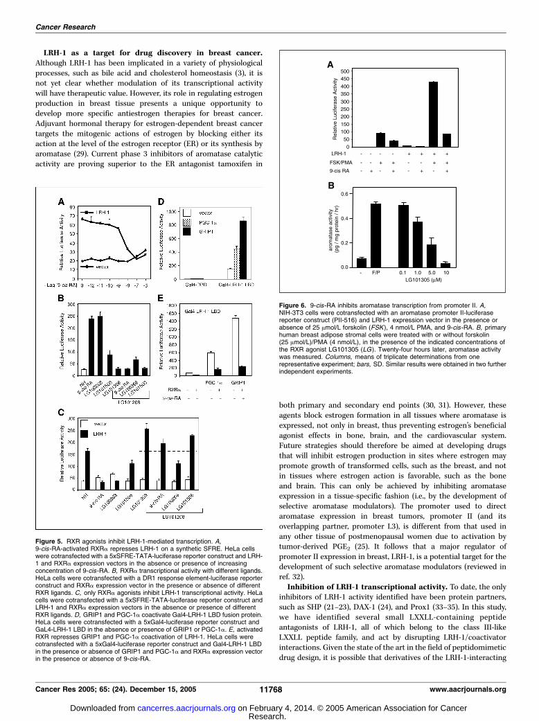
receptor A. To address the specificity of the LRH-1-interactingpeptides identified in this screen, we assessed their ability to bindan RXRa-VP16 fusion protein in a mammalian two-hybrid assay.Interestingly, all of the LRH-1-interacting peptides identified alsointeracted with activated RXR bound to 9-cis-retinoic acid (9-cis-RA) or the selective RXR agonist LG100268 (Fig. 4). Treatment withthe RXR antagonist LG101208 abolished both the basal andagonist-induced interaction of the peptides with RXR, indicatingthat they probably also interact with the canonical coactivatorbinding surface on this receptor (Fig. 4). These results suggestthat activated RXR shares identical or highly similar surfaces withLRH-1, raising the possibility that LRH-1 and RXR can be activatedby similar coregulators within target cells.Retinoid X receptor agonists inhibit LRH-1–mediated
transcription. Given that LRH-1 peptide antagonists also interactwith RXR, it is likely that activation of RXR has effects on LRH-1signaling within target cells. More specifically, we hypothesizedthat RXR ligands might inhibit LRH-1-mediated transcriptionalactivity by enabling activated RXRa to compete with LRH-1for common coactivators (i.e., antagonism independent of DNAbinding). To address this possibility, we transfected HeLa cells withLRH-1 and RXRa expression vectors, and a luciferase reporter geneconstruct containing five copies of the SFRE. As shown inFig. 5A , LRH-1 stimulated the SFRE reporter by f3-fold. Additionof increasing concentrations of 9-cis-RA inhibited LRH-1-mediatedtranscription in a concentration-dependent manner, with completeinhibition occurring at 0.1 Amol/L of 9-cis-RA. Therefore, activatedRXR can completely block LRH-1-mediated transcriptional activity,and this inhibition can occur on a promoter to which RXR does notbind in a direct manner. We next asked if LRH-1 inhibition by RXRis agonist dependent. 9-cis-RA and the selective RXR ligandLG100268 are full agonists and activate RXRa-mediated transcrip-tional activity on a DR1 response element. In contrast, LG101208 isa full antagonist, whereas LG101506 is a partial agonist (Fig. 5B).When the 5xSFRE reporter construct was cotransfected withLRH-1 and RXRa, 9-cis-RA as well as the selective RXR ligandLG100268 fully repressed LRH-1-mediated transcription, whereasthe partial agonist LG101506 had a partial inhibitory effect(Fig. 5C). Significantly, the full-antagonist LG101208 completely
Figure 3. LRH-1-binding peptides interfere with its transcriptional activity.A, affinity selection of LRH-1-binding motifs using phage display technology.Baculovirus-expressed full-length LRH-1 was immobilized on DNA containing anSFRE in 96-well Costar plates as a screening target. Phage that interactedspecifically with LRH-1 were selected, and the peptide sequences were deducedby DNA sequencing. B, Gal4-LXXLL peptide fusion interacts with LRH-1 inmammalian two-hybrid assay. HepG2 cells were cotransfected with CMVh-Gal,either the empty pM vector or the indicated fusion peptides, and either the emptyVP16 vector or VP16-LRH-1. C, Gal4-DBD fusion peptides suppressLRH-1-stimulated transcription from a synthetic SFRE. HeLa cells werecotransfected with a consensus SFRE promoter 5xSFRE-TATA-Luc reportergene and increasing amounts of the Gal4-DBD-peptide fusion constructs(as indicated) in the presence (solid columns ) or absence (open columns ) of anLRH-1 expression plasmid. D, LRH-1 peptides disrupt GRIP1 and PGC-1acoactivation of LRH-1. HeLa cells were cotransfected with an aromatasepromoter II-luciferase reporter construct, LRH-1 expression plasmid in thepresence or absence of GRIP1 or PGC-1a, and increasing concentrations of theGal4-DBD-peptide fusion constructs.
Cancer Research
Cancer Res 2005; 65: (24). December 15, 2005 11766 www.aacrjournals.org
Research. on February 4, 2014. © 2005 American Association for Cancercancerres.aacrjournals.org Downloaded from

reversed the inhibition of LRH-1 transcriptional activity by either9-cis-RA or LG100268 (Fig. 5C). Together, these data indicate thatinhibition of LRH-1 by RXR is agonist dependent, and RXR requiresan active conformation to mediate this repression. Thus, LRH-1inhibition by activated RXR may occur as a consequence of compe-tition for common coregulators used by these two transcriptionfactors.To further show that this inhibition is through direct competi-
tion for coactivators used by both receptors, we designed atransfection experiment in which Gal4-LRH-1 LBD and RXRa werecoexpressed in the presence or absence of 9-cis-RA, PGC-1a, orGRIP1. We used the Gal4 system to exclude the possibility that RXRmight interfere with LRH-1 binding to its response element. Asshown in Fig. 5D , Gal4-LRH-1 LBD activated transcription from the5xGal4 response element reporter construct, and this activity wasfurther enhanced in the presence of PGC-1a or GRIP1. In thiscontext, 9-cis-RA strongly repressed basal activity of LRH-1 LBD(Fig. 5E ). More interestingly, activated RXR repressed thestimulation of LRH-1 by either PGC-1a or GRIP1 (Fig. 5E). Theseresults indicate that in the presence of 9-cis-RA, RXR is able tocompete for coactivators present within the cell required for LRH-1transcriptional activity.9-cis-retinoic acid inhibits transcription from promoter II
of the CYP19 gene. Because LRH-1 is implicated as a keytranscription factor mediating aromatase expression in breasttissue (7), we asked whether 9-cis-RA can repress LRH-1-mediatedtranscriptional activity on aromatase promoter II. The majorinducer of aromatase expression in breast preadipocytes isprostaglandin E2 (PGE2) derived from breast tumor epitheliumor macrophage infiltration (25). Here, we used forskolin and PMAto mimic PGE2 signaling through protein kinases A and C (25).NIH-3T3 cells were transfected with an LRH-1 expression vectorin the presence or absence of forskolin and PMA. In the absenceof stimulation, LRH-1 produced a 10-fold increase in thetranscriptional activity of promoter II (Fig. 6A). In the absenceof exogenous LRH-1, forskolin and PMA treatment produced a100-fold increase in the transcriptional activity of promoter II. Incombination, however, LRH-1, forskolin, and PMA had a synergisticeffect, increasing promoter II activity by 500-fold. Addition of9-cis-RA repressed LRH-1-mediated transcription (10.9- to 2.9-fold).Moreover, 9-cis-RA was able to repress LRH-1, forskolin, andPMA synergism on promoter II (by 80%). Therefore, 9-cis-RA caninhibit hormone-induced aromatase promoter activity by inhibit-ing LRH-1 via sequestration of coactivators by endogenouslyactivated RXR.To confirm that this action results in inhibition of endogenous
aromatase activity, human breast adipose stromal cells wereincubated with forskolin plus PMA, together with the RXR ligand
LG101305, in various concentrations (Fig. 6B). This ligand inhibitedaromatase activity of the cells in a concentration-dependentfashion. Thus, the inhibitory activity of RXR agonists on theisolated aromatase promoter II is recapitulated on the endogenousgene in the absence of ectopic receptor expression.
Discussion
LRH-1 regulates diverse processes, including endodermaldevelopment, reverse cholesterol transport, bile acid homeostasis,and steroidogenesis (3). No natural agonists for LRH-1 have yetbeen identified, although the crystallized ligand binding domains ofthis receptor were found to contain phospholipids. The biologicalsignificance of these observations remains to be determined (12).It seems more likely that LRH-1 transcriptional activity is regulatedby other means (e.g., by post-translational modification, hetero-dimer interactions with NR0B family members, or by the natureand availability of coregulator proteins). Here, we show novelinteractions between LRH-1 and PGC-1a that have powerful effectson LRH-1 transcriptional activity and, more importantly, areamenable to modulation by small molecules, either positively viacAMP-induced PGC-1a expression, or negatively via rexinoid-induced RXR activation and consequent sequestration of endog-enous LRH-1 coactivators.PGC-1A is an LRH-1 coactivator. Previous studies have
suggested that LRH-1 exhibits weak binding to coregulators andindeed displays little discrimination between coactivators, such asSRC-1 and corepressors, such as SMRT (11). We show here thatPGC-1a (but not PGC-1h) is a strong coactivator of LRH-1 in thecontext of aromatase promoter II through direct interaction via itsconsensus LXXLL box. The significance of this lies in the fact thatunlike many other coregulators that are ubiquitously expressed and(in general) not hormonally regulated, PGC-1a exhibits markedtissue-specific expression that can be strongly regulated by cAMP.In mice, PGC-1a is expressed predominantly in brown adiposetissue, heart, kidney, and brain (18), whereas in humans, it is alsoexpressed in white adipose tissue (26). Although expression inwhite adipose tissue is relatively low, we show here that PGC-1amRNA levels in human breast preadipocytes can be markedlyincreased by cAMP treatment. This confirms that PGC-1aexpression is regulated by cAMP in white adipose tissue, as it isin brown adipose tissue (27) and liver (28). More importantly, PGC-1a thus provides a mechanism whereby hormonal signals, viacAMP, could regulate LRH-1 transcriptional activity in the absenceof any cognate ligand. The consequences of this for LRH-1 targetgene expression are illustrated by the marked stimulation of bothPGC-1a and aromatase mRNA expression in preadipocytes inresponse to forskolin (Fig. 2).
Figure 4. LRH-1–interacting peptides also interact with activatedRXR. HepG2 cells were cotransfected with a 5xGal4-luciferasereporter construct, a Gal4-DBD-fusion peptide, and either an emptyVP16 vector or VP16-tagged RXRa. Twenty-four hours followingtransfection, cells were treated overnight with either the fullagonists 9-cis-RA or LG100268 at 0.1 Amol/L, the partial agonistLG101506 at 0.1 Amol/L, or the full antagonist LG101208 at1 Amol/L.
LRH-1 Coactivation and Aromatase Expression
www.aacrjournals.org 11767 Cancer Res 2005; 65: (24). December 15, 2005
Research. on February 4, 2014. © 2005 American Association for Cancercancerres.aacrjournals.org Downloaded from
LRH-1 as a target for drug discovery in breast cancer.Although LRH-1 has been implicated in a variety of physiologicalprocesses, such as bile acid and cholesterol homeostasis (3), it isnot yet clear whether modulation of its transcriptional activitywill have therapeutic value. However, its role in regulating estrogenproduction in breast tissue presents a unique opportunity todevelop more specific antiestrogen therapies for breast cancer.Adjuvant hormonal therapy for estrogen-dependent breast cancertargets the mitogenic actions of estrogen by blocking either itsaction at the level of the estrogen receptor (ER) or its synthesis byaromatase (29). Current phase 3 inhibitors of aromatase catalyticactivity are proving superior to the ER antagonist tamoxifen in
both primary and secondary end points (30, 31). However, theseagents block estrogen formation in all tissues where aromatase isexpressed, not only in breast, thus preventing estrogen’s beneficialagonist effects in bone, brain, and the cardiovascular system.Future strategies should therefore be aimed at developing drugsthat will inhibit estrogen production in sites where estrogen maypromote growth of transformed cells, such as the breast, and notin tissues where estrogen action is favorable, such as the boneand brain. This can only be achieved by inhibiting aromataseexpression in a tissue-specific fashion (i.e., by the development ofselective aromatase modulators). The promoter used to directaromatase expression in breast tumors, promoter II (and itsoverlapping partner, promoter I.3), is different from that used inany other tissue of postmenopausal women due to activation bytumor-derived PGE2 (25). It follows that a major regulator ofpromoter II expression in breast, LRH-1, is a potential target for thedevelopment of such selective aromatase modulators (reviewed inref. 32).Inhibition of LRH-1 transcriptional activity. To date, the only
inhibitors of LRH-1 activity identified have been protein partners,such as SHP (21–23), DAX-1 (24), and Prox1 (33–35). In this study,we have identified several small LXXLL-containing peptideantagonists of LRH-1, all of which belong to the class III-likeLXXLL peptide family, and act by disrupting LRH-1/coactivatorinteractions. Given the state of the art in the field of peptidomimeticdrug design, it is possible that derivatives of the LRH-1-interacting
Figure 5. RXR agonists inhibit LRH-1-mediated transcription. A,9-cis-RA-activated RXRa represses LRH-1 on a synthetic SFRE. HeLa cellswere cotransfected with a 5xSFRE-TATA-luciferase reporter construct and LRH-1 and RXRa expression vectors in the absence or presence of increasingconcentration of 9-cis-RA. B, RXRa transcriptional activity with different ligands.HeLa cells were cotransfected with a DR1 response element-luciferase reporterconstruct and RXRa expression vector in the presence or absence of differentRXR ligands. C, only RXRa agonists inhibit LRH-1 transcriptional activity. HeLacells were cotransfected with a 5xSFRE-TATA-luciferase reporter construct andLRH-1 and RXRa expression vectors in the absence or presence of differentRXR ligands. D, GRIP1 and PGC-1a coactivate Gal4-LRH-1 LBD fusion protein.HeLa cells were cotransfected with a 5xGal4-luciferase reporter construct andGaL4-LRH-1 LBD in the absence or presence of GRIP1 or PGC-1a. E, activatedRXR represses GRIP1 and PGC-1a coactivation of LRH-1. HeLa cells werecotransfected with a 5xGal4-luciferase reporter construct and Gal4-LRH-1 LBDin the presence or absence of GRIP1 and PGC-1a and RXRa expression vectorin the presence or absence of 9-cis-RA.
Figure 6. 9-cis-RA inhibits aromatase transcription from promoter II. A,NIH-3T3 cells were cotransfected with an aromatase promoter II-luciferasereporter construct (PII-516) and LRH-1 expression vector in the presence orabsence of 25 Amol/L forskolin (FSK ), 4 nmol/L PMA, and 9-cis-RA. B, primaryhuman breast adipose stromal cells were treated with or without forskolin(25 Amol/L)/PMA (4 nmol/L), in the presence of the indicated concentrations ofthe RXR agonist LG101305 (LG). Twenty-four hours later, aromatase activitywas measured. Columns, means of triplicate determinations from onerepresentative experiment; bars, SD. Similar results were obtained in two furtherindependent experiments.
Cancer Research
Cancer Res 2005; 65: (24). December 15, 2005 11768 www.aacrjournals.org
Research. on February 4, 2014. © 2005 American Association for Cancercancerres.aacrjournals.org Downloaded from

peptides could be created, which directly interfere with the functionof the AF-2 pocket of the receptor. However, our data showing thatRXR and LRH-1 are likely to be regulated by similar endogenouscoactivators has highlighted a more near-term clinical opportunityfor a breast cancer therapeutic. Specifically, we show that 9-cis-RAinhibits PGC-1a and GRIP1-induced LRH-1 activity and potentlyinhibits (by up to 80%) aromatase promoter activity induced byLRH-1 and forskolin. This again provides a mechanism for themodulation, in this case, inhibition, of LRH-1 transcriptional activityand expression of LRH-1 target genes by small molecules andillustrates a level of crosstalk between the RXR and LRH-1 pathways.RXR ligands are promising molecules for both chemoprevention
and therapy of cancer (36). In particular, 9-cis-RA and RXR-selective ligands are highly efficacious in preventing breast cancerin experimental animal models (37, 38). The mechanisms by whichrexinoids suppress carcinogenesis are not fully understood, in partdue to their large spectra of action. However, it is likely thatinhibition of local estrogen synthesis by aromatase in breastpreadipocytes contributes to their efficacy. Thiazolidinediones,which are PPARg ligands, have also been shown to inhibitproliferation of breast cancer cells (39). We previously showedthat RXR and PPARg ligands inhibit aromatase expression frompromoter II and promoter I.4 in primary human preadipocytes viaan indirect mechanism that does not involve binding of either RXRor PPARg to the aromatase promoter (40). One mechanism bywhich rexinoids and PPARg ligands could exert antiproliferativeeffects in ER-positive breast cancers is the sequesteration of LRH-1coactivators by activated RXR with resulting inhibition of LRH-1-induced local aromatase expression and estrogen production.However, rexinoids are also effective growth inhibitor agents in ER-negative tumors (38). We have observed an increase in the proli-feration rate of MCF-7 breast cancer cells that modestly (3-fold)overexpress LRH-1, associated with increased expression of cyclin
D1,4 suggesting that LRH-1 directly controls cell cycle progressionin breast cancer cells, as it does in colon cancer (41). Thus, wespeculate that rexinoids may in part inhibit breast cancer cellproliferation through inhibiting LRH-1 transcriptional activity andcell cycle progression.In summary, we have identified novel hormone-regulated
coregulator interactions that modulate LRH-1 transcriptionalactivity. Because deletion of LRH-1 results in embryonic lethalityin mice (42), knowledge of the signaling pathways that modulate itsactivity, coupled with the development of highly specific peptideantagonists of LRH-1 as described here, will greatly facilitateunderstanding of the roles of LRH-1 in health and disease andvalidate LRH-1 as a therapeutic target. In particular, the identifica-tion of small molecules, such as rexinoids that inhibit LRH-1activation of aromatase in breast, suggests that suchmolecules fulfillthe criteria of selective aromatase modulators, and that LRH-1 couldbe a target for breast-specific tumor therapy.
Acknowledgments
Received 8/5/2005; revised 9/30/2005; accepted 10/12/2005.Grant support: Victorian Breast Cancer Research Consortium, Inc.; NIH ATLAS
grant DK62434 (D.P. McDonnell); Department of Defense grant DAMD17-03-1-0174 (S.Gaillard); and National Health and Medical Research Council of Australia grant 289318(C.D. Clyne).
The costs of publication of this article were defrayed in part by the payment of pagecharges. This article must therefore be hereby marked advertisement in accordancewith 18 U.S.C. Section 1734 solely to indicate this fact.
We thank Drs. Mark Leibowitz and Bill Lamph (Ligand Pharmaceuticals) for thegift of rexinoid ligands; Drs. C.M. Newgard, B.M. Spiegelman, D.J. Mangelsdorf, andM.R. Stallcup for the gift of their plasmids; and members of the McDonnell lab for theiruseful discussions.
4 Manuscript in preparation.
LRH-1 Coactivation and Aromatase Expression
www.aacrjournals.org 11769 Cancer Res 2005; 65: (24). December 15, 2005
References1. Lavorgna G, Ueda H, Clos J, Wu C. FTZ-F1, a steroidhormone receptor-like protein implicated in the activa-tion of fushi tarazu. Science 1991;252:848–51.
2. Parker KL, Schimmer BP. Steroidogenic factor 1: a keydeterminant of endocrine development and function.Endocr Rev 1997;18:361–77.
3. Fayard E, Auwerx J, Schoonjans K. LRH-1: an orphannuclear receptor involved in development, metabolismand steroidogenesis. Trends Cell Biol 2004;14:250–60.
4. Falender AE, Lanz R, Malenfant D, Belanger L,Richards JS. Differential expression of steroidogenicfactor-1 and FTF/LRH-1 in the rodent ovary. Endocri-nology 2003;144:3598–610.
5. Liu DL, Liu WZ, Li QL, et al. Expression and functionalanalysis of liver receptor homologue 1 as a potentialsteroidogenic factor in rat ovary. Biol Reprod 2003;69:508–17.
6. Pezzi V, Sirianni R, Chimento A, et al. Differentialexpression of SF-1/AD4BP and LRH-1/FTF in the rattestis: LRH-1 as a potential regulator of testiculararomatase expression. Endocrinology 2004;145:2186–96.
7. Clyne CD, Speed CJ, Zhou J, Simpson ER. Liverreceptor homologue-1 (LRH-1) regulates expression ofaromatase in preadipocytes. J Biol Chem 2002;277:20591–7.
8. Zhou J, Suzuki T, Kovacic A, et al. Interactionsbetween prostaglandin E2, liver receptor homologue-1(LRH-1) and aromatase in breast cancer. Cancer Res2005;65:657–63.
9. Agarwal VR, Bulun SE, Leitch M, Rohrich R, SimpsonER. Use of alternative promoters to express the aroma-tase cytochrome P450 (CYP19) gene in breast adipose
tissues of cancer-free and breast cancer patients. J ClinEndocrinol Metab 1996;81:3843–9.
10. McKenna NJ, O’Malley BW. Minireview: nuclearreceptor coactivators—an update. Endocrinology 2002;143:2461–5.
11. Sablin EP, Krylova IN, Fletterick RJ, Ingraham HA.Structural basis for ligand-independent activation of theorphan nuclear receptor LRH-1. Mol Cell 2003;11:1575–85.
12. Ortlund EA, Lee Y, Solomon IH, et al. Modulation ofhuman nuclear receptor LRH-1 activity by phospholi-pids and SHP. Nat Struct Mol Biol 2005;12:357–63.
13. Krylova IN, Sablin EP, Moore J, et al. Structuralanalyses reveal phosphatidyl inositols as ligands for theNR5 orphan receptors SF-1 and LRH-1. Cell 2005;120:343–55.
14. Wang W, Zhang C, Marimuthu A, et al. The crystalstructures of human steroidogenic factor-1 and liverreceptor homologue-1. Proc Natl Acad Sci U S A 2005;102:7505–10.
15. Chang C, Norris JD, Gron H, et al. Dissection of theLXXLL nuclear receptor-coactivator interaction motifusing combinatorial peptide libraries: discovery ofpeptide antagonists of estrogen receptors a and h.Mol Cell Biol 1999;19:8226–39.
16. Ackerman GE, Smith ME, Mendelson CR, MacDonaldPC, Simpson ER. Aromatization of androstenedione byhuman adipose tissue stromal cells in monolayerculture. J Clin Endocrinol Metab 1981;53:412–7.
17. Lee YK, Parker KL, Choi HS, Moore DD. Activation ofthe promoter of the orphan receptor SHP by orphanreceptors that bind DNA as monomers. J Biol Chem1999;274:20869–73.
18. Puigserver P, Wu Z, Park CW, et al. A cold-inducible
coactivator of nuclear receptors linked to adaptivethermogenesis. Cell 1998;92:829–39.
19. Lin J, Puigserver P, Donovan J, Tarr P, Spiegelman BM.Peroxisome proliferator-activated receptor g coactivator1h (PGC-1h), a novel PGC-1-related transcriptioncoactivator associated with host cell factor. J Biol Chem2002;277:1645–8.
20. Knutti D, Kaul A, Kralli A. A tissue-specific coac-tivator of steroid receptors, identified in a functionalgenetic screen. Mol Cell Biol 2000;20:2411–22.
21. Kovacic A, Speed CJ, Simpson ER, Clyne CD.Inhibition of aromatase transcription via promoter IIby short heterodimer partner (SHP) in human preadi-pocytes. Mol Endocrinol 2004;18:252–9.
22. Goodwin B, Jones SA, Price RR, et al. A regulatorycascade of the nuclear receptors FXR, SHP-1, andLRH-1 represses bile acid biosynthesis. Mol Cell 2000;6:517–26.
23. Lu TT, Makishima M, Repa JJ, et al. Molecular basisfor feedback regulation of bile acid synthesis by nuclearreceptors. Mol Cell 2000;6:507–15.
24. Suzuki T, Kasahara M, Yoshioka H, Morohashi K,Umesono K. LXXLL-related motifs in Dax-1 have targetspecificity for the orphan nuclear receptors Ad4BP/SF-1and LRH-1. Mol Cell Biol 2003;23:238–49.
25. Zhao Y, Agarwal VR, Mendelson CR, Simpson ER.Estrogen biosynthesis proximal to a breast tumor isstimulated by PGE2 via cyclic AMP, leading to activationof promoter II of the CYP19 (aromatase) gene.Endocrinology 1996;137:5739–42.
26. Larrouy D, Vidal H, Andreelli F, Laville M, Langin D.Cloning and mRNA tissue distribution of human PPARgcoactivator-1. Int J Obes Relat Metab Disord 1999;23:1327–32.
Research. on February 4, 2014. © 2005 American Association for Cancercancerres.aacrjournals.org Downloaded from

Cancer Research
Cancer Res 2005; 65: (24). December 15, 2005 11770 www.aacrjournals.org
27. Cao W, Daniel KW, Robidoux J, et al. p38 mitogen-activated protein kinase is the central regulator ofcyclic AMP-dependent transcription of the brown fatuncoupling protein 1 gene. Mol Cell Biol 2004;24:3057–67.
28. Herzig S, Long F, Jhala US, et al. CREB regulateshepatic gluconeogenesis through the coactivator PGC-1.Nature 2001;413:179–83.
29. Santen RJ. To block estrogen’s synthesis or action:that is the question. J Clin Endocrinol Metab 2002;87:3007–12.
30. Baum M, Budzar AU, Cuzick J, et al. Anastrozolealone or in combination with tamoxifen versus tamox-ifen alone for adjuvant treatment of postmenopausalwomen with early breast cancer: first results of theATAC randomised trial. Lancet 2002;359:2131–9.
31. Buzdar AU. Data from the Arimidex, tamoxifen,alone or in combination (ATAC) trial: implications foruse of aromatase inhibitors in 2003. Clin Cancer Res2004;10:355–61S.
32. Clyne CD, Kovacic A, Speed CJ, et al. Regulation of
aromatase expression by the nuclear receptor LRH-1 inadipose tissue. Mol Cell Endocrinol 2004;215:39–44.
33. Liu YW, Gao W, Teh HL, Tan JH, Chan WK. Prox1 is anovel coregulator of Ff1b and is involved in theembryonic development of the zebra fish interrenalprimordium. Mol Cell Biol 2003;23:7243–55.
34. Qin J, Gao DM, Jiang QF, et al. Prox1 is a corepressorof human liver receptor homologue-1 and suppressesthe transcription of the cholesterol 7-a-hydroxylasegene. Mol Endocrinol 2004;18:2424–39.
35. Steffensen KR, Holter E, Bavner A, et al. Functionalconservation of interactions between a homeodomaincofactor and a mammalian FTZ-F1 homologue. EMBORep 2004;5:613–9.
36. Lippman SM, Lotan R. Advances in the developmentof retinoids as chemopreventive agents. J Nutr 2000;130:479–82S.
37. Gottardis MM, Bischoff ED, Shirley MA, et al.Chemoprevention of mammary carcinoma by LGD1069(Targretin): an RXR-selective ligand. Cancer Res 1996;56:5566–70.
38. Wu K, Zhang Y, Xu XC, et al. The retinoid X receptor-selective retinoid, LGD1069, prevents the developmentof estrogen receptor-negative mammary tumors intransgenic mice. Cancer Res 2002;62:6376–80.
39. Fenner MH, Elstner E. Peroxisome proliferator-activated receptor-g ligands for the treatment of breastcancer. Expert Opin Investig Drugs 2005;14:557–68.
40. Rubin GL, Duong JH, Clyne CD, et al. Ligands forthe peroxisomal proliferator-activated receptor g andthe retinoid X receptor inhibit aromatase cytochromeP450 (CYP19) expression mediated by promoter IIin human breast adipose. Endocrinology 2002;143:2863–71.
41. Botrugno OA, Fayard E, Annicotte JS, et al. Synergybetween LRH-1 and h-catenin induces G1 cyclin-mediated cell proliferation. Mol Cell 2004;15:499–509.
42. Pare JF, Malenfant D, Courtemanche C, et al. Thefetoprotein transcription factor (FTF) gene is essentialto embryogenesis and cholesterol homeostasis, andregulated by a DR4 element. J Biol Chem 2004;279:21206–16.
Research. on February 4, 2014. © 2005 American Association for Cancercancerres.aacrjournals.org Downloaded from