Angiotensin AT2 receptor stimulation is anti-inflammatory in lipopolysaccharide-activated THP-1...
9
ORIGINAL ARTICLE Angiotensin AT 2 receptor stimulation is anti-inflammatory in lipopolysaccharide-activated THP-1 macrophages via increased interleukin-10 production Isha Dhande 1,2 , Wanshu Ma 2 and Tahir Hussain 1,2 Macrophages have an important role in the pathogenesis of hypertension and associated end-organ damage via the activation of the Toll-like receptors, such as Toll-like receptor-4 (TLR4). Accumulating evidence suggests that the angiotensin AT 2 receptor (AT 2 R) has a protective role in pathological conditions involving inflammation and tissue injury. We have recently shown that AT 2 R stimulation is renoprotective, which occurs in part via increased levels of anti-inflammatory interleukin-10 (IL-10) production in renal epithelial cells; however, the role of AT 2 R in the inflammatory activity of macrophages is not known. The present study was designed to investigate whether AT 2 R activation stimulates an anti-inflammatory response in TLR4-induced inflammation. The effects of the anti-inflammatory mechanisms that occurred following pre-treatment with the AT 2 R agonist Compound 21 (C21) (1 μmol ml - 1 ) on the cytokine profiles of THP-1 macrophages after activation by lipopolysaccharide (LPS) (1 μg ml - 1 ) were studied. The AT 2 R agonist dose-dependently attenuated LPS-induced tumor necrosis factor-α (TNF-α) and IL-6 production but increased IL-10 production. IL-10 was critical for the anti-inflammatory effects of AT 2 R stimulation because the IL-10-neutralizing antibody dose-dependently abolished the AT 2 R-mediated decrease in TNF-α levels. Further, enhanced IL-10 levels were associated with a sustained, selective increase in the phosphorylation of extracellular signal-regulated kinase (ERK1/2) but not p38 mitogen-activated protein kinase (MAPK). Blocking the activation of ERK1/2 before C21 pre-treatment completely abrogated this increased IL-10 production in response to the AT 2 R agonist C21, while there was a partial reduction in IL-10 levels following the inhibition of p38. We conclude that AT 2 R stimulation exerts a novel anti-inflammatory response in THP-1 macrophages via enhanced IL-10 production as a result of sustained, selective ERK1/2 phosphorylation, which may have protective roles in hypertension and associated tissue injury. Hypertension Research advance online publication, 11 September 2014; doi:10.1038/hr.2014.132 Keywords: AT 2 receptor; inflammation; interleukin-10; macrophage INTRODUCTION Chronic inflammation, which is characterized by elevated cytokine and chemokine expression, has been shown to have a central role in the pathophysiology of hypertension and associated end-organ damage, including renal injury. 1–4 At the cellular level, chronic inflammation is mediated largely by macrophages. Additionally, macrophage infiltration invariably accompanies hypertensive organ damage, such as that which occurs to blood vessels, 5 the heart 6 and the kidneys. 7 Moreover, circulating monocytes are activated in hyperten- sive patients and produce increased amounts of pro-inflammatory cytokines, including tumor necrosis factor-α (TNF-α), interleukin-1 (IL-1) and transforming growth factor-β. 8 Recent evidence indicates that rather than being a mere consequence of elevated blood pressure, macrophage activation is actually a causative factor in the development of hypertension. 9,10 The renin-angiotensin system is a critical hormonal system that regulates blood pressure, and it is abnormally activated in hypertensive patients. Macrophages also express all major components of the renin- angiotensin system. 11 Angiotensin II (Ang II) has been demonstrated to participate in the activation and pro-inflammatory polarization of leukocytes via the AT 1 receptor (AT 1 R). 12–14 However, the precise cellular mechanisms underlying these activities are not well defined. One potential inflammatory pathway that has been implicated in hypertension is the activation of innate immune receptors, specifically 1 Department of Pharmacological and Pharmaceutical Sciences, Heart and Kidney Institute, University of Houston, Houston, TX, USA and 2 Department of Pharmacal Sciences, Harrison School of Pharmacy, Auburn University, Auburn, AL, USA Correspondence: Professor T Hussain, Department of Pharmacological and Pharmaceutical Sciences, University of Houston, Room 555 Science and Research Building II, Houston, TX 77204, USA. E-mail: [email protected] Received 14 March 2014; revised 19 June 2014; accepted 6 July 2014 Hypertension Research (2014), 1–9 & 2014 The Japanese Society of Hypertension All rights reserved 0916-9636/14 www.nature.com/hr
-
Upload
independent -
Category
Documents
-
view
2 -
download
0
Transcript of Angiotensin AT2 receptor stimulation is anti-inflammatory in lipopolysaccharide-activated THP-1...

ORIGINAL ARTICLE
Angiotensin AT2 receptor stimulation isanti-inflammatory in lipopolysaccharide-activatedTHP-1 macrophages via increased interleukin-10production
Isha Dhande1,2, Wanshu Ma2 and Tahir Hussain1,2
Macrophages have an important role in the pathogenesis of hypertension and associated end-organ damage via the activation of
the Toll-like receptors, such as Toll-like receptor-4 (TLR4). Accumulating evidence suggests that the angiotensin AT2 receptor
(AT2R) has a protective role in pathological conditions involving inflammation and tissue injury. We have recently shown that
AT2R stimulation is renoprotective, which occurs in part via increased levels of anti-inflammatory interleukin-10 (IL-10)
production in renal epithelial cells; however, the role of AT2R in the inflammatory activity of macrophages is not known. The
present study was designed to investigate whether AT2R activation stimulates an anti-inflammatory response in TLR4-induced
inflammation. The effects of the anti-inflammatory mechanisms that occurred following pre-treatment with the AT2R agonist
Compound 21 (C21) (1 μmol ml−1) on the cytokine profiles of THP-1 macrophages after activation by lipopolysaccharide (LPS)
(1 μgml−1) were studied. The AT2R agonist dose-dependently attenuated LPS-induced tumor necrosis factor-α (TNF-α) and IL-6
production but increased IL-10 production. IL-10 was critical for the anti-inflammatory effects of AT2R stimulation because the
IL-10-neutralizing antibody dose-dependently abolished the AT2R-mediated decrease in TNF-α levels. Further, enhanced IL-10
levels were associated with a sustained, selective increase in the phosphorylation of extracellular signal-regulated kinase
(ERK1/2) but not p38 mitogen-activated protein kinase (MAPK). Blocking the activation of ERK1/2 before C21 pre-treatment
completely abrogated this increased IL-10 production in response to the AT2R agonist C21, while there was a partial reduction
in IL-10 levels following the inhibition of p38. We conclude that AT2R stimulation exerts a novel anti-inflammatory response in
THP-1 macrophages via enhanced IL-10 production as a result of sustained, selective ERK1/2 phosphorylation, which may have
protective roles in hypertension and associated tissue injury.
Hypertension Research advance online publication, 11 September 2014; doi:10.1038/hr.2014.132
Keywords: AT2 receptor; inflammation; interleukin-10; macrophage
INTRODUCTION
Chronic inflammation, which is characterized by elevated cytokineand chemokine expression, has been shown to have a central role inthe pathophysiology of hypertension and associated end-organdamage, including renal injury.1–4 At the cellular level, chronicinflammation is mediated largely by macrophages. Additionally,macrophage infiltration invariably accompanies hypertensive organdamage, such as that which occurs to blood vessels,5 the heart6 and thekidneys.7 Moreover, circulating monocytes are activated in hyperten-sive patients and produce increased amounts of pro-inflammatorycytokines, including tumor necrosis factor-α (TNF-α), interleukin-1(IL-1) and transforming growth factor-β.8 Recent evidence indicates
that rather than being a mere consequence of elevated blood pressure,macrophage activation is actually a causative factor in the developmentof hypertension.9,10
The renin-angiotensin system is a critical hormonal system thatregulates blood pressure, and it is abnormally activated in hypertensivepatients. Macrophages also express all major components of the renin-angiotensin system.11 Angiotensin II (Ang II) has been demonstratedto participate in the activation and pro-inflammatory polarization ofleukocytes via the AT1 receptor (AT1R).
12–14 However, the precisecellular mechanisms underlying these activities are not well defined.One potential inflammatory pathway that has been implicated inhypertension is the activation of innate immune receptors, specifically
1Department of Pharmacological and Pharmaceutical Sciences, Heart and Kidney Institute, University of Houston, Houston, TX, USA and 2Department of Pharmacal Sciences,Harrison School of Pharmacy, Auburn University, Auburn, AL, USACorrespondence: Professor T Hussain, Department of Pharmacological and Pharmaceutical Sciences, University of Houston, Room 555 Science and Research Building II,Houston, TX 77204, USA.E-mail: [email protected] 14 March 2014; revised 19 June 2014; accepted 6 July 2014
Hypertension Research (2014), 1–9& 2014 The Japanese Society of Hypertension All rights reserved 0916-9636/14www.nature.com/hr
Toll-like receptor-4 (TLR4) signaling,15 which leads to the productionof an array of pro-inflammatory mediators. In fact, peripheralmonocyte TLR4 expression is markedly increased in hypertensivepatients compared with normotensive controls.16 Furthermore, Ang IIhas been shown to upregulate TLR4 expression via the AT1R
17 andexacerbate pro-inflammatory cytokine production in response toTLR4 activation by lipopolysaccharide (LPS) in macrophages.18
Accumulating evidence suggests that the AT2 receptor (AT2R), whichis generally considered to be a functional antagonist of the AT1R,exerts an anti-inflammatory response.19–21 We have previously shownthat the stimulation of the AT2R by the selective agonist Compound21 (C21) attenuates pro-inflammatory signaling in response to theLPS activation of proximal tubule epithelial cells via increased IL-10production.22 However, the abilities of the AT2R to induce IL-10production and exert an anti-inflammatory response in LPS-activatedmacrophages have not been investigated to date.In macrophages, multiple pathways exist to promote IL-10
production.23 The activation of mitogen-activated protein kinases(MAPKs), specifically via p38 and extracellular signal-regulated kinase(ERK1/2), has been shown to be required to increase IL-10 levels inmacrophages, and the inhibition of either of these MAPKs impairsIL-10 production.24–28 Moreover, increased ERK1/2 activationcorrelates well with the extent of IL-10 production in this celltype.23 Because the AT2R has been linked to a sustained increase inERK1/2 phosphorylation, this may be a potential molecular mechan-ism by which the AT2R agonist can enhance IL-10 production inmacrophages.The present study was designed to test the hypothesis that the
stimulation of the AT2R attenuates TLR4-mediated pro-inflammatorycytokine production in macrophages via increased IL-10 production.We evaluated the effects of C21 on the production of TNF-α, IL-6 andIL-10 in LPS-activated THP-1 macrophages. We demonstrated thatpre-treatment with C21 significantly attenuated the levels of pro-inflammatory cytokines. This was found to be dependent uponincreased IL-10 production in response to AT2R activation. Moreover,this upregulation of IL-10 was mediated by a sustained, selectiveincrease in ERK1/2 phosphorylation.
MATERIALS AND METHODS
Cell culture and treatmentsThe human THP-1 monocytic cell line (ATCC) was cultured in RPMI-1640medium supplemented with 10% heat-inactivated fetal bovine serum and anantibiotic/antimycotic cocktail (100 Uml− 1 penicillin, 100 μgml− 1 streptomy-cin and 250 ngml− 1 amphotericin B) at 37 °C in a humidified atmosphere with5% CO2. Cell culture media and supplements were all purchased fromHyClone (ThermoFisher Scientific, Waltham, MA, USA). To differentiatemonocytes from macrophages, 5 × 10⁵ cells per well were treated with 40nmol l− 1 PMA (phorbol-12-myristate-13-acetate) (Sigma, St Louis, MO, USA)for 48 h in RPMI-1640 containing 5% fetal bovine serum. At the end of the 48-h incubation period, the medium was aspirated, and the cells were washed withRPMI-1640 without fetal bovine serum or antibiotics/antimycotics and werethen incubated in this medium for 6–8 h. To eliminate pre-existing cytokineproduction, the medium was replaced by fresh serum-free medium before thetreatments were initiated. LPS (1 μgml− 1) (E. coli O55:B5; Sigma) was used toinduce the production of pro-inflammatory cytokines. The cells were pre-treated with C21 (custom synthesized) for 60min before the addition of LPS,and the drug remained in the medium for the entire duration of the treatment.Treatments with specific inhibitors, including 1 μmol l− 1 candesartan (an AT1Rantagonist; AstraZeneca, Wilmington, DE, USA), 10 μmol l− 1 PD123319 (anAT1R antagonist; Pfizer, New York, NY, USA), 10 μmol l− 1 SB203580 (a p38inhibitor; Cell Signaling Technology, Danvers, MA, USA) and 10 μmol l− 1
PD98059 (an MEK (MAPK/ERK kinase) inhibitor; Cell Signaling Technology)were carried out as indicated in the text and figure legends.
ImmunoblottingMacrophages were washed twice with PBS and lysed on a plate using ice-coldcell lysis buffer (Cell Signaling Technology) containing protease (Roche,Indianapolis, IN, USA)- and phosphatase (Sigma)-inhibitor cocktails. Equalamounts of protein in Laemmli buffer were loaded per well (15 μg per lane forAT1R, 45 μg per lane for AT2R and 30 μg per lane for ERK1/2 and p38 MAPK)and separated by SDS-PAGE using a Tris-glycine system. Proteins were thentransferred onto a PVDF membrane using the wet transfer protocol. Themembrane was incubated in 5% non-fat dry milk in PBST for 1 h, followed byincubation overnight with primary antibodies against AT1R (BiomolecularIntegrations, Little Rock, AR, USA), AT2R (EZ Biolabs, Carmel, IN, USA),p-ERK1/2 (Cell Signaling Technology), p-p38 MAPK (Cell Signaling Technol-ogy) at 1:1000 dilutions. An antibody against inducible nitric oxide synthase(iNOS) (Cell Signaling Technology) was used at 1:500, while that againstmacrophage mannose receptor (MMR; Abcam, Cambridge, MA, USA)was used at a 1:200 dilution. This was followed by a wash with PBST
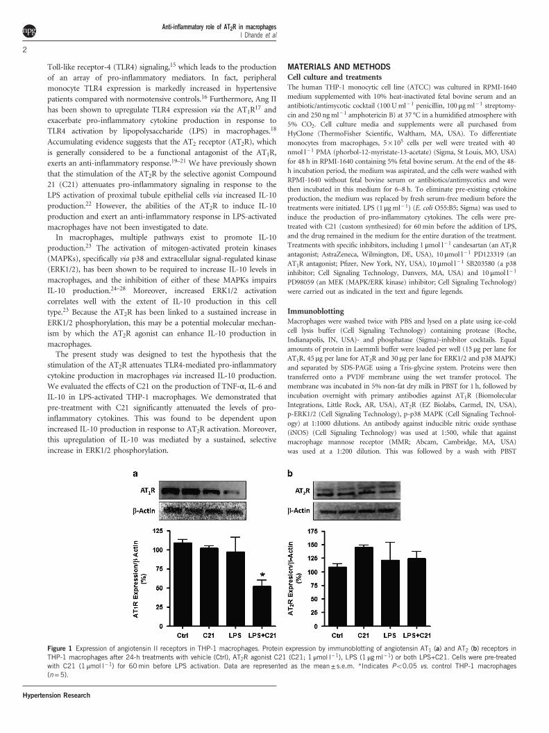
Figure 1 Expression of angiotensin II receptors in THP-1 macrophages. Protein expression by immunoblotting of angiotensin AT1 (a) and AT2 (b) receptors inTHP-1 macrophages after 24-h treatments with vehicle (Ctrl), AT2R agonist C21 (C21; 1 μmol l−1), LPS (1 μgml−1) or both LPS+C21. Cells were pre-treatedwith C21 (1 μmol l−1) for 60min before LPS activation. Data are represented as the mean± s.e.m. *Indicates Po0.05 vs. control THP-1 macrophages(n=5).
Anti-inflammatory role of AT2R in macrophagesI Dhande et al
2
Hypertension Research

and a 1-h incubation with appropriate HRP-conjugated secondary antibodies
(Santa Cruz Biotechnology). Chemiluminescence was detected by the addition
of Luminol HRP substrate (Santa Cruz Biotechnology) and quantified by a
software-assisted densitometric analysis (Alpha Innotech, San Leandro, CA,
USA). To ensure equal loading, the blots were stripped and re-probed for
β-actin (BioVision, Milpitas, CA, USA) for AT1R and AT2R and total-p38
and total-ERK1/2 for p-p38 and p-ERK1/2, respectively (Cell Signaling
Technology).
Enzyme-linked immunosorbent assayThe cytokines in the medium were assessed by a kit-based enzyme-linked
immunosorbent assay (ELISA) according to the manufacturer’s protocols
(R&D Systems, Minneapolis, MN, USA).
mRNA expression by RT–PCRTotal RNA from the cells was extracted using the RNeasy kit (Qiagen, Valencia,
CA, USA) according to the manufacturer’s protocol. cDNA was synthesized by
RT–PCR from 1 μg of total RNA using the SuperScript III First-Strand
Synthesis System (Life Technologies, Grand Island, NY, USA). This cDNA
was used as a template for the quantitative RT–PCR analysis of the gene
expressions of TNFA, IL6 and IL10 using TaqMan gene expression assays
(Applied Biosystems, Grand Island, NY, USA). Relative quantifications were
determined using the delta-delta Ct method with GAPDH as a control.
Statistical analysisThe data are presented as the mean± s.e.m. Student’s t-test was used to
compare the mean values of two groups. One-way ANOVA with a post hoc test
Figure 2 Effects of an AT2R agonist (C21) and an AT1R antagonist (candesartan) on cytokine production by LPS-activated THP-1 macrophages. THP-1macrophages were incubated with either the AT2R agonist (C21; 1 μmol l−1) or the AT1R antagonist candesartan (Can; 1 μmol l−1) for 60min beforeactivation with LPS (1 μgml−1). To demonstrate the specificity of C21, an additional group of cells were incubated with the AT2R antagonist PD123319(PD; 10 μmol l−1) for 30min before C21 pre-treatment. The cytokines TNF-α (a), IL-6 (b) and IL-10 (c) were assessed in the media at 24 h after LPSactivation by ELISA. The data are represented as the mean± s.e.m. *Indicates Po0.05 vs. control, #indicates Po0.05 vs. LPS-treated and $indicatesPo0.05 vs. LPS+C21-treated THP-1 macrophages (n=6).
Figure 3 Dose-dependent and time-dependent effects of an AT2R agonist (C21) on cytokine production by LPS-activated THP-1 macrophages. The dose-dependent effects of the AT2R agonist C21 on TNF-α (a) and IL-10 (b) levels were assessed at 24 h following LPS activation (1 μgml−1). C21 pre-treatment(0.1–10 μmol l−1) was administered 60min before LPS activation, and cytokines in the media were assessed by ELISA. An additional set of cells wereincubated with the AT2R antagonist PD123319 (PD; 10 μmol l−1) for 30min before C21 pre-treatment to demonstrate the receptor specificity of C21. TNF-α(c) and IL-10 (d) production at earlier time points was also determined in the media at the indicated times after LPS activation following C21 (1 μmolml−1)pre-treatment. The data are represented as the mean± s.e.m. *Indicates Po0.05 vs. respective control (n=6).
Anti-inflammatory role of AT2R in macrophagesI Dhande et al
3
Hypertension Research

(Tukey’s) for multiple comparisons was used to compare variations between
more than two groups. A value of Po0.05 was considered to be statistically
significant with n= 5–8 experiments per group.
RESULTS
Expression of angiotensin AT1R and AT2R in THP-1 macrophagesTHP-1 macrophages express both AT1R and AT2R. The expressions ofboth receptor subtypes were not altered by the C21 or LPS treatments(Figures 1a and b); however, the C21 pre-treatment lowered AT1Rexpression by ~ 50% in response to LPS (Figure 1a), which is inagreement with a number of previous reports demonstrating theAT2R-mediated downregulation of AT1R in pathophysiologicalconditions.29–31
Effects of AT2R agonist (C21) and AT1R antagonist (candesartan)on cytokine production by LPS-activated THP-1 macrophagesTHP-1 macrophages were treated with LPS (1 μg ml− 1) for 24 h toinduce the expression of pro-inflammatory cytokines (TNF-α andIL-6) and the anti-inflammatory cytokine IL-10. Pre-treatmentwith the AT2R agonist C21 (1 μmol l− 1) attenuated the LPS-inducedTNF-α and IL-6 production by ~ 33 and 50%, respectively, with aconcurrent 75% increase in IL-10 production. This anti-inflammatoryresponse was blocked by the AT2R antagonist PD123319 (10 μmol l− 1),suggesting the presence of an AT2R-mediated effect. However, pre-treatment with the AT1R antagonist candesartan (1 μmol l− 1) did notalter the cytokine levels in response to LPS (Figures 2a–c). In anadditional set of experiments, C21 (1 μmol l− 1) was administered 1 hafter LPS administration to determine the effects of AT2R stimulationfollowing the initiation of inflammatory pathways. IL-6, but notTNF-α, was significantly attenuated when the macrophages weretreated with C21 post-LPS administration. IL-10 levels appeared tobe only modestly increased (Supplementary Figure S1).
The anti-inflammatory effects of an AT2R agonist (C21) on cytokineproduction by LPS-activated THP-1 macrophages are mediated byincreased IL-10 productionPre-treatment with the AT2R agonist C21 dose-dependently(0.1–10 μmol l− 1) attenuated the production of TNF-α (Figure 3a),while IL-10 production was concurrently enhanced (Figure 3b)in the LPS-activated THP-1 macrophages at 24 h post LPS adminis-tration. Pre-treatment with C21 before LPS activation resultedin lower TNF-α levels, which were observed as early as 60min postLPS administration, and this trend continued for up to 4 h (Figure 3c).The cells pre-treated with C21 for 1 h produced measurableamounts of anti-inflammatory IL-10 at the time of LPS addition,and the IL-10 levels continued to be elevated for up to 4 hcompared with the LPS-treated control cells (Figure 3d). Thus, thisAT2R agonist exerts anti-inflammatory effects on early cytokineproduction in THP-1 macrophages, and this response is dosedependent.Cytokine production at the levels of mRNA and the protein
released into the media was assessed at 24 h post LPS activation todetermine the effects of the chronic stimulation of AT2R. C21pre-treatment significantly attenuated pro-inflammatory TNF-α andIL-6 mRNA levels in addition to their protein expression levels(Figures 4a, b, d and e). Conversely, the IL-10 level in the mediawas significantly higher for the C21 pre-treated cells following LPSstimulation (Figure 4f). Interestingly, IL-10 mRNA expression washigher in the C21-treated cells, even in the absence of LPS activation(Figure 4c), although this did not translate into higher IL-10 proteinexpression in this treatment group (Figure 4f). Furthermore, aneutralizing antibody to IL-10 dose-dependently ameliorated theeffects of C21 on LPS-induced TNF-α production (Figure 5). Thus,the increase in IL-10 production in response to C21 in the presence ofLPS appears to be critical for attenuating the anti-inflammatoryresponse to LPS.
Figure 4 Effects of an AT2R agonist (C21) on mRNA and protein expression of cytokines by LPS-activated THP-1 macrophages. Macrophages were treatedwith vehicle (Control), C21 (1 μmolml−1), LPS (1 μgml−1) or both LPS and C21 (LPS+C21). C21 (1 μmol l−1) pre-treatment was initiated 60min beforeLPS activation. The expression of TNF-α, IL-6 and IL-10 at the mRNA level (a–c) was determined by RT–PCR, while TNF-α, IL-6 and IL-10 protein levelswere quantified in the media (d–f) by ELISA at 24 h after LPS activation. The data are represented as the mean± s.e.m. *Indicates Po0.05 vs. control and#indicates Po0.05 vs. LPS-treated THP-1 macrophages (n=5–8).
Anti-inflammatory role of AT2R in macrophagesI Dhande et al
4
Hypertension Research

Involvement of p38 and ERK1/2 MAPKs in the AT2R-mediatedincrease in IL-10 productionThe activation of MAPKs, specifically p38 and ERK1/2, has beenshown to be a key requirement for the regulation of IL-10 productionin macrophages.25,28,32,33 LPS treatment led to the rapid phosphoryla-tion of p38 and ERK1/2; these activities peaked at 30min post LPStreatment and returned to basal levels by 2 h post LPS treatment(Figures 6a, b, 7a and b). C21 pre-treatment delayed the peak of p38phosphorylation to 1 h post LPS treatment (Figures 6a and b).
Incubation with a p38 inhibitor (SB203580; 10 μmol l− 1) before C21pre-treatment partially abolished the C21-mediated increase in IL-10at 24 h post LPS activation (Figure 6c).However, C21 pre-treatment resulted in a delayed, sustained
increase in ERK1/2 phosphorylation (Figures 7a and b), whichpersisted for up to 24 h following LPS treatment (Figure 7c). More-over, pre-incubation of the cells with an MEK inhibitor (PD98059;10 μmol l− 1), which prevented ERK1/2 activation, before C21 pre-treatment completely abolished the C21-mediated increase in IL-10 at24 h post LPS activation (Figure 7d), suggesting that the sustained,selective ERK1/2 phosphorylation associated with the AT2R agonist isessential for the increase in IL-10 production. C21 treatment alone didnot induce ERK1/2 phosphorylation at 24 h (Figure 7c) or at anyearlier time points (data not shown), suggesting that LPS-mediatedsignaling is required for C21 to exert its anti-inflammatory effects.
Effects of AT2R (C21) agonist pre-treatment on macrophagepolarizationDepending on the stimulating factors, macrophages can be polarizedto produce predominantly pro-inflammatory or anti-inflammatorymediators. These phenotypes are broadly classified as ‘classically’activated M1 and ‘alternatively’ activated M2 macrophages,respectively. M1 macrophages express high levels of iNOS, whileexpression of the macrophage mannose receptor (MMR) is upregu-lated in M2 macrophages. Consequently, these proteins may be usedas phenotypic markers. LPS (1 μg ml− 1) treatment resulted in anapproximate fivefold increase in iNOS expression, which was pre-vented when the cells were pre-incubated with C21 (1 μgml− 1) for 1 h(Figure 8a). The expression of MMR was markedly lower in LPS-treated cells but was ~ 50% higher in C21-treated macrophages, whichwas independent of LPS stimulation (Figure 8b).
Figure 5 Dose-dependent inhibition of anti-inflammatory effects of an AT2Ragonist (C21) on TNF-α production by neutralizing interleukin-10 (IL-10)antibody. Cells were incubated with increasing doses of neutralizing IL-10antibody (0.125–2.5 μgml−1) for 30min before C21 pre-treatment(1 μmol l−1). The cells were activated with LPS (1 μgml−1) 60min after theaddition of C21. Cytokine concentrations in media were measured by ELISA.The data are represented as the mean± s.e.m. *Indicates Po0.05 vs. LPS-,# indicates Po0.05 vs. LPS+C21-treated THP-1 macrophages (n=5).
Figure 6 Effects of pre-treatment with an AT2R agonist (C21) on the phosphorylation of p38 MAPK. The time course of p38 phosphorylation in responseto LPS activation with or without C21 was determined by immunoblotting, (a) and the ratio of phosphorylated to total p38 was quantified (b). C21pre-treatment (1 μmol l−1) was initiated 60min before the addition of LPS (1 μgml−1). Times indicated are post-LPS addition. The effect of p38 inhibitionon IL-10 production was also assessed (c). Macrophages were activated with LPS (1 μgml−1) 60min after the addition of C21 (1 μmol l−1). An additional setof cells were pre-treated with the p38 inhibitor SB203580 (10 μmol l−1) for 60min before C21 was added. Cytokine concentrations in the media weremeasured by ELISA 24 h after LPS treatment. The data are represented as the mean± s.e.m. *Indicates Po0.05 vs. control, #indicates Po0.05 vs.LPS-treated and $indicates Po0.05 vs. LPS+C21-treated THP-1 macrophages (n=6).
Anti-inflammatory role of AT2R in macrophagesI Dhande et al
5
Hypertension Research

Figure 7 Effects of pre-treatment with an AT2R agonist (C21) on the phosphorylation of ERK1/2. The time course of ERK1/2 phosphorylation in response toLPS-activation with or without C21 was determined by immunoblotting, (a) and the ratio of phosphorylated to total ERK1/2 was quantified (b). C21 pre-treatment (1 μmol l−1) was initiated 60min before the addition of LPS (1 μgml−1). Times indicated are post-LPS addition. To assess the ability of the AT2Ragonist to lead to a sustained increase in ERK1/2 phosphorylation, macrophages were activated with LPS (1 μgml−1) 60min after the addition of C21(1 μmol l−1), and the cells were collected at 24 h post LPS treatment. Subsequently, the ratio of phosphorylated to total ERK1/2 was quantified byimmunoblotting (c). The effect of the inhibition of ERK1/2 phosphorylation on IL-10 production was also assessed (d). Macrophages were activated with LPS(1 μgml−1) 60min after the addition of C21 (1 μmol l−1). An additional set of cells were pre-treated with the MEK inhibitor PD98059 (10 μmol l−1) for60min before C21 was added. Cytokine concentrations in the media were measured by ELISA 24 h after LPS treatment. The data are represented as themean± s.e.m. *Indicates Po0.05 vs. control, #indicates Po0.05 vs. LPS-treated and $indicates Po0.05 vs. LPS+C21-treated THP-1 macrophages (n=6).
Figure 8 Expression of the M1 marker iNOS and the M2 marker MMR in THP-1 macrophages. Protein expression by immunoblotting of iNOS (a) and MMR(b) in THP-1 macrophages after 24 h of treatment with vehicle (Control), the AT2R agonist C21 (C21; 1 μmol l−1), LPS (1 μgml−1) or both LPS+C21. Thecells were pre-treated with C21 (1 μmol l−1) for 60min before LPS activation. The data are represented as the mean± s.e.m. *Indicates Po0.05 vs. controland #indicates Po0.05 vs. LPS-treated THP-1 macrophages (n=6).
Anti-inflammatory role of AT2R in macrophagesI Dhande et al
6
Hypertension Research

DISCUSSION
Macrophages have an important role in the initiation and progressionof hypertension and associated end-organ damage via the activation ofTLR4-mediated pro-inflammatory signaling. Macrophages expressAT2Rs,
11 which have been demonstrated to attenuate TLR-mediatedpro-inflammatory cytokine production in proximal tubule epithelialcells.22 In the present study, we have demonstrated that the pre-treatment of THP-1 macrophages with the AT2R agonist C21attenuates TNF-α and IL-6 production in response to the activationof TLR4 by LPS. These effects are mainly a result of the increasedIL-10 production by macrophages via a sustained, selective increase inERK1/2 phosphorylation.Ang II has been documented to promote pro-inflammatory
cytokine and chemokine production via the AT1R in a manner similarto TLR4-mediated signaling in a number of tissues, includingendothelial cells,34,35 renal tubular epithelial cells,36 dendriticcells37–39 and T lymphocytes.40–42 However, its precise role inassociation with macrophages/monocytes is controversial. The incuba-tion of monocytes with Ang II has been shown to induce theexpression of the chemokine monocyte chemoattractant protein-1,43
while this process has not been shown to affect cytokine production.44
The renin-angiotensin system is upregulated during monocyte differ-entiation, and macrophages express relatively higher levels of AT1Rand AT2 R compared with monocytes.11 The incubation of macro-phages with Ang II via AT1R activation has been demonstrated toresult in increased IL-6 production without affecting TNF-α.18,45Further, an interaction between Ang II and enhanced TLR4 signalinghas been reported.17,46–48 However, the interpretation of these findingshas been complicated by the fact that the commonly used AT1Rblockers, including losartan and candesartan, possess anti-inflammatory activities independent of the AT1R.
18,44
Conversely, the anti-inflammatory role of the AT2R, which isgenerally considered to act as a functional antagonist of the AT1R,has been demonstrated in a number of in vitro and in vivomodels.19–22,49 Here, we report that the AT2R agonist C21 significantlylowered both TNF-α and IL-6 levels in association with increasedproduction of the anti-inflammatory cytokine IL-10. Some degree ofvariation was observed in the absolute values of cytokines elicited byLPS in our experiments, which may be attributed to batch-to-batchvariations in LPS levels. Nevertheless, in all cases, C21 treatmentresulted in an approximate 50% decrease in pro-inflammatorycytokine levels. Because this altered cytokine profile could beabrogated by the AT2R antagonist PD123319, we conclude that theseanti-inflammatory effects were associated with a specific AT2R-mediated response.Our preliminary experiments (data not shown) suggested that
Ang II did not stimulate the production of cytokines, which is inagreement with the findings by An et al.,18 who have reported thatAng II does not alter cytokine levels in the presence or absence of LPSin THP-1 macrophages. Larrayoz et al.44 have previously demonstratedthat candesartan inhibits LPS-induced pro-inflammatory cytokineproduction by human monocytes, while in our study, it did not alterthe cytokine levels. This might be explained by differences in the celltypes and LPS doses used. In the present study, we used a dose of LPSthat was 20 times higher than that used by Larrayoz et al.,44 and it ispossible that candesartan may not be effective under conditions ofexcessive immune activation. Another critical difference between thetwo studies is the time of sample collection. We collected samples forcytokine estimation at 24 h post LPS administration as opposed to 2 h.Our data simply suggest that candesartan may not possess anti-inflammatory activities in THP-1 macrophages. It is very likely that
in vivo, the blockade of AT1R results in anti-inflammatory effects dueto its collective influences on other cell types, as previouslydemonstrated.We found that pre-treatment with C21 in the presence of LPS also
attenuated AT1R expression. The downregulation of AT1R in responseto AT2R stimulation under pathophysiological conditions has beenreported in a number of experimental models.29–31 In the presentstudy, however, this observation may be unrelated to the anti-inflammatory response to the AT2R agonist because the increase inpro-inflammatory cytokine levels did not appear to be mediated byAT1R activation.We have previously shown that AT2R stimulation results in
enhanced IL-10 secretion by proximal tubule epithelial cells.22 Asimilar observation was reported in a specific subset of splenic CD8+
AT2R+ T cells, which produced uncharacteristically high levels of IL-10
and AT2R following stimulation by Ang II as well as by C21, whichfurther augmented IL-10 production.50 Here, we report that C21 aloneincreased IL-10 gene expression levels; however, this did not translateto increased IL-10 protein secretion, except in the presence of TLR4activation by LPS. This could be a result of post-transcriptionalmodifications to IL-10 mRNA that have been shown to occur inimmune cells as a means of regulating IL-10 production in the absenceof inflammatory stimuli.51
Although there is considerable evidence demonstrating the anti-inflammatory effects of AT2R stimulation, the signaling pathwaysinvolved in mediating this response remain unclear and are still asubject of debate. Moreover, the cell types and experimental condi-tions used greatly influence the downstream signaling cascadesactivated by the AT2R. Typically, AT2R stimulation results in theactivation of phosphatases, including MAP kinase phosphatase-152–54
and SH-2 domain-containing phosphatase-1,55–57 which ultimatelyleads to AT2R-mediated apoptosis. However, AT2R stimulation hasalso been shown to promote cellular differentiation via a sustainedincrease in ERK1/2 phosphorylation,58–61 which is independent ofcAMP-mediated signaling.62 In the present study, AT2R agonist pre-treatment resulted in a delayed increase in ERK1/2 phosphorylation,which was sustained for up to 24 h post LPS activation; however,treatment with the AT2R agonist alone did not promote ERK1/2phosphorylation at any of the time points studied, nor was IL-10detectable in the medium. Thus, it appears that LPS-mediatedsignaling pathways are required for the augmented IL-10 productionby the AT2R agonist. On the basis of the protein expressions of themacrophage markers iNOS and MMR, C21 pre-treatment may primemacrophages, so that in the presence of an activating signal, such asLPS, their polarization to the alternatively activated, anti-inflammatoryM2 phenotype is favored over the pro-inflammatory, classicallyactivated M1 phenotype. In the present study, we used naïve PMA-differentiated macrophages, which were then stimulated by LPS.However, in the setting of chronic inflammation in vivo, a percentageof the macrophages are likely to be polarized to the M1 state. Theeffects of AT2R activation in these differentiated macrophages warrantfurther investigation.In macrophages, multiple pathways exist that can regulate the
production of IL-10, depending upon the activating stimulus.28,63–66
Of these, activation of the p38 and ERK1/2 MAPKs has been shown tobe critical for the induction of IL-10 synthesis.23–28 We report that theinhibition of p38 activation partially abrogated the AT2R-mediatedincrease in IL-10, while the inhibition of ERK1/2 activation resulted ina complete lack of IL-10 production in response to AT2R stimulation,suggesting that p38 MAPK may contribute to, but is not essential for,AT2R-mediated IL-10 expression. This observation could also be
Anti-inflammatory role of AT2R in macrophagesI Dhande et al
7
Hypertension Research

linked to the altered kinetics of p38 MAPK phosphorylation inresponse to LPS in the presence or absence of AT2R agonist pre-treatment. However, the precise mechanisms that orchestrate thesechanges in MAPK phosphorylation over time require further inves-tigation. Overall, our results regarding MAPK activation are inagreement with the in vivo study by Kaschina et al.,67 who havereported that treatment with C21 promotes p38 and ERK1/2phosphorylation in a model of myocardial infarction.Over the past decade, AT2R stimulation has emerged as a potential
therapeutic target for the treatment of hypertension and end-organdamage,21,22,29,68,69 particularly in the presence of a concomitant AT1Rblockade.70,71 Further, AT2R activation at the level of the kidney hasbeen shown to promote vasodilation and natriuresis, thus conferringrenoprotection in the setting of hypertension.29,72,73 Although theadministration of C21 has been shown to have modest, if any, effectson lowering blood pressure,21,69,74–76 its protective effects on inflam-mation, oxidative stress, fibrosis and vascular remodeling underscorethe potential benefit of the addition of an AT2R agonist to thecurrently used classical anti-hypertensive drugs to delay the progres-sion of hypertension and end-organ damage. Here, we identifymacrophages, which have a central role in the initiation andprogression of hypertension-associated target organ damage, as anadditional target of AT2R stimulation.In conclusion, the present study demonstrated a novel anti-
inflammatory role of AT2R stimulation in macrophages involvingthe attenuation of TLR4-mediated pro-inflammatory cytokine pro-duction. We further demonstrated that the ERK1/2-dependentincrease in IL-10 production was a key event involved in mediatingthis anti-inflammatory response. Thus, stimulation of the AT2R maybe beneficial in the treatment of hypertension due to its protectiveeffects, not only on hemodynamic factors, such as vasodilation andNa+ excretion, but also as a consequence of the attenuation ofinflammation and associated end-organ injury.
ACKNOWLEDGEMENTS
This study was supported by grant R01 DK-61578 from the National Institutesof Health, which was awarded to TH. The authors also wish to thank DrJianzhong Shen (Auburn University) for providing the THP-1 cells. PD123319was a generous gift from Pfizer. Candesartan was a generous gift fromAstraZeneca.
1 Bautista LE. Inflammation, endothelial dysfunction, and the risk of high blood pressure:Epidemiologic and biological evidence. J Hum Hypertens 2003; 17: 223–230.
2 Chrysohoou C, Pitsavos C, Panagiotakos DB, Skoumas J, Stefanadis C. Associationbetween prehypertension status and inflammatory markers related to atheroscleroticdisease: The ATTICA Study. Am J Hypertens 2004; 17: 568–573.
3 Heijnen BFJ, Essen H, Schalkwijk CG, Janssen BJA, Struijker-Boudie HAJ. Renalinflammatory markers during the onset of hypertension in spontaneouslyhypertensive rats. Hypertens Res 2014; 37: 100–109.
4 Stumpf C, John S, Jukic J, Yilmaz A, Raaz D, Schmieder RE, Daniel WG, Garlichs CD.Enhanced levels of platelet P-selectin and circulating cytokines in young patients withmild arterial hypertension. J Hypertens 2005; 23: 995–1000.
5 Clozel M, Kuhn H, Hefti F, Baumgartner HR. Endothelial dysfunction and subendothe-lial monocyte macrophages in hypertension. Effect of angiotensin converting enzymeinhibition. Hypertension 1991; 18: 132–141.
6 Haller H, Behrend M, Park JK, Schaberg T, Luft FC, Distler A. Monocyte infiltration andc-fms expression in hearts of spontaneously hypertensive rats. Hypertension 1995; 25:132–138.
7 Johnson RJ, Alpers CE, Yoshimura A, Lombardi D, Pritzl P, Floege J, Schwartz SM.Renal injury from angiotensin II-mediated hypertension. Hypertension 1992; 19:464–474.
8 Chen NG, Abbasi F, Lamendola C, McLaughlin T, Cooke JP, Tsao PS, Reaven GM.Mononuclear cell adherence to cultured endothelium is enhanced by hypertension andinsulin resistance in healthy nondiabetic volunteers. Circulation 1999; 100: 940–943.
9 Machnik A, Neuhofer W, Jantsch J, Dahlmann A, Tammela T, Machura K,Park JK, Beck FX, Müller DN, Derer W, Goss J, Ziomber A, Dietsch P, Wagner H, vanRooijen N, Kurtz A, Hilgers KF, Alitalo K, Eckardt KU, Luft FC, Kerjaschki D, Titze J.Macrophages regulate salt-dependent volume and blood pressure by a vascularendothelial growth factor-C-dependent buffering mechanism. Nat Med 2009; 15:545–552.
10 Kriska T, Cepura C, Gauthier KM, Campbell WB. Role of macrophage PPARγ inexperimental hypertension. Am J Physiol 2014; 306: H26–H32.
11 Okamura A, Rakugi H, Ohishi M, Yanagitani Y, Takiuchi S, Moriguchi K, Fennessy PA,Higaki J, Ogihara T. Upregulation of renin-angiotensin system during differentiation ofmonocytes to macrophages. J Hypertens 1999; 17: 537–545.
12 Ma LJ, Corsa BA, Zhou J, Yang H, Li H, Tang YW, Babaev VR, Major AS, Linton MF,Fazio S, Hunley TE, Kon V, Fogo AB. Angiotensin type 1 receptor modulatesmacrophage polarization and renal injury in obesity. Am J Physiol Renal Physiol2011; 300: F1203–F1213.
13 Rafatian N, Milne RW, Leenen FH, Whitman SC. Role of renin-angiotensin system inactivation of macrophages by modified lipoproteins. Am J Physiol Heart Circ Physiol2013; 305: H1309–H1320.
14 Yamamoto S, Yancey PG, Zuo Y, Ma LJ, Kaseda R, Fogo AB, Ichikawa I, Linton MF,Fazio S, Kon V. Macrophage polarization by angiotensin II-type 1 receptor aggravatesrenal injury-acceleration of atherosclerosis. Arterioscler Thromb Vasc Biol 2011; 31:2856–2864.
15 Bomfim GF, Dos Santos RA, Oliveira MA, Giachini FR, Akamine EH, Tostes RC,Fortes ZB, Webb RC, Carvalho MHC. Toll-like receptor 4 contributes to blood pressureregulation and vascular contraction in spontaneously hypertensive rats. Clin Sci 2012;122: 535–543.
16 Marketou ME, Kontaraki JE, Zacharis EA, Kochiadakis GE, Giaouzaki A, Chlouverakis G,Vardas PE. TLR2 and TLR4 gene expression in peripheral monocytes in nondiabetichypertensive patients: the effect of intensive blood pressure-lowering. J Clin Hypertens(Greenwich) 2012; 14: 330–335.
17 Wolf G, Bohlender J, Bondeva T, Roger T, Thaiss F, Wenzel UO. Angiotensin IIupregulates toll-like receptor 4 on mesangial cells. J Am Soc Nephrol 2006; 17:1585–1593.
18 An J, Nakajima T, Kuba K, Kimura A. Losartan inhibits LPS-induced inflammatorysignaling through a PPARgamma-dependent mechanism in human THP-1 macro-phages. Hypertens Res 2010; 33: 831–835.
19 Rompe F, Artuc M, Hallberg A, Alterman M, Stroder K, Thone-Reineke C,Reichenback A, Schacherl J, Dahlof B, Bader M, Alenina N, Schwaninger M,Zuberbier T, Funke-Kaiser H, Schmidt C, Schunck W, Unger T, Steckelings UM.Direct angiotensin II type 2 receptor stimulation acts as anti-inflammatory throughepoxyeicosatrienoic acid and inhibition of nuclear factor κB. Hypertension 2010; 55:924–931.
20 Abadir PM, Walton JD, Carey RM, Siragy HM. Angiotensin II type 2 receptors modulateinflammation through signal transducer and activator transcription proteins 3phosphorylation and TNF-α production. J Interferon Cytokine Res 2011; 31:471–474.
21 Matavelli LC, Jiang H, Siragy HM. Angiotensin AT2 receptor stimulation inhibits earlyrenal inflammation in renovascular hypertension. Hypertension 2011; 57: 308–313.
22 Dhande IS, Ali Q, Hussain T. Proximal tubule Angiotensin AT2 receptors mediate ananti-inflammatory response via interleukin-10: role in renoprotection in obese rats.Hypertension 2013; 61: 1218–1226.
23 Saraiva M, O’Garra A. The regulation of Interleukin-10 production by immune cells.Nat Rev Immunol 2010; 10: 170–181.
24 Zhang P, Martin M, Michalek SM, Katz J. Role of mitogen-activated protein kinases andNF-κB in the regulation of proinflammatory and anti-inflammatory cytokines byPorphyromonas gingivalis hemagglutinin B. Infect Immun 2005; 73: 3990–3998.
25 Ma W, Lim W, Gee K, Aucoin S, Nandan D, Kozlowski M, Diaz-Mitoma F, Kumar A. Thep38 mitogen-activated kinase pathway regulates the human interleukin-10 promoter viathe activation of Sp1 transcription factor in lipopolysaccharide-stimulated humanmacrophages. J Biol Chem 2001; 276: 13664–13674.
26 Yi AK, Yoon JG, Yeo SJ, Hong SC, English BK, Krieg AM. Role of mitogen-activatedprotein kinases in CpG DNA-mediated IL-10 and IL-12 production: central roleof extracellular signal-regulated kinase in the negative feedback loop of the CpGDNA-mediated Th1 response. J Immunol 2002; 168: 4711–4720.
27 Koscsó B, Csóka B, Selmeczy Z, Himer L, Pacher P, Virág L, Haskó G. Adenosineaugments IL-10 production by microglial cells through an A2B adenosine receptor-mediated process. J Immunol 2012; 188: 445–453.
28 Chanteux H, Guisset AC, Pilette C, Sibille Y. LPS induces IL-10 production by humanalveolar macrophages via MAPKinases- and Sp1-dependent mechanisms. Respir Res2007; 8: 71.
29 Ali Q, Wu Y, Hussain T. Chronic AT2 receptor activation increases renal ACE2 activity,attenuates AT1 receptor function and blood pressure in obese Zucker rats. Kidney Int2013; 84: 931–939.
30 Shum M, Pinard S, Guimond MO, Labbé SM, Roberge C, Baillargeon JP, Langlois MF,Alterman M, Wallinder C, Hallberg A, Carpentier AC, Gallo-Payet N. Angiotensin II type2 receptor promotes adipocyte differentiation and restores adipocyte size in high-fat/high-fructose diet-induced insulin resistance in rats. Am J Physiol Endocrinol Metab2013; 304: E197–E210.
31 Yang J, Chen C, Ren H, Han Y, He D, Zhou L, Hopfer U, Jose PA, Zeng C. Angiotensin IIAT(2) receptor decreases AT(1) receptor expression and function via nitric oxide/cGMP/Sp1 in renal proximal tubule cells from Wistar-Kyoto rats. J Hypertens 2012; 30:1176–1184.
Anti-inflammatory role of AT2R in macrophagesI Dhande et al
8
Hypertension Research

32 Lucas M, Zhang X, Prasanna V, Mosser DM. ERK activation following macrophageFcgammaR ligation leads to chromatin modifications at the IL-10 locus. J Immunol2005; 175: 469–477.
33 Kaiser F, Cook D, Papoutsopoulou S, Rajsbaum R, Wu X, Yang HT, Grant S,Ricciardi-Castagnoli P, Tsichlis PN, Ley SC, O'Garra A. TPL-2 negatively regulatesinterferon-beta production in macrophages and myeloid dendritic cells. J Exp Med2009; 206: 1863–1871.
34 Pastore L, Tessitore A, Martinotti S, Toniato E, Alesse E, Bravi MC, Ferri C, Desideri G,Gulino A, Santucci A. Angiotensin II stimulates intercellular adhesion molecule-1(ICAM-1) expression by human vascular endothelial cells and increases soluble ICAM-1release in vivo. Circulation 1999; 100: 1646–1652.
35 Pueyo ME, Gonzalez W, Nicoletti A, Savoie F, Arnal JF, Michel JB. Angiotensin IIstimulates endothelial vascular cell adhesion molecule-1 via nuclear factor-kappaBactivation induced by intracellular oxidative stress. Arterioscler Thromb Vasc Biol 2000;20: 645–651.
36 Rice EK, Tesch GH, Cao Z, Cooper ME, Metz Christine N, Bucala R, Atkins RC,Nikolic-Paterson DJ. Induction of MIF synthesis and secretion by tubular epithelialcells: a novel action of angiotensin II. Kidney Int 2003; 63: 1265–1275.
37 Lapteva N, Ide K, Nieda M, Ando Y, Hatta-Ohashi Y, Minami M, Dymshits G, Egawa K,Juji T, Tokunaga K. Activation and suppression of renin-angiotensin system in humandendritic cells. Biochem Biophys Res Commun 2002; 296: 194–200.
38 Muller DN, Shagdarsuren E, Park JK, Dechend R, Mervaala E, Hampich F, Fiebeler A,Ju X, Finckenberg P, Theuer J, Viedt C, Kreuzer J, Heidecke H, Haller H,Zenke M, Luft FC. Immunosuppressive treatment protects against angiotensinII-induced renal damage. Am J Pathol 2002; 161: 1679–1693.
39 Nahmod KA, Vermeulen ME, Raiden S, Salamone G, Gamberale R, Fernandez-Calotti P,Alvarez A, Nahmod V, Giordano M, Geffner JR. Control of dendritic cell differentiationby angiotensin II. FASEB J 2003; 17: 491–493.
40 Crowley SD, Frey CW, Gould SK, Griffiths R, Ruiz P, Burchette JL, Howell DN,Makhanova N, Yan M, Kim HS, Tharaux PL, Coffman TM. Stimulation of lymphocyteresponses by angiotensin II promotes kidney injury in hypertension. Am J Physiol RenalPhysiol 2008; 295: F515–F524.
41 Jurewicz M, McDermott DH, Sechler JM, Tinckam K, Takakura A, Carpenter CB,Milford E, Abdi R. Human T and natural killer cells possess a functional renin-angiotensin system: further mechanisms of angiotensin II-induced inflammation.J Am Soc Nephrol 2007; 18: 1093–1102.
42 Kvakan H, Kleinewietfeld M, Qadri F, Park JK, Fischer R, Schwarz I, Rahn HP,Plehm R, Wellner M, Elitok S, Gratze P, Dechend R, Luft FC, Muller DN. RegulatoryT cells ameliorate angiotensin II-induced cardiac damage. Circulation 2009; 119:2904–2912.
43 Dai Q, Xu M, Yao M, Sun B. Angiotensin AT1 receptor antagonists exert anti-inflammatory effects in spontaneously hypertensive rats. Br J Pharmacol 2007; 152:1042–1048.
44 Larrayoz IM, Pang T, Benicky J, Pavel J, Sánchez-Lemus E, Saavedra JM. Candesartanreduces the innate immune response to lipopolysaccharide in human monocytes.J Hypertens 2009; 27: 2365–2376.
45 Nakamura A, Johns EJ, Imaizumi A, Yanagawa Y, Kohsaka T. Effect of beta(2)-adrenoceptor activation and angiotensin II on tumour necrosis factor and interleukin 6gene transcription in the rat renal resident macrophage cells. Cytokine 1999; 11:759–765.
46 Ji Y, Liu J, Wang Z, Liu N. Angiotensin II induces inflammatory response partly viatoll-like receptor 4-dependent signaling pathway in vascular smooth muscle cells. CellPhysiol Biochem 2009; 23: 265–276.
47 Bondeva T, Roger T, Wolf G. Differential regulation of Toll-Like Receptor 4 geneexpression in renal cells by angiotensin ii: dependency on AP1 and PU.1transcriptional sites. Am J Nephrol 2007; 27: 308–314.
48 Wu J, Yang X, Zhang YF, Zhou SF, Zhang R, Dong XQ, Fan JJ, Liu M, Yu XQ.Angiotensin II up-regulated Toll-like receptor 4 and enhances lipopolysaccharide-induced CD40 expression in rat peritoneal mesothelial cells. Inflamm Res 2009; 58:473–482.
49 Sabuhi R, Ali Q, Asghar M, Al-Zamily NRH, Hussain T. Role of angiotensin II AT2receptor in inflammation and oxidative stress: Opposing effects in lean and obese rats.Am J Renal Physiol 2011; 300: F700–F706.
50 Curato C, Slavic S, Dong J, Skorska A, Altarche-Xifro W, Miteva K, Kaschina E, Thiel A,Imboden H, Wang I, Steckelings U, Steinhoff G, Unger T, Li J. Identification of non-cytotoxic and IL-10 producing CD8+AT2R+ T-cell population in response to ischemicheart injury. J Immunol 2010; 185: 6286–6293.
51 Powell MJ, Thompson SA, Tone Y, Waldmann H, Tone M. Posttranscriptional regulationof IL-10 gene expression through sequences in the 3'-untranslated region. J Immunol2000; 165: 292–296.
52 Hayashida W, Horiuchi M, Dzau VJ. Intracellular third loop domain of angiotensin IItype-2 receptor. Role in mediating signal transduction and cellular function. J BiolChem 1996; 271: 21985–21992.
53 Yamada T, Horiuchi M, Dzau VJ. Angiotensin II type 2 receptor mediates programmedcell death. Proc Natl Acad Sci 1996; 93: 156–160.
54 Horiuchi M, Hayashida W, Kambe T, Yamada T, Dzau VJ. Angiotensin type 2 receptordephosphorylates Bcl-2 by activating mitogen-activated protein kinase phosphatase-1and induces apoptosis. J Biol Chem 1997; 272: 19022–19026.
55 Bedecs K, Elbaz N, Sutren M, Masson M, Susini C, Strosberg AD, Nahmias C.Angiotensin II type 2 receptors mediate inhibition of mitogen-activated protein kinasecascade and functional activation of SHP-1 tyrosine phosphatase. Biochem J 1997;325: 449–454.
56 Elbaz N, Bedecs K, Masson M, Sutren M, Strosberg AD, Nahmias C. Functional trans-inactivation of insulin receptor kinase by growth-inhibitory angiotensin II AT2 receptor.Mol Endocrinol 2000; 14: 795–804.
57 Shibasaki Y, Matsubara H, Nozawa Y, Mori Y, Masaki H, Kosaki A, Tsutsumi Y,Uchiyama Y, Fujiyama S, Nose A, Iba O, Tateishi E, Hasegawa T, Horiuchi M, NahmiasC, Iwasaka T. Angiotensin II type 2 receptor inhibits epidermal growth factor receptortransactivation by increasing association of SHP-1 tyrosine phosphatase. Hypertension2001; 38: 367–372.
58 Gendron L, Laflamme L, Rivard N, Asselin C, Payet MD, Gallo-Payet N. Signals from theAT2 (angiotensin type 2) receptor of angiotensin II inhibit p21ras and activate MAPK(mitogen-activated protein kinase) to induce morphological neuronal differentiation inNG108-15 cells. Mol Endocrinol 1999; 13: 1615–1626.
59 Stroth U, Blume A, Mielke K, Unger T. Angiotensin AT(2) receptor stimulates ERK1 andERK2 in quiescent but inhibits ERK in NGF-stimulated PC12W cells. Brain Res MolBrain Res 2000; 78: 175–180.
60 Hansen JL, Servant G, Baranski TJ, Fujita T, Iiri T, Sheikh SP. Functional reconstitutionof the angiotensin II type 2 receptor and G(i) activation. Circ Res 2000; 87: 753–759.
61 De Paolis P, Porcellini A, Savoia C, Lombardi A, Gigante B, Frati G, Rubattu S,Musumeci B, Volpe M. Functional cross-talk between angiotensin II and epidermalgrowth factor receptors in NIH3T3 fibroblasts. J Hypertens 2002; 20: 693–699.
62 Gendron L, Oligny JF, Payet MD, Gallo-Payet N. Cyclic AMP-independent involvementof Rap1/B-Raf in the angiotensin II AT2 receptor signaling pathway in NG108-15 cells.J Biol Chem 2003; 278: 3606–3614.
63 MacKenzie KF, Clark K, Naqvi S, McGuire VA, Nöehren G, Kristariyanto Y,van den Bosch M, Mudaliar M, McCarthy PC, Pattison MJ, Pedrioli PG, Barton GJ,Toth R, Prescott A, Arthur JS. PGE(2) induces macrophage IL-10 production and aregulatory-like phenotype via a protein kinase A-SIK-CRTC3 pathway. J Immunol 2013;190: 565–577.
64 Okenwa C, Kumar A, Rego D, Konarski Y, Nilchi L, Wright K, Kozlowski M. SHP-1-Pyk2-Src protein complex and p38 MAPK pathways independently regulate IL-10 productionin lipopolysaccharide-stimulated macrophages. J Immunol 2013; 191: 2589–2603.
65 Elcombe SE, Naqvi S, Van Den Bosch MW, MacKenzie KF, Cianfanelli F,Brown GD, Arthur JS. Dectin-1 regulates IL-10 production via a MSK1/2 and CREBdependent pathway and promotes the induction of regulatory macrophage markers.PLoS ONE 2013; 8: e60086.
66 Iyer SS, Ghaffari AA, Cheng G. Lipopolysaccharide-mediated IL-10 transcriptionalregulation requires sequential induction of type I IFNs and IL-27 in macrophages.J Immunol 2010; 185: 6599–6607.
67 Kaschina E, Grzesiak A, Li J, Foryst-Ludwig A, Timm M, Rompe F, Sommerfeld M,Kemnitz UR, Curato C, Namsolleck P, Tschöpe C, Hallberg A, Alterman M, Hucko T,Paetsch I, Dietrich T, Schnackenburg B, Graf K, Dahlöf B, Kintscher U,Unger T, Steckelings UM. Angiotensin II type 2 receptor stimulation: a novel optionof therapeutic interference with the renin-angiotensin system in myocardial infarction?Circulation 2008; 118: 2523–2532.
68 Carey RM, Padia SH. Role of angiotensin AT(2) receptors in natriuresis: intrarenalmechanisms and therapeutic potential. Clin Exp Pharmacol Physiol 2013; 40:527–534.
69 Rehman A, Leibowitz A, Yamamoto N, Rautureau Y, Paradis P, Schiffrin EL.Angiotensin type 2 receptor agonist compound 21 reduces vascular injury andmyocardial fibrosis in stroke-prone spontaneously hypertensive rats. Hypertension2012; 59: 291–299.
70 Carey RM, Howell NL, Jin XH, Siragy HM. Angiotensin type 2 receptor-mediatedhypotension in angiotensin type-1 receptor-blocked rats. Hypertension 2001; 38:1272–1277.
71 Barber MN, Sampey DB, Widdop RE. AT(2) receptor stimulation enhances antihyper-tensive effect of AT(1) receptor antagonist in hypertensive rats. Hypertension 1999; 34:1112–1116.
72 Ali Q, Hussain T. AT2 receptor non-peptide agonist C21 promotes natriuresis in obeseZucker rats. Hypertens Res 2012; 35: 654–660.
73 Hilliard LM, Jones ES, Steckelings UM, Unger T, Widdop RE, Denton KM. Sex-specificinfluence of angiotensin type 2 receptor stimulation on renal function: a noveltherapeutic target for hypertension. Hypertension 2012; 59: 409–414.
74 Bosnyak S, Welungoda IK, Hallberg A, Alterman M, Widdop RE, Jones ES. Stimulationof angiotensin AT2 receptors by the non-peptide agonist, Compound 21, evokesvasodepressor effects in conscious spontaneously hypertensive rats. Br J Pharmacol2010; 159: 709–716.
75 Paulis L, Becker ST, Lucht K, Schwengel K, Slavic S, Kaschina E, Thöne-Reineke C,Dahlöf B, Baulmann J, Unger T, Steckelings UM. Direct angiotensin II type 2 receptorstimulation in Nω-nitro-L-arginine-methyl ester-induced hypertension: the effect onpulse wave velocity and aortic remodeling. Hypertension 2012; 59: 485–492.
76 Gelosa P, Pignieri A, Fändriks L, de Gasparo M, Hallberg A, Banfi C, Castiglioni L,Turolo L, Guerrini U, Tremoli E, Sironi L. Stimulation of AT2 receptor exerts beneficialeffects in stroke-prone rats: focus on renal damage. J Hypertens 2009; 27:2444–2451.
Supplementary Information accompanies the paper on Hypertension Research website (http://www.nature.com/hr)
Anti-inflammatory role of AT2R in macrophagesI Dhande et al
9
Hypertension Research