Activation of lung toll-like receptors does not exacerbate sickness responses to lipopolysaccharide...
9
Activation of lung toll-like receptors does not exacerbate sickness responses to lipopolysaccharide in mice Adam K. Walker a,⇑ , Jennifer Hsieh a , Katherine V. Luu a , Aiat A. Radwan a , Gabriella R. Valverde b , Burton F. Dickey b , Michael J. Tuvim b , Robert Dantzer a a Department of Symptom Research, Laboratory of Neuroimmunology of Cancer-Related Symptoms (NICRS), Division of Internal Medicine, The University of Texas MD Anderson Cancer Center, Houston, TX 77030, USA b Department of Pulmonary Medicine, Division of Internal Medicine, The University of Texas MD Anderson Cancer Center, Houston, TX 77030, USA article info Article history: Received 24 September 2013 Received in revised form 24 January 2014 Accepted 6 February 2014 Available online 15 February 2014 Keywords: Pneumonia Innate immunity Toll-like receptor Systemic lipopolysaccharide Cytokines abstract Pneumonia represents a leading cause of death. Recently, a novel treatment strategy for pneumonia has involved enhancing the host pulmonary innate immune response by pre-exposure to aerosolized toll-like receptor (TLR)9 and TLR2/6 agonists, known as O/P. O/P inhalation in mice has been demonstrated to stimulate innate lung immunity, and thus increase survival against subsequent pneumonia infection while producing barely detectable increases in systemic cytokines. Here, we examined the safety of O/ P treatment when used in mice that are inflamed systemically. Swiss-Webster mice were treated with two doses of aerosolized O/P (1 or 8) vs phosphate buffered saline (PBS) either immediately before intraperitoneal injection of 0.1 mg/kg lipopolysaccharide (LPS) or PBS (equivolume) or 2 h after. Sickness responses (reduced body weight, food intake, activity and social interaction) were examined at 2 and 5.5 h post-treatment. Immediately following behavioral testing, mice were euthanized, perfused with PBS, and brains, spleens, livers and lungs snap frozen for assessment of pro-inflammatory cytokine mRNAs. While O/P treatment alone increased lung IL-1b, IFNc and TNF-a, no such effects were observed in the brain, spleen or liver. Furthermore, there was no evidence that O/P treatment administered before or after LPS had any synergizing effect to potentiate the cytokine response to LPS in any compartment measured. Supportive of these findings were the measures of sickness behaviors that did not show any increased sickness response in O/P-treated mice exposed to LPS, suggestive that the cytokine signal pro- duced in the lungs from O/P inhalation did not propagate to the brain and synergize with LPS-induced neuroinflammation. These findings support the safety of the use of O/P inhalation as a preventative mea- sure against pneumonia and demonstrate a unique ability of the lungs to compartmentalize pulmonary inflammation and limit propagation of the cytokine signal to the brain. Ó 2014 Elsevier Inc. All rights reserved. 1. Introduction Pneumonia is one of the leading causes of death worldwide from infection (File, 2003; Liu et al., 2010). Recent efforts to devel- op novel therapeutic strategies have demonstrated that exploita- tion of the lung’s innate defense mechanisms can enhance resistance to pulmonary infections in preclinical murine models. Specifically, inhalation of synthetic toll-like receptor (TLR)9 and TLR2/6 agonists combined, known as oligodeoxynucleotide and Pam2 (O/P), can significantly accelerate pathogen killing in the lungs and reduce mortality rates to pneumonia (Duggan et al., 2011). While this O/P treatment can induce robust pulmonary inflammation, it appears that the pro-inflammatory cytokines in- duced by inhalation of innate immune ligands are primarily con- tained within the lungs (Clement et al., 2008; Tuvim et al., 2009; Evans et al., 2010). Indeed, preliminary efforts to explore the safety of this O/P treatment determined almost no adverse events from O/ P inhalation even when administered well above the anticipated clinical dose. Specifically, a 1 O/P dose was found to be maximally effective in inducing resistance to lung infection. However, even at an 8 dosage, minimal, if any, adverse physiological or behavioral responses could be observed (Alfaro et al., 2014). While these findings are promising, pneumonia often develops on a background of systemic inflammation where individuals are http://dx.doi.org/10.1016/j.bbi.2014.02.004 0889-1591/Ó 2014 Elsevier Inc. All rights reserved. ⇑ Corresponding author. Address: Department of Symptom Research Laboratory of Neuroimmunology of Cancer-Related Symptoms at the Institute of Biosciences and Technology, Texas A&M Health Sciences Center, 2121 W Holcombe Boulevard, Room 1025, Houston, TX 77030, USA. Tel.: +1 713 794 4854; fax: +1 713 745 3475. E-mail address: [email protected] (A.K. Walker). Brain, Behavior, and Immunity 38 (2014) 211–219 Contents lists available at ScienceDirect Brain, Behavior, and Immunity journal homepage: www.elsevier.com/locate/ybrbi
-
Upload
independent -
Category
Documents
-
view
1 -
download
0
Transcript of Activation of lung toll-like receptors does not exacerbate sickness responses to lipopolysaccharide...
Brain, Behavior, and Immunity 38 (2014) 211–219
Contents lists available at ScienceDirect
Brain, Behavior, and Immunity
journal homepage: www.elsevier .com/locate /ybrbi
Activation of lung toll-like receptors does not exacerbate sicknessresponses to lipopolysaccharide in mice
http://dx.doi.org/10.1016/j.bbi.2014.02.0040889-1591/� 2014 Elsevier Inc. All rights reserved.
⇑ Corresponding author. Address: Department of Symptom Research Laboratoryof Neuroimmunology of Cancer-Related Symptoms at the Institute of Biosciencesand Technology, Texas A&M Health Sciences Center, 2121 W Holcombe Boulevard,Room 1025, Houston, TX 77030, USA. Tel.: +1 713 794 4854; fax: +1 713 745 3475.
E-mail address: [email protected] (A.K. Walker).
Adam K. Walker a,⇑, Jennifer Hsieh a, Katherine V. Luu a, Aiat A. Radwan a, Gabriella R. Valverde b,Burton F. Dickey b, Michael J. Tuvim b, Robert Dantzer a
a Department of Symptom Research, Laboratory of Neuroimmunology of Cancer-Related Symptoms (NICRS), Division of Internal Medicine, The University of Texas MD AndersonCancer Center, Houston, TX 77030, USAb Department of Pulmonary Medicine, Division of Internal Medicine, The University of Texas MD Anderson Cancer Center, Houston, TX 77030, USA
a r t i c l e i n f o a b s t r a c t
Article history:Received 24 September 2013Received in revised form 24 January 2014Accepted 6 February 2014Available online 15 February 2014
Keywords:PneumoniaInnate immunityToll-like receptorSystemic lipopolysaccharideCytokines
Pneumonia represents a leading cause of death. Recently, a novel treatment strategy for pneumonia hasinvolved enhancing the host pulmonary innate immune response by pre-exposure to aerosolized toll-likereceptor (TLR)9 and TLR2/6 agonists, known as O/P. O/P inhalation in mice has been demonstrated tostimulate innate lung immunity, and thus increase survival against subsequent pneumonia infectionwhile producing barely detectable increases in systemic cytokines. Here, we examined the safety of O/P treatment when used in mice that are inflamed systemically. Swiss-Webster mice were treated withtwo doses of aerosolized O/P (1� or 8�) vs phosphate buffered saline (PBS) either immediately beforeintraperitoneal injection of 0.1 mg/kg lipopolysaccharide (LPS) or PBS (equivolume) or 2 h after. Sicknessresponses (reduced body weight, food intake, activity and social interaction) were examined at 2 and5.5 h post-treatment. Immediately following behavioral testing, mice were euthanized, perfused withPBS, and brains, spleens, livers and lungs snap frozen for assessment of pro-inflammatory cytokinemRNAs. While O/P treatment alone increased lung IL-1b, IFNc and TNF-a, no such effects were observedin the brain, spleen or liver. Furthermore, there was no evidence that O/P treatment administered beforeor after LPS had any synergizing effect to potentiate the cytokine response to LPS in any compartmentmeasured. Supportive of these findings were the measures of sickness behaviors that did not show anyincreased sickness response in O/P-treated mice exposed to LPS, suggestive that the cytokine signal pro-duced in the lungs from O/P inhalation did not propagate to the brain and synergize with LPS-inducedneuroinflammation. These findings support the safety of the use of O/P inhalation as a preventative mea-sure against pneumonia and demonstrate a unique ability of the lungs to compartmentalize pulmonaryinflammation and limit propagation of the cytokine signal to the brain.
� 2014 Elsevier Inc. All rights reserved.
1. Introduction
Pneumonia is one of the leading causes of death worldwidefrom infection (File, 2003; Liu et al., 2010). Recent efforts to devel-op novel therapeutic strategies have demonstrated that exploita-tion of the lung’s innate defense mechanisms can enhanceresistance to pulmonary infections in preclinical murine models.Specifically, inhalation of synthetic toll-like receptor (TLR)9 andTLR2/6 agonists combined, known as oligodeoxynucleotide and
Pam2 (O/P), can significantly accelerate pathogen killing in thelungs and reduce mortality rates to pneumonia (Duggan et al.,2011). While this O/P treatment can induce robust pulmonaryinflammation, it appears that the pro-inflammatory cytokines in-duced by inhalation of innate immune ligands are primarily con-tained within the lungs (Clement et al., 2008; Tuvim et al., 2009;Evans et al., 2010). Indeed, preliminary efforts to explore the safetyof this O/P treatment determined almost no adverse events from O/P inhalation even when administered well above the anticipatedclinical dose. Specifically, a 1� O/P dose was found to be maximallyeffective in inducing resistance to lung infection. However, even atan 8� dosage, minimal, if any, adverse physiological or behavioralresponses could be observed (Alfaro et al., 2014).
While these findings are promising, pneumonia often developson a background of systemic inflammation where individuals are

212 A.K. Walker et al. / Brain, Behavior, and Immunity 38 (2014) 211–219
already immunocompromised or inflamed, such as with cancerpatients (Joos and Tamm, 2005; Fekrazad et al., 2010). This can,in turn, exacerbate the symptoms of pneumonia and increase mor-tality rates. Therefore, while minimal to no adverse events or sick-ness responses have been observed with the O/P treatment alonein healthy animals, it is necessary to confirm that activation oftoll-like receptors (TLRs) and the induction of pro-inflammatorycytokines in the lungs does not synergize with pre-existing or sub-sequent systemic inflammation and further potentiate sicknessand cytokine responses. Therefore, we examined the extent of ad-verse outcomes from O/P treatment in a more clinically relevantmodel, in which mice were treated with either a 1� or 8� O/Paerosol dose and with an intraperitoneal injection of lipopolysac-charide (LPS) to mimic systemic inflammation. Sickness responsesincluding body weight loss, reductions in food consumption,locomotor activity and social interaction were assessed alongsidepro-inflammatory cytokine responses in several compartments ofthe body.
2. Methods
2.1. Animals and treatments
Swiss Webster (6–8 weeks old, n = 42) (Charles River) femaleswere used in these experiments. Mice were housed individuallyin standard shoebox cages in a temperature (23 �C) and humidity(45–55%) controlled environment with a 12/12-h modified dark-light cycle (lights on at 2200 h). Food and water were availablead libitum. Mice were handled and habituated to the aerosol cham-ber daily for 1 week prior to experimentation.
For O/P treatment mice were placed in a 10 L polyethyleneexposure chamber downstream of an Aerotech II nebulizer (BiodexMedical Systems) driven by 10 L/min of air supplemented with 5%CO2 to promote deep breathing, connected by polyethylene tubing(30 cm � 22 mm). The exposure chamber was vented to a biosafetyhood by another segment of polyethylene tubing. Mice were ex-posed to aerosolized synthetic TLR agonists (2,3-bis(palmitoyl-oxy)-2-propyl-Cys-Ser-Lys-Lys-Lys-Lys-OH (Pam2CSK4 or Pam2)as the trifluoroacetic acid salt was purchased from Bachem, and50 TCG TCG TCG TTC GAA CGA CGT TGA T 30 oligodeoxynucleotideas the sodium salt on a nuclease-resistant phosphorothioate back-bone (ODN M362 or ODN) from TriLink BioTechnologies) dissolvedin 6 mL of endotoxin-free sterile water at 1� (1 lM ODN and 4 lMPam2) or 8� concentrations for 20 min, with �4 mL of solutiontypically delivered. Controls were exposed to aerosolized phos-phate buffered saline (PBS) for 20 min.
LPS treatment involved an intraperitoneal injection of freshlymade solutions of 0.1 mg/kg LPS (L-020M4062, serotype0127:B8; Sigma, St. Louis, MO) prepared with sterile endotoxin-free isotonic PBS or PBS (equivolume). This dose of LPS was chosento induce mild sickness behaviors and cytokine activation at thetime point of measurement, while still providing room for the po-tential of elevated responses for the combined effect of the TLRagonist treatment with the LPS.
For Experiment 1 mice were treated with aerosolized 1�, 8� orPBS and then injected with LPS or PBS immediately after. ForExperiment 2 mice were pretreated with LPS or PBS and then 2 hlater treated with aerosolized 8� O/P or PBS. Mice were euthanizedafter CO2 anesthesia 6 h post-LPS. This time interval was chosen toallow us to carry out behavioral observation of the experimentalanimals during the sickness response to LPS and still be able tomeasure activation of the inflammatory response at the peripheryand in the brain (Dantzer et al., 2008). The brain, lung, liver andspleen were removed after perfusion with sterile PBS. All biochem-ical assays on brain tissue were conducted on entire half brains cut
down the midline to include a representation of all brain regions.Fig. 1 outlines the experimental design and timing of behavioralexperiments.
All protocols in this study were approved by the InstitutionalAnimal Care and Use Committees (IACUC) of Texas A&M Universityor The University of Texas MD Anderson Cancer Center.
2.2. Behavior
To assess whether localized pulmonary inflammation caused byO/P inhalation was capable of potentiating the inflammatory sick-ness and cytokine response caused by a systemic inflammatorychallenge, we examined body weight loss, food disappearanceand sickness behaviors following aerosolized treatment with TLR9 and TLR 2/6 agonists and intraperitoneal injection of LPS. Allbehavioral experiments were performed during the first 6 h ofthe dark phase of the light cycle. Changes in locomotor activityand social exploration were assessed 2 and 5.5 h after drug admin-istration. We selected behavioral endpoints that we had alreadydemonstrated to be very sensitive to systemic inflammation-in-duced sickness (Kent and Bluthe, 1992).
2.2.1. Locomotor activityLocomotor activity was assessed as previously described
(Walker et al., 2013). Briefly, mice were individually placed inclean, novel cages identical to the home cage but without beddingor litter. The cage was divided into four virtual quadrants andactivity was measured by counting the number of quadrant entries,operationalized as when all four paws entered the quadrant, over afive-min period. Rearing, including wall climbing was also counted.Counting was conducted by a well-trained observer who was blindto the treatments. A new cage was used for each mouse.
2.2.2. Social explorationImmediately following testing for locomotor activity, a naïve,
untreated mouse of the same sex and age was placed into the novelcage. Time spent interacting with the novel mouse was scored overa 5 min period. Interaction time was counted only if the experi-mental mouse initiated contact with the novel conspecific, whichincluded nose to nose interaction, climbing over or under, andchasing the novel mouse.
2.3. Biochemistry
2.3.1. RNA extraction and reverse transcriptionTotal RNA from liver, lung, spleen and whole brain samples
were extracted in TRIzol� reagent (Life Technologies Corporation,Carlsbad, CA). All reverse transcription reactions were performedin a Bio-Rad C1000 Touch™ Thermal Cycler, using an Ambion�
PureLink� RNA Mini reverse transcriptase kit (Life TechnologiesCorporation, Carlsbad, CA; cat # 12183018A) according to manu-facturer’s instructions, with random decamer primers for eachreaction.
2.3.2. Real-time reverse transcription polymerase chain reaction (RT-PCR)
Real-time RT-PCR was carried out on an Applied BiosystemsViiA7 TM Real-Time PCR System using Taqman� gene expressionassays for IL-6 (cat: Mm.PT.51.12387735), IL-1b (cat:Mm.PT.51.17215823), TNFa (cat: Mm.PT.51.16622079), IFNc (cat:Mm.PT.45.13380517), IL-10 (Mm.PT.56a.13531087) and glyceral-dehyde-3-phosphate dehydrogenase (GAPDH; cat: Mm.PT.39.1)purchased from Integrated DNA Technologies as previously de-scribed (O’Connor et al., 2009). Reactions were performed in dupli-cate. Relative quantitative measurement of target gene levels wasperformed using the DDCt method, where Ct is the threshold
O/P aerosol LPS i.p.
0 h 2 h
BW Food LMA Social explora�on
5.5 h
BW Food LMA Social explora�on
6 h
Tissue collec�on
LPS i.p.
0 h 2 h
8 x O/P aerosol BW Food
5.5 h
BW Food LMA Social explora�on
6 h
Tissue collec�on
Experiment 1:
Experiment 2:
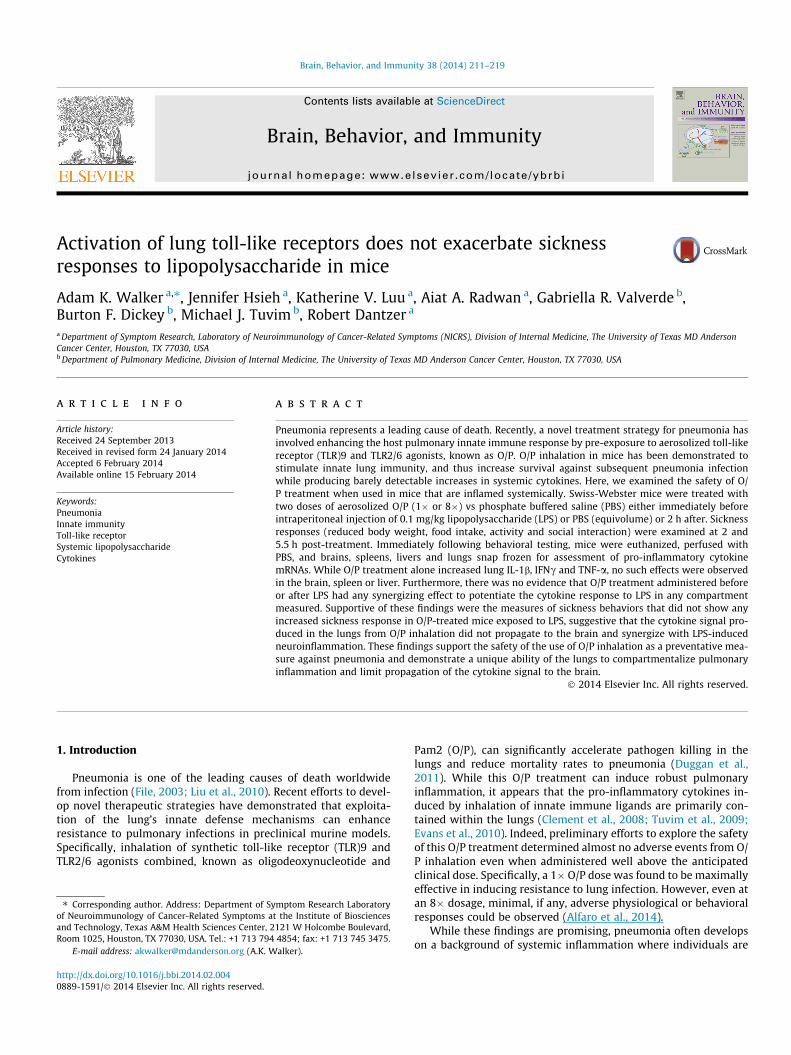
Fig. 1. Experimental timeline. Mice were treated with aerosolized oligodeoxynucleotide and Pam2 (O/P) and lipopolysaccharide LPS (ip) and assessed for measures ofsickness such a body weight loss (BW), food disappearance, locomotor activity (LMA) and social exploration.
A.K. Walker et al. / Brain, Behavior, and Immunity 38 (2014) 211–219 213
concentration (O’Connor et al., 2009). GAPDH was used as theendogenous housekeeping control gene.
2.4. Bronchoalveoloar lavage (BAL) fluid preparation and cell analysis
For Experiment 2 cell counts of BAL fluid were assessed. Bron-choalveolar lavage (BAL) fluid was obtained by instilling andcollecting two aliquots of 1 mL each of PBS through a luer stubadapter cannula (BD Biosciences) inserted through rings of the ex-posed trachea at the indicated time points. Total leukocyte count
0
10
20
30
40
50
PBS LPS
Lung
IL-1
βm
RNA
(fol
d ch
ange
)
PBS
1x O/P
8x O/P
0
1
2
3
4
PBS LPS
Lung
TN
F-α
mRN
A (f
old
chan
ge)
PBS
1x O/P
8x O/P
**
*
*
Fig. 2. Oligodeoxynucleotide and Pam2 (O/P) inhalation and systemic lipopolysaccharideIFNc mRNA fold differences (± SEM) of mice treated with aerosolized O/P or PBS immed
was determined with a hemocytometer (Bright-Line, HausserScientific) and slides for differential cell count were prepared bycytospin of 200 ll of BAL fluid at 300�g for 5 min, followed byWright-Giemsa staining.
2.5. Statistical analysis
Two-way analyses of variances (ANOVAs) (LPS vs PBS � O/P (0,1 or 8�)) were performed for all measures of biochemistry for tis-sue collected at 6 h. Three-way repeated measures ANOVAs (LPS vs
0
5
10
15
20
PBS LPS
Lung
IL-6
mRN
A (f
old
chan
ge)
PBS
1x O/P
8x O/P
0
2
4
6
8
PBS LPS
Lung
IFN
γm
RNA
(fol
d ch
ange
)
PBS
1x O/P
8x O/P
*
**
**
*
*
(LPS) increases pro-inflammatory cytokines in the lung. Mean IL-1b, IL-6, TNF-a andiately before LPS or PBS (ip). ⁄p < 0.05; ⁄⁄p < 0.01; ⁄⁄⁄p < 0.0001.
0
1
2
3
4
PBS LPS
Brai
n IL
-6 m
RNA
(f
old
chan
ge)
PBS
1x O/P
8x O/P
***
0
1
2
3
4
PBS LPS
Brai
n TN
F-α
mRN
A
(fol
d ch
ange
)
PBS
1x O/P
8x O/P
***
0
2
4
6
8
PBS LPS
Brai
n IL
-1β
mRN
A(f
old
chan
ge)
PBS
1x O/P
8x O/P
***
0
1
2
3
4
5
6
PBS LPS
Sple
en Il
-1β
mRN
A(f
old
chan
ge)
PBS
1x O/P
8x O/P
***
0
2
4
6
8
10
12
14
PBS LPS
Sple
en IL
- 6 m
RNA
(f
old
chan
ge)
PBS
1x O/P
8x O/P
***
0
1
2
3
PBS LPS
Sple
en T
NF-
αm
RNA
(f
old
chan
ge)
PBS
1x O/P
8x O/P
**
0
10
20
30
40
50
PBS LPS
Live
r Il-1
βm
RNA
(fol
d ch
ange
)
PBS
1x O/P
8x O/P
***
0
40
80
120
160
200
PBS LPS
Live
r TN
F-α
mRN
A
( fol
d ch
ange
)PBS
1x O/P
8x O/P
***
0
100
200
300
400
PBS LPS
Live
r IL-
6 m
RNA
(f
old
chan
ge)
PBS
1x O/P
8x O/P
* *
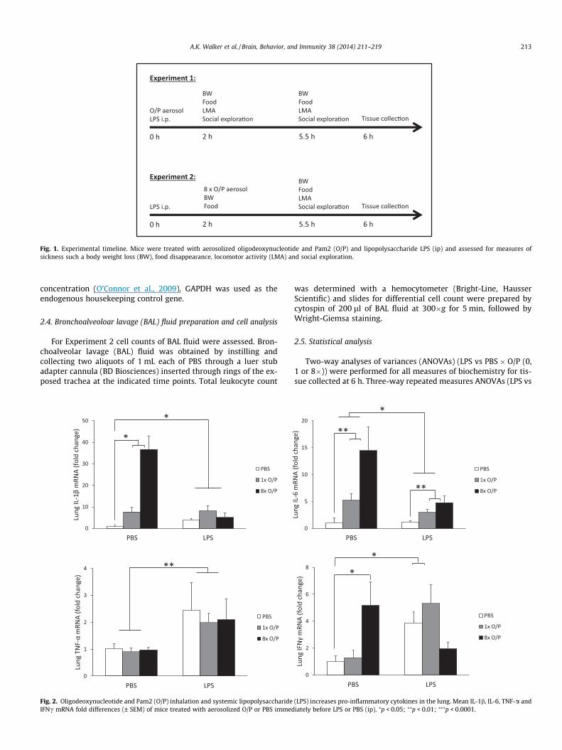
Fig. 3. Oligodeoxynucleotide and Pam2 (O/P) inhalation immediately before intraperitoneal lipopolysaccharide (LPS) administration does not potentiate spleen, liver andbrain cytokine responses to LPS. Mean IL-1b, IL-6 and TNF-a mRNA fold differences (± SEM) of mice treated with aerosolized O/P or PBS and LPS or PBS (ip). ⁄p < 0.05;⁄⁄p < 0.01; ⁄⁄⁄p < 0.0001.
214 A.K. Walker et al. / Brain, Behavior, and Immunity 38 (2014) 211–219
PBS � O/P (0, 1 or 8�) vs PBS � time as a repeated factor) were per-formed for measures of sickness behaviors, body weight loss andfood consumption. Where applicable, planned comparisons werealways performed against PBS/PBS negative control to determinechanges in behavior and elevations in cytokine concentrationsfrom baseline levels. Planned comparisons were determined usingFisher’s PLSD adjusted for the number of comparisons to control forfamily wise error. Only relevant significant comparisons betweengroups are indicated on figures.
3. Results
3.1. Aerosolized TLR treatment activates lung pro-inflammatorycytokines (Fig. 2)
LPS increased lung IL-6 (F(1,29) = 4.29, p < 0.05) and TNF-a(F(1,32) = 7.44, p = 0.01) mRNA concentrations. O/P treatment alsoincreased lung IL-6 mRNA (F(1,29) = 6.06, p < 0.01). A significantinteraction between LPS and O/P treatment was observed for lungIL-1b mRNA (F(2,28) = 17.65, p < 0.0001). LPS treatment significantlyincreased IL-1b compared to PBS/PBS controls and O/P treatmentalone dose-dependently increased IL-1b mRNA compared to PBS/PBS controls (p < 0.05 for all). A significant interaction betweenLPS and O/P treatment was also observed for lung IFNc mRNA(F(2,28) = 5.59, p < 0.01). O/P treatment alone significantly increasedlung IFNc mRNA at the 8� dosage (p < 0.05). LPS treatmentincreased lung IFNc mRNA, which reached significance for
LPS-treated mice independently of O/P treatment (p < 0.05 forall). Interestingly, 8� O/P significantly decreased the response toLPS for all cytokines except TNF-a (p < 0.05 for all).
3.2. Aerosolized TLR treatment does not potentiate spleen, liver andbrain pro-inflammatory cytokine responses to systemic LPS (Fig. 3)
LPS significantly increased spleen IL-1b (F(1,33) = 19.64,p < 0.0001), IL-6 (F(1,32) = 28.28, p < 0.0001) and TNF-a(F(1,33) = 8.85, p = 0.005) mRNA. O/P treatment had no effect onspleen cytokine mRNA levels either alone or in interaction withLPS.
LPS also significantly increased brain IL-1b (F(1,32) = 24.30,p < 0.0001), IL-6 (F(1,32) = 7.44, p = 0.01) and TNF-a (F(1,32) = 7.44,p = 0.01) mRNA. O/P treatment had no effect on brain cytokinemRNA levels either alone or in interaction with LPS.
LPS significantly increased liver IL-1b (F(1,32) = 50.30, p < 0.0001)and TNF-a (F(1,33) = 50.11, p < 0.0001) mRNA with no effect of O/Ptreatment. A significant interaction between LPS and O/P treatmentwas observed for liver IL-6 mRNA (F(2,33) = 5.23, p = 0.01). Post hoccomparisons revealed that all LPS-treated groups had significantlyhigher hepatic IL-6 mRNA than their respective controls, and IL-6mRNA was significantly higher for mice treated with 1� O/P andLPS compared to those treated with 8� O/P and LPS (p < 0.05 forall), suggestive of a suppression of the response to LPS by 8� O/P.O/P controls did not significantly differ from PBS/PBS controls.

0
0.2
0.4
0.6
0.8
1
1.2
0-2 h 2-5.5 h
Food
dis
appe
aran
ce (g
)
***
***
0
20
40
60
80
100
0-2 h 2-5.5 h
Qua
dran
t ent
ries
******
5.5 h 2 h 0
20
40
60
80
100
0-2 h 2-5.5 h
Rear
s
***
***
2 h 5.5 h
0
40
80
120
160
200
0-2 h 2-5.5 h
Soci
al e
xplo
ra�o
n (s
)
*** ***
2 h 5.5 h
-1.5
-1
-0.5
0
0.5
1
0-2 h 2-5.5 h
Body
wei
ght c
hang
e (g
)
***
***
a b
c d e
Fig. 4. Oligodeoxynucleotide and Pam2 (O/P) inhalation immediately before intraperitoneal lipopolysaccharide (LPS) administration does not synergize with LPS-inducedsickness. (a, b) Mean body weight change (g) and food disappearance (g) (+ SEM) for mice treated with aerosolized oligodeoxynucleotide and Pam2 (O/P) or PBS and LPS orPBS (ip) between 0–2 and 2–5.5 h. (c–e) Mean number of rears, quadrant entries, and time engaged in social exploration (± SEM) for mice treated with aerosolized O/P or PBSand LPS or PBS (ip) at 2 and 5.5 h. ⁄⁄⁄p < 0.0001.
A.K. Walker et al. / Brain, Behavior, and Immunity 38 (2014) 211–219 215
3.3. Aerosolized TLR treatment does not induce IL-10 in distalcompartments to attenuate the response to systemic LPS
Given that there are some instances where O/P inhalationappeared to reduce the pro-inflammatory response to LPS, weexamined whether O/P treatment may actually induce an anti-inflammatory response. LPS significantly increased IL-10 mRNAin the spleen (F(1,34) = 8.33, p < 0.01), brain (F(1,33) = 3.69, p < 0.05)and liver (F(1,33) = 19.22, p = 0.0001). However, O/P treatment hadno effect on IL-10 either alone or in interaction with LPS in anyof these organs. No effect of LPS or O/P treatment was observedfor IL-10 mRNA in the lung.
3.4. Aerosolized TLR treatment does not potentiate sickness responsesto systemic LPS
The effects of O/P treatment with or without LPS administrationare presented in Fig. 4.
3.4.1. Body weight and food consumptionLPS reduced body weight between 0–2 h and 2–5.5 h
(F(1,36) = 24.88, p < 0.0001). A main effect of O/P treatment was ob-served whereby mice treated with 8� O/P lost more weight than1� O/P-treated mice (F(2,36) = 3.91, p < 0.05). LPS reduced food con-sumption (F(1,36) = 68.01, p < 0.0001). Aerosolized O/P treatment,
irrespective of dose, did not alter LPS-induced reductions in foodconsumption.
3.4.2. Locomotor activityMain effects of LPS and O/P treatment were observed for loco-
motor activity across time, although no interaction was found.LPS significantly reduced quadrant entries and rears at 2 and5.5 h (F(1,35) = 20.34, p < 0.0001 and F(1,35) = 30.51, p < 0.0001,respectively). A significant O/P treatment � time interaction wasobserved for quadrant entries (F(2,35) = 5.41, p = 0.009). Plannedcomparisons revealed that 8� O/P treatment and 1� O/P treatmentactually increased quadrant entries at 2 and 5.5 h respectively(p < 0.05 for both). A main effect of O/P treatment was observedfor rears such that both doses of O/P treatment significantly in-creased rearing compared to controls (F(2,35) = 5.04, p = 0.01).
3.4.3. Social explorationLPS significantly reduced social exploration time (F(1,36) = 57.83,
p < 0.0001), whereas O/P treatment significantly increased socialexploration compared to controls (F(2,36) = 3.95, p = 0.03).
3.5. Pre-treatment with systemic LPS does not potentiate aerosolizedTLR-induced lung inflammation
In the previous experiments we demonstrated that treatmentwith O/P immediately before systemic LPS treatment did not exac-

0
1
2
3
4
5
PBS LPS
Lung
IFN
γm
RNA
(fol
d ch
ange
)PBS
8x O/P
0
10
20
30
40
50
PBS LPS
Lung
IL-1
βm
RNA
(fol
d ch
ange
)
PBS
8x O/P
*
*
0
20
40
60
80
100
120
140
PBS LPS
Lung
IL-6
mRN
A (f
old
chan
ge)
PBS
8x O/P
*
*
0
5
10
15
20
25
30
PBS LPS
Lung
TN
F-α
mRN
A (f
old
chan
ge)
PBS
8x O/P
*
*
Fig. 5. Oligodeoxynucleotide and Pam2 (O/P) inhalation does not potentiate pre-existing systemic lipopolysaccharide (LPS)-induced increases in pro-inflammatory cytokinesin the lung. Mean IL-1b, IL-6, TNF-a and IFNc mRNA fold differences (± SEM) of mice treated with aerosolized O/P or PBS 2 h after LPS or PBS (ip). ⁄p < 0.05.
216 A.K. Walker et al. / Brain, Behavior, and Immunity 38 (2014) 211–219
erbate symptoms of LPS-induced sickness or inflammatory re-sponses. Here we examined a more clinically relevant questionas to whether the maximal 8� O/P dose potentiates sickness whengiven after LPS-induced systemic inflammation has alreadyreached its peak.
3.5.1. Changes in lung leukocyte levelsWe examined changes in lung leukocyte levels in the same mice
by measuring total and differential cell counts in BAL fluid. A sig-nificant interaction between LPS and O/P treatment was observedfor the total number of cells in BAL fluid (F(1,15) = 5.03, p < 0.05),whereby O/P treatment significantly increased neutrophil levelswhich was attenuated in the presence of LPS (p < 0.05). Of the cellsinduced by 8� O/P almost 90% were neutrophils with almost noneutrophils observed in the control groups. Only approximately40% of total cells were neutrophils however in the mice pre-treatedwith LPS. The remaining percentage of total cells in each case weremacrophages. Minimal to no lymphocytes or eosinophils were ob-served in BAL fluid.
3.5.2. Lung cytokine levelsFig. 5 depicts the pro-inflammatory cytokine mRNA levels
observed in the lungs of mice treated with 8� O/P 2 h after LPStreatment. Significant O/P � LPS interactions were observed forlung IL-1b (F(1,14) = 11.84, p < 0.01) and TNF-a (F(1,14) = 7.96,p < 0.05) mRNA whereby both O/P and LPS alone or in combinationincreased mRNA levels above that of PBS-treated controls (p < 0.05for all). The same trend was observed for IL-6 mRNA but only O/P
treatment reached significance (F(1,14) = 8.15, p < 0.05). No signifi-cant effects for IL-10 and IFNc mRNA were observed.
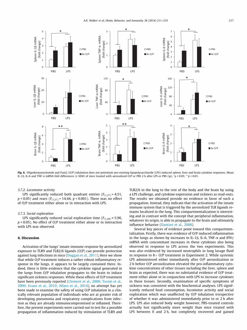
3.6. Aerosolized TLR treatment does not potentiate spleen, liver andbrain cytokine responses to pre-treatment with LPS (Fig. 6)
LPS increased spleen IL-1b (F(1,15) = 10.73, p < 0.05), TNF-a(F(1,15) = 4.89, p < 0.05) and IL-6 (F(1,14) = 4.90, p < 0.05) mRNA withno effect of O/P treatment. No significant differences were ob-served for IL-10.
LPS increased brain IL-1b (F(1,13) = 15.37, p < 0.01), TNF-a(F(1,12) = 12.24, p < 0.01) and IL-6 (F(1,12) = 4.81, p < 0.05) mRNAwith no effect of O/P treatment. No significant differences were ob-served for IL-10.
LPS increased liver IL-6 (F(1,13) = 5.33, p < 0.05) and TNF-a(F(1,14) = 6.74, p < 0.05) mRNA with no effect of O/P treatment.Trends also indicated LPS-induced increases in liver IL-1b but didnot reach significance.
3.7. Aerosolized TLR treatment does not potentiate sickness responsesto pre-existing inflammation caused by systemic LPS (Fig. 7)
3.7.1. Body weight and food consumptionLPS reduced body weight between from 2–5.5 h (F(1,28) = 50.27,
p < 0.0001). LPS reduced food consumption between 2 and 5.5 h(F(1,28) = 22.54, p < 0.0001) with no effect of pre-treatment with8� O/P.
0
1
2
3
4
5
6
7
PBS LPS
Sple
en IL
-6 m
RNA
(f
old
chan
ge)
PBS
8x O/P
0
1
2
3
PBS LPS
Sple
en T
NF-
αm
RNA
( f
old
chan
ge)
PBS
8x O/P
0
1
2
3
4
5
PBS LPS
Sple
en IL
-1β
mRN
A
(fol
d ch
ange
)
PBS
8x O/P
0
1
2
3
4
PBS LPSBr
ain
IL-6
mRN
A
(fol
d ch
ange
)
PBS
8x O/P
0
1
2
3
4
5
PBS LPS
Brai
n TN
F-α
mRN
A
( fol
d ch
ange
)
PBS
8x O/P
0
2
4
6
8
PBS LPS
Brai
n IL
-1β
mRN
A
(fol
d ch
ange
)
PBS
8x O/P
* ****
* * *
0
40
80
120
160
PBS LPS
Live
r IL-
6 m
RNA
(f
old
chan
ge)
PBS
8x O/P
0
40
80
120
PBS LPS
Live
r TN
F-α
mRN
A
(fol
d ch
ange
) PBS
8x O/P
*
0
10
20
30
PBS LPS
Live
r IL-
1βm
RNA
(f
old
chan
ge)
PBS
8x O/P
*
Fig. 6. Oligodeoxynucleotide and Pam2 (O/P) inhalation does not potentiate pre-existing lipopolysaccharide (LPS)-induced spleen, liver and brain cytokine responses. MeanIL-1b, IL-6 and TNF-a mRNA fold differences (± SEM) of mice treated with aerosolized O/P or PBS 2 h after LPS or PBS (ip). ⁄p < 0.05; ⁄⁄p < 0.01.
A.K. Walker et al. / Brain, Behavior, and Immunity 38 (2014) 211–219 217
3.7.2. Locomotor activityLPS significantly reduced both quadrant entries (F(1,27) = 4.51,
p < 0.05) and rears (F(1,27) = 14.44, p < 0.001). There was no effectof O/P treatment either alone or in interaction with LPS.
3.7.3. Social explorationLPS significantly reduced social exploration time (F(1,28) = 5.96,
p < 0.05). No effect of O/P treatment either alone or in interactionwith LPS was observed.
4. Discussion
Activation of the lungs’ innate immune response by aerosolizedexposure to TLR9 and TLR2/6 ligands (O/P) can provide protectionagainst lung infections in mice (Duggan et al., 2011). Here we showthat while O/P treatment induces a rather robust inflammatory re-sponse in the lungs, it appears to be largely contained there. In-deed, there is little evidence that the cytokine signal generated inthe lungs from O/P inhalation propagates to the brain to inducesignificant sickness responses. While these effects of O/P treatmenthave been previously reported (Clement et al., 2008; Tuvim et al.,2009; Evans et al., 2010; Alfaro et al., 2014), no attempt has yetbeen made to examine the safety of using O/P inhalation in a clin-ically relevant population of individuals who are at greater risk ofdeveloping pneumonia and respiratory complications from infec-tion as they are already immunocompromised or inflamed. There-fore, the present experiments were carried out to test for a possiblepropagation of inflammation induced by stimulation of TLR9 and
TLR2/6 in the lung to the rest of the body and the brain by usinga LPS challenge, and cytokine expression and sickness as read-outs.The results we obtained provide no evidence in favor of such apropagation. Instead, they indicate that the activation of the innateimmune system that is triggered by the aerosolized TLR ligands re-mains localized in the lung. This compartmentalization is interest-ing and in contrast with the concept that peripheral inflammation,whatever its origin, is able to propagate to the brain and ultimatelyinfluence behavior (Dantzer et al., 2008).
Several key pieces of evidence point toward this compartmen-talization. Firstly, there was evidence of O/P-induced inflammationin the lungs as shown by increases in IL-1b, IL-6, TNF-a and IFNcmRNA with concomitant increases in these cytokines also beingobserved in response to LPS across the two experiments. Thiswas also evidenced by increased neutrophils in lung lavage fluidin response to 8� O/P treatment in Experiment 2. While systemicLPS administered either immediately after O/P aerosolization or2 h before O/P aerosolization elevated the pro-inflammatory cyto-kine concentrations of other tissues including the liver, spleen andbrain as expected, there was no substantial evidence of O/P treat-ment either alone or in conjunction with LPS to increase cytokinesin these tissues. Secondly, examination of specific symptoms ofsickness was consistent with the biochemical analyses. LPS signif-icantly reduced food consumption, locomotor activity and socialexploration which was unaffected by O/P inhalation irrespectiveof whether it was administered immediately prior to or 2 h afterLPS. LPS also reduced body weight however, PBS-treated controlsactually lost significantly more weight than mice treated withLPS between 0 and 2 h, but completely recovered and gained

0
0.2
0.4
0.6
0.8
1
1.2
1.4
0-2 h 2-5.5 h
Food
dis
appe
aran
ce (g
)
* b
0
20
40
60
80
PBS LPS
Rear
s
0
40
80
120
160
200
PBS LPSSo
cial
exp
lora
�on
(s)
0
20
40
60
80
100
PBS LPS
Qua
dran
t ent
ries
* c d e ** *
-1.5
-1
-0.5
0
0.5
1
1.5
0-2 h 2-5.5 h
Body
wei
ght c
hang
e (g
)a
*
Fig. 7. Oligodeoxynucleotide and Pam2 (O/P) inhalation 2 h after intraperitoneal lipopolysaccharide (LPS) administration does not synergize with LPS-induced sickness. (a, b)Mean body weight change (g) and food disappearance (g) (+ SEM) for mice treated with aerosolized oligodeoxynucleotide and Pam2 (O/P) or PBS 2 h after LPS or PBS (ip)between 0–2 and 2–5.5 h. (c–e) Mean number of rears, quadrant entries, and time engaged in social exploration (± SEM) for mice treated with aerosolized O/P or PBS 2 h afterLPS or PBS (ip) at 2 and 5.5 h. ⁄p < 0.05; ⁄⁄p < 0.01.
218 A.K. Walker et al. / Brain, Behavior, and Immunity 38 (2014) 211–219
weight between 2 and 5.5 h. This body weight loss between 0–2 hin the mice receiving aerosols is likely accounted for by the factthat the mice huddle in a corner shortly after aerosol administra-tion, and are not observed to drink or eat during this time.
Curiously, the data from Experiment 1 and Experiment 2 notonly dispute the notion that activation of pulmonary TLR9 andTLR2/6 potentiates LPS-induced sickness and systemic inflamma-tion, but even indicates a dose-dependent attenuation by aerosol-ized O/P of the response to LPS in some instances. For instance,there are consistent trends showing an increase in activity follow-ing O/P treatment in Experiment 1. While this does not negate thefact that O/P inhalation does not appear to induce sickness, it ispossible that O/P inhalation influences brain activity outside ofinflammatory pathways. Also, 8� O/P when administered immedi-ately prior to LPS particularly shows a reduced response in thepresence of systemic LPS for IFNc in the lung and for TNF-a andIL-6 in the liver. Again, 8� O/P when administered 2 h after sys-temic LPS shows a reduced response for IL-1b in the lung as wellas a decrease in the number of neutrophils in BAL fluid comparedto when 8� O/P was administered without LPS. To determinewhether these effects were due to O/P inhalation actually inducingan increase in anti-inflammatory cytokines we measured IL-10mRNA in each compartment. No significant relationship betweenIL-10 and O/P treatment was observed in the lungs, liver, spleenor brain and cannot explain the O/P-induced attenuation of the re-sponse to LPS. Nor is it likely that a tolerance effect can explain thisphenomenon as the cytokine response to LPS in the lungs was notlower than in other organs. It is possible that the activation of lung
TLRs by the high dose of O/P activated the cholinergic anti-inflam-matory reflex that then attenuates the response to LPS, howeverthis requires confirmation. Similarly, the reduction in neutrophilsin BAL fluid in Experiment 2 when mice were treated with 8� O/P and LPS can be accounted for by the known increase in neutro-phil and monocyte adhesion to endothelial cells in response toLPS. LPS has been demonstrated to increase numerous adhesionmolecules and a rapid reduction in the number of circulating neur-trophils (Jersmann et al., 2001; Bradfield et al., 2007; Hickey andKubes, 2009). This has been well demonstrated in the lungs, andit has been suggested that activated neutrophils may becomelodged in the narrow lung capillaries making it difficult for migra-tion (Hickey and Kubes, 2009). This is likely to account for the re-duced number of total cells observed in the BAL fluid of micetreated with 8� O/P and LPS compared to the BAL fluid of micetreated with 8� O/P alone and may not actually represent an atten-uated response.
Containment of O/P-induced inflammation to the lungs indi-cates that the lung microenvironment is capable of regulating itsinflammatory response somewhat independently of the rest ofthe body. This idea is supported by recent findings showing thatalveolar macrophages are not influenced by systemic events andrespond differently than circulating monocytes. For instance, Phil-ippart et al. (2012) demonstrated that alveolar macrophages do notdevelop endotoxin tolerance unlike monocytes and peritonealmacrophages after injection with LPS in mice. This suggestslocalized mechanisms specific to the lung microenvironment. Themechanism responsible for containment within the lungs in the

A.K. Walker et al. / Brain, Behavior, and Immunity 38 (2014) 211–219 219
present study most likely reflects the difference between themolecular responses to TLR 9 and TLR 2/6 activation by O/P treat-ment compared to conditions of pulmonary infection such as influ-enza. That is, both the TLR agonists and influenza, for example, actat the level of the epithelial cells. However, unlike with the TLRagonist inhalation, invading pathogens will cause epithelial celldeath and activate ‘danger’ signals. This in turn, will lead to a highdegree of macrophage recruitment and hence systemic spill over.Therefore, lung endothelial inflammation in the absence of celldeath, as is the case with exposure to aerosolized TLR 9 and TLR2/6 agonists, can be considered safe from propagating the cytokinesignal to other compartments and inducing sickness.
5. Conclusion
The evidence provided here supports the safety of using TLR 9and TLR 2/6 agonists as a protective therapy against pneumoniain both healthy at-risk individuals and already immunocompro-mised or inflamed patients. We report no evidence of O/P-inducedexacerbation of the LPS response either behaviorally or in regardsto cytokine activation in distal peripheral and central compart-ments of the body. Furthermore, we demonstrate the ability ofthe lung microenvironment to compartmentalize itself from therest of the body and contain inflammation to the lungs withoutpropagation to the brain under conditions that lack endothelial celldeath and macrophage recruitment. This compartmentalizationchallenges the existing beliefs that peripheral inflammation, what-ever its origin, is able to propagate to the brain and ultimatelyinfluence behavior.
Conflicts of interest
Robert Dantzer works as a consultant for Ironwoods Pharma-ceuticals. Michael J. Tuvim and Burton F. Dickey have receivedGrant support from the US National Institutes of Health for studieson inducible innate resistance; they are inventors of technologiesrelated to inducible resistance for which patent applications havebeen made; and they have ownership interests in Pulmotect, Inc.,which has licensed technologies from their employer, MD Ander-son Cancer Center, for stimulating innate immunity to preventrespiratory infections and has sponsored research in their laborato-ries. All other authors declare no competing interests.
Authors’ contributions
A.K.W. participated in the study conception, design, coordina-tion, data collection, statistical analysis and drafting of the manu-script. J.H., K.V.L. and A.R. participated in the behavioral testingand qRT-PCR. G.R.V. was responsible for the O/P treatment prepa-ration and administration, and lung lavage and cell counts of BALfluid. B.F.D., M.J.T. and R.D. participated in the study conception,design, coordination, and drafting of the manuscript. All authorsread and approved the final manuscript.
Acknowledgments
This work was supported by the University of Texas MD Ander-son Cancer Center and Grant National Institutes of Health GrantR01 NS073939 to and supported in part by the MD Anderson Can-cer Center support Grant CA016672 from the National Institutes ofHealth and P01 CA124787 from NCI RD. These funding sources hadno further involvement in this study.
References
Alfaro, V., Goldblatt, D.L., Valverde, G.R., Munsell, M., Quinton, L.J., Walker, A.K.,Dantzer, R., Varadhachary, A., Scott, B.L., Evans, S.E., Tuvim, M.J., Dickey, B.F.,2014. Safety, tolerability, and biomarkers of the treatment of mice withaerosolized toll-like receptor ligands. Front. Pharmacol. 5. http://dx.doi.org/10.3389/fphar.2014.00008.
Bradfield, P.F., Johnson-Leger, C.A., Zimmerli, C., Imhof, B.A., 2007. LPS differentiallyregulates adhesion and transendothelial migration of human monocytes understatic and flow conditions. Int. Immunol. 20, 247–257.
Clement, C.G., Evans, S.E., Evans, C.M., Hawke, D., Kobayashi, R., Reynolds, P.R.,Moghaddam, S.J., Scott, B.L., Melicoff, E., Adachi, R., Dickey, B.F., Tuvim, M.J.,2008. Stimulation of lung innate immunity protects against lethalpneumococcal pneumonia in mice. Am. J. Respir. Crit. Care Med. 177, 1322–1330.
Dantzer, R., O’Connor, J.C., Freund, G.G., Johnson, R.W., Kelley, K.W., 2008. Frominflammation to sickness and depression: when the immune system subjugatesthe brain. Nat. Rev. Neurosci. 9, 46–56.
Duggan, J.M., You, D., Cleaver, J.O., Larson, D.T., Garza, R.J., Guzman Pruneda, F.A.,Tuvim, M.J., Zhang, J., Dickey, B.F., Evans, S.E., 2011. Synergistic interactions ofTLR2/6 and TLR9 induce a high level of resistance to lung infection in mice. J.Immunol. 186, 5916–5926.
Evans, S.E., Scott, B.L., Clement, C.G., Larson, D.T., Kontoyiannis, D., Lewis, R.E.,LaSala, P.R., Pawlik, J., Peterson, J.W., Chopra, A.K., Klimpel, G., Bowden, G., Höök,M., Xu, Y., Tuvim, M.J., Dickey, B.F., 2010. Stimulation of lung innate immunityprotects mice broadly against bacterial and fungal pneumonia. Am. J. Respir.Cell. Mol. Biol. 42, 40–50.
Fekrazad, M.H., Eberhardt, S., Jones, D.V., Lee, F.C., 2010. Development ofbronchiolitis obliterans organizing pneumonia with platinum-basedchemotherapy for metastatic rectal cancer. Clin. Colorectal Cancer 9, 177–178.
File, T.M., 2003. Community-acquired pneumonia. Lancet 362, 1991–2001.Joos, L., Tamm, M., 2005. Breakdown of pulmonary host defense in the
immunocompromised host: cancer chemotherapy. Proc. Am. Thorac. Soc. 2,445–448.
Hickey, M.J., Kubes, P., 2009. Intravascular immunity: the host–pathogen encounterin blood vessels. Nat. Rev. Immunol. 9, 364–375.
Jersmann, H.R., Hii, C.S.T., Ferrante, J.V., Ferrante, A., 2001. Bacteriallipopolysaccharide and tumor necrosis factor alpha synergistically increaseexpression of human endothelial adhesion molecules through activation ofNF-KB and p38 mitogen-activated protein kinase signaling pathways. Infect.Immun. 69, 1273–1279.
Kent, S., Bluthe, R.-M., Kelley, K.W., Dantzer, R., 1992. Sickness behavior as a newtarget for drug development. Trends Pharmacol. Sci. 13, 24–28.
Liu, L., Johnson, H.L., Cousens, S., Perin, J., Scott, S., Lawn, J.E., Rudan, I., Campbell, H.,Cibulskis, R., Li, M., Mathers, C., Black, R.E., 2010. Global, regional, and nationalcauses of child mortality: an updated systematic analysis for 2010 with timetrends since 2000. Lancet 379, 2151–2161.
O’Connor, J.C., Andre, C., Wang, Y., Lawson, M.A., Szegedi, S.S., Lestage, J., Castanon,N., Kelley, K.W., Dantzer, R., 2009. Interferon-c and tumor necrosis factor-amediate the upregulation of indoleamine 2,3-dioxygenase and the induction ofdepressive-like behavior in mice in response to Bacillus Calmette-Guerin. J.Neurosci. 29, 4200–4209.
Philippart, F., Fitting, C., Cavaillon, J.-M., 2012. Lung microenvironment contributesto the resistance of alveolar macrophages to develop tolerance to endotoxin.Crit. Care Med. 40, 2987–2996.
Tuvim, M.J., Evans, S.E., Clement, C.G., Dickey, B.F., Gilbert, B.E., 2009. Augmentedlung inflammation protects against influenza A pneumonia. PLoS One 4, e4176.
Walker, A.K., Budac, D.P., Bisulco, S., Lee, A.W., Smith, R.A., Beenders, B., Kelley, K.W.,Dantzer, R., 2013. NMDA receptor blockade by ketamine abrogateslipopolysaccharide-induced depressive-like behavior in C57BL/6J mice.Neuropsychopharmacology 38, 1609–1616.