The GPI biosynthetic pathway as a therapeutic target for African sleeping sickness
14
Review The GPI biosynthetic pathway as a therapeutic target for African sleeping sickness Michael A.J. Ferguson a ; *, John S. Brimacombe a , Jillian R. Brown a , Arthur Crossman a , Alexander Dix a , Robert A. Field b , M. Lucia S. Gu « ther a , Kenneth G. Milne a , Deepak K. Sharma a , Terry K. Smith a a Division of Molecular Parasitology and Biological Chemistry, Departments of Biochemistry and Chemistry, University of Dundee, Dundee DD1 5EH, UK b School of Chemistry, University of St Andrews, Fife, UK Received 8 February 1999; received in revised form 23 April 1999; accepted 23 April 1999 Abstract African sleeping sickness is a debilitating and often fatal disease caused by tsetse fly transmitted African trypanosomes. These extracellular protozoan parasites survive in the human bloodstream by virtue of a dense cell surface coat made of variant surface glycoprotein. The parasites have a repertoire of several hundred immunologically distinct variant surface glycoproteins and they evade the host immune response by antigenic variation. All variant surface glycoproteins are anchored to the plasma membrane via glycosylphosphatidylinositol membrane anchors and compounds that inhibit the assembly or transfer of these anchors could have trypanocidal potential. This article compares glycosylphosphatidylinositol biosynthesis in African trypanosomes and mammalian cells and identifies several steps that could be targets for the development of parasite-specific therapeutic agents. ß 1999 Elsevier Science B.V. All rights reserved. Keywords : Glycosylphosphatidylinositol ; GPI ; Trypanosome ; Biosynthesis ; Glycosyltransferase Contents 1. Introduction .......................................................... 328 2. GPI biosynthesis ....................................................... 328 2.1. The UDP-GlcNAc :PI K1-6 N-acetylglucosaminyltransferase complex ............ 329 2.2. The GlcNAc-PI de-N-acetylase ........................................ 331 2.3. The Dol-P-Man :GlcN-PI/GlcN-(acyl)PI K1-4 mannosyltransferase (MT-I) ......... 331 2.4. Inositol acyltransferases and inositol deacylases ............................ 333 2.5. The Dol-P-Man:Man 1 GlcN-( þ acyl)PI K1-6 mannosyltransferase (MT-II) ......... 333 2.6. The Dol-P-Man:Man 2 GlcN-( þ acyl)PI K1-2 mannosyltransferase (MT-III) ........ 335 2.7. Ethanolamine phosphotransferases ...................................... 335 2.8. Other GPI glycosyltransferases ......................................... 336 0925-4439 / 99 / $ ^ see front matter ß 1999 Elsevier Science B.V. All rights reserved. PII:S0925-4439(99)00058-7 * Corresponding author. Fax : +44-1382-345764 ; E-mail : [email protected] Biochimica et Biophysica Acta 1455 (1999) 327^340 www.elsevier.com/locate/bba
-
Upload
independent -
Category
Documents
-
view
0 -
download
0
Transcript of The GPI biosynthetic pathway as a therapeutic target for African sleeping sickness

Review
The GPI biosynthetic pathway as a therapeutic target for Africansleeping sickness
Michael A.J. Ferguson a;*, John S. Brimacombe a, Jillian R. Brown a,Arthur Crossman a, Alexander Dix a, Robert A. Field b, M. Lucia S. Gu«ther a,
Kenneth G. Milne a, Deepak K. Sharma a, Terry K. Smith a
a Division of Molecular Parasitology and Biological Chemistry, Departments of Biochemistry and Chemistry, University of Dundee,Dundee DD1 5EH, UK
b School of Chemistry, University of St Andrews, Fife, UK
Received 8 February 1999; received in revised form 23 April 1999; accepted 23 April 1999
Abstract
African sleeping sickness is a debilitating and often fatal disease caused by tsetse fly transmitted African trypanosomes.These extracellular protozoan parasites survive in the human bloodstream by virtue of a dense cell surface coat made ofvariant surface glycoprotein. The parasites have a repertoire of several hundred immunologically distinct variant surfaceglycoproteins and they evade the host immune response by antigenic variation. All variant surface glycoproteins areanchored to the plasma membrane via glycosylphosphatidylinositol membrane anchors and compounds that inhibit theassembly or transfer of these anchors could have trypanocidal potential. This article compares glycosylphosphatidylinositolbiosynthesis in African trypanosomes and mammalian cells and identifies several steps that could be targets for thedevelopment of parasite-specific therapeutic agents. ß 1999 Elsevier Science B.V. All rights reserved.
Keywords: Glycosylphosphatidylinositol ; GPI; Trypanosome; Biosynthesis ; Glycosyltransferase
Contents
1. Introduction . . . . . . . . . . . . . . . . . . . . . . . . . . . . . . . . . . . . . . . . . . . . . . . . . . . . . . . . . . 328
2. GPI biosynthesis . . . . . . . . . . . . . . . . . . . . . . . . . . . . . . . . . . . . . . . . . . . . . . . . . . . . . . . 3282.1. The UDP-GlcNAc:PI K1-6 N-acetylglucosaminyltransferase complex . . . . . . . . . . . . 3292.2. The GlcNAc-PI de-N-acetylase . . . . . . . . . . . . . . . . . . . . . . . . . . . . . . . . . . . . . . . . 3312.3. The Dol-P-Man:GlcN-PI/GlcN-(acyl)PI K1-4 mannosyltransferase (MT-I) . . . . . . . . . 3312.4. Inositol acyltransferases and inositol deacylases . . . . . . . . . . . . . . . . . . . . . . . . . . . . 3332.5. The Dol-P-Man:Man1GlcN-( þ acyl)PI K1-6 mannosyltransferase (MT-II) . . . . . . . . . 3332.6. The Dol-P-Man:Man2GlcN-( þ acyl)PI K1-2 mannosyltransferase (MT-III) . . . . . . . . 3352.7. Ethanolamine phosphotransferases . . . . . . . . . . . . . . . . . . . . . . . . . . . . . . . . . . . . . . 3352.8. Other GPI glycosyltransferases . . . . . . . . . . . . . . . . . . . . . . . . . . . . . . . . . . . . . . . . . 336
0925-4439 / 99 / $ ^ see front matter ß 1999 Elsevier Science B.V. All rights reserved.PII: S 0 9 2 5 - 4 4 3 9 ( 9 9 ) 0 0 0 5 8 - 7
* Corresponding author. Fax: +44-1382-345764; E-mail : [email protected]
BBADIS 61863 15-9-99
Biochimica et Biophysica Acta 1455 (1999) 327^340
www.elsevier.com/locate/bba
2.9. GPI lipid remodelling . . . . . . . . . . . . . . . . . . . . . . . . . . . . . . . . . . . . . . . . . . . . . . . 3362.10. The GPI-transamidase complex . . . . . . . . . . . . . . . . . . . . . . . . . . . . . . . . . . . . . . . . 337
3. Perspectives . . . . . . . . . . . . . . . . . . . . . . . . . . . . . . . . . . . . . . . . . . . . . . . . . . . . . . . . . . . 337
Acknowledgements . . . . . . . . . . . . . . . . . . . . . . . . . . . . . . . . . . . . . . . . . . . . . . . . . . . . . . . . . 337
References . . . . . . . . . . . . . . . . . . . . . . . . . . . . . . . . . . . . . . . . . . . . . . . . . . . . . . . . . . . . . . . 337
1. Introduction
Sleeping sickness a¡ects millions of people in sub-Saharan Africa and the WHO estimates that thereare currently over 300 000 new cases a year. Thereare no vaccines and current drugs are relatively toxicand di¤cult to administer. The African trypano-somes are extracellular protozoan parasites that livein the blood, lymph and interstitial £uids of the in-fected mammalian host where they divide by binary¢ssion. The ability of the trypanosome population toevade the host immune response is due to a denseand uniform protective coat [1] made of about 107
copies/cell of a 55 kDa variant surface glycoprotein(VSG) [2] (Fig. 1). This dense palisade of VSG gly-coprotein forms a di¡usion barrier that prevents theapproach of macromolecules to the parasite plasmamembrane and protects the parasite from lysis by thealternative- and lectin-mediated complement path-ways. An individual trypanosome expresses onlyone VSG gene at a time but contains about a thou-sand VSG genes encoding structurally similar [3] butimmunologically distinct VSGs. In natural and ex-perimental infections, the mammalian host mountsa potent humoral immune response to the prevailingpopulation of VSG coats that leads to almost com-plete clearance of the parasites. However, the switch-ing of VSG genes by some parasites gives rise to anew population that replicates unchecked until an-other speci¢c immune response is elicited. This proc-ess of antigenic variation [4] is repeated until thedeath of the host.
Depending on the VSG variant, VSG monomerscontain one, two or three N-glycosylation sites occu-pied by oligomannose and/or complex oligosaccha-rides [5^7]. All VSGs form homodimers anchored tothe plasma membrane via glycosylphosphatidylinosi-tol (GPI) membrane anchors (one per VSG mono-mer) of conserved structure that vary only in the
extent of their side chain galactosylation [7]. In con-trast to the extremely high density of GPI-anchoredglycoproteins in the trypanosome (Fig. 1), mamma-lian cells contain mostly transmembrane and periph-eral glycoproteins at the cell surface and GPI-an-chored proteins are in the minority. To put this intocontext, one of the most abundant mammalian GPI-anchored proteins is rat thymocyte Thy-1 (present atabout 6U105 copies per cell [8]) and a piece of mem-brane like that modelled (Fig. 1) for the trypanosomewould contain less than one molecule of Thy-1. De-spite their lower density, mammalian GPI-anchoredproteins are clearly vital since GPI knockouts areembryo-lethal in mice and tissue-speci¢c GPI knock-out causes signi¢cant tissue-speci¢c defects [9].
In conclusion, the absolute dependence of the tryp-anosome on its protective VSG coat, and the de-pendence of the latter on its GPI anchors, makesthe parasite GPI biosynthetic pathway an attractivetherapeutic target. However, the presence, and im-portance, of GPI anchored glycoproteins in mam-mals suggest that parasite-speci¢c inhibitors of GPIbiosynthetic enzymes are desirable. This article fo-cuses on the known and suggested di¡erences be-tween Trypanosoma brucei and mammalian GPI bio-synthesis.
2. GPI biosynthesis
The conserved EtNP-6ManK1-2ManK1-6ManK1-4GlcNK1-6myo-inositol1-P-lipid core region of allprotein-linked GPI membrane anchors [10^45] (Fig.2) suggests conservation in the GPI biosynthetic ma-chinery. The African trypanosome played a key rolein the development of methodologies to study GPIbiosynthesis. Intact GPI anchor precursors were ¢rstidenti¢ed and characterised in T. brucei [46^48] andit was used to develop the ¢rst cell-free system to
BBADIS 61863 15-9-99
M.A.J. Ferguson et al. / Biochimica et Biophysica Acta 1455 (1999) 327^340328

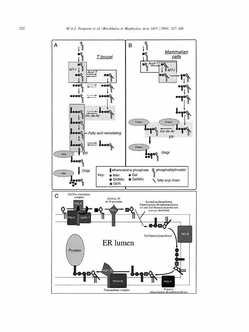
delineate the GPI biosynthetic pathway [49]. Thecell-free system is based on washed membranesprimed with UDP-GlcNAc and GDP-[3H]Man inthe presence of tunicamycin to suppress the synthesisof dolichol cycle intermediates. The radiolabelledGPI intermediates formed in the membranes fromendogenous phosphatidylinositol (PI) are extractedin solvent and analysed by thin-layer chromatogra-phy. Enzyme and chemical treatments are then usedto assign the chemical structures of the GPI inter-mediates. This system has been adapted to studyGPI biosynthesis in a variety of organisms, includ-ing mammalian cells and yeast ([50^54], and referen-ces therein). The essential features of GPI biosyn-thesis in the bloodstream form of T. brucei [49,55]and mammalian cells [50,51] are shown in Fig.3A,B.
2.1. The UDP-GlcNAc:PI K1-6N-acetylglucosaminyltransferase complex
The biosynthesis of GPI anchors starts with thetransfer of GlcNAc from UDP-GlcNAc to PI[56,57] on the cytoplasmic face of the ER [58,59].In mammalian cells, this simple reaction requires atleast four proteins (PIG-A, PIG-C, PIG-H andhGPI-1) that form a complex in the ER membrane(Fig. 3C) [60,61]. The PIG-A component is mostlikely the catalytic subunit since it has homologywith a bacterial GlcNAc-transferase. Homologuesof the PIG-A, PIG-C and hGPI-1 genes (GPI3,GPI2 and GPI1, respectively) exist in the yeast Sac-charomyces cerevisiae [62^64] but a PIG-H homo-logue is missing in that organism.
There is relatively little information on the specif-
Fig. 1. T. brucei, the VSG coat and the VSG GPI membrane anchor. A scanning electron micrograph (courtesy of M. Duszenko) ofa bloodstream form T. brucei trypomastigote (top left) is shown next to a cartoon model (top right) of a 20 nmU20 nm section ofthe plasma membrane [7]. The structure of a VSG dimer, based on the N-terminal crystal structure [3], is shown (bottom right) nextto the primary structure of the GPI anchor [11] (bottom left). The boxed section of the GPI anchor structure is conserved throughoutthe eukaryotes. The galactose side chains and the fatty acids of the PI moiety, both myristate, are unique to VSG.
BBADIS 61863 15-9-99
M.A.J. Ferguson et al. / Biochimica et Biophysica Acta 1455 (1999) 327^340 329
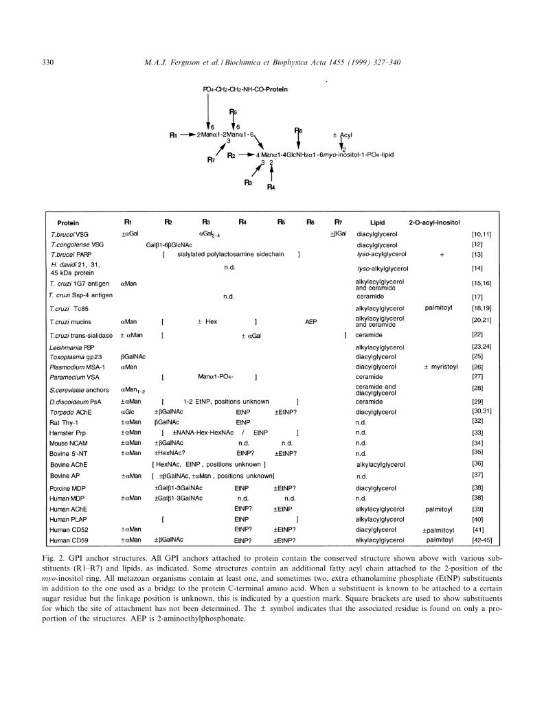
Fig. 2. GPI anchor structures. All GPI anchors attached to protein contain the conserved structure shown above with various sub-stituents (R1^R7) and lipids, as indicated. Some structures contain an additional fatty acyl chain attached to the 2-position of themyo-inositol ring. All metazoan organisms contain at least one, and sometimes two, extra ethanolamine phosphate (EtNP) substituentsin addition to the one used as a bridge to the protein C-terminal amino acid. When a substituent is known to be attached to a certainsugar residue but the linkage position is unknown, this is indicated by a question mark. Square brackets are used to show substituentsfor which the site of attachment has not been determined. The þ symbol indicates that the associated residue is found on only a pro-portion of the structures. AEP is 2-aminoethylphosphonate.
BBADIS 61863 15-9-99
M.A.J. Ferguson et al. / Biochimica et Biophysica Acta 1455 (1999) 327^340330
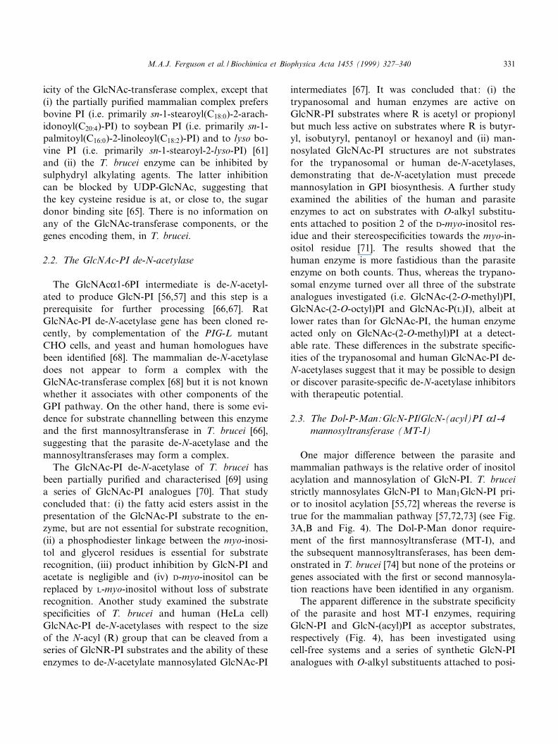
icity of the GlcNAc-transferase complex, except that(i) the partially puri¢ed mammalian complex prefersbovine PI (i.e. primarily sn-1-stearoyl(C18:0)-2-arach-idonoyl(C20:4)-PI) to soybean PI (i.e. primarily sn-1-palmitoyl(C16:0)-2-linoleoyl(C18:2)-PI) and to lyso bo-vine PI (i.e. primarily sn-1-stearoyl-2-lyso-PI) [61]and (ii) the T. brucei enzyme can be inhibited bysulphydryl alkylating agents. The latter inhibitioncan be blocked by UDP-GlcNAc, suggesting thatthe key cysteine residue is at, or close to, the sugardonor binding site [65]. There is no information onany of the GlcNAc-transferase components, or thegenes encoding them, in T. brucei.
2.2. The GlcNAc-PI de-N-acetylase
The GlcNAcK1-6PI intermediate is de-N-acetyl-ated to produce GlcN-PI [56,57] and this step is aprerequisite for further processing [66,67]. RatGlcNAc-PI de-N-acetylase gene has been cloned re-cently, by complementation of the PIG-L mutantCHO cells, and yeast and human homologues havebeen identi¢ed [68]. The mammalian de-N-acetylasedoes not appear to form a complex with theGlcNAc-transferase complex [68] but it is not knownwhether it associates with other components of theGPI pathway. On the other hand, there is some evi-dence for substrate channelling between this enzymeand the ¢rst mannosyltransferase in T. brucei [66],suggesting that the parasite de-N-acetylase and themannosyltransferases may form a complex.
The GlcNAc-PI de-N-acetylase of T. brucei hasbeen partially puri¢ed and characterised [69] usinga series of GlcNAc-PI analogues [70]. That studyconcluded that: (i) the fatty acid esters assist in thepresentation of the GlcNAc-PI substrate to the en-zyme, but are not essential for substrate recognition,(ii) a phosphodiester linkage between the myo-inosi-tol and glycerol residues is essential for substraterecognition, (iii) product inhibition by GlcN-PI andacetate is negligible and (iv) D-myo-inositol can bereplaced by L-myo-inositol without loss of substraterecognition. Another study examined the substratespeci¢cities of T. brucei and human (HeLa cell)GlcNAc-PI de-N-acetylases with respect to the sizeof the N-acyl (R) group that can be cleaved from aseries of GlcNR-PI substrates and the ability of theseenzymes to de-N-acetylate mannosylated GlcNAc-PI
intermediates [67]. It was concluded that: (i) thetrypanosomal and human enzymes are active onGlcNR-PI substrates where R is acetyl or propionylbut much less active on substrates where R is butyr-yl, isobutyryl, pentanoyl or hexanoyl and (ii) man-nosylated GlcNAc-PI structures are not substratesfor the trypanosomal or human de-N-acetylases,demonstrating that de-N-acetylation must precedemannosylation in GPI biosynthesis. A further studyexamined the abilities of the human and parasiteenzymes to act on substrates with O-alkyl substitu-ents attached to position 2 of the D-myo-inositol res-idue and their stereospeci¢cities towards the myo-in-ositol residue [71]. The results showed that thehuman enzyme is more fastidious than the parasiteenzyme on both counts. Thus, whereas the trypano-somal enzyme turned over all three of the substrateanalogues investigated (i.e. GlcNAc-(2-O-methyl)PI,GlcNAc-(2-O-octyl)PI and GlcNAc-P(L)I), albeit atlower rates than for GlcNAc-PI, the human enzymeacted only on GlcNAc-(2-O-methyl)PI at a detect-able rate. These di¡erences in the substrate speci¢c-ities of the trypanosomal and human GlcNAc-PI de-N-acetylases suggest that it may be possible to designor discover parasite-speci¢c de-N-acetylase inhibitorswith therapeutic potential.
2.3. The Dol-P-Man:GlcN-PI/GlcN-(acyl)PI K1-4mannosyltransferase (MT-I)
One major di¡erence between the parasite andmammalian pathways is the relative order of inositolacylation and mannosylation of GlcN-PI. T. bruceistrictly mannosylates GlcN-PI to Man1GlcN-PI pri-or to inositol acylation [55,72] whereas the reverse istrue for the mammalian pathway [57,72,73] (see Fig.3A,B and Fig. 4). The Dol-P-Man donor require-ment of the ¢rst mannosyltransferase (MT-I), andthe subsequent mannosyltransferases, has been dem-onstrated in T. brucei [74] but none of the proteins orgenes associated with the ¢rst or second mannosyla-tion reactions have been identi¢ed in any organism.
The apparent di¡erence in the substrate speci¢cityof the parasite and host MT-I enzymes, requiringGlcN-PI and GlcN-(acyl)PI as acceptor substrates,respectively (Fig. 4), has been investigated usingcell-free systems and a series of synthetic GlcN-PIanalogues with O-alkyl substituents attached to posi-
BBADIS 61863 15-9-99
M.A.J. Ferguson et al. / Biochimica et Biophysica Acta 1455 (1999) 327^340 331
BBADIS 61863 15-9-99
M.A.J. Ferguson et al. / Biochimica et Biophysica Acta 1455 (1999) 327^340332
tion 2 of the D-myo-inositol residue. The ¢rst ofthese, GlcN-(2-O-methyl)PI [75], was shown to be agood substrate for T. brucei MT-I but neither a sub-strate nor an inhibitor of HeLa cell MT-I [72]. Theidenti¢cation of a selective substrate for T. bruceiMT-I suggested that parasite-speci¢c inhibitorsshould also be attainable. This prediction was con-¢rmed recently when GlcN-(2-O-octyl)PI and GlcN-(2-O-hexadecyl)PI were tested and found to be para-site-speci¢c GPI pathway inhibitors with di¡erentmodes of action: GlcN-(2-O-hexadecyl)PI inhibitsT. brucei MT-I while GlcN-(2-O-octyl)PI inhibitsthe T. brucei inositol acyltransferase [76] (Fig. 4).Although these compounds are unlikely to work invivo, since they will probably not pass through theplasma membrane, the existence of parasite-speci¢cinhibitors that are active in vitro is encouraging andfeatures of these compounds are being combinedwith other information on the ¢ne speci¢city of T.brucei MT-I [66] to design smaller and less polarinhibitors.
2.4. Inositol acyltransferases and inositol deacylases
The role of inositol acylation (i.e., the esteri¢cationof the hydroxyl group at the 2-position of the D-myo-inositol residue with a fatty acid, most often palmiticacid [41,77^79] (Fig. 2)) in GPI biosynthesis is largelyunknown. It is not an essential step in all organismssince, for example, the Leishmania do not appear toperform this reaction [80,81]. Furthermore, in manycases (Fig. 2) the ¢nal protein-linked GPI anchor isnot inositol acylated. Nevertheless, in mammaliancells and in yeast, most of the GPI intermediatesare inositol acylated [50^54] and in many mammali-an cells, and in yeast, the fatty acid is removed fromthe inositol residue immediately after transfer of themature GPI precursor to protein in the ER [82,83].
The timing, nature and role of inositol acylationand inositol deacylation in T. brucei appears to be
quite unique. Thus, as noted above, whereas inositolacylation of GlcN-PI precedes mannosylation inmammalian cells [57,72,73] and in yeast [52] it onlyoccurs after the action of MT-I in T. brucei [55,72].Furthermore, (i) inositol acylation is CoA and/oracyl-CoA dependent in mammalian cells [73,84] andyeast [52], but independent of these cofactors in T.brucei [49,55]; (ii) inositol acylation is inhibited byphenylmethylsulphonyl £uoride (PMSF) in T. brucei,but not in mammalian cells [85]; (iii) the fatty acidstransferred to inositol are more heterogeneous in T.brucei than they are in mammalian cells [86] and (iv)most of the GPI intermediates in bloodstream formsof T. brucei are in a dynamic equilibrium betweeninositol acylated and inositol deacylated forms (Fig.3A) via the actions of a PMSF-sensitive inositol acyl-transferase [85] and a diisopropyl£uorophosphate(DFP)-sensitive inositol deacylase [55]. The abilityto manipulate the acylation status of GPI intermedi-ates in T. brucei with PMSF and DFP has shownthat, in that organism, inositol acylation is necessaryfor e¤cient addition of the ethanolamine phosphatebridge and that subsequent inositol deacylation isessential for the fatty acid remodelling reactions[55,85] (see Fig. 3A). The former point was con-¢rmed in the study with GlcN-(2-O-methyl)PI whichwas e¤ciently converted to Man3GlcN-(2-O-methyl)-PI by the cell-free system but which failed to receivethe ethanolamine phosphate bridge [72]. Given theimportance of GPI fatty acid remodelling for T. bru-cei (see below) and the essential nature of the etha-nolamine phosphate bridge for GPI addition to pro-tein, both the T. brucei inositol acyltransferase andthe inositol deacylase are potential therapeutic tar-gets.
2.5. The Dol-P-Man:Man1GlcN-( þ acyl)PI K1-6mannosyltransferase (MT-II)
Essentially nothing is known about the proteins or
6
Fig. 3. GPI biosynthesis in T. brucei bloodstream forms and mammalian cells. The general schemes of GPI biosynthesis in T. brucei(A) and HeLa cells (B) are adapted from [55], and references therein, and [50,51], and references therein. The di¡erences in the orderof mannosylation by the ¢rst K-mannosyltransferase (MT-I) and inositol acylation by inositol acyltransferase (Acyl-T) and the di¡er-ence in the timing of inositol deacylation by inositol deacylase (Ino de-Ac) are highlighted. Other signi¢cant di¡erences include the ad-dition of an extra ethanolamine phosphate group to the ¢rst Man residue in the mammalian pathway and the nature of the side chaincarbohydrate modi¢cations that occur after GPI addition to protein. The topology [58,59] and known complexation, see text, of thecomponents of the mammalian GPI biosynthetic pathway are summarised in C.
BBADIS 61863 15-9-99
M.A.J. Ferguson et al. / Biochimica et Biophysica Acta 1455 (1999) 327^340 333

BBADIS 61863 15-9-99
M.A.J. Ferguson et al. / Biochimica et Biophysica Acta 1455 (1999) 327^340334
genes involved in this step of the pathway in anyorganism.
2.6. The Dol-P-Man:Man2GlcN-( þ acyl)PI K1-2mannosyltransferase (MT-III)
A gene (PIG-B) encoding a protein with homologyto a yeast K-mannosyltransferase was cloned [87] bycomplementation of a murine thymoma mutant de-¢cient in adding the third KMan residue to GPI in-termediates [51]. The PIG-B gene is believed to en-code MT-III and a yeast homologue (GPI10) hasbeen identi¢ed and shown to be essential for GPIbiosynthesis in yeast [53,54]. Expression of humanPIG-B cDNA rescued a GPI103 conditional mutantunder non-permissive conditions and restored GPIbiosynthesis, showing that the two genes are func-tional homologues [54]. BLAST searches have alsorevealed a T. brucei PIG-B homologue but thereare no functional data on this gene. The PIG-Bgene product is predicted to span the ER membraneseven or nine times, which is a similar topology toother ER resident glycosyltransferases [88] and di¡er-ent from the type-2 membrane protein structure typ-ical of Golgi glycosyltransferases.
MT-III activity has been detected in the T. bruceicell-free system using the simple glycoside ManK1-6ManK1-O-(CH2)7CH3 as an exogenous acceptor[89] and the mechanism of the inhibition of GPIbiosynthesis by mannosamine [90,91] has beenstudied in that organism [92]. Essentially, mannos-amine is incorporated in GDP-Man and Dol-P-Man and then into GPI intermediates to produceManNH2ManGlcN-PI. The latter has an aminogroup in place of the hydroxyl acceptor site forMT-III, and therefore cannot act as an acceptor,and appears to inhibit the action of MT-III on en-dogenous Man2GlcN-PI intermediates [92]. Unfortu-nately, the e¡ects of mannosamine are not speci¢c totrypanosomes and, because trypanosomes synthesise
an excess of GPI precursors [92,93], the addition ofGPI anchors to VSG is not prevented by mannos-amine.
2.7. Ethanolamine phosphotransferases
All GPI anchors utilise an ethanolamine phos-phate (EtNP) bridge between the third Man residueand the COOH-terminal amino acid of the GPI-anchored protein (except for a small proportion ofTrypanosoma cruzi anchors that utilise the EtNPanalogue 2-aminoethylphosphonate [20]). The originof this EtNP group has been shown to be the phos-pholipid phosphatidylethanolamine (PE) in yeast [94]and in T. brucei [95]. A mammalian gene (PIG-F)required for the addition of the EtNP bridge hasbeen cloned by complementation but it is not knownwhether this encodes the transferase itself or an es-sential accessory element [96].
T. brucei, and many other protozoan organismssuch as Leishmania and Plasmodium falciparum, donot express additional EtNP groups on the ¢rst andsecond Man residues of the conserved GPI core. Incontrast, many other organisms express EtNP on the¢rst Man residue and on some proportion of thesecond Man residue (Fig. 2). It has recently cometo light that these additional EtNP groups are alsoadded during GPI biosynthesis in yeast [53,54]. Thishas stimulated a reinterpretation [54] of the activityof the terpenoid lactone compound YW3548, a po-tent inhibitor of GPI biosynthesis in mammaliancells and yeast but not in P. falciparum or T. brucei[97]. Thus, it is possible that the accumulation ofMan2GlcN-(acyl)PI in yeast treated with YW3548is due to the inhibition of EtNP addition to the ¢rstMan residue rather than direct inhibition of MT-III.This model assumes that yeast and mammalian MT-III require the presence of the EtNP group on the¢rst Man residue for substrate recognition [54]whereas T. brucei MT-III does not; a di¡erence
6
Fig. 4. Di¡erences in the substrates and inhibitors of T. brucei and human GPI de-N-acetylases, mannosyltransferases and inositolacyltransferases. The alternative routes from GlcNAc-PI to Man1GlcN-(acyl)PI taken by the T. brucei and mammalian (e.g. humanHeLa cell) and yeast GPI biosynthetic pathways are indicated at the top. The abilities of synthetic GlcN-PI, and analogues thereof, toact as substrates (S) or inhibitors (I) of T. brucei and HeLa cell de-N-acetylase, MT-I and inositol acyltransferase are shown at thebottom. Compounds that are neither substrates nor inhibitors are indicated by 3. *The de-N-acetylase results refer to the N-acetyl de-rivatives of the compounds shown.
BBADIS 61863 15-9-99
M.A.J. Ferguson et al. / Biochimica et Biophysica Acta 1455 (1999) 327^340 335
that may be exploitable for the inhibition of trypa-nosome MT-III. In any case, the selective inhibitionof GPI biosynthesis in yeast and mammalian cells byYW3548 [97] is another indication of fundamentaldi¡erences between parasite and mammalian/yeastGPI biosynthesis.
2.8. Other GPI glycosyltransferases
The presence of additional Man residues in GPIanchors is quite common (Fig. 2). In yeast, a fourthMan residue is added to the GPI precursor beforetransfer to protein [82] and the ¢fth Man residue(that can be either K1-2 or K1-3 linked) is added inthe Golgi apparatus [98]. The origin of the fourthMan residue in mammalian cells is less clear and itmay be added before or after GPI transfer to theprotein. The signi¢cance of the additional Man res-idues is yet to be reported. Additional carbohydrateside chains are quite common in mammalian cells,e.g. the addition of GalNAc and/or GalL1-3GalNAcin L1-4 linkage to the ¢rst Man residue (Fig. 2).There is no information on the glycosyltransferasesresponsible for these modi¢cations.
The GPI anchors on T. brucei VSG can be modi-¢ed with several Gal residues (Figs. 1 and 2). Theproteins and genes responsible for these modi¢ca-tions have not been identi¢ed but the activities oftwo of them, the UDP-Gal:Man3GlcN-PI K1-3 ga-lactosyltransferase [99,100] and the UDP-Gal:Gal1Man3GlcN-PI K1-2 galactosyltransferase [101],can be detected using the exogenous glycoside sub-strates ManK1-2ManK1-S-(CH2)7CH3 and ManK1-2(GalK1-3)ManK1-O-(CH2)7CH3, respectively. TheK- and L-galactosyltransferases that add Gal residuesdirectly to the trimannosyl core appear to be locatedin the ER [11,102] while the others are most likely inthe Golgi apparatus [11,103].
The VSGs can be classi¢ed into four classes basedon COOH-terminal sequence homology [104]. MostVSG variants belong to class-1 and class-2 and onlyone class-4 variant and two class-3 variants areknown. The mean Gal content of the GPI glycansof class-1 VSGs is 3.1 residues per mol GPI [10]whereas this ¢gure is about 4.9 residues per molfor the class-2 VSG [11] and the class-3 VSGs donot contain any Gal [105]. These di¡erences in GPIanchor galactosylation appear to re£ect steric con-
straints imposed by the VSG COOH-terminal struc-ture on galactosyltransferase activity. This conclu-sion is consistent with the original suggestion [106]that GPI anchor galactosylation, that is unique toAfrican trypanosomes, is a mechanism for ¢llingspace close to the plasma membrane beneath andaround the VSG COOH-terminus. This processingmay be important in maintaining a functional pro-tective barrier on the surface of the parasite regard-less of which VSG variant is being expressed. Sincethe GPI-modifying galactosyltransferases are uniqueto the African trypanosomes, and since the repertoireof non-galactosylated class-3 VSG variants is sosmall, these enzymes could be considered potentialtherapeutic targets.
2.9. GPI lipid remodelling
Lipid exchange occurs in yeast where diacylglycer-ol is exchanged for ceramide in the ER and wherethis ceramide can be exchanged for another ceramidein the Golgi [82,107,108]. Furthermore, the presenceof exclusively distearoyl-PI in some mammalian GPIanchors (e.g., porcine membrane dipeptidase and hu-man CD52-I [38,41]) suggests that lipid remodellingmay also occur in some mammalian tissues. Thetrypanosome VSG GPI anchors contain exclusivelymyristic (C14:0) acid in the form of dimyristoylglycer-ol [10] and lipid remodelling of GPI anchors was ¢rstdescribed in this organism [109]. The GPI intermedi-ates in T. brucei are based on sn-1-stearoyl-2-acyl-PI(where the acyl group at the sn-2 position is a mix-ture [110]) up until the fatty acid remodelling stepswhen the fatty acids are sequentially removed andreplaced by myristate (Fig. 3A). The trypanosomealso performs proofreading myristate exchange reac-tions on the GPI precursor [111] and on the VSG-linked mature GPI anchor [112] to ensure the exclu-sivity of dimyristoylglycerol in the VSG anchor. Thistype of myristate-speci¢c fatty acid remodelling hasbeen reported in Leishmania GPIs, where longerchain fatty acids in the sn-2 position of sn-1-alkyl-2-acyl-PI are replaced by myristate [81], but not inmammalian cells. The need for myristate in the VSGanchor remains obscure but myristate analogues thatbecome incorporated into the VSG anchor are toxicto trypanosomes [113,114]. Thus, the enzymes offatty acid remodelling/myristate exchange and the
BBADIS 61863 15-9-99
M.A.J. Ferguson et al. / Biochimica et Biophysica Acta 1455 (1999) 327^340336
other trypanosome systems required for scavengingmyristate [115] are possible targets for anti-trypano-some chemotherapy.
2.10. The GPI-transamidase complex
The transfer of the mature GPI precursor to pro-tein involves a transamidation reaction whereby aCOOH-terminal GPI addition signal peptide (typi-cally 17^35 amino acids in length) is directly replacedby the formation of an amide linkage to the aminogroup of the ethanolamine phosphate bridge[116,117]. In yeast the transamidase is made of atleast two components encoded by the GAA1 andGPI8 genes [118,119]. The latter gene, which has ho-mology with some plant endopeptidases, is believedto encode the catalytic subunit while data presentedby Kinoshita and colleagues (unpublished work) sug-gest that GAA1 encodes a subunit that is involved inrecognising the site of GPI addition in the proteinprecursor. Species-speci¢c di¡erences in GPI-trans-amidase speci¢city have been noted from site-di-rected mutagenesis studies at and around the trans-amidation (g) site [116,120]. For example, Cys is apoor g amino acid in yeast but an acceptable residuein mammalian cells. The g+1 and g+2 residues (i.e.,the amino terminal and subterminal residues of thecleaved GPI signal peptide) are also important deter-minants for GPI-transamidation and these, like the gamino acid, are generally relatively small residues.Interestingly, the g/g+1/g+2 triplet of T. bruceiclass-1 VSGs does not function in COS cells, suggest-ing that there may be exploitable di¡erences betweenmammalian and parasite GPI-transamidases [121].Proteins that fail to acquire a GPI anchor are re-tarded in the ER [122] and degraded by a protea-some-dependent pathway [123,124].
3. Perspectives
Considerable progress has been made on the bio-chemistry and molecular biology of GPI biosynthe-sis. Despite the overall conservation of GPI corestructure, signi¢cant di¡erences in the substrate spe-ci¢cities of trypanosome and mammalian GPI bio-synthetic enzymes have been demonstrated. Thus, theGlcNAc-PI de-N-acetylase, the MT-I and MT-III
mannosyltransferases, the inositol acyltransferase,the inositol deacylase, the trypanosome-speci¢c fattyacid remodelling/exchange enzymes, the GPI-trans-amidase and the trypanosome-speci¢c side chain ga-lactosyltransferases are all potential therapeutic tar-gets. The fact that parasite-speci¢c inhibitors arebeginning to emerge is encouraging but the cloningof T. brucei (and other pathogen) GPI pathway genesand structural information on GPI pathway compo-nents remain important goals for the future. Finally,many other protozoan parasites, such as T. cruzi,Leishmania, Plasmodium and Toxoplasma, are richin GPI-anchored glycoproteins and/or GPI-relatedglycoconjugates. Therefore, GPI pathway inhibitorsdeveloped with African trypanosomes may also havetherapeutic potential in a number of other diseases.
Acknowledgements
The authors' work is supported by a ProgrammeGrant (054491) from The Wellcome Trust.
References
[1] K. Vickerman, A.G. Luckins, Nature 224 (1969) 1125^1126.[2] G.A. Cross, Parasitology 71 (1975) 393^417.[3] M.L. Blum, J.A. Down, A.M. Gurnett, M. Carrington, M.J.
Turner, D.C. Wiley, Nature 362 (1993) 603^609.[4] G.A. Cross, Bioessays 18 (1996) 283^291.[5] S.E. Zamze, E.W. Wooten, D.A. Ashford, M.A. Ferguson,
R.A. Dwek, T.W. Rademacher, Eur. J. Biochem. 187 (1990)657^663.
[6] S.E. Zamze, D.A. Ashford, E.W. Wooten, T.W. Rademach-er, R.A. Dwek, J. Biol. Chem. 266 (1991) 20244^20261.
[7] A. Mehlert, N. Zitzmann, J.M. Richardson, A. Treumann,M.A. Ferguson, Mol. Biochem. Parasitol. 91 (1998) 145^152.
[8] A.F. Williams, Immunol. Ser. 45 (1989) 49^69.[9] M. Tarutani, S. Itami, M. Okabe, M. Ikawa, T. Tezuka, K.
Yoshikawa, T. Kinoshita, J. Takeda, Proc. Natl. Acad. Sci.USA 94 (1997) 7400^7405.
[10] M.A. Ferguson, S.W. Homans, R.A. Dwek, T.W. Rade-macher, Science 239 (1988) 753^759.
[11] A. Mehlert, J.M. Richardson, M.A. Ferguson, J. Mol. Biol.277 (1998) 379^392.
[12] P. Gerold, B. Striepen, B. Reitter, H. Geyer, R. Geyer, E.Reinwald, H.J. Risse, R.T. Schwarz, J. Mol. Biol. 261 (1996)181^194.
[13] A. Treumann, N. Zitzmann, A. Hulsmeier, A.R. Prescott, A.
BBADIS 61863 15-9-99
M.A.J. Ferguson et al. / Biochimica et Biophysica Acta 1455 (1999) 327^340 337

Almond, J. Sheehan, M.A. Ferguson, J. Mol. Biol. 269(1997) 529^547.
[14] P. Butikofer, M. Boschung, Mol. Biochem. Parasitol. 74(1995) 65^75.
[15] M.L. Guther, M.L. de Almeida, N. Yoshida, M.A. Fergu-son, J. Biol. Chem. 267 (1992) 6820^6828.
[16] N. Heise, M.L. de Almeida, M.A. Ferguson, Mol. Biochem.Parasitol. 70 (1995) 71^84.
[17] L.E. Bertello, N.W. Andrews, R.M. de Lederkremer, Mol.Biochem. Parasitol. 79 (1996) 143^151.
[18] A.S. Couto, R.M. De Lederkremer, W. Colli, M.J. Alves,Eur. J. Biochem. 217 (1993) 597^602.
[19] G. Abuin, A.S. Couto, R.M. de Lederkremer, O.L. Casal, C.Galli, W. Colli, M.J. Alves, Exp. Parasitol. 82 (1996) 290^297.
[20] J.O. Previato, C. Jones, M.T. Xavier, R. Wait, L.R. Tra-vassos, A.J. Parodi, L. Mendonca-Previato, J. Biol. Chem.270 (1995) 7241^7250.
[21] A.A. Serrano, S. Schenkman, N. Yoshida, A. Mehlert, J.M.Richardson, M.A. Ferguson, J. Biol. Chem. 270 (1995)27244^27253.
[22] R. Agusti, A.S. Couto, O. Campetella, A.C. Frasch, R.M.Lederkremer, Mol. Biochem. Parasitol. 97 (1998) 123^131.
[23] P. Schneider, M.A. Ferguson, M.J. McConville, A. Mehlert,S.W. Homans, C. Bordier, J. Biol. Chem. 265 (1990) 16955^16964.
[24] M.J. McConville, T.A. Collidge, M.A. Ferguson, P.Schneider, J. Biol. Chem. 268 (1993) 15595^15604.
[25] S. Tomavo, J.F. Dubremetz, R.T. Schwarz, Biol. Cell 78(1993) 155^162.
[26] P. Gerold, L. Scho¢eld, M.J. Blackman, A.A. Holder, R.T.Schwarz, Mol. Biochem. Parasitol. 75 (1996) 131^143.
[27] N. Azzouz, B. Striepen, P. Gerold, Y. Capdeville, R.T.Schwarz, EMBO J. 14 (1995) 4422^4433.
[28] C. Fankhauser, S.W. Homans, J.E. Thomas-Oates, M.J.McConville, C. Desponds, A. Conzelmann, M.A. Ferguson,J. Biol. Chem. 268 (1993) 26365^26374.
[29] P.A. Haynes, A.A. Gooley, M.A. Ferguson, J.W. Redmond,K.L. Williams, Eur. J. Biochem. 216 (1993) 729^737.
[30] P. Butikofer, F.A. Kuypers, C. Shackleton, U. Brodbeck, S.Stieger, J. Biol. Chem. 265 (1990) 18983^18987.
[31] A. Mehlert, L. Varon, I. Silman, S.W. Homans, M.A. Fer-guson, Biochem. J. 296 (1993) 473^479.
[32] S.W. Homans, M.A. Ferguson, R.A. Dwek, T.W. Rade-macher, R. Anand, A.F. Williams, Nature 333 (1988) 269^272.
[33] N. Stahl, M.A. Baldwin, D.B. Teplow, L. Hood, B.W. Gib-son, A.L. Burlingame, S.B. Prusiner, Biochemistry 32 (1993)1991^2002.
[34] R. Mukasa, M. Umeda, T. Endo, A. Kobata, K. Inoue,Arch. Biochem. Biophys. 318 (1995) 182^190.
[35] R. Taguchi, N. Hamakawa, M. Harada-Nishida, T. Fukui,K. Nojima, H. Ikezawa, Biochemistry 33 (1994) 1017^1022.
[36] R. Haas, B.C. Jackson, B. Reinhold, J.D. Foster, T.L.Rosenberry, Biochem. J. 314 (1996) 817^825.
[37] J. Armesto, E. Hannappel, K. Leopold, W. Fischer, R. Bu-blitz, L. Langer, G.A. Cumme, A. Horn, Eur. J. Biochem.238 (1996) 259^269.
[38] I.A. Brewis, M.A. Ferguson, A. Mehlert, A.J. Turner, N.M.Hooper, J. Biol. Chem. 270 (1995) 22946^22956.
[39] M.A. Deeg, D.R. Humphrey, S.H. Yang, T.R. Ferguson,V.N. Reinhold, T.L. Rosenberry, J. Biol. Chem. 267 (1992)18573^18580.
[40] C.A. Redman, J.E. Thomas-Oates, S. Ogata, Y. Ikehara,M.A. Ferguson, Biochem. J. 302 (1994) 861^865.
[41] A. Treumann, M.R. Lifely, P. Schneider, M.A. Ferguson,J. Biol. Chem. 270 (1995) 6088^6099.
[42] Y. Nakano, K. Noda, T. Endo, A. Kobata, M. Tomita,Arch. Biochem. Biophys. 311 (1994) 117^126.
[43] Y. Sugita, Y. Nakano, E. Oda, K. Noda, T. Tobe, N.H.Miura, M. Tomita, J. Biochem. 114 (1993) 473^477.
[44] S. Meri, T. Lehto, C.W. Sutton, J. Tyynela, M. Baumann,Biochem. J. 316 (1996) 923^935.
[45] P.M. Rudd, B.P. Morgan, M.R. Wormald, D.J. Harvey,C.W. van den Berg, S.J. Davis, M.A. Ferguson, R.A.Dwek, J. Biol. Chem. 272 (1997) 7229^7244.
[46] J.L. Krakow, T.L. Doering, W.J. Masterson, G.W. Hart,P.T. Englund, Mol. Biochem. Parasitol. 36 (1989) 263^270.
[47] S. Mayor, A.K. Menon, G.A. Cross, M.A. Ferguson, R.A.Dwek, T.W. Rademacher, J. Biol. Chem. 265 (1990) 6164^6173.
[48] S. Mayor, A.K. Menon, G.A. Cross, J. Biol. Chem. 265(1990) 6174^6181.
[49] W.J. Masterson, T.L. Doering, G.W. Hart, P.T. Englund,Cell 56 (1989) 793^800.
[50] S. Hirose, G.M. Prince, D. Sevlever, L. Ravi, T.L. Rose-nberry, E. Ueda, M.E. Medof, J. Biol. Chem. 267 (1992)16968^16974.
[51] A. Puoti, A. Conzelmann, J. Biol. Chem. 268 (1993) 7215^7224.
[52] L.C. Costello, P. Orlean, J. Biol. Chem. 267 (1992) 8599^8603.
[53] E. Canivenc-Gansel, I. Imhof, F. Reggiori, P. Burda, A.Conzelmann, A. Benachour, Glycobiology 8 (1998) 761^770.
[54] C. Sutterlin, M.V. Escribano, P. Gerold, Y. Maeda, M.J.Mazon, T. Kinoshita, R.T. Schwarz, H. Riezman, Biochem.J. 332 (1998) 153^159.
[55] M.L. Guther, M.A. Ferguson, EMBO J. 14 (1995) 3080^3093.
[56] T.L. Doering, W.J. Masterson, P.T. Englund, G.W. Hart,J. Biol. Chem. 264 (1989) 11168^11173.
[57] S. Hirose, L. Ravi, S.V. Hazra, M.E. Medof, Proc. Natl.Acad. Sci. USA 88 (1991) 3762^3766.
[58] J. Vidugiriene, A.K. Menon, J. Cell Biol. 121 (1993) 987^996.
[59] J. Vidugiriene, A.K. Menon, J. Cell Biol. 127 (1994) 333^341.
[60] R. Watanabe, T. Kinoshita, R. Masaki, A. Yamamoto, J.Takeda, N. Inoue, J. Biol. Chem. 271 (1996) 26868^26875.
[61] R. Watanabe, N. Inoue, B. Westfall, C.H. Taron, P. Orlean,J. Takeda, T. Kinoshita, EMBO J. 17 (1998) 877^885.
BBADIS 61863 15-9-99
M.A.J. Ferguson et al. / Biochimica et Biophysica Acta 1455 (1999) 327^340338
[62] S.D. Leidich, Z. Kostova, R.R. Latek, L.C. Costello, D.A.Drapp, W. Gray, J.S. Fassler, P. Orlean, J. Biol. Chem. 270(1995) 13029^13035.
[63] S.D. Leidich, P. Orlean, J. Biol. Chem. 271 (1996) 27829^27837.
[64] A. Tiede, J. Schubert, C. Nischan, I. Jensen, B. Westfall,C.H. Taron, P. Orlean, R.E. Schmidt, Biochem. J. 334(1998) 609^616.
[65] K.G. Milne, M.A. Ferguson, W.J. Masterson, Eur. J. Bio-chem. 208 (1992) 309^314.
[66] T.K. Smith, S. Cottaz, J.S. Brimacombe, M.A.J. Ferguson,J. Biol. Chem. 271 (1996) 6476^6482.
[67] D.K. Sharma, T.K. Smith, A. Crossman, J.S. Brimacombe,M.A.J. Ferguson, Biochem. J. 328 (1997) 171^177.
[68] N. Nakamura, N. Inoue, R. Watanabe, M. Takahashi, J.Takeda, V.L. Stevens, T. Kinoshita, J. Biol. Chem. 272(1997) 15834^15840.
[69] K.G. Milne, R.A. Field, W.J. Masterson, S. Cottaz, J.S.Brimacombe, M.A.J. Ferguson, J. Biol. Chem. 269 (1994)16403^16408.
[70] S. Cottaz, J.S. Brimacombe, M.A.J. Ferguson, J. Chem. Soc.Perkin Trans. 1 (1995) 1673^1678.
[71] D.K. Sharma, T.K. Smith, C.T. Weller, A. Crossman, J.S.Brimacombe, M.A. Ferguson, Glycobiology 9 (1999) 415^422.
[72] T.K. Smith, D.K. Sharma, A. Crossman, A. Dix, J.S. Bri-macombe, M.A. Ferguson, EMBO J. 16 (1997) 6667^6675.
[73] W.T. Doerrler, J. Ye, J.R. Falck, M.A. Lehrman, J. Biol.Chem. 271 (1996) 27031^27038.
[74] A.K. Menon, S. Mayor, R.T. Schwarz, EMBO J. 9 (1990)4249^4258.
[75] A. Crossman, J.S. Brimacombe, M.A.J. Ferguson, J. Chem.Soc. Perkin Trans. 1 (1997) 2769^2774.
[76] T.K. Smith, D.K. Sharma, A. Crossman, J.S. Brimacombe,M.A.J. Ferguson, (1999) submitted.
[77] W.L. Roberts, B.H. Kim, T.L. Rosenberry, Proc. Natl.Acad. Sci. USA 84 (1987) 7817^7821.
[78] W.L. Roberts, S. Santikarn, V.N. Reinhold, T.L. Rose-nberry, J. Biol. Chem. 263 (1988) 18776^18784.
[79] D. Sevlever, D.R. Humphrey, T.L. Rosenberry, Eur. J. Bio-chem. 233 (1995) 384^394.
[80] T.K. Smith, F.C. Milne, D.K. Sharma, A. Crossman, J.S.Brimacombe, M.A. Ferguson, Biochem. J. 326 (1997) 393^400.
[81] J.E. Ralton, M.J. McConville, J. Biol. Chem. 273 (1998)4245^4257.
[82] G. Sipos, A. Puoti, A. Conzelmann, EMBO J. 13 (1994)2789^2796.
[83] R. Chen, E.I. Walter, G. Parker, J.P. Lapurga, J.L. Millan,Y. Ikehara, S. Udenfriend, M.E. Medof, Proc. Natl. Acad.Sci. USA 95 (1998) 9512^9517.
[84] V.L. Stevens, H. Zhang, J. Biol. Chem. 269 (1994) 31397^31403.
[85] M.L. Guther, W.J. Masterson, M.A. Ferguson, J. Biol.Chem. 269 (1994) 18694^18701.
[86] M.L. Guther, A. Treumann, M.A. Ferguson, Mol. Bio-chem. Parasitol. 77 (1996) 137^145.
[87] M. Takahashi, N. Inoue, K. Ohishi, Y. Maeda, N. Naka-mura, Y. Endo, T. Fujita, J. Takeda, T. Kinoshita, EMBOJ. 15 (1996) 4254^4261.
[88] N. Dan, M.A. Lehrman, J. Biol. Chem. 272 (1997) 14214^14219.
[89] J.R. Brown, M.L. Guther, R.A. Field, M.A. Ferguson,Glycobiology 7 (1997) 549^558.
[90] M.P. Lisanti, M.C. Field, I.W. Caras, A.K. Menon, E.Rodriguez-Boulan, EMBO J. 10 (1991) 1969^1977.
[91] Y.T. Pan, T. Kamitani, C. Bhuvaneswaran, Y. Hallaq,C.D. Warren, E.T. Yeh, A.D. Elbein, J. Biol. Chem. 267(1992) 21250^21255.
[92] J.E. Ralton, K.G. Milne, M.L. Guther, R.A. Field, M.A.Ferguson, J. Biol. Chem. 268 (1993) 24183^24189.
[93] W.J. Masterson, M.A. Ferguson, EMBO J. 10 (1991) 2041^2045.
[94] A.K. Menon, V.L. Stevens, J. Biol. Chem. 267 (1992)15277^15280.
[95] A.K. Menon, M. Eppinger, S. Mayor, R.T. Schwarz,EMBO J. 12 (1993) 1907^1914.
[96] N. Inoue, T. Kinoshita, T. Orii, J. Takeda, J. Biol. Chem.268 (1993) 6882^6885.
[97] C. Sutterlin, A. Horvath, P. Gerold, R.T. Schwarz, Y.Wang, M. Dreyfuss, H. Riezman, EMBO J. 16 (1997)6374^6383.
[98] G. Sipos, A. Puoti, A. Conzelmann, J. Biol. Chem. 270(1995) 19709^19715.
[99] S. Pingel, R.A. Field, M.L. Guther, M. Duszenko, M.A.Ferguson, Biochem. J. 309 (1995) 877^882.
[100] T. Ziegler, R. Dettmann, M. Duszenko, V. Kolb, Carbo-hydr. Res. 295 (1996) 7^23.
[101] J.R. Brown, T.K. Smith, M.A.J. Ferguson, R.A. Field, Bio-org. Med. Chem. Lett. 8 (1998) 2051^2054.
[102] S. Mayor, A.K. Menon, G.A. Cross, J. Biol. Chem. 267(1992) 754^761.
[103] J.D. Bangs, T.L. Doering, P.T. Englund, G.W. Hart,J. Biol. Chem. 263 (1988) 17697^17705.
[104] M. Carrington, N. Miller, M. Blum, I. Roditi, D. Wiley, M.Turner, J. Mol. Biol. 221 (1991) 823^835.
[105] M.L. Guther, M.A. Ferguson, Methods Mol. Biol. 14(1993) 99^117.
[106] S.W. Homans, C.J. Edge, M.A. Ferguson, R.A. Dwek,T.W. Rademacher, Biochemistry 28 (1989) 2881^2887.
[107] F. Reggiori, E. Canivenc-Gansel, A. Conzelmann, EMBOJ. 16 (1997) 3506^3518.
[108] F. Reggiori, A. Conzelmann, J. Biol. Chem. 273 (1998)30550^30559.
[109] W.J. Masterson, J. Raper, T.L. Doering, G.W. Hart, P.T.Englund, Cell 62 (1990) 73^80.
[110] T.L. Doering, M.S. Pessin, G.W. Hart, D.M. Raben, P.T.Englund, Biochem. J. 299 (1994) 741^746.
[111] L.U. Buxbaum, J. Raper, F.R. Opperdoes, P.T. Englund,J. Biol. Chem. 269 (1994) 30212^30220.
BBADIS 61863 15-9-99
M.A.J. Ferguson et al. / Biochimica et Biophysica Acta 1455 (1999) 327^340 339
[112] L.U. Buxbaum, K.G. Milne, K.A. Werbovetz, P.T. En-glund, Proc. Natl. Acad. Sci. USA 93 (1996) 1178^1183.
[113] T.L. Doering, T. Lu, K.A. Werbovetz, G.W. Gokel, G.W.Hart, J.I. Gordon, P.T. Englund, Proc. Natl. Acad. Sci.USA 91 (1994) 9735^9739.
[114] T.L. Doering, J. Raper, L.U. Buxbaum, S.P. Adams, J.I.Gordon, G.W. Hart, P.T. Englund, Science 252 (1991)1851^1854.
[115] K.A. Werbovetz, P.T. Englund, Mol. Biochem. Parasitol.85 (1997) 1^7.
[116] K. Kodukula, S.E. Maxwell, S. Udenfriend, Methods En-zymol. 250 (1995) 536^547.
[117] S. Ramalingam, S.E. Maxwell, M.E. Medof, R. Chen, L.D.Gerber, S. Udenfriend, Proc. Natl. Acad. Sci. USA 93(1996) 7528^7533.
[118] D. Hamburger, M. Egerton, H. Riezman, J. Cell Biol. 129(1995) 629^639.
[119] M. Benghezal, A. Benachour, S. Rusconi, M. Aebi, A.Conzelmann, EMBO J. 15 (1996) 6575^6583.
[120] C. Nuo¡er, P. Jeno, A. Conzelmann, H. Riezman, Mol.Cell. Biol. 11 (1991) 27^37.
[121] P. Moran, I.W. Caras, J. Cell Biol. 125 (1994) 333^343.[122] M.C. Field, P. Moran, W. Li, G.A. Keller, I.W. Caras,
J. Biol. Chem. 269 (1994) 10830^10837.[123] K. Oda, Y. Ikehara, S. Omura, Biochem. Biophys. Res.
Commun. 219 (1996) 800^805.[124] B. Wilbourn, D.N. Nesbeth, L.J. Wainwright, M.C. Field,
Biochem. J. 332 (1998) 111^118.
BBADIS 61863 15-9-99
M.A.J. Ferguson et al. / Biochimica et Biophysica Acta 1455 (1999) 327^340340