Lipopolysaccharide-Induced Loss of Cultured Rat Myenteric Neurons - Role of AMP-Activated Protein...
17
RESEARCH ARTICLE Lipopolysaccharide-Induced Loss of Cultured Rat Myenteric Neurons - Role of AMP-Activated Protein Kinase Ulrikke Voss*, Eva Ekblad Department of Experimental Medical Science, Lund University, Lund, Sweden * [email protected] Abstract Objective: Intestinal barrier function is vital for homeostasis. Conditions where the mucosal barrier is compromised lead to increased plasma content of lipopolysaccharide (LPS). LPS acts on Toll-like receptor 4 (TLR4) and initiates cellular inflammatory responses. TLR4 receptors have been identified on enteric neurons and LPS exposure causes neuronal loss, counteracted by vasoactive intestinal peptide (VIP), by unknown mechanisms. In addition AMP activated protein kinase (AMPK) stimulation causes loss of enteric neurons. This study investigated a possible role of AMPK activation in LPS-induced neuronal loss. Design: Primary cultures of myenteric neurons isolated from rat small intestine were used. Cultures were treated with LPS (0.2–20 mg/mL) with and without TAK1- inhibitor (5Z)-7-Oxozeaenol (10 26 M) or AMPK inhibitor compound C (10 25 M). AMPK-induced neuronal loss was verified treating cultures with three different AMPK activators, AICAR (10 24 23610 23 M), metformin (0.2–20 mg/mL) and A- 769662 (10 25 23610 24 M) with or without the presence of compound C (10 25 M). Upstream activation of AMPK-induced neuronal loss was tested by treating cultures with AICAR (10 23 M) in the presence of TAK1 inhibitor (5Z)-7-Oxozeaenol (10 26 M). Neuronal survival and relative numbers of neurons immunoreactive (IR) for VIP were evaluated using immunocytochemistry. Results: LPS caused a concentration dependent loss of neurons. All AMPK activators induced loss of myenteric neurons in a concentration dependent manner. LPS-, AICAR- and metformin-,but not A-769662-, induced neuronal losses were inhibited by presence of compound C. LPS, AICAR or metformin exposure increased the relative number of VIP-IR neurons; co-treatment with (5Z)-7- Oxozeaenol or compound C reversed the relative increase in VIP-IR neurons induced by LPS. (5Z)-7-Oxozeaenol, compound C or A-769662 did not per se change neuronal survival or relative numbers of VIP-IR neurons. OPEN ACCESS Citation: Voss U, Ekblad E (2014) Lipopolysaccharide-Induced Loss of Cultured Rat Myenteric Neurons - Role of AMP- Activated Protein Kinase. PLoS ONE 9(12): e114044. doi:10.1371/journal.pone.0114044 Editor: Yvette Tache, University of California, Los Angeles, United States of America Received: June 13, 2014 Accepted: November 3, 2014 Published: December 2, 2014 Copyright: ß 2014 Voss, Ekblad. This is an open- access article distributed under the terms of the Creative Commons Attribution License, which permits unrestricted use, distribution, and repro- duction in any medium, provided the original author and source are credited. Data Availability: The authors confirm that all data underlying the findings are fully available without restriction. All relevant data are within the paper and its Supporting Information files. Funding: This study was supported by the Pa ˚hlsson Foundation, Royal Physiographic Society and Faculty of Medicine, Lund University. The funders had no role in study design, data collection and analysis, decision to publish or preparation of the manuscript. Competing Interests: The authors have declared that no competing interest exist. PLOS ONE | DOI:10.1371/journal.pone.0114044 December 2, 2014 1 / 17
Transcript of Lipopolysaccharide-Induced Loss of Cultured Rat Myenteric Neurons - Role of AMP-Activated Protein...

RESEARCH ARTICLE
Lipopolysaccharide-Induced Loss ofCultured Rat Myenteric Neurons - Role ofAMP-Activated Protein KinaseUlrikke Voss*, Eva Ekblad
Department of Experimental Medical Science, Lund University, Lund, Sweden
Abstract
Objective: Intestinal barrier function is vital for homeostasis. Conditions where the
mucosal barrier is compromised lead to increased plasma content of
lipopolysaccharide (LPS). LPS acts on Toll-like receptor 4 (TLR4) and initiates
cellular inflammatory responses. TLR4 receptors have been identified on enteric
neurons and LPS exposure causes neuronal loss, counteracted by vasoactive
intestinal peptide (VIP), by unknown mechanisms. In addition AMP activated
protein kinase (AMPK) stimulation causes loss of enteric neurons. This study
investigated a possible role of AMPK activation in LPS-induced neuronal loss.
Design: Primary cultures of myenteric neurons isolated from rat small intestine
were used. Cultures were treated with LPS (0.2–20 mg/mL) with and without TAK1-
inhibitor (5Z)-7-Oxozeaenol (1026 M) or AMPK inhibitor compound C (1025 M).
AMPK-induced neuronal loss was verified treating cultures with three different
AMPK activators, AICAR (10242361023 M), metformin (0.2–20 mg/mL) and A-
769662 (10252361024 M) with or without the presence of compound C (1025 M).
Upstream activation of AMPK-induced neuronal loss was tested by treating cultures
with AICAR (1023 M) in the presence of TAK1 inhibitor (5Z)-7-Oxozeaenol
(1026 M). Neuronal survival and relative numbers of neurons immunoreactive (IR)
for VIP were evaluated using immunocytochemistry.
Results: LPS caused a concentration dependent loss of neurons. All AMPK
activators induced loss of myenteric neurons in a concentration dependent manner.
LPS-, AICAR- and metformin-,but not A-769662-, induced neuronal losses were
inhibited by presence of compound C. LPS, AICAR or metformin exposure
increased the relative number of VIP-IR neurons; co-treatment with (5Z)-7-
Oxozeaenol or compound C reversed the relative increase in VIP-IR neurons
induced by LPS. (5Z)-7-Oxozeaenol, compound C or A-769662 did not per se
change neuronal survival or relative numbers of VIP-IR neurons.
OPEN ACCESS
Citation: Voss U, EkbladE (2014) Lipopolysaccharide-Induced Loss ofCultured Rat Myenteric Neurons - Role of AMP-Activated Protein Kinase. PLoS ONE 9(12):e114044. doi:10.1371/journal.pone.0114044
Editor: Yvette Tache, University of California, LosAngeles, United States of America
Received: June 13, 2014
Accepted: November 3, 2014
Published: December 2, 2014
Copyright:� 2014 Voss, Ekblad. This is an open-access article distributed under the terms of theCreative Commons Attribution License, whichpermits unrestricted use, distribution, and repro-duction in any medium, provided the original authorand source are credited.
Data Availability: The authors confirm that all dataunderlying the findings are fully available withoutrestriction. All relevant data are within the paperand its Supporting Information files.
Funding: This study was supported by thePahlsson Foundation, Royal Physiographic Societyand Faculty of Medicine, Lund University. Thefunders had no role in study design, data collectionand analysis, decision to publish or preparation ofthe manuscript.
Competing Interests: The authors have declaredthat no competing interest exist.
PLOS ONE | DOI:10.1371/journal.pone.0114044 December 2, 2014 1 / 17

Conclusion: AMPK activation mimics LPS-induced loss of cultured myenteric
neurons and LPS-induced neuronal loss is counteracted by TAK1 and AMPK
inhibition. This suggests enteric neuroimmune interactions involving AMPK
regulation.
Introduction
The gastrointestinal (GI) tract comprises the body’s largest surface to the outside
environment. It is vital for nutrient uptake and contains the human microbiome,
consisting of more than 100 trillion microorganisms with different properties.
[1, 2] The importance of a functional barrier is highlighted in conditions such as
post-operative ileus, functional bowel disorders and obesity, where a compro-
mised barrier causes inflammatory responses of different severity. [3–5] Increased
permeability of the intestinal barrier commonly leads to increased plasma levels of
lipopolysaccharide (LPS), a major component of gram negative bacteria
membranes. LPS binds to toll like receptor 4 (TLR4) and initiates an
inflammatory response. [6] The transforming growth factor-b-activated kinase 1
(TAK1) is an important regulator of cellular responses initiated by environmental
stress. [7] As a downstream effector-molecule common to e.g. TLR4-, interleukin-
1- and tumor necrosis factor-receptor stimulation it is closely linked to the innate
immune response. [6, 7]
A key player in regulating digestive, in particular intestinal, functions is the
enteric nervous system (ENS). The ENS is optimally situated within and along the
digestive tract where it is pivotal in regulating intestinal motility, blood flow and
secretion. Dysregulation of ENS causes GI symptoms and jeopardizes intestinal
barrier integrity. LPS exposure in vitro has previously been shown to cause loss of
porcine and rat enteric neurons, probably through TLR4 activation since this
receptors is expressed on a subpopulation of enteric neurons. [8, 9] Furthermore,
vasoactive intestinal peptide (VIP) has been highlighted as being protective in the
response to LPS mediated TLR4 activation. It reduces LPS-induced inflammation
and enteric neuronal loss. [9, 10]
The evolutionarily well conserved AMP-activated protein kinase (AMPK) is
central in cellular metabolism and energy regulation. It acts as a metabolic switch,
conveying cellular and hormonal responses both short and long term. AMPK is a
heterotrimeric complex consisting of a catalytic a subunit and two regulatory b/c
subunits. It is activated by allosteric binding of AMP to domains on the c subunit
and phosphorylation of Thr172 on the a subunit. Depending on the combination
of subunit isoforms AMPK can display different signalling properties. [11, 12]
Studies investigating AMPK in inflammation have suggested diverse roles. In
microglia cultures and cell lines LPS has been shown to activate AMPK thereby
mediating cytokine release. [13–15] In macrophages, however, AMPK activation
inhibits LPS-induced activation, causing reduced inflammation. [16, 17] AMPK
Lipopolysaccharide and AMP-Activated Protein Kinase
PLOS ONE | DOI:10.1371/journal.pone.0114044 December 2, 2014 2 / 17

activation, using AICAR has even been shown to reduce the pro-inflammatory
cytokine response in TNBS-induced colitis and LPS-induced lung injury. [16, 17]
Current study using pharmacologic in vitro experimentation was designed to
investigate mechanisms underlying LPS-induced enteric neuronal loss.
Methods
Ethics statement
Procedures were approved by the regional Malmo/Lund committee for
experimental animal ethics, under the Swedish board of Agriculture, (diary
number M152-12). Animals were used in accordance with the European
Community Council Directive (2010/63/EU) and the Swedish Animal Welfare Act
(SFS 1988:534).
Animals and tissue preparations
Female Sprauge-Dawley rats (Charles River, DE), (n523, 130–180 g) were used.
Primary myenteric neuronal cultures from the small intestine were prepared as
described previously. [18] From each animal 6 culture plates of 8 wells (BD
Bioscience, SE) were prepared, animals were never pooled. The resulting cultures
containing both myenteric neurons and enteric glia were grown 4 days in medium
(neurobasal A, containing 10% fetal bovine serum, 0.5 mM L-glutamine, 50 U/
mL penicillin and 50 mg/mL streptomycin, all from Life Technologies, SE). Fresh
medium containing applicable experimental test agents was then added and
incubation for an additional 4 day period followed. Control wells were cultured in
parallel. Cells were fixed in Stefaninis fixative, rinsed in Tyrode solution, frozen,
thawed and subjected to immunocytochemistry. [18]
Pharmacological agents and experimental set-ups
Stock solutions of 5-amino-b-D-ribofuranosyl-imidazole-4-carboxamide
(AICAR, Sigma-Aldrich, SE), 6,7-Dihydro-4-hydroxy-3-(2’-hydroxy[1,1’-biphe-
nyl]-4-yl)-6-oxo-thieno[2,3-b]pyridine-5-carbonitrile (A-769662, Tocris, UK),
metformin hydrochloride (Cayman Chemicals, UK), 6-[4-(2-Piperidin-1-
ylethoxy)phenyl]-3-pyridin-4-ylpyrazolo[1,5-a]pyrimidine (compound C, Sigma-
Aldrich, SE), (5Z)-7-Oxozeaenol (Tocris Bioscience, UK) and Escherichia coli
serotype O111:B4 derived LPS (Sigma-Aldrich, SE) were prepared according to
manufactures recommendations, aliquoted and stored at 20 C.
Various sets of experiments were performed. Incubations were for 4 days.
Cultures were exposed to 1. LPS (0.2–20 mg/mL) with or without (5Z)-7-
Oxozeaenol (1026 M) or compound C (1025 M), 2. (5Z)-7-Oxozeaenol (1027–
1025 M), 3. compound C (361027–361025 M), 4. AICAR (1024–361023 M),
A-769662 (1025–361024 M) or metformin (1027–1023 M), 5. AICAR (1023 M),
A-769662 (1024 M) or metformin (1024 M) together with compound C
Lipopolysaccharide and AMP-Activated Protein Kinase
PLOS ONE | DOI:10.1371/journal.pone.0114044 December 2, 2014 3 / 17

(1025 M), 6. AICAR (1023 M), metformin (1024 M) or LPS (20 mg/mL) together
with (5Z)-7-Oxozeaenol (1026 M). Controls were always run in parallel.
Immunocytochemistry
For details on primary and secondary antibodies see table 1. All antibodies were
diluted in phosphate buffered saline containing 0.25% Triton X-100 and 0.25%
BSA. Double immunolabelling of cultures was performed by overnight incubation
in moist chambers at 4 C with a mixture of primary antibodies. [9] Secondary
antibodies were mixed and incubated 1 h at RT. Hoechst (Life Technologies, SE)
cell nuclei counter staining was performed according to manufacturer’s protocol.
Mounting was in PBS:glycerol 1:1 followed by fluorescence microscopy (Olympus
BX43, LRI, SE) with appropriate filter setting.
Neuronal analyses
Neuronal survival was estimated according to previously described protocol.
[18, 21] In brief, neuronal survival after exposure to the various treatments was
calculated by counting the total number of HuC/HuD-immunoreactive (IR)
neurons in the entire culture well (69 mm2) and expressed as percentage of the
number of total neurons in the control well run in parallel (% neuronal survival of
control). Relative number of neurons immunoreactive for VIP was estimated
from cultures double immunolabeled for HuC/HuD and VIP. Results were
expressed as the percentage of HuC/HuD-IR neurons also positive for VIP (%
VIP-IR neurons).
Statistical analyses
Data are presented as means ¡ SEM and analyzed by GraphPad Prism (GraphPad
Software Inc, USA). Every experimental group contain n53257 repeats from a
minimum of 3 different animals. Statistical significances were determined using
one-way-analysis-of-variance followed by Dunnet’s post hoc test towards
controls. A confidence level of 95% was considered significant.
Results
All investigations included a 4 day pre-culture period followed by 4 days in treatment
conditions. Untreated controls were always run in parallel and neuronal survival was
calculated and expressed as percentage of control. Control wells displayed a large
number (3.3¡0.03 neurons/mm2, n557, average seeding density 226 neurons/well) of
evenly distributed neurons, with a dense varicose fibre network.
LPS effect on myenteric neurons
Presence of LPS (0.2–20 mg/mL) caused a concentration dependent loss of
cultured myenteric neurons. At highest concentrations tested (20 mg/mL) survival
Lipopolysaccharide and AMP-Activated Protein Kinase
PLOS ONE | DOI:10.1371/journal.pone.0114044 December 2, 2014 4 / 17

was reduced by 45%, figure 1A. Presence of LPS also increased the relative
number of VIP-IR neurons, in a concentration dependent manner, figure 1B.
Representable micrographs of control and LPS treated cultures are shown in
figure 1C–H.
TAK1 inhibitor (5Z)-7-Oxozeaenol exposure
(5Z)-7-Oxozeaenol is a selective and irreversible inhibitor of TAK1. [22] Presence
of TAK1 inhibitor (1027–1025 M) did not alter neuronal survival, (figure 2A) or
the relative number of VIP-IR neurons (figure 2B) compared to control. Based on
this pharmacological profile and profiles described in, [22] 1026 M was chosen as
working concentration in the following experiments.
Compound C has a biphasic pharmacological profile on neuronal
survival
Compound C is demonstrated as a competitive AMPK antagonist. [11] Exposing
cultures to low concentrations (361027–361026 M) of compound C caused a
slight increase in neuronal survival, compared to controls. Higher concentrations
(1025–361025 M) resulted in a concentration dependent reduction in neuronal
survival, figure 2C. Compound C (361027–361025 M) exposure caused no
change in the relative number of VIP-IR neurons, figure 2D. Based on the here
shown pharmacological profile of compound C, 1025 M was chosen as working
concentration in the following experiments.
LPS-induced neuronal loss and relative increase in VIP-IR
neurons are reversed by (5Z)-7-Oxozeaenol or compound C
Simultaneous exposure of LPS (20 mg/mL) and (5Z)-7-Oxozeaenol (1026 M) or
compound C (1025 M) did not change either neuronal survival or the relative
number of VIP-IR neurons within the treated cultures compared to control,
figure 1. Thus, presence of (5Z)-7-Oxozeaenol or compound C abolished the
previously described LPS-induced myenteric neuronal loss and relative increase in
the number of VIP-IR neurons.
Table 1. Overview of primary and secondary antibodies used in immunocytochemistry.
Raised against Dilution Code Source Host References
Human neuronal protein, (HuC/HuD) 1:600 A21272 Life Technologies, SE Mouse [18, 19]
Human gene product 9.5, (PGP 9.5), purifiedhuman brain
1:1.200 RA95101 Ultraclone, UK Rabbit [19]
Vasoactive intestinal peptide, purified porcine 1:1200 7852 Euro-Diagnostica, SE Rabbit [20]
Mouse IgG 1:1.000 115-545-166 Jackson Lab Inc, USA Goat
Rabbit IgG 1:1.000 711-585-152 Jackson Lab Inc, USA Donkey
doi:10.1371/journal.pone.0114044.t001
Lipopolysaccharide and AMP-Activated Protein Kinase
PLOS ONE | DOI:10.1371/journal.pone.0114044 December 2, 2014 5 / 17

AMPK activators mimic the effect of LPS on myenteric neurons.
AICAR acts as an AMP analogue and A-769662 acts as a reversible activator of the
AMPK b/c subunits. [23, 24] Metformin is suggested to interact with the c-AMPK
subunit causing a conformational change, [25] or to activate AMPK indirect
through inhibition of the respiratory chain altering cellular energy status. [26]
Exposing cultures to increasing concentrations of AICAR (1024–361023 M), A-
Figure 1. LPS- induced effects on survival and relative numbers of VIP-immunoreactive (IR) myenteric neurons are attenuated by simultaneousaddition of (5Z)-7-Oxozeaenol or compound C. A. shows neuronal survival, expressed as % of controls, after exposure to LPS (0.2–20 mg/mL) and aftersimultaneous exposure to LPS (2–20 mg/mL) and either TAK-1 inhibitor (5Z)-7-Oxozeaenol (1026 M) or compound C (1025 M). Nerve cell bodies wereidentified using immunostaining against HuC/HuD. Exposure of LPS reduces neuronal survival in a concentration dependent manner. Both (5Z)-7-Oxozeaenol and compound C protect against LPS induced neuronal loss. B. shows the relative numbers of VIP-IR neurons, expressed in percentage ofHuC/HuD-IR neurons, after exposure to LPS (0.2–20 mg/mL) and after simultaneous exposure to LPS (2–20 mg/mL) and either TAK-1 inhibitor (5Z)-7-Oxozeaenol (1026 M) or compound C (1025 M). LPS increases the relative numbers of VIP-IR neurons compared to controls. This effect is blocked bysimultaneous exposure with (5Z)-7-Oxozeaenol (1026 M) or compound C (1025 M). Data are expressed as mean ¡ SEM, ** p,0.01, n53–30. C–Hrepresentative micrographs of cultured myenteric neurons double immunolabled with (C and F) HuC/HuD and (D and G) VIP and (E and H) merged. C–Econtrol culture, F–H LPS (20 mg/mL) treated culture. Arrowheads in E and H mark neurons IR for both Huc/HuD and VIP. Bar represents 20 mm.
doi:10.1371/journal.pone.0114044.g001
Lipopolysaccharide and AMP-Activated Protein Kinase
PLOS ONE | DOI:10.1371/journal.pone.0114044 December 2, 2014 6 / 17

Figure 2. (5Z)-7-Oxozeaenol- and compound C-induced effects on survival and relative numbers of VIP-immunoreactive (IR) neurons. A. shows neuronal survival, expressed as % of controls, after exposure to TAK-1inhibitor (5Z)-7-Oxozeaenol (1027–1025 M). Nerve cell bodies were identified using immunostaining against HuC/HuD. Exposure to (5Z)-7-Oxozeaenol does not change neuronal survival in cultures. B. shows that the relative
Lipopolysaccharide and AMP-Activated Protein Kinase
PLOS ONE | DOI:10.1371/journal.pone.0114044 December 2, 2014 7 / 17

769662 (1025–361024 M) or metformin (1026–1023 M) caused a concentration
dependent loss of neurons, figure 3A. A-769662 (1025–361024 M) or metfor-
min (1026–1024 M) exposure did not change the relative proportion of neurons
IR for VIP, while AICAR (1023 M) exposure caused an increase, figure 3B.
number of VIP-IR neurons expressed as percentage of HuC/HuD-IR are unchanged after (5Z)-7-Oxozeaenol(1027–1025 M), compared to control. C. shows neuronal survival, expressed as % of controls, after exposure tocompoundC (361027–361025 M). Exposure to compoundC displays a biphasic pharmacological profile with lowconcentrations (361026 M) increasing and high concentrations (361025) decreasing neuronal survival. D. showsthat the relative numbers of VIP-IR neurons are unchanged after exposure to compound C (361027–361025 M),compared to controls. Data are expressed as mean ¡ SEM, ** p,0.01, n55–20.
doi:10.1371/journal.pone.0114044.g002
Figure 3. Effects of AMPK activation on survival and relative numbers of VIP-immunoreactive (IR)myenteric neurons. A. Neuronal survival, expressed as % of controls, after 4 days of exposure to AICAR(1024–361023 M), A-769662 (1025–361024 M) or metformin (1026–1023 M). Nerve cell bodies wereidentified using immunostaining against HuC/HuD. AICAR, A-769662 and metformin induced a concentrationdependent loss of neurons. B. shows the relative numbers of VIP-IR neurons, expressed in percentage ofHuC/HuD-IR neurons, after exposure to AICAR (1023 M), A-769662 (1025–361024 M) or metformin (1026–1023 M). AICAR increases the relative numbers of VIP-IR neurons while A-769662 and metformin does not,compared to control. Data expressed as mean ¡ SEM, ** p,0.01, n54–24.
doi:10.1371/journal.pone.0114044.g003
Lipopolysaccharide and AMP-Activated Protein Kinase
PLOS ONE | DOI:10.1371/journal.pone.0114044 December 2, 2014 8 / 17
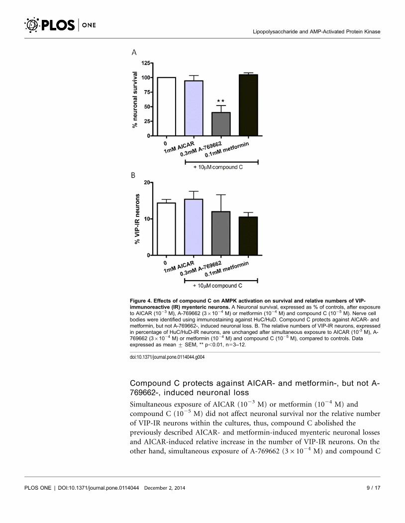
Compound C protects against AICAR- and metformin-, but not A-
769662-, induced neuronal loss
Simultaneous exposure of AICAR (1023 M) or metformin (1024 M) and
compound C (1025 M) did not affect neuronal survival nor the relative number
of VIP-IR neurons within the cultures, thus, compound C abolished the
previously described AICAR- and metformin-induced myenteric neuronal losses
and AICAR-induced relative increase in the number of VIP-IR neurons. On the
other hand, simultaneous exposure of A-769662 (361024 M) and compound C
Figure 4. Effects of compound C on AMPK activation on survival and relative numbers of VIP-immunoreactive (IR) myenteric neurons. A Neuronal survival, expressed as % of controls, after exposureto AICAR (1023 M), A-769662 (361024 M) or metformin (1024 M) and compound C (1025 M). Nerve cellbodies were identified using immunostaining against HuC/HuD. Compound C protects against AICAR- andmetformin, but not A-769662-, induced neuronal loss. B. The relative numbers of VIP-IR neurons, expressedin percentage of HuC/HuD-IR neurons, are unchanged after simultaneous exposure to AICAR (10-3 M), A-769662 (361024 M) or metformin (1024 M) and compound C (1025 M), compared to controls. Dataexpressed as mean ¡ SEM, ** p,0.01, n53–12.
doi:10.1371/journal.pone.0114044.g004
Lipopolysaccharide and AMP-Activated Protein Kinase
PLOS ONE | DOI:10.1371/journal.pone.0114044 December 2, 2014 9 / 17

(1025 M) caused loss of myenteric neurons in the same magnitude as A-769662
alone. Results are summarized in figure 4.
(5Z)-7-Oxoaeaenol does not inhibit AICAR- or metformin-induced
neuronal loss
Simultaneous exposure of AICAR (1023 M) or metformin (1024 M) and the
TAK1 inhibitor (5Z)-7-Oxozeaenol (1026 M) caused loss of cultured myenteric
neurons in the same magnitude as AICAR (1023 M) or metformin (1024 M)
alone. Presence of (5Z)-7-Oxozeaenol (1026 M) did not affect AICAR-induced
up-regulation of the relative number of VIP-IR neurons. Results are summarized
in figure 5.
Figure 5. Effects of (5Z)-7-Oxozeaenol on AMPK activation on survival and relative numbers of VIP-immunoreactive (IR) myenteric neurons. A. neuronal survival, expressed as % of controls, after exposureto AICAR (1023 M) or metformin (1024 M) and TAK-1 inhibitor (5Z)-7-Oxozeaenol (1026 M). Nerve cell bodieswere identified using immunostaining against HuC/HuD. (5Z)-7-Oxozeaenol does not protect against eitherAICAR- or metformin-induced neuronal loss. B. (5Z)-7-Oxozeaenol (1025 M) are unable to reduce the AICAR(1023 M)-induced relative numbers of VIP-IR neurons, expressed in percentage of HuC/HuD-IR neurons.VIP-IR after simultaneous exposure of 5Z)-7-Oxozeaenol (1025 M) and metformin (1024 M) are unchangedcompared to controls. Data expressed as mean ¡ SEM, ** p,0.01, n57–14.
doi:10.1371/journal.pone.0114044.g005
Lipopolysaccharide and AMP-Activated Protein Kinase
PLOS ONE | DOI:10.1371/journal.pone.0114044 December 2, 2014 10 / 17

Discussion
Current study re-affirmed previous findings showing that Escherichia coli derived
LPS induces neuronal loss in primary cultures of adult myenteric neurons. [8, 9]
The novel findings presented here are that presence of the AMPK inhibitor
compound C protects myenteric neurons against LPS-induced neuronal loss and
that, by using three independent AMPK activators, AMPK activation was found to
mimic LPS exposure in that it induced loss of cultured myenteric neurons. The
link between LPS-induced TLR4 stimulation and AMPK activation was further
strengthened by the finding that TAK1 is involved. This since the presence of
TAK1 inhibitor (5Z)-7-Oxozeaenol protected against LPS-induced neuronal loss
but not against AICAR-induced neuronal loss.
LPS-induced AMPK activation
Elevated fat and energy intake increases plasma LPS levels in both patients and
mice, leading to increased expressions of TLR4 and exacerbation of inflammation.
[3, 27] Current study revealed LPS-induced neuronal loss to be mediated by
AMPK activation. The reduced neuronal survival evident after LPS exposure was
blocked by simultaneous presence of the AMPK inhibitor compound C. These
findings are corroborated by previous findings on primary microglia cultures and
cell lines in which LPS was found to cause AMPK activation and cytokine release,
through TLR4 activation. [13–15] The link between TLR4 activation and AMPK
activation is suggested to be TAK1, a key player in regulating the cellular response
to environmental and cytokine-induced stress. TAK1 is able to regulate several
transcription factors including IKKb, p38/JNK and NF-kB, [7, 28] and has
previously been shown to mediate AMPK activation. [29–31] Enteric neurons are
sensitive to LPS and express TLR4. [9] The significance of such expression is
suggested to be regulation of the innate tolerance response to commensal bacteria
in the intestine. [9] This ensures a balanced immune response with respect to
luminal content and homeostasis. Supporting this, is the finding that TLR4-/- mice
display a delayed transit time. [32] In contrast to previous, [8, 9] and current
findings, LPS has been reported to increase survival of enteric neuronal cultures
through TLR4 signalling. [32] This discrepancy is suggested due to differences in
LPS concentration; low concentrations are protective while high ones are
deleterious. It may also be due to differences in TLR4 expression and LPS
sensitivity between embryonic (gut not colonized with microbiota), [32] and
adult [8, 9] enteric neurons. Also LPS exposure duration and potency are probably
of importance. In vivo the adaptive immune response primed by bacterial
exposure is, in the healthy intestine with a functional barrier, probably mediated
by low mucosal permeability and hence low LPS concentrations. TLR4 activation
may under such healthy conditions promote maintenance of neurons to ensure
survival. In conditions of compromised barrier function and permeability,
systemic LPS reach higher concentrations. This change in LPS concentration may
cause hyper-activation of TLR4 and imminent dysregulation of the adaptive
Lipopolysaccharide and AMP-Activated Protein Kinase
PLOS ONE | DOI:10.1371/journal.pone.0114044 December 2, 2014 11 / 17

immune response eventually leading to death. Another intriguing possibility that
should be considered is the origin of LPS. Though many of the gram negative
bacterial strains are the source of adverse infections some are considered
beneficial. [33, 34] The molecular part of LPS interacting with TLR4 is the
membrane anchoring region, called lipid A. [35] This region consists of a
phosphorylated N-acetylglucosamine dimer with a varying amount of saturated
fatty acid tails in both tail number and chain length attached. [35] The ability of
this region to interact with the TLR4 complex elicits a fine-tuned immune
response. [35, 36] The properties of the LPS variants used in previous studies
investigating LPS-induced effects on ENS need to be taken into consideration
when interpreting and comparing results.
AMPK activation in neurons
The results show AMPK activation to be detrimental to myenteric neurons; this
was confirmed using three different AMPK activators, AICAR, metformin and A-
769662. AMPK-induced neuronal loss has previously been suggested to occur in
enteric, [37] as well as in central [38] neurons. In the CNS compound C was
found to protect against stroke-induced neuronal loss. [38] The suggested
mechanism behind was that compound C attenuated the AMPK activated energy
processes causeing increased cellular stress in already compromised neurons. A
balanced AMPK response is necessary in models of ischemic stress, which induces
a transient increase of AMPK activation and autophagy. This is believed to be part
of the protective response that, if unbalanced, leads to cell death. [39] Further,
AMPK activation has, in neuronal cell lines, been shown to decrease neurite
growth causing loss of contact and eventually neurodegeneration. [40] These
studies are well in line with the here presented data, showing that activation of
AMPK is detrimental to enteric neuronal survival. Interestingly TAK1 inhibition,
like AMPK inhibition, is suggested to mediate neuroprotection also of central
neurons after ischemic or traumatic brain injury. [41, 42]
The here noted difference in sensitivity to compound C by AICAR, metformin
and A-769662 induced neuronal is likely explained by their molecular targets.
AICAR is an AMP analogue activating AMPK in a the same manner as AMP; an
allosteric conformational change and activation. [23] Compound C is a
competitive inhibitor binding to the same ligand site as AMP and AICAR. [11]
On the other hand, A-769662 has a different AMPK activation mechanism
involving ligand site separate from AMP and depends on the presence of the b1
subunit. [24, 43] These differences in ligand binding and activation patterns may
well explain why compound C blocks AICAR, but not A-769662, induced
neuronal loss. Metformin- like AICAR-induced loss was blocked by compound C.
Whether this, in analogy with AICAR, can be explained by a competitive binding
is more uncertain. The mechanism by which metformin activate AMPK is
currently debated. Both a direct interaction with AMPK, [25] as well as several
possible indirect mechanisms have been suggested involving both AMP dependent
and independent mechanism. Metformin is a commonly used drug in the
Lipopolysaccharide and AMP-Activated Protein Kinase
PLOS ONE | DOI:10.1371/journal.pone.0114044 December 2, 2014 12 / 17

treatment of type 2-diabetes and its positive effects on this disease may be ascribed
to other signalling pathways than AMPK activation. [26] It is worth noting that in
the present study while using a concentration within the reported physiological
range, [47, 48] metformin was used as an AMPK activator.
LPS- and AMPK activation-induced VIP expression
The LPS-induced increase in the relative proportion of VIP immunoreactive
myenteric neurons has previously been reported, together with the finding that
exogenous VIP protects against LPS-induced neuronal loss. [8, 9] These reports
further speculated if the physiological significance of an increased number of VIP-
IR neurons was due to VIP’s immunmodulatory effects, or due to its general
neuroprotective properties. [9, 49, 50] Current study showed LPS and AICAR, but
not metformin or A-769662, to increase the relative numbers of VIP
immunoreactive neurons, an increase that could be attenuated by simultaneous
exposure to compound C. The differences between AICAR, metformin and A-
769662 in evoking an increased relative number of VIP-IR neurons are enigmatic.
AICAR has been shown to have intracellular secondary activation targets. [51, 52]
This may provide one explanation since A-769662, which is believed to be a more
precise AMPK activator, [24] did not up-regulate VIP. The unresolved
mechanism of metformin’s activation of AMPK makes interpretation of the
metformin effect on VIP-IR neurons harder to discuss. However, as compound C
was able to reverse both the LPS- and AICAR-induced upregulation of VIP-IR
neurons the possibility of a direct AMPK-induced effect can’t be completely
disregarded. In muscle cells AMPK is able to phosphorylate the transcription
factor cAMP responsive element binding protein (CREB) at the same site as
protein kinase A. [53] AMPK is thereby, through phosphorylation events, able to
cause an increase in CREB-dependent transcription leading to increased VIP
expression. [54] The mechanism behind the lack of VIP up-regulation by A-
769662 and metformin needs further investigation taking into account the diverse
regulatory factors both on transcriptional and translational levels. The rationale
behind such AMPK-induced neurotransmitter up-regulation needs further
investigations. VIP is a neuroimmunopeptide that besides acting as an inhibitory
neurotransmitter in ENS also is able to regulate TLR expression. [10] VIP has
been shown to down regulate LPS-induced TLR4 expression and subsequent the
inflammatory response. [10] It can be speculated that LPS- and AMPK activation-
induced VIP up-regulation is part of a protective neuronal response that may, as
previously described, play a role in modulating the innate immune-response to
commensal bacteria or inflammation. [9]
Conclusion
LPS exposure and activation of AMPK cause neuronal loss of cultured myenteric
neurons. LPS-induced neuronal loss is blocked by the presence of compound C
Lipopolysaccharide and AMP-Activated Protein Kinase
PLOS ONE | DOI:10.1371/journal.pone.0114044 December 2, 2014 13 / 17

and therefore suggested to be executed through activation of AMPK. The link
between LPS and AMPK is suggested to be TAK1 acting downstream of TLR4 and
upstream of AMPK. This suggestion is supported by the finding that (5Z)-7-
Oxozeaenol is able to attenuate LPS- but not AICAR- or metformin-induced
neuronal loss. Further, both LPS and AICAR cause significant increase in the
relative numbers of VIP-IR neurons. The rationale behind upregulation of VIP is
suggested assigned both to activate adaptive innate neuroimmune-responses to
intestinal commensal bacteria and to provide neuroprotection.
Supporting Information
Dataset S1. Neuronal survival of control and relative VIP-IR data.
doi:10.1371/journal.pone.0114044.s001 (XLSX)
Acknowledgments
We thank Olga Goransson, Department of Experimental Medical Sciences, Lund
University for sharing A-769662 and for fruitful discussions on AMPK regulation.
We thank Anna Themner-Persson for excellent technical assistance.
Author ContributionsConceived and designed the experiments: UV EE. Performed the experiments: UV
EE. Analyzed the data: UV EE. Contributed reagents/materials/analysis tools: UV
EE. Wrote the paper: UV EE.
References
1. Backhed F, Ley RE, Sonnenburg JL, Peterson DA, Gordon JI (2005) Host-bacterial mutualism in thehuman intestine. Science 307: 1915–1920.
2. Clemente JC, Ursell LK, Parfrey LW, Knight R (2012) The Impact of the Gut Microbiota on HumanHealth: An Integrative View. Cell 148: 1258–1270.
3. Kim KA, Gu W, Lee IA, Joh EH, Kim DH (2012) High Fat Diet-Induced Gut Microbiota ExacerbatesInflammation and Obesity in Mice via the TLR4 Signaling Pathway. Plos One 7.
4. Camilleri M, Madsen K, Spiller R, Van Meerveld BG, Verne GN (2012) Intestinal barrier function inhealth and gastrointestinal disease. Neurogastroenterology and Motility 24: 503–512.
5. Wouters MM, Boeckxstaens GE (2011) Neuroimmune mechanisms in functional bowel disorders.Netherlands Journal of Medicine 69: 55–61.
6. West AP, Koblansky AA, Ghosh S (2006) Recognition and signaling by toll-like receptors. AnnualReview of Cell and Developmental Biology. Palo Alto: Annual Reviews. pp. 409–437.
7. Landstrom M (2010) The TAK1-TRAF6 signalling pathway. Int J Biochem Cell Biol 42: 585–589.
8. Arciszewski M, Pierzynowski S, Ekblad E (2005) Lipopolysaccharide induces cell death in culturedporcine myenteric neurons. Digestive Diseases and Sciences 50: 1661-1668.
9. Arciszewski MB, Sand E, Ekbad E (2008) Vasoactive intestinal peptide rescues cultured rat myentericneurons from lipopolysaccharide induced cell death. Regulatory Peptides 146: 218–223.
Lipopolysaccharide and AMP-Activated Protein Kinase
PLOS ONE | DOI:10.1371/journal.pone.0114044 December 2, 2014 14 / 17

10. Gomariz RP, Arranz A, Juarranz Y, Gutierrez-Canas I, Garcia-Gomez M, et al. (2007) Regulation ofTLR expression, a new perspective for the role of VIP in immunity. Peptides 28: 1825–1832.
11. Viollet B, Horman S, Leclerc J, Lantier L, Foretz M, et al. (2010) AMPK inhibition in health anddisease. Critical Reviews in Biochemistry and Molecular Biology 45: 276–295.
12. Leff T (2003) AMP-activated protein kinase regulates gene expression by direct phosphorylation ofnuclear proteins. Biochemical Society Transactions 31: 224–227.
13. Labuzek K, Liber S, Gabryel B, Okopien B (2010) AICAR (5-aminoimidazole-4-carboxamide-1-beta-4-ribofuranoside) increases the production of toxic molecules and affects the profile of cytokines release inLPS-stimulated rat primary microglial cultures. Neurotoxicology 31: 134–146.
14. Labuzek K, Liber S, Gabryel B, Buldak L, Okopien B (2010) Ambivalent effects of compound C(dorsomorphin) on inflammatory response in LPS-stimulated rat primary microglial cultures. Naunyn-Schmiedebergs Archives of Pharmacology 381: 41–57.
15. Kim SY, Jeong S, Jung E, Baik KH, Chang MH, et al. (2012) AMP-activated protein kinase-alpha 1 asan activating kinase of TGF-beta-activated kinase 1 has a key role in inflammatory signals. Cell Death &Disease 3: 1–13.
16. Bai AP, Ma AG, Yong M, Weiss CR, Ma YB, et al. (2010) AMPK agonist downregulates innate andadaptive immune responses in TNBS-induced murine acute and relapsing colitis. BiochemicalPharmacology 80: 1708–1717.
17. Salminen A, Hyttinen JMT, Kaarniranta K (2011) AMP-activated protein kinase inhibits NF-kappa Bsignaling and inflammation: impact on healthspan and lifespan. Journal of Molecular Medicine-Jmm 89:667–676.
18. Voss U, Sand E, Hellstrom PM, Ekblad E (2012) Glucagon-like peptides 1 and 2 and vasoactiveintestinal peptide are neuroprotective on cultured and mast cell co-cultured rat myenteric neurons. BmcGastroenterology 12: 30.
19. Sand E, Voss U, Hammar O, Alm R, Nordin Fredrikson G, et al. (2012) Gonadotropin-releasinghormone analog buserelin causes neuronal loss in rat gastrointestinal tract. Cell Tissue Res.
20. Ekblad E, Ekman R, Hakanson R, Sundler F (1988) Projections of peptide-containing neurons in ratcolon. Neuroscience 27: 655–674.
21. Voss U, Sand E, Olde B, Ekblad E (2013) Enteric neuropathy can be induced by high fat diet in vivo andpalmitic acid exposure in vitro. PLoS One 8: e81413.
22. Wu J, Powell F, Larsen NA, Lai Z, Byth KF, et al. (2013) Mechanism and in vitro pharmacology of TAK1inhibition by (5Z)-7-Oxozeaenol. ACS Chem Biol 8: 643–650.
23. Corton JM, Gillespie JG, Hawley SA, Hardie DG (1995) 5-aminoimidazole-4-carboxamideribonucleoside - a specific method for activating AMP-activated protein-kinase in intact-cells.European Journal of Biochemistry 229: 558–565.
24. Goransson O, McBride A, Hawley SA, Ross FA, Shpiro N, et al. (2007) Mechanism of action of A-769662, a valuable tool for activation of AMP-activated protein kinase. Journal of Biological Chemistry282: 32549–32560.
25. Zhang Y, Wang Y, Bao C, Xu Y, Shen H, et al. (2012) Metformin interacts with AMPK through binding toc subunit. Mol Cell Biochem 368: 69–76.
26. Hardie DG, Ross FA, Hawley SA (2012) AMP-activated protein kinase: a target for drugs both ancientand modern. Chem Biol 19: 1222–1236.
27. Ghanim H, Abuaysheh S, Sia CL, Korzeniewski K, Chaudhuri A, et al. (2009) Increase in PlasmaEndotoxin Concentrations and the Expression of Toll-like Receptors and Suppressor of CytokineSignaling-3 in Mononuclear Cells After a High-Fat, High-Carbohydrate Meal Implications for insulinresistance. Diabetes Care 32: 2281–2287.
28. Adhikari A, Xu M, Chen ZJ (2007) Ubiquitin-mediated activation of TAK1 and IKK. Oncogene 26: 3214–3226.
29. Xie M, Zhang D, Dyck JR, Li Y, Zhang H, et al. (2006) A pivotal role for endogenous TGF-beta-activated kinase-1 in the LKB1/AMP-activated protein kinase energy-sensor pathway. Proc Natl AcadSci U S A 103: 17378–17383.
Lipopolysaccharide and AMP-Activated Protein Kinase
PLOS ONE | DOI:10.1371/journal.pone.0114044 December 2, 2014 15 / 17

30. Momcilovic M, Hong SP, Carlson M (2006) Mammalian TAK1 activates Snf1 protein kinase in yeastand phosphorylates AMP-activated protein kinase in vitro. Journal of Biological Chemistry 281: 25336–25343.
31. Chen ZY, Shen XL, Shen FY, Zhong W, Wu H, et al. (2013) TAK1 activates AMPK-dependent celldeath pathway in hydrogen peroxide-treated cardiomyocytes, inhibited by heat shock protein-70.Molecular and Cellular Biochemistry 377: 35–44.
32. Anitha M, Vijay-Kumar M, Sitaraman SV, Gewirtz AT, Srinivasan S (2012) Gut Microbial ProductsRegulate Murine Gastrointestinal Motility via Toll-Like Receptor 4 Signaling. Gastroenterology 143:1006–+.
33. Everard A, Belzer C, Geurts L, Ouwerkerk JP, Druart C, et al. (2013) Cross-talk betweenAkkermansia muciniphila and intestinal epithelium controls diet-induced obesity. Proceedings of theNational Academy of Sciences of the United States of America 110: 9066–9071.
34. Wexler HM (2007) Bacteroides: the good, the bad, and the nitty-gritty. Clinical Microbiology Reviews 20:593–+.
35. Maeshima N, Fernandez RC (2013) Recognition of lipid A variants by the TLR4-MD-2 receptorcomplex. Frontiers in Cellular and Infection Microbiology 3: 13.
36. Miller SI, Ernst RK, Bader MW (2005) LPS, TLR4 and infectious disease diversity. Nat Rev Microbiol 3:36–46.
37. Voss U, Sand E, Olde B, Ekblad E (2013) Enteric neuropathy can be induced by high fat diet in vivo andpalmitic acid exposure in vitro. PLOS ONE SUBMITTED.
38. McCullough LD, Zeng ZY, Li H, Landree LE, McFadden J, et al. (2005) Pharmacological inhibition ofAMP-activated protein kinase provides neuroprotection in stroke. Journal of Biological Chemistry 280:20493–20502.
39. Poels J, Spasic MR, Callaerts P, Norga KK (2009) Expanding roles for AMP-activated protein kinase inneuronal survival and autophagy. Bioessays 31: 944–952.
40. Jiang PZ, Gan M, Ebrahim AS, Castanedes-Casey M, Dickson DW, et al. (2013) Adenosinemonophosphate-activated protein kinase overactivation leads to accumulation of alpha-synucleinoligomers and decrease of neurites. Neurobiology of Aging 34: 1504–1515.
41. Neubert M, Ridder DA, Bargiotas P, Akira S, Schwaninger M (2011) Acute inhibition of TAK1 protectsagainst neuronal death in cerebral ischemia. Cell Death and Differentiation 18: 1521–1530.
42. Zhang D, Hu Y, Sun Q, Zhao J, Cong Z, et al. (2013) Inhibition of transforming growth factor beta-activated kinase 1 confers neuroprotection after traumatic brain injury in rats. Neuroscience 238: 209–217.
43. Scott JW, van Denderen BJW, Jorgensen SB, Honeyman JE, Steinberg GR, et al. (2008)Thienopyridone Drugs Are Selective Activators of AMP-Activated Protein Kinase beta 1-ContainingComplexes. Chemistry & Biology 15: 1220–1230.
44. Hawley SA, Gadalla AE, Olsen GS, Hardie DG (2002) The antidiabetic drug metformin activates theAMP-activated protein kinase cascade via an adenine nucleotide-independent mechanism. Diabetes 51:2420–2425.
45. Sakamoto K, Goransson O, Hardie DG, Alessi DR (2004) Activity of LKB1 and AMPK-related kinasesin skeletal muscle: Effects of contraction, phenformin, and AICAR. American Journal of Physiology-Endocrinology and Metabolism 287: E310–E317.
46. Fryer LGD, Parbu-Patel A, Carling D (2002) The anti-diabetic drugs rosiglitazone and metforminstimulate AMP-activated protein kinase through distinct signaling pathways. Journal of BiologicalChemistry 277: 25226–25232.
47. Zhou G, Myers R, Li Y, Chen Y, Shen X, et al. (2001) Role of AMP-activated protein kinase inmechanism of metformin action. J Clin Invest 108: 1167–1174.
48. Wilcock C, Bailey CJ (1994) Accumulation of metformin by tissues of the normal and diabetic mouse.Xenobiotica 24: 49–57.
49. Ekblad E, Mulder H, Sundler F (1996) Vasoactive intestinal peptide expression in enteric neurons isupregulated by both colchicine and axotomy. Regulatory Peptides 63: 113–121.
Lipopolysaccharide and AMP-Activated Protein Kinase
PLOS ONE | DOI:10.1371/journal.pone.0114044 December 2, 2014 16 / 17

50. Sandgren K, Lin Z, Svenningsen AF, Ekblad E (2003) Vasoactive intestinal peptide and nitric oxidepromote survival of adult rat myenteric neurons in culture. Journal of Neuroscience Research 72: 595–602.
51. Gadalla AE, Pearson T, Currie AJ, Dale N, Hawley SA, et al. (2004) AICA riboside both activatesAMP-activated protein kinase and competes with adenosine for the nucleoside transporter in the CA1region of the rat hippocampus. Journal of Neurochemistry 88: 1272–1282.
52. Lanner JT, Georgiou DK, Dagnino-Acosta A, Ainbinder A, Cheng Q, et al. (2012) AICAR preventsheat-induced sudden death in RyR1 mutant mice independent of AMPK activation. Nature Medicine 18:244–251.
53. Canto C, Auwerx J (2010) AMP-activated protein kinase and its downstream transcriptional pathways.Cellular and Molecular Life Sciences 67: 3407–3423.
54. Goodman RH (1990) REGULATION OF NEUROPEPTIDE GENE-EXPRESSION. Annual Review ofNeuroscience 13: 111–127.
Lipopolysaccharide and AMP-Activated Protein Kinase
PLOS ONE | DOI:10.1371/journal.pone.0114044 December 2, 2014 17 / 17