Anergy to Development of CD4+ T Cell Dendritic Cell Function Contributes Receptor {gamma} Control of...
10
Peroxisome Proliferator-Activated Receptor Control of Dendritic Cell Function Contributes to Development of CD4 T Cell Anergy Luisa Klotz, 1 * † Indra Dani,* Frank Edenhofer, ‡ Lars Nolden, ‡ Bernd Evert, † Bianca Paul, § Waldemar Kolanus, § Thomas Klockgether, † Percy Knolle,* and Linda Diehl* There is increasing evidence that dendritic cell (DC) immunogenicity is not only positively regulated by ligands of pattern recognition receptors, but also negatively by signals that prevent DC activation and full functional maturation. Depending on their activation status, DCs can induce either immunity or tolerance. In this study, we provide molecular evidence that the transcription factor peroxisome proliferator-activated receptor (PPAR) is a negative regulator of DC maturation and function. Sustained PPAR activation in murine DCs reduced maturation-induced expression of costimulatory molecules and IL-12, and profoundly inhibited their capacity to prime naive CD4 T cells in vitro. Using PPAR-deficient DCs, generated by Cre-mediated ablation of the PPAR gene, agonist-mediated suppression of maturation-induced functional changes were abrogated. Moreover, absence of PPAR increased DC immunogenicity, suggesting a constitutive regulatory function of PPAR in DCs. Adoptive transfer of PPAR-activated Ag-presenting DCs induced CD4 T cell anergy, char- acterized by impaired differentiation resulting in absent Th1 and Th2 cytokine production and failure of secondary clonal expansion upon restimulation. Collectively, our data support the notion that PPAR is an efficient regulator of DC immu- nogenicity that may be exploited to deliberately target CD4 T cell-mediated immune responses. The Journal of Immu- nology, 2007, 178: 2122–2131. D endritic cells (DCs) 2 are professional APCs with a unique capacity to stimulate naive T cells (1). The func- tional properties of DCs are closely linked to their mat- uration status, because maturation-induced expression of costimu- latory molecules and production of proinflammatory cytokines, in particular IL-12, are essential for DCs to induce strong T cell- mediated immunity (2, 3). Critical to the functional maturation of DCs is their activation through pattern-recognition receptors, e.g., TLRs that recognize conserved microbial structures (4). Induction of T cell-mediated immunity is characterized by massive clonal expansion and differentiation of naive T cells into cells with ef- fector function, i.e., cytotoxicity for CD8 T cells and helper func- tion for CD4 T cells. Under steady-state conditions, i.e., in the absence of inflammation, immature DCs take up tissue Ags, mi- grate to draining lymph nodes, and present Ag in the absence of proper costimulation and IL-12 secretion, thereby inducing spe- cific immune tolerance toward tissue Ags (3, 5). Several mediators contribute to the maintenance of tolerance by negative interference with DC maturation, such as IL-10 or TGF- derived from regu- latory T cells, as well as DC-derived mediators like prostanoids (6, 7). However, the molecular mechanisms responsible for the main- tenance of a tolerogenic state in DCs are not fully understood. The nuclear receptor peroxisome proliferator-activated receptor (PPAR) is a transcription factor that has been shown to mediate anti-inflammatory effects on several immune cell types (8, 9). Upon ligand binding, PPAR heterodimerizes with the retinoid X receptor and binds to PPAR response elements located in the promoter region of target genes. Additionally, important anti- inflammatory effects of PPAR are mediated by negative interfer- ence with proinflammatory cell signaling, either via competition for coactivators or via transrepression through physical interaction with proinflammatory transcription factors, in particular NF-B (9, 10). PPAR agonists include endogenous ligands such as polyun- saturated fatty acids and prostanoids like the PGD 2 metabolite 15- deoxy- (11, 12) PGJ 2 , as well as several synthetic ligands such as the antidiabetic thiazolidinediones, e.g., pioglitazone (PIO) and rosiglitazone, which are currently being used for the correction of metabolic disturbances in type II diabetes (13). In monocytes and macrophages, PPAR agonists inhibit the expression of a number of proinflammatory cytokines (8, 9), and attenuate the oxidative burst (11). In T lymphocytes, PPAR activation inhibits both Ag- specific and nonspecific T cell proliferation and the production of several proinflammatory cytokines (12, 14). Recently, it has been shown that PPAR agonists prevent expression of IL-12 in murine DCs (15) and affect maturation of human monocyte-derived DCs as evidenced by altered surface expression levels of costimulatory molecules (16). However, because PPAR knockout (k.o.) is embryonally le- thal, formal evidence for involvement of PPAR in immune con- trol mechanisms is lacking (17). Moreover, several reports raised *Institute of Molecular Medicine and Experimental Immunology, University of Bonn, Bonn, Germany; † Department of Neurology, University of Bonn, Bonn, Germany; ‡ Institute of Reconstructive Neurobiology, Life and Brain Center, University of Bonn and Hertie Foundation, Bonn, Germany; and § Life and Medical Sciences Institute, Program Unit Molecular Immune and Cell Biology, University of Bonn, Bonn, Germany Received for publication July 13, 2006. Accepted for publication November 15, 2006. The costs of publication of this article were defrayed in part by the payment of page charges. This article must therefore be hereby marked advertisement in accordance with 18 U.S.C. Section 1734 solely to indicate this fact. 1 Address correspondence and reprint requests to Dr. Luisa Klotz, Department of Neurology, University of Bonn, Sigmund-Freud-Strasse 25, 53105 Bonn, Germany. E-mail address: [email protected] 2 Abbreviations used in this paper: DC, dendritic cell; PPAR, peroxisome prolifera- tor-activated receptor ; PIO, pioglitazone; k.o., knockout; BMDC, bone marrow DC; 7-AAD, 7-aminoactinomycin D; wt, wild type. Copyright © 2007 by The American Association of Immunologists, Inc. 0022-1767/07/$2.00 The Journal of Immunology www.jimmunol.org
-
Upload
independent -
Category
Documents
-
view
1 -
download
0
Transcript of Anergy to Development of CD4+ T Cell Dendritic Cell Function Contributes Receptor {gamma} Control of...

Peroxisome Proliferator-Activated Receptor � Control ofDendritic Cell Function Contributes to Development ofCD4� T Cell Anergy
Luisa Klotz,1*† Indra Dani,* Frank Edenhofer,‡ Lars Nolden,‡ Bernd Evert,† Bianca Paul,§
Waldemar Kolanus,§ Thomas Klockgether,† Percy Knolle,* and Linda Diehl*
There is increasing evidence that dendritic cell (DC) immunogenicity is not only positively regulated by ligands of patternrecognition receptors, but also negatively by signals that prevent DC activation and full functional maturation. Dependingon their activation status, DCs can induce either immunity or tolerance. In this study, we provide molecular evidence thatthe transcription factor peroxisome proliferator-activated receptor � (PPAR�) is a negative regulator of DC maturation andfunction. Sustained PPAR� activation in murine DCs reduced maturation-induced expression of costimulatory moleculesand IL-12, and profoundly inhibited their capacity to prime naive CD4� T cells in vitro. Using PPAR�-deficient DCs,generated by Cre-mediated ablation of the PPAR� gene, agonist-mediated suppression of maturation-induced functionalchanges were abrogated. Moreover, absence of PPAR� increased DC immunogenicity, suggesting a constitutive regulatoryfunction of PPAR� in DCs. Adoptive transfer of PPAR�-activated Ag-presenting DCs induced CD4� T cell anergy, char-acterized by impaired differentiation resulting in absent Th1 and Th2 cytokine production and failure of secondary clonalexpansion upon restimulation. Collectively, our data support the notion that PPAR� is an efficient regulator of DC immu-nogenicity that may be exploited to deliberately target CD4� T cell-mediated immune responses. The Journal of Immu-nology, 2007, 178: 2122–2131.
D endritic cells (DCs)2 are professional APCs with aunique capacity to stimulate naive T cells (1). The func-tional properties of DCs are closely linked to their mat-
uration status, because maturation-induced expression of costimu-latory molecules and production of proinflammatory cytokines, inparticular IL-12, are essential for DCs to induce strong T cell-mediated immunity (2, 3). Critical to the functional maturation ofDCs is their activation through pattern-recognition receptors, e.g.,TLRs that recognize conserved microbial structures (4). Inductionof T cell-mediated immunity is characterized by massive clonalexpansion and differentiation of naive T cells into cells with ef-fector function, i.e., cytotoxicity for CD8� T cells and helper func-tion for CD4� T cells. Under steady-state conditions, i.e., in theabsence of inflammation, immature DCs take up tissue Ags, mi-grate to draining lymph nodes, and present Ag in the absence ofproper costimulation and IL-12 secretion, thereby inducing spe-cific immune tolerance toward tissue Ags (3, 5). Several mediators
contribute to the maintenance of tolerance by negative interferencewith DC maturation, such as IL-10 or TGF-� derived from regu-latory T cells, as well as DC-derived mediators like prostanoids (6,7). However, the molecular mechanisms responsible for the main-tenance of a tolerogenic state in DCs are not fully understood.
The nuclear receptor peroxisome proliferator-activated receptor� (PPAR�) is a transcription factor that has been shown to mediateanti-inflammatory effects on several immune cell types (8, 9).Upon ligand binding, PPAR� heterodimerizes with the retinoid Xreceptor and binds to PPAR response elements located in thepromoter region of target genes. Additionally, important anti-inflammatory effects of PPAR� are mediated by negative interfer-ence with proinflammatory cell signaling, either via competitionfor coactivators or via transrepression through physical interactionwith proinflammatory transcription factors, in particular NF-�B (9,10). PPAR� agonists include endogenous ligands such as polyun-saturated fatty acids and prostanoids like the PGD2 metabolite 15-deoxy-� (11, 12) PGJ2, as well as several synthetic ligands such asthe antidiabetic thiazolidinediones, e.g., pioglitazone (PIO) androsiglitazone, which are currently being used for the correction ofmetabolic disturbances in type II diabetes (13). In monocytes andmacrophages, PPAR� agonists inhibit the expression of a numberof proinflammatory cytokines (8, 9), and attenuate the oxidativeburst (11). In T lymphocytes, PPAR� activation inhibits both Ag-specific and nonspecific T cell proliferation and the production ofseveral proinflammatory cytokines (12, 14). Recently, it has beenshown that PPAR� agonists prevent expression of IL-12 in murineDCs (15) and affect maturation of human monocyte-derived DCsas evidenced by altered surface expression levels of costimulatorymolecules (16).
However, because PPAR� knockout (k.o.) is embryonally le-thal, formal evidence for involvement of PPAR� in immune con-trol mechanisms is lacking (17). Moreover, several reports raised
*Institute of Molecular Medicine and Experimental Immunology, University of Bonn,Bonn, Germany; †Department of Neurology, University of Bonn, Bonn, Germany;‡Institute of Reconstructive Neurobiology, Life and Brain Center, University of Bonnand Hertie Foundation, Bonn, Germany; and §Life and Medical Sciences Institute,Program Unit Molecular Immune and Cell Biology, University of Bonn, Bonn,Germany
Received for publication July 13, 2006. Accepted for publication November 15, 2006.
The costs of publication of this article were defrayed in part by the payment of pagecharges. This article must therefore be hereby marked advertisement in accordancewith 18 U.S.C. Section 1734 solely to indicate this fact.1 Address correspondence and reprint requests to Dr. Luisa Klotz, Department ofNeurology, University of Bonn, Sigmund-Freud-Strasse 25, 53105 Bonn, Germany.E-mail address: [email protected] Abbreviations used in this paper: DC, dendritic cell; PPAR�, peroxisome prolifera-tor-activated receptor �; PIO, pioglitazone; k.o., knockout; BMDC, bone marrow DC;7-AAD, 7-aminoactinomycin D; wt, wild type.
Copyright © 2007 by The American Association of Immunologists, Inc. 0022-1767/07/$2.00
The Journal of Immunology
www.jimmunol.org

the question of whether the anti-inflammatory effects of PPAR�agonists are really receptor-mediated; for instance, it has beenshown that at least some effects of both natural and syntheticPPAR� agonists are not due to activation of the receptor (18, 19).Using a conditional k.o. strategy, we in this study provide directmolecular evidence that PPAR� controls DC function both in thesteady-state situation and under inflammatory conditions. The con-sequence of PPAR� activation in DCs is the conservation of afunctionally immature state, which is associated with failure topromote activation of naive CD4� T cells and subsequent clonalexpansion and differentiation into effector cells.
Materials and MethodsMice
Female BALB/c mice and C57BL/6 mice (6–10 wk old) were purchasedfrom Charles River Laboratories. MHC-class II-restricted TCR-transgenicmice OT II (H-2b background) and DO11.10 (H-2d background), both spe-cific for OVA peptide (323–339), as well as PPAR�fl/fl mice (20) (pur-chased from The Jackson Laboratory) were bred in our animal facility. Allexperiments were performed according to guidelines of the animal ethicscommittee.
Oral treatment with PIO
Seven days before splenic DC isolation, mice were treated with 30 mg/kgbody weight per day PIO (Actos; Takeda Pharmaceuticals) suspended in2% carboxymethylcellulose by daily oral gavage. Control DCs were iso-lated from mice fed for 7 days with vehicle only.
Generation of bone marrow DCs (BMDCs)
Bone marrow was flushed with IMDM (PAA) from femurs and tibiae ofC57BL/6 mice or from PPAR�fl/fl mice. Cells were washed, quantified, andplated in 100-mm diameter petri dishes at a density of 5 � 106 in IMDMsupplemented with 30% supernatant from the GM-CSF-producing cell lineAg 8653, 10% FCS, 1% glutamine, and antibiotics. After medium changeat day 3, cells were trypsinized at day 4, resuspended, and plated at adensity of 5 � 106 until day 7. A total of 10 �M PIO (Axxora) was addedto the culture during the whole differentiation procedure from day 0 to 7.Percentage of CD11c� DCs at day 7, routinely �90%, was not altered bytreatment with PIO. For maturation, BMDCs were stimulated with 100ng/ml LPS for 20 h at day 7.
Isolation of splenic T cells and splenic DCs and in vitrococulture assays
Splenic CD4� T cells from OT-II mice and CD11c� DCs from C57BL/6mice were isolated by immunomagnetic separation using CD4� andCD11c�-labeled MACS microbeads (Miltenyi Biotec). Purity of isolatedcells was controlled by FACS staining of CD4, and CD11c, respectively.CD4� T cells were cocultured with either CD11c� splenic DCs or BMDCsin triplicates for 5 days in RPMI 1640 medium (Invitrogen Life Technol-ogies) containing 8% FCS, 1% glutamine, and antibiotics. OVA (Serva)was added at a concentration of 0.5 mg/ml. For restimulation experiments,CD4� T cells were separated by density centrifugation using lymphoprep(PAA), washed twice, and cultured in triplicates for another 20 h in thepresence of anti-CD3 Ab (clone 145-2C11) coated on 96-well plates. Fortitration assays, fixed amounts of naive OVA-specific CD4� T cells werecocultured with BMDCs at ratios from 2:1 to 10:1 (T cells:BMDCs) intriplicates on 96-well plates in the presence of Ag. For analysis of T cellproliferation in vitro, naive freshly isolated CD4� T cells were labeledfor 15 min with 1 �M CFSE, washed twice with PBS, and coculturedwith BMDCs in 96-well plates for 5 days in the presence of OVA;proliferation was assessed by flow cytometric analysis of CFSE dilutionin CD4� cells.
Conditional k.o. of PPAR� in BMDCs
Conditional k.o. of PPAR� in vitro was achieved by site-specific recom-bination of loxP sites integrated into the PPAR� locus of PPAR�fl/fl miceusing a membrane-permeable Cre recombinase (HTNCre). RecombinantHTNCre protein was purified from Escherichia coli as described previ-ously (21). Bone marrow precursor cells were isolated from PPAR�fl/fl
mice or C57BL/6 mice as described above, and incubated with 3 �MHTNCre in IMDM containing 1% FCS for 6 h. Afterward, cells werewashed twice and cultured in 100-mm diameter petri dishes at a density of
5 � 106 in IMDM supplemented with GM-CSF-containing supernatant for3 days. At day 3, Cre transduction with 3 �M HTNCre was performedagain for 6 h, and cells were cultured for another 4 days in IMDM plusGM-CSF as done before. A total of 10 �M PIO was added to the cultureduring the whole differentiation procedure from day 0 to 7. Treatment ofBMDCs with HTNCre did not induce significant cell death as assessed byflow cytometric analysis of 7-aminoactinomycin D (7-AAD) incorporation.Additionally, percentage of CD11c� DCs at day 7 as well as DC phenotype(baseline levels of CD40, CD80, CD86) were not altered by Crerecombination.
In vivo experiments
Naive CD4� T cells from DO11.10 mice were isolated by immunomag-netic separation, labeled with 1 �M CFSE for 5 min, and injected into thelateral tail vein of BALB/c mice (5 � 106 viable cells per animal) at day1. At day 0, mice received a s.c. immunization with 1 � 106 OVA 323–339-loaded CD11c� splenic DCs from animals treated for 7 days with PIOor vehicle. At day 5, draining lymph nodes and spleens were isolated;proliferation of CD4� T cells was assessed by flow cytometric analysis ofCFSE profiles. For ex vivo restimulation experiments, cells (200,000 cellsper 96-well in triplicates) were cultivated in the presence of 5 �M OVA323–339 (MWG Biotec). Supernatants were analyzed by ELISA after 20 hfor the amount of IL-2 and IFN-� and after 72 h for the amount of IL-4 andIL-10. For recall assays, spleen cells from immunized mice were restim-ulated for 7 days with OVA (0.5 mg/ml); at day 4, cells were separated bydensity centrifugation, and cultured for another 3 days in the presence ofIL-2 (2 ng/ml). At day 7, equal cell numbers were restimulated with OVA(323–339)-loaded CD19� syngeneic B cells for 20 h; afterward, superna-tants were analyzed for cytokine content. Flow cytometric analysis of reg-ulatory T cells was performed directly ex vivo, at day 3 after Ag-specificrestimulation, and at day 2 after recall stimulation. For analysis of apopto-sis, lymph node cells and spleen cells were cultured for 1 h in the presenceof the FLICA reagent FAM-DEVD-FMK, which specifically and irrevers-ibly binds to active caspases 3 and 7 (Axxora). After extensive washing,cells were stained for CD4 and the Ag-specific TCR KJ1-26; unspecificcell death was assessed by additional staining with 7-AAD.
Cytokine ELISA
Supernatants were collected at the indicated time points. QuantitativeELISA for IL-2, IL-4, IL-10, and IFN-� were performed using commer-cially available Abs (BD Biosciences) according to the manufacturer’srecommendations.
Flow cytometry
Single-cell suspensions were stained with fluorescently conjugated Abs toCD11c, CD40, CD80, and CD86 for DCs, and to CD4, CD25, and KJ1-26for T cells (all obtained from BD Biosciences). Appropriate isotype con-trols were always included. Cell death was measured by 7-AAD incorpo-ration. Ag uptake was assessed by quantification of Alexa647-labeled OVAin CD11c� cells after 10-min incubation. For intracellular staining ofBMDCs, cells were stimulated for 5 h with 100 ng/ml LPS in the pres-ence of Golgi Plug and Golgi Stop (BD Biosciences). After surfacestaining, cells were fixed with 4% paraformaldehyde, permeabilizedwith 0.25% saponin, and stained with Abs against IL-12 and IL-10 (BDBiosciences). For intracellular staining of lymphocytes, cells were fixedwith 2% paraformaldehyde, permeabilized with 0.5% triton, and stainedwith an Ab against FoxP3 (eBioscience). Cells were measured using aFACSCalibur (BD Biosciences) and results were analyzed usingFlowJo software (Tree Star).
RNA extraction and RT-PCR
Total RNA was extracted from BMDCs at day 7 after HNTCre treatmentand subsequent differentiation by using TRIzol (Invitrogen Life Technol-ogies) according to standard protocols. Reverse transcription was per-formed with SuperScript (Invitrogen Life Technologies). Sense (5�-GTCACGTTCTGACAGGACTGTGTGAC-3�) and antisense (5�-TATCACTGGAGATCTCCGCCAACAGC-3�) primers were designed to anneal toregions in exons A1 and 4 of PPAR�, respectively, which detect bothfull-length (700 bp) and recombined (300 bp) transcripts (20). PCR wasperformed by 40 cycles of 94°C for 20 s, 60°C for 30 s, and 72°C for 60 s.
Nuclear protein extraction
Cells were washed with PBS, centrifuged at 2,000 � g for 5 min, washedin 1 ml of buffer A (10 mM HEPES (pH 7.9), 1.5 mM MgCl2, 10 mM KCl,0.1% Nonidet P-40, and 0.5 mM DTT), and pelleted at 2,000 � g for 5min. Pellets were resuspended in 80 �l of buffer A containing 0.1% Triton
2123The Journal of Immunology

X-100, incubated at 4°C for 10 min, and centrifuged at 2,000 � g for 5min. Afterward, the nuclear pellet was resuspended in 60 �l of 20 mMHEPES (pH 7.9), 0.42 M NaCl, 25% (v/v) glycerol, 1.5 mM MgCl2, and0.2 mM EDTA, incubated on ice for 30 min, and centrifuged at 15,000 �g for 20 min at 4°C. Extracts were stored at �80°C until use.
EMSA
For detection of NF-�B DNA binding, the following sequence was used:5�-AGT TGA GGG GAC TTT CCC AGG C-3� and 3�-TCA ACT CCCCTG AAA GGG TCC G-5�. Oligonucleotides were annealed in 10 mMTris (pH 8.0) at 65°C for 1 min and slow cooling at room temperature.Double-stranded oligonucleotides (10 pmol) were 5�-end labeled with[�-32P]ATP (5000 Ci/mmol; Amersham Biosciences) and T4 polynucle-
otide kinase (New England Biolabs), purified using a G25 quick spin col-umn (Roche Molecular Biochemicals), and diluted to a final volume of 150�l. Experiments were performed using the Gel Shift Assay System fromPromega: nuclear extracts (10 �g per reaction) were preincubated in bind-ing buffer (4% glycerol, 1 mM MgCl2, 0.5 mM EDTA, 0.5 mM DTT, 50mM NaCl, 10 mM Tris-HCl (pH 7.5), 0.5 mg/ml poly(dI:dC)� poly(dI:dC))for 1 h on ice. For supershift and competition assays, 3 �l of Ab (poly-clonal anti-p50-Ab, sc-8414X, and anti-p65-Ab, sc-372X, from SantaCruz Biochemicals) or an excess of unlabeled double-stranded oligo-nucleotides were added. After addition of 1 �l of labeled probe to eachreaction and 20-min incubation on ice, DNA protein complexes were separatedon 8% native polyacrylamide gels at 220 V with 0.5 � Tris-buffered EDTA asrunning buffer. Gels were vacuum-dried and visualized by autoradiography.
FIGURE 1. PIO impairs Ag-specific CD4� T cellpriming by DCs in vitro. BMDCs were generated in thepresence (BMDC � Pio) or absence (BMDC) of 10 �MPIO for 7 days (a and c). Alternatively, splenic CD11c�
DCs were isolated from animals treated p.o. for 7 dayswith PIO (splenic DC � Pio) or vehicle (splenic DC) (band d). Afterward, DCs were cocultured with naiveOVA-specific CD4� T cells in the presence of OVA for5 days. Coculture in the absence of Ag served as con-trol. a and b, Cytokine levels (IL-2 and IFN-�) at day 3of coculture. c and d, Time course of IL-2 secretionduring 5 days of coculture of DCs and CD4� T cells.Cytokine content in supernatants was measured in trip-licates by ELISA; �, cytokine secretion compared withDC (�, p � 0.05 and ��, p � 0.01, Student’s t test).
FIGURE 2. CD4� T cells primed by PIO-treatedDCs exhibit impaired effector function upon restimula-tion. Cytokine responses upon anti-CD3 restimulationof OVA-specific CD4� T cells that have been cocul-tured for 5 days either with PIO-treated or untreatedBMDCs (a) or with splenic DCs from PIO-treated ormock-treated animals (b). Restimulation of T cellscocultured for 5 days with DCs in the absence of Agserved as control. Cytokine content in supernatants wasmeasured in triplicates by ELISA; �, cytokine secretioncompared with T cells primed by DCs (�, p � 0.05 and��, p � 0.01, Student’s t test).
2124 PPAR�-ACTIVATED DCs INDUCE CD4� T CELL ANERGY

PAGE and Western blot analysisAfter appropriate stimulation cells were pelleted, lysed in SDS-samplebuffer (100 mM Tris-HCl (pH 6.8), 4% SDS, 0.1% bromphenolblue, 20%glycerol, 200 mM DTT), boiled, and sonicated. Lysates were then sub-
jected to 10% SDS-PAGE followed by Western blotting. Proteins weredetected with rabbit anti-phospho p44/42 Ab (Cell Signaling Technology),mouse anti-Hsc70 Ab (Stressgen), and HRP-conjugated anti-rabbit and anti-mouse secondary Abs.
FIGURE 3. PPAR� k.o. abrogates PIO-induced changes in BMDC phenotype. BMDCs were generated in the presence or absence of 10 �M PIO. a,At day 7, cells were stained for CD11c and 7-AAD and the percentage of dead cells was determined by flow cytometry (black line, BMDC; gray shadow,BMDC � PIO). b–e, PPAR�-k.o. BMDCs were generated by treatment of bone-marrow precursor cells from PPAR�fl/fl mice with a cell-permeant Crerecombinase (HTNCre) and subsequent differentiation in GM-CSF containing medium for 7 days in the presence or absence of 10 �M PIO. As wt controls,either PPAR�fl/fl bone marrow was left untreated or C57BL/6 (B6)-derived bone marrow was treated with HTNCre. b, Cre-mediated recombination wasdetermined by RT-PCR of wt (700 bp) and deleted (300 bp) PPAR� transcripts. c, At day 7, BMDCs were incubated with 10 �g/ml Alexa647-labeled OVAfor 10 min and Ag-uptake by CD11c� cells was determined by flow cytometry. d, Surface staining of CD40, CD80, and CD86 on CD11c� LPS-maturatedBMDCs within a 7-AAD-negative life gate. Relative expression levels of CD40, CD80, and CD86 are shown as percentage of expression levels onmaturated wt BMDCs without PIO. e, IL-12 production of CD11c� 7-AAD-negative BMDCs was assessed after 5-h LPS stimulation; IL-10 productionwas assessed after 20-h LPS-stimulation by flow cytometry; percentage of cytokine-producing cells is indicated.
2125The Journal of Immunology

Statistical analysisStatistical analysis was performed using unpaired Student’s t test; p values�0.05 were considered significant. Cell division index, which indicates theaverage number of cell divisions undergone by the responding T cells, wascalculated from CFSE� cells using FlowJo software according to the man-ufacturer’s protocol.
ResultsPIO impairs Ag-specific CD4� T cell priming by DCs in vitroWe used the PPAR� agonist PIO to investigate the effect ofPPAR� on the capacity of DCs to prime naive Ag-specific CD4�
T cells. BMDCs were generated over 7 days in the presence or
FIGURE 3. (continued)
FIGURE 4. PPAR� k.o. increases DC-priming capacity. PPAR� k.o. and wtBMDCs were generated as described inFig. 3. Afterward, cells were coculturedwith naive OVA-specific CD4� T cells inthe presence of OVA. a, After 3 days, IL-2secretion was measured in triplicates byELISA. b, After 5 days of coculture, CD4�
T cells were restimulated with anti-CD3, andIL-2 content in supernatants was determinedafter 20 h by ELISA. c, Priming of OVA-specific CD4� T cells by PPAR� wt and k.o.BMDCs at ratios ranging from 1:10 to 1:2(DC:T cell) in the presence of Ag. IL-2 con-tent in supernatants was analyzed at day 3 intriplicates by ELISA. a–c, Values are de-picted as mean � SEM; significant differ-ences are indicated (�, p � 0.05; ��, p �
0.01; ##, p � 0.01; Student’s t test). d,CFSE-labeled OVA-specific CD4� T cellswere cocultured with BMDCs for 5 days inthe presence of Ag, and proliferation profileswere analyzed by flow cytometry.
2126 PPAR�-ACTIVATED DCs INDUCE CD4� T CELL ANERGY

absence of PIO (10 �M), and subsequently cocultured with OVA-specific CD4� T cells from TCR-transgenic animals (OT-II) in thepresence or absence of OVA for 5 days (Fig. 1a). Ag-specificactivation of naive CD4� T cells, as determined by measuring Tcell release of IL-2 and IFN-� in culture supernatants, was signif-icantly impaired when T cells encountered their Ag on PIO-treatedBMDCs. Direct effects of PIO on CD4� T cells were excludedbecause only BMDCs were exposed to the PPAR� agonist. Toinvestigate the effects of PPAR� activation on DCs in vivo, splenicDCs were isolated from mice that received PIO p.o. for 7 days ata dosage corresponding to the one used for antidiabetic treatmentin humans (22). Similar to results obtained from BMDCs, PIOtreatment of splenic DCs lead to strongly diminished OVA-spe-cific T cell priming (Fig. 1b). Importantly, reduced CD4� T cellcytokine production upon priming by PIO-treated DCs was not dueto delayed T cell responses (Fig. 1, c and d). Thus, PIO treatmentof DCs both in vitro and in vivo strongly impairs Ag-specific prim-ing of naive CD4� T cells.
CD4� T cells primed by PIO-treated DCs show reducedcytokine release upon restimulation
To investigate the effect of priming by PIO-treated DCs for sub-sequent function of CD4� T cells, we restimulated the T cells viaactivation of their TCR. Equal numbers of OVA-specific CD4� Tcells that were primed by PIO-treated BMDCs or splenic DCs over5 days were restimulated with anti-CD3, and cytokine productionwas determined after 20 h. T cells that were primed by PIO-treatedsplenic DCs or BMDCs produced significantly less IL-2 and IFN-�than T cells primed by untreated DCs (Fig. 2, a and b). This resultsuggests that naive T cells primed by PIO-treated DCs did notundergo functional differentiation into Th1 cells but rather exhibitimpaired effector functions upon restimulation.
PPAR� is required for PIO-induced changes in DC phenotype
A direct cytotoxic effect of PIO on BMDCs was excluded becausewe did not observe 7-AAD incorporation in PIO-treated vs un-treated BMDCs by flow cytometric analysis (Fig. 3a). Similar re-sults were obtained for splenic DCs from PIO-treated vs mock-treated animals (data not shown). Because the functional effects ofPPAR� agonists on DCs may be caused by receptor-independentmechanisms, we next analyzed whether PIO-induced changes inDC function are indeed mediated by activation of PPAR�. To di-rectly demonstrate involvement of PPAR� in the functional mod-ulation of DCs, we generated PPAR� k.o. BMDCs by means ofsite-specific recombination using a cell-permeant Cre recombinasein vitro. Ablation of PPAR� was achieved by treatment of BMDCsfrom PPARfl/fl mice with HTNCre, resulting in deletion of exon 1and 2 of the PPAR� gene (21). BMDCs from PPARfl/fl mice with-out HTNCre treatment as well as BMDCs from C57BL/6 micetreated with HTNCre served as wild-type (wt) controls in all ex-periments. Recombination was detected by RT-PCR of PPAR�transcripts from wt and k.o. BMDCs (700 bp and 300 bp, respec-tively; Fig. 3b). We next investigated DC functions relevant forAg-specific T cell activation; i.e., Ag-uptake, costimulation, andcytokine production. Surprisingly, uptake of fluorescently labeledOVA was significantly increased in PIO-treated BMDCs (Fig. 3c).However, this effect was equally pronounced in PPAR� wt andk.o. BMDCs and therefore does not represent a specific PPAR�-mediated property of PIO. The costimulatory molecules CD40,CD80, and CD86 were equally expressed in LPS-maturated k.o.compared with wt BMDCs in the absence of PIO. Interestingly,PIO treatment of wt BMDCs prevented maturation-induced up-regulation of the costimulatory molecules CD40, CD80, and CD86(Fig. 3d). Additionally, the LPS-induced production of IL-12 was
strongly suppressed by PIO treatment (Fig. 3e). These effects werenot present in PPAR� k.o. BMDCs, indicating that PIO-inducedphenotypic changes in BMDCs are mediated via activation ofPPAR�. An induction in BMDC IL-10 production by PIO was notobserved (Fig. 3e).
These results demonstrate that despite increased PIO-inducedAg-uptake, DC activation properties characterized by expressionof costimulatory molecules and IL-12 production are negativelyregulated by PPAR� activation.
k.o. of PPAR� increases priming capacity of DCs
We next addressed the question of whether PPAR� also regulatesAPC function in DCs. We therefore compared PPAR�-activatedwt vs k.o. BMDCs in their ability to prime naive CD4� T cells(Fig. 4a). Interestingly, the priming capacity of PPAR�-k.o.BMDCs was significantly increased compared with wt BMDCs,indicating that PPAR� is involved in the attenuation of DC-prim-ing capacity (Fig. 4a). Moreover, PIO treatment of PPAR�-k.o.BMDCs did not significantly decrease their enhanced priming ca-pacity, demonstrating that the functional effects of PIO on DCs aremediated via activation of PPAR� and not via receptor-indepen-dent effects. The respective T cell activation status obtained bypriming with PIO-treated wt and k.o. BMDCs was conserved after
FIGURE 5. PPAR� activation interferes with NF-�B DNA-binding ac-tivity and MAPK signaling in DCs. a, Determination of NF-�B DNA-binding activity by EMSA in PIO-treated or untreated PPAR� wt and k.o.BMDCs stimulated with 100 ng/ml LPS for 20 h. Immature wt and k.o.BMDCs served as control. Supershift experiments with Abs directedagainst p50 and p65 and cold competition with excessive unlabeled oligo-nucleotide (cp) demonstrate the specificity of the reaction. b, PIO-treated oruntreated PPAR� wt and k.o. BMDCs were stimulated with 100 ng/ml LPSfor 15 min. Phosphorylated MAPK ERK1 (pp42) was detected by Westernblot using an Ab against phosphorylated ERK. Western blot analysis ofhsc70 served as control.
2127The Journal of Immunology

TCR-mediated restimulation (Fig. 4b). The changes in DC-prim-ing capacity by modulation of PPAR� activity are consistent overvarious DC:T cell ratios, thus demonstrating an intrinsic dysfunc-tion of PPAR�-activated DCs independent from cell numbers dur-ing interaction (Fig. 4c). Interestingly, Ag-specific proliferation ofCD4� T cells primed by PPAR�-activated wt and k.o. BMDCswas not altered (Fig. 4d). In conclusion, these data provide directevidence that PPAR� activity influences APC function of DCs.
PPAR� activation inhibits NF-�B DNA binding and MAPKsignaling in DCs
NF-�B is known to play an important role in DC maturation bydifferent stimuli; for example, it induces expression of costimula-
tory molecules and IL-12. We therefore investigated whetherPPAR� activation in DCs impairs NF-�B DNA-binding activity asa putative molecular mechanism of PPAR�-mediated interferencewith DC immunogenicity. As depicted in Fig. 5a, untreated wtBMDCs showed a strong induction in NF-�B DNA-binding ac-tivity upon LPS stimulation, whereas PIO-treated DCs failed toup-regulate NF-�B DNA binding upon stimulation with LPS. Im-portantly, NF-�B DNA-binding activity in LPS-treated PPAR�-k.o. DCs was even more pronounced compared with wt BMDCs,and this increase in NF-�B DNA-binding activity was not abro-gated by treatment of ko BMDCs with PIO, thus indicating thatPIO-induced impairment of NF-�B DNA binding in DCs is de-pendent on PPAR�.
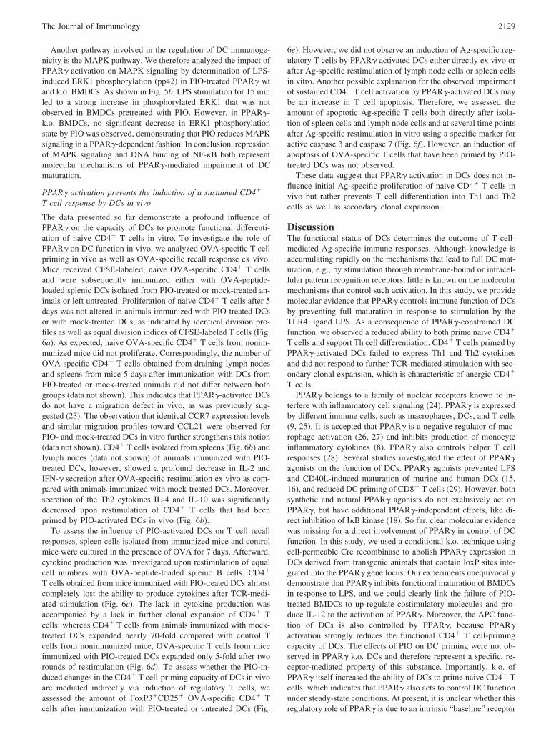
FIGURE 6. PPAR� activation in DCs prevents the induction of a sustained CD4� T cell response in vivo. At day �1, 5 � 106 CFSE-labeled (a) orunlabeled (b–f) DO11.10 CD4� T cells were injected i.v. into BALB/c mice. At day 0, these mice were immunized s.c. with 1 � 106 OVA-loaded splenicDC from mice treated with PIO or vehicle for 7 days; unimmunized mice served as controls. a, After 5 days, spleen cells were isolated and proliferationprofile and cell division index (CDI) of CFSE� KJ1-26� T cells was determined by flow cytometry. b, Five days after DC immunization, spleen cells wereisolated and equal cell numbers were restimulated with 10 �M OVA323–339 peptide. IL-2 and IFN-� content as well as IL-4 and IL-10 content in culturesupernatants were determined by ELISA after 20 and 72 h, respectively. c, Spleen cells from immunized mice were cultured in the presence of 0.5 mg/mlOVA for 7 days. Afterward, CD4� T cells were harvested and restimulated with splenic B cells loaded with OVA323–339 peptide (recall assay). After 20 h,IL-2 and IFN-� contents in culture supernatants were determined by ELISA. d, After 7 days restimulation of splenic T cells in vitro, numbers ofOVA-specific T cells were assessed by flow cytometric quantification of KJ1-26-positive T cells in total CD4� T cells; graph shows relative increase inAg-specific T cells compared with nonimmunized control animals. a–d, Values are depicted as mean � SEM; significant differences compared with DCwithout PIO are indicated (�, p � 0.05; ��, p � 0.01, Student’s t test). e, For analysis of regulatory T cells, spleen cells isolated 5 days after immunizationwere either stained ex vivo or first cultured in the presence of OVA for 3 days and afterward stained for CD4, KJ1-26, CD25, and intracellular FoxP3. Dotplots show CD25�, FoxP3� cells in the CD4� T cell population. The table depicts the percentage of CD25�FoxP3� cells in total KJ1-26� cells ex vivo,3 days after culture with OVA (restim) and 2 days after secondary restimulation with splenic B cells loaded with OVA323–339 peptide (recall). f, Fordetermination of apoptosis, spleen cells were labeled for active caspase 3 � 7 (FAM) for 1 h, and afterward stained for CD4, KJ1-26, and 7-AAD. Dotplots depict the percentage of early apoptotic (FAM single positive) and late apoptotic (FAM and 7-AAD double positive) cells in total CD4� T cells 1day after OVA-specific restimulation. The table depicts the percentage of FAM-positive cells in total KJ1-26� cells ex vivo, 1 day after culture with OVA(restim) and 1 day after secondary restimulation with splenic B cells loaded with OVA323–339 peptide (recall).
2128 PPAR�-ACTIVATED DCs INDUCE CD4� T CELL ANERGY
Another pathway involved in the regulation of DC immunoge-nicity is the MAPK pathway. We therefore analyzed the impact ofPPAR� activation on MAPK signaling by determination of LPS-induced ERK1 phosphorylation (pp42) in PIO-treated PPAR� wtand k.o. BMDCs. As shown in Fig. 5b, LPS stimulation for 15 minled to a strong increase in phosphorylated ERK1 that was notobserved in BMDCs pretreated with PIO. However, in PPAR�-k.o. BMDCs, no significant decrease in ERK1 phosphorylationstate by PIO was observed, demonstrating that PIO reduces MAPKsignaling in a PPAR�-dependent fashion. In conclusion, repressionof MAPK signaling and DNA binding of NF-�B both representmolecular mechanisms of PPAR�-mediated impairment of DCmaturation.
PPAR� activation prevents the induction of a sustained CD4�
T cell response by DCs in vivo
The data presented so far demonstrate a profound influence ofPPAR� on the capacity of DCs to promote functional differenti-ation of naive CD4� T cells in vitro. To investigate the role ofPPAR� on DC function in vivo, we analyzed OVA-specific T cellpriming in vivo as well as OVA-specific recall response ex vivo.Mice received CFSE-labeled, naive OVA-specific CD4� T cellsand were subsequently immunized either with OVA-peptide-loaded splenic DCs isolated from PIO-treated or mock-treated an-imals or left untreated. Proliferation of naive CD4� T cells after 5days was not altered in animals immunized with PIO-treated DCsor with mock-treated DCs, as indicated by identical division pro-files as well as equal division indices of CFSE-labeled T cells (Fig.6a). As expected, naive OVA-specific CD4� T cells from nonim-munized mice did not proliferate. Correspondingly, the number ofOVA-specific CD4� T cells obtained from draining lymph nodesand spleens from mice 5 days after immunization with DCs fromPIO-treated or mock-treated animals did not differ between bothgroups (data not shown). This indicates that PPAR�-activated DCsdo not have a migration defect in vivo, as was previously sug-gested (23). The observation that identical CCR7 expression levelsand similar migration profiles toward CCL21 were observed forPIO- and mock-treated DCs in vitro further strengthens this notion(data not shown). CD4� T cells isolated from spleens (Fig. 6b) andlymph nodes (data not shown) of animals immunized with PIO-treated DCs, however, showed a profound decrease in IL-2 andIFN-� secretion after OVA-specific restimulation ex vivo as com-pared with animals immunized with mock-treated DCs. Moreover,secretion of the Th2 cytokines IL-4 and IL-10 was significantlydecreased upon restimulation of CD4� T cells that had beenprimed by PIO-activated DCs in vivo (Fig. 6b).
To assess the influence of PIO-activated DCs on T cell recallresponses, spleen cells isolated from immunized mice and controlmice were cultured in the presence of OVA for 7 days. Afterward,cytokine production was investigated upon restimulation of equalcell numbers with OVA-peptide-loaded splenic B cells. CD4�
T cells obtained from mice immunized with PIO-treated DCs almostcompletely lost the ability to produce cytokines after TCR-medi-ated stimulation (Fig. 6c). The lack in cytokine production wasaccompanied by a lack in further clonal expansion of CD4� Tcells: whereas CD4� T cells from animals immunized with mock-treated DCs expanded nearly 70-fold compared with control Tcells from nonimmunized mice, OVA-specific T cells from miceimmunized with PIO-treated DCs expanded only 5-fold after tworounds of restimulation (Fig. 6d). To assess whether the PIO-in-duced changes in the CD4� T cell-priming capacity of DCs in vivoare mediated indirectly via induction of regulatory T cells, weassessed the amount of FoxP3�CD25� OVA-specific CD4� Tcells after immunization with PIO-treated or untreated DCs (Fig.
6e). However, we did not observe an induction of Ag-specific reg-ulatory T cells by PPAR�-activated DCs either directly ex vivo orafter Ag-specific restimulation of lymph node cells or spleen cellsin vitro. Another possible explanation for the observed impairmentof sustained CD4� T cell activation by PPAR�-activated DCs maybe an increase in T cell apoptosis. Therefore, we assessed theamount of apoptotic Ag-specific T cells both directly after isola-tion of spleen cells and lymph node cells and at several time pointsafter Ag-specific restimulation in vitro using a specific marker foractive caspase 3 and caspase 7 (Fig. 6f). However, an induction ofapoptosis of OVA-specific T cells that have been primed by PIO-treated DCs was not observed.
These data suggest that PPAR� activation in DCs does not in-fluence initial Ag-specific proliferation of naive CD4� T cells invivo but rather prevents T cell differentiation into Th1 and Th2cells as well as secondary clonal expansion.
DiscussionThe functional status of DCs determines the outcome of T cell-mediated Ag-specific immune responses. Although knowledge isaccumulating rapidly on the mechanisms that lead to full DC mat-uration, e.g., by stimulation through membrane-bound or intracel-lular pattern recognition receptors, little is known on the molecularmechanisms that control such activation. In this study, we providemolecular evidence that PPAR� controls immune function of DCsby preventing full maturation in response to stimulation by theTLR4 ligand LPS. As a consequence of PPAR�-constrained DCfunction, we observed a reduced ability to both prime naive CD4�
T cells and support Th cell differentiation. CD4� T cells primed byPPAR�-activated DCs failed to express Th1 and Th2 cytokinesand did not respond to further TCR-mediated stimulation with sec-ondary clonal expansion, which is characteristic of anergic CD4�
T cells.PPAR� belongs to a family of nuclear receptors known to in-
terfere with inflammatory cell signaling (24). PPAR� is expressedby different immune cells, such as macrophages, DCs, and T cells(9, 25). It is accepted that PPAR� is a negative regulator of mac-rophage activation (26, 27) and inhibits production of monocyteinflammatory cytokines (8). PPAR� also controls helper T cellresponses (28). Several studies investigated the effect of PPAR�agonists on the function of DCs. PPAR� agonists prevented LPSand CD40L-induced maturation of murine and human DCs (15,16), and reduced DC priming of CD8� T cells (29). However, bothsynthetic and natural PPAR� agonists do not exclusively act onPPAR�, but have additional PPAR�-independent effects, like di-rect inhibition of I�B kinase (18). So far, clear molecular evidencewas missing for a direct involvement of PPAR� in control of DCfunction. In this study, we used a conditional k.o. technique usingcell-permeable Cre recombinase to abolish PPAR� expression inDCs derived from transgenic animals that contain loxP sites inte-grated into the PPAR� gene locus. Our experiments unequivocallydemonstrate that PPAR� inhibits functional maturation of BMDCsin response to LPS, and we could clearly link the failure of PIO-treated BMDCs to up-regulate costimulatory molecules and pro-duce IL-12 to the activation of PPAR�. Moreover, the APC func-tion of DCs is also controlled by PPAR�, because PPAR�activation strongly reduces the functional CD4� T cell-primingcapacity of DCs. The effects of PIO on DC priming were not ob-served in PPAR� k.o. DCs and therefore represent a specific, re-ceptor-mediated property of this substance. Importantly, k.o. ofPPAR� itself increased the ability of DCs to prime naive CD4� Tcells, which indicates that PPAR� also acts to control DC functionunder steady-state conditions. At present, it is unclear whether thisregulatory role of PPAR� is due to an intrinsic “baseline” receptor
2129The Journal of Immunology

activity that is ligand-independent, or is mediated by yet unknownendogenous PPAR�-ligands derived either from the DC itself orfrom interacting T cells. Interestingly, the increase in CD4� Tcell-priming capacity after k.o. of PPAR� was not accompanied byan increase in steady-state expression levels of the costimulatorymolecules CD40, CD80, and CD86, or by enhanced IL-12 pro-duction, suggesting that PPAR� may additionally regulate DC ex-pression of other costimulatory or perhaps coinhibitory molecules.Ag uptake was strongly increased by PIO, as has already beendescribed by others (30). However, this increase was not depen-dent on PPAR�, because PPAR� wt and k.o. BMDCs exhibit equalAg uptake both in the presence or absence of PIO.
A control of DC function is important to regulate Ag-specificimmune responses. Several molecular mechanisms involving in-terference with signal transduction pathways have been character-ized that prevent full activation of APCs in response to TLR-me-diated signals (31, 32). We demonstrate in this study that PPAR�interferes with LPS-induced NF-�B DNA-binding activity in DCs,which is known to be important for full functional maturation (33,34). Additionally, we show that PPAR� activation decreased LPS-induced phosphorylation of ERK1, thus interfering with MAPKsignaling. This is in line with observations made by Appel et al.(35), who showed that the PPAR� agonist troglitazone interferedwith TLR ligand-induced MAPK signaling in human DCs result-ing in reduced DC activation. Thus, it appears that the immunos-timulatory capacity of DCs is stringently controlled by regulatorymechanisms that involve signaling via PPAR�.
Given the potent activity of PPAR� on DC function in vitro, weinvestigated whether this is also true for the in vivo situation. Anal-ysis of CD4� T cell priming in vivo revealed that Ag-specificCD4� T cell proliferation was induced to the same extent both byPPAR�-activated and untreated DCs. However, CD4� T cellsprimed by PPAR�-activated DCs exhibited impaired effector func-tion, and secondary clonal expansion was severely blunted uponrestimulation. It has already been shown that initial proliferation ofnaive CD8� T cells can occur in the absence of APC-derivedsignals that are essential for full T cell activation and differentia-tion (36). A lack of sufficient IL-2 signaling during the priming ofCD8� T cells is associated with failure to differentiate into effectorcells and to undergo secondary clonal expansion (37). Accord-ingly, proliferation of CD4� T cells primed by PPAR�-activatedDCs was not impaired. However, low IL-2 production by these Tcells during priming suggests that the observed lack of differenti-ation and clonal expansion may be due to insufficient IL-2 signal-ing. We therefore propose that PPAR� controls DC function in acomplex manner that permits survival of Ag-reactive CD4� Tcells, but induces CD4� T cell anergy instead of immunity uponsecondary Ag encounter. The observed lack in both Th1 and Th2cytokines produced by CD4� T cells that were primed by PPAR�-activated DCs in vivo is in contrast to a switch toward Th2 re-sponses by PPAR�-activated DCs observed by Gosset et al. (16).However, Gosset et al. (16) used shorter treatment periods withPPAR� agonists, probably resulting in less pronounced control ofDC function.
Interestingly, it has been shown that agonists of the vitamin Dreceptor, another member of the steroid hormone receptor super-family, also inhibit maturation and Ag-presenting capacity of DCsin a NF-�B-dependent fashion (38, 39). However, this processinvolves induction of IL-10 secretion by DCs that in turn proved tobe essential for induction of regulatory T cells (40). In contrast tothese findings, we did not observe an increase in IL-10 productionby PPAR�-activated DCs. Correspondingly, we did not find anincrease in regulatory T cells upon encounter with PPAR�-acti-vated DCs in vivo. Furthermore, it has been shown that vitamin D
receptor-treated DCs are capable of selective induction of T cellapoptosis (41). Increased cell death could also contribute to theobserved lack in effector function in CD4� T cells primed byPPAR�-activated DCs. We did not, however, observe increased Tcell apoptosis in vitro (data not shown) or ex vivo, therefore ex-cluding this possibility. These observations demonstrate that de-spite a close relationship between these receptor families and sim-ilar net effects on the immunostimulatory capacity of DCs, theunderlying mechanisms differ considerably.
If PPAR� activity dominates DC function, it is difficult to en-visage how sufficient immunity to infections arises. However, pa-tients taking PPAR� agonists for metabolic control of type II di-abetes are not immune-suppressed. One possible explanation forinflammation-induced reversal of PPAR�-mediated effects on DCsis that proinflammatory mediators like IFN-� and TNF-� are ableto reduce PPAR� protein levels both by increased proteasomaldegradation and by reduced gene expression (42, 43). It will beimportant to determine in future experiments, which molecularmechanisms override PPAR�-mediated control of DC function invivo and thus contribute to the induction of immunity in responseto infection.
Collectively, our data provide clear molecular evidence thatPPAR� controls DC function in response to proinflammatory stim-uli, but also under steady-state conditions. PPAR� activation re-strains costimulatory signals in DCs resulting in inhibition of naiveCD4� T cells to undergo differentiation and subsequent secondaryclonal expansion. The elucidation of the molecular mechanismscontrolling DC function will be crucial for the development oftreatment strategies, on the one hand to improve tolerogenic DCfunction for treatment of autoimmune diseases, and on the otherhand for the targeted disinhibition of DC function to improve vac-cination strategies.
DisclosuresThe authors have no financial conflict of interest.
References1. Banchereau, J., and R. M. Steinman. 1998. Dendritic cells and the control of
immunity. Nature 392: 245–252.2. Banchereau, J., F. Briere, C. Caux, J. Davoust, S. Lebecque, Y. J. Liu, B.
Pulendran, and K. Palucka. 2000. Immunobiology of dendritic cells. Annu. Rev.Immunol. 18: 767–811.
3. Heath, W. R., and F. R. Carbone. 2001. Cross-presentation, dendritic cells, tol-erance and immunity. Annu. Rev. Immunol. 19: 47–64.
4. Medzhitov, R., and C. Janeway, Jr. 2000. Innate immune recognition: mecha-nisms and pathways. Immunol. Rev. 173: 89–97.
5. Dhodapkar, M. V., R. M. Steinman, J. Krasovsky, C. Munz, and N. Bhardwaj.2001. Antigen-specific inhibition of effector T cell function in humans after in-jection of immature dendritic cells. J. Exp. Med. 193: 233–238.
6. Martin, E., B. O’Sullivan, P. Low, and R. Thomas. 2003. Antigen-specific sup-pression of a primed immune response by dendritic cells mediated by regulatoryT cells secreting interleukin-10. Immunity 18: 155–167.
7. McIlroy, A., G. Caron, S. Blanchard, I. Fremaux, D. Duluc, Y. Delneste, A.Chevailler, and P. Jeannin. 2006. Histamine and prostaglandin E up-regulate theproduction of Th2-attracting chemokines (CCL17 and CCL22) and down-regu-late IFN-�-induced CXCL10 production by immature human dendritic cells. Im-munology 117: 507–516.
8. Jiang, C., A. T. Ting, and B. Seed. 1998. PPAR-� agonists inhibit production ofmonocyte inflammatory cytokines. Nature 391: 82–86.
9. Ricote, M., A. C. Li, T. M. Willson, C. J. Kelly, and C. K. Glass. 1998. Theperoxisome proliferator-activated receptor-� is a negative regulator of macro-phage activation. Nature 391: 79–82.
10. Yang, X. Y., L. H. Wang, T. Chen, D. R. Hodge, J. H. Resau, L. DaSilva, andW. L. Farrar. 2000. Activation of human T lymphocytes is inhibited by peroxi-some proliferator-activated receptor � (PPAR�) agonists: PPAR� co-associationwith transcription factor NFAT. J. Biol. Chem. 275: 4541–4544.
11. Von Knethen, A., and B. Brune. 2002. Activation of peroxisome proliferator-activated receptor � by nitric oxide in monocytes/macrophages down-regulatesp47phox and attenuates the respiratory burst. J. Immunol. 169: 2619–2626.
12. Cunard, R., Y. Eto, J. T. Muljadi, C. K. Glass, C. J. Kelly, and M. Ricote. 2004.Repression of IFN-� expression by peroxisome proliferator-activated receptor �.J. Immunol. 172: 7530–7336.
2130 PPAR�-ACTIVATED DCs INDUCE CD4� T CELL ANERGY

13. Lehmann, J. M., J. M. Lenhard, B. B. Oliver, G. M. Ringold, and S. A. Kliewer.1997. Peroxisome proliferator-activated receptors � and � are activated by indo-methacin and other non-steroidal anti-inflammatory drugs. J. Biol. Chem. 272:3406–3410.
14. Clark, R. B., D. Bishop-Bailey, T. Estrada-Hernandez, T. Hla, L. Puddington, andS. J. Padula. 2000. The nuclear receptor PPAR� and immunoregulation: PPAR�mediates inhibition of helper T cell responses. J. Immunol. 164: 1364–1371.
15. Faveeuw, C., S. Fougeray, V. Angeli, J. Fontaine, G. Chinetti, P. Gosset, P.Delerive, C. Maliszewski, M. Capron, B. Staels, et al. 2000. Peroxisome prolif-erator-activated receptor � activators inhibit interleukin-12 production in murinedendritic cells. FEBS Lett. 486: 261–266.
16. Gosset, P., A. S. Charbonnier, P. Delerive, J. Fontaine, B. Staels, J. Pestel, A. B.Tonnel, and F. Trottein. 2001. Peroxisome proliferator-activated receptor � ac-tivators affect the maturation of human monocyte-derived dendritic cells. Eur.J. Immunol. 31: 2857–2865.
17. Akiyama, T. E., S. Sakai, G. Lambert, C. J. Nicol, K. Matsusue, S. Pimprale,Y. H. Lee, M. Ricote, C. K. Glass, H. B. Brewer, Jr., and F. J. Gonzalez. 2002.Conditional disruption of the peroxisome proliferator-activated receptor � gene inmice results in lowered expression of ABCA1, ABCG1, and apoE in macro-phages and reduced cholesterol efflux. Mol. Cell. Biol. 22: 2607–2619.
18. Rossi, A., P. Kapahi, G. Natoli, T. Takahashi, Y. Chen, M. Karin, and M. G.Santoro. Anti-inflammatory cyclopentenone prostaglandins are direct inhibitorsof I�B kinase. Nature 403: 103–108.
19. Chawla, A., Y. Barak, L. Nagy, D. Liao, P. Tontonoz, and R. M. Evans. 2001.PPAR-� dependent and independent effects on macrophage-gene expression inlipid metabolism and inflammation. Nat. Med. 7: 48–52.
20. He, W., Y. Barak, A. Hevener, P. Olson, D. Liao, J. Le, M. Nelson, E. Ong,J. M. Olefsky, and R. M. Evans. 2003. Adipose-specific peroxisome proliferator-activated receptor � knockout causes insulin resistance in fat and liver but not inmuscle. Proc. Natl. Acad. Sci. USA 100: 15712–15717.
21. Nolden, L., F. Edenhofer, S. Haupt, P. Koch, F. T. Wunderlich, H. Siemen, andO. Brustle. 2006. Site-specific recombination in human embryonic stem cellsinduced by cell-permeant Cre recombinase. Nat. Methods 3: 461–467.
22. Yajima, K., H. Hirose, H. Fujita, Y. Seto, H. Fujita, K. Ukeda, K. Miyashita,T. Kawai, Y. Yamamoto, T. Ogawa, et al. 2003. Combination therapy withPPAR� and PPAR� agonists increases glucose-stimulated insulin secretion indb/db mice. Am. J. Physiol. 284: E966–E971.
23. Angeli, V., H. Hammad, B. Staels, M. Capron, B. N. Lambrecht, and F. Trottein.2003. Peroxisome proliferator-activated receptor � inhibits the migration of den-dritic cells: consequences for the immune response. J. Immunol. 170: 5295–5301.
24. Daynes, R. A., and D. C. Jones. 2002. Emerging roles of PPARs in inflammationand immunity. Nat. Rev. Immunol. 2: 748–759.
25. Clark, R. B. 2002. The role of PPARs in inflammation and immunity. J.Leukocyte Biol. 71: 388–400.
26. Ricote, M., J. Huang, L. Fajas, A. Li, J. Welch, J. Najib, J. L. Witztum, J. Auwerx,W. Palinski, and C. K. Glass. 1998. Expression of the peroxisome proliferator-acti-vated receptor � (PPAR�) in human atherosclerosis and regulation in macrophagesby colony stimulating factors and oxidized low density lipoprotein. Proc. Natl. Acad.Sci. USA 95: 7614–7619.
27. Nagy, L., P. Tontonoz, J. G. Alvarez, H. Chen, and R. M. Evans. 1998. OxidizedLDL regulates macrophage gene expression through ligand activation of PPAR�.Cell 93: 229–240.
28. Clark, R. B., D. Bishop-Bailey, T. Estrada-Hernandez, T. Hla, L. Puddington, andS. J. Padula. 2000. The nuclear receptor PPAR� and immunoregulation: PPAR�mediates inhibition of helper T cell responses. J. Immunol. 164: 1364–1371.
29. Nencioni, A., F. Grunebach, A. Zobywlaski, C. Denzlinger, W. Brugger, and P.Brossart. 2002. Dendritic cell immunogenicity is regulated by peroxisome pro-liferator-activated receptor �. J. Immunol. 169: 1228–1235.
30. Szatmari, I., P. Gogolak, J. S. Im, B. Dezso, E. Rajnavolgyi, and L. Nagy. 2004.Activation of PPAR� specifies a dendritic cell subtype capable of enhanced in-duction of iNKT cell expansion. Immunity 21: 95–106.
31. Kobayashi, K., L. D. Hernandez, J. E. Galan, C. A. Janeway, Jr., R. Medzhitov,and R. A. Flavell. 2002. IRAK-M is a negative regulator of Toll-like receptorsignaling. Cell 110: 191–202.
32. Wald, D., J. Qin, Z. Zhao, Y. Qian, M. Naramura, L. Tian, J. Towne, J. E. Sims,G. R. Stark, and X. Li. 2003. SIGIRR, a negative regulator of Toll-like receptor-interleukin 1 receptor signaling. Nat. Immunol. 4: 920–927.
33. O’Sullivan, B. J., and R. Thomas. 2002. CD40 ligation conditions dendritic cellantigen-presenting function through sustained activation of NF-�B. J. Immunol.168: 5491–5498.
34. Murphy, T. L., M. G. Cleveland, P. Kulesza, J. Magram, and K. M. Murphy.1995. Regulation of interleukin 12 p40 expression through an NF-�B half-site.Mol. Cell. Biol. 15: 5258–5267.
35. Appel, S., V. Mirakaj, A. Bringmann, M. M. Weck, F. Grunebach, and P.Brossart. 2005. PPAR-� agonists inhibit Toll-like receptor-mediated activation ofdendritic cells via the MAP kinase and NF-�B pathways. Blood 106: 3888–3894.
36. Limmer, A., J. Ohl, C. Kurts, H. G. Ljunggren, Y. Reiss, M. Groettrup, F.Momburg, B. Arnold, and P. A. Knolle. 2000. Efficient presentation of exogenousantigen by liver endothelial cells to CD8� T cells results in antigen-specificT-cell tolerance. Nat. Med. 6: 1348–1354.
37. Williams, M. A., A. J. Tyznik, and M. J. Bevan. 2006. Interleukin-2 signalsduring priming are required for secondary expansion of CD8� memory T cells.Nature 441: 890–893.
38. Penna, G., and L. Adorini. 2000. 1�,25-Dihydroxyvitamin D3 inhibits differen-tiation, maturation, activation, and survival of dendritic cells leading to impairedalloreactive T cell activation. J. Immunol. 164: 2405–2411.
39. Dong, X., W. Lutz, T. M. Schroeder, L. A. Bachman, J. J. Westendorf, R. Kumar,and M. D. Griffin. 2005. Regulation of relB in dendritic cells by means of mod-ulated association of vitamin D receptor and histone deacetylase 3 with the pro-moter. Proc. Natl. Acad. Sci. USA 102: 16007–16012.
40. Penna, G., A. Roncari, S. Amuchastegui, K. C. Daniel, E. Berti, M. Colonna, andL. Adorini. 2005. Expression of the inhibitory receptor ILT3 on dendritic cells isdispensable for induction of CD4�Foxp3� regulatory T cells by 1,25-dihy-droxyvitamin D3. Blood 106: 3490–3497.
41. van Halteren, A. G., O. M. Tysma, E. van Etten, C. Mathieu, and B. O. Roep.2004. 1�,25-Dihydroxyvitamin D3 or analogue treated dendritic cells modulatehuman autoreactive T cells via the selective induction of apoptosis.J. Autoimmun. 23: 233–239.
42. Waite, K. J., Z. E. Floyd, P. Arbour-Reily, and J. M. Stephens. 2001. Interferon-�-induced regulation of peroxisome proliferator-activated receptor � and STATsin adipocytes. J. Biol. Chem. 276: 7062–7068.
43. Xing, H., J. P. Northrop, J. R. Grove, K. E. Kilpatrick, J. L. Su, and G. M.Ringold. 1997. TNF�-mediated inhibition and reversal of adipocyte differentia-tion is accompanied by suppressed expression of PPAR� without effects onPref-1 expression. Endocrinology 138: 2776–2783.
2131The Journal of Immunology