Mechanisms of divergent effects of activated peroxisome proliferator-activated receptor-γ on...
10
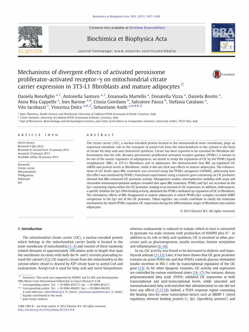
Mechanisms of divergent effects of activated peroxisome proliferator-activated receptor-γ on mitochondrial citrate carrier expression in 3T3-L1 fibroblasts and mature adipocytes ☆ Daniela Bonofiglio a, 1 , Antonella Santoro a, 1 , Emanuela Martello a , Donatella Vizza a , Daniela Rovito a , Anna Rita Cappello a , Ines Barone a, b , Cinzia Giordano b , Salvatore Panza b , Stefania Catalano a , Vito Iacobazzi c , Vincenza Dolce a, ⁎ , 2 , Sebastiano Andò a, b, ⁎⁎ , 2 a Dept. Pharmacy, Health Sciences and Nutritional, University of Calabria 87036 Arcavacata di Rende (Cosenza), Italy b Centro Sanitario, University of Calabria 87036 Arcavacata di Rende (Cosenza), Italy c Dept of Biosciences, Biotechnology and Farmacological Sciences, and Center of Excellence in Comparative Genomics, University of Bari, 70125 Bari, Italy abstract article info Article history: Received 9 July 2012 Received in revised form 16 January 2013 Accepted 18 January 2013 Available online 28 January 2013 Keywords: Citrate carrier Mitochondrion PPARgamma Adipocyte Sp1 The citrate carrier (CIC), a nuclear-encoded protein located in the mitochondrial inner membrane, plays an important metabolic role in the transport of acetyl-CoA from the mitochondrion to the cytosol in the form of citrate for fatty acid and cholesterol synthesis. Citrate has been reported to be essential for fibroblast dif- ferentiation into fat cells. Because peroxisome proliferator-activated receptor-gamma (PPARγ) is known to be one of the master regulators of adipogenesis, we aimed to study the regulation of CIC by the PPARγ ligand rosiglitazone (BRL) in 3T3-L1 fibroblasts and in adipocytes. We demonstrated that BRL up-regulated CIC mRNA and protein levels in fibroblasts, while it did not elicit any effects in mature adipocytes. The enhance- ment of CIC levels upon BRL treatment was reversed using the PPARγ antagonist GW9662, addressing how this effect was mediated by PPARγ. Functional experiments using a reporter gene containing rat CIC promoter showed that BRL enhanced CIC promoter activity. Mutagenesis studies, electrophoretic-mobility-shift assay and chromatin-immunoprecipitation analysis revealed that upon BRL treatment, PPARγ and Sp1 are recruited on the Sp1-containing region within the CIC promoter, leading to an increase in CIC expression. In addition, mithramycin, a specific inhibitor for Sp1-DNA binding activity, abolished the PPARγ-mediated up-regulation of CIC in fibroblasts. The stimulatory effects of BRL disappeared in mature adipocytes in which PPARγ/Sp1 complex recruited SMRT corepressor to the Sp1 site of the CIC promoter. Taken together, our results contribute to clarify the molecular mechanisms by which PPARγ regulates CIC expression during the differentiation stages of fibroblasts into mature adipocytes. © 2013 Elsevier B.V. All rights reserved. 1. Introduction The mitochondrial citrate carrier (CIC), a nuclear-encoded protein which belongs to the mitochondrial carrier family is located in the inner membrane of mitochondria [1–3] and consists of three tandemly related domains of approximately 100 amino acids in length that span the membrane six times with both the N- and C-termini protruding to- ward the cytosol [4,5]. CIC exports citrate from the mitochondria to the cytosol where citrate is cleaved by ATP-citrate lyase to acetyl-CoA and oxaloacetate. Acetyl-CoA is used for fatty acid and sterol biosynthesis; whereas oxaloacetate is reduced to malate, which in turn is converted to pyruvate via malic enzyme with production of NADPH plus H + . In addition to its role in fatty acid synthesis, CIC is involved in other pro- cesses such as gluconeogenesis, insulin secretion, histone acetylation and inflammation [6–10]. In rat, CIC activity was found to be decreased in diabetic and hypo- thyroid animals [11,12]. Later, it has been shown that CIC gene promoter contains an active FOXA site and that FOXA1 controls glucose-stimulated insulin secretion in INS-1 cells by transcriptional regulation of the CIC gene [13]. As for other lipogenic enzymes, CIC activity and expression are controlled by various nutritional states [14–17]. For instance, dietary polyunsaturated fatty acids (PUFA) inhibited CIC expression at both transcriptional and post-transcriptional levels, while saturated and monounsaturated fatty acid-enriched diet administration to rats did not have any effects [7,17,18]. Indeed, a PUFA response region containing the binding sites for some transcription factors such as SREBP-1 (sterol regulatory element binding protein-1), Sp1 (Specificity protein1) and Biochimica et Biophysica Acta 1831 (2013) 1027–1036 ☆ Footnotes: This work was supported by MURST and Ex 60% and Reintegration AIRC/Marie Curie International Fellowship in Cancer Research to IB. ⁎ Corresponding author. Tel.: +39 0984 493177; fax: +39 0984 493271. ⁎⁎ Corresponding author. Tel.: +39 0984 496201; fax: +39 0984 496203. E-mail addresses: [email protected] (V. Dolce), [email protected] (S. Andò). 1 Equally contributed to the work. 2 Joint Senior Authors. 1388-1981/$ – see front matter © 2013 Elsevier B.V. All rights reserved. http://dx.doi.org/10.1016/j.bbalip.2013.01.014 Contents lists available at SciVerse ScienceDirect Biochimica et Biophysica Acta journal homepage: www.elsevier.com/locate/bbalip
-
Upload
independent -
Category
Documents
-
view
1 -
download
0
Transcript of Mechanisms of divergent effects of activated peroxisome proliferator-activated receptor-γ on...
Biochimica et Biophysica Acta 1831 (2013) 1027–1036
Contents lists available at SciVerse ScienceDirect
Biochimica et Biophysica Acta
j ourna l homepage: www.e lsev ie r .com/ locate /bba l ip
Mechanisms of divergent effects of activated peroxisomeproliferator-activated receptor-γ on mitochondrial citratecarrier expression in 3T3-L1 fibroblasts and mature adipocytes☆
Daniela Bonofiglio a,1, Antonella Santoro a,1, Emanuela Martello a, Donatella Vizza a, Daniela Rovito a,Anna Rita Cappello a, Ines Barone a,b, Cinzia Giordano b, Salvatore Panza b, Stefania Catalano a,Vito Iacobazzi c, Vincenza Dolce a,⁎,2, Sebastiano Andò a,b,⁎⁎,2
a Dept. Pharmacy, Health Sciences and Nutritional, University of Calabria 87036 Arcavacata di Rende (Cosenza), Italyb Centro Sanitario, University of Calabria 87036 Arcavacata di Rende (Cosenza), Italyc Dept of Biosciences, Biotechnology and Farmacological Sciences, and Center of Excellence in Comparative Genomics, University of Bari, 70125 Bari, Italy
☆ Footnotes: This work was supported by MURST andAIRC/Marie Curie International Fellowship in Cancer Re⁎ Corresponding author. Tel.: +39 0984 493177; fax:
⁎⁎ Corresponding author. Tel.: +39 0984 496201; fax:E-mail addresses: [email protected] (V. Dolce), sebast
1 Equally contributed to the work.2 Joint Senior Authors.
1388-1981/$ – see front matter © 2013 Elsevier B.V. Allhttp://dx.doi.org/10.1016/j.bbalip.2013.01.014
a b s t r a c t
a r t i c l e i n f oArticle history:Received 9 July 2012Received in revised form 16 January 2013Accepted 18 January 2013Available online 28 January 2013
Keywords:Citrate carrierMitochondrionPPARgammaAdipocyteSp1
The citrate carrier (CIC), a nuclear-encoded protein located in the mitochondrial inner membrane, plays animportant metabolic role in the transport of acetyl-CoA from the mitochondrion to the cytosol in the formof citrate for fatty acid and cholesterol synthesis. Citrate has been reported to be essential for fibroblast dif-ferentiation into fat cells. Because peroxisome proliferator-activated receptor-gamma (PPARγ) is known tobe one of the master regulators of adipogenesis, we aimed to study the regulation of CIC by the PPARγ ligandrosiglitazone (BRL) in 3T3-L1 fibroblasts and in adipocytes. We demonstrated that BRL up-regulated CICmRNA and protein levels in fibroblasts, while it did not elicit any effects in mature adipocytes. The enhance-ment of CIC levels upon BRL treatment was reversed using the PPARγ antagonist GW9662, addressing howthis effect was mediated by PPARγ. Functional experiments using a reporter gene containing rat CIC promotershowed that BRL enhanced CIC promoter activity. Mutagenesis studies, electrophoretic-mobility-shift assay andchromatin-immunoprecipitation analysis revealed that upon BRL treatment, PPARγ and Sp1 are recruited on theSp1-containing regionwithin the CIC promoter, leading to an increase in CIC expression. In addition, mithramycin,a specific inhibitor for Sp1-DNAbinding activity, abolished the PPARγ-mediated up-regulation of CIC infibroblasts.The stimulatory effects of BRL disappeared in mature adipocytes in which PPARγ/Sp1 complex recruited SMRTcorepressor to the Sp1 site of the CIC promoter. Taken together, our results contribute to clarify the molecularmechanisms by which PPARγ regulates CIC expression during the differentiation stages of fibroblasts into matureadipocytes.
© 2013 Elsevier B.V. All rights reserved.
1. Introduction
The mitochondrial citrate carrier (CIC), a nuclear-encoded proteinwhich belongs to the mitochondrial carrier family is located in theinner membrane of mitochondria [1–3] and consists of three tandemlyrelated domains of approximately 100 amino acids in length that spanthe membrane six times with both the N- and C-termini protruding to-ward the cytosol [4,5]. CIC exports citrate from the mitochondria to thecytosol where citrate is cleaved by ATP-citrate lyase to acetyl-CoA andoxaloacetate. Acetyl-CoA is used for fatty acid and sterol biosynthesis;
Ex 60% and Reintegrationsearch to IB.+39 0984 493271.+39 0984 [email protected] (S. Andò).
rights reserved.
whereas oxaloacetate is reduced to malate, which in turn is convertedto pyruvate via malic enzyme with production of NADPH plus H+. Inaddition to its role in fatty acid synthesis, CIC is involved in other pro-cesses such as gluconeogenesis, insulin secretion, histone acetylationand inflammation [6–10].
In rat, CIC activity was found to be decreased in diabetic and hypo-thyroid animals [11,12]. Later, it has been shown that CIC gene promotercontains an active FOXA site and that FOXA1 controls glucose-stimulatedinsulin secretion in INS-1 cells by transcriptional regulation of the CICgene [13]. As for other lipogenic enzymes, CIC activity and expressionare controlled by various nutritional states [14–17]. For instance, dietarypolyunsaturated fatty acids (PUFA) inhibited CIC expression at bothtranscriptional and post-transcriptional levels, while saturated andmonounsaturated fatty acid-enriched diet administration to rats did nothave any effects [7,17,18]. Indeed, a PUFA response region containingthe binding sites for some transcription factors such as SREBP-1 (sterolregulatory element binding protein-1), Sp1 (Specificity protein1) and
1028 D. Bonofiglio et al. / Biochimica et Biophysica Acta 1831 (2013) 1027–1036
NF-Y (Nuclear Factor-Y) has been identified in the CIC gene promoter[7,19,20]. SREBP-1 activates the expression of CIC in HepG2 cells [7],hepatocytes [20] and mammary epithelium [21]. Recently, Damianoet al. have demonstrated that CIC expression, in hepatocytes and adipo-cytes, is regulated by ligands of peroxisome proliferator-activated recep-tor (PPAR) α and γ, identifying a peroxisome proliferator-activatedreceptor responsive element (PPRE) motif at −625 bp of CIC pro-moter [22].
PPARs are members of nuclear hormone receptors superfamilythat function as ligand-dependent transcription factors [23]. ThreePPAR isoforms, α, β/δ, and γ, are expressed in multiple species ina tissue-specific manner [24,25]. Among them, PPARγ integratesthe control of energy, lipid and glucose homeostasis participatingin the transcriptional activation of several genes important for adi-pocyte maturation, lipid accumulation, and insulin-sensitive glu-cose transport, as adipocyte fatty acid binding protein aP2, CCAAT/enhancer-binding protein-α (C/EBPα), Perilipin, and GLUT4 [26–29],phosphoenolpyruvate carboxykinase [30,31] and glycerol kinase [32].Moreover, it has been demonstrated a role for PPARγ in cell differenti-ation, growth arrest and apoptosis in a large variety of cells [33–36].Natural ligands of PPARγ include fatty acids and prostaglandin de-rivatives, while synthetic ligands of PPARγ are the insulin-sensitizingthiazolidinediones, as rosiglitazone (BRL).
Due to this knowledge, aim of our study was to define whetherPPARγmay differently regulate CIC expression during the differentia-tion of 3T3-L1 fibroblasts into mature adipocyte cells. We have pro-vided evidence, for the first time, that activated PPARγ up-regulatesCIC expression in fibroblasts through Sp1 site present within CICpromoter region. The stimulatory effects of BRL disappear in matureadipocytes in which PPARγ/Sp1 complex recruits SMRT corepressorto the Sp1 site of the CIC promoter. Our results contribute to clarifythe molecular mechanisms by which PPARγ during adipogenesisregulates CIC expression, which represents a crucial cross-point forseveral metabolic pathways.
2. Materials and methods
2.1. Reagents
BRL49653 (BRL) was purchased from Alexis (San Diego, CA, USA),the irreversible PPARγ antagonist GW9662 (GW) and mithramycin(M) were purchased from Sigma (Milan, Italy).
2.2. Cell culture and differentiation
The 3T3-L1 fibroblasts were maintained in Dulbecco's modifiedEagle's medium (DMEM) supplemented with 10% (v/v) fetal bovineserum, 2 mM L-glutamine, 100 U penicillin and 100 μg/ml streptomy-cin (Sigma) in an atmosphere of 5% CO2 at 37 °C. The cells werecultured until confluence had been reached, and then differentiationwas induced 2 days thereafter (designated as “day 0”) by adding3-isobutyl-1-methylxanthine, dexamethasone and insulin (Sigma) tomake their final concentrations of 0.5 mM, 1 μM and 1 μg/ml, respec-tively. After 72 h, the medium was changed to maturation mediumsupplemented with 1 μM dexamethasone and 1 μg/ml insulin. Cellswere fed with maturation medium every 48 h, obtaining adipocytesat an early stage of differentiation (preadipocytes) after 7 days andmature adipocytes after 14 days from day 0. Cell differentiation wasmonitored by evaluating cell morphology under phase-contrastmicroscopy. Cells were considered to be adipocytes when numerouslipid droplets were observed in the cytoplasm. More than 90% ofcells expressed the adipocyte phenotype. Cells were switched toserum-free medium the day before each experiment and then treatedas indicated.
2.3. Mitochondrial isolation
3T3-L1 fibroblasts and mature adipocytes were grown in 10-cmdishes and exposed to treatments in serum-free medium as indicatedbefore fractionation.Mitochondriawere isolated as described previous-ly [37]. Briefly, cells were washed with ice cold PBS, collected by scrap-ping in cold PBS and, after centrifugation (600 ×g, 4 °C, 10 min),resuspended in 200 mM sucrose, 10 mM Tris–MOPS and 1 mM EDTA/Tris, pH 7.4 (STE buffer). Cells were homogenized by glass Potterhomogenization and mitochondria were then isolated by serial centri-fugations. The mitochondrial pellet was resuspended in lysis buffer forimmunoblotting analysis and in STE buffer for transportmeasurements.
2.4. Immunoblot analysis
Cells were grown in 10-cm dishes to 70% to 80% confluence andexposed to treatments in serum-free medium as indicated. Cellswere then harvested in cold PBS and resuspended in lysis buffercontaining 50 mM Tris–HCl (pH 8), 150 mM NaCl, 2 mM EDTA, 20%glycerol, 1% NP-40, and inhibitors (0.1 mM sodium orthovanadate,1% phenylmethylsulfonylfluoride, and 20 mg/ml aprotinin). Equalamounts of total protein lysates or mitochondrial extracts, isolatedas described above, were resolved on 10% SDS-polyacrylamide gel,transferred onto a nitrocellulose membrane (Amersham Biosciences,Milan, Italy), and probed with the antibody directed against humanC terminal-CIC [5] or PPARγ (Santa Cruz Biotechnology, Inc., SantaCruz, CA, USA). A mouse monoclonal antibody against the β-subunitof human F1-ATPase (β-ATPase) (BD Biosciences, San Josè, CA, USA)was used as a loading control to ensure that any differences in proteinexpression between pre- and post-differentiation cells were not dueto the increase in number of mitochondria, typically occurring to ma-ture adipocytes during differentiation. Antigen–antibody complexeswere detected using anti-rabbit or anti-mouse IgG-coupled horserad-ish peroxidase (Pierce, Rockford, IL, USA) and revealed using the ECLWestern Blotting Analysis System (Amersham). The bands of interestwere quantified by the Scion Image laser densitometry scanningprogram.
2.5. Mitochondria reconstitution and transport measurements
Isolated mitochondria from 3T3-L1 fibroblasts and mature adipo-cytes were solubilized in a buffer containing 3% Triton X-114, 4 mg/mlcardiolipin, 10 mM Na2SO4, 0.5 mM EDTA, and 5 mM PIPES, pH 7using modifications of the method described previously by Jordenset al. [38]. After incubation for 20 min at 4 °C, the mixture wascentrifuged at 138,000 ×g for 10 min. The supernatant was incorporat-ed into phospholipid vesicles by cyclic removal of the detergent [39].The reconstitution mixture consisted of 0.04 mg protein solution,10% Triton X-114, 10% phospholipids (egg lecithin from Fluka, Milan,Italy) as sonicated liposomes, 10 mM citrate, 0.85 mg/ml cardiolipin(Sigma) and 20 mM PIPES; pH 7.0. The mixture was recycled 13 timesthrough an Amberlite column. All phases were performed at 4 °C,except for the passages through Amberlite, which were carried out atroom temperature. To measure citrate transport, external substratewas removed from the proteoliposomes on Sephadex G-75 columnspre-equilibrated with buffer A (50 mM NaCl and 10 mM PIPES,pH 7.0). Transport at 25 °C was started by the addition of 0.5 mM[14C]citrate (Amersham) to the eluted proteoliposomes and terminatedby the ‘inhibitor-stop’ method with the addition of 20 mM 1,2,3-benzene-tricarboxylate [40,41]. In control samples, the inhibitor wasadded simultaneously to the labeled substrate. Finally, the externalradioactivitywas removed from the Sephadex G-75 columns and radio-activity in the liposomes wasmeasured [39]. Transport activity was cal-culated by subtracting the control values from the experimental values.
1029D. Bonofiglio et al. / Biochimica et Biophysica Acta 1831 (2013) 1027–1036
2.6. RT-PCR assay
Total RNA was extracted from 3T3-L1 fibroblasts, pre-adipocytesand mature adipocytes using a Trizol reagent (Invitrogen, Milan,Italy) according to the manufacturer's protocol. RNA was quantifiedspectrophotometrically and its quality was checked by electrophore-sis through agarose gels stained with ethidium bromide. CIC andPPARγ expression were analyzed by the real-time polymerase chainreaction (RT-PCR) method as described previously [42], using thefollowing primers: CIC forward 5′-CTGTCAGGTTTGGGATGTTC-3′ andreverse 5′-GTGGGTTCATAGGTTTGTTG-3′; PPARγ forward 5′ GGTGAAACTCTGGGAGATTC-3′ and reverse 5′-CAACCATTGGGTCAGCTCTT-3′;β-actin forward 5′-AGGCATCCTGACCCTGAAGTAC-3′ and reverse 5′-TCTTCATGAGGTAGTCTGTCAG-3′. PCR was performed for 34 cycles forCIC (94 °C 1 min, 66 °C 1 min, 72 °C 1 min), 32 cycles for PPARγ(94 °C 1 min, 67 °C 1 min, 72 °C 1 min) and 24 cycles for β-actin(94 °C 1 min, 60 °C 1 min, 72 °C 1 min).
2.7. Plasmid and reporter vector construction
The plasmid pCIC1437 containing the rat CIC gene promoter regionspanning from −1473 to +35 bp was amplified from Rat GenomicDNA (Novagen,Merck Bioscience, Germany) by nested PCR as previouslydescribed [42] using the following primers: sense 5′-AGAGCTCCAGACCATGTGC-3′ and antisense 5′-AGTTTGGCTTTCCCGGACC-3′; nested-sense (pCIC1473for) 5′-TGAGGTACCAACAAGCCCCTCAGAGGCTG-3′ andnested-antisense (pCICrev) 5′-TGAAAGCTTTCGACCTCGGGTCCGAGCC-3′.The amplifiedDNA fragmentwas digestedwithKpnI andHindIII and thencloned into the pGL3 basic vector (Promega, Milan, Italy). The pCIC1473plasmid was used as template to generate the different deletedconstructs: pCIC284 (−284 to +35 bp), pCIC145 (−145 to +35 bp),pCIC115 (−115 to +35 bp) and pCIC82 (−82 to +35 bp).
Forward primers are listed in Table 1; the reverse primer waspCICrev for all the constructs.
The mutation of Sp1 site included from −115 to −82 region wasobtained by site-directed mutagenesis using QuickChange kit (Strata-gene, La Jolla, CA) performed on pCIC115 plasmid. The mutagenicprimers to construct the pCIC115-Sp1mut are listed in Table 1. The plas-mid pCIC-3xSp1, containing a threefold repeat of wild type responsiveSp1 site, was constructed by annealing between the following forwardand reverse primers: forward: 5′-CATGGTACCTAATGCGGGGCGGATGCGGGGCGGAAGCGGGGCGGATCCAAGCTTTAG-3′; reverse: 5′-CTAAAGCTTGGATCCGCCCCGCTTCCGCCCCGCATCCGCCCCGCATTAGGTACCATG-3′.The fragment obtained by annealing was used as template in a PCRreaction conducted with the following forward and reverse primers5′-CATGGTACCTAATGCGGG-3′ and 5′-CTAAAGCTTGGATCCGCC-3′, re-spectively. The DNA fragment was digested with KpnI and HindIII andthen cloned into the pGL3 basic vector (Promega, Milan, Italy).
The sequence of the different constructs was verified by nucleotidesequence analysis.
2.8. Transient transfection assays
3T3-L1 fibroblasts were transiently transfected using theLipofectamine 2000 reagent (Invitrogen, Milan, Italy) with the
Table 1Oligonucleotides used for CIC promoter constructs.
Construct Oligonucleotide sequence
pCIC1473 5′-TGAGGTACCAACAAGCCCCTCAGAGGCTG-3′pCIC284 5′-TGAGGTACCTACCCGCTTTGGCAAAGAGTTGC-3′pCIC145 5′-TAGGGTACCAGTTTCCCGGCTGGCAC-3′pCIC115 5′-TAGGGTACCGGCGGGGCTCAGCTCAG-3′pCIC82 5′-TAGGGTACCCCGGGGAGCTGACGTGA-3′pCIC115Sp1mut For 5′-GCTCAGGCCACGCGGATCCGAGCCGGGGAGCTGAC-3′pCIC115Sp1mut Rev 5′-GTCAGCTCCCCGGCTCGGATCCGCGTGGCCTGAGC-3′
described rat CIC promoter constructs for 18 h. After transfection,cells were treated as described for 12 h. Thymidine kinase–Renillaluciferase plasmid was used to normalize the efficiency of the trans-fection. Firefly and Renilla luciferase activities were measured withthe Dual Luciferase Kit (Promega) according to the manufacturer'srecommendations.
2.9. Electrophoretic mobility shift assays (EMSA)
Nuclear extracts from 3T3-L1 fibroblasts were prepared as previ-ously described [43]. The probe was generated by annealingsingle-stranded oligonucleotides labeled with [32P]ATP and tyrosinepolynucleotide kinase and then purified using Sephadex G-50 spincolumns (Sigma). The DNA sequence used as probe or as cold com-petitor was as follows (the nucleotide motif of interest is underlinedand mutations are shown as lowercase letters): Sp1 5′-AGGCCACGCGGGGCGGAGCCCGGGA-3′, mutated Sp1 5′-AGGCCACGCGattaGGAGCCCGGGA-3′.
The protein-binding reactions were carried out in 20 μl of buffer[20 mM HEPES (pH 8), 1 mM EDTA, 50 mM KCl, 10 mM dithiothreitol,10% glycerol, 1 mg/ml BSA, 50 μg/ml poly(dI/dC)] with 50,000 cpmof la-beled probe, and 5 μg of fibroblast nuclear protein. Themixtureswere in-cubated at room temperature for 20 min in the presence or absence ofunlabeled competitor oligonucleotides. For the experiments involvinganti-PPARγ and anti-Sp1 antibodies (Santa Cruz Biotechnology), the re-action mixture was incubated with these antibodies at 4 °C for 30 minbefore addition of the labeled probe. The entire reaction mixturewas electrophoresed through a 6% polyacrylamide gel in 0.25×Tris-borate-EDTA for 3 h at 150 V. Gel was dried and subjected toautoradiography at −80 °C.
2.10. Chromatin immunoprecipitation (ChIP) and re–ChIP assays
3T3-L1 fibroblasts and mature adipocytes were grown in 10-cmdishes to 50%–60% confluence, starved with serum-free medium for24 h and then treated with BRL. Thereafter, cells were washed twicewith PBS and cross-linked with 1% formaldehyde and sonicated. Su-pernatants were immunocleared with salmon spermDNA/protein A aga-rose for 1 h at 4 °C. The precleared chromatin was immunoprecipitatedwith specific anti-PPARγ, anti-Sp1, or anti-polymerase II (POLII) anti-bodies (Santa Cruz Biotechnology). The anti-PPARγ samples werereimmunoprecipitated with anti-Sp1, anti-ARA70, anti-PCG1α (SantaCruz Biotechnology), anti-SMRT or anti-NCoR (Novus Biologicals, Milan,Italy) antibodies. The anti-Sp1 samples were reimmunoprecipitatedwith anti-SMRT antibody. A normal mouse serum IgG was used as nega-tive control. Pellets were washed, eluted with elution buffer (1% SDS,0.1 mol/l NaHCO3), and digested with proteinase K. DNA was obtainedby phenol/chloroform/isoamyl alcohol extractions and was precipitatedwith ethanol. Five microliters of each sample and input were used forPCR with the primers flanking the Sp1 sequence present in the CIC pro-moter region: 5′-TAGCGTTGCTGTCCGGAGACCA-3′ and 5′-GAGACCACGACCAATTCTGGT-3′. The amplification products obtained were analyzedin 2% agarose gel and visualized by ethidium bromide staining.
2.11. RNA silencing
3T3-L1 fibroblasts and mature adipocytes were transfected withRNA duplex of stealth siRNA targeted for the mouse SMRT mRNA se-quence (Ambion, ID:s74031) or with a control siRNA used as a controlfor non-sequence-specific effects to a final concentration of 100 nMusing Lipofectamine 2000 as recommended by the manufacturer.After 5 h the transfection medium was changed with serum-freemedium and then the cells were exposed to treatments.
1030 D. Bonofiglio et al. / Biochimica et Biophysica Acta 1831 (2013) 1027–1036
2.12. Statistical analysis
Statistical analysis was performed using ANOVA followed byNewman–Keuls' testing to determine differences in means. Pb0.05was considered as statistically significant.
3. Results
3.1. Functional characterization of CIC in 3T3-L1 cells
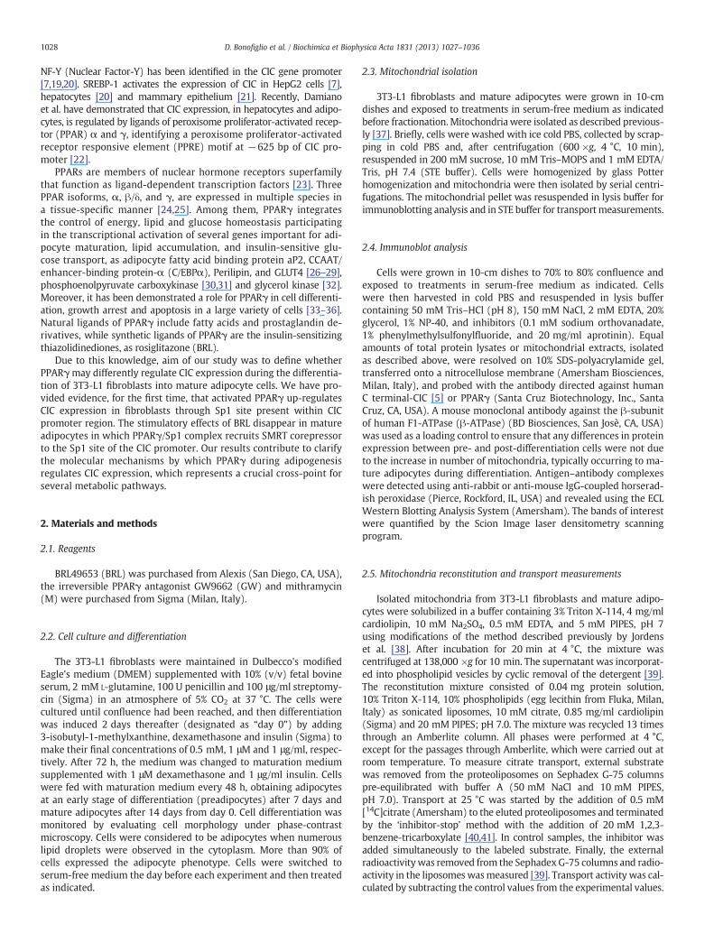
We first aimed to investigate the expression and activity of CIC inmitochondrial extracts from 3T3-L1 fibroblasts (F) and mature adipo-cytes (A, 14 days after differentiation induction). Immunoblot analy-sis, using an antibody raised against the carboxy-terminus of themature CIC protein, revealed a weak immunoreactive band in fibro-blasts at 34 kDa, corresponding to the mitochondrial CIC, while a4.5-fold increase in band intensity was observed in mature adipocytes(Fig. 1A). A similar pattern of CIC expression was also found in totalextracts of both fibroblasts and adipocytes (Fig. 1B). The activity ofCIC in mitochondrial extracts from 3T3-L1 fibroblasts and mature ad-ipocyte cells was tested by assaying the rate of the [14C]citrate/citrateexchange in reconstituted liposomes [40,41]. As shown in Fig. 1C, theuptake of radioactive L-citrate in liposomes reconstituted with mito-chondrial extracts from fibroblasts was approximately 45% lower com-pared to liposomes reconstituted with the mitochondrial extracts frommature adipocyte cells (132±14.4 versus 238±25.0 nmol citrate/mgprotein, respectively).
3.2. The PPARγ ligand BRL up-regulates CIC expression in 3T3-L1fibroblasts
Since PPARγ is considered to be one of the master regulatorsof adipocyte differentiation, we evaluated the involvement of this nu-clear receptor in the modulation of CIC expression during adipocyte
Fig. 1. CIC expression and activity in 3T3-L1 cells. Immunoblots for CIC expression from mitsubunit of mitochondrial ATPase (β-ATPase) was used as loading control. Numbers represenin fibroblast and adipocyte mitochondria. Transport was initiated by adding 0.5 mM [14C]citisolated from either fibroblasts (square) or mature adipocytes (circle). The transport reactioexperiments.
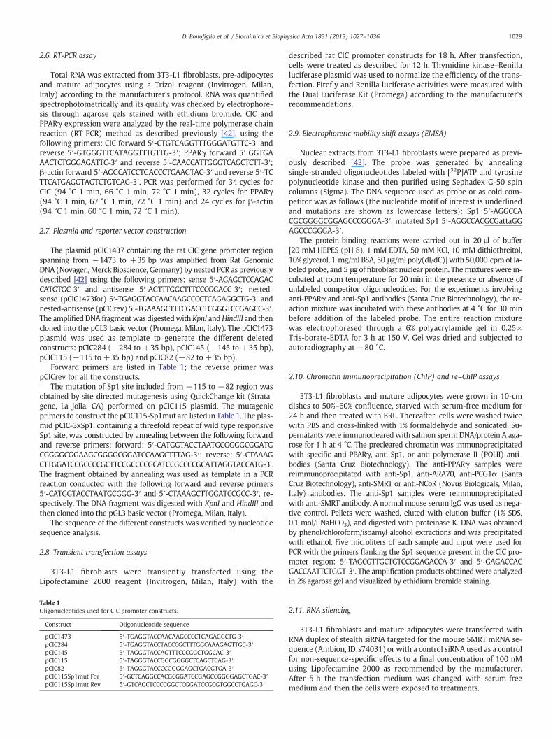
differentiation. We tested the effects of BRL49653 (BRL), a syntheticand specific ligand of PPARγ, in 3T3-L1 fibroblasts (F), inpre-adipocytes (P), which are adipocytes at an early stage of differen-tiation, and in mature adipocytes (A). The results obtained demon-strated that BRL treatment up-regulated CIC mRNA expression infibroblasts and to a lesser extent in pre-adipocytes, while it did notelicit any effects on mature adipocytes (Fig. 2A). As previouslyreported [44], the expression level of PPARγ mRNA was enhancedin BRL-treated fibroblasts and pre-adipocytes and reduced inBRL-treated mature adipocytes (Fig. 2A). Moreover, CIC protein con-tent in fibroblasts increased 4-fold after treatment with BRL for 24 hcompared to untreated fibroblasts (Fig. 2B). This up-regulation wasabrogated by GW9662 (GW), an irreversible PPARγ antagonist, dem-onstrating a direct involvement of PPARγ (Fig. 2B). As expected, inmature adipocytes BRL treatment did not modulate CIC protein levels,while it down-regulated PPARγ protein expression, which was re-versed in the presence of GW (Fig. 2C).
Finally, we investigated the effects of the PPARγ ligand BRL on CICactivity in mitochondrial extracts from fibroblasts and adipocytes. Wefound that the uptake of [14C]citrate in BRL-treated fibroblasts wasenhanced as compared to untreated cells (192±21.2 versus 130±15 nmol citrate/mg protein, respectively), reaching the CIC activitylevels measured in mature adipocytes (235±24 nmol citrate/mgprotein), while BRL did not exert any effects in mature adipocytes(Fig. 2D). Taken together, these data suggest that activated PPARγis able to induce CIC expression and increase CIC activity only infibroblasts.
3.3. BRL transactivates CIC gene promoter in 3T3-L1 fibroblasts
The aforementioned observations prompted us to investigatewhether BRL is able to modulate CIC transcriptional activity. Thus,we performed functional assays by transiently transfecting 3T3-L1
ochondria (A) and total extracts (B) of fibroblasts (F) and mature adipocytes (A). Betat the average fold change of CIC/β-ATPase levels. C: Rate of [14C]citrate/citrate exchangerate to proteoliposomes containing 10 mM citrate and reconstituted with mitochondrian was stopped at the indicated times. The data represent means of three independent
Fig. 2. Activated PPARγ up-regulates CIC expression and activity in fibroblasts. A: CIC and PPARγ mRNA expression in 3T3-L1 fibroblasts (F), preadipocytes (P) and mature adipo-cytes (A) untreated (−) or treated with 10 μM BRL for 24 h. β-ACTIN was used as loading control. Numbers represent the average fold change of CIC or PPARγ/β-ACTIN levels.Immunoblots for CIC and PPARγ expression from total extracts of fibroblast (B) and mature adipocyte cells (C) untreated (−) or treated with 10 μM BRL in the presence or notof 10 μM GW for 24 h. β-ATPase was used as loading control. Numbers represent the average fold change of CIC or PPARγ/β-ATPase levels. D: [14C]citrate/citrate exchange in fibro-blast and adipocyte mitochondria untreated or treated with BRL. Transport was initiated by adding 0.5 mM [14C]citrate to proteoliposomes containing 10 mM citrate andreconstituted with mitochondria isolated from untreated fibroblasts (square), BRL-treated fibroblasts (down-pointing triangle), adipocytes (circle) and BRL-treated adipocytes(up-pointing triangle). The transport reaction was stopped at the indicated times. The data represent means of three independent experiments.
1031D. Bonofiglio et al. / Biochimica et Biophysica Acta 1831 (2013) 1027–1036
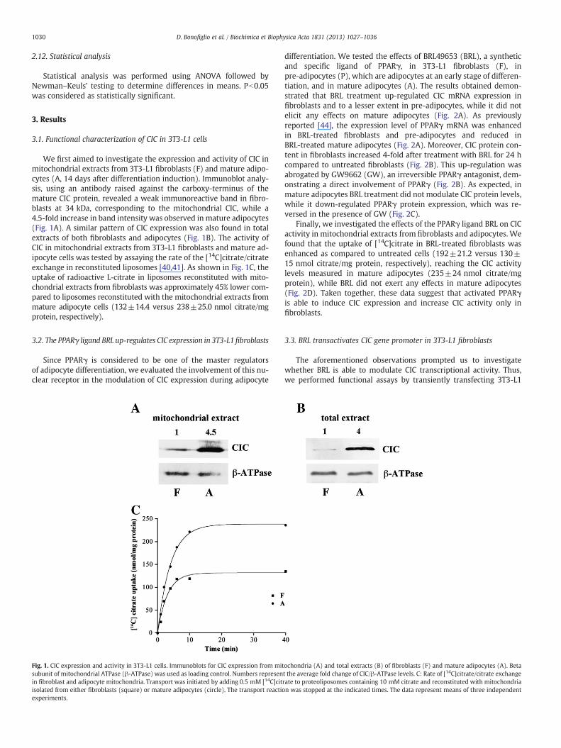
fibroblasts with a plasmid containing rat CIC regulatory sequencepCIC1473 (−1473/+35) and found that BRL significantly inducedluciferase activity (Fig. 3A). This effect was no longer noticeable inthe presence of GW, confirming that the transactivation of CIC byBRL occurred in a PPARγ-dependent manner (data not shown). Therat CIC promoter contains multiple responsive elements for differenttranscription factors, including glucocorticoid receptor (GR), estrogenreceptor alpha (ERα), PPARγ and α, c/EBPα and Sp1 (Fig. 3A). Toidentify the region within the CIC promoter responsible for the BRL-induced transactivation, the activity of the different CIC promoter–deleted constructs pCIC284 (−284/+35), pCIC145 (−145/+35),pCIC115 (−115/+35) and pCIC82 (−82/+35) was tested. In trans-fection experiments performed using the aforementioned plasmidspCIC284, pCIC145 and pCIC115, responsiveness to BRL was stillobserved (Fig. 3A). Of note, BRL was able to transactivate all testedconstructs independently of the PPRE site, which was recently identi-fied at −625 bp [22]. In contrast, in cells transfected with thepromoter–deleted construct pCIC82 we did not detect any increasein luciferase activity (Fig. 3A). Consequently, the region from −115
to −82, which contains the Sp1 motif, was the minimal region ofCIC promoter responsible for BRL induction.
Thus, we performed site-directed mutagenesis on the minimalresponsive Sp1 domain (pCIC115-Sp1mut) within the CIC promoter(Fig. 3A). Mutation of this domain abrogated BRL effects (Fig. 3A)demonstrating that the integrity of Sp1-binding site is necessaryfor PPARγ modulation of CIC promoter activity. To strengthen theimportance of the Sp1 site in CIC promoter modulation by BRL, weperformed transfection experiments using a construct (pCIC-3xSp1)bearing threefold repeat of wild type responsive Sp1 site locatedin the minimal region of CIC promoter. BRL treatment induced a1.7 fold increase in luciferase activity respect to untreated cells(Fig. 3B).
In addition, functional experiments and RT-PCR analysis wereperformed using mithramycin that binds to GC boxes and preventssequential Sp1 binding to its consensus sequence [45]. Our resultsshowed that mithramycin was able to abrogate the BRL-induced CICtranscriptional activity as well as its mRNA expression in fibroblastcells (Fig. 3C and D).
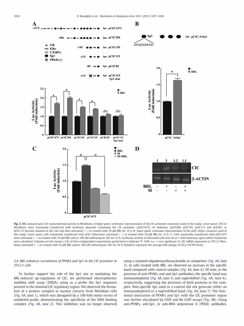
Fig. 3. BRL transactivates CIC transcriptional activity in fibroblasts. A:Upper panel, schematic representation of the CIC promoter constructs used in this study. Lower panel, 3T3-L1fibroblasts were transiently transfected with luciferase plasmids containing the CIC promoter (pCIC1473), its deletions (pCIC284, pCIC145, pCIC115 and pCIC82) orpCIC115-Sp1mut mutated in Sp1 site and then untreated (−) or treated with 10 μM BRL for 12 h. B: Upper panel, schematic representation of the pCIC-3xSp1 construct used inthis study. Lower panel, cells transiently transfected with pCIC-3xSp1were untreated (−) or treated with 10 μM BRL for 12 h. C: Cells transiently transfected with pCIC1473were untreated (−) or treated with 10 μM BRL and/or 100 nM mithramycin (M) for 12 h. Luciferase activity of untreated cells was set as 1-fold induction, upon which treatmentswere calculated. Columns are the means±S.D. of three independent experiments performed in triplicate.*Pb0.05, n.s. = non significant. D: CIC mRNA expression in 3T3-L1 fibro-blasts untreated (−) or treated with 10 μM BRL and/or 100 nM mithramycin (M) for 24 h. Numbers represent the average fold change of CIC/β-ACTIN levels.
1032 D. Bonofiglio et al. / Biochimica et Biophysica Acta 1831 (2013) 1027–1036
3.4. BRL enhances recruitment of PPARγ and Sp1 to the CIC promoter in3T3-L1 cells
To further support the role of the Sp1 site in mediating theBRL-induced up-regulation of CIC, we performed electrophoreticmobility shift assay (EMSA) using as a probe the Sp1 sequencepresent in the minimal CIC regulatory region. We observed the forma-tion of a protein complex in nuclear extracts from fibroblast cells(Fig. 4A, lane 1), which was abrogated by a 100-fold molar excess ofunlabeled probe, demonstrating the specificity of the DNA bindingcomplex (Fig. 4A, lane 2). This inhibition was no longer observed
using a mutated oligodeoxyribonucleotide as competitor (Fig. 4A, lane3). In cells treated with BRL, we observed an increase in the specificband compared with control samples (Fig. 4A, lane 4). Of note, in thepresence of anti-PPARγ and anti-Sp1 antibodies, the specific band wasimmunodepleted (Fig. 4A, lane 5) and supershifted (Fig. 4A, lane 6),respectively, suggesting the presence of both proteins in the com-plex. Non-specific IgG used as a control did not generate either animmunodepleted or a supershifted band (Fig. 4A, lane 7). The func-tional interaction of PPARγ and Sp1 with the CIC promoter regionwas further elucidated by ChIP and Re-ChIP assays (Fig. 4B). Usinganti-PPARγ, anti-Sp1, or anti-RNA polymerase II (POLII) antibodies,
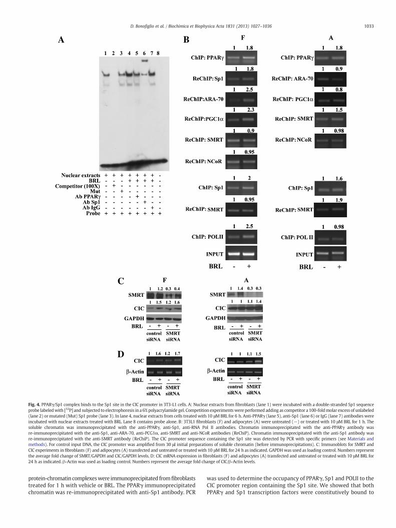
Fig. 4. PPARγ/Sp1 complex binds to the Sp1 site in the CIC promoter in 3T3-L1 cells. A: Nuclear extracts from fibroblasts (lane 1) were incubated with a double-stranded Sp1 sequenceprobe labeledwith [32P] and subjected to electrophoresis in a 6% polyacrylamide gel. Competition experimentswere performed adding as competitor a 100-foldmolar excess of unlabeled(lane 2) ormutated (Mut) Sp1 probe (lane 3). In lane 4, nuclear extracts from cells treatedwith 10 μMBRL for 6 h. Anti-PPARγ (lane 5), anti-Sp1 (lane 6) or IgG (lane 7) antibodies wereincubated with nuclear extracts treated with BRL. Lane 8 contains probe alone. B: 3T3L1 fibroblasts (F) and adipocytes (A) were untreated (−) or treated with 10 μM BRL for 1 h. Thesoluble chromatin was immunoprecipitated with the anti-PPARγ, anti-Sp1, anti-RNA Pol II antibodies. Chromatin immunoprecipitated with the anti-PPARγ antibody wasre-immunoprecipitated with the anti-Sp1, anti-ARA-70, anti-PCG1α, anti-SMRT and anti-NCoR antibodies (ReChiP). Chromatin immunoprecipitated with the anti-Sp1 antibody wasre-immunoprecipitated with the anti-SMRT antibody (ReChiP). The CIC promoter sequence containing the Sp1 site was detected by PCR with specific primers (see Materials andmethods). For control input DNA, the CIC promoter was amplified from 30 μl initial preparations of soluble chromatin (before immunoprecipitations). C: Immunoblots for SMRT andCIC experiments in fibroblasts (F) and adipocytes (A) transfected and untreated or treatedwith 10 μMBRL for 24 h as indicated. GAPDHwas used as loading control. Numbers representthe average fold change of SMRT/GAPDH and CIC/GAPDH levels. D: CIC mRNA expression in fibroblasts (F) and adipocytes (A) transfected and untreated or treated with 10 μM BRL for24 h as indicated. β-Actin was used as loading control. Numbers represent the average fold change of CIC/β-Actin levels.
1033D. Bonofiglio et al. / Biochimica et Biophysica Acta 1831 (2013) 1027–1036
protein-chromatin complexeswere immunoprecipitated fromfibroblaststreated for 1 h with vehicle or BRL. The PPARγ immunoprecipitatedchromatin was re-immunoprecipitated with anti-Sp1 antibody. PCR
was used to determine the occupancy of PPARγ, Sp1 and POLII to theCIC promoter region containing the Sp1 site. We showed that bothPPARγ and Sp1 transcription factors were constitutively bound to
1034 D. Bonofiglio et al. / Biochimica et Biophysica Acta 1831 (2013) 1027–1036
the CIC promoter in untreated cells and that this recruitment was in-creased upon BRL exposure (Fig. 4B, left panel). Similar results werealso obtained by PPARγ/Sp1 Re-ChIP assay (Fig. 4B, left panel). In ad-dition, the positive regulation of the CIC transcriptional activity in-duced by BRL was demonstrated by an increased recruitment ofRNA POLII (Fig. 4B, left panel). Although protein–chromatin com-plexes from adipocytes treated with BRL showed an enhanced re-cruitment of PPARγ and Sp1 to the CIC regulatory region, nochanges in the association of RNA POLII to the Sp1 site were detected(Fig. 4B, right panel).
To assess whether the divergent effects exerted by BRL on CIC ex-pression during adipocyte differentiation might be caused by the coop-erative interaction between PPARγ and positive (PCG1α and ARA-70)or negative (SMRT and NCoR) transcriptional regulators, we performedRe-ChIP assays in both cell lines. We found, after BRL exposure, anenhanced recruitment of PCG1α and ARA-70 coactivators in theSp1-containing region of the CIC promoter in fibroblast cells (Fig. 4B,left panel), while an increased SMRT occupancywas observed in adipo-cyte cells (Fig. 4B, right panel). Finally, to better define the role of SMRTin the PPARγ-dependent modulation of the CIC mRNA and proteinlevels, RNA silencing technologieswere used to knockdown the expres-sion of endogenous SMRT in both fibroblast and adipocyte cells. SMRTexpressionwas effectively silenced as revealed by immunoblot analysisafter 24 h of siRNA transfection in both cell lines (Fig. 4C). As expected,silencing of the SMRT gene had no effects on the up-regulation of CICprotein content and mRNA levels (Fig. 4C and D, left panels) inducedby the specific PPARγ ligand in fibroblast cells. In contrast, BRL wasable to increase CIC expression in SMRT silenced adipocyte cells (Fig. 4C and D, right panels) highlighting a crucial role of SMRT corepressorin regulating CIC activity under adipocyte differentiation.
4. Discussion
In this study, we have demonstrated that activated PPARγ modu-lates the expression and the activity of the mitochondrial CIC duringthe differentiation stages of fibroblasts into adipocytes.
PPARγ, a ligand-activated transcription factor, plays a key role inadipocyte biology by regulating their differentiation, maintenance,and lipid metabolism [46–48]. Actually, PPARγ is considered the mas-ter regulator of adipogenesis participating in the transcriptional acti-vation of several adipogenic and lipogenic genes [49,50]. It is knownthat cellular fat synthesis is regulated at various steps [51,52]. Partic-ularly, the regulation of fatty acid synthesis via CIC or dicarboxylatecarriers is essential for adipocyte differentiation from the early differ-entiation stage of 3T3-L1 fibroblasts into mature fat cells [53].
Using the cultured 3T3-L1 cell system, we have shown that BRLup-regulated CIC expression and activity in fibroblasts through PPARγactivation, while BRL was not able to modulate CIC levels in matureadipocytes. These data contradict previous findings indicating thatPPARγ ligands increased CIC expression in adipocytes [22], althoughthe latter measurements were performed at 7 days after differentiationinduction.
From our study, the specific involvement of PPARγ in up-regulatingCIC expression in fibroblasts was proved by the observation that thePPARγ effect was completely abrogated in the presence of GW, a potentand selective antagonist of PPARγ. The molecular events responsiblefor CIC induction by the PPARγ ligand BRL were consistent with the en-hanced transcriptional activation of this gene as it raised by the capabil-ity of BRL to activate CIC promoter.Multiple transcription factor bindingsites within the rat and human CIC promoter have been described, in-cluding FOXA, SRE, GR, C/EBP, ER and Sp1 binding sequences. Functionalstudies using different CIC-promoter-deleted constructs identifiedthe region of CIC promoter, spanning from −115/−82, as the minimalregion responsible for BRL induction. Of note, this region of rat CICpromoter shows a very high degree of sequence similarity with the cor-responding portion of the mouse CIC genes (approximately 94%
identity). Although it has been recently demonstrated that CIC expres-sion is regulated by PPARγ ligands through a PPRE site, identified at −625 bp of the CIC promoter [22], our results showed that CICtransactivation occurs independently of the PPRE site, suggesting thatother transcription factors are involved in PPARγ-mediated CIC induc-tion. Indeed, analysis of the minimal CIC promoter region reveals thepresence of a GC-box sequence, and deletion as well as mutation of thissite results in the abrogation of PPARγ transactivating activity. The abilityof activated PPARγ to transactivate CIC promoter through Sp1 was con-firmed in functional assays using a construct carrying threefold repeatof the cognate Sp1motif within theminimal region of CIC gene promoter.It could be clearly seen that luciferase expression was significantly in-creased upon BRL treatment.
Furthermore, when Sp1-DNA binding activity was blocked by aselective inhibitor, both PPARγ-mediated transactivation and inductionof CIC expression were subsequently abolished. In line with our results,an interesting observation is that in the presence of the Sp1mutation at−92 bp the basal activity of CIC promoter is reduced when comparedwith the transcriptional activity of the wild-type CIC promoter [20].
Sp1 has been considered traditionally as a ubiquitous factor asso-ciated closely with core promoter activities; it has recently been ob-served that it participates in the regulation of gene transcriptiontriggered by multiple signaling pathways and metabolic or differenti-ation conditions. Moreover, Sp1 interacts physically and cooperatesfunctionally with several sequence-specific activators includingNF-kB, GATA, YY1, E2F1, Rb, SREBP-1 and PPARγ [54–59] to modulategene expression. In addition, it has been shown that the activation of CICgene expression by Sp1 is virtually abolished by methylation of theSp1-binding elements which are present in the promoters of all CICgenes sequenced inmammalswithin the CpG island located immediate-ly upstream the translocation start codon [19]. For the first time, ourresults have revealed a novel important regulatory role of Sp1 in regulat-ing CIC promoter activity. In nuclear extracts fromfibroblast cells treatedwith BRL, EMSA showed a strong increase in the Sp1-DNA bindingthat was immunodepleted by anti-Sp1 and anti-PPARγ antibodies,suggesting the presence of both proteins in the complex. In addition,ChIP and Re-ChIP assays demonstrated that PPARγ/Sp1 occupancy ofthe Sp1-containing promoter region induced by BRL treatment wasconcomitant with an increase in RNA-Pol II, addressing a positive CICtranscriptional regulation mediated by PPARγ.
It is known that members of the nuclear hormone receptor su-perfamily, including PPARγ, once activated, can interact physicallyand modulate target gene transcription. PPARγ can regulate tran-scription by several distinct mechanisms, and its function seems todepend not only on ligand binding, which is known to regulate receptorconformation, but also on the context of the gene and associatedpromot-er factors that contribute to create a gene-specific topography, achievingspecific profiles of gene expression. Several studies have examined therole of coregulators in adipogenesis and demonstrated that coactivatorssuch as PGC-1α or steroid receptor coactivators (SRCs) are essential[60]; whereas NCoR, SMRT and histone deacetylases act as negative reg-ulators of differentiation [61–63]. The physiological relevance of theirimplication in metabolic regulation has been demonstrated in the con-text of PPARγ-mediated adipogenesis, during which they promote atarget-gene specific repression of PPARγ activity [64]. A negative actionof SMRT and NCoR on fat storage has been suggested by the enhancedadipogenesis and increased expression of proadipogenic PPARγ targetgenes after RNAi-mediated inhibition of these corepressors [62].
Our results evidenced in fibroblast cells, after BRL stimulation, anenhanced recruitment of PGC-1α and ARA-70 on the Sp1 site of theCIC promoter. In contrast, we observed that mature adipocytes treat-ed with BRL showed an increased recruitment of SMRT corepressor tothe Sp1 site within the CIC promoter along with no changes in the oc-cupancy of RNA POLII. Finally, we demonstrated a direct involvementof SMRT in the loss of CIC promoter responsiveness to the BRL in ma-ture adipocytes using a specific SMRT siRNA.
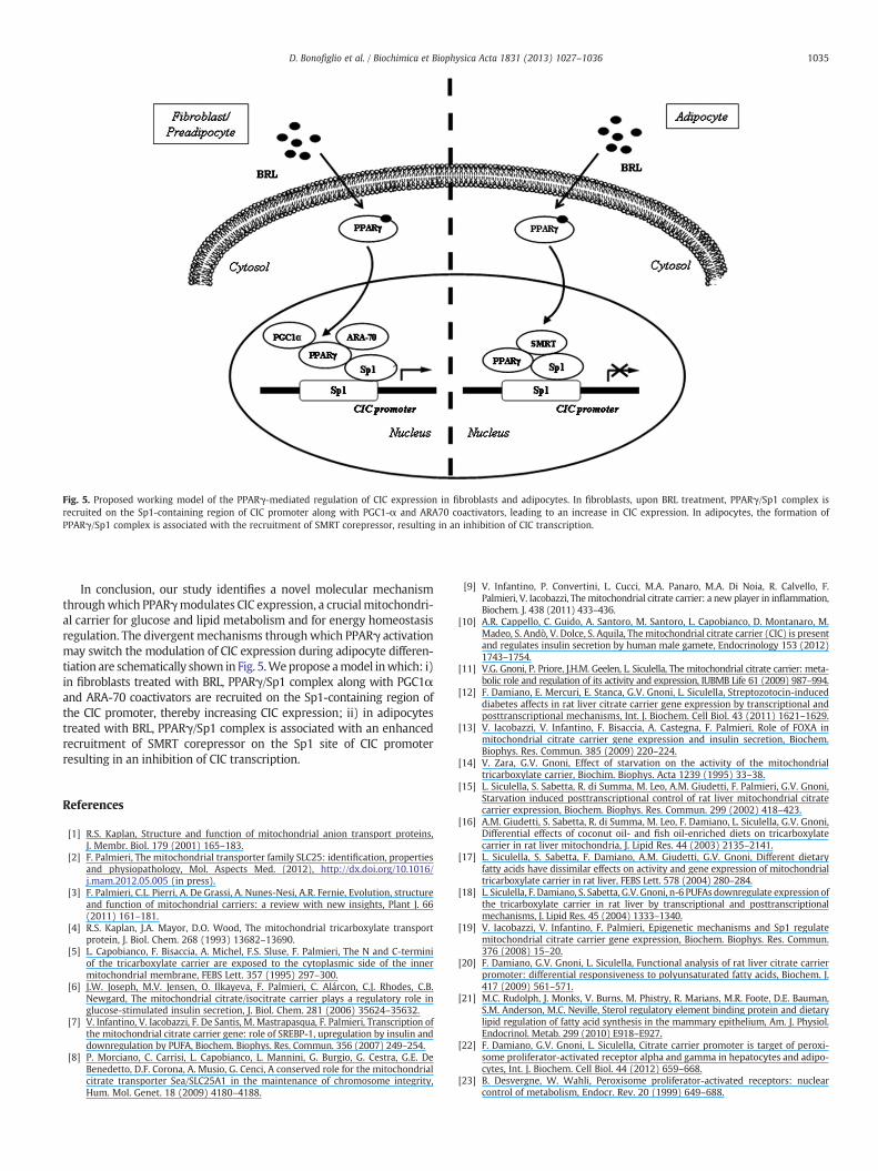
Fig. 5. Proposed working model of the PPARγ-mediated regulation of CIC expression in fibroblasts and adipocytes. In fibroblasts, upon BRL treatment, PPARγ/Sp1 complex isrecruited on the Sp1-containing region of CIC promoter along with PGC1-α and ARA70 coactivators, leading to an increase in CIC expression. In adipocytes, the formation ofPPARγ/Sp1 complex is associated with the recruitment of SMRT corepressor, resulting in an inhibition of CIC transcription.
1035D. Bonofiglio et al. / Biochimica et Biophysica Acta 1831 (2013) 1027–1036
In conclusion, our study identifies a novel molecular mechanismthroughwhich PPARγmodulates CIC expression, a crucialmitochondri-al carrier for glucose and lipid metabolism and for energy homeostasisregulation. The divergentmechanisms throughwhich PPARγ activationmay switch the modulation of CIC expression during adipocyte differen-tiation are schematically shown in Fig. 5.Wepropose amodel inwhich: i)in fibroblasts treated with BRL, PPARγ/Sp1 complex along with PGC1αand ARA-70 coactivators are recruited on the Sp1-containing region ofthe CIC promoter, thereby increasing CIC expression; ii) in adipocytestreated with BRL, PPARγ/Sp1 complex is associated with an enhancedrecruitment of SMRT corepressor on the Sp1 site of CIC promoterresulting in an inhibition of CIC transcription.
References
[1] R.S. Kaplan, Structure and function of mitochondrial anion transport proteins,J. Membr. Biol. 179 (2001) 165–183.
[2] F. Palmieri, The mitochondrial transporter family SLC25: identification, propertiesand physiopathology, Mol. Aspects Med. (2012), http://dx.doi.org/10.1016/j.mam.2012.05.005 (in press).
[3] F. Palmieri, C.L. Pierri, A. De Grassi, A. Nunes-Nesi, A.R. Fernie, Evolution, structureand function of mitochondrial carriers: a review with new insights, Plant J. 66(2011) 161–181.
[4] R.S. Kaplan, J.A. Mayor, D.O. Wood, The mitochondrial tricarboxylate transportprotein, J. Biol. Chem. 268 (1993) 13682–13690.
[5] L. Capobianco, F. Bisaccia, A. Michel, F.S. Sluse, F. Palmieri, The N and C-terminiof the tricarboxylate carrier are exposed to the cytoplasmic side of the innermitochondrial membrane, FEBS Lett. 357 (1995) 297–300.
[6] J.W. Joseph, M.V. Jensen, O. Ilkayeva, F. Palmieri, C. Alárcon, C.J. Rhodes, C.B.Newgard, The mitochondrial citrate/isocitrate carrier plays a regulatory role inglucose-stimulated insulin secretion, J. Biol. Chem. 281 (2006) 35624–35632.
[7] V. Infantino, V. Iacobazzi, F. De Santis, M. Mastrapasqua, F. Palmieri, Transcription ofthe mitochondrial citrate carrier gene: role of SREBP-1, upregulation by insulin anddownregulation by PUFA, Biochem. Biophys. Res. Commun. 356 (2007) 249–254.
[8] P. Morciano, C. Carrisi, L. Capobianco, L. Mannini, G. Burgio, G. Cestra, G.E. DeBenedetto, D.F. Corona, A. Musio, G. Cenci, A conserved role for the mitochondrialcitrate transporter Sea/SLC25A1 in the maintenance of chromosome integrity,Hum. Mol. Genet. 18 (2009) 4180–4188.
[9] V. Infantino, P. Convertini, L. Cucci, M.A. Panaro, M.A. Di Noia, R. Calvello, F.Palmieri, V. Iacobazzi, Themitochondrial citrate carrier: a new player in inflammation,Biochem. J. 438 (2011) 433–436.
[10] A.R. Cappello, C. Guido, A. Santoro, M. Santoro, L. Capobianco, D. Montanaro, M.Madeo, S. Andò, V. Dolce, S. Aquila, Themitochondrial citrate carrier (CIC) is presentand regulates insulin secretion by human male gamete, Endocrinology 153 (2012)1743–1754.
[11] V.G. Gnoni, P. Priore, J.H.M. Geelen, L. Siculella, The mitochondrial citrate carrier: meta-bolic role and regulation of its activity and expression, IUBMB Life 61 (2009) 987–994.
[12] F. Damiano, E. Mercuri, E. Stanca, G.V. Gnoni, L. Siculella, Streptozotocin-induceddiabetes affects in rat liver citrate carrier gene expression by transcriptional andposttranscriptional mechanisms, Int. J. Biochem. Cell Biol. 43 (2011) 1621–1629.
[13] V. Iacobazzi, V. Infantino, F. Bisaccia, A. Castegna, F. Palmieri, Role of FOXA inmitochondrial citrate carrier gene expression and insulin secretion, Biochem.Biophys. Res. Commun. 385 (2009) 220–224.
[14] V. Zara, G.V. Gnoni, Effect of starvation on the activity of the mitochondrialtricarboxylate carrier, Biochim. Biophys. Acta 1239 (1995) 33–38.
[15] L. Siculella, S. Sabetta, R. di Summa, M. Leo, A.M. Giudetti, F. Palmieri, G.V. Gnoni,Starvation induced posttranscriptional control of rat liver mitochondrial citratecarrier expression, Biochem. Biophys. Res. Commun. 299 (2002) 418–423.
[16] A.M. Giudetti, S. Sabetta, R. di Summa, M. Leo, F. Damiano, L. Siculella, G.V. Gnoni,Differential effects of coconut oil- and fish oil-enriched diets on tricarboxylatecarrier in rat liver mitochondria, J. Lipid Res. 44 (2003) 2135–2141.
[17] L. Siculella, S. Sabetta, F. Damiano, A.M. Giudetti, G.V. Gnoni, Different dietaryfatty acids have dissimilar effects on activity and gene expression of mitochondrialtricarboxylate carrier in rat liver, FEBS Lett. 578 (2004) 280–284.
[18] L. Siculella, F. Damiano, S. Sabetta, G.V. Gnoni, n-6 PUFAs downregulate expression ofthe tricarboxylate carrier in rat liver by transcriptional and posttranscriptionalmechanisms, J. Lipid Res. 45 (2004) 1333–1340.
[19] V. Iacobazzi, V. Infantino, F. Palmieri, Epigenetic mechanisms and Sp1 regulatemitochondrial citrate carrier gene expression, Biochem. Biophys. Res. Commun.376 (2008) 15–20.
[20] F. Damiano, G.V. Gnoni, L. Siculella, Functional analysis of rat liver citrate carrierpromoter: differential responsiveness to polyunsaturated fatty acids, Biochem. J.417 (2009) 561–571.
[21] M.C. Rudolph, J. Monks, V. Burns, M. Phistry, R. Marians, M.R. Foote, D.E. Bauman,S.M. Anderson, M.C. Neville, Sterol regulatory element binding protein and dietarylipid regulation of fatty acid synthesis in the mammary epithelium, Am. J. Physiol.Endocrinol. Metab. 299 (2010) E918–E927.
[22] F. Damiano, G.V. Gnoni, L. Siculella, Citrate carrier promoter is target of peroxi-some proliferator-activated receptor alpha and gamma in hepatocytes and adipo-cytes, Int. J. Biochem. Cell Biol. 44 (2012) 659–668.
[23] B. Desvergne, W. Wahli, Peroxisome proliferator-activated receptors: nuclearcontrol of metabolism, Endocr. Rev. 20 (1999) 649–688.
1036 D. Bonofiglio et al. / Biochimica et Biophysica Acta 1831 (2013) 1027–1036
[24] S.A. Kliewer, B.M. Forman, B. Blumberg, E.S. Ong, U. Borgmeyer, D.J. Mangelsdorf, K.Umesono, R.M. Evans, Differential expression and activation of a family of murine per-oxisome proliferator-activated receptors, Proc. Natl. Acad. Sci. 91 (1994) 7355–7359.
[25] O. Braissant, F. Foufelle, C. Scotto, M. Dauca, W. Wahli, Differential expression ofperoxisome proliferator activated receptor (PPARs): tissue distribution ofPPAR-α, -β, and -γ in the adult rat, Endocrinology 137 (1996) 354–366.
[26] P. Tontonoz, E. Hu, R.A. Graves, A.I. Budavari, B.M. Spiegelman, mPPAR gamma 2:tissue-specific regulator of an adipocyte enhancer, Genes Dev. 8 (1994) 1224–1234.
[27] A. Chawla, J.J. Repa, R.M. Evans, D.J. Mangelsdorf, Nuclear receptors and lipidphysiology: opening the X-files, Science 294 (2001) 1866–1870.
[28] G.A. Francis, E. Fayard, F. Picard, J. Auwerx, Nuclear receptors and the control ofmetabolism, Annu. Rev. Physiol. 65 (2003) 261–311.
[29] B.G. Shearer, W.J. Hoekstra, Recent advances in peroxisome proliferator-activatedreceptor science, Curr. Med. Chem. 10 (2003) 267–280.
[30] J.M.Way,W.W.Harrington, K.K. Brown,W.K. Gottschalk, S.S. Sundseth, T.A.Mansfield,R.K. Ramachandran, T.M.Willson, S.A. Kliewer, Comprehensivemessenger ribonucleicacid profiling reveals that PPARγ activation has coordinate effects on gene expressionin multiple insulin-sensitive tissues, Endocrinology 142 (2001) 1269–1277.
[31] J. Huang, S.H. Hsia, T. Imamura, I. Usui, J.M. Olefsky, Annexin II is a thiazolidinedioneresponsive gene involved in insulin-induced glucose transporter isoform 4 translo-cation in 3T3-L1 adipocytes, Endocrinology 145 (2004) 1579–1586.
[32] V. Ribon, J.H. Johnson, H.S. Camp, A.R. Saltiel, Thiazolidinediones and insulin resis-tance: PPARγ activation stimulates expression of the CAP gene, Proc. Natl. Acad.Sci. 95 (1998) 14751–14756.
[33] C. Grommes, G.E. Landreth, M.T. Heneka, Antineoplastic effects of peroxisomeproliferator-activated receptor gamma agonists, Lancet Oncol. 5 (2004) 419–429.
[34] D. Bonofiglio, S. Aquila, S. Catalano, S. Gabriele, M. Belmonte, E. Middea, H. Qi, C.Morelli, M. Gentile, M. Maggiolini, S. Andò, Peroxisome proliferator-activatedreceptor-gamma activates p53 gene promoter binding to the nuclear factor-kappaBsequence in humanMCF7 breast cancer cells,Mol. Endocrinol. 20 (2006) 3083–3092.
[35] D. Bonofiglio, S. Gabriele, S. Aquila, H. Qi, M. Belmonte, S. Catalano, S. Andò,Peroxisome proliferator-activated receptor gamma activates fas ligand gene pro-moter inducing apoptosis in human breast cancer cells, Breast Cancer Res. Treat.113 (2009) 423–434.
[36] D. Bonofiglio, E. Cione, D. Vizza, M. Perri, A. Pingitore, H. Qi, S. Catalano, D. Rovito, G.Genchi, S. Andò, Bid as a potential target of apoptotic effects exerted by low doses ofPPARγ and RXR ligands in breast cancer cells, Cell Cycle 10 (2011) 2344–2354.
[37] C. Frezza, S. Cipolat, L. Scorrano, Organelle isolation: functional mitochondriafrommouse liver, muscle and cultured fibroblasts, Nat. Protoc. 2 (2007) 287–295.
[38] E.Z. Jordens, L. Palmieri, M. Huizing, L.P. van den Heuvel, R.C. Sengers, A. Dörner,W. Ruitenbeek, F.J. Trijbels, J. Valsson, G. Sigfusson, F. Palmieri, J.A. Smeitink,Adenine nucleotide translocator 1 deficiency associated with Sengers syndrome,Ann. Neurol. 52 (2002) 95–99.
[39] F. Palmieri, C. Indiveri, F. Bisaccia, V. Iacobazzi, Mitochondrial metabolite carrierproteins: purification, reconstitution, and transport studies, Methods Enzymol.260 (1995) 349–369.
[40] F. Bisaccia, A. De Palma, F. Palmieri, Identification and purification of the tricarboxylatecarrier from rat liver mitochondria, Biochim. Biophys. Acta 977 (1989) 171–176.
[41] F. Bisaccia, A. De Palma, G. Prezioso, F. Palmieri, Kinetic characterization of thereconstituted tricarboxylate carrier from rat liver mitochondria, Biochim. Biophys.Acta 1019 (1990) 250–256.
[42] D. Iacopetta, R. Lappano, A.R. Cappello, M. Madeo, E.M. De Francesco, A. Santoro,R. Curcio, L. Capobianco, V. Pezzi, M. Maggiolini, V. Dolce, SLC37A1 gene expression isup-regulated by epidermal growth factor in breast cancer cells, Breast Cancer Res.Treat. 122 (2010) 755–764.
[43] N.C. Andrews, D.V. Faller, A rapid micropreparation technique for extraction ofDNA-binding proteins from limiting numbers of mammalian cells, Nucleic AcidsRes. 19 (1991) 2499.
[44] T. Takamura, E. Nohara, Y. Nagai, K. Kobayashi, Stage-specific effects of athiazolidinedione on proliferation, differentiation and PPARgamma mRNA ex-pression in 3T3-L1 adipocytes, Eur. J. Pharmacol. 422 (2001) 23–29.
[45] S.W. Blume, R.C. Snyder, R. Ray, S. Thomas, C.A. Koller, D.M. Miller, Mithramycin in-hibits Sp1 binding and selectively inhibits transcriptional activity of the dihydrofolatereductase gene in vitro and in vivo, J. Clin. Invest. 88 (1991) 1613–1621.
[46] F. Picard, J. Auwerx, PPARγ and glucose homeostasis, Annu. Rev. Nutr. 22 (2002)167–197.
[47] J.N. Feige, L. Gelman, L. Michalik, B. Desvergne, W. Wahli, From molecular actionto physiological outputs: peroxisome proliferator-activated receptors are nuclearreceptors at the crossroads of key cellular functions, Prog. Lipid Res. 45 (2006)120–159.
[48] P. Tontonoz, B.M. Spiegelman, Fat and beyond: the diverse biology of PPARγ,Annu. Rev. Biochem. 77 (2008) 289–312.
[49] W. He, Y. Barak, A. Hevener, P. Olson, D. Liao, J. Le, M. Nelson, E. Ong, J.M. Olefsky,R.M. Evans, Adipose-specific peroxisome proliferator-activated receptor γ knock-out causes insulin resistance in fat and liver but not in muscle, Proc. Natl. Acad.Sci. 100 (2003) 15712–15717.
[50] J.B. Seo, H.M. Moon, W.S. Kim, Y.S. Lee, H.W. Jeong, E.J. Yoo, J. Ham, H. Kang, M.Park, K.R. Steffensen, T.M. Stulnig, J. Gustafsson, S.D. Park, J.B. Kim, Activatedliver X receptors stimulate adipocyte differentiation through induction of perox-isome proliferator-activated receptor γ expression, Mol. Cell. Biol. 24 (2004)3430–3444.
[51] R.A. Coleman, T.M. Lewin, D.M. Muoio, Physiological and nutritional regulation ofenzymes of triacylglycerol synthesis, Annu. Rev. Nutr. 20 (2000) 77–103.
[52] R.W. Hanson, L. Reshef, Glyceroneogenesis revisited, Biochimie 85 (2003)1199–1205.
[53] K. Kajimoto, H. Terada, Y. Baba, Y. Shinohara, Essential role of citrate exportfrom mitochondria at early differentiation stage of 3T3-L1 cells for their effectivedifferentiation into fat cells, as revealed by studies using specific inhibitors ofmitochondrial di- and tricarboxylate carriers, Mol. Genet. Metab. 85 (2005) 46–53.
[54] V. Noé, C. Alemany, L.A. Chasin, C.J. Ciudad, Retinoblastoma protein associateswith Sp1 and activates the hamster dihydrofolate reductase promoter, Oncogene16 (1998) 1931–1938.
[55] H. Rotheneder, S. Geymayer, E. Haidweger, Transcription factors of the Sp1 family:interaction with E2F and regulation of the murine thymidine kinase promoter, J.Mol. Biol. 293 (1999) 1005–1015.
[56] A. Sugawara, A. Uruno, M. Kudo, Y. Ikeda, K. Sato, Y. Taniyama, S. Ito, K. Takeuchi,Transcription suppression of thromboxane receptor gene by peroxisomeproliferator-activated receptor-c via an interaction with Sp1 in vascular smoothmuscle cells, J. Biol. Chem. 277 (2002) 9676–9683.
[57] C.E. Flück, W.L. Miller, GATA-4 and GATA-6 modulate tissue-specific transcriptionof the human gene for P450c17 by direct interaction with Sp1, Mol. Endocrinol.18 (2004) 1144–1157.
[58] B. Teferedegne, M.R. Green, Z. Guo, J.M. Boss, Mechanism of action of a distalNF-kB-dependent enhancer, Mol. Cell. Biol. 26 (2006) 5759–5770.
[59] D. Bonofiglio, H. Qi, S. Gabriele, S. Catalano, S. Aquila, M. Belmonte, S. Andò,Peroxisome proliferator-activated receptor gamma inhibits follicular and ana-plastic thyroid carcinoma cells growth by upregulating p21Cip1/WAF1 gene ina Sp1-dependent manner, Endocr. Relat. Cancer 15 (2008) 545–557.
[60] J.N. Feige, J. Auwerx, Transcriptional coregulators in the control of energy homeostasis,Trends Cell Biol. 6 (2007) 292–301.
[61] F. Picard, M. Kurtev, N. Chung, A. Topark-Ngarm, T. Senawong, R. Machado DeOliveira, M. Leid, M.W. McBurney, L. Guarente, Sirt1 promotes fat mobilizationin white adipocytes by repressing PPAR-gamma, Nature 429 (2004) 771–776.
[62] C. Yu, K. Markan, K.A. Temple, D. Deplewski, M.J. Brady, R.N. Cohen, The nuclearreceptor corepressors NCoR and SMRT decrease peroxisome proliferator-activatedreceptor gamma transcriptional activity and repress 3T3-L1 adipogenesis, J. Biol.Chem. 280 (2005) 13600–13605.
[63] E. Jing, S. Gesta, C.R. Kahn, SIRT2 regulates adipocyte differentiation throughFoxO1 acetylation/deacetylation, Cell Metab. 6 (2007) 105–114.
[64] H.P. Guan, T. Ishizuka, P.C. Chui, M. Lehrke, M.A. Lazar, Corepressors selectivelycontrol the transcriptional activity of PPARgamma in adipocytes, Genes Dev. 19(2005) 453–461.