Lipid droplets hypertrophy: a crucial determining factor in insulin regulation by adipocytes
Upload
independentCategory
view
2download
0
www.elsevier.com/locate/cellbi
Cell Biology International 29 (2005) 449e457
Interleukin-15 stimulates adiponectin secretion by3T3-L1 adipocytes: Evidence for a skeletal
muscle-to-fat signaling pathway
LeBris S. Quinn a,b,*, Lena Strait-Bodey c, Barbara G. Anderson b,c,Josep M. Argiles d, Peter J. Havel e
a Geriatric Research, Education, and Clinical Center, 151 American Lake Division, VA Puget Sound Health Care System,
Tacoma, WA 98493, USAb Division of Gerontology and Geriatric Medicine, Department of Medicine, University of Washington, Seattle, WA, USA
c Research Service, VA Puget Sound Health Care System, Tacoma, WA, USAd Department of Biochemistry and Molecular Biology, Faculty of Biology, University of Barcelona, Barcelona, Spain
e Department of Nutrition, University of California, Davis, CA, USA
Received 18 November 2004; accepted 8 February 2005
Abstract
Interleukin-15 (IL-15) is a cytokine which is highly expressed in skeletal muscle tissue, and which has anabolic effects on skeletalmuscle protein dynamics both in vivo and in vitro. Additionally, administration of IL-15 to rats and mice inhibits white adipose
tissue deposition. To determine if the action of IL-15 on adipose tissue is direct, the capacity of cultured murine 3T3-L1preadipocytes and adipocytes to respond to IL-15 was examined. IL-15 administration inhibited lipid accumulation indifferentiating 3T3-L1 preadipocytes, and stimulated secretion of the adipocyte-specific hormone adiponectin by differentiated
3T3-L1 adipocytes. The latter observation constitutes the first report of a cytokine or growth factor which stimulates adiponectinproduction. IL-15 mRNA expression by cultured 3T3-L1 adipogenic cells and C2C12 murine skeletal myogenic cells was alsoexamined. Quantitative real-time PCR indicated IL-15 mRNA was expressed by C2C12 skeletal myogenic cells, and was
upregulated more than 10-fold in differentiated skeletal myotubes compared to undifferentiated myoblasts. In contrast, 3T3-L1 cellsexpressed little or no IL-15 mRNA at either the undifferentiated preadipocyte or differentiated adipocyte stages. These findingsprovide support for the hypothesis that IL-15 functions in a muscle-to-fat endocrine axis which modulates fat:lean body
composition and insulin sensitivity.� 2005 International Federation for Cell Biology. Published by Elsevier Ltd. All rights reserved.
Keywords: Interleukin-15; Adipocytes; Skeletal muscle; Adiponectin; Cytokines
1. Introduction
The cytokine interleukin-15 (IL-15) is highly expressedat the mRNA level in skeletal muscle tissue (Grabstein
* Corresponding author. 151 American Lake Division, VA Puget
Sound Health Care System, Tacoma, WA 98493, USA. Tel.: C1 253
582 8440x76724; fax: C1 253 589 4004.
E-mail address: [email protected] (L.S. Quinn).
1065-6995/$ - see front matter � 2005 International Federation for Cell B
doi:10.1016/j.cellbi.2005.02.005
et al., 1994). Previous in vitro experiments havedemonstrated that IL-15 has anabolic effects on skeletalmuscle protein dynamics (Quinn et al., 1995, 2002),conferred by a mechanism which is distinct from that ofthe well-characterized anabolic factor insulin-like growthfactor-I (IGF-I). In addition, IL-15 administrationsignificantly reduced the rate of muscle protein degrada-tion and inhibited skeletal muscle wasting in an in vivorodent model of cancer cachexia (Carbo et al., 2000).
iology. Published by Elsevier Ltd. All rights reserved.
450 L.S. Quinn et al. / Cell Biology International 29 (2005) 449e457
These findings suggest that IL-15 may function as anautocrine anabolic mediator of skeletal muscle growth.
In studies investigating the effects of IL-15 on skeletalmuscle, it was also observed that IL-15 administrationinduced 33% reductions in adipose tissue deposition ingrowing rats, with no reduction in food intake (Carboet al., 2000, 2001). Such observations cannot distinguishbetween a direct effect of IL-15 on adipose tissue, oran indirect effect on other tissues and/or hormonesregulating adipogenesis or energy expenditure. Anotherin vivo study utilizing obese rodent models (obeseZucker rats and ob/ob mice) reported that sensitivity tothe fat-inhibiting effects of IL-15 was associated withexpression levels of mRNA for a key signaling subunitof the IL-15 receptor in adipose tissue (Alvarez et al.,2002). These data suggest that IL-15 reduces adiposetissue mass by a direct mechanism.
The present study was performed to determinewhether IL-15 has direct effects on a cultured adipogeniccell line. To this end, we examined the effects ofrecombinant human (rh) IL-15 on lipid deposition andadiponectin secretion (Wolf, 2003) in cultured murine3T3-L1 cells. We also examined IL-15 mRNA expres-sion by cultured murine 3T3-L1 and C2C12 skeletalmyogenic cells. The present data provide evidencesupporting the involvement of IL-15 in a muscle-to-fatendocrine axis (Carbo et al., 2001), and may contributeto a better understanding of the control of lean/fat bodycomposition, with implications for the pathophysiologyand treatment of associated conditions such as obesity,insulin resistance, and type 2 diabetes.
2. Materials and methods
2.1. Cell culture
C2C12 skeletal myogenic and 3T3-L1 adipogenicmurine cell lines were obtained from the American Type
Nomenclature
DMEM Dulbecco’s Minimal Essential MediumDMSO dimethylsulfoxideFBS fetal bovine serumIL-15 interleukin-15IGF-I insulin-like growth factor-IMMLV murine Maloney leukemia virusPBS phosphate-buffered salinePCR polymerase chain reactionPEG polyethylene glycolPPAR-g peroxisome proliferator-activated recep-
tor-gammarh recombinant humanRIA radioimmunoassayRT reverse transcriptase
Culture Collection (ATCC; Manassas, VA, USA). Cellsfrom both lines were used for experiments at 2e3passages. For expansion and storage, cell lines werecultured in 10% fetal bovine serum (FBS; Hyclone,Logan, UT, USA) in high glucose formulation Dulbecco’sMinimal Essential Medium (DMEM; Sigma, St. Louis,MO, USA) and cells stored frozen at �135 �C in 20%FBS/DMEM containing 10% dimethylsulfoxide (DMSO;Sigma). For analysis, 3T3-L1 preadipocytes were platedin 1 ml 10% FBS/DMEM in 24-well plates (Costar,Cambridge, MA, USA) at 105 cells per well, resulting inconfluent cultures by the next day, considered day 0 ofculture. To induce differentiation (Engelman et al., 1998),the medium was changed to 10% FBS/DMEM containing160 nM insulin (Sigma), 0.25 mM dexamethasone (Sigma),and 0.5 mM 3-isobutyl-1-methylxanthine (Sigma). Afteran additional 48 h (day 2), medium was changed to 10%FBS/DMEM containing 160 nM insulin. Cultures weretreated with 0e10 ng/ml rhIL-15 (R&D Systems, Minne-apolis, MN, USA) either continuously for 4 days (0e96 hof culture) with replenishment of rhIL-15 at 48 h with themedium change, or for 48-h periods, days 0e2, 2e4, or 4e6 of culture. The latter protocol was utilized to determinethe effects of rhIL-15 on lipid deposition and adiponectinsecretion at specific periods of adipogenic differentiation,with days 0e2 representing undifferentiated preadipocytes,days 2e4 representing mid-differentiation preadipocytes,and days 4e6 representing differentiated adipocytes.
For analysis of skeletal myogenic cultures, C2C12myoblasts were plated onto 24-well culture plates at105 cells per well in 10% FBS/DMEM and allowed toattach overnight. To induce differentiation, medium waschanged to a lower serum concentration, 0.5% FBS/DMEM, at 24 h after plating (considered day 0). Cellscommenced differentiation and fusion into post-mitoticmyotubes on day 2, and cultures contained a prepon-derance of differentiated myotubes by day 5.
2.2. Assay of lipid deposition
Neutral lipid deposition was assessed in 3T3-L1cultures using a modification of the procedure ofKasturi and Joshi (1982). At specific time points (days2, 4, or 6), cultures were rinsed 1! with phosphate-buffered saline (PBS) at 37 �C and fixed with 10%formalin in PBS overnight at RT. Cultures were rinsedsequentially with dH2O and 70% ethanol, then stainedfor 25 min with filtered oil Red-O solution, 6 parts oilRed-O-saturated isopropanol (Sigma) in 4 parts dH2O.Stained cultures were rinsed sequentially with 70%ethanol and dH2O, and lysed with 4% nonidet P-40(Calbiochem, La Jolla, CA, USA) in isopropanol ona rotary shaker for 15 min. Absorbance of the disperseddye at 544 nm was read photometrically using a Victor2microplate reader (PerkineElmer Wallac, Gaithersburg,MD, USA). Results shown represent the mean of
451L.S. Quinn et al. / Cell Biology International 29 (2005) 449e457
3 replicates at each time point and/or concentration ofrhIL-15, with differences between control (no IL-15)and experimental treatments determined using Student’st-tests.
2.3. DNA assay
DNA content per well was assessed in 3T3-L1adipogenic cultures by measuring Hoechst 33258 dye(bisbenzimide; Sigma) fluorescence as previously de-scribed (Haugk et al., 2000). Briefly, cells were rinsed1! with DMEM, fixed for 5 min with 70% ethanol/formalin/acetic acid (20:2:1) at 4 �C, then rinsed 3!with DMEM and 2! with PBS. Cells were exposed to1 mg/ml Hoechst 33258 dye in standard saline citratefor 30 min and analyzed using the fluorescence featureof the Victor2 microplate reader. Results shown repre-sent the mean values for 3 wells at each concentration ofrhIL-15, with differences between control and eachconcentration of rhIL-15 determined using Student’st-tests.
2.4. Adiponectin secretion
Adiponectin was assayed in culture medium collectedfrom differentiating 3T3-L1 adipogenic cultures after48 h periods of rhIL-15 exposure (0e10 ng/ml), days0e2, 2e4, or 4e6 of culture in 10% FBS/DMEM, plusthe induction factors added according to the scheduleoutlined above. This protocol was utilized to separatethe effects of rhIL-15 on 3T3-L1 differentiation (whichcould in turn affect adiponectin secretion) from theeffects of rhIL-15 on adiponectin secretion at each stageof differentiation. At each time point, medium sampleswere collected, stored frozen at �20 �C, and thawedimmediately prior to analysis. Culture wells from whichmedium samples were collected were rinsed and used foranalysis of lipid deposition as outlined above. Mediaadiponectin concentrations were determined usinga mouse adiponectin radioimmunoassay (RIA) kit(Linco, St. Charles, MO, USA) utilizing 125I-labeledmurine adiponectin and an anti-adiponectin rabbitantiserum to measure adiponectin concentrations bythe double antibody/PEG (polyethylene glycol) tech-nique. Standards over the range of 1e100 ng/mlwere prepared using recombinant murine adiponectin.Medium samples were assayed undiluted or diluted 1:4,yielding an effective range of 1e400 ng/ml. The intra-assay and inter-assay coefficients of variation atadiponectin concentrations in the range of 3e12 mg/mlwere 3.7e4.4% and 6.6e8.2%, respectively. Mediumcontrol samples consisted of 10% FBS/DMEM in-cubated in 24-well plates with no cells along withthe experimental samples. All medium control samplesexhibited immunoreactivity of less than 2 ng/ml adipo-nectin. Results shown represent the mean values for
3 wells at each concentration of rhIL-15 added to thecultures for each time period, with differences betweencontrol (0 ng/ml IL-15 added) and each concentration ofrhIL-15 determined using Student’s t-tests.
2.5. Quantitative real-time PCR analysis of IL-15mRNA expression
C2C12 myogenic cultures and 3T3-L1 adipogeniccultures were established and induced to differentiate asdescribed above. RNA was harvested at the undifferen-tiated preadipocyte (day 0) or myoblast (day 0) stages,mid-differentiation stages (day 2 for 3T3-L1 and day3 for C2C12), and differentiated adipocyte or myotubestages (day 4 for 3T3-L1 and day 5 for C2C12) of culture.Total RNA was isolated and purified from C2C12 and3T3-L1 cultures using the RNeasy Fibrous Tissue orLipid Tissue Kits (Qiagen, Valencia, CA, USA), re-spectively. RNA samples were treated on column with30 Kunitz units DNase I (Qiagen) to remove residualgenomic DNA, then quantified using the RiboGreenRNA Quantitation Kit (Molecular Probes, Eugene, OR,USA). cDNA was reverse transcribed from 1 mg totalRNA using the Bio-Rad iScript cDNA Synthesis Kit(Bio-Rad, Hercules, CA, USA). Quantitative real-timepolymerase chain reaction (PCR) analysis of IL-15mRNA expression was performed using a Bio-RadiCycler, and IL-15 mRNA expression normalized to18S ribosomal RNA expression for each sample. TotalRNA (1 mg) was run in 20 ml reactions using a blend ofoligo dT and random hexamer primers and a modifiedMMLV-derived reverse transcriptase (RT; Bio-Rad).Reaction cycles were 5 min at 25 �C, 30 min at 42 �C,and 5 min at 85 �C. Each cDNA reaction was diluted5!, along with a calibrator sample containing thetranscript of interest, and run in 50 ml Bio-Rad SYBRGreen reactions using 200 mM primers designed byBeacon Designer software (BioSoft International, PaloAlto, CA, USA). Primers for murine IL-15 mRNA,which detected either of two RNA splice variants codingfor identical mature IL-15 peptides (Fehniger andCalgiuri, 2001), were: FORWARD 5#-CAG CAC TCTGTC TTC TAA CAA GAA-3#; REVERSE 5#-TCTGTG AAG GTT TTC TCC TCC A-3#. Primers for 18Sribosomal RNA, used as the ‘‘housekeeping’’ gene, were:FORWARD 5#-GCGAATGGC TCATTAAATCAGTTA-3#; REVERSE 5#-TGG TTT TGA TCT GATAAATGCACG-3#. Reactions were processed using oneinitial denaturation cycle (5 min at 95 �C), then 40 cyclesof denaturation (30 s at 95 �C), annealing (30 s at 61 �C)and amplification (30 s at 72 �C) followed by melt curvedetermination consisting of one denaturation cycle(1 min at 95 �C), annealing (one cycle for 1 min at62 �C), and then 68 cycles (10 s each at 61 �C to 95 �C).The 18S ribosomal RNA primer pair reaction was runon every sample for template content normalization
452 L.S. Quinn et al. / Cell Biology International 29 (2005) 449e457
purposes. A calibrator RT sample containing the cDNAof interest was run to yield a standard curve for eachprimer set, and individual primer reaction efficiencieswere calculated from this curve using the iCyclersoftware. The iCycler software calculated a thresholdcycle for each sample; threshold cycles and primer pairefficiencies were used in the 2�DDCT method (Livak andSchmittgen, 2001) to yield differences in IL-15 mRNAexpression as normalized to the 18S housekeeping gene.
3. Results
3.1. Effects of IL-15 on lipid deposition in 3T3-L1adipogenic cultures
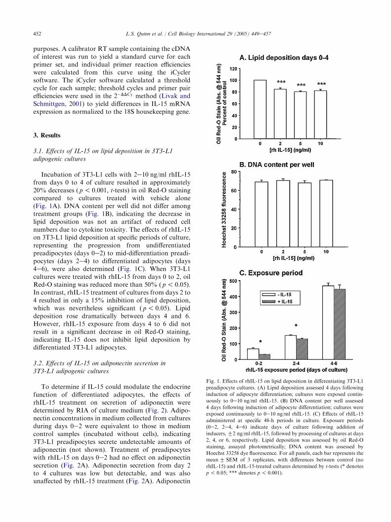
Incubation of 3T3-L1 cells with 2e10 ng/ml rhIL-15from days 0 to 4 of culture resulted in approximately20% decreases ( p! 0.001, t-tests) in oil Red-O stainingcompared to cultures treated with vehicle alone(Fig. 1A). DNA content per well did not differ amongtreatment groups (Fig. 1B), indicating the decrease inlipid deposition was not an artifact of reduced cellnumbers due to cytokine toxicity. The effects of rhIL-15on 3T3-L1 lipid deposition at specific periods of culture,representing the progression from undifferentiatedpreadipocytes (days 0e2) to mid-differentiation preadi-pocytes (days 2e4) to differentiated adipocytes (days4e6), were also determined (Fig. 1C). When 3T3-L1cultures were treated with rhIL-15 from days 0 to 2, oilRed-O staining was reduced more than 50% ( p! 0.05).In contrast, rhIL-15 treatment of cultures from days 2 to4 resulted in only a 15% inhibition of lipid deposition,which was nevertheless significant ( p! 0.05). Lipiddeposition rose dramatically between days 4 and 6.However, rhIL-15 exposure from days 4 to 6 did notresult in a significant decrease in oil Red-O staining,indicating IL-15 does not inhibit lipid deposition bydifferentiated 3T3-L1 adipocytes.
3.2. Effects of IL-15 on adiponectin secretion in3T3-L1 adipogenic cultures
To determine if IL-15 could modulate the endocrinefunction of differentiated adipocytes, the effects ofrhIL-15 treatment on secretion of adiponectin weredetermined by RIA of culture medium (Fig. 2). Adipo-nectin concentrations in medium collected from culturesduring days 0e2 were equivalent to those in mediumcontrol samples (incubated without cells), indicating3T3-L1 preadipocytes secrete undetectable amounts ofadiponectin (not shown). Treatment of preadipocyteswith rhIL-15 on days 0e2 had no effect on adiponectinsecretion (Fig. 2A). Adiponectin secretion from day 2to 4 cultures was low but detectable, and was alsounaffected by rhIL-15 treatment (Fig. 2A). Adiponectin
Fig. 1. Effects of rhIL-15 on lipid deposition in differentiating 3T3-L1
preadipocyte cultures. (A) Lipid deposition assessed 4 days following
induction of adipocyte differentiation; cultures were exposed contin-
uously to 0e10 ng/ml rhIL-15. (B) DNA content per well assessed
4 days following induction of adipocyte differentiation; cultures were
exposed continuously to 0e10 ng/ml rhIL-15. (C) Effects of rhIL-15
administered at specific 48-h periods in culture. Exposure periods
(0e2, 2e4, 4e6) indicate days of culture following addition of
inducers,G2 ng/ml rhIL-15, followed by processing of cultures at days
2, 4, or 6, respectively. Lipid deposition was assessed by oil Red-O
staining, assayed photometrically; DNA content was assessed by
Hoechst 33258 dye fluorescence. For all panels, each bar represents the
meanG SEM of 3 replicates, with differences between control (no
rhIL-15) and rhIL-15-treated cultures determined by t-tests (* denotes
p ! 0.05; *** denotes p! 0.001).

453L.S. Quinn et al. / Cell Biology International 29 (2005) 449e457
Fig. 2. Effects of IL-15 on adiponectin secretion in 3T3-L1 adipogenic
cultures. (A) Effects of 10 ng/ml rhIL-15 administered at specific 48-h
periods in culture. Exposure periods (0e2, 2e4, 4e6) indicate days ofculture following addition of inducers,G10 ng/ml rhIL-15, followed by
processing of cultures at days 2, 4, or 6, respectively. (B) Adiponectin
secretion by 3T3-L1 adipocytes exposed to 0e10 ng/ml rhIL-15 on days
4e6 following induction of adipocyte differentiation. (C) Lipid content
of 3T3-L1 adipocytes exposed to 0e10 ng/ml rhIL-15 on days 4e6
following induction of adipocyte differentiation. Adiponectin secretion
was determined by RIA of culture medium; lipid deposition was
determined by microplate-based assay of oil Red-O staining. For all
panels, each bar represents the mean G SEM of 3 replicates, with
differences between control (no rhIL-15) and rhIL-15-treated cultures
determined by t-tests (* denotes p! 0.05; *** denotes p! 0.001).
secretion increased dramatically by days 4e6, coincidentwith adipocyte differentiation (Fig. 2A). Treatment ofday 4e6 cultures with 10 ng/ml rhIL-15 significantlyincreased adiponectin secretion from 71.5G 3.8 ng/ml to128.8G 4.0 ng/ml ( p! 0.001). The effect of rhIL-15 onadiponectin secretion by 3T3-L1 adipocytes was concen-tration-dependent with a threshold of 5 ng/ml (Fig. 2B),while lipid content in adipocyte cultures treated withrhIL-15 on days 4e6 was unchanged (Fig. 2C).
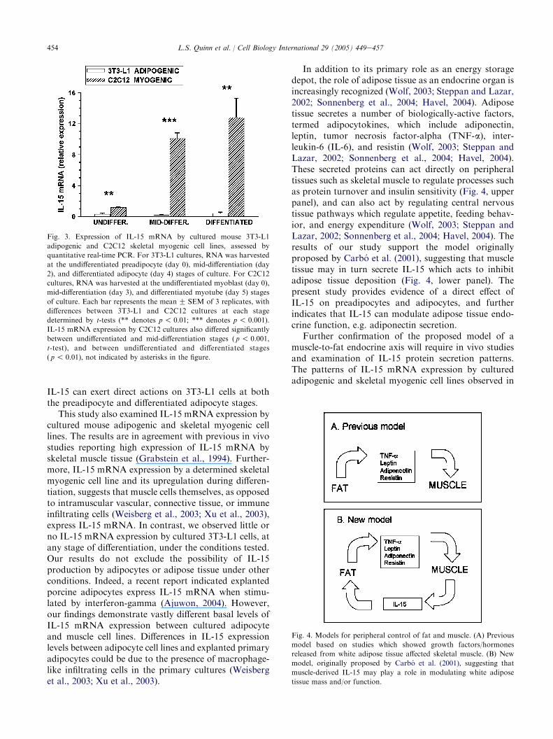
3.3. Expression of IL-15 mRNA in cultured skeletalmyogenic and adipogenic cells
IL-15 mRNA expression in cultured skeletal myo-genic and adipogenic cells has not been characterizedwith respect to differentiation stages. Fig. 3 showsreal-time quantitative PCR analysis of IL-15 mRNAexpression by cultured 3T3-L1 adipogenic cells andC2C12 skeletal myogenic cells. For both cell types, IL-15 mRNA expression was assessed at the undifferenti-ated precursor cell (preadipocyte, myoblast) stages,at mid-differentiation, and at the differentiated cell(adipocyte, myotube) stages. C2C12 myoblasts (cultureday 0) expressed IL-15 mRNA which was detectableafter 32.5 cycles of PCR. By the mid-differentiation anddifferentiated myotube stages (days 3e5 of culture), IL-15 mRNA expression increased more than 10-fold, andwas significantly different from the level of IL-15mRNA expression in undifferentiated myoblast cultures( p! 0.01, t-tests; not indicated in Fig. 3). In contrast,IL-15 mRNA expression by 3T3-L1 cells was detected at34.7e38 cycles of PCR, equivalent to background levelsand significantly lower than the levels detected in C2C12cells at equivalent stages of differentiation ( p! 0.01,t-tests). These findings indicate cultured skeletal myo-genic cells express IL-15 mRNA which is upregulatedconcurrent with differentiation into myotubes, whereascultured 3T3-L1 preadipocytes and adipocytes expresslittle or no IL-15 mRNA under these conditions.
4. Discussion
Previous in vivo studies suggested that IL-15 inhibitsfat deposition via a direct action on adipose tissue inrodents (Alvarez et al., 2002). The present study utilizedthe murine 3T3-L1 preadipocyte cell line to investigatethe direct actions of IL-15 on cultured adipogenic cells.Incubation of 3T3-L1 preadipocytes with rhIL-15inhibited lipid deposition by more than 50%, an effectwhich decreased as differentiation progressed. IL-15had no effect on lipid content in differentiated 3T3-L1adipocytes; however, IL-15 instead acted to stimulatesecretion of the adipocyte-specific hormone, adiponec-tin, by these cells. These observations demonstrate that
454 L.S. Quinn et al. / Cell Biology International 29 (2005) 449e457
IL-15 can exert direct actions on 3T3-L1 cells at boththe preadipocyte and differentiated adipocyte stages.
This study also examined IL-15 mRNA expression bycultured mouse adipogenic and skeletal myogenic celllines. The results are in agreement with previous in vivostudies reporting high expression of IL-15 mRNA byskeletal muscle tissue (Grabstein et al., 1994). Further-more, IL-15 mRNA expression by a determined skeletalmyogenic cell line and its upregulation during differen-tiation, suggests that muscle cells themselves, as opposedto intramuscular vascular, connective tissue, or immuneinfiltrating cells (Weisberg et al., 2003; Xu et al., 2003),express IL-15 mRNA. In contrast, we observed little orno IL-15 mRNA expression by cultured 3T3-L1 cells, atany stage of differentiation, under the conditions tested.Our results do not exclude the possibility of IL-15production by adipocytes or adipose tissue under otherconditions. Indeed, a recent report indicated explantedporcine adipocytes express IL-15 mRNA when stimu-lated by interferon-gamma (Ajuwon, 2004). However,our findings demonstrate vastly different basal levels ofIL-15 mRNA expression between cultured adipocyteand muscle cell lines. Differences in IL-15 expressionlevels between adipocyte cell lines and explanted primaryadipocytes could be due to the presence of macrophage-like infiltrating cells in the primary cultures (Weisberget al., 2003; Xu et al., 2003).
Fig. 3. Expression of IL-15 mRNA by cultured mouse 3T3-L1
adipogenic and C2C12 skeletal myogenic cell lines, assessed by
quantitative real-time PCR. For 3T3-L1 cultures, RNA was harvested
at the undifferentiated preadipocyte (day 0), mid-differentiation (day
2), and differentiated adipocyte (day 4) stages of culture. For C2C12
cultures, RNA was harvested at the undifferentiated myoblast (day 0),
mid-differentiation (day 3), and differentiated myotube (day 5) stages
of culture. Each bar represents the meanG SEM of 3 replicates, with
differences between 3T3-L1 and C2C12 cultures at each stage
determined by t-tests (** denotes p! 0.01; *** denotes p! 0.001).
IL-15 mRNA expression by C2C12 cultures also differed significantly
between undifferentiated and mid-differentiation stages ( p! 0.001,
t-test), and between undifferentiated and differentiated stages
( p ! 0.01), not indicated by asterisks in the figure.
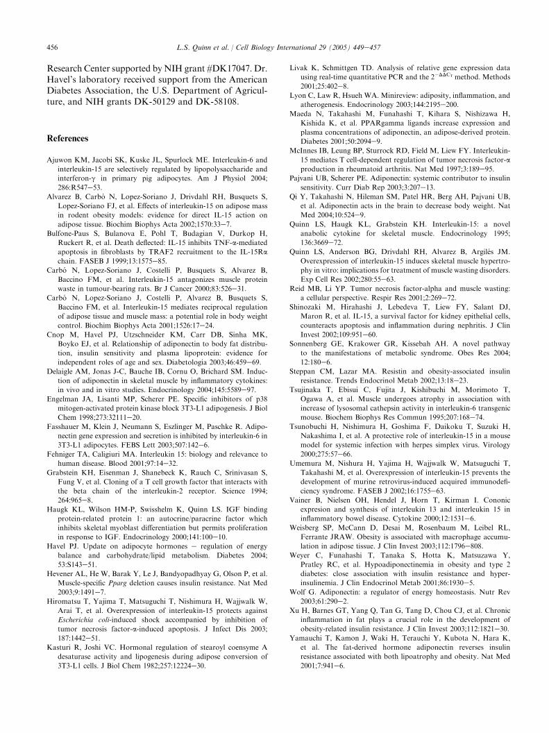
In addition to its primary role as an energy storagedepot, the role of adipose tissue as an endocrine organ isincreasingly recognized (Wolf, 2003; Steppan and Lazar,2002; Sonnenberg et al., 2004; Havel, 2004). Adiposetissue secretes a number of biologically-active factors,termed adipocytokines, which include adiponectin,leptin, tumor necrosis factor-alpha (TNF-a), inter-leukin-6 (IL-6), and resistin (Wolf, 2003; Steppan andLazar, 2002; Sonnenberg et al., 2004; Havel, 2004).These secreted proteins can act directly on peripheraltissues such as skeletal muscle to regulate processes suchas protein turnover and insulin sensitivity (Fig. 4, upperpanel), and can also act by regulating central nervoustissue pathways which regulate appetite, feeding behav-ior, and energy expenditure (Wolf, 2003; Steppan andLazar, 2002; Sonnenberg et al., 2004; Havel, 2004). Theresults of our study support the model originallyproposed by Carbo et al. (2001), suggesting that muscletissue may in turn secrete IL-15 which acts to inhibitadipose tissue deposition (Fig. 4, lower panel). Thepresent study provides evidence of a direct effect ofIL-15 on preadipocytes and adipocytes, and furtherindicates that IL-15 can modulate adipose tissue endo-crine function, e.g. adiponectin secretion.
Further confirmation of the proposed model of amuscle-to-fat endocrine axis will require in vivo studiesand examination of IL-15 protein secretion patterns.The patterns of IL-15 mRNA expression by culturedadipogenic and skeletal myogenic cell lines observed in
Fig. 4. Models for peripheral control of fat and muscle. (A) Previous
model based on studies which showed growth factors/hormones
released from white adipose tissue affected skeletal muscle. (B) New
model, originally proposed by Carbo et al. (2001), suggesting that
muscle-derived IL-15 may play a role in modulating white adipose
tissue mass and/or function.

455L.S. Quinn et al. / Cell Biology International 29 (2005) 449e457
the present study support this model. Moreover,multiple tissue Northern blots indicated skeletal muscleand placenta are the major sites of IL-15 mRNAexpression in vivo (Grabstein et al., 1994). The IL-15mRNA isoform expressed in skeletal muscle is the formbelieved to be more readily secreted (Fehniger andCaligiuri, 2001). However, it is not yet known whetheradipose tissue in vivo is exposed to biologically-relevantconcentrations of IL-15 produced by muscle. Recentstudies of mice which carried a muscle-specific deletionof the peroxisome proliferator-activated receptor-gamma (PPAR-g) gene suggested that this deletionindirectly affected secretion of an unidentified, muscle-derived hormone which in turn affected adipose tissue(Hevener et al., 2003). Our findings suggest that IL-15could be this putative factor.
IL-15 mRNA is widely expressed at lower levels ina number of non-lymphoid tissues, including heart, lung,liver, kidney, brain, and testes (Fehniger and Caliguiri,2001). Along with interleukin-2, an important role forIL-15 is to support the development of several types of Tlymphocytes, particularly natural killer and CD8C(‘‘memory T’’) cells (Fehniger and Caligiuri, 2001).Due to these actions, IL-15 inhibits lethality in murinemodels of infectious diseases such as retrovirus-inducedacquired immunodeficiency syndrome (Umemura et al.,2002) and herpes simplex virus infection (Tsunobuchiet al., 2000). Additionally, IL-15 has proinflammatoryactivity in several mouse models of autoimmune diseasessuch as rheumatoid arthritis and inflammatory boweldisease (McInnes et al., 1997; Vainer et al., 2000).However, in other systems, such as a murine model ofnephritis, IL-15 exerts anti-inflammatory activity (Shi-nozaki et al., 2002). In several in vitro and in vivosystems, IL-15 inhibits the proinflammatory and pro-apoptotic actions of TNF-a (e.g., Bulfone-Paus et al.,1999; Hiromatsu et al., 2003).
Increased production of the proinflammatory adipo-cytokines IL-6 and TNF-a during obesity has beenmechanistically linked to insulin resistance (Weisberget al., 2003; Xu et al., 2003; Ajuwon et al., 2004;Sonnenberg et al., 2004; Lyon et al., 2003). TNF-a andIL-6 inhibit adiponectin mRNA and protein secretion(Havel, 2004; Maeda et al., 2001; Fasshauer et al., 2003),while the present study showed IL-15 stimulatesadiponectin secretion from cultured adipocytes. Inskeletal muscle, IL-6 and TNF-a exert catabolic actions,inhibiting skeletal myoblast differentiation and stimu-lating muscle fiber protein degradation (Reid and Li,2001; Tsujinaka et al., 1995). In contrast, IL-15 exertsanabolic actions in skeletal muscle in vivo and in vitrovia inhibition of muscle fiber protein degradation(Quinn et al., 2002; Carbo et al., 2000). The opposingactions of IL-15 and TNF-a on muscle proteindegradation and on adiponectin secretion by adipocytessuggests that IL-15 may function to inhibit or oppose
TNF-a-mediated pathways in both skeletal muscle andadipose tissue.
An important finding of this study was the stimula-tion of adiponectin secretion observed during incuba-tion of differentiated 3T3-L1 cells in the presence ofIL-15. Adiponectin is a secreted protein producedexclusively by adipocytes in most physiological con-ditions (Havel, 2004; Pajvani and Scherer, 2003;Delaigle et al., 2004). Circulating levels of adiponectinare negatively correlated with visceral adiposity andinsulin resistance in both rodents and humans (Wolf,2003; Sonnenberg et al., 2004; Havel, 2004; Weyer et al.,2001; Cnop et al., 2003). Adiponectin exerts actions onboth liver and skeletal muscle, where it activates AMP-activated protein kinase and stimulates fatty acidoxidation, resulting in improved insulin sensitivity(Yamauchi et al., 2001, 2002). Adiponectin may alsohave actions in the central nervous system to increaseenergy expenditure (Qi et al., 2004). In addition,adiponectin has direct anti-inflammatory actions onvascular endothelium and may protect against theprogression of atherosclerosis (Havel, 2004; Yamauchiet al., 2003). Due to the known insulin-sensitizing andanti-atherosclerotic actions of adiponectin, the regula-tion of adiponectin expression and secretion is ofconsiderable interest. The insulin-sensitizing thiazolide-nedione class of PPAR-g agonists appear to function inpart by stimulating adiponectin expression (Sonnenberget al., 2004; Havel, 2004; Maeda et al., 2001). Theobservation reported here that IL-15 stimulates adipo-nectin secretion by cultured 3T3-L1 adipocytes repre-sents the first report of an endogenous cytokine orgrowth factor which stimulates adiponectin secretion.These data suggest that IL-15 could potentially act invivo to stimulate adiponectin production by adiposetissue, which would in turn modulate skeletal musclemetabolism and insulin sensitivity.
The present data demonstrating that IL-15 has directeffects on cultured 3T3-L1 preadipocytes and adipocytesprovides evidence supporting the involvement of IL-15in a muscle-to-fat endocrine axis (Carbo et al., 2001).Further investigation of this hypothesis is likely to pro-vide a better understanding control of body compositionand substrate metabolism, with implications for thepathophysiology and treatment of associated conditionssuch as obesity, insulin resistance, and type 2 diabetes.
Acknowledgements
We thank Kimber Stanhope and James Graham forexpert technical assistance in performing the adiponectinassays. This work was funded by grant #98-35206-6463from the U.S. Department of Agriculture (LSQ, JMA)and by a Pilot and Feasibility Award (LSQ) from theUniversity of Washington Diabetes Endocrinology
456 L.S. Quinn et al. / Cell Biology International 29 (2005) 449e457
Research Center supported by NIH grant #DK17047. Dr.Havel’s laboratory received support from the AmericanDiabetes Association, the U.S. Department of Agricul-ture, and NIH grants DK-50129 and DK-58108.
References
Ajuwon KM, Jacobi SK, Kuske JL, Spurlock ME. Interleukin-6 and
interleukin-15 are selectively regulated by lipopolysaccharide and
interferon-g in primary pig adipocytes. Am J Physiol 2004;
286:R547e53.
Alvarez B, Carbo N, Lopez-Soriano J, Drivdahl RH, Busquets S,
Lopez-Soriano FJ, et al. Effects of interleukin-15 on adipose mass
in rodent obesity models: evidence for direct IL-15 action on
adipose tissue. Biochim Biophys Acta 2002;1570:33e7.
Bulfone-Paus S, Bulanova E, Pohl T, Budagian V, Durkop H,
Ruckert R, et al. Death deflected: IL-15 inhibits TNF-a-mediated
apoptosis in fibroblasts by TRAF2 recruitment to the IL-15Ra
chain. FASEB J 1999;13:1575e85.
Carbo N, Lopez-Soriano J, Costelli P, Busquets S, Alvarez B,
Baccino FM, et al. Interleukin-15 antagonizes muscle protein
waste in tumour-bearing rats. Br J Cancer 2000;83:526e31.
Carbo N, Lopez-Soriano J, Costelli P, Alvarez B, Busquets S,
Baccino FM, et al. Interleukin-15 mediates reciprocal regulation
of adipose tissue and muscle mass: a potential role in body weight
control. Biochim Biophys Acta 2001;1526:17e24.
Cnop M, Havel PJ, Utzschneider KM, Carr DB, Sinha MK,
Boyko EJ, et al. Relationship of adiponectin to body fat distribu-
tion, insulin sensitivity and plasma lipoprotein: evidence for
independent roles of age and sex. Diabetologia 2003;46:459e69.
Delaigle AM, Jonas J-C, Bauche IB, Cornu O, Brichard SM. Induc-
tion of adiponectin in skeletal muscle by inflammatory cytokines:
in vivo and in vitro studies. Endocrinology 2004;145:5589e97.
Engelman JA, Lisanti MP, Scherer PE. Specific inhibitors of p38
mitogen-activated protein kinase block 3T3-L1 adipogenesis. J Biol
Chem 1998;273:32111e20.
Fasshauer M, Klein J, Neumann S, Eszlinger M, Paschke R. Adipo-
nectin gene expression and secretion is inhibited by interleukin-6 in
3T3-L1 adipocytes. FEBS Lett 2003;507:142e6.
Fehniger TA, Caligiuri MA. Interleukin 15: biology and relevance to
human disease. Blood 2001;97:14e32.
Grabstein KH, Eisenman J, Shanebeck K, Rauch C, Srinivasan S,
Fung V, et al. Cloning of a T cell growth factor that interacts with
the beta chain of the interleukin-2 receptor. Science 1994;
264:965e8.
Haugk KL, Wilson HM-P, Swisshelm K, Quinn LS. IGF binding
protein-related protein 1: an autocrine/paracrine factor which
inhibits skeletal myoblast differentiation but permits proliferation
in response to IGF. Endocrinology 2000;141:100e10.
Havel PJ. Update on adipocyte hormones e regulation of energy
balance and carbohydrate/lipid metabolism. Diabetes 2004;
53:S143e51.
Hevener AL, He W, Barak Y, Le J, Bandyopadhyay G, Olson P, et al.
Muscle-specific Pparg deletion causes insulin resistance. Nat Med
2003;9:1491e7.
Hiromatsu T, Yajima T, Matsuguchi T, Nishimura H, Wajjwalk W,
Arai T, et al. Overexpression of interleukin-15 protects against
Escherichia coli-induced shock accompanied by inhibition of
tumor necrosis factor-a-induced apoptosis. J Infect Dis 2003;
187:1442e51.
Kasturi R, Joshi VC. Hormonal regulation of stearoyl coensyme A
desaturase activity and lipogenesis during adipose conversion of
3T3-L1 cells. J Biol Chem 1982;257:12224e30.
Livak K, Schmittgen TD. Analysis of relative gene expression data
using real-time quantitative PCR and the 2�DDCT method. Methods
2001;25:402e8.
Lyon C, Law R, Hsueh WA. Minireview: adiposity, inflammation, and
atherogenesis. Endocrinology 2003;144:2195e200.
Maeda N, Takahashi M, Funahashi T, Kihara S, Nishizawa H,
Kishida K, et al. PPARgamma ligands increase expression and
plasma concentrations of adiponectin, an adipose-derived protein.
Diabetes 2001;50:2094e9.
McInnes IB, Leung BP, Sturrock RD, Field M, Liew FY. Interleukin-
15 mediates T cell-dependent regulation of tumor necrosis factor-a
production in rheumatoid arthritis. Nat Med 1997;3:189e95.
Pajvani UB, Scherer PE. Adiponectin: systemic contributor to insulin
sensitivity. Curr Diab Rep 2003;3:207e13.
Qi Y, Takahashi N, Hileman SM, Patel HR, Berg AH, Pajvani UB,
et al. Adiponectin acts in the brain to decrease body weight. Nat
Med 2004;10:524e9.
Quinn LS, Haugk KL, Grabstein KH. Interleukin-15: a novel
anabolic cytokine for skeletal muscle. Endocrinology 1995;
136:3669e72.
Quinn LS, Anderson BG, Drivdahl RH, Alvarez B, Argiles JM.
Overexpression of interleukin-15 induces skeletal muscle hypertro-
phy in vitro: implications for treatment of muscle wasting disorders.
Exp Cell Res 2002;280:55e63.
Reid MB, Li YP. Tumor necrosis factor-alpha and muscle wasting:
a cellular perspective. Respir Res 2001;2:269e72.
Shinozaki M, Hirahashi J, Lebedeva T, Liew FY, Salant DJ,
Maron R, et al. IL-15, a survival factor for kidney epithelial cells,
counteracts apoptosis and inflammation during nephritis. J Clin
Invest 2002;109:951e60.Sonnenberg GE, Krakower GR, Kissebah AH. A novel pathway
to the manifestations of metabolic syndrome. Obes Res 2004;
12:180e6.
Steppan CM, Lazar MA. Resistin and obesity-associated insulin
resistance. Trends Endocrinol Metab 2002;13:18e23.
Tsujinaka T, Ebisui C, Fujita J, Kishibuchi M, Morimoto T,
Ogawa A, et al. Muscle undergoes atrophy in association with
increase of lysosomal cathepsin activity in interleukin-6 transgenic
mouse. Biochem Biophys Res Commun 1995;207:168e74.
Tsunobuchi H, Nishimura H, Goshima F, Daikoku T, Suzuki H,
Nakashima I, et al. A protective role of interleukin-15 in a mouse
model for systemic infection with herpes simplex virus. Virology
2000;275:57e66.
Umemura M, Nishura H, Yajima H, Wajjwalk W, Matsuguchi T,
Takahashi M, et al. Overexpression of interleukin-15 prevents the
development of murine retrovirus-induced acquired immunodefi-
ciency syndrome. FASEB J 2002;16:1755e63.
Vainer B, Nielsen OH, Hendel J, Horn T, Kirman I. Cononic
expresion and synthesis of interleukin 13 and interleukin 15 in
inflammatory bowel disease. Cytokine 2000;12:1531e6.
Weisberg SP, McCann D, Desai M, Rosenbaum M, Leibel RL,
Ferrante JRAW. Obesity is associated with macrophage accumu-
lation in adipose tissue. J Clin Invest 2003;112:1796e808.Weyer C, Funahashi T, Tanaka S, Hotta K, Matsuzawa Y,
Pratley RC, et al. Hypoadiponectinemia in obesity and type 2
diabetes: close association with insulin resistance and hyper-
insulinemia. J Clin Endocrinol Metab 2001;86:1930e5.
Wolf G. Adiponectin: a regulator of energy homeostasis. Nutr Rev
2003;61:290e2.
Xu H, Barnes GT, Yang Q, Tan G, Tang D, Chou CJ, et al. Chronic
inflammation in fat plays a crucial role in the development of
obesity-related insulin resistance. J Clin Invest 2003;112:1821e30.
Yamauchi T, Kamon J, Waki H, Terauchi Y, Kubota N, Hara K,
et al. The fat-derived hormone adiponectin reverses insulin
resistance associated with both lipoatrophy and obesity. Nat Med
2001;7:941e6.

457L.S. Quinn et al. / Cell Biology International 29 (2005) 449e457
Yamauchi T, Kamon J, Minokoshi Y, Ito Y, Waki H, Uchida S, et al.
Adiponectin stimulates glucose utilization and fatty-acid oxidation
by activating AMP-activated protein kinase. Nat Med
2002;8:1288e95.
Yamauchi T, Hara K, Kubota N, Terauchi Y, Tobe K, Froguel P,
et al. Dual roles of adiponectin/Acrp30 in vivo as an anti-diabetic
and anti-atherogenic adipokine. Curr Drug Targets Immune
Endocr Metabol Disord 2003;3:243e54.
Copyright © 2022 FDOKUMEN