Genetic variability affects the development of brown adipocytes in white fat but not in...
11
Genetic variability affects the development of brown adipocytes in white fat but not in interscapular brown fat Bingzhong Xue, 1 Jong-Seop Rim, Jessica C. Hogan, Ann A. Coulter, Robert A. Koza, and Leslie P. Kozak 2 Pennington Biomedical Research Center, Baton Rouge, LA 70808 Abstract Cold exposure induces brown adipocytes in ret- roperitoneal fat (RP) of adult A/J mice but not in C57BL/6J (B6) mice. In contrast, induction of the mitochondrial un- coupling protein 1 gene (Ucp1) in interscapular brown ad- ipose tissue (iBAT) shows no strain dependence. We now show that unlike iBAT, in which Ucp1 was expressed in the fetus and continued throughout life, in RP, Ucp1 was tran- siently expressed between 10 and 30 days of age and then disappeared. Similar to the lack of genetic variation in the expression of Ucp1 in iBAT during cold induction of adult mice, no genetic variation in Ucp1 expression in iBAT was detected during development. In contrast, UCP1-positive multilocular adipocytes, together with corresponding in- creases in Ucp1 expression, appeared in RP at 10 days of age in A/J and B6 mice, but with much higher expression in A/J mice. At 20 days of age, brown adipocytes represent the major adipocyte present in RP of A/J mice. The disappear- ance of brown adipocytes by 30 days of age suggested that tissue remodeling occurred in RP. Genetic variability in Ucp1 expression could not be explained by variation in the expression of selective transcription factors and signaling molecules of adipogenesis. In summary, the existence of genetic variability between A/J and B6 mice during the de- velopment of brown adipocyte expression in RP, but not in iBAT, suggests that developmental mechanisms for the brown adipocyte differentiation program are different in these adipose tissues.—Xue, B., J-S. Rim, J. C. Hogan, A. A. Coulter, R. A. Koza, and L. P. Kozak. Genetic variability af- fects the development of brown adipocytes in white fat but not in interscapular brown fat. J. Lipid Res. 2007. 48: 41–51. Supplementary key words white fat development & mitochondrial uncoupling protein 1 & transcription factors of adipogenesis & immunoblot analysis & quantitative reverse transcription polymerase chain reaction & immunohistology & peroxisome proliferator-activated receptor a knockout & peroxisome proliferator-activated receptor g coactivator-1a & A/J mice & C57BL/6J mice Since the cloning of leptin, we have come to appreciate that the adipocyte is not just a place for storing energy as fat but is also an endocrine cell, secreting cytokines asso- ciated with the control of energy balance (1, 2). Adipose tissue has been shown to have additional potentialities at the cellular level, as shown by the presence of mesenchy- mal stem cells in the tissues that are able to differentiate into several different cell types (3). However, a potentiality of particular interest to obesity research is the striking cellular transformation that occurs when a fat depot com- posed almost exclusively of white adipocytes, in which fat is stored, is converted to a site enriched in brown adipocytes, in which fat is oxidized to produce heat (4, 5). Thus, two diametrically opposed functions can be performed by the same adipose tissue, depending on the cellular environ- ment and genetic constitution of the animal. Heat generated through the uncoupling of oxidative phosphorylation via uncoupling protein 1 (UCP1) in brown adipose tissue maintains body temperature in small and newborn mammals exposed to cold (6, 7). Consistent with this function, the defined interscapular brown adipose tis- sue (iBAT) depots develop precociously during fetal devel- opment to provide the newborn with protection from cold exposure (8). Brown adipocytes, although not present to any extent in traditional white fat depots of adult rodents maintained at 258C, can be induced in these depots in adult animals (9, 10). Because the development of white fat de- pots occurs almost exclusively during the postnatal period immediately after birth (11), the developmental dynamics during this period that provide white fat depots with the potentiality to activate a brown adipocyte differentiation program in the adult animal represent an intriguing area for investigation. Several lines of evidence suggest that a Manuscript received 5 July 2006 and in revised form 26 September 2006. Published, JLR Papers in Press, October 14, 2006. DOI 10.1194/jlr.M600287-JLR200 Abbreviations: ATF, activating transcription factor; B6, C57BL/6J; COXI, cytochrome c oxidase subunit I; CREB, cAMP response element binding protein; DIO2, type 2 T4 deiodinase; iBAT, interscapular brown adipose tissue; PGC1a, peroxisome proliferator-activated receptor g coactivator-1a; PKA, protein kinase A; PPAR, peroxisome proliferator- activated receptor; QTL, quantitative trait locus; RP, retroperitoneal fat; UCP1, uncoupling protein 1. 1 Present address of B. Xue: Division of Endocrinology, Diabetes, and Metabolism, Beth Israel Deaconess Medical Center, Harvard Medical School, Boston, MA 02215. 2 To whom correspondence should be addressed. e-mail: [email protected] The online version of this article (available at http://www.jlr.org) contains supplemental data. Copyright D 2007 by the American Society for Biochemistry and Molecular Biology, Inc. This article is available online at http://www.jlr.org Journal of Lipid Research Volume 48, 2007 41 by guest, on August 28, 2014 www.jlr.org Downloaded from
Transcript of Genetic variability affects the development of brown adipocytes in white fat but not in...
Genetic variability affects the development of brown
adipocytes in white fat but not in interscapular brown fat
Bingzhong Xue,1 Jong-Seop Rim, Jessica C. Hogan, Ann A. Coulter, Robert A. Koza,and Leslie P. Kozak2
Pennington Biomedical Research Center, Baton Rouge, LA 70808
Abstract Cold exposure induces brown adipocytes in ret-roperitoneal fat (RP) of adult A/J mice but not in C57BL/6J(B6) mice. In contrast, induction of the mitochondrial un-coupling protein 1 gene (Ucp1) in interscapular brown ad-ipose tissue (iBAT) shows no strain dependence. We nowshow that unlike iBAT, in which Ucp1 was expressed in thefetus and continued throughout life, in RP, Ucp1 was tran-siently expressed between 10 and 30 days of age and thendisappeared. Similar to the lack of genetic variation in theexpression of Ucp1 in iBAT during cold induction of adultmice, no genetic variation in Ucp1 expression in iBAT wasdetected during development. In contrast, UCP1-positivemultilocular adipocytes, together with corresponding in-creases in Ucp1 expression, appeared in RP at 10 days of agein A/J and B6 mice, but with much higher expression in A/Jmice. At 20 days of age, brown adipocytes represent themajor adipocyte present in RP of A/J mice. The disappear-ance of brown adipocytes by 30 days of age suggested thattissue remodeling occurred in RP. Genetic variability inUcp1 expression could not be explained by variation in theexpression of selective transcription factors and signalingmolecules of adipogenesis. In summary, the existence ofgenetic variability between A/J and B6 mice during the de-velopment of brown adipocyte expression in RP, but notin iBAT, suggests that developmental mechanisms for thebrown adipocyte differentiation program are different inthese adipose tissues.—Xue, B., J-S. Rim, J. C. Hogan, A. A.Coulter, R. A. Koza, and L. P. Kozak. Genetic variability af-fects the development of brown adipocytes in white fat butnot in interscapular brown fat. J. Lipid Res. 2007. 48: 41–51.
Supplementary key words white fat development & mitochondrialuncoupling protein 1 & transcription factors of adipogenesis &immunoblot analysis & quantitative reverse transcription polymerasechain reaction & immunohistology & peroxisome proliferator-activatedreceptor a knockout & peroxisome proliferator-activated receptor gcoactivator-1a & A/J mice & C57BL/6J mice
Since the cloning of leptin, we have come to appreciatethat the adipocyte is not just a place for storing energy asfat but is also an endocrine cell, secreting cytokines asso-ciated with the control of energy balance (1, 2). Adipose
tissue has been shown to have additional potentialities atthe cellular level, as shown by the presence of mesenchy-mal stem cells in the tissues that are able to differentiateinto several different cell types (3). However, a potentialityof particular interest to obesity research is the strikingcellular transformation that occurs when a fat depot com-posed almost exclusively of white adipocytes, in which fat isstored, is converted to a site enriched in brown adipocytes,in which fat is oxidized to produce heat (4, 5). Thus, twodiametrically opposed functions can be performed by thesame adipose tissue, depending on the cellular environ-ment and genetic constitution of the animal.
Heat generated through the uncoupling of oxidativephosphorylation via uncoupling protein 1 (UCP1) in brownadipose tissue maintains body temperature in small andnewborn mammals exposed to cold (6, 7). Consistent withthis function, the defined interscapular brown adipose tis-sue (iBAT) depots develop precociously during fetal devel-opment to provide the newborn with protection from coldexposure (8). Brown adipocytes, although not present toany extent in traditional white fat depots of adult rodentsmaintained at 258C, can be induced in these depots in adultanimals (9, 10). Because the development of white fat de-pots occurs almost exclusively during the postnatal periodimmediately after birth (11), the developmental dynamicsduring this period that provide white fat depots with thepotentiality to activate a brown adipocyte differentiationprogram in the adult animal represent an intriguing areafor investigation. Several lines of evidence suggest that a
Manuscript received 5 July 2006 and in revised form 26 September 2006.
Published, JLR Papers in Press, October 14, 2006.DOI 10.1194/jlr.M600287-JLR200
Abbreviations: ATF, activating transcription factor; B6, C57BL/6J;COXI, cytochrome c oxidase subunit I; CREB, cAMP response elementbinding protein; DIO2, type 2 T4 deiodinase; iBAT, interscapular brownadipose tissue; PGC1a, peroxisome proliferator-activated receptor g
coactivator-1a; PKA, protein kinase A; PPAR, peroxisome proliferator-activated receptor; QTL, quantitative trait locus; RP, retroperitoneal fat;UCP1, uncoupling protein 1.
1Present address of B. Xue: Division of Endocrinology, Diabetes, andMetabolism, Beth Israel Deaconess Medical Center, Harvard MedicalSchool, Boston, MA 02215.
2 To whom correspondence should be addressed.e-mail: [email protected] online version of this article (available at http://www.jlr.org)
contains supplemental data.
Copyright D 2007 by the American Society for Biochemistry and Molecular Biology, Inc.
This article is available online at http://www.jlr.org Journal of Lipid Research Volume 48, 2007 41
by guest, on August 28, 2014
ww
w.jlr.org
Dow
nloaded from

function for brown adipocytes in white fat depots lies in theregulation of energy balance and body weight throughenhanced thermogenesis. Since the initial hypothesis ondiet-induced thermogenesis by Rothwell and Stock (12) in1979, many pharmacological and genetic studies in animalshave shown correlations between increased UCP1 in whitefat depots and reduced obesity. Ghorbani and Himms-Hagen (13) showed that the reversal of obesity and diabetesin Zucker fa/fa rats by the b3-adrenergic agonist CL 316,243is accompanied by the appearance of brown adipocytes inwhite fat. Studies in inbred strains A/J and C57BL/6J (B6)also revealed that the ability of CL 316,243 treatment toprevent diet-induced obesity in A/J mice correlated with theinduction of brown adipocytes in white fat regions (5, 14).In addition, several transgenic studies have demonstratedthat increasingUcp1 expression in white fat is associated withthe reduction of adiposity (15–19).
The induction of Ucp1 expression in iBAT of adult A/Jmice is quantitatively indistinguishable from that of B6mice (20). In contrast, A/J mice show a strong inductionof brown adipocytes in retroperitoneal fat (RP) depots byadrenergic stimulation, whereas B6 mice are largely re-sistant to this induction (5). These patterns of expressionsuggest that if differences in nonshivering thermogenesisprovide the mechanistic bases for susceptibility to obesityand insulin resistance between the two strains, then it isthe brown adipocytes in white fat depots that are respon-sible for the phenotypic variation in obesity, and not iBAT.Based upon this rationale, our laboratory pursued a quan-titative trait locus (QTL) analysis of Ucp1 induction in theRP (5, 21, 22). As many as eight individual QTLs thatcontrol Ucp1 mRNA levels in RP after cold exposure for7 days were mapped to the mouse genome. With the ex-ception of the peroxisome proliferator-activated receptor(PPAR) a gene on chromosome 15, candidates for theother QTLs have not been identified. However, severalcomponents in both signaling and transcription, includ-ing p38 mitogen-activated protein kinase, cAMP responseelement binding protein (CREB), type 2 T4 deiodinase(Dio2), peroxisome proliferator-activated receptor g co-activator-1a (Pgc1a), and Ppara, were shown to be onlymodestly, but significantly, upregulated by these QTLs.Therefore, it was proposed that these regulatory factorsinteract synergistically to effect a large 80-fold differencein expression of Ucp1 between A/J and B6 mice (22).
It is intriguing that genetic variation is observed forbrown adipocyte induction in the RP, whereas there is novariation in the induction of Ucp1 in iBAT of adult miceduring cold exposure. One explanation is that the geneticdifferences in the induction of the brown adipocytes in RPof A/J and B6 adult mice reflect the recapitulation of dif-ferences in the mechanisms controlling the induction ofthe brown adipocyte phenotype that was established dur-ing early postnatal development, whereas induction ofUcp1 in iBAT is simply a physiological response of maturedifferentiated brown adipocytes. A second related ques-tion concerns the origin of those adipocytes in RP thatbecome differentiated brown adipocytes. There is mor-phological evidence that during the induction of brown
adipocytes in adult mice, cell division is essentially un-detectable (23) and the induction of Ucp1 reaches max-imal levels within 2 days, a kinetic response that is moreconsistent with the conversion of mature white adipocytesinto brown adipocytes (5) than with de novo brown adi-pocyte differentiation from preadipocytes.
To address these questions, it is necessary to have basicinformation on the molecular features of the develop-mental program for adipogenesis in the RP depot in A/Jand B6 mice that is comparable to what we now have forthe RP in adult mice during cold exposure (5, 22). Thisdevelopmental analysis has revealed that an unexpectedcellular remodeling of a white fat depot occurs duringpostnatal development, in which a majority of the ad-ipocytes in the tissue between 10 and 30 days of age aremultilocular and express UCP1, followed by the loss ofthe multilocular brown adipocytes and the reemergenceof unilocular white adipocytes. This leads to the formationof traditional white fat in adult animals that appears tobe composed exclusively of white adipocytes and devoidof brown adipocytes; subsequently, adrenergic stimula-tion of the adult mouse can lead to the reappearance ofbrown adipocytes.
MATERIALS AND METHODS
Animals
Breeding pairs of the A/J, B6, and 129Sv/ImJ strains and thePpara knockout mice were obtained from the Jackson Laboratory(Bar Harbor, ME) and bred at the Pennington Biomedical Re-search Center. All parental mice and their pups were kept atroom temperature (23 6 18C) with a 12 h light/dark cycle.
Mice were ad libitum fed a chow diet (11.9 kcal% fat and64.5 kcal% carbohydrate; Picolab 5053 Rodent Diet 20), andpups were weaned at 21 days of age onto the same chow diet. Tostudy brown adipose tissue development during the perinatalstage, male and female A/J or B6 mice were mated, and theday of conception was determined by the presence of a vaginalplug. iBAT was collected from fetuses after 18–20 days of ges-tation, newborn pups (0 h), and 24 h old pups (24). Male micewith ages ranging from 5 days to 4 months were euthanizedby cervical dislocation, and iBAT and RP were removed for vari-ous measurements.
Real-time RT-PCR
Total RNA was isolated from tissues using Tri-Reagent (Mo-lecular Research Center, Cincinnati, OH). RNA expression levelsfor Ucp1 and other genes were measured by quantitative real-timeRT-PCR using the ABI Prism 7700 and 7900 sequence detectionsystems (Applied Biosystems, Foster City, CA), as described pre-viously (21). Briefly, RNA was diluted to 50 ng/ml (RP) or 20 ng/ml(iBAT) in formamide and then diluted 1:100 in deionized waterjust before use. Ten microliters of diluted RNA (5 ng for RP and2 ng for iBAT) was used in a 50 ml reaction with a single reportermeasurement. The sequences for the primers and probes areavailable upon request. RNA from liver was used as a standard forPPARa, and pooled iBAT RNA from 18–20 day fetus was used as astandard for Preadipocyte factor 1. Standards for the remaininggenes were prepared as described previously (21). Both standardsand samples were run in duplicate. Each transcript was correctedby the level of cyclophilin mRNA.
42 Journal of Lipid Research Volume 48, 2007
by guest, on August 28, 2014
ww
w.jlr.org
Dow
nloaded from

Mitochondrial DNA measurement
Total DNA was isolated using Tri-Reagent after the isolationof RNA according to the manufacturer’s instructions. Mitochon-drial mass was determined by measuring mitochondrial DNA-encoded cytochrome c oxidase subunit I (COXI) by real-timePCR using a Taqman probe and primers for COXI. COXI levelswere normalized to UCP2, a nuclear DNA-encoded gene DNA.IBAT DNA was used as a standard.
Western blot analysis
Analysis of iBAT and RP tissues was carried out as describedby Xue et al. (22). Rabbit anti-UCP1 (1:10,000) and anti-PGC1a(1:4,000) polyclonal antibodies were kindly provided byDr. Thomas Gettys (25). Rabbit anti-PPARa (1:1,000) and anti-PPARg (1:1,000) polyclonal antibodies were obtained from SantaCruz Biotechnology (Santa Cruz, CA). Rabbit antibodies againstprotein kinase A (PKA) subunits (Ca, RIa, RIIa, pRIIb, and RIIb)were obtained from BD Transduction Laboratories (San Jose,CA). Rabbit antibodies against CREB, Activating TranscriptionFactor (ATF), and mitogen-activated protein kinase were ob-tained from Cell Signaling Technology (Beverly, MA).
Immunohistochemistry
RP tissues were fixed in 4% paraformaldehyde, paraffin-embedded, cut into 3 mm thick sections, and mounted ontoSuperFrost Microscopy Slides (Fisher Scientific, Pittsburgh, PA).Immunohistochemistry for UCP1 was performed according toFekete et al. (26) with some modifications (5).
Dio2 activity measurement
Dio2 activity was measured with 2 nM 125I-T4 (specific activity,1,000 mCi/mg; Perkin-Elmer, Boston, MA) purified using LH-20column chromatography (Amersham Pharmacia Biotech, Inc.,Piscataway, NJ) on the day of the experiment, according toSchneider et al. (27). Nonenzymatic deiodination was correctedfor by subtracting I2 released in tissue-free measurements. Dio2activity was reported as fmol I2 released/mg/h. Results weremultiplied by 2 to correct for random labeling at the equivalent39 and 59 positions.
RESULTS
Body weight and fat pad mass during development
No strain differences were observed in body weights andiBAT or RP mass between A/J and B6 mice from late ges-tational development through the postnatal period anduntil mice were 2 months of age (Fig. 1). Slight differenceswere found at 4 months of age. In addition, there was nodifference in litter size between the two strains (7 6 2 forA/J and 7 6 1 for B6), which excludes the possible in-fluence of litter size on sympathetic nervous system de-velopment of the pups (28).
Induction of UCP1 expression in iBATduring development
Ucp1 mRNA and protein levels were first detected infetal iBAT in both A/J and B6 mice after 18 days of ges-tation and showed an equally rapid increase toward birth(Fig. 2A, B). UCP1 mRNA reached maximal levels at 1 dayof age, and protein levels reached peak values at z10 daysafter birth and then both remained constant, although a
slight decrease was observed in B6 mice. In addition, therewas a parallel increase in mitochondrial content in iBATduring this period, as measured by the amount of COXIDNA, a mitochondrial DNA-encoded gene (Fig. 2C). Thesepatterns of brown adipocyte expression are consistent withthose observed by others (24, 29–32) and with previouslypublished results from our laboratory with B6 mice (33). Nostrain-dependent variations in either Ucp1 expression or mi-tochondrial amount in iBAT were detected between A/Jand B6 mice from 18 days of gestation to 4 months of age.
Induction of brown adipocytes in RP during development
We next investigated the appearance of brown adipo-cytes in white fat during early postnatal development. Ucp1mRNA and protein levels in RP of both A/J and B6 micewere very low at 5 days of age, although they were 10-foldhigher in A/J mice. Ucp1 mRNA levels increased in bothstrains, reaching a maximum for B6 mice at 10 days of ageand for A/J mice at 20 days of age, after which it remained
Fig. 1. Growth of A/J and C57BL/6J (B6) mice during prenataland postnatal developments. A, B: Body weight (A) and interscap-ular brown adipose tissue (iBAT) (B) weight measured 2 daysbefore birth and up to 120 days of age. C: Retroperitoneal fat (RP)weight measured from 5 to 120 days of age. * P , 0.05 versus A/Jmice. Each time point is based upon the analysis of 8–10 mice. Datawere expressed as mean 6 SE. Differences between strains wereanalyzed for statistical significance by student’s t-test.
Brown adipocytes in white fat 43
by guest, on August 28, 2014
ww
w.jlr.org
Dow
nloaded from

increased until 30 days of age before declining (Fig. 3A).The expression of UCP1 protein generally agreed with themRNA data, although the level of protein, measured in aseparate set of mice, began its decline after 20 days of age(Fig. 3B). Although increases in Ucp1 expression initiatedat approximately the same time for both strains, the in-duction in B6 seemed to abort early, somewhere between10 and 20 days of age, and declined thereafter. On theother hand, expression in A/J mice increased until 20 daysof age, resulting in an z5-fold higher level of expressionthan in B6 mice; however, expression eventually decreasedin the A/J mice as well (Fig. 3A, B). Mitochondrial DNAin RP also showed a parallel increase during the earlypostnatal stage, reached peak levels at 1 month of age, anddecreased slightly after that (Fig. 3C). The level of mi-tochondrial DNA was higher in A/J mice than in B6 mice(Fig. 3C).
The Ucp1 expression data and the analysis of COXIDNA for mitochondrial content indicating a higher brownadipocyte content in A/J mice were confirmed by UCP1immunohistochemistry. A field of cells dominated by thosewith the morphology of brown adipocytes indicates thatbrown adipocytes transiently became the major adipocyteof RP between z20 and 30 days of age (Figs. 4, 5). A mon-
Fig. 2. Development of iBAT in A/J and B6 mice. Uncouplingprotein 1 (Ucp1) mRNA (A), UCP1 protein (B), and mitochondrialDNA (C) expression in iBAT of A/J and B6 mice during prenataland postnatal development. For Ucp1 mRNA, each time point is theaverage value from four to nine individual animals analyzed in du-plicate and is expressed relative to cyclophilin. Cytochrome c oxi-dase subunit I (CoxI ) mitochondrial DNA is expressed relative tothe nuclear gene Ucp2. Four mice were analyzed in duplicate. AU,arbitrary units.
Fig. 3. Induction of Ucp1 in RP of A/J and B6 mice during post-natal development. Ucp1 mRNA (A), UCP1 protein (B), andmitochondrial DNA (C) expression. * P , 0.05 versus B6 mice.Ucp1 mRNA data are based upon the analysis of three to sixanimals, and CoxI DNA data are based upon the analysis of fourmice per time point. A.U., arbitrary units.
Fig. 4. Immunohistochemical assessment of the appearance ofUCP1-positive multilocular brown adipocytes in RP of A/J and B6mice during postnatal development. Magnification, 320.
44 Journal of Lipid Research Volume 48, 2007
by guest, on August 28, 2014
ww
w.jlr.org
Dow
nloaded from

tage of microscopic fields from A/J and B6 mice shows thatthe majority of cells in the RP are multilocular (Fig. 5). InRP of A/J mice, these cells are clearly positive for UCP1 byimmunohistology; however, in B6 mice, it is difficult todiscern staining for UCP1 because of the low UCP1 ex-pression. Low-power micrographs of RP from four miceshow that the majority of adipocytes in the RP of 20 day oldmice were brown adipocytes (see supplementary Fig. I).Brown adipocyte induction in RP of adult mice is mosaic,with some strains (AXB8) showing an abundance of re-gions with brown adipocytes and others (AXB15) havingfew brown adipocytes (5) (see supplementary Fig. II). Ac-cordingly, the dominant appearance of brown adipocytesin the RP of 20 day old mice was surprising. The ac-quisition of brown adipocytes in B6 mice occurred moretransiently, as there were no multilocular brown adipo-cytes present in RP by the time they were 1 month old,
whereas an abundance of brown adipocytes still existed inRP of A/J mice at the same age. However, by the time A/Jmice were 2 months of age, all brown adipocytes disap-peared and were replaced by unilocular white adipocytes(Fig. 4). Cold exposure of 2 month old animals for 7 daysresulted in the induction of brown adipocytes in RP of A/Jmice but not B6 mice (5, 22). Immunohistology of whiteadipose tissue in adult tissues of mice shows a broad range ofmultilocular cells with UCP1, but rarely if ever do multi-locular cells not express UCP1. Immunohistology of iBATwith an antibody that is highly specific for UCP1, as shownby the absence of staining with control serum and in tissuefrom Ucp1-deficient mice (see supplementary Fig. III), alsoshows variation in the intensity of staining, but multilocularcells have protein that almost always reacts with the antibody.
To provide a comparison of Ucp1 expression in iBATand RP in two strains of mice and at two stages of devel-
Fig. 5. A montage of photomicrographs of A/J and B6 RP immunohistograms stained with antibody against UCP1 and counterstained withhematoxylin. More than 80% of the RP depot is present in the figure. Magnification, 203. (See supplementary data for patterns of stainingin additional mice.)
Brown adipocytes in white fat 45
by guest, on August 28, 2014
ww
w.jlr.org
Dow
nloaded from

opment, its expression at the mRNA level, as determinedby quantitative RT-PCR, is presented in Table 1. In iBAT,Ucp1 expression in A/J and B6 mice is similar at 20 and60 days of age at 238C, and it is comparably induced inA/J and B6 mice by cold at 60 days of age. In contrast,Ucp1 expression in RP of A/J mice is always higher thanin B6 mice, and induction by cold exposure is very lowin the RP of B6 mice but very strong in A/J mice, inwhich levels reach 71% of those observed in iBAT atambient temperature (238C). We do not know the levelsof Ucp1 expression that could be induced in RP by coldexposure of 20 day old mice.
Expression of transcription factors in RPduring development
The genetic analysis of Ucp1 induction in RP has ledto a model for regulation that occurs on two levels (22).On the first level, allelic differences in eight QTLs deter-mine variation in selective signaling and transcriptionfactors in RP of A/J and B6 mice. Because the magnitudeof the increase in these factors did not approach that ob-served for cold induction of Ucp1, we proposed a secondlevel of regulation in which modest differences in thetranscription factors and signaling molecules achieve amuch higher effect in transcribing a target gene if theyinteracted synergistically in the regulation of Ucp1. Giventhat variation in Ucp1 expression between A/J and B6mice also exists in developing RP, if the same mechanismsregulating gene expression in the adult mice during cold-induced gene expression were also operating in the de-veloping RP depots, then variation in these factors mightalso exist there. Accordingly, we have determined theprofiles of these molecules in the developing RP of A/Jand B6 mice.
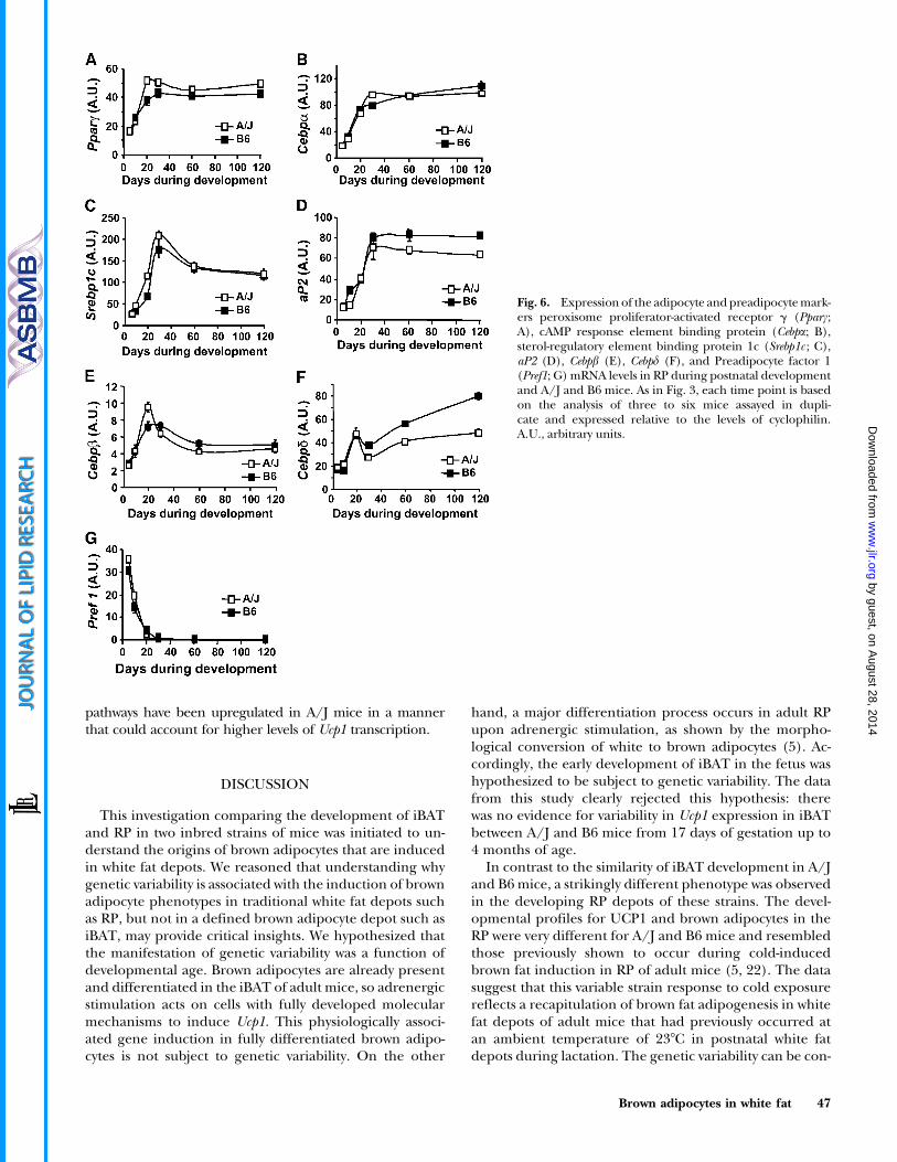
The developmental profiles of several transcription fac-tors showed the expected increase in expression duringpostnatal RP development, consistent with roles for thesefactors in adipogenesis, as established previously from stud-ies with the 3T3-L1 cell culture model. A peak in theexpression at z20 days of age was observed for Pparc, sterol-regulatory element binding protein 1c, Cebpa, -b, and -d,adipocyte P2 (aP2), Pgc1a, and Ppara; although none ofthem returned to the baseline as Ucp1 did (Figs. 6, 7). This
is consistent with the brown adipocyte specificity of Ucp1,as the other genes are also expressed in white adipocytesand possibly other cells in the developing fat tissue. Pre-adipocyte factor 1, a putative marker for the preadipocyte,decreased to very low levels by 20 days of age after high ex-pression at 5 days of age in RP tissue from A/J and B6 mice(Fig. 6G).
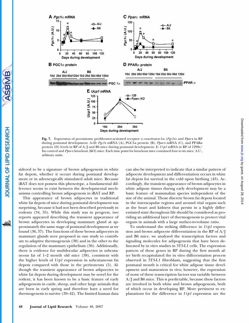
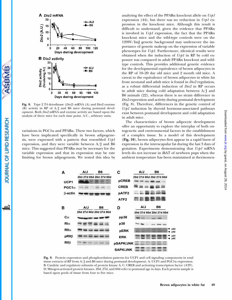
Besides CoxI DNA (Fig. 3C), only Pgc1a and Pparashowed significant increases in expression at both mRNAand protein levels in the RP from A/J mice compared withB6 mice (Fig. 7). The 4-fold induction of Ppara, com-parable to the magnitude of Ucp1 induction, suggested asignificant role for PPARa in the developmental regula-tion of RP remodeling. However, the induction of Ucp1in Ppara knockout mice on the 129Sv/ImJ backgroundat 20 days of age was not significantly different from thatin wild-type controls (Fig. 7E). 129Sv/ImJ mice have in-creased Ucp1 expression during cold acclimation, similarto the A/J strain. With the exception of Ucp1 expression,Dio2 expression showed the most robust differencesbetween A/J and B6 mice during the cold adaptationof adult mice (22); however, no differences could be de-tected in the RP during postnatal development, and itis surprising that there was not even a spike of activityat 20 days of age when the expression of Ucp1 was maxi-mal (Fig. 8).
PKA-associated signaling
We previously showed that during cold induction ofUcp1 in RP of 2 month old mice, subunits of PKA showedno change in expression between A/J and B6 mice byimmunoblot analysis. Yet, modest 3- to 5-fold increasesin phospho-CREB and phosphorylated p38 mitogen-activatedprotein kinase were detected in A/J mice after 1 and 7 days ofcold exposure compared with B6 mice (22). A similar analysisof RP from A/J and B6 mice during postnatal developmentwas performed. Figure 9A confirms the data in Fig. 3B show-ing that the levels of UCP1 were highly increased in A/J micecompared with B6 mice. In this set of tissues, differences inPGC1a protein levels were small. Analyses of subunits ofPKA, CREB, ATF, p38 mitogen-activated protein kinase, ex-tracellular regulated kinase, and stress-activated proteinkinase/c-Jun kinase do not indicate that these signaling
TABLE 1. Ucp1 mRNA levels in iBAT and RP in A/J and B6 mice at 20 and 60 days of age and in mice maintainedat 238C or 48C
Ucp1 Relative Percent
Tissue Age/Conditions A/J B6 A/J B6
arbitrary units/cyclophilin mRNA
iBAT 20 days/238C 773 6 31 (n 5 11) 789 6 9 (n 5 7) 100 10260 days/238C 998 6 27 (n 5 3) 598 6 58 (n 5 6) 129 7760 days/48C, 1 day 1,766 6 27 (n 5 4) 1,352 6 28 (n 5 3) 229 17560 days/48C, 7 days 1,847 6 73 (n 5 5) 1,834 6 70 (n 5 3) 239 237
RP 20 days/238C 63 6 14 (n 5 5) 6 6 1.4 (n 5 4) 8 0.860 days/238C 7 6 1.4 (n 5 5) 0.2 6 0.04 (n 5 6) 0.9 0.0260 days/48C, 1 day 234 6 63 (n 5 5) 3 6 1.6 (n 5 3) 30 0.460 days/48C, 7 days 547 6 59 (n 5 5) 6 6 1.0 (n 5 3) 71 0.8
B6, C57BL/6J; iBAT, interscapular brown adipose tissue; RP, retroperitoneal fat;Ucp1, uncoupling protein 1 gene.
46 Journal of Lipid Research Volume 48, 2007
by guest, on August 28, 2014
ww
w.jlr.org
Dow
nloaded from
pathways have been upregulated in A/J mice in a mannerthat could account for higher levels of Ucp1 transcription.
DISCUSSION
This investigation comparing the development of iBATand RP in two inbred strains of mice was initiated to un-derstand the origins of brown adipocytes that are inducedin white fat depots. We reasoned that understanding whygenetic variability is associated with the induction of brownadipocyte phenotypes in traditional white fat depots suchas RP, but not in a defined brown adipocyte depot such asiBAT, may provide critical insights. We hypothesized thatthe manifestation of genetic variability was a function ofdevelopmental age. Brown adipocytes are already presentand differentiated in the iBAT of adult mice, so adrenergicstimulation acts on cells with fully developed molecularmechanisms to induce Ucp1. This physiologically associ-ated gene induction in fully differentiated brown adipo-cytes is not subject to genetic variability. On the other
hand, a major differentiation process occurs in adult RPupon adrenergic stimulation, as shown by the morpho-logical conversion of white to brown adipocytes (5). Ac-cordingly, the early development of iBAT in the fetus washypothesized to be subject to genetic variability. The datafrom this study clearly rejected this hypothesis: therewas no evidence for variability in Ucp1 expression in iBATbetween A/J and B6 mice from 17 days of gestation up to4 months of age.
In contrast to the similarity of iBAT development in A/Jand B6 mice, a strikingly different phenotype was observedin the developing RP depots of these strains. The devel-opmental profiles for UCP1 and brown adipocytes in theRP were very different for A/J and B6 mice and resembledthose previously shown to occur during cold-inducedbrown fat induction in RP of adult mice (5, 22). The datasuggest that this variable strain response to cold exposurereflects a recapitulation of brown fat adipogenesis in whitefat depots of adult mice that had previously occurred atan ambient temperature of 238C in postnatal white fatdepots during lactation. The genetic variability can be con-
Fig. 6. Expression of the adipocyte and preadipocyte mark-ers peroxisome proliferator-activated receptor g (Pparc;A), cAMP response element binding protein (Cebpa; B),sterol-regulatory element binding protein 1c (Srebp1c ; C),aP2 (D), Cebpb (E), Cebpd (F), and Preadipocyte factor 1(Pref1; G) mRNA levels in RP during postnatal developmentand A/J and B6 mice. As in Fig. 3, each time point is basedon the analysis of three to six mice assayed in dupli-cate and expressed relative to the levels of cyclophilin.A.U., arbitrary units.
Brown adipocytes in white fat 47
by guest, on August 28, 2014
ww
w.jlr.org
Dow
nloaded from
sidered to be a signature of brown adipogenesis in whitefat depots, whether it occurs during postnatal develop-ment or in adrenergically stimulated adult mice. BecauseiBAT does not possess this phenotype, a fundamental dif-ference seems to exist between the developmental mech-anisms controlling brown adipogenesis in iBAT and RP.
This appearance of brown adipocytes in traditionalwhite fat depots of mice during postnatal development wassurprising, because it had not been described previously inrodents (34, 35). While this study was in progress, tworeports appeared describing the transient appearance ofbrown adipocytes in developing mammary gland at ap-proximately the same stage of postnatal development as wefound (36, 37). The functions of these brown adipocytes inmammary glands were proposed in one study to contrib-ute to adaptive thermogenesis (38) and in the other to theregulation of the mammary epithelium (36). Additionally,there is evidence for multilocular adipocytes in subcuta-neous fat of 1–2 month old mice (38), consistent withthe higher levels of Ucp1 expression in subcutaneous fatdepots compared with those in the peritoneum (5). Al-though the transient appearance of brown adipocytes inwhite fat depots during development may be novel for therodent, it has been known to be a basic feature of earlyadipogenesis in cattle, sheep, and other large animals thatare born in early spring and therefore have a need forthermogenesis to survive (39–42). The limited human data
can also be interpreted to indicate that a similar pattern ofadipocyte development and differentiation occurs in whitefat depots for survival in the cold upon birthing (43). Ac-cordingly, the transient appearance of brown adipocytes inwhite adipose tissues during early development may be abasic feature of mammalian species independent of thesize of the animal. Those discrete brown fat depots locatedin the interscapular regions and around vital organs suchas the heart and kidneys that persist in a highly differ-entiated state throughout life should be considered as pro-viding an additional layer of thermogenesis to protect vitalorgans in animals with a large surface-to-volume ratio.
To understand the striking difference in Ucp1 expres-sion and brown adipocyte differentiation in the RP of A/Jand B6 mice, we analyzed the transcription factors andsignaling molecules for adipogenesis that have been de-lineated by in vitro studies in 3T3-L1 cells. The expressionpattern of these genes in RP during the first month af-ter birth recapitulated the in vitro differentiation processobserved in 3T3-L1 fibroblasts, suggesting that the firstpostnatal month is critical for white adipose tissue devel-opment and maturation in vivo; however, the expressionof none of these transcription factors was variable betweenA/J and B6 mice. This is predictable, because these factorsare involved in both white and brown adipogenesis, bothof which occur in developing RP. More pertinent to ex-planations for the difference in Ucp1 expression are the
Fig. 7. Expression of peroxisome proliferator-activated receptor g coactivator-1a (Pgc1a) and Ppara in RPduring postnatal development. A–D: Pgc1a mRNA (A), PGC1a protein (B), Ppara mRNA (C), and PPARaprotein (D) levels in RP of A/J and B6 mice during postnatal development. E: Ucp1 mRNA in RP of 129Sv/Im control and Ppara knockout (KO) mice. Each time point for knockout mice contained four to six mice. A.U.,arbitrary units.
48 Journal of Lipid Research Volume 48, 2007
by guest, on August 28, 2014
ww
w.jlr.org
Dow
nloaded from
variations in PGC1a and PPARa. These two factors, whichhave been implicated specifically in brown adipogene-sis, were expressed with a pattern that resembled Ucp1expression, and they were variable between A/J and B6mice. This suggested that PPARa may be necessary for thevariable expression and that its expression may be ratelimiting for brown adipogenesis. We tested this idea by
analyzing the effect of the PPARa knockout allele on Ucp1expression (44), but there was no reduction in Ucp1 ex-pression in the knockout mice. Although this result isdifficult to understand, given the evidence that PPARais involved in Ucp1 expression, the fact that the PPARaknockout mice and the wild-type controls were on the129SV/ImJ genetic background may underscore the im-portance of genetic makeup on the expression of variablephenotypes for Ucp1. Furthermore, identical results wereobtained when the induction of Ucp1 in RP by cold ex-posure was compared in adult PPARa knockout and wild-type controls. This provides additional genetic evidencefor the developmental equivalence of brown adipocytes inthe RP of 10–20 day old mice and 2 month old mice. Acaveat to the equivalence of brown adipocytes in white fatfrom neonatal and adult mice is found in Dio2 regulation,as a robust differential induction of Dio2 in RP occursin adult mice during cold adaptation between A/J andB6 animals (22), whereas there is no strain difference inDio2 expression and activity during postnatal development(Fig. 8). Therefore, differences in the genetic control ofUcp1 induction by thyroid hormone-associated pathwaysexist between postnatal development and cold adaptationin adult mice.
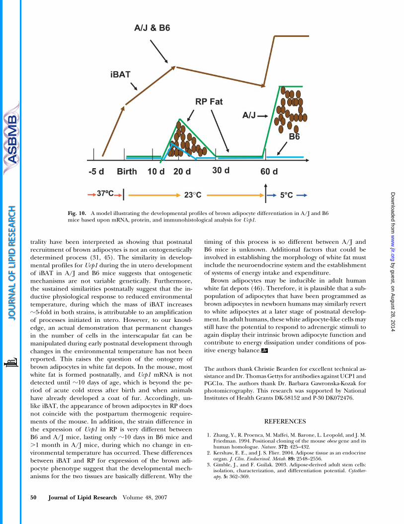
The characteristics of brown adipocyte developmentoffer an opportunity to explore the interplay of both on-togenetic and environmental factors in the establishmentof a complex tissue. In a model of this development(Fig. 10), brown adipocytes first appear in a rapid burst ofexpression in the interscapular fat during the last 3 days ofgestation. Experiments demonstrating that Ucp1 mRNAlevels do not increase in iBAT of newborn pups when theambient temperature has been maintained at thermoneu-
Fig. 8. Type 2 T4 deiodinase (Dio2) mRNA (A) and Dio2 enzyme(B) activity in RP of A/J and B6 mice during postnatal devel-opment. Both Dio2 mRNA and enzyme activity are based upon theanalysis of three mice for each time point. A.U., arbitrary units.
Fig. 9. Protein expression and phosphorylation patterns for UCP1 and cell signaling components in totaltissue extracts of RP from A/J and B6 mice during postnatal development. A: UCP1 and PGC1a expression.B: Catalytic and regulatory subunits of protein kinase A. C: CREB and activating transcription factor (ATF).D: Mitogen-activated protein kinases. 20d, 27d, and 60d refer to postnatal age in days. Each protein sample isbased upon pools of tissue from four to five mice.
Brown adipocytes in white fat 49
by guest, on August 28, 2014
ww
w.jlr.org
Dow
nloaded from
trality have been interpreted as showing that postnatalrecruitment of brown adipocytes is not an ontogeneticallydetermined process (31, 45). The similarity in develop-mental profiles for Ucp1 during the in utero developmentof iBAT in A/J and B6 mice suggests that ontogeneticmechanisms are not variable genetically. Furthermore,the sustained similarities postnatally suggest that the in-ductive physiological response to reduced environmentaltemperature, during which the mass of iBAT increasesz5-fold in both strains, is attributable to an amplificationof processes initiated in utero. However, to our knowl-edge, an actual demonstration that permanent changesin the number of cells in the interscapular fat can bemanipulated during early postnatal development throughchanges in the environmental temperature has not beenreported. This raises the question of the ontogeny ofbrown adipocytes in white fat depots. In the mouse, mostwhite fat is formed postnatally, and Ucp1 mRNA is notdetected until z10 days of age, which is beyond the pe-riod of acute cold stress after birth and when animalshave already developed a coat of fur. Accordingly, un-like iBAT, the appearance of brown adipocytes in RP doesnot coincide with the postpartum thermogenic require-ments of the mouse. In addition, the strain difference inthe expression of Ucp1 in RP is very different betweenB6 and A/J mice, lasting only z10 days in B6 mice and.1 month in A/J mice, during which no change in en-vironmental temperature has occurred. These differencesbetween iBAT and RP for expression of the brown adi-pocyte phenotype suggest that the developmental mech-anisms for the two tissues are basically different. Why the
timing of this process is so different between A/J andB6 mice is unknown. Additional factors that could beinvolved in establishing the morphology of white fat mustinclude the neuroendocrine system and the establishmentof systems of energy intake and expenditure.
Brown adipocytes may be inducible in adult humanwhite fat depots (46). Therefore, it is plausible that a sub-population of adipocytes that have been programmed asbrown adipocytes in newborn humans may similarly revertto white adipocytes at a later stage of postnatal develop-ment. In adult humans, these white adipocyte-like cells maystill have the potential to respond to adrenergic stimuli toagain display their intrinsic brown adipocyte function andcontribute to energy dissipation under conditions of pos-itive energy balance.
The authors thank Christie Bearden for excellent technical as-sistance and Dr. Thomas Gettys for antibodies against UCP1 andPGC1a. The authors thank Dr. Barbara Gawronska-Kozak forphotomicrography. This research was supported by NationalInstitutes of Health Grants DK-58152 and P-30 DK072476.
REFERENCES
1. Zhang, Y., R. Proenca, M. Maffei, M. Barone, L. Leopold, and J. M.Friedman. 1994. Positional cloning of the mouse obese gene and itshuman homologue. Nature. 372: 425–432.
2. Kershaw, E. E., and J. S. Flier. 2004. Adipose tissue as an endocrineorgan. J. Clin. Endocrinol. Metab. 89: 2548–2556.
3. Gimble, J., and F. Guilak. 2003. Adipose-derived adult stem cells:isolation, characterization, and differentiation potential. Cytother-apy. 5: 362–369.
Fig. 10. A model illustrating the developmental profiles of brown adipocyte differentiation in A/J and B6mice based upon mRNA, protein, and immunohistological analysis for Ucp1.
50 Journal of Lipid Research Volume 48, 2007
by guest, on August 28, 2014
ww
w.jlr.org
Dow
nloaded from

4. Himms-Hagen, J. 1989. Brown adipose tissue thermogenesis andobesity. Prog. Lipid Res. 28: 67–115.
5. Guerra, C., R. A. Koza, H. Yamashita, K. Walsh, and L. P. Kozak.1998. Emergence of brown adipocytes in white fat in mice is undergenetic control. Effects on body weight and adiposity. J. Clin. Invest.102: 412–420.
6. Nicholls, D. G., and R. M. Locke. 1984. Thermogenic mechanismsin brown fat. Physiol. Rev. 64: 1–64.
7. Ricquier, D., and J. C. Kader. 1976. Mitochondrial protein alter-ation in active brown fat: a sodium dodecyl sulfate-polyacrylamide gelelectrophoretic study. Biochem. Biophys. Res. Commun. 73: 577–583.
8. Skala, J., T. Barnard, and O. Lindberg. 1970. Changes in inter-scapular brown adipose tissue of the rat during perinatal and earlypostnatal development and after cold acclimation. II. Mitochon-drial changes. Comp. Biochem. Physiol. 33: 509–528.
9. Cousin, B., S. Cinti, M. Morroni, S. Raimbault, D. Ricquier, L.Penicaud, and L. Casteilla. 1992. Occurrence of brown adipocytesin rat white adipose tissue: molecular and morphological charac-terization. J. Cell Sci. 103: 931–942.
10. Young, P., J. R. S. Arch, and M. Ashwell. 1984. Brown adipose tissuein the parametrial fat pad of the mouse. FEBS Lett. 167: 10–14.
11. Ailhaud, G., and H. Hauner. 2004. Development of White AdiposeTissue. 2nd edition. Marcel Decker, Inc., New York.
12. Rothwell, N. J., and M. J. Stock. 1979. A role for brown adiposetissue in diet-induced thermogenesis. Nature. 281: 31–35.
13. Ghorbani, M., and J. Himms-Hagen. 1997. Appearance of brownadipocytes in white adipose tissue during CL 316,243-induced rever-sal of obesity and diabetes in Zucker fa/fa rats. Int. J. Obes. 21: 465–475.
14. Collins, S., K. W. Daniel, A. E. Petro, and R. S. Surwit. 1997. Strain-specific response to beta 3-adrenergic receptor agonist treatmentof diet-induced obesity in mice. Endocrinology. 138: 405–413.
15. Cederberg, A., L. M. Gronning, B. Ahren, P. Tasken, P. Carlsson,and S. Enerback. 2001. FOXC2 is a winged helix gene that counter-acts obesity, hypertriglyceridemia, and diet-induced insulin resis-tance. Cell. 106: 563–573.
16. Cummings, D. E., E. P. Brandon, J. V. Planas, K. Motamed, R. L.Idzerda, and G. S. McKnight. 1996. Genetically lean mice resultfrom targeted disruption of the RII beta subunit of protein kinaseA. Nature. 382: 622–626.
17. Kopecky, J., G. Clarke, S. Enerback, B. Spiegelman, and L. P. Kozak.1995. Expression of the mitochondrial uncoupling protein genefrom the aP2 gene promoter prevents genetic obesity. J. Clin. Invest.96: 2914–2923.
18. Soloveva, V., R. A. Graves, M. M. Rasenick, B. M. Spiegelman, and S. R.Ross. 1997. Transgenic mice overexpressing the b1-adrenergic recep-tor in adipose tissue are resistant to obesity. Mol. Endocrinol. 11: 27–38.
19. Tsukiyama-Kohara, K., F. Poulin, M. Kohara, C. T. DeMaria, A.Cheng, Z. Wu, A. C. Gingras, A. Katsume, M. Elchebly, B. M.Spiegelman, et al. 2001. Adipose tissue reduction in mice lackingthe translational inhibitor 4E-BP1. Nat. Med. 7: 1128–1132.
20. Coulter, A. A., C. M. Bearden, X. Liu, R. A. Koza, and L. P. Kozak.2003. Dietary fat interacts with QTLs controlling induction of Pgc-1alpha and Ucp1 during conversion of white to brown fat. Physiol.Genomics. 14: 139–147.
21. Koza, R. A., S. M. Hohmann, C. Guerra, M. Rossmeisl, and L. P.Kozak. 2000. Synergistic gene interactions control the induction ofthe mitochondrial uncoupling protein (Ucp1) gene in white fat tis-sue. J. Biol. Chem. 275: 34486–34492.
22. Xue, B., A. Coulter, J. S. Rim, R. A. Koza, and L. P. Kozak. 2005.Transcriptional synergy and the regulation of Ucp1 during brownadipocyte induction in white fat depots. Mol. Cell. Biol. 25: 8311–8322.
23. Himms-Hagen, J., A. Melnyk, M. C. Zingaretti, E. Ceresi, G.Barbatelli, and S. Cinti. 2000. Multilocular fat cells in WAT of CL-316243-treated rats derive directly from white adipocytes. Am. J.Physiol. Cell Physiol. 279: C670–C681.
24. Manchado, C., P. Yubero, O. Vinas, R. Iglesias, F. Villarroya, T.Mampel, and M. Giralt. 1994. CCAAT/enhancer-binding proteins aand b in brown adipose tissue: evidence for a tissue-specific patternof expression during development. Biochem. J. 302: 695–700.
25. Commins, S. P., D. J. Marsh, S. A. Thomas, P. M. Watson, M. A.Padgett, R. Palmiter, and T. W. Gettys. 1999. Norepinephrine isrequired for leptin effects on gene expression in brown and whiteadipose tissue. Endocrinology. 140: 4772–4778.
26. Fekete, C., G. Legradi, E. Mihaly, Q. H. Huang, J. B. Tatro, W. M.Rand, C. H. Emerson, and R. M. Lechan. 2000. a-Melanocyte-stimulating hormone is contained in nerve terminals innervating
thyrotropin-releasing hormone-synthesizing neurons in the hypo-thalamic paraventricular nucleus and prevents fasting-inducedsuppression of prothyrotropin-releasing hormone gene expression.J. Neurosci. 20: 1550–1558.
27. Schneider, M. J., S. N. Fiering, S. E. Pallud, A. F. Parlow, D. L. St.Germain, and V. A. Galton. 2001. Targeted disruption of the type 2selenodeiodinase gene (DIO2) results in a phenotype of pituitaryresistance to T4. Mol. Endocrinol. 15: 2137–2148.
28. Young, J. B. 2002. Effects of litter size on sympathetic activity inyoung adult rats. Am. J. Physiol. Regul. Integr. Comp. Physiol. 282:R1113–R1121.
29. Giralt, M., I. Martin, R. Iglesias, O. Vinas, F. Villarroya, and T.Mampel. 1990. Ontogeny and perinatal modulation of geneexpression in rat brown adipose tissue. Unaltered iodothyronine59-deiodinase activity is necessary for the response to environmen-tal temperature at birth. Eur. J. Biochem. 193: 297–302.
30. Houstek, J., J. Kopecky, R. Zdenek, and T. Soukup. 1988. Un-coupling protein in embryonic brown adipose tissue—existence ofnonthermogenic and thermogenic mitochondria. Biochim. Biophys.Acta. 935: 19–25.
31. Teruel, T., A. M. Valverde, A. Alvarez, M. Benito, and M. Lorenzo.1995. Differentiation of rat brown adipocytes during late foetal de-velopment: role of insulin-like growth factor I.Biochem. J. 310: 771–776.
32. Villena, J. A., M. C. Carmona, M. Rodriguez de la Concepcion, M.Rossmeisl, O. Vinas, T. Mampel, R. Iglesias, M. Giralt, and F.Villarroya. 2002. Mitochondrial biogenesis in brown adipose tissueis associated with differential expression of transcription regulatoryfactors. Cell Mol. Life Sci. 59: 1934–1944.
33. Rim, J. S., B. Xue, B. Gawronska-Kozak, and L. P. Kozak. 2004.Sequestration of thermogenic transcription factors in the cyto-plasm during development of brown adipose tissue. J. Biol. Chem.279: 25916–25926.
34. Cinti, S. 1999. The Adipose Organ. Editrice Kurtis, Milano, Italy.35. Nedergaand, J. and B. Cannon. 2004. Brown adipose tissue: de-
velopment and function. In Fetal and Neonatal Physiology. R. A.Poulin, W. W. Fox and S. H. Abman, editors. Saunders. 404–415.
36. Gouon-Evans, V., and J. W. Pollard. 2002. Unexpected depositionof brown fat in mammary gland during postnatal development.Mol. Endocrinol. 16: 2618–2627.
37. Master, S. R., J. L. Hartman, C. M. D’Cruz, S. E. Moody, E. A.Keiper, S. I. Ha, J. D. Cox, G. K. Belka, and L. A. Chodosh. 2002.Functional microarray analysis of mammary organogenesis revealsa developmental role in adaptive thermogenesis. Mol. Endocrinol.16: 1185–1203.
38. Rossmeisl, M., G. Barbatelli, P. Flachs, P. Brauner, M. C. Zingaretti,M. Marelli, P. Janovska, M. Horakova, I. Syrovy, S. Cinti, et al.2002. Expression of the uncoupling protein 1 from the aP2 genepromoter stimulates mitochondrial biogenesis in unilocular ad-ipocytes in vivo. Eur. J. Biochem. 269: 19–28.
39. Alexander, G., J. W. Bennett, and R. T. Gemmell. 1975. Brownadipose tissue in the new-born calf (Bos taurus). J. Physiol. 244:223–234.
40. Casteilla, L., C. Forest, J. Robelin, D. Ricquier, A. Tombet, and G.Ailhaud. 1987. Characterization of mitochondrial uncouplingprotein in bovine fetus and newborn calf. Am. J. Physiol. 252:E627–E636.
41. Gemmell, R. T., and G. Alexander. 1978. Ultrastructural develop-ment of adipose tissue in foetal sheep. Aust. J. Biol. Sci. 31: 505–515.
42. Gemmell, R. T., A. W. Bell, and G. Alexander. 1972. Morphologyof adipose cells in lambs at birth and during subsequent transitionof brown to white adipose tissue in cold and in warm conditions.Am. J. Anat. 133: 143–164.
43. Lean, M. E., W. P. James, and P. Trayhurn. 1986. Brown adiposetissue in patients with phaeochromocytoma. Int. J. Obes. 10: 219–227.
44. Kersten, S., J. Seydoux, J. M. Peters, F. J. Gonzalez, B. Desvergne,and W. Wahli. 1999. Peroxisome proliferator-activated receptoralpha mediates the adaptive response to fasting. J. Clin. Invest. 103:1489–1498.
45. Obregon, M. L., A. Jacobsson, T. Kirchgessner, M. L. Schotz, B.Cannon, and J. Nedergaand. 1989. Postnatal recruitment of brownadipose tissue is induced by the cold stress experienced by thepups. Biochem. J. 259: 341–346.
46. Digby, J. E., C. T. Montague, C. P. Sewter, L. Sanders, W. O.Wilkison, S. O’Rahilly, and J. B. Prins. 1998. Thiazolidinedioneexposure increases the expression of uncoupling protein 1 in cul-tured human preadipocytes. Diabetes. 47: 138–141.
Brown adipocytes in white fat 51
by guest, on August 28, 2014
ww
w.jlr.org
Dow
nloaded from