Protective effects of noradrenaline against tumor necrosis factor-alpha-induced apoptosis in...
10
PAPER Protective effects of noradrenaline against tumor necrosis factor-a-induced apoptosis in cultured rat brown adipocytes: role of nitric oxide-induced heat shock protein 70 expression E Nisoli 1 *, L Regianini 1 , A Bulbarelli 1 , L Briscini 1 , R Breacale 1 and MO Carruba 1,2 1 Center for Study and Research on Obesity, Department of Preclinical Sciences, LITA Vialba, L Sacco Hospital, Milan University, Milan, Italy; and 2 Istituto Auxologics Italiano, Milan, Italy OBJECTIVE: To elucidate the effects and molecular mechanism(s) by means of which noradrenaline (NA) protects against the tumor necrosis factor (TNF)-a-induced apoptosis of brown adipocytes. DESIGN: Brown fat precursor cells were isolated from young rats; 2.5 million cells were added to each 24-well culture plate and cultured in a defined culture medium. On day 8, the cultured cells were exposed to murine recombinant TNF-a and=or cycloheximide (CHX; 10 mg=ml) and=or NA and=or nitric oxide (NO) donors and=or the NO synthase inhibitor N G -nitro-L- arginine methyl ester (L-NAME) and=or 10 mM heat shock protein 70 (HSP70) antisense or sense oligomers. MEASUREMENTS: Analysis of DNA fragmentation on agarose gel as a marker of apoptosis; reverse transcriptase-polymerase chain reaction analysis of mRNA levels; Western blotting analysis of protein levels. RESULTS: Pretreatment of primary cultures of rat brown fat cells with micromolar concentrations of NA or the NO-donor S- nitroso-N-acetylpenicillamine (SNAP) induced the expression of HSP70 mRNA and protein, which was associated with cytoprotection against TNF-a plus CHX-induced apoptosis. The L-NAME inhibitor of NO synthase activity inhibited both NA- stimulated HSP70 expression and cytoprotection. Furthermore, pretreatment of brown adipocytes with an antisense oligo- nucleotide to HSP70 antagonized both SNAP- and NA-induced cytoprotection. CONCLUSION: These findings demonstrate that the NO produced by NA stimulation can induce resistance to the TNF-a- induced apoptosis of brown adipocytes, possibly by means of the expression of HSP70. International Journal of Obesity (2001) 25, 1421 – 1430 Keywords: apoptosis; noradrenaline; brown adipocytes; nitric oxide; heat shock protein Introduction Obesity in rodents is related to functional atrophy of ther- mogenically active brown fat and the consequent impair- ment of adaptive thermogenesis; 1,2 in addition to defects in the molecular mechanisms of adaptive thermogenesis, this atrophy may be due to an increase in the cell death rate. As the white fat of obese subjects is a significant source of endogenous tumor necrosis factor (TNF)-a, 3,4 which induces apoptosis in many cell types, 5–7 this cytokine could be involved in brown adipose tissue (BAT) atrophy. TNF-a induces the apoptosis of cultured rat brown adipocytes 8,9 and a larger number of apoptotic nuclei can be seen in the BAT of obese rats than in that of lean controls; 8 furthermore, a significant reduction in brown adipocyte apoptosis can be obtained in genetically obese mice by partially or completely abolishing TNF-a signaling and function. 10 Noradrenaline (NA) increases the number of viable brown fat cells, and correspondingly decreases the number of apop- totic cells when added before TNF-a. 8 Moreover, the cold exposure stimulation of sympathetic activity to BAT signifi- cantly decreases the number of apoptotic cells in comparison with that observed in obese rats kept at room temperature. 8 *Correspondence: E Nisoli, Center for Study and Research on Obesity, LITA Vialba, L. Sacco Hospital, Via G.B. Grassi, 74-20157 Milano, Italy. E-mail: [email protected] Received 21 August 2000; revised 7 March 2001; accepted 20 April 2001 International Journal of Obesity (2001) 25, 1421–1430 ß 2001 Nature Publishing Group All rights reserved 0307–0565/01 $15.00 www.nature.com/ijo International Journal of Obesity (2001) 25, 1421–1430 ß 2001 Nature Publishing Group All rights reserved 0307–0565/01 $15.00 www.nature.com/ijo
Transcript of Protective effects of noradrenaline against tumor necrosis factor-alpha-induced apoptosis in...

PAPER
Protective effects of noradrenaline against tumornecrosis factor-a-induced apoptosis in cultured ratbrown adipocytes: role of nitric oxide-induced heatshock protein 70 expression
E Nisoli1*, L Regianini1, A Bulbarelli1, L Briscini1, R Breacale1 and MO Carruba1,2
1Center for Study and Research on Obesity, Department of Preclinical Sciences, LITA Vialba, L Sacco Hospital, Milan University,Milan, Italy; and 2Istituto Auxologics Italiano, Milan, Italy
OBJECTIVE: To elucidate the effects and molecular mechanism(s) by means of which noradrenaline (NA) protects against thetumor necrosis factor (TNF)-a-induced apoptosis of brown adipocytes.DESIGN: Brown fat precursor cells were isolated from young rats; 2.5 million cells were added to each 24-well culture plate andcultured in a defined culture medium. On day 8, the cultured cells were exposed to murine recombinant TNF-a and=orcycloheximide (CHX; 10 mg=ml) and=or NA and=or nitric oxide (NO) donors and=or the NO synthase inhibitor NG-nitro-L-arginine methyl ester (L-NAME) and=or 10mM heat shock protein 70 (HSP70) antisense or sense oligomers.MEASUREMENTS: Analysis of DNA fragmentation on agarose gel as a marker of apoptosis; reverse transcriptase-polymerasechain reaction analysis of mRNA levels; Western blotting analysis of protein levels.RESULTS: Pretreatment of primary cultures of rat brown fat cells with micromolar concentrations of NA or the NO-donor S-nitroso-N-acetylpenicillamine (SNAP) induced the expression of HSP70 mRNA and protein, which was associated withcytoprotection against TNF-a plus CHX-induced apoptosis. The L-NAME inhibitor of NO synthase activity inhibited both NA-stimulated HSP70 expression and cytoprotection. Furthermore, pretreatment of brown adipocytes with an antisense oligo-nucleotide to HSP70 antagonized both SNAP- and NA-induced cytoprotection.CONCLUSION: These findings demonstrate that the NO produced by NA stimulation can induce resistance to the TNF-a-induced apoptosis of brown adipocytes, possibly by means of the expression of HSP70.International Journal of Obesity (2001) 25, 1421 – 1430
Keywords: apoptosis; noradrenaline; brown adipocytes; nitric oxide; heat shock protein
IntroductionObesity in rodents is related to functional atrophy of ther-
mogenically active brown fat and the consequent impair-
ment of adaptive thermogenesis;1,2 in addition to defects in
the molecular mechanisms of adaptive thermogenesis, this
atrophy may be due to an increase in the cell death rate. As
the white fat of obese subjects is a significant source of
endogenous tumor necrosis factor (TNF)-a,3,4 which induces
apoptosis in many cell types,5 – 7 this cytokine could be
involved in brown adipose tissue (BAT) atrophy. TNF-ainduces the apoptosis of cultured rat brown adipocytes8,9
and a larger number of apoptotic nuclei can be seen in the
BAT of obese rats than in that of lean controls;8 furthermore,
a significant reduction in brown adipocyte apoptosis can be
obtained in genetically obese mice by partially or completely
abolishing TNF-a signaling and function.10
Noradrenaline (NA) increases the number of viable brown
fat cells, and correspondingly decreases the number of apop-
totic cells when added before TNF-a.8 Moreover, the cold
exposure stimulation of sympathetic activity to BAT signifi-
cantly decreases the number of apoptotic cells in comparison
with that observed in obese rats kept at room temperature.8
*Correspondence: E Nisoli, Center for Study and Research on Obesity,
LITA Vialba, L. Sacco Hospital, Via G.B. Grassi, 74-20157 Milano, Italy.
E-mail: [email protected]
Received 21 August 2000; revised 7 March 2001;
accepted 20 April 2001
International Journal of Obesity (2001) 25, 1421–1430ß 2001 Nature Publishing Group All rights reserved 0307–0565/01 $15.00
www.nature.com/ijo
International Journal of Obesity (2001) 25, 1421–1430ß 2001 Nature Publishing Group All rights reserved 0307–0565/01 $15.00
www.nature.com/ijo

These results suggest that TNF-a produced by white fat cells
can induce the apoptosis of brown adipocytes in obese
animals and that the noradrenergic system may counteract
this phenomenon. Micromolar NA concentrations markedly
increase the Bcl-2=Bax mRNA and protein ratios and protect
against the serum deprivation-induced apoptosis of brown
adipocytes.11 Furthermore, NA stimulates the expression of
the inducible form of nitric oxide (NO) synthase, an enzyme
that ensures NO production, in cultured brown adipocytes
and BAT.12
Interestingly, it has been found that NO is involved in
both cell death13 and cell survival,14,15 different behaviours
that seem to be related to the cell type. In particular,
extensive research has shown that NO can induce resistance
to TNF-a-induced hepatotoxicity, possibly as a result of an
increased expression of the inducible form of heat shock
protein 70 (HSP70).16
The aim of this study was to investigate the role of NO in
mediating the anti-apoptotic effects of NA in brown fat cells
treated with TNF-a plus cycloheximide (CHX), which we
found to be partially mediated by the induction of cyto-
protective stress proteins such as inducible HSP70.
Materials and methodsAnimals
Normal-weight male Zucker rats aged 5 – 8 weeks were
obtained from Charles River (Calco, Como, Italy). All of
the animal experiments were conducted in accordance
with the highest standards of humane animal care.
Brown adipocyte isolation
Brown fat precursor cells were isolated from young rats as
previously described.8,10 The BAT fragments were carefully
dissected out under sterile conditions and placed in a Hepes-
buffered solution (pH 7.4) containing 2 mg=ml type II col-
lagenase. After 30 min of enzyme treatment at 37�C, the
tissue remnants were removed by filtration through a
250 mm nylon screen, and the mature adipocytes were
allowed to float to the surface (30 min on ice). The infra-
natant containing the adipocyte precursor cells was then
collected, filtered through a 25 mm nylon screen, pelletted
by centrifugation for 10 min at 700 g in 10 ml of culture
medium (see below), and diluted to 20 ml.
Adipose cell culture and treatments
Two-and-a-half million cells were added to each 24-well
culture plate (Nunclon Delta, Milan, Italy) and cultured in
2.0 ml of a culture medium consisting of Dulbecco’s mod-
ified Eagle’s medium (DMEM) supplemented with 4 mM
glutamine, 10% newborn calf serum, 4 nM insulin, 10 mM
Hepes, and 50 IU of penicillin, 50 mg of streptomycin and
25 mg of sodium ascorbate per ml (all from Flow Laboratories,
Milan, Italy), at 37�C in a water-saturated atmosphere of 6%
CO2 in air. The medium was completely exchanged with
prewarmed fresh medium on day 1 (when the cultures were
first washed with 5 ml of prewarmed DMEM) and every each
day (without washing). On day 8, the cultured cells were
exposed to murine recombinant TNF-a (freshly diluted in
buffers; Genzyme, Cinisello Balsamo, Italy) with a specific
activity of 5�107 U=mg, and=or CHX 10 mg=ml and=or NA
freshly diluted in a buffer containing 0.1% ascorbic acid (in
order to prevent NA oxidation for the times indicated in the
figure legends), and then harvested. During the long-term
experiments (24 h), the cells were exposed to the drugs twice
a day.
In order to characterize the effects of NO donors, the
brown adipocyte cultures were treated with 100 – 750 mM S-
nitroso-N-acetylpenicillamine (SNAP; Sigma, Milan, Italy) for
2 – 24 h. The NOS inhibitor NG-Nitro-L-arginine methyl ester
(L-NAME; Sigma, Milan, Italy) was used at a concentration
of 1 mM.
In order to evaluate the involvement of inducible HSP70
in the cytoprotective effects of SNAP and NA, brown fat cells
were incubated with 10 mM HSP70 antisense or sense oligo-
mers during 24 h pretreatments with SNAP 500 mM or NA
10 mM, followed by TNF-a plus CHX exposure for a further
4 h in the continued presence of medium containing the
oligonucleotides as described by Kim et al.16 The inducible
HSP70 antisense (TGTTTTCTTGGCCAT) and sense oligomers
(ATGGCCAAGAAAACA) were synthesized from sequences
complementary to the initiation and four downstream
codons of rat inducible HSP70 mRNA.17
Analysis of DNA fragmentation
The confluent brown adipocytes (at 8 days in culture) were
lysed in 20 mM Tris – HCl (pH 7.5), 10 mM EDTA and 0.5%
Triton X 100 (pH 8), and occasionally shaken while on ice for
30 min. The cytosolic fraction was obtained by centrifuga-
tion at maximum speed (13 000 g) for 20 min at 4�C, and the
cytosolic DNA was extracted with a mixture of phenol and
chloroform; one-tenth volume of 5 mM NaCl was added to
the solution and DNA was then precipitated overnight by
adding an equal volume of isopropanol. After storing over-
night at 720�C a DNA pellet was recovered by centrifuga-
tion, resuspended in water, and treated with RNase 50 mg=ml.
The DNA (3 – 5 mg) was then loaded on 1.2% agarose gels and
stained with ethidium bromide after migration.
RT-PCR analysis
Total RNA was isolated from the cultured cells after drug
treatments using RNazol method (TM Cinna Scientific,
Friendswood, Texas, USA). For PCR analysis, the RNAs were
treated for 1 h at 37�C with 6 U of RNase-free DNaseI per mg
of RNA in 100 mM Tris – HCl, pH 7.5, and 50 mM MgCl2 in
the presence of 2 U=ml of placenta RNase inhibitor. One
microgram of total RNA was reverse transcribed with 200 U
of Moloney murine leukemia virus reverse transcriptase
(Promega, Milan, Italy) in 20 ml of Promega supplied buffer
Anti-apoptotic effects of NA in brown adipocytesE Nisoli et al
1422
International Journal of Obesity

containing 0.4 mM dNTP, 2 U=ml RNase inhibitor, and 0.8 mg
oligo(dT)15 primer (Promega). A control experiment without
reverse transcriptase was performed for each sample in order
to verify that the amplification did not originate from
residual genomic DNA. The PCR was performed using trun-
cated Thermophilus acquaticus DNA polymerase (Biotaq2,
Bioprobe Systems, Milan, Italy) in 50 ml of standard buffer
(20 mM Tris – HCl, pH 8.55, 16 mM (NH4)2SO4, 2.5 mM
MgCl2, 150 mg=ml bovine serum albumin, and 200 mM
dNTP) containing 10 ng of cDNA from the various prepara-
tions, and 1mM of the sense and antisense oligonucleotide
primers specific for inducible HSP70 (50
-ACATCAGCCAGAA-
CAAGCG-30
, amino acids 932 – 950, and 50
-TGATGC-
TCTTGTTCAGGTCG-30
, amino acids 1248 – 1267),18 Bcl-2
(50
-AGAGGGGCTACGAGTGGGAT-30
, amino acids 1895 –
1914, and 50
-CTCAGTCATCCACAGGGCG-A-30
, amino
acids 2330 – 2349),19 and Bax (50
-GGTTTCATCCAGGATCGA-
GACGG-30
, amino acids 84 – 106, and 50
-ACAAAGATGGT-
CACGGTCTGCC-30
, amino acids 509 – 530).20 The mRNA
for constitutive b-actin was examined as the reference cell
transcript. b-actin mRNA amplification products were pre-
sent at equivalent levels in the lysates of treated and
untreated cells at the different time points. The reaction
was performed using specific primers described elsewhere.12
b-Actin was separately co-amplified with HSP70, Bcl-2 and
Bax cDNA, with the b-Actin primers being added after 10
amplification cycles in order to avoid saturation. The PCR
conditions were as follows: denaturation at 94�C for 30 s,
annealing at 61�C (63�C for HSP70) for 30 s, and polymerisa-
tion at 72�C for 30 s. After 28 cycles, 5ml of the reaction
mixture were separated by electrophoresis (1.2% agarose gel
in Tris-Borate-EDTA buffer) and made visible by means of
ethidium bromide staining. The number of cycles and the
reaction temperature of the semiquantitative RT-PCR assay
were previously established in our laboratory.11 Indeed, we
observed a linear relationship between the amount of input
template and the amount of PCR product generated over a
wide concentration range (0.5 – 10 mg of total RNA) also after
30 cycles of amplification. For this reason we judged as an
optimal experimental condition for linearity to perform 28
cycles of amplification.
The RT-PCR products were semiquantitatively analysed
using a QuickImage densitometer (Canberra, Packard, Milan,
Italy) and a Phoretix 1D (version 3.0.1) software image
analyser. In order to reduce the detection limits of the
densitometric analysis, the measurements were made
within the linear response range of the image capturing
device (optical density 0 – 3.0); for this purpose, a standard
image was acquired at the same time as the experimental
images. Each experiment was densitometrically analysed at
least four times; the coefficient of analytical variation
between the different analyses was less than 5%. If the
density of the b-actin band of a given sample differed
by>10% from the mean density of the samples in a gel, it
was removed from the analysis. The HSP70 mRNA levels
were expressed relative to the levels of the co-amplified b-
actin mRNA. The Bcl-2 and Bax mRNA levels were expressed
as the ratio of signal intensity for the target genes in relation
to that for the coamplified b-actin, and the Bcl-2=Bax mRNA
ratio was calculated. In order to normalize between-experi-
ment densitometer differences, the data were corrected for
background and expressed as a percentage of a specific
within-experiment reference value of one (see figure
legends).
Western blot analysis
Western blot analysis was performed as previously
described.12 Briefly, 70 mg total protein extracts were resolved
by 8% SDS – PAGE and transferred to nitrocellulose filter
papers. The primary antibody (monoclonal antibody direc-
ted against the mouse inducible HSP70 at a dilution of
1 : 1000; StressGen, Lexington, KY, Canada) was diluted in
Tris-buffered saline, pH 7.6, and incubated with membrane
for 2 h at room temperature. The secondary antibody (horse-
radish peroxidase-conjugated anti-mouse, Pierce, Rockford,
IL, USA) was incubated with membrane for 1 h at room
temperature. Immunoreactive bands were visualized by
enhanced chemiluminescence according to the specifica-
tions of the manufacturer (Super Signal Substrate, Pierce)
and then densitometrically analysed.
Statistical methods
The data are presented as the mean values� s.d. of at least
three separate experiments. The comparisons between two
values were made by means of one-way ANOVA followed by
the Student – Newman – Kauls post-hoc comparison. A P-value
of 0.05 was considered significant.
ResultsSNAP pretreatment protects brown adipocytes against
TNF-a plus CHX-induced apoptosis
We have previously demonstrated that the pretreatment of
brown adipocytes with NA provides protection against TNF-aplus CHX-induced apoptosis.8 Since NA increases NO pro-
duction by stimulating the inducible form of NO synthase in
brown adipocytes,12 we investigated whether NO pretreat-
ment may offer similar protection against TNF-a-induced
toxicity. For this aim, brown adipocytes differentiated in
culture were treated with 100 mM SNAP for 24 h, washed
twice with fresh medium, and then added with TNF-a(1 nM) plus CHX (10 mg=ml) for 4 – 6 h.
Apoptosis was determined by measuring DNA fragmenta-
tion on agarose gels. Figure 1A shows that 4 h treatment with
TNF-a plus CHX (but neither TNF-a nor CHX alone) induced
a marked DNA fragmentation, thus confirming our previous
results.8 Twenty-four-hour pretreatment with 100 mM SNAP
protected the brown adipocytes from TNF-a plus CHX toxi-
city to the same extent as 10 mM NA (Figure 1B). In addition,
the anti-apoptotic effects of NA were antagonized by 1 mM
Anti-apoptotic effects of NA in brown adipocytesE Nisoli et al
1423
International Journal of Obesity

of the NOS inhibitor L-NAME (Figure 1B, lane 7). Both SNAP
and L-NAME alone did not induce any DNA fragmentation.
Pretreatment with SNAP 2 h before the addition of TNF-aplus CHX had no effect; however, if SNAP was added 4 – 12 h
before TNF-a plus CHX treatment, the degree of DNA frag-
mentation was markedly reduced, and 24 h before apoptosis
was almost completely inhibited (Figure 2A). When SNAP
concentrations of 100 – 750 mM were added to the brown
adipocytes 24 h before TNF-a plus CHX, protection against
apoptosis was evident even at the lowest dose (Figure 2B).
Effects of 8 Br-cGMP on TNF-a plus CHX-induced
apoptosis of brown adipocytes
It is known that the inhibitory or stimulatory effects of NO
on the apoptosis of different cell types take place through
cyclic GMP (cGMP)-dependent or -independent mechan-
isms.21 – 23 In order to investigate the role of this cyclic
nucleotide in inhibiting the TNF-a plus CHX-induced
Figure 1 NA and SNAP pretreatments antagonize TNF-a plus CHX-induced apoptosis in cultured brown adipocytes. (A) Four-hour treatment with 1 nMTNF-a plus 10 mg=ml CHX induced apoptosis of brown fat cells. (B) Brown fat cells were pretreated with either NA 10 mM or SNAP 100mM or L-NAME1 mM for 24 h and incubated with TNF-a (1 nM) plus CHX (10 mg=ml) for 4 h. Cytosolic DNA was isolated as described in Materials and methods andelectrophoresed on agarose gel after normalizing the amount of DNA by protein concentration. The DNA was visualized using UV light andphotographed. M, molecular weight markers (A¼ Standard 6, Boehringer, Mannheim, Germany; B¼Gene Ruler 100 bp DNA ladder, Genenco, Milan,Italy). The gels shown are representative of the data obtained from six independent experiments.
Figure 2 SNAP pretreatment protects brown adipocytes from TNF-aplus CHX-mediated apoptosis in a time- (A) and dose-dependent (B)manner. The brown fat cells were pretreated with 500 mM (A) or differentconcentrations (B) of SNAP for various time periods (A) or 24 h (B),washed twice with fresh medium, and then incubated with TNF-a (1 nM)plus CHX (10 mg=ml) for 4 h. DNA fragmentation was measured asdescribed in Figure 1.
Anti-apoptotic effects of NA in brown adipocytesE Nisoli et al
1424
International Journal of Obesity

apoptosis in brown adipocytes, we studied the effects of the
membrane-permeable cyclic GMP analogue, 8 Br-cGMP.
Figure 3 shows that the pretreament of brown adipocytes
with either 250 (lane 4) or 500 mM (lane 5) 8 Br-cGMP for
24 h did not affect TNF-a plus CHX-induced DNA fragmen-
tation, as well as 500mM 8 Br-cGMP alone (lane 6) did
not induce DNA fragmentation, thus suggesting that the
anti-apoptotic effects of NO on brown fat cells are cGMP-
independent.
SNAP pretreatments do not increase the Bcl-2=Bax mRNA
ratio
Given that NA increases in a concentration-dependent
manner the Bcl-2 mRNA and protein levels, leaving
unchanged Bax mRNA and protein levels in brown fat cells
(ie the Bcl-2=Bax mRNA and protein ratios are increased),11
which suggests that its cytoprotective effects may be partly
due to a modulation of the balance between survival (Bcl-2)
and death factors (Bax), we investigated the effects of SNAP
on Bcl-2 and Bax gene expression. Figure 4 shows that SNAP
treatment (100 – 750 mM for 24 h) did not change the levels of
either Bcl-2 or Bax mRNA, and so the Bcl-2=Bax mRNA ratio
also remained unchanged. This suggests that the anti-apop-
totic effects of the NO donor do not seem to depend on the
modulation of bcl-2 gene family proteins.
NA and SNAP pretreatments induce HSP70 expression
The cytotoxic effects of TNF-a are antagonized by the indu-
cible heat shock protein HSP70 in various cell types.21,24 – 26
In particular, inducible (but not constitutive) HSP70 is
involved in the SNAP-induced protection of hepatocytes
from TNF-a-mediated cytotoxicity.21 In order to determine
whether SNAP or NA increase inducible HSP70 expression in
brown adipocytes, we exposed cultured brown fat cells to
increasing concentrations for different times and examined
HSP70 mRNA levels by means of RT-PCR analysis. HSP70
mRNA expression increased at SNAP concentrations as low as
100 mM and was maximal at 500 mM, and at NA concentra-
tions as low as 1 mM (maximal at 100 mM; Figure 5A). Figure
5B shows that, after exposure to SNAP 500 mM and NA 10 mM,
HSP70 mRNA levels progressively increased for 4 h and then
declined. All of these results were confirmed at protein level
by Western blot analysis by means of a monoclonal antibody
specific for the inducible HSP70 isoform. In particular, Figure
5C shows that SNAP and NA increased HSP70 protein levels
in a dose-dependent manner after 8 h treatment, and that
these increases were blunted after 24 h treatment. In addi-
tion, Figure 5A and B show that exposure to increasing
concentrations of 8 Br-cGMP for different times did not
change HSP70 expression.
Interestingly, even if the time-dependent NA-induced
increases in HSP70 mRNA levels were markedly antagonized
by 1 mM of L-NAME, this anatgonism was not complete
(Figure 6). These findings may suggest an additional protec-
tive mechanism induced by NA, since it is unlikely that there
is a concentration effect due to insufficient amounts of L-
NAME being used. Indeed, L-NAME concentrations up to
5 mM did not antagonize fully the NA effects.
Antisense oligomer to HSP70 blocks HSP70 expression
and its protection against TNF-a plus CHX-induced
apoptosis
In order to investigate the role of HSP70 in the SNAP- and
NA-induced inhibition of apoptosis mediated by TNF-a plus
CHX, its expression was suppressed using an antisense
HSP70 oligomer. Treatment with antisense did not influence
cell morphology. The brown fat cells were incubated for 24 h
with 10 mM of HSP70 antisense or sense oligonucleotides
during 500mM SNAP or 10 mM NA pretreatment, followed
by TNF-a plus CHX exposure for a further 4 h in the con-
tinued presence of the oligonucleotides. Figure 7A shows
that HSP70 antisense, differently from the sense oligomer,
inhibited either SNAP- or NA-induced protection against
TNF-a plus CHX-mediated apoptosis. In addition, neither
HSP70 antisense or sense alone induced apoptosis. These
Figure 3 8 Br-cGMP does not affect TNF-a plus CHX-induced apoptosisof cultured brown adipocytes. The brown fat cells were pretreated with250 (lane 4) or 500 mM of 8 Br-cGMP (lane 5) for 24 h, washed twice withfresh medium, and incubated with TNF-a (1 nM) plus CHX (10 mg=ml) for4 h. Lane 6, no apoptotic effect was evident after treatment with 500 mM8 Br-cGMP alone. DNA fragmentation was measured as described inFigure 1. M, molecular weight marker (Gene Ruler 100 bp DNA ladder,Genenco, Milan, Italy). The gel shown is representative of the dataobtained from three independent experiments.
Anti-apoptotic effects of NA in brown adipocytesE Nisoli et al
1425
International Journal of Obesity

findings clearly suggest that NO-induced HSP70 expression is
involved in the NO and NA cytoprotection of brown fat cells.
The efficacy and selectivity of the antisense strategy was
proved by Western blot analysis. Indeed, Figure 7B shows
that the antisense oligomer blocked the SNAP-induced
HSP70 expression at protein level, while the sense oligomer
had no effect, as expected. Since NA has been found to
increase inducible NO synthase expression and NO produc-
tion in brown fat cells,12 the present findings suggest that the
HSP70-mediated cytoprotective effects of NA may be partly
due to NA-stimulated NO production. As described above
1 mM of L-NAME counteracted the anti-apoptotic effects of
NA (Figure 1B), by confirming this hypothesis.
DiscussionThe aim of this study was to elucidate the role of NO and
HSP70 in the NA protection of brown adipocytes from TNF-
a-induced apoptosis.8 As an experimental model, rat brown
fat precursor cells were grown in culture because they divide
rapidly and their differentiation at confluence (8 – 10 days)
makes them reminiscent of mature brown fat cells. Various
lines of evidence indicate that a complex array of different
cell signaling pathways are involved in the balance between
cell survival and death, one of which is the modulation of
the Bcl-2 protein family.27 Pro- and anti-apoptotic family
members such as Bcl-2 and Bax can heterodimerize and
apparently titrate each other’s function, thus suggesting
that their relative concentrations may act as a rheostat for
the suicide program.28,29 We have previously demonstrated
that NA can stimulate Bcl-2 gene expression in cultured
brown adipocytes in such a way as to increase the Bcl-
2=Bax mRNA and protein ratios in a dose-dependent
manner,11 which suggested that it may counteract TNF-aplus CHX-induced apoptosis through this signaling path-
way; on the contrary, TNF-a treatment did not lead to any
change in the Bcl-2=Bax ratio,11 thus indicating that its
cytotoxic action on brown fat cells may be associated with
the activation of alternative pathways.
In another work NA was demonstrated to stimulate
expression of inducible NO synthase, ensuring NO produc-
tion in brown adipocytes.12 NO was implied as an anti-
apoptotic signal in several cell models.14,15 In the present
work, exposure of the differentiated cells to the NO-generat-
ing compound clearly showed that SNAP offers protection
against TNF-a plus CHX-induced apoptosis. We therefore
investigated whether the protective effects of NO are due
to the modulation of Bcl-2 and Bax gene expression.
As pretreatment with NO donors does not change
the Bcl-2=Bax mRNA and protein ratios in cultured brown
Figure 4 Effects of NA and SNAP on the Bcl-2=Bax mRNA ratio. The brown adipocytes were exposed to NA 10 mM or different concentrations of SNAPfor 24 h. The bars represent the mean values� s.d. of three separate experiments plotted in relation to the area under the curve for the Bcl-2=Bax ratio ofthe untreated cells (C) taken as one. *P<0.05 vs untreated cells.
Anti-apoptotic effects of NA in brown adipocytesE Nisoli et al
1426
International Journal of Obesity

adipocytes (present and unpublished results), it is unlikely
that this system is involved in the cytoprotective effects of
NO. Thus, we investigated whether HSP70 may play a role in
these effects.
Indeed, both time-course and dose – response studies have
revealed that the induction of HSP70 mRNA and protein
after cell exposure to both NA and chemically generated NO
protects against apoptosis. Our results confirm previous
findings published by others indicating that the exposure
of rodents to cold ambient temperatures leads to a tissue-
selective induction of HSP70 in BAT, which coincides with
the induction of UCP-1 synthesis.30,31 It has been found that
adrenergic receptor antagonists block cold-induced HSP70
expression in BAT, whereas adrenergic agonists induce BAT
HSP70 expression even in the absence of cold exposure.30,31
These in vivo and our own in vitro findings suggest that NA
induces HSP70 expression in BAT, and that this may affect
cell survival by counteracting cytokine-induced apoptosis;
this hypothesis is also supported by other recent evidence
highlighting the substantial involvement of HSPs in
protecting cells against apoptotic stimuli.32 – 34
In order to demonstrate the assumed role of HSP70 in
protecting brown adipocytes against cytokine-induced toxi-
city, DNA fragmentation was studied in cells treated with
the HSP70 antisense oligomer in which its expression was
Figure 5 RT-PCR and Western blot analyses of NA- and SNAP-mediatedHSP70 induction. (A, B) RT-PCR analysis: the brown adipocytes weretreated with increasing concentrations of SNAP (closed circles) or NA(open circles) for 24 h (A) or different time periods with 500 mM SNAP(closed circles) or 10 mM NA (open circles) (B) and HSP70 mRNA levelswere measured. The effects of different concentrations (A) or 500 mM of 8Br-cGMP (B) for 24 h (A) or for different time periods (B) are shown asclosed squares. (C) Immunoblot obtained by separating 70 mg of proteinon 8% SDS – polyacrylamide gel under reducing conditions (top) anddensitometric analysis (bottom). The points and bars represent the meanvalues� s.d. of three separate experiments plotted in relation to the areaunder the curve for the HSP70 mRNA and protein levels in the untreatedcells taken as one. *P<0.001 vs untreated cells.
Figure 6 L-NAME antagonizes the time-dependent NA-inducedincreases in HSP70 mRNA levels. The brown adipocytes were exposedto NA 10 mM (open circles) or NA 10 mM plus L-NAME 1 mM (closedcircles) for 24 h. L-NAME alone had no effect. The bars represent themean values� s.d. of three separate experiments plotted in relation tothe area under the curve for the HSP70 mRNA levels in the untreated cellstaken as one. *P<0.005 vs NA-treated cells.
Anti-apoptotic effects of NA in brown adipocytesE Nisoli et al
1427
International Journal of Obesity

markedly reduced. This suppression was highly specific for
inducible HSP70. HSP70 antisense mRNA (but not the HSP70
sense oligomer) inhibited NO-induced HSP70 expression and
restored the susceptbility of brown adipocytes to TNF-a plus
CHX-induced apoptosis. It was particularly interesting to
find that NA, which has been shown to induce NO produc-
tion in brown adipocytes and protect them against TNF-aplus CHX-induced apoptosis,8,12 lost its anti-apoptotic
effects when the cells were pretreated with the NOS inhibitor
L-NAME or the antisense oligomer to HSP70. These results
suggest that the antiapoptotic effect of NO in brown fat cells
is mediated by HSP70 induction, and that NA can at least
partially antagonize TNF-a plus CHX-induced apoptosis by
means of the NO-mediated induction of HSP70 expression.
The decomposition products of SNAP 1 mM (unpublished
data), and 8 Br-cGMP 250 and 500 mM had no effect on
HSP70 induction or cytoprotection, thus suggesting that
the NO released by SNAP acted as a mediator by means of
a cGMP-independent mechanism. It has been previously
shown that NO inhibits TNF-a-induced apoptosis by means
of mechanisms that are both dependent on, and independent
of, guanylyl cyclase activation and cGMP formation.21 – 23
The cGMP-independent mechanism may be due to S-nitro-
sylation and the consequent inhibition of various active
caspases.21,22 It has been found that NO inhibits both early
and late apoptosis: its late-stage protection is cGMP-inde-
pendent and seems to depend on the oxidative state of the
cells, as is suggested by the fact that it is inhibited by the
reducing agent DTT.35 It is known that NO can protect cells
against apoptosis by directly scavenging O27,36 but this
seems to be unlikely in our cell system because the NO
donor was added 16 – 24 h before TNF-a plus CHX, and no
protective effect against DNA fragmentation was observed
when brown adipocytes were simultaneously treated with
500mM of the NO donor SNAP and TNF-a.
Taken together, our present and previous results suggest
that NA can counteract the TNF-a-induced apoptosis of
brown adipocytes through different signaling pathways,
two of which seem to be independent of each other and to
involve Bcl-2 protein family and NO systems. In turn, NO
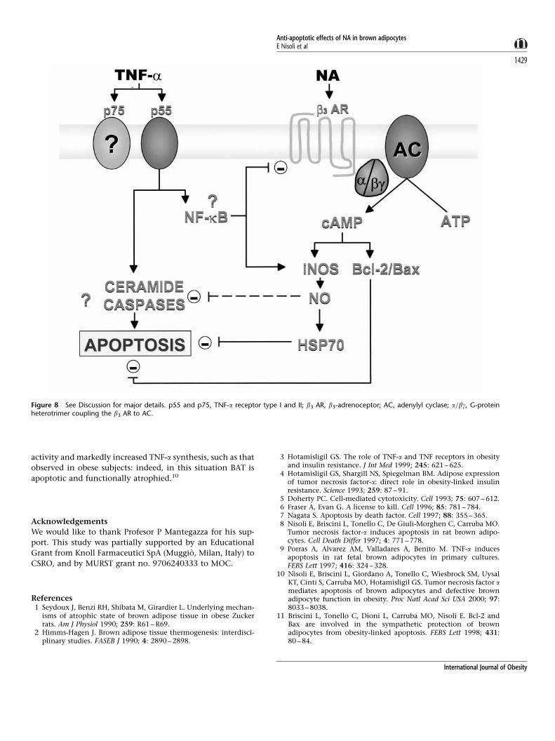
may act through HSP70 induction (see Figure 8). Further
experiments will be required to establish whether these
signaling processes actually play a role in the pathophysio-
logy of obesity and related disorders, but it is interesting to
note that TNF-a induces NO production in BAT.37 – 39 We can
therefore speculate that this induction is insufficient to
replace the anti-apoptotic NO signaling system triggered by
NA in the case of defective sympathetic nervous system
Figure 7 Effects of the HSP70 antisense oligomer on SNAP- and NA-induced protection against TNF-a plus CHX-mediated apoptosis, and on SNAP-induced HSP70 expression. (A) The brown adipocytes were pretreated with SNAP 500 mM or NA 10 mM, in the presence or absence of the HSP70 sense (S)or antisense (AS) oligomers, or with S or AS oligomers alone, for 24 h. The cells were washed twice with fresh medium, and then exposed to TNF-a (1 nM)plus CHX (10 mg=ml) for 4 h in the continued presence of the oligonucleotides. DNA fragmentation was measured as described in Figure 1. M, molecularweight marker (Gene Ruler 100 bp DNA ladder, Genenco, Milan, Italy). The gel shown is representative of the data obtained from three independentexperiments. (B) Western blot analysis of SNAP-induced HSP70 expression in brown adipocytes treated as decribed above. The bars represent the meanvalues� s.d. of three separate experiments plotted in relation to the area under the curve for the HSP70 protein levels in the untreated cells (lane 1) takenas one. *P<0.005 vs untreated-cells; **P<0.005 vs SNAP-treated cells.
Anti-apoptotic effects of NA in brown adipocytesE Nisoli et al
1428
International Journal of Obesity
activity and markedly increased TNF-a synthesis, such as that
observed in obese subjects: indeed, in this situation BAT is
apoptotic and functionally atrophied.10
Acknowledgements
We would like to thank Profesor P Mantegazza for his sup-
port. This study was partially supported by an Educational
Grant from Knoll Farmaceutici SpA (Muggio, Milan, Italy) to
CSRO, and by MURST grant no. 9706240333 to MOC.
References1 Seydoux J, Benzi RH, Shibata M, Girardier L. Underlying mechan-
isms of atrophic state of brown adipose tissue in obese Zuckerrats. Am J Physiol 1990; 259: R61 – R69.
2 Himms-Hagen J. Brown adipose tissue thermogenesis: interdisci-plinary studies. FASEB J 1990; 4: 2890 – 2898.
3 Hotamisligil GS. The role of TNF-a and TNF receptors in obesityand insulin resistance. J Int Med 1999; 245: 621 – 625.
4 Hotamisligil GS, Shargill NS, Spiegelman BM. Adipose expressionof tumor necrosis factor-a: direct role in obesity-linked insulinresistance. Science 1993; 259: 87 – 91.
5 Doherty PC. Cell-mediated cytotoxicity. Cell 1993; 75: 607 – 612.6 Fraser A, Evan G. A license to kill. Cell 1996; 85: 781 – 784.7 Nagata S. Apoptosis by death factor. Cell 1997; 88: 355 – 365.8 Nisoli E, Briscini L, Tonello C, De Giuli-Morghen C, Carruba MO.
Tumor necrosis factor-a induces apoptosis in rat brown adipo-cytes. Cell Death Differ 1997; 4: 771 – 778.
9 Porras A, Alvarez AM, Valladares A, Benito M. TNF-a inducesapoptosis in rat fetal brown adipocytes in primary cultures.FEBS Lett 1997; 416: 324 – 328.
10 Nisoli E, Briscini L, Giordano A, Tonello C, Wiesbrock SM, UysalKT, Cinti S, Carruba MO, Hotamisligil GS. Tumor necrosis factor amediates apoptosis of brown adipocytes and defective brownadipocyte function in obesity. Proc Natl Acad Sci USA 2000; 97:8033 – 8038.
11 Briscini L, Tonello C, Dioni L, Carruba MO, Nisoli E. Bcl-2 andBax are involved in the sympathetic protection of brownadipocytes from obesity-linked apoptosis. FEBS Lett 1998; 431:80 – 84.
Figure 8 See Discussion for major details. p55 and p75, TNF-a receptor type I and II; b3 AR, b3-adrenoceptor; AC, adenylyl cyclase; a=bg, G-proteinheterotrimer coupling the b3 AR to AC.
Anti-apoptotic effects of NA in brown adipocytesE Nisoli et al
1429
International Journal of Obesity

12 Nisoli E, Tonello C, Briscini L, Carruba MO. Inducible nitric oxidesynthase in rat brown adipocytes: implications for blood flow tobrown adipose tissue. Endocrinology 1997; 138: 676 – 682.
13 Fehsel K, Kroncke KD, Meyer KL, Huber H, Wahn V, Kolb-Bachofen V. Nitric oxide induces apoptosis in mouse thymocytes.J Immunol 1995; 155: 2858 – 2865.
14 Matthys P, Froyen G, Verdot L, Huang S, Sobis H, Van Damme J,Vray B, Aguet M, Billiau A. IFN-g receptor-deficient mice arehypersensitive to the anti-CD3-induced cytokine release syn-drome and thymocyte apoptosis. Protective role of endogenousnitric oxide. J Immunol 1995; 155: 3823 – 3829.
15 Mannick JB, Miao XQ, Stamler JS. Nitric oxide inhibits Fas-induced apoptosis. J Biol Chem 1997; 272: 24125 – 24128.
16 Kim YM, de Vera ME, Watkins SC, Billiar TR. Nitric oxide protectscultured rat hepatocytes from tumor necrosis factor-a-inducedapoptosis by inducing heat shock protein 70 expression. J BiolChem 1997; 272: 1402 – 1411.
17 Mestril R, Chi SH, Sayen MR, Dillmann WH. Isolation of a novelinducible rat heat-shock protein (HSP70) gene and its expressionduring ischaemia=hypoxia and heat shock. Biochem J 1994; 298:561 – 569.
18 Longo FM, Wang S, Narasimhan P, Zhang JS, Chen J, Massa SM,Sharp FR. cDNA cloning and expression of stress-inducible ratHSP70 in normal and injured rat brain. J Neurosci Res 1993; 36:325 – 335.
19 Levine B, Huang Q, Isaacs JT, Reed JC, Griffin DE, Hardwick JM.Conversion of lytic to persistent alphavirus infection by the Bcl-2cellular oncogene. Nature 1993; 361: 739 – 742.
20 Tilly JT, Tilly KI, Kenton ML, Johnson AL. Expression of membersof the Bcl-2 gene family in the immature rat ovary: equinechorionic gonadotropin-mediated inhibition of granulosa cellapoptosis is associated with decreased bax and constitutive bcl-xlong messenger ribonucleic acid levels. Endocrinol 1995; 136:232 – 241.
21 Kim YM, Talanian RV, Billiar TR. Nitric oxide inhibits apoptosisby preventing increases in caspase-3-like activity via two distinctmechanisms. J Biol Chem 1997; 272: 31138 – 31148.
22 Dimmeler S, Haendeler J, Sause A., Zeiher AM. Nitric oxideinhibits APO-1=Fas-mediated cell death. Cell Growth Differ 1998;9: 415 – 422.
23 Shen YH, Wang XL, Wilcken DE. Nitric oxide induces andinhibits apoptosis through different pathways. FEBS Lett 1998;433: 125 – 131.
24 Jaattela M, Wissing D, Kakholm K, Kallunki T, Egeblad M. HSP70exerts its anti-apoptotic function downstream of caspase-3-likeproteases. EMBO J 1998; 17: 6124 – 6134.
25 Syken J, De-Medina T, Munger K. TID1, a human homologue ofthe Drosophila tumor suppressor 1(2)tid, encodes two mitochon-drial modulators of apoptosis with opposing functions. Proc NatlAcad Sci USA 1999; 96: 8499 – 8504.
26 Ahn JH, Ko YG, Park WY, Kang YS, Chung HY, Seo JS. Suppressionof ceramide-mediated apoptosis by HSP70. Mol Cells 1999; 9:200 – 206.
27 Antonsson B, Martimou JC. The Bcl-2 protein family. Exp Cell Res2000; 256: 50 – 57.
28 Oltvai ZN, Milliman CL, Korsmeyer SJ. Bcl-2 heterodimerizes invivo with a conserved homolog, Bax, that accelerates pro-grammed cell death. Cell 1993; 74: 609 – 619.
29 Knudson MC, Korsmeyer SJ. Bcl-3 and Bax function indepen-dently to regulate cell death. Nature Genet 1997; 16: 358 – 363.
30 Matz JM, Blake MJ, Tatelman HM, LaVoi KP, Holbrook NJ.Characterization and regulation of cold-induced heat shock pro-tein expression in mouse brown adipose tissue. Am J Physiol 1995;269: R38 – R47.
31 Matz JM, LaVoi KP, Moen RJ, Blake MJ. Cold-induced heat shockprotein expression in rat aorta and brown adipose tissue. PhysiolBehav 1996; 60: 1369 – 1374.
32 Robertson JD, Datta K, Biswal SS, Kehrer JP. Heat-shock protein 70antisense oligomers enhance proteosome inhibitor-inducedapoptosis. Biochem J 1999; 344: 477 – 485.
33 Creagh EM, Carmody RJ, Cotter TG. Heat shock protein 70inhibits caspase-dependent and -independent apoptosis inJurkat T cells. Exp Cell Res 2000; 257: 58 – 66.
34 Kobayashi Y, Kume A, Li M, Doyu M, Hata M, Ohtsuka K, SobueG. Chaperones HSP70 and HSP40 suppress aggregate formationand apoptosis in cultured neuronal cell expressing truncatedandrogen receptor protein with expanded polyglutamine tract.J Biol Chem 2000; 275: 8772 – 8778.
35 DeNadai C, Sestili P, Cantoni O, Lievremont JP, Sciorati C,Barsacchi R, Moncada S, Meldolesi J, Clementi E. Nitric oxideinhibts tumor necrosis factor-a-induced apoptosis by reducingthe generation of ceramide. Proc Natl Acad Sci USA 2000; 97:5480 – 5485.
36 Gutteridge JM, Hallkiwell B. Free radicals and antioxidants in theyear 2000. A historical look to the future. Ann NY Acad Sci 2000;899: 136 – 147.
37 Uchida Y, Tsukahara F, Ohba K, Ogawa A, Irie K, Fujii E, Yoshi-moto T, Yoshioka T, Muraki T. Nitric oxide mediates downregulation of lipoprotein lipase activity induced by tumor necro-sis factor-a in brown adipocyes. Eur J Pharmac 1997; 335: 235 –243.
38 Uchida Y, Tsukahara F, Ohba K, Ogawa A, Nomato T, Muraki T.Augmentation of tumor necrosis factor-a-induced suppression oflipoprotein lipase by nitric oxide donors in cultured brownadipocytes. Ann NY Acad Sci 1997; 813: 369 – 372.
39 Kapur S, Marcotte B, Marette A. Mechanism of adipose tissueiNOS induction in endotoxemia. Am J Physiol 1999; 276: E635 –E641.
Anti-apoptotic effects of NA in brown adipocytesE Nisoli et al
1430
International Journal of Obesity

![4Aminopyridine stimulates B50 (GAP43) phosphorylation and [3H]-noradrenaline release in rat hippocampal slices](https://static.fdokumen.com/doc/165x107/6314ccec3ed465f0570b4d12/4aminopyridine-stimulates-b50-gap43-phosphorylation-and-3h-noradrenaline-release.jpg)