Stim1, an endoplasmic reticulum Ca 2+ sensor, negatively regulates 3T3-L1 pre-adipocyte...
20
Stim1, an endoplasmic reticulum Ca 2+ sensor, negatively regulates 3T3-L1 pre-adipocyte differentiation Sarah J.L. Graham a , Melony J. Black a , Jonathan Soboloff b , Donald L. Gill b , Marie A. Dziadek a,c , and Lorna S. Johnstone a,* a School of Biological Sciences, The University of Auckland, Private Bag 92019, Auckland, New Zealand b Department of Biochemistry, Temple University School of Medicine, Philadelphia, PA 19140-5104, USA c Cancer Research Program, Garvan Institute of Medical Research, Darlinghurst NSW 2010, Australia Abstract Ca 2+ plays a complex role in the differentiation of committed pre-adipocytes into mature, fat laden adipocytes. Stim1 is a single pass transmembrane protein that has an essential role in regulating the influx of Ca 2+ ions through specific plasma membrane store-operated Ca 2+ channels. Stim1 is a sensor of endoplasmic reticulum Ca 2+ store content and when these stores are depleted ER- localized Stim1 interacts with molecular components of store-operated Ca 2+ channels in the plasma membrane to activate these channels and induce Ca 2+ influx. To investigate the potential role of Stim1 in Ca 2+ -mediated adipogenesis, we investigated the expression of Stim1 during adipocyte differentiation and the effects of altering Stim1 expression on the differentiation process. Western blotting revealed that Stim1 was expressed at low levels in 3T3-L1 pre- adipocytes and was upregulated 4 days following induction of differentiation. However, overexpression of Stim1 potently inhibited their ability to differentiate and accumulate lipid, and reduced the expression of C/EBP alpha and adiponectin. Stim1-mediated differentiation was shown to be dependent on store-operated Ca 2+ entry, which was increased upon overexpression of Stim1. Overexpression of Stim1 did not disrupt cell proliferation, mitotic clonal expansion or subsequent growth arrest. siRNA-mediated knockdown of endogenous Stim1 had the opposite effect, with increased 3T3-L1 differentiation and increased expression of C/EBP alpha and adiponectin. We thus demonstrate for the first time the presence of store-operated Ca 2+ entry in 3T3-L1 adipocytes, and that Stim1-mediated Ca 2+ entry negatively regulates adipocyte differentiation. We suggest that increased expression of Stim1 during 3T3-L1 differentiation may act, through its ability to modify the level of Ca 2+ influx through store-operated channels, to balance the level of differentiation in these cells in vitro. Keywords Stim1; 3T3-L1; Adipocyte; Store-operated Ca 2+ entry; BTP2; Differentiation 1. Introduction Adipocytes play a fundamental role in the maintenance of energy balance in mammals and, via the secretion of a diverse variety of hormones and peptides, also play a critical role in © 2008 International Society of Differentiation. Published by Elsevier Ltd. All rights reserved. * Corresponding author. Tel.: +64 9 3737599x83748; fax: +64 9 3737668. [email protected] (L.S. Johnstone). NIH Public Access Author Manuscript Differentiation. Author manuscript; available in PMC 2013 March 19. Published in final edited form as: Differentiation. 2009 March ; 77(3): 239–247. doi:10.1016/j.diff.2008.10.013. NIH-PA Author Manuscript NIH-PA Author Manuscript NIH-PA Author Manuscript
-
Upload
independent -
Category
Documents
-
view
0 -
download
0
Transcript of Stim1, an endoplasmic reticulum Ca 2+ sensor, negatively regulates 3T3-L1 pre-adipocyte...

Stim1, an endoplasmic reticulum Ca2+ sensor, negativelyregulates 3T3-L1 pre-adipocyte differentiation
Sarah J.L. Grahama, Melony J. Blacka, Jonathan Soboloffb, Donald L. Gillb, Marie A.Dziadeka,c, and Lorna S. Johnstonea,*
aSchool of Biological Sciences, The University of Auckland, Private Bag 92019, Auckland, NewZealand bDepartment of Biochemistry, Temple University School of Medicine, Philadelphia, PA19140-5104, USA cCancer Research Program, Garvan Institute of Medical Research,Darlinghurst NSW 2010, Australia
AbstractCa2+ plays a complex role in the differentiation of committed pre-adipocytes into mature, fat ladenadipocytes. Stim1 is a single pass transmembrane protein that has an essential role in regulatingthe influx of Ca2+ ions through specific plasma membrane store-operated Ca2+ channels. Stim1 isa sensor of endoplasmic reticulum Ca2+ store content and when these stores are depleted ER-localized Stim1 interacts with molecular components of store-operated Ca2+ channels in theplasma membrane to activate these channels and induce Ca2+ influx. To investigate the potentialrole of Stim1 in Ca2+-mediated adipogenesis, we investigated the expression of Stim1 duringadipocyte differentiation and the effects of altering Stim1 expression on the differentiationprocess. Western blotting revealed that Stim1 was expressed at low levels in 3T3-L1 pre-adipocytes and was upregulated 4 days following induction of differentiation. However,overexpression of Stim1 potently inhibited their ability to differentiate and accumulate lipid, andreduced the expression of C/EBP alpha and adiponectin. Stim1-mediated differentiation wasshown to be dependent on store-operated Ca2+ entry, which was increased upon overexpression ofStim1. Overexpression of Stim1 did not disrupt cell proliferation, mitotic clonal expansion orsubsequent growth arrest. siRNA-mediated knockdown of endogenous Stim1 had the oppositeeffect, with increased 3T3-L1 differentiation and increased expression of C/EBP alpha andadiponectin. We thus demonstrate for the first time the presence of store-operated Ca2+ entry in3T3-L1 adipocytes, and that Stim1-mediated Ca2+ entry negatively regulates adipocytedifferentiation. We suggest that increased expression of Stim1 during 3T3-L1 differentiation mayact, through its ability to modify the level of Ca2+ influx through store-operated channels, tobalance the level of differentiation in these cells in vitro.
KeywordsStim1; 3T3-L1; Adipocyte; Store-operated Ca2+ entry; BTP2; Differentiation
1. IntroductionAdipocytes play a fundamental role in the maintenance of energy balance in mammals and,via the secretion of a diverse variety of hormones and peptides, also play a critical role in
© 2008 International Society of Differentiation. Published by Elsevier Ltd. All rights reserved.*Corresponding author. Tel.: +64 9 3737599x83748; fax: +64 9 3737668. [email protected] (L.S. Johnstone).
NIH Public AccessAuthor ManuscriptDifferentiation. Author manuscript; available in PMC 2013 March 19.
Published in final edited form as:Differentiation. 2009 March ; 77(3): 239–247. doi:10.1016/j.diff.2008.10.013.
NIH
-PA Author Manuscript
NIH
-PA Author Manuscript
NIH
-PA Author Manuscript
such diverse processes as hematopoiesis, vascular remodeling, insulin sensitivity and theimmune response (Morrison and Farmer, 1999). Excess accumulation of triglycerides inobesity hampers normal adipocyte function and results in a significant risk in thedevelopment of metabolic disorders (Pi-Sunyer, 1993). In many models of obesity, de novoadipocyte differentiation contributes to an increased total number of adipocytes andincreased adipose stores (Hirsch and Batchelor, 1976). Understanding the molecularmechanisms regulating adipocyte differentiation has thus been the subject of intenseinvestigation. An increase in intracellular Ca2+ concentration ([Ca2+]i) in adipocytes oftenaccompanies the development of obesity (Draznin et al., 1988). In vitro, increasing [Ca2+]iby overexpressing neuronatin (Suh et al., 2005), a putative Ca2+-ATPase regulator, acts toincrease the differentiation of the 3T3-L1 pre-adipocyte cell line, a well-characterized invitro model of adipocyte differentiation. Paradoxically, directly raising [Ca2+]i in 3T3-L1pre-adipocytes by calcium mobilizing agents efficiently inhibits differentiation, diminishesadipocyte-specific gene expression and reduces lipid accumulation (Ntambi and Takova,1996). These inhibitory effects can be mimicked either by enhancing the activity of theCa2+/calmodulin-dependent serine/ threonine phosphatase calcineurin (Neal and Clipstone,2002) or by constitutive activation of calcineurin effectors such as members of the nuclearfactor of activated T cell (NFAT) family (Neal and Clipstone, 2003). Conversely, inhibitionof calcineurin activity by cyclosporin A (CsA) increases adipocyte differentiation and lipidaccumulation (Neal and Clipstone, 2002), mimicking the obesogenic effects of CsAtreatment in humans (Mathieu et al., 1994). These apparently contradictory effects suggestthat elevating [Ca2+]i in discrete cellular microdomains likely has diverse effects onadipocyte differentiation, similar to that seen in other cell types (Berridge, 2006).
Ca2+ influx through plasma membrane store-operated Ca2+ channels (SOCs) provides forlocalized sub-plasma membrane increase in [Ca2+]i critical for sustained activity of severalintracellular enzymes (Cooper et al., 1998), including calcineurin (Gwack et al., 2007).SOCs are uniquely activated by a mechanism critically dependent on the depletion ofendoplasmic reticulum (ER) Ca2+ stores (Venkatachalam et al., 2002). The predominantlyER membrane protein Stromal interaction molecule-1 (Stim1) (Williams et al., 2002; Liou etal., 2005) plays a critical role in sensing ER [Ca2+] via its ER luminal N-terminal unpairedEF hand domain and activating SOCs (Liou et al., 2005). When ER [Ca2+] drops followingreceptor-mediated IP3 signaling, Ca2+ is no longer bound to the low affinity EF handdomain of Stim1, inducing a conformational change and aggregation of Stim1 within the ERmembrane (Liou et al., 2005). These aggregates, forming within 10–25nm of the plasmamembrane (Wu et al., 2006), initiate clustering of the plasma membrane store-operated Ca2+
channel component, Orai1 (Xu et al., 2006). Through an, as yet, unidentified mechanism,Stim1 and Orai1 together induce localized Ca2+ influx from the extracellular space(Soboloff et al., 2006b). In mice, targeted inactivation of Stim1 in T-cells severely impairsCa2+ entry through SOCs, abolishes NFAT-mediated gene expression and impairsregulatory T-cell development and function (Oh-hora et al., 2008).
It is now clear that Stim1 mediates Ca2+ entry through SOCs in a varied selection of celltypes, and that the expression level of Stim1 profoundly affects the level of Ca2+ influx(Roos et al., 2005; Soboloff et al., 2006a). The ubiquitous tissue expression pattern of Stim1(Dziadek and Johnstone, 2007) and the perinatal lethal phenotype of Stim1−/− mice (Oh-hora et al., 2008) also suggest that Stim1 is critical for correct development and/or functionof multiple cell types. However, it is not clear how Stim1 regulates developmental processesat a cellular level. Given that the activity of calcineurin in modifying 3T3-L1 adipocytedifferentiation suggests the presence of functional SOCs in these cells, we investigated therole of Stim1 in 3T3-L1 pre-adipocyte differentiation. By modulating the expression level ofStim1 we demonstrate that increasing Ca2+ entry through SOCs via upregulation of Stim1acts to inhibit 3T3-L1 differentiation without affecting proliferation, and downregulation of
Graham et al. Page 2
Differentiation. Author manuscript; available in PMC 2013 March 19.
NIH
-PA Author Manuscript
NIH
-PA Author Manuscript
NIH
-PA Author Manuscript
Stim1 promotes 3T3-L1 differentiation. These results suggest that Stim1 is a negativemodulator of 3T3-L1 differentiation, and that its expression may act to balance the level ofdifferentiation in 3T3-L1 cells in vitro.
2. Materials and methods2.1. Cell culture and adipocyte differentiation
Mouse 3T3-L1 pre-adipocytes (ATCC) were maintained in Dulbecco’s modified Eagle’smedium with high glucose supplemented with 10% (v/v) fetal bovine serum, 100 units/mlpenicillin G, 100 µg/ml streptomycin and 2 mM L-glutamine (all from Invitrogen). To induceadipocyte differentiation, cells were grown until 2 days postconfluence (day 0) and thentreated for 2 days with growth medium plus MDI (0.5 mM methylisobutylxanthine, 1 µMdexamethasone and 10 µg/ml insulin; all from Sigma). Subsequently, cells were incubated ingrowth medium containing 10 µg/ml insulin for 2 days and then refed every 2 days ingrowth medium for the remainder of the culture period. Where indicated, differentiation wasinduced with 0.25 mM methylisobutylxanthine and 0.5 µM dexamethasone (0.5MD) for 2days, and thereafter cells were refed every 2 days in growth medium. At the end of theculture period, cells were fixed in 4% paraformaldehyde and stained with 2.1 mg/ml Oil RedO (Sigma) in 60% isopropanol. Cells were photographed and Oil Red O eluted in 100%isopropanol for spectrophotometric quantification at OD520. Where indicated, cells weretreated with 3 or 10 µM N-(4-[3,5-bis(trifluoromethyl)-1H-pyrazol-1-yl]phenyl)-4-methyl-1,2,3-thiadiazole-5-carboxamide (BTP2) (Merck), 1 µg/ml cyclosporin A (Sigma), 5ng/ml FK-506 (Sigma) or vehicle control (DMSO, ethanol).
2.2. Generation of L1-control and L1-STIM1 cellsTo create 3T3-L1 lines stably overexpressing STIM1 (L1-STIM1 lines), cells weretransfected with a human pIRES::STIM1 expression plasmid (Williams et al., 2001) orempty pIRES plasmid (L1-control lines) using Lipofectamine 2000 according to themanufacturers instructions (Invitrogen). Stably overexpressing cell lines were selected inG418 (Sigma) and single clonal lines obtained by limiting dilution in 96 well plates.
2.3. Ca2+ measurementsCells grown on coverslips were placed in “cation-safe” medium free of sulfate andphosphate anions (107 mM NaCl/7.2 mM KCl/1.2 mM MgCl2/11.5 mM glucose/20 mMHepes-NaOH, pH 7.2) and loaded with 4 µM fura-2/acetoxymethyl ester (Molecular Probes)for 30 min at 20 °C in the presence of 0.01% Pluronic F-127 (Invitrogen). Cells werewashed and dye was allowed to deesterify for a minimum of 30 min at 20 °C beforemeasurement of Ca2+ using an InCyt dual-wavelength fluorescence imaging system(Intracellular Imaging, Cincinnati). Fluorescence emission at 505 nm was monitored withexcitation at 340 and 380 nm; intracellular Ca2+ measurements are shown as 340- to 380-nmratios. Store-operated Ca2+ entry was induced by incubation with 1 µM Tg (EMDBiosciences) or 100 µUTP (Sigma) in zero Ca2+ conditions. All traces are averages frommultiple (20–50) cells and are representative of at least three separate experiments.
2.4. Western blot analysisTotal 3T3-L1 protein extracts were prepared and quantified as previously described(Williams et al., 2001). Protein samples (10–20 µg) were separated on 12% SDS-PAGE or4–16% gradient (Invitrogen) gels under reducing conditions and Western blotting performedas previously described (Williams et al., 2001). Primary antibodies were affinity purifiedrabbit anti-STIM1-CT at 1:1000 (Williams et al., 2001), mouse anti-β-actin (Sigma) at1:2000, rabbit anti-C/EBPα (14AA), C/EBPβ (C-19) and PPARγ (H-100) (all Santa Cruz
Graham et al. Page 3
Differentiation. Author manuscript; available in PMC 2013 March 19.
NIH
-PA Author Manuscript
NIH
-PA Author Manuscript
NIH
-PA Author Manuscript

Biotechnology) all at 1:500, and rabbit anti-Adpn (Sigma) at 1:1000. Secondary antibodies(anti-mouse-AP, anti-rabbit-AP, anti-rabbit-BIO) were from Jackson Immunochemicalsused at 1:2000 and ExtrAvidin-AP (Sigma) at 1:300,000. Alkaline phosphatase was detectedusing NBT-BCIP substrate (Invitrogen) or by CDP-Star chemiluminescent substrate solution(Sigma). Where indicated, image capture and quantification of Western blots wereperformed with the LAS-3000 imaging system and MultiGuage software (Fujifilm).
2.5. siRNA design and transfectionPre-designed Stealth™ siRNA duplexes to mouse Stim1 (MSS209660, MSS209661) andcontrol medium GC siRNA duplexes (12935-300) were purchased from Invitrogen. 3T3-L1cells at 30–50% confluency were transfected with Lipofectamine 2000 (Invitrogen) in 6-wellplates according to the protocol described by Fox et al. (2006). Following transfection andrecovery, cells were harvested in 0.25% trypsin (Invitrogen) and re-seeded at 2 × 10⁴/well in24 well plates for differentiation.
2.6. Real-time RT-PCRTotal RNA was prepared using TRIzol Reagent (Invitrogen) and genomic DNA-free RNAconverted to cDNA using Superscript Vilo cDNA synthesis kit (Invitrogen). Samplesprepared without reverse transcriptase were used as negative controls. Analysis of Stim1 andβ-actin mRNA levels in pentuplicate wells were performed using Express Two-Step SYBRGreenER (Invitrogen) on a ABI-7700HT (Applied Biosystems) using fast cycling conditionsand the exon spanning PCR primers 5′-ttgccaagcaggaagctc-3′,5′-ctccttctctgctttcctcaag-3′(Stim1) and 5′-aaggccaaccgtgaaaagat-3′,5′-gtggtacgaccagaggcatac-3′ (β-actin). Thepresence of single products were confirmed by melting point analysis of every sample.Stim1 gene expression was analyzed using the 2−ddCT method (Livak and Schmittgen, 2001)relative to β-actin levels.
2.7. Data analysisStatistical analysis was performed using a Student’s t-test with a p-value of 0.05 consideredsignificant. All data shown are representative of at least 3 individual experiments.
3. Results3.1. Stim1 is expressed in pre-adipocytes and is upregulated following adipocytedifferentiation
The 3T3-L1 pre-adipocyte cell line efficiently differentiates into morphologically distinct,triglyceride laden adipocytes over a 10-day differentiation period when confluent cells aretreated with a differentiation cocktail comprising methylisobutylxanthine (IBMX),dexamethasone and insulin (MDI) (Cowherd et al., 1999). Western blotting showed celllysates prepared from cells prior to differentiation contained very low levels of Stim1 (Fig.1A, top panel). Stim1 levels increased markedly 4 days following stimulation with MDI, andremained stable throughout the remaining differentiation period. Adiponectin (Adpn), aknown marker of terminal 3T3-L1 differentiation, was robustly induced at day 4 post MDItreatment (Fig. 1A, middle panel) and continued to increase throughout the remainingdifferentiation period as expected (Hu et al., 1996). This analysis indicated that Stim1 isupregulated during adipogenic differentiation of 3T3-L1 cells.
3.2. Overexpression of STIM1 in 3T3-L1 cells increases Ca2+ entry through SOCsTo determine the role of Stim1 in the differentiation process, 3T3-L1 pre-adipocytes werestably transfected with a human pIRES::STIM1 expression construct (Williams et al., 2001).Ten clonally derived STIM1-overexpressing cell lines (L1-STIM1) and 10 control empty
Graham et al. Page 4
Differentiation. Author manuscript; available in PMC 2013 March 19.
NIH
-PA Author Manuscript
NIH
-PA Author Manuscript
NIH
-PA Author Manuscript

vector pIRES-transfected lines (L1-control) were generated. Western blot analysis revealeda moderate upregulation of STIM1 expression levels in the C8 clonal line, and higherexpression in the C1, C6 and C7 clonal lines compared to wild-type 3T3-L1 cells and the β-actin loading control (Fig. 1B).
To determine whether STIM1 overexpression in 3T3-L1 pre-adipocytes increased the levelof Ca2+ entry in response to store-depletion, the sarcoplasmic/endoplasmic reticulum Ca2+
ATPase Ca2+ pump blocker thapsigargin (Tg) was used to deplete ER Ca2+ stores, andfura-2 was used to monitor cytosolic Ca2+ levels (Soboloff et al., 2006a). In the absence ofexternal Ca2+, Tg induced a large and transient increase in intracellular Ca2+ as stores wereinitially emptied, followed by a return to baseline values as plasma membrane Ca2+ pumpsfacilitated extrusion into the extracellular medium (Fig. 2A). The level of Tg-induced ERCa2+ release was similar in both L1-control and L1-STIM1 pre-adipocytes. Addition ofextracellular Ca2+ caused a rapid entry of Ca2+ through SOCs similar to that described inother cells (Venkatachalam et al., 2002), the extent of which was almost 2-fold greater inL1-STIM1 cells compared to L1-controls (p < 0.0001); these increases correlated with thegain in Tg-induced Ca2+ entry in single HEK293 cells overexpressing STIM1 (Soboloff etal., 2006a). Pre-incubation of L1-STIM1 cells with the store-operated Ca2+ channel blockerBTP2 for 10 min prior to Tg treatment reduced Ca2+ entry to approximately 25% of thatseen in vehicle control-treated cells (p < 0.0001) (Fig. 2B), correlating with the BTP2-mediated decrease in Tg-induced Ca2+ entry in B-lymphocytes (He et al., 2005). Thisanalysis indicated that the increased Ca2+ entry seen in L1-STIM1 cells could be attributedto activity of store-operated Ca2+ channels.
To demonstrate that STIM1-mediated SOC activation could contribute to a physiologicalsignal, we tested the responses of L1-control and L1-STIM1 cells to UTP. In whiteadipocytes this nucleotide likely activates the P2Y purinergic receptor subclass (Lee et al.,2005), a typical phospholipase C-linked G-protein coupled receptor. In both L1-control (Fig.2C) and L1-STIM1 (Fig. 2D) pre-adipocytes, UTP rapidly induced InsP3-mediated Ca2+
release from stores in the absence of extracellular Ca2+. Addition of external Ca2+ resultedin rapid entry of Ca2+ in L1-control cells, which was greatly enhanced in L1-STIM1 pre-adipocytes. Thus, enhanced activation of SOCs by STIM1 was not limited topharmacological ER Ca2+ depletion; Ca2+ entry in L1-STIM1 cells was also enhanced inresponse to physiological signals.
3.3. STIM1 overexpression inhibits 3T3-L1 pre-adipocyte differentiationTo investigate the effects of increasing Ca2+ entry through SOCs on the differentiation of3T3-L1 cells, confluent L1-STIM1 or L1-control pre-adipocytes were induced todifferentiate by MDI and the extent of differentiation was quantified by staining lipid withOil Red O (Kutt and Tsaltas, 1959). At the end of the 8-day differentiation period, bothwild-type 3T3-L1 and L1-control cells had accumulated a comparable amount of lipid (Fig.3A, top panels). In the higher expressing L1-STIM1 C7 line, lipid accumulation wasinhibited by 90% (Fig. 3A, bottom left panel; p = 0.0002) whereas in the lower expressingL1-STIM1 C8 line, lipid accumulation was reduced by 67% (Fig. 3A, bottom right panel; p= 0.0005). This experiment was repeated with four individually created L1-STIM1 and threeL1-control cell lines over a 10-day differentiation period (Fig. 3B). Poor differentiation ofL1-STIM1 lines (closed symbols) was observed in every experiment compared to the L1-control lines (open symbols).
BTP2 was used to determine whether inhibition of Ca2+ entry through SOCs in L1-STIM1cells would reverse the inhibitory effects of STIM1 overexpression on the differentiationprocess. BTP2 concentrations of up to 10 µM have been successfully used in extendedculture conditions to inhibit hypertrophy-associated gene expression in cardiomyocytes
Graham et al. Page 5
Differentiation. Author manuscript; available in PMC 2013 March 19.
NIH
-PA Author Manuscript
NIH
-PA Author Manuscript
NIH
-PA Author Manuscript

(Bush et al., 2006). Confluent L1-STIM1 C7 or L1-control cells were incubated with 3 µMor 10 µM BTP2 or an equivalent final dilution of the DMSO vehicle control, and followingthe standard differentiation protocol, cells were stained with Oil Red O. Incubation withBTP2 had no measurable effect on lipid accumulation in L1-control cells (Fig. 3C, top rightpanel) when compared to cells treated with vehicle control (Fig. 3C, top left panel), whereasL1-STIM1 C7 cells treated with vehicle control did not accumulate lipid (Fig. 3C, bottomleft panel). In contrast, 10 µM BTP2 partially reversed the inability of the L1-STIM1 C7cells to differentiate, with an increase in both the size and number of adipogenic clusters(Fig. 3C, bottom right panel), whereas 3 µM BTP2 had little effect (not shown). 10 µMBTP2 also reversed the differentiation block in several other individually created L1-STIM1lines (not shown). Together, these results indicated that overexpression of STIM1 inhibitsadipocyte differentiation, that the level of inhibition correlates with the expression level ofSTIM1 and that this inhibition of differentiation could be partially reversed by inhibition ofstore-operated Ca2+ channel activity with BTP2.
3.4. Overexpression of STIM1 does not affect 3T3-L1 proliferation, MCE or growth arrestTo investigate the cellular and molecular mechanisms by which STIM1 overexpressioninhibited differentiation, several critical phases of the differentiation process were examined.Confluent 3T3-L1 cells undergo one to two rounds of cell division (Cowherd et al., 1999),known as mitotic clonal expansion (MCE) over the 4 days following MDI treatmentfollowed by growth arrest, and this process is critical for robust differentiation (Tang et al.,2003). To test the effects of STIM1 overexpression on MCE, confluent L1-STIM1 andcontrol pre-adipocytes were induced to differentiate and total cell numbers quantified at day0 (MDI addition), day 2 and day 4 (Fig. 4A). An identical 2.5-fold increase in cell numberwas observed at day 2 in both L1-STIM1 and control cells, indicating that each cell line hadundergone at least one round of cell division. No further increase in cell number in eithercell line between days 2 and 4 indicated that both lines had once again growth arrested.Therefore STIM1 overexpression did not appear to inhibit differentiation by blocking post-confluent MCE or subsequent growth arrest. The intrinsic proliferative capacity of L1-STIM1 cells was also examined since aberrant proliferation reduces 3T3-L1 differentiation(Neal and Clipstone, 2003). L1-STIM1 or control pre-adipocytes were seeded at low densityand cell numbers counted 2 and 3 days post seeding (Fig. 4B). No difference in proliferationrates between the two cell lines could be discerned, with cell numbers increasing 6-fold atday 2 and 13-fold at day 3 in both cell lines.
3.5. STIM1 overexpression results in downregulation of C/EBPα and adiponectinTo determine the effects of STIM1 overexpression on adipogenic gene regulation, theexpression of transcription factors essential for 3T3-L1 differentiation were determined. Thetranscription factors CCAAT/enhancer binding protein (C/EBP) β and C/EBPδ whichactivate transcription of peroxisome proliferators-activated receptor gamma (PPARγ), areinduced by differentiation (Salma et al., 2006). C/EBPβ and PPARγ induce expression of C/EBPα, which reinforces expression of PPARγ in a positive feedback loop, and also inducesexpression of other proteins such as adiponectin (Adpn) involved in terminal differentiation(Park et al., 2004). The expression of C/EBPβ, C/EBPα and PPARγ 1 and 2 isoforms wereexamined by Western blot during differentiation of confluent L1-STIM1 and control cells(Fig. 4C). Levels of C/EBPβ and PPARγ1 and 2 isoforms were indistinguishable in controland L1-STIM1 cells over the 6-day differentiation period while both C/EBPα and Adpnexpression were visibly reduced in L1-STIM1 cells compared to controls at all time-pointsexamined, by 99% and 91%, respectively at day 6. Together, these data, and the datapresented in Fig. 4A and B, suggested that STIM1 overexpression results in thedownregulation of C/EBPα and Adpn expression.
Graham et al. Page 6
Differentiation. Author manuscript; available in PMC 2013 March 19.
NIH
-PA Author Manuscript
NIH
-PA Author Manuscript
NIH
-PA Author Manuscript

3.6. The effects of STIM1 overexpression can be reversed by calcineurin inhibitorsSTIM1 overexpression activates calcineurin/NFAT signaling in lymphocytes (Huang et al.,2006), and overexpression of activated calcineurin potently inhibits 3T3-L1 differentiation(Neal and Clipstone, 2002). To determine whether the effects of STIM1 overexpression onadipocyte differentiation could be mediated by calcineurin, L1-STIM1 cells were induced todifferentiate at day 0 with MDI, or with MDI and the specific calcineurin inhibitorscyclosporin A or FK506 (Neal and Clipstone, 2002). Compared with L1-STIM1 cells treatedwith vehicle control (Fig. 4D, top panel), lipid accumulation in FK506 or cyclosporin A-treated L1-STIM1 cells (Fig. 4D, middle, bottom panels) increased 5.5-fold (p = 0.001) and6.5-fold (p = 0.0008), respectively. These data indicate that calcineurin activity is required tomediate the inhibitory effects of STIM1 overexpression on adipocyte differentiation.
3.7. Reducing endogenous STIM1 expression enhances 3T3-L1 differentiation, resulting inupregulation of C/EBPα and adiponectin
To determine the effects of reducing endogenous Stim1 expression on 3T3-L1differentiation, 3T3-L1 cells were transfected with Stim1-specific Stealth™ siRNA (Stim1–60) or control siRNA and induced to differentiate under sub-optimal differentiationconditions by modifying the concentration of the MDI adipogenic cocktail (Neal andClipstone, 2002). This modification resulted in the expected progressive reduction in bothsize and number of adipogenic colonies detected by Oil Red O staining (Fig. 5A). ControlsiRNA-transfected 3T3-L1 cells differentiated fully under optimal MDI conditions (Fig. 5A,top left) and accumulated 3.5-fold less lipid in sub-optimal 0.5MD conditions (Fig. 5A,bottom left). An increased number of Stim1–60-transfected cells appeared to stain Oil RedO positive than control siRNA-transfected cells in MDI (Fig. 5A, top right), butquantification of extracted lipid showed that this difference was not significant. In contrast,in 0.5MD, a greater number of Stim1–60-transfected cells stained Oil Red O positive (Fig.5A, bottom right) and accumulated 2.5-fold more lipid than control siRNA-transfected cells(p = 0.0002). To confirm specificity, a second siRNA was tested. The Stim1–61 siRNA alsoincreased lipid accumulation in 0.5MD conditions (Fig. 5B), by 1.4-fold (p = 0.05). Real-time RT-PCR confirmed that the Stim1–61 siRNA was less efficient at reducing endogenousStim1 mRNA levels compared to the Stim1–60 siRNA (Fig. 5C), indicating that theenhancing effects of reduced Stim1 levels on adipocyte differentiation were dose dependent.
We confirmed by Western blotting that the Stim1–60 siRNA virtually abolished endogenousStim1 expression at day 4 post 0.5MD treatment compared to control siRNA-transfectedcells (Fig. 5D, top panel). Expression of PPARγ isoforms appeared relatively unaffected inStim1-siRNA-treated cells (Fig. 5D, middle panel) whereas C/EBPα levels were increasedby 5.7-fold and Adpn by 2.3-fold (Fig. 5E) in Stim1 siRNA-transfected cells at day 4. Thesedata indicated that reducing endogenous Stim1 expression increases adipocytedifferentiation, accompanied by an increase in expression of C/EBPα and Adpn.
4. DiscussionIn the current study, we provide several lines of evidence indicating that Ca2+ entry viastore-operated channels is functional in 3T3-L1 pre-adipocytes and, through a Stim1-mediated pathway, acts to negatively regulate adipocyte differentiation. We demonstrate thatoverexpression of Stim1 in 3T3-L1 pre-adipocytes specifically increases Ca2+ influxthrough SOCs and consequently inhibits differentiation associated with reduced expressionof C/EBPα and Adpn. In contrast, we find that reducing Stim1 levels with siRNA markedlyincreases differentiation in response to sub-optimal adipogenic stimuli, associated withincreased expression of C/EBPα and Adpn. However, in apparent contradiction, we find thatendogenous Stim1 is itself upregulated during adipocyte differentiation. Altogether, our data
Graham et al. Page 7
Differentiation. Author manuscript; available in PMC 2013 March 19.
NIH
-PA Author Manuscript
NIH
-PA Author Manuscript
NIH
-PA Author Manuscript

suggests that the level of Ca2+ influx though SOCs may be intrinsically regulated duringdifferentiation by the expression level of Stim1, and that this in turn influences adipocytegene expression, and ultimately may set the level of commitment of pre-adipocytes to theterminal differentiation phase.
We have found that the inhibitory effects of STIM1 over-expression on adipocytedifferentiation requires calcineurin activity. The calcineurin/NFAT signaling pathway is animportant downstream effector of Ca2+ entry via SOCs in lymphocytes (Gwack et al., 2007).Ca2+-activated calcineurin is best known for it’s ability to dephosphorylate cytosolic NFATand trigger its nuclear translocation where it acts both as a positive and negative regulator oftarget gene transcription (Crabtree and Olson, 2002). Activation of the calcineurin/NFATpathway in 3T3-L1 cells by prostaglandin F2a (Liu and Clipstone, 2007) or by theintroduction of constitutively active calcineurin or NFATC1 (Neal and Clipstone, 2002,2003) potently inhibits their differentiation, similar to the effects of Stim1 overexpression.In addition, cyclosporin A markedly increases adipocyte differentiation in response tosuboptimal differentiation stimuli (Neal and Clipstone, 2002), similar to the effects ofsiRNA-mediated knockdown of Stim1. Modulation of Stim1 levels directly affects theactivity of the calcineurin/NFAT pathway in epithelial and hematopoietic cells (Wang et al.,2006; Huang et al., 2006; Liou et al., 2005), and this study suggests that Stim1 is able toexert a similar influence in 3T3-L1 adipocytes. However, this does not preclude theinvolvement of additional signaling pathways in Stim1-mediated inhibition of differentiationsince cyclosporin A and FK506, at the concentrations used here, did not completely rescuethe effects of STIM1 overexpression. In some cells, Ca2+ is known to enhance the activationof transcription factors such as NFκB (Crabtree, 2001), also an inhibitor of adipocytedifferentiation (Chae and Kwak, 2003). It remains to be tested whether modulation of Stim1levels directly affects additional signaling pathways in 3T3-L1 cells.
Overexpression of Stim1 virtually abolished C/EBPα and Adpn expression. Antisenseknockdown of C/EBPα alone in 3T3-L1 pre-adipocytes is sufficient to inhibit triglycerideaccumulation and terminal gene expression (Lin and Lane, 1992) and C/EBPα is a criticalregulator of Adpn expression (Park et al., 2004) therefore it is likely that reduced C/EBPαexpression is the major contributor to the reduced Adpn expression levels and inhibition ofadipocyte differentiation observed in this study. While expression and activity of C/EBPβand PPARγ are required for efficient induction of C/EBPα expression (Park et al., 2004;Zuo et al., 2006b) we show that Stim1 overexpression does not affect MDI-induced C/EBPβor PPARγ expression, nor does it affect C/EBPβ-dependent mitotic clonal expansion or cellcycle arrest. However, Stim1-mediated Ca2+ entry may interfere with C/EBPβphosphorylation, which is critical for induction of C/EBPα expression but not required forinduction of PPARγ expression (Park et al., 2004). Alternatively, Stim1-mediated effectsmay be distal to C/EBPβ activity but instead may inhibit PPARγ activity, since selectiveantagonists of PPARγ activity reduce C/EBPα expression and inhibit 3T3-L1 adipogenesis(Zuo et al., 2006a, b). Another possible mechanism underlying Stim1-mediated effects isdirect repression of C/EBPα expression after Ca2+ entry independently of C/EBPβ andPPARγ activity. Prostaglandin F2a-mediated inhibition of 3T3-L1 adipo-genesis via Ca2+/calcineurin can be attenuated by a histone deacetylase inhibitor, which rescues PPARγ andC/EBPα expression, suggesting that activation of Ca2+/calcineurin signaling in 3T3-L1 cellslikely promotes transcriptional repression (Liu and Clipstone, 2007). However, Stim1overexpression does not result in complete abolition of C/EBPα and PPARγ expression asis seen with sustained Prostaglandin F2a treatment, or expression of constitutively activecalcineurin. Whilst the reason for this discrepancy is unclear, we and others have found thatStim1-mediated SOC entry in cells stably overexpressing STIM1 is still regulated by ERCa2+ store content (Soboloff et al., 2006a), whereas constitutively active calcineurin andNFAT are likely not subject to normal regulatory processes and may have additional effects
Graham et al. Page 8
Differentiation. Author manuscript; available in PMC 2013 March 19.
NIH
-PA Author Manuscript
NIH
-PA Author Manuscript
NIH
-PA Author Manuscript
on cell behavior and gene expression (Neal and Clipstone, 2003). This study has shown thatreducing Stim1 levels increased expression of Adpn and C/EBPα. Both C/EBPα and Adpnare capable of increasing triglyceride accumulation in 3T3-L1 cells when overexpressed(Lin and Lane, 1994; Fu et al., 2005). Interestingly, 2-fold overexpression of Adpn increasestriglyceride accumulation almost 3-fold at day 9 of differentiation which correlates well withthe level of Adpn upregulation and increased triglyceride accumulation observed in thisstudy.
It is becoming increasingly understood that Stim1 can act as a multifunctional regulator ofCa2+ influx through several classes of SOCs, including Orai1, Orai2 and transient receptorpotential C1 (TRPC1) (Huang et al., 2006) and that its presence can dictate the level ofexpression of some channels, such as TRPC1 in HEK293 cells (Huang et al., 2006). Theexpression of Orai or TRP channels in 3T3-L1 pre-adipocytes has not been reported,however, transcript profiling of adipocytes in vivo indicate expression of Orai and TRPC1genes (Su et al., 2004). It will now be important to characterize Orai and TRP channelexpression in 3T3-L1 pre-adipocytes and determine through which of these Stim1 exerts itsanti-adipogenic effects. Recent evidence also suggests that Stim1 situated in the plasmamembrane may regulate non-store-operated Ca2+ channels such as the arachidonic acidregulated channels (ARC) (Mignen et al., 2007). AA is a potent inhibitor of 3T3-L1 pre-adipocyte differentiation, acting through the cyclooxygenase-dependent generation of AAeicosanoid metabolites (Petersen et al., 2003). However, activation of Ca2+ influx throughARC channels is likely a direct action of AA on the channel itself, independently of AAmetabolites and cyclooxygenase activity (Shuttleworth and Thompson, 1998), arguingagainst a role for ARC channels in Stim1-mediated adipocyte differentiation.
Our study has, for the first time, provided evidence that adipocytes utilize a store-operatedCa2+ entry mechanism that may control commitment to differentiation via upregulation ofthe endoplasmic reticulum Ca2+ sensor Stim1. That Stim1 exerts an inhibitory effect ondifferentiation is perhaps surprising given its upregulation during the differentiation process.However, the functional attributes of mature adipocytes are one of lipid storage and lipidrelease and, by necessity, adipocytes obtain the proteins required to perform these functionsduring the differentiation process. Indeed, expression of another negative regulator ofadipocyte differentiation, Sirtuin 1, is also upregulated during the differentiation process andappears to play a major role in promoting fat mobilization in mature adipocytes (Picard etal., 2004). Interestingly, NFAT proteins have recently been found to facilitate fatmobilization and metabolic gene expression in mature adipocytes (Holowachuk, 2007).Whether Stim1 plays a similar role remains to be determined. In conclusion, as in othercells, the subcellular source of Ca2+ is likely an important regulator of adipocytedifferentiation. This could be especially important in human disease such as obesity wherederegulation of calcium handling is evident. It will now be important to examine whetherSOC entry play a role in the increased adipocyte Ca2+ levels and development of obesity inhumans.
AcknowledgmentsWe thank Iain MacDonald and Vivian Ward for photography and graphics assistance, Annette Lasham for technicaladvice and Lance Xu for the kind gift of the anti-Adpn antibody. This work was supported by grants from theHealth Research Council, New Zealand, the Auckland Medical Research Foundation, New Zealand and the Schoolof Biological Sciences, The University of Auckland.
ReferencesBerridge MJ. Calcium microdomains: organization and function. Cell Calcium. 2006; 40:405–412.
[PubMed: 17030366]
Graham et al. Page 9
Differentiation. Author manuscript; available in PMC 2013 March 19.
NIH
-PA Author Manuscript
NIH
-PA Author Manuscript
NIH
-PA Author Manuscript

Bush EW, Hood DB, Papst PJ, Chapo JA, Minobe W, Bristow MR, Olson EN, McKinsey TA.Canonical transient receptor potential channels promote cardiomyocyte hypertrophy throughactivation of calcineurin signaling. J. Biol. Chem. 2006; 281:33487–33496. [PubMed: 16950785]
Chae GN, Kwak SJ. NF-kappaB is involved in the TNF-alpha induced inhibition of the differentiationof 3T3-L1 cells by reducing PPARgamma expression. Exp. Mol. Med. 2003; 35:431–437.[PubMed: 14646597]
Cooper DM, Karpen JW, Fagan KA, Mons NE. Ca(2+)-sensitive adenylyl cyclases. Adv. SecondMessenger Phosphoprotein Res. 1998; 32:23–51. [PubMed: 9421584]
Cowherd RM, Lyle RE, McGehee RE Jr. Molecular regulation of adipocyte differentiation. Semin.Cell Dev. Biol. 1999; 10:3–10. [PubMed: 10355023]
Crabtree GR. Calcium, calcineurin, and the control of transcription. J. Biol. Chem. 2001; 276:2313–2316. [PubMed: 11096121]
Crabtree GR, Olson EN. NFAT signaling: choreographing the social lives of cells. Cell. 2002;109:S67–S79. [PubMed: 11983154]
Draznin B, Sussman KE, Eckel RH, Kao M, Yost T, Sherman NA. Possible role of cytosolic freecalcium concentrations in mediating insulin resistance of obesity and hyperinsulinemia. J. Clin.Investigat. 1988; 82:1848–1852.
Dziadek MA, Johnstone LS. Biochemical properties and cellular localisation of STIM proteins. CellCalcium. 2007
Fox KE, Fankell DM, Erickson PF, Majka SM, Crossno JT Jr, Klemm DJ. Depletion of cAMP-response element-binding protein/ATF1 inhibits adipogenic conversion of 3T3-L1 cellsectopically expressing CCAAT/enhancer-binding protein (C/EBP) alpha, C/EBP beta, or PPARgamma 2. J. Biol. Chem. 2006; 281:40341–40353. [PubMed: 17071615]
Fu Y, Luo N, Klein RL, Garvey WT. Adiponectin promotes adipocyte differentiation, insulinsensitivity, and lipid accumulation. J. Lipid Res. 2005; 46:1369–1379. [PubMed: 15834118]
Gwack Y, Feske S, Srikanth S, Hogan PG, Rao A. Signalling to transcription: store-operated Ca2+
entry and NFAT activation in lymphocytes. Cell Calcium. 2007; 42:145–156. [PubMed:17572487]
He LP, Hewavitharana T, Soboloff J, Spassova MA, Gill DL. A functional link between store-operatedand TRPC channels revealed by the 3,5- bis(trifluoromethyl)pyrazole derivative, BTP2. J. Biol.Chem. 2005; 280:10997–11006. [PubMed: 15647288]
Hirsch J, Batchelor B. Adipose tissue cellularity in human obesity. Clin. Endocrinol. Metab. 1976;5:299–311. [PubMed: 1085232]
Holowachuk EW. Nuclear factor of activated T cell (NFAT) transcription proteins regulate genesinvolved in adipocyte metabolism and lipolysis. Biochem. Biophys. Res. Commun. 2007;361:427–432. [PubMed: 17651692]
Hu E, Liang P, Spiegelman BM. AdipoQ is a novel adipose-specific gene dysregulated in obesity. J.Biol. Chem. 1996; 271:10697–10703. [PubMed: 8631877]
Huang GN, Zeng W, Kim JY, Yuan JP, Han L, Muallem S, Worley PF. STIM1 carboxyl-terminusactivates native SOC, I(crac) and TRPC1 channels. Nat. Cell Biol. 2006; 8:1003–1010. [PubMed:16906149]
Kutt H, Tsaltas TT. Staining properties of Oil Red O and a method of partial purification of thecommercial product. Clin. Chem. 1959; 5:149–160. [PubMed: 13639310]
Lee H, Jun DJ, Suh BC, Choi BH, Lee JH, Do MS, Suh BS, Ha H, Kim KT. Dual roles of P2purinergic receptors in insulin-stimulated leptin production and lipolysis in differentiated rat whiteadipocytes. J. Biol. Chem. 2005; 280:28556–28563. [PubMed: 15955812]
Lin FT, Lane MD. Antisense CCAAT/enhancer-binding protein RNA suppresses coordinate geneexpression and triglyceride accumulation during differentiation of 3T3-L1 preadipocytes. GenesDev. 1992; 6:533–544. [PubMed: 1373117]
Lin FT, Lane MD. CCAAT/enhancer binding protein alpha is sufficient to initiate the 3T3-L1adipocyte differentiation program. Proc. Natl. Acad. Sci. USA. 1994; 91:8757–8761. [PubMed:8090719]
Graham et al. Page 10
Differentiation. Author manuscript; available in PMC 2013 March 19.
NIH
-PA Author Manuscript
NIH
-PA Author Manuscript
NIH
-PA Author Manuscript

Liou J, Kim ML, Heo WD, Jones JT, Myers JW, Ferrell JE Jr, Meyer T. STIM is a Ca2+ sensoressential for Ca2+-store-depletion-triggered Ca2+ influx. Curr. Biol. 2005; 15:1235–1241.[PubMed: 16005298]
Liu L, Clipstone NA. Prostaglandin F2alpha inhibits adipocyte differentiation via a G alpha q-calcium-calcineurin-dependent signaling pathway. J. Cell Biochem. 2007; 100:161–173. [PubMed:16888802]
Livak KJ, Schmittgen TD. Analysis of relative gene expression data using real-time quantitative PCRand the 2(-Delta Delta C(T)) method. Methods (San Diego, CA). 2001; 25:402–408.
Mathieu RL, Casez JP, Jaeger P, Montandon A, Peheim E, Horber FF. Altered body composition andfuel metabolism in stable kidney transplant patients on immuno-suppressive monotherapy withcyclosporine A. Eur. J. Clin. Invest. 1994; 24:195–200. [PubMed: 8033954]
Mignen O, Thompson JL, Shuttleworth TJ. STIM1 regulates Ca2+ entry via arachidonate-regulatedCa2+-selective (ARC) channels without store depletion or translocation to the plasma membrane.J. Physiol. 2007; 579:703–715. [PubMed: 17158173]
Morrison RF, Farmer SR. Insights into the transcriptional control of adipocyte differentiation. J. CellBiochem. Suppl. 1999; 32-33:59–67.
Neal JW, Clipstone NA. Calcineurin mediates the calcium-dependent inhibition of adipocytedifferentiation in 3T3-L1 cells. J. Biol. Chem. 2002; 277:49776–49781. [PubMed: 12351639]
Neal JW, Clipstone NA. A constitutively active NFATc1 mutant induces a transformed phenotype in3T3-L1 fibroblasts. J. Biol. Chem. 2003; 278:17246–17254. [PubMed: 12598522]
Ntambi JM, Takova T. Role of Ca2+ in the early stages of murine adipocyte differentiation asevidenced by calcium mobilizing agents. Differentiation. 1996; 60:151–158. [PubMed: 8766594]
Oh-hora M, Yamashita M, Hogan PG, Sharma S, Lamperti E, Chung W, Prakriya M, Feske S, Rao A.Dual functions for the endoplasmic reticulum calcium sensors STIM1 and STIM2 in T cellactivation and tolerance. Nat. Immunol. 2008; 9:432–443. [PubMed: 18327260]
Park B-H, Qiang L, Farmer SR. Phosphorylation of C/EBP{beta} at a consensus extracellular signal-regulated kinase/glycogen synthase kinase 3 site is required for the induction of adiponectin geneexpression during the differentiation of mouse fibroblasts into adipocytes. Mol. Cell. Biol. 2004;24:8671–8680. [PubMed: 15367685]
Petersen RK, Jorgensen C, Rustan AC, Froyland L, Muller-Decker K, Furstenberger G, Berge RK,Kristiansen K, Madsen L. Arachidonic acid-dependent inhibition of adipocyte differentiationrequires PKA activity and is associated with sustained expression of cyclooxygenases. J. LipidRes. 2003; 44:2320–2330. [PubMed: 12923227]
Picard F, Kurtev M, Chung N, Topark-Ngarm A, Senawong T, Machado De Oliveira R, Leid M,McBurney MW, Guarente L. Sirt1 promotes fat mobilization in white adipocytes by repressingPPAR-gamma. Nature. 2004; 429:771–776. [PubMed: 15175761]
Pi-Sunyer FX. Medical hazards of obesity. Ann. Intern. Med. 1993; 119:655–660. [PubMed: 8363192]
Roos J, DiGregorio PJ, Yeromin AV, Ohlsen K, Lioudyno M, Zhang S, Safrina O, Kozak JA, WagnerSL, Cahalan MD, Velicelebi G, Stauderman KA. STIM1, an essential and conserved component ofstore-operated Ca2+ channel function. J. Cell Biol. 2005; 169:435–445. [PubMed: 15866891]
Salma N, Xiao H, Imbalzano AN. Temporal recruitment of CCAAT/ enhancer-binding proteins toearly and late adipogenic promoters in vivo. J. Mol. Endocrinol. 2006; 36:139–151. [PubMed:16461934]
Shuttleworth TJ, Thompson JL. Muscarinic receptor activation of arachidonate-mediated Ca2+ entry inHEK293 cells is independent of phospholipase C. J. Biol. Chem. 1998; 273:32636–32643.[PubMed: 9830003]
Soboloff J, Spassova MA, Hewavitharana T, He LP, Xu W, Johnstone LS, Dziadek MA, Gill DL.STIM2 is an inhibitor of STIM1-mediated storeoperated Ca2+ entry. Curr. Biol. 2006a; 16:1465–1470. [PubMed: 16860747]
Soboloff J, Spassova MA, Tang XD, Hewavitharana T, Xu W, Gill DL. Orai1 and STIM reconstitutestore-operated calcium channel function. J. Biol. Chem. 2006b; 281:20661–20665. [PubMed:16766533]
Su AI, Wiltshire T, Batalov S, Lapp H, Ching KA, Block D, Zhang J, Soden R, Hayakawa M,Kreiman G, Cooke MP, Walker JR, Hogenesch JB. A gene atlas of the mouse and human protein-
Graham et al. Page 11
Differentiation. Author manuscript; available in PMC 2013 March 19.
NIH
-PA Author Manuscript
NIH
-PA Author Manuscript
NIH
-PA Author Manuscript

encoding transcriptomes. Proc. Natl. Acad. Sci. U S A. 2004; 101:6062–6067. [PubMed:15075390]
Suh YH, Kim WH, Moon C, Hong YH, Eun SY, Lim JH, Choi JS, Song J, Jung MH. Ectopicexpression of Neuronatin potentiates adipogenesis through enhanced phosphorylation of cAMP-response element-binding protein in 3T3- L1 cells. Biochem. Biophys. Res. Commun. 2005;337:481–489. [PubMed: 16223607]
Tang QQ, Otto TC, Lane MD. Mitotic clonal expansion: a synchronous process required foradipogenesis. Proc. Natl. Acad. Sci. U S A. 2003; 100:44–49. [PubMed: 12502791]
Venkatachalam K, van Rossum DB, Patterson RL, Ma HT, Gill DL. The cellular and molecular basisof store-operated calcium entry. Nat. Cell Biol. 2002; 4:E263–E272. [PubMed: 12415286]
Wang J, Wang N, Xie J, Walton SC, McKown RL, Raab RW, Ma P, Beck SL, Coffman GL, HussainiIM, Laurie GW. Restricted epithelial proliferation by lacritin via PKCalpha-dependent NFAT andmTOR pathways. J. Cell Biol. 2006; 174:689–700. [PubMed: 16923831]
Williams RT, Manji SS, Parker NJ, Hancock MS, Van Stekelenburg L, Eid JP, Senior PV,Kazenwadel JS, Shandala T, Saint R, Smith PJ, Dziadek MA. Identification and characterizationof the STIM (stromal interaction molecule) gene family: coding for a novel class oftransmembrane proteins. Biochem. J. 2001; 357:673–685. [PubMed: 11463338]
Williams RT, Senior PV, Van Stekelenburg L, Layton JE, Smith PJ, Dziadek MA. Stromal interactionmolecule 1 (STIM1), a transmembrane protein with growth suppressor activity, contains anextracellular SAM domain modified by N-linked glycosylation. Biochim. Biophys. Acta. 2002;1596:131–137. [PubMed: 11983428]
Wu MM, Buchanan J, Luik RM, Lewis RS. Ca2+ store depletion causes STIM1 to accumulate in ERregions closely associated with the plasma membrane. J. Cell Biol. 2006; 174:803–813. [PubMed:16966422]
Xu P, Lu J, Li Z, Yu X, Chen L, Xu T. Aggregation of STIM1 underneath the plasma membraneinduces clustering of Orai1. Biochem. Biophys. Res. Commun. 2006; 350:969–976. [PubMed:17045966]
Zuo Y, Qiang L, Farmer SR. Activation of CCAAT/enhancer-binding protein (C/EBP) {alpha}expression by C/EBPbeta during adipogenesis requires a peroxisome proliferator-activatedreceptor-{gamma}-associated repression of HDAC1 at the C/ebp{alpha} gene promoter. J. Biol.Chem. 2006a; 281:7960–7967. [PubMed: 16431920]
Zuo Y, Qiang L, Farmer SR. Activation of CCAAT/enhancer-binding protein (C/EBP) alphaexpression by C/EBP beta during adipogenesis requires a peroxisome proliferator-activatedreceptor-gamma-associated repression of HDAC1 at the C/ebp alpha gene promoter. J. Biol.Chem. 2006b; 281:7960–7967. [PubMed: 16431920]
Graham et al. Page 12
Differentiation. Author manuscript; available in PMC 2013 March 19.
NIH
-PA Author Manuscript
NIH
-PA Author Manuscript
NIH
-PA Author Manuscript

Fig. 1.Stim1 expression is upregulated during differentiation of 3T3-L1 cells. (A) Western blottingfor Stim1, Adpn and β-actin in cell lysates from 3T3-L1 cells collected at the proliferativephase (p), day-2 of differentiation (d-2), at day 0 = MDI addition (d0) and at days 2, 4, 6 and13 post MDI treatment (d2, d4, d6, d13). (B) Western blotting for Stim1 and β-actin inlysates from proliferating wild-type 3T3-L1 cells and L1-STIM1 clones.
Graham et al. Page 13
Differentiation. Author manuscript; available in PMC 2013 March 19.
NIH
-PA Author Manuscript
NIH
-PA Author Manuscript
NIH
-PA Author Manuscript
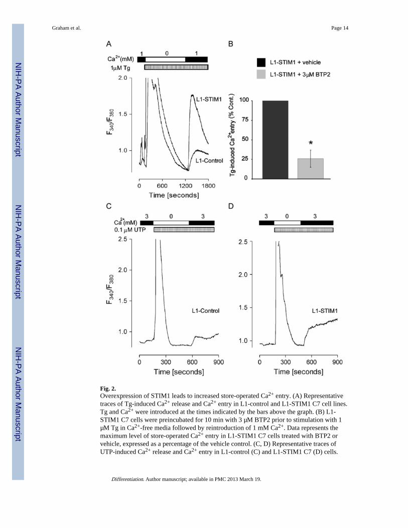
Fig. 2.Overexpression of STIM1 leads to increased store-operated Ca2+ entry. (A) Representativetraces of Tg-induced Ca2+ release and Ca2+ entry in L1-control and L1-STIM1 C7 cell lines.Tg and Ca2+ were introduced at the times indicated by the bars above the graph. (B) L1-STIM1 C7 cells were preincubated for 10 min with 3 µM BTP2 prior to stimulation with 1µM Tg in Ca2+-free media followed by reintroduction of 1 mM Ca2+. Data represents themaximum level of store-operated Ca2+ entry in L1-STIM1 C7 cells treated with BTP2 orvehicle, expressed as a percentage of the vehicle control. (C, D) Representative traces ofUTP-induced Ca2+ release and Ca2+ entry in L1-control (C) and L1-STIM1 C7 (D) cells.
Graham et al. Page 14
Differentiation. Author manuscript; available in PMC 2013 March 19.
NIH
-PA Author Manuscript
NIH
-PA Author Manuscript
NIH
-PA Author Manuscript

UTP and Ca2+ were introduced at the times indicated by the bars above the graphs, *p <0.0001.
Graham et al. Page 15
Differentiation. Author manuscript; available in PMC 2013 March 19.
NIH
-PA Author Manuscript
NIH
-PA Author Manuscript
NIH
-PA Author Manuscript

Fig. 3.STIM1 overexpression inhibits 3T3-L1 adipogenesis. (A) Oil Red O staining of fixed wild-type 3T3-L1, L1-control, L1-STIM1 C7 and C8 cells at day 10 post MDI induction. (B)Spectrophometric quantitation of Oil Red O in cell extracts at various timepoints post MDItreatment of individual L1-STIM1 cell lines (closed symbols) and individual L1-controllines (open symbols). (C) Oil Red O staining of fixed L1-control and L1-STIM1 C7 cells atday 10 following incubation with either DMSO or 10 µM BTP2 between days 0 and 6 postMDI treatment.
Graham et al. Page 16
Differentiation. Author manuscript; available in PMC 2013 March 19.
NIH
-PA Author Manuscript
NIH
-PA Author Manuscript
NIH
-PA Author Manuscript

Fig. 4.STIM1 overexpression leads to downregulation of C/EBPα and adiponectin expression. (A)3T3-L1-control (gray bars) and L1-STIM1 C7 (black bars) cell numbers at day 0 (MDIaddition), days 2 and 4 post MDI treatment as determined by cell counting with ahaemocytometer. For each cell line, data is expressed as the cell number relative to that of3T3-L1-control and L1-STIM1 C7 cell numbers at day 0, each of which were normalized to1. Error bars represent the standard error of triplicate wells. (B) 3T3-L1-control (gray bars)and L1-STIM1 C7 (black bars) cell numbers at 0 (day of seeding), 2 and 3 days ofproliferation as determined by cell counting with a haemocytometer. Data is expressed as for(A). Error bars represent the standard error of triplicate wells. (C) Western blotting for C/
Graham et al. Page 17
Differentiation. Author manuscript; available in PMC 2013 March 19.
NIH
-PA Author Manuscript
NIH
-PA Author Manuscript
NIH
-PA Author Manuscript

EBPβ, C/EBPα, PPARγ1 and 2, Adpn and β-actin in cell lysates collected at days 0, 2, 4and 6 post MDI treatment in 3T3-L1-control and L1-STIM1 C7 cell lines. (D) Oil-red-Ostaining of fixed L1-STIM1 C7 cells at day 8 post MDI induction. Cells were treated withvehicle (ethanol), FK506 or cyclosporin A from day 0.
Graham et al. Page 18
Differentiation. Author manuscript; available in PMC 2013 March 19.
NIH
-PA Author Manuscript
NIH
-PA Author Manuscript
NIH
-PA Author Manuscript

Fig. 5.Stim1 downregulation enhances adipogenesis in 3T3-L1 cells and leads to upregulation ofC/EBPα and Adpn expression. (A) Oil Red O staining of 3T3-L1 cells at day 8 followingtransfection with standard control Stealth™ siRNA duplexes or Stim1–60 Stealth™ siRNAduplexes and induced to differentiate with either MDI, or 0.5MD. (B) Quantification atOD520 of lipid extracted from triplicate wells of 3T3-L1 cells at day 8. Cells weretransfected with standard control, Stim1–60 or Stim1–61 Stealth™ siRNA duplexes andinduced to differentiate with 0.5MD. (C) Relative Stim1 mRNA levels in Stim1 or controlsiRNA transfected 3T3-L1 cells harvested 48 h post transfection. Western blotting for (D)Stim1, PPARγ1 and γ2, β-actin and (E) C/EBPα, Adpn, β-actin in cell lysates collected
Graham et al. Page 19
Differentiation. Author manuscript; available in PMC 2013 March 19.
NIH
-PA Author Manuscript
NIH
-PA Author Manuscript
NIH
-PA Author Manuscript

from Stealth™ Stim1-60 (S1–60) or control (Con) siRNA duplex-transfected cells at 4 dayspost 0.5MD treatment.
Graham et al. Page 20
Differentiation. Author manuscript; available in PMC 2013 March 19.
NIH
-PA Author Manuscript
NIH
-PA Author Manuscript
NIH
-PA Author Manuscript