Apoptosis and non-inflammatory phagocytosis can be induced by mitochondrial damage without caspases

STIM1 Juxtaposes ER to Pha
Current Biology 22, 1990–1997, November 6, 2012 ª2012 Elsevier Ltd All rights reserved http://dx.doi.org/10.1016/j.cub.2012.08.049
2+
Articlegosomes,
Generating Ca Hotspotsthat Boost Phagocytosis
Paula Nunes,1 Daniela Cornut,1 Vanessa Bochet,2
Udo Hasler,1,3 Masatsugu Oh-Hora,4
Jean-Marc Waldburger,2 and Nicolas Demaurex1,*1Department of Cell Physiology and Metabolism2Department of Pathology and Immunology3Department of Medical SpecialtiesUniversity of Geneva, 1211 Geneva 4, Switzerland4Department of Cell Signaling, Tokyo Medical and DentalUniversity, Yushima, Bunkyo-ku, Tokyo 113-8549, Japan
Summary
Background: Endoplasmic reticulum (ER) membranes are re-cruited to phagosomes, but the mechanism and functionalsignificance of this ER recruitment is not known. Here, weshow that the ER Ca2+ sensor stromal interaction molecule 1(STIM1) sustains high-efficiency phagocytosis by recruitingthin ER cisternae that interact productively but do not fusewith phagosomes.Results: Endogenous STIM1 was recruited to phagosomesupon ER Ca2+ depletion in mouse neutrophils, and exogenousYFP-STIM1 puncta coincided with localized Ca2+ elevationsaround phagosomes in fibroblasts expressing phagocyticreceptors. STIM1 ablation decreased phagocytosis, ER-phag-osome contacts, and periphagosomal Ca2+ elevations in bothneutrophils and fibroblasts, whereas STIM1 re-expression inStim12/2 fibroblasts rescued these defects, promoted theformation and elongation of tight ER-phagosome contactsupon ER Ca2+ depletion and increased the shedding of periph-agosomal actin rings. Re-expression of a signaling-deficientSTIM1 mutant unable to open Ca2+ channels recruited ERcisternae to the vicinity of phagosomes but failed to rescuephagocytosis, actin shedding, and periphagosomal Ca2+ ele-vations. The periphagosomal Ca2+ hotspots were decreasedby extracellular Ca2+ chelation and by Ca2+ channels inhibi-tors, revealing that the Ca2+ ions originate at least in partfrom phagosomes.Conclusions: Our findings indicate that STIM1 recruits ERcisternae near phagosomes for signaling purposes and thatthe opening of phagosomal Ca2+ channels generates localizedCa2+ elevations that promote high-efficiency phagocytosis.
Introduction
Phagocytosis is a fundamental cellular event of great clinicalrelevance, and many microorganisms disrupt intracellular sig-nals controlling phagocytosis to evade destruction by innateimmune cells [1]. In neutrophils, Ca2+ elevations boost phago-cytosis by promoting phagolysosome fusion [2], shedding ofthe actin coat [3], and NAPDH oxidase activation [4], but themechanism driving localized Ca2+ elevations around phago-somes [5] remains elusive [6]. Stromal interaction molecule 1(STIM1) was recently identified as a ubiquitous ER Ca2+ sensorthat, uponCa2+ depletion of the ER, induces the formation near
*Correspondence: [email protected]
the plasma membrane (PM) of thin ER cisternae known ascortical ER [7]. STIM1 molecules on the cortical ER interactwith store-operated Ca2+ entry (SOCE) channels on the PM,sustaining long-lasting Ca2+ signals required for the activationof T cells [8]. ER remodeling is a prominent feature of phago-cytosis whose functional significance was much debated.Although ER recruitment to phagosomes had been previouslyshown [9], more in-depth studies proposed that the ERprovided membranes to phagosomes and supplied ER resi-dent proteins for antigen cross-presentation [10, 11]. However,because these functions require the ER to fuse with phago-somes, these proposals were strongly contested [12].WhetherSTIM1-mediated ER remodeling occurs during phagocyto-sis is not known, but engagement of phagocytic receptorsreleases Ca2+ from internal stores [6] and the ER structuresaccumulating near phagosomes bear a striking similarity tothe cortical ER structures induced by STIM1. Furthermore,STIM1 ablation decreases phagocytosis in mouse peritonealmacrophages [13] and STIM1 knockdown reduces intrapha-gosomal ROS production in HL60 cells [14]. We thereforepostulated that STIM1 could recruit ER cisternae to phago-somes for signaling purposes and confirmed this by light andelectron microscopy by comparing wild-type (WT) fibroblaststo fibroblasts from STIM1 knockout mice rendered phagocyticby expression of FcgRIIA receptors, and WT neutrophils toneutrophils from mice bearing a myeloid-specific ablationof the STIM1 gene. By re-expressing a signaling-deficientSTIM1 mutant, we further show that STIM1 interacts withCa2+ channels on phagosomes to promote localized Ca2+
elevations that drive periphagosomal actin shedding andhigh-efficiency phagocytosis.
Results
STIM1 Is Recruited to Nascent Phagosomes
To test whether STIM1 is recruited to phagosomes, we as-sessed the intracellular location of endogenous and exoge-nous STIM1 in mouse neutrophils and promyeloid leukemiadHL60 cells ingesting IgG-opsonized sheep red blood cells(RBC) and zymosan particles (Zym), respectively, and inmouse embryonic fibroblasts (MEFs) derived from Stim12/2
knockout (STIM1 KO) mice and rendered phagocytic byectopic expression of FcgRIIA receptors. YFP-STIM1 localizedto nascent phagosomes in dHL60 cells (Figure 1A), as early asjust before cup closure, with dynamic puncta observed up to15 min postingestion (see Movie S1 available online).In phagocytic MEFs, mCherry-STIM1 puncta were observed
in close proximity to the FcgRIIA-GFP signal lining the phago-somal membrane (Figure 1B), confirming that the STIM1mole-cules accumulated around phagosomes. The phagocytic KOMEFs completely lacked STIM1 but had normal levels ofSTIM2 (Figure S1A) and exhibited a near complete SOCEdefect that was restored by STIM1 re-expression (Figure S1B).STIM1-containing cortical-ER junctions are specialized ERsubdomains connected to the bulk ER by a constrictioncapable of excluding ‘‘classic’’ ER markers such as BiP, aKDEL-containing protein [7]. Phagosomes in MEFs were notdecorated by the ER resident protein GFP-KDEL (Figure S1C),

Slice 3D Projection A YFP-STIM1 568-Zym
*
dHL6
0
+ 1μM Tg
Mou
se N
eutr
ophi
ls
Anti-STIM1
RBC
Hoechst
C
CTR +Tg % P
hago
som
es w
ith
asso
ciat
ed S
TIM
1 pu
ncta
Orai1-GFP mCherry-STIM1 RBC
E
KO MEF (+myc-Fcγ-RIIA )
D + 1μM Tg
RBC
YFP-STIM1 K
O M
EF
(+m
yc-F
cγ-R
IIA )
**
Fc RIIA-GFP
RBC
mCherry-STIM1
KO
MEF
B
p = 0.0091
61 76
Hoechst
Figure 1. STIM1 Is Recruited to Phagosomes
upon Ca2+ Depletion of the ER
(A) 3D projection of a confocal z stack shows that
YFP-STIM1 (green) is recruited to zymosan-con-
taining phagosomes (red) in dHL60 cells. STIM1
accumulates around nascent phagosomes
(arrow) and is shed from mature phagosomes
(star). Right-hand zoom represent single-plane
confocal slices. See also Movie S1.
(B) Similar to dHL60, mCherry-STIM1 (red) local-
izes near phagosomes (blue) when re-expressed
in STIM1 KO MEFs expressing FcgRIIA-GFP
receptors (green).
(C) Endogenous STIM1 (green) staining near
phagosomes (red, arrows) is enhanced by ER
Ca2+ depletion (1mM Tg) in mouse neutrophils.
(D) ER Ca2+depletion (1mM Tg) increases STIM1
(green) recruitment (arrows) to phagosomes
(blue) in STIM1 KO MEFs expressing phagocytic
receptors.
(E) mCherry-STIM1 (red) colocalizes with its
partner channel Orai1-GFP (green) on phago-
somes (blue). See also Figure S1. Scale bars
represent 3 mm. Data are means 6 SEM of three
independent experiments, and numbers in bars
indicate the total number of measured phago-
somes.
STIM1 Activates Ca2+ Channels on Phagosomes1991
indicating that STIM1 was indeed enriched in specializedperiphagosomal ER domains. In mouse neutrophils, STIM1immunoreactive puncta were detected around phagosomesand their numbers increased upon treatment with thapsigargin(Tg, Figure 1C), a SERCA pump inhibitor that increases STIM1oligomerization and activation by depleting ER Ca2+ stores [8].ER Ca2+ depletion also increased the number and size of pe-riphagosomal STIM1puncta aswell as thepercentageof phag-osomes associated with STIM1 clusters in STIM1-rescuedphagocytic MEFs (Figure 1D). Because sorting of plasmamembrane proteins into phagosomes occurs even beforephagosome closure [15, 16], we then needed to checkwhetherCa2+-permeable channels gatedbySTIM1canbe incorporatedinto phagosomes. All three Orai isoforms localized to phago-somes when expressed in phagocytic MEFs and dHL60 (Fig-ure S1D), and mCherry-STIM1 colocalized with GFP-taggedOrai1 in these periphagosomal structures (Figure 1E). Thesedata indicate that STIM1 is recruited to nascent phagosomesupon store depletion together with its partner channel Orai1,the major SOCE channel of immune cells.
STIM1 Promotes the Formation of Tight Phagosomal-ERJunctions
Because we were unable to find a good marker for immuno-staining the specialized ER subdomains generated by STIM1,
we next confirmed that STIM1 indeedrecruits ER cisternae to phagosomesby morphological examination, usingelectron microscopy. ER membranesappeared as dark flattened cisternae,located close to phagosomal mem-branes in w37% of phagosomes fromWT MEFs, a percentage that droppedto 10%upon STIM1 ablation (Figure 2A).The residual periphagosomal ER struc-tures forming in STIM1-deficient cellswere similar in length to those found in
WT MEFs, and STIM1 re-expression augmented both theirfrequency and length byw70% (Figure 2A). The periphagoso-mal ER structures were still connected with conventional ERcisternae (Figure S2A) and were detected already at the cupstage (Figure S2B), consistent with dHL60 live-cell imaging.Importantly, ER Ca2+ depletion dramatically increased thefrequency and length of periphagosomal ER cisternae in WTas well as STIM1-Rescued, but not in KO cells (Figure 2A,right). To test whether endogenous STIM1 also recruits thinER cisternae near phagosomes in neutrophils, we bred floxedStim1fl/fl mice to LysM-Creki/ki knockin mice to restrict STIM1ablation to myeloid cells and avoid the perinatal death asso-ciated with whole-animal STIM1 ablation in mice [17]. STIM1expression was reduced by w90% in bone-marrow derivedneutrophils isolated by FACs sorting (Figure S2C) fromStim1fl/fl, LysM-Creki/ki mice compared to Stim1+/+ LysM-Creki/ki littermates (Figures S2D and S2E). ER cisternae juxta-posed to phagosomes were observed in 50% of phagosomesfrom control neutrophils, a proportion that decreased to 30%in neutrophils from Stim1fl/fl, LysM-Creki/ki mice (Figure 2B).The juxtaposed ER cisternae were slightly, but not sig-nificantly, shorter in STIM1-ablated neutrophils (Figure 2B),and their frequency and length did not increase upon ERCa2+ stores depletion in either genotype (data not shown).Because periphagosomal STIM1 puncta increased upon Tg
Ph
*
*
+STI
M1
Control
KO
Phagocytosis of IgG-RBC in MEFs (+myc-Fcγ-RIIA )
Ph
Ph Ph
1 μM Tg
Ph Ph
WT
Ph
*
Stim1fl/fl, LysM-Creki/ki
Ph
*
Stim1+/+, LysM-Creki/ki
ER ju
nctio
ns/
100
phag
osom
es
**
p = 0.08
ns p = 0.008
93 94 45 27
ER ju
nctio
n le
ngth
/ ph
agos
ome
perim
eter
A
B ER
junc
tion
leng
th/
phag
osom
e pe
rimet
er
* *
* *
* *
210
117
104
89
34
39
10
8
51
19
18
ER ju
nctio
ns/
100
phag
osom
es
p = 0.038
*
p = 0.005
**
p = 0.03 *
p = 0.005
**
p = 0.0003 ***
p < 0.0001 ***
p = 0.197 n.s.
p = 0.006 **
33
Figure 2. STIM1 Promotes the Formation of Tight Phagosomal-ER Junctions
(A) Electron micrographs illustrating ER cisternae (*) juxtaposed to phagosomes (Ph) in STIM1WT (top), KO (middle), and STIM1-rescued phagocytic MEFs
(bottom) treated or not with 1 mMTg. Note the long periphagosomal cisternae in STIM1-rescued cells treatedwith Tg (bottom right). Bar graphs: Quantitative
EM analysis shows that STIM1 ablation decreases the frequency (top) and length (bottom) of phagosomal-ER contacts whereas STIM1 rescue increased
their occurrence and promoted their formation and elongation upon ER Ca2+ depletion.
(B) Myeloid-specific STIM1 ablation decreased the number of periphagosomal ER cisternae (*) in mouse neutrophils without significantly decreasing their
length. See also Figure S2. Scale bars represent 200 nm. Data aremeans6 SEMof three independent experiments, with the total number ofmeasured phag-
osomes indicated in bars.
Current Biology Vol 22 No 211992
addition (Figure 1C), this finding suggests that store deple-tion either increases STIM1 immunoreactivity or recruitsadditional STIM1 molecules to already docked ER cisternaein neutrophils. The high levels of ER-phagosome contactsin neutrophils (which are similar to WT MEFs stimulatedwith Tg), combined with their insensitivity to ER Ca2+ deple-tion suggest that the ER is very efficiently recruited duringphagocytosis in these professional phagocytes. Together,these EM data indicate that ER-phagosomal contacts canform in STIM1-deficient cells irrespective of Ca2+ store deple-tion but that STIM1 expression is required for the store-oper-ated recruitment and elongation of ER cisternae to nascentphagosomes.
STIM1 Enhances Phagocytosis and Promotes Phagosomal
Actin SheddingTo determine whether STIM1-mediated ER recruitment hasa functional impact on phagocytosis, we determined theability of WT and STIM1 KO MEFs to ingest IgG-opsonizedRBCs, using cells with matching FcgRIIA-GFP fluorescenceto ensure similar amounts of phagocytic receptors. WhileWT and KO cells exposed to a low target:cell ratio phagocy-tosed to a similar extent, increasing the target:cell ratiorevealed a defect in phagocytic ingestion of w30% inKO cells (Figure 3A). Concordantly, when YFP-STIM1 ormCherry-STIM1 (see below) were re-expressed in KO MEFsexpressing myc-FcgRIIA, phagocytosis at a high target:cell

Figure 3. STIM1-Mediated SOCE Channel Activation Is Required for High-Level Phagocytosis
(A) STIM1 ablation decreased phagocytosis at high but not low target loads in MEFs expressing matching levels of FcgRIIA-GFP receptors (n = 3).
(B) YFP-STIM1 but not YFP-STIM2 or ER-targeted KDEL-GFP enhances phagocytosis in STIM1 KO MEFs expressing myc-FcgRIIA receptors (n = 4, 10:1
target:cell ratio).
(C) Myeloid-specific STIM1 ablation reduces phagocytosis by 50% in mouse neutrophils (n = 6, 10:1 target:cell ratio).
(D) STIM1-4K-mRFP lacking polybasic residues (red) required for channel activation is recruited to phagosomes (green). Scale bar represents 3 mm.
(E) STIM1-4K-mRFP promotes the formation and elongation of tight phagosomal-ER contacts (compare with Figure 2A). Ph, phagosome; scale bar repre-
sents 200 nm.
(F) STIM1-4K-mRFP does not enhance phagocytosis and removal of extracellular Ca2+ abrogates mCherry-STIM1 prophagocytic effects (n = 4, 10:1 target:
cell ratio). Data are means 6 SEM of 3–6 independent experiments, and numbers in bars indicate total measured cells (A, B, C, F) or phagosomes (E).
STIM1 Activates Ca2+ Channels on Phagosomes1993
ratio was w40% higher than in cells expressing either cyto-plasmic eGFP, ER-targeted KDEL-GFP, or YFP-STIM2 (Fig-ure 3B). These data indicate that although STIM1 is notrequired for basal levels of particle ingestion, it facilitatesphagocytosis as the particle load increases, and that thiseffect is specific for STIM1. Myeloid-specific STIM1 ablationreduced phagocytosis by nearly 50% in mouse neutrophils(Figure 3C), indicating that endogenous STIM1 also sustainshigh-level phagocytosis in professional phagocytes. To testwhether the prophagocytic effects of STIM1 require pro-ductive interactions at the ER-phagosome interface, weexpressed a STIM1 mutant lacking the polybasic domainrequired for Orai channel activation [18] that did not triggerSOCE in MEFs (Figure S1B). This ‘‘STIM1-4K’’ signaling-defi-cient mutant accumulated around phagosomes (Figure 3D)and increased the frequency and length of periphagosomalER cisternae when expressed in MEFs (Figure 3E). Storedepletion did not further increase either parameter, suggestingthat ER recruitment may already be maximal in cells express-ing the 4K mutant which, by inhibiting Ca2+ influx, is expectedto inhibit disassembly of STIM1 oligomers [19]. Notably,STIM1-4K expression did not rescue phagocytosis (Figure 3F),indicating that electrostatic interactions between STIM1 and
its target channel(s) are required for the prophagocytic func-tion of STIM1. Consistent with activation of Ca2+-permeablechannels, STIM1 re-expression did not augment phagocytosisin Ca2+-free physiological saline (Figure 3F).Because shedding of the actin coat is an early event during
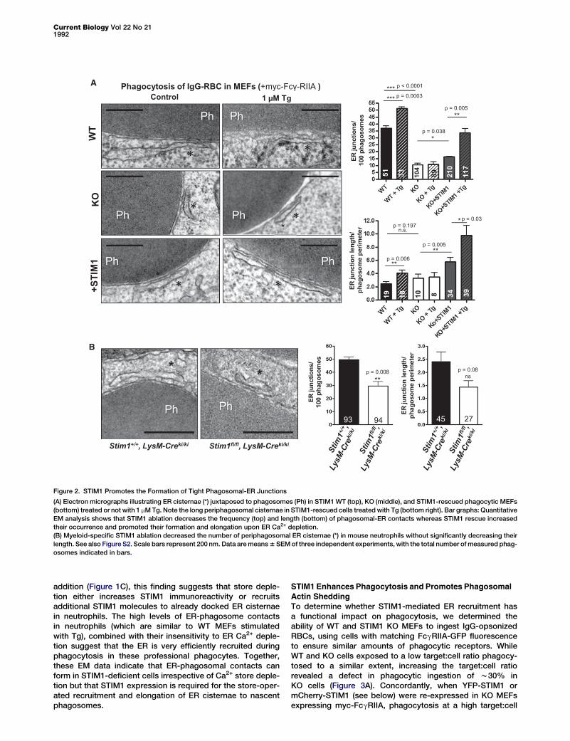
phagocytic maturation that is controlled by Ca2+ elevations [3]and reduces phagocytic index when impaired [20–22], we nextexamined whether STIM1 expression impacted periphagoso-mal actin structures. Periphagosomal actin rings were moreprominent in STIM1 KO cells expressing KDEL-GFP or YFP-STIM1-4K than in cells expressing YFP-STIM1 (Figure 4A),and fluorescence quantification showed that re-expressionof WT STIM1, but not of signaling-deficient STIM1-4K, de-creased periphagosomal filamentous (F)-actin content, in ex-periments where STIM1-YFP puncta decorated phagosomesto similar extents (Figure 4B).
STIM1 Promotes Periphagosomal Ca2+ ElevationsSTIM1-mediated Orai1 channel activation restricts Ca2+ entryto distinct sites at the PM [23, 24]. However, whether STIM1is capable of gating Ca2+ channels in internal membranecompartments is unknown. To test whether STIM1 alsogenerates Ca2+ hotspots around phagosomes, we measured
Actin
KD
EL-
GFP
YFP-
ST
IM1
YFP-
ST
IM1-
4K
B A
**
Nor
mal
ized
phag
ogom
alac
tin
KO MEF (+myc-Fcγ-RIIA )
ns
p = 0.003
56 51 56
% P
hago
som
esw
ith
asso
ciat
ed S
TIM
1 pu
ncta
ns
51 56
KO MEF (+myc-Fcγ-RIIA )
Figure 4. STIM1-Mediated Interactions Promote
Periphagosomal Actin Shedding
(A) Periphagosomal F-actin rings (red) were
decreased by YFP-STIM1 but not YFP-STIM1-4K
re-expression in MEFs.
(B) Average phalloidin intensity in a 1 mm ring
surrounding the midsection of phagosomes,
normalized to the total cellular phalloidin staining
(top) and percentage of phagosomes bearing
YFP-STIM1 or YFP-STIM1-4K puncta (bottom).
Scale bar represents 3 mm. Data are means 6
SEM of four independent experiments, and
numbers in bars indicate total measured phago-
somes.
Current Biology Vol 22 No 211994
Ca2+ concentration changes by visualizing cells loaded withthe Ca2+-binding dye Fluo8 using confocal microscopy. InMEFs, a low concentration of the Ca2+ chelator BAPTA-AMwas used to decrease the lateral diffusion of Ca2+ and improvehotspots visualization [24]. Ca2+ hotspots were observedaround w50% of phagosomes in WT cells, a proportion thatdecreased to 20% upon STIM1 ablation and that was nearlyrestored by STIM1 re-expression, whereas STIM1-4K expres-sion increased Ca2+ hotspot occurrence only to 28% (Figures5A and 5B; Table S1). The same hotspot proportion (28%) wasobserved in STIM1-rescued cells exposed to the SOCEchannel inhibitor La3+ (5 mM) whereas Ca2+ chelation de-creased hotspot occurrence to 17% in STIM1-rescued cells(Figure 5B; Table S1). This indicates that periphagosomal ERCa2+ stores and phagosomal Ca2+ channels both contributeto the Ca2+ hotspots observed in the vicinity of phagosomes,with STIM-gated Ca2+ channels contributing more than halfof the signal. Periphagosomal Ca2+ hotspots were alreadyvisible as early as 2 min after particle contact and usuallyoccurred repeatedly at the same location, with some singlehotspots persisting for several minutes (Figure 5C; MovieS2). Strikingly, the formation of STIM1 puncta coincidedtemporally and spatially with the appearance of periphagoso-mal Ca2+ microdomains (Figure 5D; Movie S3), implying thatSTIM1 recruitment to phagosomes initiates the localizedintracellular Ca2+ elevations. Periphagosomal Ca2+ hotspotscoincident with mCherry-STIM1 puncta were also observedin cells treated with 1 mM Tg after switching to Ca2+-free con-ditions (Movie S4), further validating the phagosomal originof the Ca2+ ions. That STIM1 relies on phagosomal Ca2+ andon polybasic residues gating Orai channels to promote bothperiphagosomal Ca2+ hotspots and phagocytosis indicatethat the opening of phagosomal SOCE channels is the majormechanism by which STIM1 boosts phagocytosis. In agree-ment with the results obtained in MEFs, the occurrence ofCa2+ hotspots was decreased by w50% in STIM1-depletedneutrophils, the residual hotspots exhibiting the same ampli-tude as in WT neutrophils over identical resting cytosolicCa2+ levels (Figure 5E). These data indicate that STIM1interacts with Ca2+ channels on phagosomes via electrostatic
interactions, to initiate localized Ca2+
elevation in their immediate vicinity.
Discussion
Our data show that the Ca2+-signalingmolecule STIM1 recruits the ER to anintracellular target, the phagosome, to
promote productive interactions required for high efficiencyphagocytosis. STIM1 does not mediate ER-phagosome fusionbut generates localized Ca2+ elevations required for actinshedding by activating SOCE channels on phagosomes, anactivity that was only described at the PM so far. This mecha-nism likely explains the defective phagocytosis of STIM1knockout peritoneal macrophages [13] and may contribute tothe impaired phagosomal ROS production of dHL60 cellsdepleted of STIM1 and Orai1 [14]. These earlier studies indi-cated that STIM1-Orai interactions were important for thephagocytic process but inferred that the interactions occurredat the PM to sustain global Ca2+ elevations rather than atthe ER/phagosome interface. Our findings that STIM-Oraiinteractions trigger SOCE across the phagosomal membraneestablish STIM1 as a key effector molecule that regulatesphagocytosis, clarify a long-standing debate on the functionof ER recruitment to phagosomes, and have potential thera-peutic implications. Intracellular pathogens like Mycobacte-rium tuberculosis and Leishmenia subvert Ca2+-dependentprocesses to survive phagocytic killing [25] and STIM1 muta-tions are associated with defective clearance of apoptoticcells in nematodes [26] and with immunodeficiencies in miceand humans [27]. The discovery that SOCE molecules controlthe highly localized Ca2+ signals associated with phagocytosiscould therefore provide new potential targets for the treatmentof infections caused by intracellular pathogens. It also sug-gests that defective phagocytosis could be part of the clinicalpicture of STIM1 dependent immunodeficiencies.
Experimental Procedures
Reagents
STIM1 KO MEFs, generated by targeted gene disruption [28], and WT MEF
controls were a kind gift from Dr. Marek Michalak (University of Alberta,
Canada). Human FcgRIIA-GFP and c-myc constructs were a kind gift from
Dr. Sergio Grinstein (University of Toronto, Canada). GFP-tagged Orai1,
Orai2, and Orai3 were a kind gift from Drs. Ivan Bogeski and Barbara Nie-
meyer (Saarland University, Germany). YFP and mRFP-tagged STIM1-4K
mutants were a kind gift from Dr. Tamas Balla (National Institute of Child
Health andHumanDevelopment, Bethesda, USA). Please see Supplemental
Information for other reagent sources.

B
Time (sec)
Ca2
+ (F
/F0
ave)
Ho
tsp
ots
***
**
% P
hago
som
esw
ithas
soci
ated
Ca2
+ho
tspo
ts
D
STIM1- mCherry
Ca2+
(F/F0 ave)
KO +STIM1KOA KO +STIM1-4K
Stim1+/+, LysM-Creki/ki
Stim1fl/fl, LysM-Creki/ki
E Stim1+/+, LysM-Creki/ki
Stim1fl/fl, LysM-Creki/ki
% P
hago
som
esw
ith a
ssoc
iate
dho
tspo
ts
***
0 sec 252 sec 684 sec
C
p < 0.0001
p =
0.008
ns
p =
0.19
p =
0.001
**
p =
0.0005
Ca2+
Hot
spot
ampl
itude
(n
M)
Cyt
osol
icC
a2+ (n
M)
***p < 0.0001 (+myc-Fcγ-RIIA )
WT
00:00 00:12 00:24 00:36 00:48 01:00 01:12 02:24 02:36
RBC- Alx633
103 122 12 10 11 6
Figure 5. STIM1-Mediated Interactions Promote Periphagosomal Ca2+ Signaling
(A) The occurrence of periphagosomal Ca2+ elevations was decreased by STIM1 ablation and restored by STIM1 re-expression, whereas STIM1-4K has
a smaller effect (arrows, see also quantification in B).
(B) Inhibition of SOCE channels with 5 mM LaCl3 and extracellular Ca2+ chelation with 3 mM EGTA reduces periphagosomal Ca2+ microdomains in STIM1-
rescued cells to levels similar to that of STIM1-4K rescued and KO cells, respectively. See also Table S1.
STIM1 Activates Ca2+ Channels on Phagosomes1995
Current Biology Vol 22 No 211996
Cell Culturing and Transfection
MEFs were transfected using Lipofectamine 2000. HL60 cells were differen-
tiated by exposure to 1.3% DMSO for 6 days (dHL60) and transfected using
the Amaxa T Nucleofection kit. Transfected cells were allowed to recover for
24 hr prior to manipulation. Please see Supplemental Experimental Proce-
dures for details.
Generation of Myeloid-Specific STIM1 Ablation and FACs Sorting
Mice deficient for STIM1 in the myeloid lineage were generated by crossing
Stim1fl/fl animals in the C57BL/6 background [29] with the LysM-Creki/ki
strain [30]. All animal manipulations were performed in accordance to the
guidelines approved by the animal research committee at the University
of Geneva. Genotyping was performed from tail biopsies as described
[29, 31]. Neutrophils were isolated from bone marrow of mouse femurs
and tibias using Percoll gradient centrifugation as previously described
[32]. For FACs sorting, cells were blocked with anti-mouse-Fc-receptor
antibodies for 30 min, and incubated with anti-GR-1-APC and anti B220-
FITC on ice for 1 hr in 2% BSA, 20 mM EDTA buffer. Sorting was performed
on a FACs Aria I sorter (BD Biosciences, Franklin-Lakes, NJ).
Phagocytosis
RBCs were opsonized in rabbit-anti-sheep RBC and zymosan was opson-
ized in rabbit-anti-zymosan at 37�C for 1 hr. For MEFs and neutrophils, tar-
gets were added directly to coverslips at indicated target:cell ratios. For
dHL60s, targets were resuspended with cells at a 10:1 target:cell and
centrifuged at 8,000 g for 30 s. Cells were then incubated at 37�C/5% CO2
for indicated times. All phagocytosis experiments were performed in
serum-containingmedia with the exception of actin experiments and exper-
iments comparing Ca2+-containing and Ca2+-free conditions, where serum-
free physiological buffer (please see Supplemental Information) was used
instead.
Immunolabelling and Actin Staining
Immunolabelling and actin staining were performed according to standard
protocols. Please see Supplemental Information for details.
Imaging
All live-cell imaging microscopes were maintained at 37�C by a microscope
temperature control system (Life Imaging Services, Basel, Switzerland) and
performed in physiological buffer where Ca2+-free solution contained 1 mM
EGTA instead of 2 mM CaCl2. Please see Supplemental Information
for microscope details. Fura-2-AM (2 mM) was loaded in physiological
buffer +0.02%pluronic and visualized using 340/380 nm alternate excitation
and 510 6 40 nm emission. Frames were acquired every 2 s. Fluo8-AM
(4 mM) was loaded in physiological buffer/250 mM sulfinpyrazone at 37�Cfor 30 min. For MEFs, this was followed by addition of BAPTA-AM (5 mM)
and incubation for 30 min at room temperature. For neutrophils, cells were
instead incubated for an additional 30 min at room temperature in the pres-
ence of 1 mM ER-Tracker Red. Simultaneous excitation at 488, 543 nm, and
650 nm and emission collection in three separate channels for green (Fluo8),
red (mCherry or mRFP), and far-red (RBC autofluorescence or RBC-Alexa
633 fluorescence, used to define the phagosomal border) was used. For
quantification of periphagosomal Ca2+ hotspots, images averaged over
6 s and captured between 20 and 30 min after the addition of IgG coated
sRBCwere used. For time-lapse imaging of periphagosomal Ca2+ hotpsots,
cells were imaged only with 488 nm laser light, although red channel emis-
sion was also collected. To minimize toxicity, we imaged cells for 30 s at
1 s frame rate during particle addition and then again after 5 min of particle
addition at 12 s frame rate for an additional 15 min. For Ca2+ chelation and
Ca2+ channel blocking experiments, 3 mM EGTA or 5 mM LaCl3 were added
1 min prior to addition of particles. Unless otherwise indicated, all images
shown represent single-plane confocal slices.
(C) Periphagosomal Ca2+ hotspots are detected early during phagocytosis and
trace is the spatially averaged intensities of an w750 nm wide periphagosom
intensity. Colored dots below the trace represent occurrences of Ca2+ hotspo
time point are represented by different colors (illustrated in the insets by color
(D) Periphagosomal Ca2+ hotspots (top) colocalize spatiotemporally with STIM
also Movies S3 and S4.
(E) Myeloid-specific STIM1 ablation reduces the occurrence of periphagosom
amplitude or the average cytosolic Ca2+ concentration. White bars represent 3
by the initial average cytosolic fluorescence (F/F0ave). Data are means 6 SEM
osomes is detailed in Table S1 or represented inside bars.
Image Quantification
All images were background subtracted by subtracting the average inten-
sity of a small region of interest (ROI) that does not contain cells from all
intensity values in the same image, prior to analysis. Phagocytic index
was defined as the number of internalized phagosomes divided by the total
number of (transfected where applicable) cells and was determined by
counting internalized phagosomes in confocal z stacks encompassing
w1 mm above and below cell monolayers using ImageJ software. FcgRIIA
marks the plasma membranes and nascent phagosomes, and internalized
particles were easily distinguished in 3D reconstructions. Peripheral phag-
osomes were deemed internalized if more than 75% of the target surface
showed visible FcgRIIA. FcgRIIA-GFP fluorescence was visualized directly
and myc-FcgRIIA was visualized by immunostaining. Total FcgRIIA and
total F-actin were defined as the average cellular FcgRIIA-GFP or TRITC-
phalloidin fluorescence intensity (FI) respectively of 32-bit sum projections
of confocal stacks. FcgRIIA-GFP expressing cells were thresholded to
ensure populations of similar total FcgRIIA-GFP. Phagosomal F-actin was
estimated using a previously described approach [33]. Briefly, the TRITC-
phalloidin FI was measured within 1 mm of the phagosomal border (defined
using the RBC fluorescence) in single confocal slices sectioning the
middle of the phagosome. Normalized phagosomal F-actin is phagosomal
F-actin/total F-actin of the same cell. Phagosomal F-actin was quantified
for all internalized phagosomes and peripheral phagosomes whose tar-
get surface showed more than 50% association with F-actin 10 min after
target exposure. At least five confocal stacks of equal dimensions per
genotype per experiment and at least three independent experiments
were quantified.
InMEFs, periphagosomal Ca2+ hotspots were defined as areasR500 nm2
(4 pixels) within a distance of w750 nm (3 pixels) from the phagosomal
border displaying a FI at least 2 SDs higher than the average cytoplasmic
Fluo8 FI. For neutrophils, ER-Tracker Red reliably demarcated nonnuclear
areas and was less toxic than Hoechst during live imaging and thus was
used as guide to avoid the nucleus and areas of ER-associated Ca2+ activity
when defining cytosolic ROIs. In addition, because in neutrophils BAPTA-
AM was not used to increase contrast, a cutoff of 2.5 SD above the mean
cytoplasmic Fluo8 FI was used instead. Periphagosomal Ca2+ traces were
generated by tracking phagosomes with a 3-pixel-wide (w750 nm) periph-
agosomal ROI in 12 s frame time-lapse series. Tracking began 3 frames prior
to phagosome arrival in the plane of focus, and average fluorescence values
of the first ROI were used to normalize subsequent values. The SD of
this first ROI was used to set a threshold of 2.5 SD to identify hotspots,
again defined as being above-threshold regions R4 contiguous pixels
(w500 nm2).
All statistical analyses were performed using Prism software (GraphPad,
La Jolla, USA). Significance between two sets of experiments was deter-
mined using a student’s t test, whereas group sets were analyzed using
ANOVA and Tukey’s post test.
Electron Microscopy and Western Blotting
Electron microscopy and western blot analysis were performed according
to standard protocols. Please see Supplemental Information for details.
Supplemental Information
Supplemental Information includes two figures, one table, Supplemental
Experimental Procedures, and fourmovies and can be foundwith this article
online at http://dx.doi.org/10.1016/j.cub.2012.08.049.
Acknowledgments
This study was supported by operating grant N� 310030B_133126 from
the Swiss National Science Foundation (to N.D.) and N� 310030_122477
occur sporadically around phagosomes for up to 15 min. The representative
al circle (illustrated in the insets) normalized to the average cytosolic Fluo8
ts in the measured region, where multiple hotspots occurring at the same
ed arrows and red above-threshold regions). See also Movie S2.
1 puncta (bottom). Images were acquired 5 min after particle addition. See
al Ca2+ hotspots (arrow) in mouse neutrophils by 50% without altering their
mm, and colored bars represent the color-coded Fluo8 fluorescence divided
of 3–12 independent experiments, and the number of total measured phag-

STIM1 Activates Ca2+ Channels on Phagosomes1997
(to J.-M.W.). The graphical abstract contains illustrations made by Servier
Medical Art (http://www.servier.fr/servier-medical-art).
Received: May 21, 2012
Revised: August 14, 2012
Accepted: August 23, 2012
Published online: October 4, 2012
References
1. Aderem, A., and Underhill, D.M. (1999). Mechanisms of phagocytosis in
macrophages. Annu. Rev. Immunol. 17, 593–623.
2. Jaconi, M.E., Lew, D.P., Carpentier, J.L., Magnusson, K.E., Sjogren, M.,
and Stendahl, O. (1990). Cytosolic free calcium elevation mediates the
phagosome-lysosome fusion during phagocytosis in human neutro-
phils. J. Cell Biol. 110, 1555–1564.
3. Bengtsson, T., Jaconi, M.E., Gustafson, M., Magnusson, K.E., Theler,
J.M., Lew, D.P., and Stendahl, O. (1993). Actin dynamics in human
neutrophils during adhesion and phagocytosis is controlled by changes
in intracellular free calcium. Eur. J. Cell Biol. 62, 49–58.
4. Dewitt, S., Laffafian, I., and Hallett, M.B. (2003). Phagosomal oxidative
activity during beta2 integrin (CR3)-mediated phagocytosis by neutro-
phils is triggered by a non-restricted Ca2+ signal: Ca2+ controls time
not space. J. Cell Sci. 116, 2857–2865.
5. Sawyer, D.W., Sullivan, J.A., and Mandell, G.L. (1985). Intracellular free
calcium localization in neutrophils during phagocytosis. Science 230,
663–666.
6. Nunes, P., and Demaurex, N. (2010). The role of calcium signaling in
phagocytosis. J. Leukoc. Biol. 88, 57–68.
7. Orci, L., Ravazzola, M., Le Coadic, M., Shen, W.-W., Demaurex, N., and
Cosson, P. (2009). From the Cover: STIM1-induced precortical and
cortical subdomains of the endoplasmic reticulum. Proc. Natl. Acad.
Sci. USA 106, 19358–19362.
8. Lewis, R.S. (2007). The molecular choreography of a store-operated
calcium channel. Nature 446, 284–287.
9. Stendahl, O., Krause, K.H., Krischer, J., Jerstrom, P., Theler, J.M., Clark,
R.A., Carpentier, J.L., and Lew, D.P. (1994). Redistribution of intracel-
lular Ca2+ stores during phagocytosis in human neutrophils. Science
265, 1439–1441.
10. Gagnon, E., Duclos, S., Rondeau, C., Chevet, E., Cameron, P.H., Steele-
Mortimer, O., Paiement, J., Bergeron, J.J.M., and Desjardins, M. (2002).
Endoplasmic reticulum-mediated phagocytosis is amechanism of entry
into macrophages. Cell 110, 119–131.
11. Houde, M., Bertholet, S., Gagnon, E., Brunet, S., Goyette, G., Laplante,
A., Princiotta, M.F., Thibault, P., Sacks, D., and Desjardins, M. (2003).
Phagosomes are competent organelles for antigen cross-presentation.
Nature 425, 402–406.
12. Touret, N., Paroutis, P., Terebiznik, M., Harrison, R.E., Trombetta, S.,
Pypaert, M., Chow, A., Jiang, A., Shaw, J., Yip, C., et al. (2005).
Quantitative and dynamic assessment of the contribution of the ER to
phagosome formation. Cell 123, 157–170.
13. Braun, A., Gessner, J.E., Varga-Szabo, D., Syed, S.N., Konrad, S.,
Stegner, D., Vogtle, T., Schmidt, R.E., and Nieswandt, B. (2009).
STIM1 is essential for Fcgamma receptor activation and autoimmune
inflammation. Blood 113, 1097–1104.
14. Steinckwich, N., Schenten, V., Melchior, C., Brechard, S., and
Tschirhart, E.J. (2011). An essential role of STIM1, Orai1, and S100A8-
A9 proteins for Ca2+ signaling and FcgR-mediated phagosomal oxida-
tive activity. J. Immunol. 186, 2182–2191.
15. Mercanti, V., Charette, S.J., Bennett, N., Ryckewaert, J.J., Letourneur,
F., and Cosson, P. (2006). Selective membrane exclusion in phagocytic
and macropinocytic cups. J. Cell Sci. 119, 4079–4087.
16. Goodridge, H.S., Reyes, C.N., Becker, C.A., Katsumoto, T.R., Ma, J.,
Wolf, A.J., Bose, N., Chan, A.S., Magee, A.S., Danielson, M.E., et al.
(2011). Activation of the innate immune receptor Dectin-1 upon forma-
tion of a ‘phagocytic synapse’. Nature 472, 471–475.
17. Baba, Y., Nishida, K., Fujii, Y., Hirano, T., Hikida, M., and Kurosaki, T.
(2008). Essential function for the calcium sensor STIM1 in mast cell
activation and anaphylactic responses. Nat. Immunol. 9, 81–88.
18. Korzeniowski, M.K., Manjarres, I.M., Varnai, P., and Balla, T. (2010).
Activation of STIM1-Orai1 involves an intramolecular switching mecha-
nism. Sci. Signal. 3, ra82.
19. Shen,W.W., Frieden, M., and Demaurex, N. (2011). Local cytosolic Ca2+
elevations are required for stromal interaction molecule 1 (STIM1) de-
oligomerization and termination of store-operated Ca2+ entry. J. Biol.
Chem. 286, 36448–36459.
20. Araki, N., Johnson, M.T., and Swanson, J.A. (1996). A role for phosphoi-
nositide 3-kinase in the completion of macropinocytosis and phagocy-
tosis by macrophages. J. Cell Biol. 135, 1249–1260.
21. Coppolino, M.G., Dierckman, R., Loijens, J., Collins, R.F., Pouladi, M.,
Jongstra-Bilen, J., Schreiber, A.D., Trimble, W.S., Anderson, R., and
Grinstein, S. (2002). Inhibition of phosphatidylinositol-4-phosphate
5-kinase Ialpha impairs localized actin remodeling and suppresses
phagocytosis. J. Biol. Chem. 277, 43849–43857.
22. Scott, C.C., Dobson, W., Botelho, R.J., Coady-Osberg, N., Chavrier,
P., Knecht, D.A., Heath, C., Stahl, P., and Grinstein, S. (2005).
Phosphatidylinositol-4,5-bisphosphate hydrolysis directs actin re-
modeling during phagocytosis. J. Cell Biol. 169, 139–149.
23. Lioudyno, M.I., Kozak, J.A., Penna, A., Safrina, O., Zhang, S.L., Sen, D.,
Roos, J., Stauderman, K.A., and Cahalan, M.D. (2008). Orai1 and STIM1
move to the immunological synapse and are up-regulated during T cell
activation. Proc. Natl. Acad. Sci. USA 105, 2011–2016.
24. Luik, R.M., Wu, M.M., Buchanan, J., and Lewis, R.S. (2006). The elemen-
tary unit of store-operated Ca2+ entry: local activation of CRAC chan-
nels by STIM1 at ER-plasma membrane junctions. J. Cell Biol. 174,
815–825.
25. Malik, Z.A., Thompson, C.R., Hashimi, S., Porter, B., Iyer, S.S., and
Kusner, D.J. (2003). Cutting edge: Mycobacterium tuberculosis blocks
Ca2+ signaling and phagosome maturation in human macrophages
via specific inhibition of sphingosine kinase. J. Immunol. 170, 2811–
2815.
26. Gronski, M.A., Kinchen, J.M., Juncadella, I.J., Franc, N.C., and
Ravichandran, K.S. (2009). An essential role for calcium flux in phago-
cytes for apoptotic cell engulfment and the anti-inflammatory response.
Cell Death Differ. 16, 1323–1331.
27. Feske, S. (2009). ORAI1 and STIM1 deficiency in human and mice: roles
of store-operated Ca2+ entry in the immune system and beyond.
Immunol. Rev. 231, 189–209.
28. Prins, D., Groenendyk, J., Touret, N., and Michalak, M. (2011).
Modulation of STIM1 and capacitative Ca2+ entry by the endoplasmic
reticulum luminal oxidoreductase ERp57. EMBO Rep. 12, 1182–1188.
29. Oh-Hora, M., Yamashita, M., Hogan, P.G., Sharma, S., Lamperti, E.,
Chung, W., Prakriya, M., Feske, S., and Rao, A. (2008). Dual functions
for the endoplasmic reticulum calcium sensors STIM1 and STIM2 in
T cell activation and tolerance. Nat. Immunol. 9, 432–443.
30. Clausen, B.E., Burkhardt, C., Reith, W., Renkawitz, R., and Forster, I.
(1999). Conditional gene targeting in macrophages and granulocytes
using LysMcre mice. Transgenic Res. 8, 265–277.
31. Lamacchia, C., Palmer, G., Seemayer, C.A., Talabot-Ayer, D., and
Gabay, C. (2010). Enhanced Th1 and Th17 responses and arthritis
severity in mice with a deficiency of myeloid cell-specific interleukin-1
receptor antagonist. Arthritis Rheum. 62, 452–462.
32. El Chemaly, A., Okochi, Y., Sasaki, M., Arnaudeau, S., Okamura, Y., and
Demaurex, N. (2010). VSOP/Hv1 proton channels sustain calcium entry,
neutrophil migration, and superoxide production by limiting cell depo-
larization and acidification. J. Exp. Med. 207, 129–139.
33. Serrander, L., Skarman, P., Rasmussen, B., Witke, W., Lew, D.P.,
Krause, K.H., Stendahl, O., and Nusse, O. (2000). Selective inhibition
of IgG-mediated phagocytosis in gelsolin-deficient murine neutrophils.
J. Immunol. 165, 2451–2457.
Copyright © 2022 FDOKUMEN