Modulation of Rab5 and Rab7 Recruitment to Phagosomes by Phosphatidylinositol 3-Kinase
14
MOLECULAR AND CELLULAR BIOLOGY, Apr. 2003, p. 2501–2514 Vol. 23, No. 7 0270-7306/03/$08.000 DOI: 10.1128/MCB.23.7.2501–2514.2003 Copyright © 2003, American Society for Microbiology. All Rights Reserved. Modulation of Rab5 and Rab7 Recruitment to Phagosomes by Phosphatidylinositol 3-Kinase Otilia V. Vieira, 1 Cecilia Bucci, 2 Rene E. Harrison, 1 William S. Trimble, 1 Letizia Lanzetti, 3 Jean Gruenberg, 4 Alan D. Schreiber, 5 Philip D. Stahl, 6 and Sergio Grinstein 1 * Cell Biology Program, Hospital for Sick Children and Department of Biochemistry, University of Toronto, Ontario M5G 1X8, Canada 1 ; Dipartimento di Scienze e Tecnologie Biologiche ed Ambientali, Universita degli Studi di Lecce, 73100 Lecce, 2 and Department of Experimental Oncology, European Institute of Oncology, 20141 Milan, 3 Italy; Department of Biochemistry, University of Geneva, 1211 Geneva 4, Switzerland 4 ; Department of Medicine, University of Pennsylvania School of Medicine, Philadelphia, Pennsylvania 19104-4283 5 ; and Department of Cell Biology and Physiology, Washington University School of Medicine, St. Louis, Missouri 63110 6 Received 4 September 2002/Returned for modification 29 November 2002/Accepted 30 December 2002 Phagosomal biogenesis is central for microbial killing and antigen presentation by leukocytes. However, the molecular mechanisms governing phagosome maturation are poorly understood. We analyzed the role and site of action of phosphatidylinositol 3-kinases (PI3K) and of Rab GTPases in maturation using both professional and engineered phagocytes. Rab5, which is recruited rapidly and transiently to the phagosome, was found to be essential for the recruitment of Rab7 and for progression to phagolysosomes. Similarly, functional PI3K is required for successful maturation. Remarkably, inhibition of PI3K did not preclude Rab5 recruitment to phagosomes but instead enhanced and prolonged it. Moreover, in the presence of PI3K inhibitors Rab5 was found to be active, as deduced from measurements of early endosome antigen 1 binding and by photobleaching recovery determinations. Though their ability to fuse with late endosomes and lysosomes was virtually elim- inated by wortmannin, phagosomes nevertheless recruited a sizable amount of Rab7. Moreover, Rab7 recruited to phagosomes in the presence of PI3K antagonists retained the ability to bind its effector, Rab7-interacting ly- sosomal protein, suggesting that it is functionally active. These findings imply that (i) dissociation of Rab5 from phagosomes requires products of PI3K, (ii) PI3K-dependent effectors of Rab5 are not essential for the recruit- ment of Rab7 by phagosomes, and (iii) recruitment and activation of Rab7 are insufficient to induce fusion of phagosomes with late endosomes and lysosomes. Accordingly, transfection of constitutively active Rab7 did not bypass the block of phagolysosome formation exerted by wortmannin. We propose that Rab5 activates both PI3K-dependent and PI3K-independent effectors that act in parallel to promote phagosome maturation. Engulfment of pathogens into phagocytic vacuoles is an es- sential component of the innate immune response (1, 36). Following formation, the vacuoles or nascent phagosomes ac- quire microbicidal properties by a maturation process that involves sequential fusion events with compartments of the endocytic pathway, culminating with the formation of a hybrid organelle, the phagolysosome (12, 13, 37). The factors that initiate and coordinate the maturation process are poorly un- derstood. By analogy with the progression of the endocytic pathway, it is thought that Rab GTPases orchestrate the se- quence of fusion events. Accordingly, Rab5 has been detected in early phagosomes, where it resides transiently (2, 12, 20, 33) and Rab7 is present at later stages of maturation (13). In addition, proteomic analysis identified also Rab2, Rab3, Rab10, Rab11, and Rab14 as components of the phagosomal membrane (17). Rab5 directs the fusion of early endosomes. It is recruited to endocytic vesicles and is also present in sorting endosomes (5, 18, 26). Bridging between the two compartments, which leads to their fusion, is thought to be the role of early endosome antigen 1 (EEA1), an effector that contains two spatially sep- arate Rab5-binding domains (7, 34). While Rab5 is constitu- tively associated with sorting endosomes, it is scarce at the plasmalemma, and it is unclear what mediates its recruitment to newly formed endocytic vesicles, though a role for RIN1 has been suggested (35). Even less is known about the mechanism leading to Rab5 association with nascent phagosomes, which are equivalent to endocytic vesicles in the maturation se- quence. It is likely that signals emanating from phagocytic receptors direct the recruitment of Rab5 to the nascent phago- some. Prominent among these is the activation of phospha- tidylinositol 3-kinases (PI3K), which have been recently re- ported to be essential for proper phagosomal maturation (16, 33, 37). Phagosomes formed by cells treated with wortmannin had a reduced content of EEA1 (16, 37), consistent with the notion that Rab5, which serves to anchor it to the membrane, may not be properly recruited to phagosomes when PI3K is impaired. However, in addition to its dual Rab5-binding do- mains, EEA1 has a FYVE domain that binds to phosphatidyl- inositol 3-phosphate [PI(3)P], a product of the class III PI3K. Because these kinases are also sensitive to wortmannin, loss of EEA1 from the phagosomes may have resulted from depletion of PI(3)P and is not necessarily an indication of Rab5 density or activity at the membrane. For these reasons, the role of PI3K in the recruitment and activation of Rab5 remains to be defined. Rab7 appears on the phagosomal membrane after Rab5 has * Corresponding author. Mailing address: Division of Cell Biology, Hospital for Sick Children, 555 University Ave., Toronto, Ontario M5G 1X8, Canada. Phone: (416) 813-5727. Fax: (416) 813-5028. E- mail: [email protected]. 2501
-
Upload
unisalento -
Category
Documents
-
view
1 -
download
0
Transcript of Modulation of Rab5 and Rab7 Recruitment to Phagosomes by Phosphatidylinositol 3-Kinase
MOLECULAR AND CELLULAR BIOLOGY, Apr. 2003, p. 2501–2514 Vol. 23, No. 70270-7306/03/$08.00�0 DOI: 10.1128/MCB.23.7.2501–2514.2003Copyright © 2003, American Society for Microbiology. All Rights Reserved.
Modulation of Rab5 and Rab7 Recruitment to Phagosomes byPhosphatidylinositol 3-Kinase
Otilia V. Vieira,1 Cecilia Bucci,2 Rene E. Harrison,1 William S. Trimble,1 Letizia Lanzetti,3Jean Gruenberg,4 Alan D. Schreiber,5 Philip D. Stahl,6 and Sergio Grinstein1*
Cell Biology Program, Hospital for Sick Children and Department of Biochemistry, University of Toronto, Ontario M5G 1X8,Canada1; Dipartimento di Scienze e Tecnologie Biologiche ed Ambientali, Universita degli Studi di Lecce, 73100 Lecce,2 and
Department of Experimental Oncology, European Institute of Oncology, 20141 Milan,3 Italy; Department of Biochemistry,University of Geneva, 1211 Geneva 4, Switzerland4; Department of Medicine, University of Pennsylvania
School of Medicine, Philadelphia, Pennsylvania 19104-42835; and Department of Cell Biologyand Physiology, Washington University School of Medicine, St. Louis, Missouri 631106
Received 4 September 2002/Returned for modification 29 November 2002/Accepted 30 December 2002
Phagosomal biogenesis is central for microbial killing and antigen presentation by leukocytes. However, themolecular mechanisms governing phagosome maturation are poorly understood. We analyzed the role and siteof action of phosphatidylinositol 3-kinases (PI3K) and of Rab GTPases in maturation using both professionaland engineered phagocytes. Rab5, which is recruited rapidly and transiently to the phagosome, was found tobe essential for the recruitment of Rab7 and for progression to phagolysosomes. Similarly, functional PI3K isrequired for successful maturation. Remarkably, inhibition of PI3K did not preclude Rab5 recruitment tophagosomes but instead enhanced and prolonged it. Moreover, in the presence of PI3K inhibitors Rab5 wasfound to be active, as deduced from measurements of early endosome antigen 1 binding and by photobleachingrecovery determinations. Though their ability to fuse with late endosomes and lysosomes was virtually elim-inated by wortmannin, phagosomes nevertheless recruited a sizable amount of Rab7. Moreover, Rab7 recruitedto phagosomes in the presence of PI3K antagonists retained the ability to bind its effector, Rab7-interacting ly-sosomal protein, suggesting that it is functionally active. These findings imply that (i) dissociation of Rab5 fromphagosomes requires products of PI3K, (ii) PI3K-dependent effectors of Rab5 are not essential for the recruit-ment of Rab7 by phagosomes, and (iii) recruitment and activation of Rab7 are insufficient to induce fusion ofphagosomes with late endosomes and lysosomes. Accordingly, transfection of constitutively active Rab7 did notbypass the block of phagolysosome formation exerted by wortmannin. We propose that Rab5 activates bothPI3K-dependent and PI3K-independent effectors that act in parallel to promote phagosome maturation.
Engulfment of pathogens into phagocytic vacuoles is an es-sential component of the innate immune response (1, 36).Following formation, the vacuoles or nascent phagosomes ac-quire microbicidal properties by a maturation process thatinvolves sequential fusion events with compartments of theendocytic pathway, culminating with the formation of a hybridorganelle, the phagolysosome (12, 13, 37). The factors thatinitiate and coordinate the maturation process are poorly un-derstood. By analogy with the progression of the endocyticpathway, it is thought that Rab GTPases orchestrate the se-quence of fusion events. Accordingly, Rab5 has been detectedin early phagosomes, where it resides transiently (2, 12, 20, 33)and Rab7 is present at later stages of maturation (13). Inaddition, proteomic analysis identified also Rab2, Rab3,Rab10, Rab11, and Rab14 as components of the phagosomalmembrane (17).
Rab5 directs the fusion of early endosomes. It is recruited toendocytic vesicles and is also present in sorting endosomes (5,18, 26). Bridging between the two compartments, which leadsto their fusion, is thought to be the role of early endosomeantigen 1 (EEA1), an effector that contains two spatially sep-
arate Rab5-binding domains (7, 34). While Rab5 is constitu-tively associated with sorting endosomes, it is scarce at theplasmalemma, and it is unclear what mediates its recruitmentto newly formed endocytic vesicles, though a role for RIN1 hasbeen suggested (35). Even less is known about the mechanismleading to Rab5 association with nascent phagosomes, whichare equivalent to endocytic vesicles in the maturation se-quence. It is likely that signals emanating from phagocyticreceptors direct the recruitment of Rab5 to the nascent phago-some. Prominent among these is the activation of phospha-tidylinositol 3-kinases (PI3K), which have been recently re-ported to be essential for proper phagosomal maturation (16,33, 37). Phagosomes formed by cells treated with wortmanninhad a reduced content of EEA1 (16, 37), consistent with thenotion that Rab5, which serves to anchor it to the membrane,may not be properly recruited to phagosomes when PI3K isimpaired. However, in addition to its dual Rab5-binding do-mains, EEA1 has a FYVE domain that binds to phosphatidyl-inositol 3-phosphate [PI(3)P], a product of the class III PI3K.Because these kinases are also sensitive to wortmannin, loss ofEEA1 from the phagosomes may have resulted from depletionof PI(3)P and is not necessarily an indication of Rab5 densityor activity at the membrane. For these reasons, the role ofPI3K in the recruitment and activation of Rab5 remains to bedefined.
Rab7 appears on the phagosomal membrane after Rab5 has
* Corresponding author. Mailing address: Division of Cell Biology,Hospital for Sick Children, 555 University Ave., Toronto, OntarioM5G 1X8, Canada. Phone: (416) 813-5727. Fax: (416) 813-5028. E-mail: [email protected].
2501

been recruited and likely resides downstream in the matu-ration sequence. Indeed, the sole known effector of Rab7,termed Rab7-interacting lysosomal protein (RILP), is thoughtto mediate fusion of late endosomes with lysosomes (8, 21),and preliminary evidence indicates that it also functions inphagolysosome formation (R. E. Harrison, C. Bucci, and S.Grinstein, unpublished observations). Because fusion of pha-gosomes with lysosomes is precluded by wortmannin, it is con-ceivable that Rab7 fails to bind or become activated at thephagosomal membrane, either because Rab5 recruitment isdefective under these conditions or, alternatively, because theputative connection between Rab5 and the acquisition of Rab7has been disrupted. These predictions were tested experimen-tally in the present report. Specifically, we used fluorescentchimeric constructs of Rab5, Rab7, and RILP, in combinationwith fluorescence recovery after photobleaching (FRAP), todefine the kinetics of association and the state of activation ofthe GTPases in control and wortmannin-treated phagosomes.Contrary to our predictions, we found that both Rab5 andRab7 are recruited and activated in phagosomes of wortman-nin-treated cells, implying that PI3K is not required for theseevents and is instead involved in parallel and/or downstreamevents that are essential for phagosomal maturation.
MATERIALS AND METHODS
Reagents, antibodies, and constructs. Dulbecco’s minimal Eagle’s mediumand fetal bovine serum were from Wisent Inc. HEPES-buffered RPMI, wortmannin,and human immunoglobulin G (IgG) were from Sigma. The 3-�m-diameter latexbeads were from Bangs Laboratories. Sheep red blood cells (RBC) and rabbitanti-RBC IgG were from ICN-Cappel. N-(3-Triethylammoniumpropyl)-4-(6-[4-{diethylamino}phenyl]hexatrienyl)pyridinium dibromide (FM4-64) and tetrameth-ylrhodamine dextran (10,000 kDa) were from Molecular Probes. Anti-lysosomalassociated membrane protein 1 (anti-LAMP-1) antibodies were from the Develop-mental Studies Hybridoma Bank (Iowa City, Iowa). Anti-c-Myc antibody and goatanti-EEA1 antibody were from Santa Cruz Biotechnology. The monoclonal anti-hemagglutinin (anti-HA) antibody was from BabCo (Berkeley, Calif.). The antibod-ies to lysobisphosphatidic acid (LBPA) have been described earlier (22, 23). Fluoro-chrome-conjugated secondary anti-human, anti-mouse, anti-rat, anti-rabbit, andanti-goat antibodies were all from Jackson ImmunoResearch Laboratories.
Cell culture and transfection. Culture conditions for macrophage RAW 264.7and Chinese hamster ovary cells stably transfected with Fc�RIIA receptors(CHO-IIA) have been previously described (37). Where specified, the cells weretreated with 100 nM wortmannin for 30 min at 37°C.
The vector directing the expression of a fusion of syntaxin 13 with greenfluorescent protein (GFP) was described by Collins et al. (10a). The plasmidencoding RN-tre-GFP was described in reference 24. The vector encodingRILP-HA was generated by Cantalupo et al. (8). The generation of the plasmidsused for expression of wild-type and mutant forms of either epitope-tagged orGFP-conjugated Rab5 (S34N and Q79L) and Rab7 (Q67L) is described in detailelsewhere (6, 32). In all cases the cells were transiently transfected using Fu-GENE 6 (Roche Molecular Biochemicals) as suggested by the manufacturer.
Phagocytosis assays and wortmannin treatment. Fresh or fixed sheep RBCwere opsonized with rabbit anti-sheep RBC antibody (1:50). Latex beads (3-�mdiameter) were opsonized by incubation with human IgG (1 mg/ml) for 1 h atroom temperature or overnight at 4°C. Before phagocytosis the cells were treatedwith or without wortmannin in the absence of serum. The cells were incubated at37°C with either RBC or latex beads for the specified time, excess particles werewashed away with phosphate-buffered saline (PBS), and the cells were incubatedfurther in culture medium at 37°C for the indicated times. To identify adherentopsonized RBC or opsonized latex beads that were not internalized, the cellswere incubated for 10 min at 4°C in HEPES-buffered medium containing Cy5-labeled donkey anti-rabbit IgG or anti-human IgG (1:1,000). Where indicated,the onset of phagocytosis was synchronized by allowing the particles to bind tocells on ice for 5 min, and ingestion was then initiated by incubation at 37°C.
Immunofluorescence and confocal microscopy. Prior to immunostaining thecells were fixed for 60 min in 4% formaldehyde in PBS, permeabilized by
incubation in 0.1% Triton X-100 (vol/vol) in PBS for 20 min, and blocked for 30min with 5% donkey serum (Gibco-BRL) in PBS (vol/vol). All steps were carriedout at room temperature. For LAMP-1 staining, which is incompatible with theabove protocol, CHO-IIA cells were fixed and permeabilized with methanol.Permeabilized cells were incubated with primary antibody for 1 h at roomtemperature, washed extensively, and then incubated with secondary antibodiesfor 1 h at room temperature and washed again. The following primary antibodieswere used at the indicated dilutions: EEA1, 1:50; LAMP-1, 1:4; Myc, 1:200; andHA, 1:1,000. LBPA immunostaining was performed using a 1:400 dilution, aspreviously described (23). Coverslips were then mounted and fixed onto glassslides using Dako mounting reagent (Dako Corporation). Both live and fixedcells were analyzed by confocal microscopy using a LSM 510 laser scanningconfocal microscope (Zeiss) with a 100� oil immersion objective. Digital imageswere prepared using Adobe Photoshop 6 and Adobe Illustrator 10 (AdobeSystems Inc.). Quantification of immunofluorescence was performed using NIHScion Image software.
To estimate FRAP CHO-IIA cells transfected with WT-Rab5-GFP or CA-Rab5-GFP were allowed to internalize opsonized 3-�m-diameter beads. Theselected phagosome was photobleached using the 488-nm laser line of the ZeissLSM 510 confocal microscope at full power, resulting in a 50 to 80% reductionin the fluorescence intensity. The bleached area was 4 �m in diameter, exceedingthe diameter of the phagosome (3 �m). The recovery of fluorescence was thenmonitored over time by scanning the bleached area at the conventional (low)laser power to minimize photobleaching during sampling. To analyze the rate ofrecovery, we compared the fluorescence of the bleached area to that of anadjacent unbleached area of the same cell with similar fluorescence intensity. Foreach time point, the fluorescence of the bleached area was normalized to that ofthe corresponding control (unbleached) area to correct for possible drift of thefocal plane or photobleaching incurred during the low light sampling. A similarprotocol was used to measure FRAP in endosomes. All FRAP measurementswere performed at 37°C.
Uptake of FM4-64. Two types of experiments were performed using the sol-vochromic dye FM4-64. In one case FM4-64 was used to determine whether earlyendosomes had access to preformed phagosomes. For these experiments cellswere allowed to internalize IgG-opsonized particles for 20 min. After phagocy-tosis, the cells were washed to remove extracellular particles and then cooled andtreated with fluorescein isothiocyanate-labeled donkey anti-human IgG (1:1,000)for 10 min to identify adherent, incompletely internalized beads. Next, the cellswere pulsed with FM4-64 (1.5 �M) for 10 min and immediately analyzed byfluorescence microscopy.
In the second type of experiment, the lysosomes of RAW or CHO-IIA cellswere prelabeled by incubation with FM4-64 (1.5 �M) for 1 h, and this wasfollowed by a 3-h chase in the absence of the dye. The cells were then challengedwith IgG-opsonized particles. After 20 min at 37°C, the cells were washed,cooled, and treated as described above to label extracellular beads. Finally, thephagosomes were allowed to mature by a further incubation for 30 or 60 min at37°C. As before, the cells were analyzed immediately by fluorescence microscopyto determine the percentage of FM4-64-positive phagosomes.
All quantitative data are presented as means � standard errors (SE) of thenumber of experiments indicated.
RESULTS
Effect of wortmannin on fusion of phagosomes with lateendosomes and lysosomes. Two model systems were employedthroughout this manuscript to analyze phagosome maturation:RAW264.7 cells, a murine line of monocytes/macrophages,and CHO-IIA cells. RAW cells were chosen because they areclosely related to primary macrophages, which are professionalphagocytes. However, myeloid cells are notoriously difficult totransfect, limiting the number of transfected cells available foranalysis. To overcome this problem we also used CHO-IIAcells that, unlike RAW cells, can be transfected efficiently andexpress higher levels of ectopic gene products. CHO-IIA is aline of engineered phagocytes generated by stable transfectionof cDNA encoding the human Fc�IIA receptor into Chinesehamster ovary cells. As described earlier (14), expression ofFc� receptors on the surface of nonphagocytic cells confers
2502 VIEIRA ET AL. MOL. CELL. BIOL.

onto them the ability to internalize IgG-coated particles (19).Moreover, the resulting phagosomes proceed to mature in amanner that is indistinguishable from that of professionalphagocytes (14).
We tested the effect of wortmannin on the maturationof phagosomes in CHO-IIA cells. Although phagocytosis isknown to be impaired by wortmannin (4, 11), the effect isdirectly proportional to the size of the particles used (11) sothat phagocytosis of beads with diameters of �3 �m proceedsat sufficiently high rates to permit analysis of maturation. We
therefore used 3-�m-diameter latex beads opsonized with IgGfor all our studies and identified fully internalized beads bytheir inaccessibility to anti-IgG antibodies. Lysosomes werelabeled by pulsing the cells with FM4-64, and this was followedby a prolonged chase period. FM4-64 is an impermeant solvo-chromic dye that fluoresces brightly when associated with bi-ological membranes and can be used to label components ofthe endocytic pathway. Lysosomes stained with FM4-64 couldbe readily seen to merge with phagosomes within 30 min incontrol cells (not shown). By contrast, a profound inhibition of
FIG. 1. Effect of wortmannin on Rab5 acquisition by phagosomes. (A to F) Time-lapse recordings of the distribution of Rab5-GFP duringphagosome maturation. RAW cells were transiently transfected with Rab5-GFP. Untreated (A to C) or wortmannin-pretreated (D to F) cells wereallowed to internalize IgG-opsonized beads while fluorescence images were acquired at regular intervals. The arrows in panels B and E point tophagosomes that had formed within 1 min of acquisition of the image. (A and D) Corresponding differential interference contrast images. (C andF) Images of the corresponding fields acquired after an additional 3 min. Scale bars � 10 �m. (G and H) Quantification of the fraction ofRab5-positive phagosomes in control (open bars) and wortmannin-treated (black bars) cells as a function of time after phagosome sealing in RAW(10-min phagocytosis pulse [G]) and CHO-IIA cells (20-min phagocytosis pulse [H]). Note that the time scales are different in these panels. Dataare means � SE (error bars) of four separate experiments each with at least 100 individual phagosomes from �30 different cells expressing lowlevels of Rab5-GFP counted per condition.
VOL. 23, 2003 Rab5 AND Rab7 IN PHAGOSOMES 2503
phagolysosomal fusion was noted in cells pretreated with wort-mannin. After 60 min only 13% � 6.4% of the phagosomescontained the lysosomal marker. The maturation process wasseemingly arrested at a comparatively early stage, becausewortmannin also blocked the recruitment to phagosomes ofLBPA, a marker of late endosomes (22, 23). These resultsresemble those obtained using professional phagocytes (16,
37), validating the use of CHO-IIA cells to study the role ofPI3K in maturation. It is noteworthy that, while primary mono-cytes and macrophages express three types of Fc� receptors (I,IIB, and III), the engineered CHO-IIA line expresses only one(Fc�IIA). Nevertheless, all the aspects of maturation analyzedto date in both systems are, at least qualitatively, very similar.These include phagosomal acquisition of Rab5, PI(3)P, EEA1,Hrs, Rab7, RILP, LAMP-1, LBPA, and other markers; vacu-olar ATPase-mediated luminal acidification; and centripetalmotion along microtubules; etc. This implies that a virtuallyidentical maturation program is triggered by engagement ofdifferent Fc� receptors and confirms the usefulness of CHO-IIA cells in the study of phagosome maturation.
Effect of wortmannin on acquisition of Rab5 by phagosomes.The role of PI3K in the phagosomal acquisition and activationof Rab5 was studied next, by transient transfection of a fluo-rescent fusion protein, Rab5-GFP. As illustrated in Fig. 1Aand C and reported earlier (31), in otherwise-untreated cellsRab5-GFP can be seen to associate transiently with earlyphagosomes. In RAW cells �60% of the phagosomes dis-played Rab5 following a 10-min phagocytosis pulse, but lessthan 10% were stained following a 10-min chase (Fig. 1G). Thekinetics of disappearance was slightly slower in CHO-IIA cells(Fig. 1H).
We used the irreversible inhibitor, wortmannin, to addressthe role of PI3K in Rab5 recruitment. As shown in Fig. 1D toF, Rab5 also associated with phagosomes formed by wortman-nin-treated cells. In fact, the fluorescence intensity was rou-tinely greater in these cells than in untreated control cells(compare Fig. 1B and E). More strikingly, Rab5 remainedassociated with phagosomes for much longer periods in wort-mannin-treated than in control cells (Fig. 1E to H). After a10-min chase 30% � 5.5% of the phagosomes were Rab5positive in wortmannin-treated RAW cells, while only 7% �2.5% were labeled in the controls. The effect was even morepronounced in CHO-IIA cells, where little disappearance wasapparent even after a 20-min chase. Thus, the inhibition ofphagosome maturation induced by wortmannin cannot be at-tributed to the failure to recruit Rab5 and may instead be dueto differences in its state of activation.
Rab5 is active in phagosomes of wortmannin-treated cells.Rab proteins exist in active (GTP-bound) and inactive (GDP-bound) states. We investigated whether wortmannin preventsthe activation of the Rab5 detected on the phagosomal mem-brane. Several criteria were applied to assess the state ofRab5 activation in situ. First, we measured the interaction ofEEA1 with phagosomes. Because the Rab5-binding domainsof EEA1 recognize only the GTP-bound form of the protein,the extent of association can be used to monitor the state ofactivation of Rab5. It must be borne in mind, however, thatEEA1 can attach to PI(3)P on the phagosomal membrane viaits FYVE domain. However, elimination of PI(3)P by inhibi-tion of PI3K results only in partial displacement of EEA1 (Fig.2E), implying that the latter has additional sites of attachment,likely via active Rab5. This assumption was tested by transfect-ing cells with Rab5(S34N), a dominant-negative form of theGTPase (DN-Rab5). The effectiveness of this inhibitory con-struct was first validated by monitoring the rate of fluid phaseendocytosis, which is known to require active Rab5 (25). Ex-pression of DN-Rab5 prevented uptake of labeled dextran
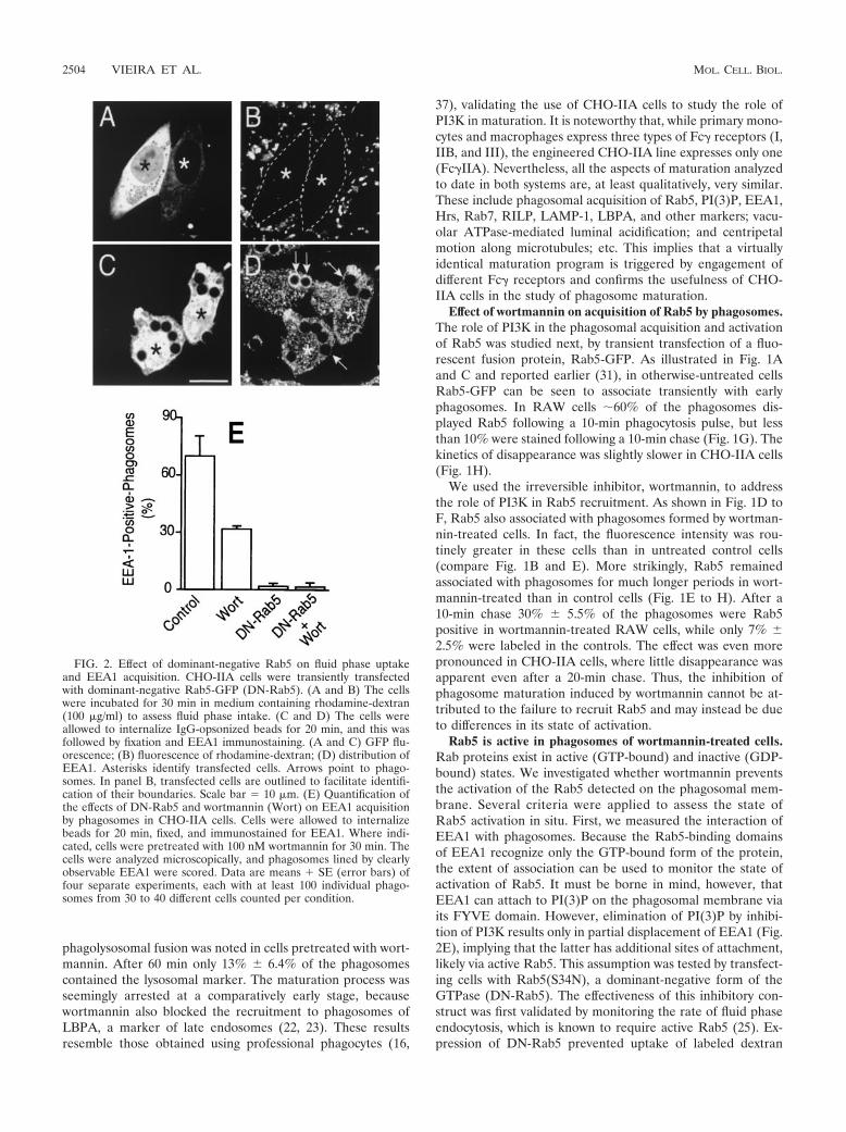
FIG. 2. Effect of dominant-negative Rab5 on fluid phase uptakeand EEA1 acquisition. CHO-IIA cells were transiently transfectedwith dominant-negative Rab5-GFP (DN-Rab5). (A and B) The cellswere incubated for 30 min in medium containing rhodamine-dextran(100 �g/ml) to assess fluid phase intake. (C and D) The cells wereallowed to internalize IgG-opsonized beads for 20 min, and this wasfollowed by fixation and EEA1 immunostaining. (A and C) GFP flu-orescence; (B) fluorescence of rhodamine-dextran; (D) distribution ofEEA1. Asterisks identify transfected cells. Arrows point to phago-somes. In panel B, transfected cells are outlined to facilitate identifi-cation of their boundaries. Scale bar � 10 �m. (E) Quantification ofthe effects of DN-Rab5 and wortmannin (Wort) on EEA1 acquisitionby phagosomes in CHO-IIA cells. Cells were allowed to internalizebeads for 20 min, fixed, and immunostained for EEA1. Where indi-cated, cells were pretreated with 100 nM wortmannin for 30 min. Thecells were analyzed microscopically, and phagosomes lined by clearlyobservable EEA1 were scored. Data are means � SE (error bars) offour separate experiments, each with at least 100 individual phago-somes from 30 to 40 different cells counted per condition.
2504 VIEIRA ET AL. MOL. CELL. BIOL.

(Fig. 2A and B) yet had no discernible effects on phagocyticefficiency; in four experiments the phagocytic index was statis-tically indistinguishable in control and DN-Rab5 expressingcells (362 � 41 and 359 � 35 particles/100 cells, respectively).More importantly, the dominant-negative Rab5 also elimi-nated the wortmannin-resistant fraction of EEA1 binding tophagosomes (Fig. 2C, D, and E), implying that this componentis mediated by the active, GTP-bound form of Rab5. Similareffects were noted 10 and 20 min after phagocytosis. The ef-fects of DN-Rab5 could be studied only in CHO-IIA cells,because the low levels of expression attained in RAW cellswere insufficient to prevent fusion of endosomes with phago-somes or the clathrin-mediated endocytosis of aggregated IgG,
which is blocked by the dominant-negative construct in CHO-IIA cells (not illustrated). These findings stress the usefulnessof CHO-IIA in instances where high levels of ectopic expres-sion of a protein are required.
The ability of wortmannin-treated phagosomes to bindEEA1 in a DN-Rab5-sensitive manner suggests that Rab5 is atleast partly active in cells treated with the inhibitor. In anattempt to confirm this notion, we measured the ability ofphagosomes to fuse with early endosomes in control and wort-mannin-treated cells. Because Rab5 is thought to mediate fu-sion of phagosomes with early endosomes (2, 20), we reasonedthat delivery to the phagosomal lumen of a marker trapped inearly endosomes could be used as an index of Rab5 activity.
FIG. 3. Effect of wortmannin on fusion of nascent phagosomes with early endosomes. (A to F) CHO-IIA cells that were untreated (A to C)or pretreated with wortmannin (D to F) were allowed to internalize IgG-opsonized latex beads for 20 min. After washing excess beads, the cellswere incubated briefly on ice with anti-human antibody conjugated with fluorescein isothiocyanate to identify adherent, incompletely internalizedbeads (B and E). The cells were then incubated for an additional 10 min at 37°C with FM4-64.The red fluorescence of FM4-64 is shown in A andD (arrows). C and F are the differential interference contrast images corresponding to A and D, respectively. (G to I) CHO-IIA cells transientlytransfected with syntaxin 13-GFP were treated with wortmannin (H and black columns in I) or left untreated (G and empty columns in I) andallowed to internalize particles for 20 min. Adherent, incompletely internalized beads were then stained as above using Texas red-conjugatedantibodies. Lastly, the cells were chased for the indicated times and fixed, and confocal fluorescence microscopy was used to visualize thedistribution of syntaxin 13. (G and H) representative fluorescence images, with corresponding differential interference contrast insets. Arrows pointto phagosomes. Scale bars �10 �m. (I) Quantitation of the effect of wortmannin on syntaxin 13 acquisition by phagosomes. Data are means �SE (error bars) of three separate experiments, each with at least 100 individual phagosomes from 30 to 50 different cells counted.
VOL. 23, 2003 Rab5 AND Rab7 IN PHAGOSOMES 2505

Such experiments are illustrated in Fig. 3. The cells were ini-tially allowed to internalize opsonized latex beads for 20 min.Next, beads that were bound but not yet internalized wereidentified by addition of anti-IgG at 4°C (e.g., Fig. 3B and E).This step was essential to differentiate delivery of endosomalmarkers to a preformed phagosome, from merely trapping thefluid-phase marker during phagosome formation. Then, earlyendosomes were loaded with FM4-64 and fusion of newlyformed endosomes with preformed phagosomes was moni-tored by confocal microscopy for the next 10 min. Fusion ofearly endosomes with phagosomes, revealed by the appearanceof a rim of FM4-64 fluorescence around the phagocytic parti-cle, was readily detectable in control cells (Fig. 3A to C) butalso in wortmannin-treated cells (Fig. 3D to F). Similar resultswere obtained using RAW cells (not illustrated). That fusionof phagosomes with early endosomes persists after treatmentwith wortmannin was also demonstrated by monitoring thedistribution of the early endosomal marker syntaxin 13 (28).Cells were transfected with a GFP fusion of this early endo-somal SNARE and analyzed after ingestion of opsonizedbeads. As illustrated in Fig. 3G and H and summarized in Fig.3I, endosomes containing syntaxin 13 merged with phagosomesto a similar extent and comparable kinetics in both control andwortmannin-treated cells. Very similar results were obtainedwith RAW cells (not shown). Together, these results suggestthat fusion-competent, presumably GTP-bound Rab5 is pres-ent on the membrane of wortmannin-treated phagosomes.
FRAP was used as a final criterion to assess the state ofactivation of Rab5 on wortmannin-treated phagosomes. Be-cause the rate of dissociation of Rab GTPases from mem-branes is thought to be determined by the conversion of theGTP-bound to the GDP-bound forms, measurements of therate of Rab5 exchange from the phagosome should provide anindication of its activation state. Dissociation rates can beinferred from the rate of fluorescence recovery after an or-ganelle expressing a labeled protein has been photobleached.This approach was recently used successfully to assess the
effect of RILP on the activation state of Rab7 (21). We there-fore compared the rate and extent of FRAP of Rab5 in phago-somes and endosomes. The latter were used to validate thesensitivity of the assay. Cells were transfected with GFP fusionsof either wild-type Rab5-GFP or Rab5(Q79L), a constitutivelyactive mutant (CA-Rab5). Typical FRAP measurements inendosomes are illustrated in Fig. 4A. Following bleaching ofwild-type Rab5, recovery was fast (half-life � 5.2 s) and nearlycomplete (fractional recovery � 92.3% � 4%; see Fig. 4A).This implies that most of the endosomal Rab5 turns overrapidly, dissociating when GTP is hydrolyzed and reassociatingwhen GDP is exchanged for GTP. Accordingly, when consti-tutively active Rab5-GFP was expressed, recovery after photo-bleaching was very poor. Only 35.8% � 4% recovery wasachieved after 200 s, the longest time studied. Having validatedthe use of FRAP, we proceeded to analyze the behavior ofphagosomes treated with wortmannin (normal phagosomescould not be studied because the short time of residence ofwild-type Rab5 in untreated cells precludes FRAP studies). Asshown in Fig. 4B, the recovery of fluorescence was poor inwortmannin-treated phagosomes (48.4% � 5%), implying thatRab5 is largely in the active state. In accordance with thisinterpretation, the extent of the recovery was only slightlygreater than that of constitutively active Rab5 (32.5% � 3%).In both instances, part of the recovery is likely due to lateraldiffusion of a fraction of fluorescent Rab5 that remains un-bleached at the poles of the phagosome, which lie outside thefocal plane of the bleaching laser. Under the conditions used,a depth of only about 1.8 �m, less than the diameter of thephagosome, was effectively bleached (unpublished observa-tions). Of interest, treatment with wortmannin also greatlyreduced the fraction of endosomal Rab5 that recovered afterphotobleaching (Fig. 4A), implying that products of PI3K arealso required for deactivation of this GTPase in endosomes.
Jointly, the EEA1, FM4-64, syntaxin 13, and FRAP experi-ments indicate that Rab5 is largely in the active state in phago-somes formed by wortmannin-treated cells.
FIG. 4. Rab5-GFP FRAP. CHO-IIA cells were transfected with either wild-type (WT) or constitutively active (CA) Rab5-GFP. Whereindicated, the cells had been pretreated with wortmannin (Wort). (A) Labeled endosomes were identified and their initial fluorescence quantifiedand defined as 100%. Bleaching was then performed and fluorescence recovery monitored as described in Materials and Methods. (B) Cellstransfected with WT-Rab5 were treated with wortmannin, while cells transfected with CA-Rab5 were left untreated. The cells were then allowedto ingest opsonized beads, fluorescent phagosomes were identified, and FRAP was measured as above. Representative experiments are shown inthe main panels. The inset tables show the corresponding times for 50% recovery (T1/2) and the fractional recovery (R). Data in the tables aremeans � SE (error bars) of 8 to 10 determinations, each involving 10 different cells.
2506 VIEIRA ET AL. MOL. CELL. BIOL.

Overexpression of DN-Rab5 arrests phagosome maturation.While earlier evidence had demonstrated that wortmanninblocks phagolysosome formation (16, 37) (see above), the ex-periments summarized above showed that Rab5 is active in themembrane of phagosomes treated with the PI3K inhibitor.Two explanations may account for these observations: (i) thePI3K-dependent step may lie downstream or on an arm of the
fusogenic pathway that is parallel to that involving Rab5, or (ii)Rab5 may not be part of the pathway required for phagosomefusion with lysosomes. While Rab5 is known to be an essentialcomponent of progression along the endocytic pathway (5; fora review, see reference 39), the role of Rab5 in phagosomematuration has not been addressed directly.
The requirement for Rab5 in phagosome maturation wastested by overexpression of DN-Rab5. CHO-IIA cells wereused for these studies, to attain higher levels of the inhibitoryprotein. As shown in Fig. 5A and B incorporation of LAMP-1,a late endosomal/lysosomal marker, into the phagosomalmembrane was greatly reduced by expression of DN-Rab5. In3 experiments, inhibition averaged 72% � 3.9% (Fig. 5E).
Rab7 associates with phagosomes following acquisition ofRab5 and, by analogy with the endosomal pathway, is likelyinvolved in directing fusion with lysosomes. We therefore an-ticipated that recruitment of Rab7 by phagosomes would re-quire the prior activation of Rab5. This prediction was verifiedin the experiments summarized in Fig. 6. Cells were cotrans-fected with myc epitope-tagged Rab7 and either GFP or DN-Rab5-GFP. Rab7 association with late phagosomes was readilydetectable in cells transfected with GFP (Fig. 6B), while thoseexpressing the inhibitory Rab5 had much reduced staining(Fig. 6E). In four separate experiments, Rab7 recruitment wasdepressed by �66% at all the times analyzed (Fig. 6G). There-fore, Rab5 seems to be essential for normal phagosome mat-uration.
Overexpression of a Rab5-GTPase activating protein ar-rests phagosome maturation. The role of Rab5 in maturationwas confirmed using RN-tre, a Rab5 GTPase-activating pro-tein (GAP) (24). Overexpression of a GTPase activator isexpected to minimize the ability of Rab5-GTP to interact withits effectors. CHO-IIA cells were transfected with RN-tre-GFPand successful expression was verified by direct fluorescencemicroscopy (Fig. 5C). Fluid phase pinocytosis was depressed incells expressing RN-tre, implying that the concentrations at-tained were sufficient to inhibit Rab5 (not illustrated). Expres-sion of RN-tre-GFP did not affect the phagocytic efficiencyand the GTPase was detectable on the membrane of nascentphagosomes. Importantly, overexpression of RN-tre-GFPprofoundly depressed the ability of phagosomes to acquireLAMP-1 (Fig. 5D and E). The extent of inhibition was similarto that observed in DN-Rab5 transfected cells (Fig. 5E).Jointly, these findings imply that active Rab5 is required forfusion of phagosomes with late endosomes and/or lysosomes.
Effect of wortmannin on Rab7 acquisition. As Rab5 acqui-sition and activation are not dependent on the presence ofPI(3)P on the phagosome, we next determined whether therecruitment of Rab7 to the phagosome is affected by wortman-nin. RAW and CHO-IIA cells were transiently transfected witha construct encoding Rab7-GFP. When appropriate levels ofthe fusion protein were expressed, the cells were either treatedwith wortmannin or left untreated and next challenged withopsonized beads. The presence of the fluorescent chimera onphagosomes was monitored by confocal microscopy. Rab7could be readily detected on the membrane of control phago-somes (Fig. 7A). In CHO-IIA cells 79% � 6.4% of the phago-somes were Rab7-positive 20 min after initiation of ingestionand this number increased, approaching 100% over the next 30min (Fig. 7E). Very similar results were obtained in RAW cells
FIG. 5. Role of Rab5 in fusion of phagosomes with late endocyticcompartments. CHO-IIA cells were transfected with DN-Rab5-GFP(A, B and black column in E) or with RN-tre-GFP (C, D, and graycolumn in E). Transfected cells (labeled by asterisks) are identifiableby their green fluorescence (A and D). The cells were allowed tointernalize opsonized beads for 20 min. After washing unbound beads,incubation at 37°C proceeded for 30 more min. The cells were thenfixed, permeabilized with methanol at �20°C, and stained for LAMP-1(B, D, and E). Arrows and arrowheads point to LAMP-1-positive and-negative phagosomes, respectively. Scale bars � 10 �m. (E) Quanti-fication of the effect of DN-Rab5 (black column) and RN-tre (graycolumn) overexpression on LAMP-1 acquisition by phagosomes, com-pared to untransfected control cells (open bar). Results shown aremeans � SE (error bars) of three separate experiments, each with atleast 100 phagosomes from 20 to 40 different cells counted.
VOL. 23, 2003 Rab5 AND Rab7 IN PHAGOSOMES 2507

(Fig. 7F). As illustrated in Fig. 8C and summarized in Fig. 7Eand F (black columns), wortmannin treatment attenuated, butdid not prevent, the acquisition of Rab7 by the phagosome.Over 50% of phagosomes were Rab7 positive immediatelyafter the ingestion pulse (a 40% inhibition compared to con-trol), and this fraction changed only marginally thereafter inboth CHO-IIA (Fig. 7E) and RAW cells (Fig. 7F).
PI3K activity is not necessary for Rab7 activation. The mod-est decrease in Rab7 recruitment documented in Fig. 7 cannotfully explain the much greater (�90%) inhibition of lysosomal
fusion produced by wortmannin. Two possible mechanismscould explain this quantitative discrepancy: products of PI3Kmay be required for Rab7 activation, independently of its re-cruitment, or alternatively, the kinase may be required for aseparate event that, in parallel with Rab7, may be essential forsuccessful phagolysosome formation.
To differentiate these hypotheses, we tested the state ofactivation of Rab7 in cells treated with wortmannin. Membersof the Rab family bind their effectors only when bound to GTP,i.e., in their active configuration. We therefore determined
FIG. 6. Effect of dominant-negative Rab5 on Rab7 acquisition. CHO-IIA cells were cotransfected with wild-type Rab7-myc and either GFP (Ato C and empty columns in G) or with DN-Rab5-GFP (D to F). The cells were incubated for 20 min with IgG-opsonized beads, chased for theindicated times, and then fixed, permeabilized, and immunostained. (A and D) Green fluorescence; (B and E) distribution of Rab7, revealed byimmunostaining the myc epitope; (C and F) corresponding differential interference contrast images. Arrows and arrowheads point to Rab7-mycpositive and -negative phagosomes, respectively. Scale bars � 10 �m. (G) quantification of the effect of DN-Rab5 on Rab7 recruitment byphagosomes, from experiments like those in panels A to F. Empty columns, control; black columns, cells transfected with DN-Rab5-GFP. Resultsshown are means � SE (error bars) of four separate experiments, each with at least 100 individual phagosomes counted per condition in cells withhigh expression levels of DN-Rab5.
2508 VIEIRA ET AL. MOL. CELL. BIOL.

whether Rab7 is capable of recruiting effectors following treat-ment with the PI3K inhibitor. To date, only one effector ofRab7, termed RILP, has been identified (8, 21). Experimentsanalyzing the distribution of RILP are depicted in Fig. 8.CHO-IIA and RAW cells were cotransfected with Rab7-GFPand with an epitope-tagged form of RILP and their distribu-tion was monitored by confocal microscopy. As reported (8,21), RILP accumulated in a juxtanuclear complex, near themicrotubule-organizing center. In addition, in cells that hadingested particles but were otherwise untreated, RILP wasfound in the vast majority of phagosomes that contained Rab7(Fig. 8A to C). Of note, RILP was also found in most of the
Rab7-positive phagosomes in wortmannin-treated cells (Fig.8D to F). This observation implies that PI3K is not essential forthe activation of Rab7 and that the large fraction of Rab7recruited to phagosomes in wortmannin-treated cells is capa-ble of binding effectors.
The preceding data imply that the site of action of wortman-nin is at a step that occurs in parallel with, or after activationof Rab7. This conclusion was validated using Rab7(Q67L), aGTPase-deficient and therefore constitutively active form ofRab7 (CA-Rab7). We predicted that if wortmannin preventsphagolysosome formation by interfering with the activation ofRab7, expression of the constitutively active mutant would
FIG. 7. Effect of wortmannin on Rab7 acquisition by phagosomes. CHO-IIA cells (A to E) or RAW cells (F) were transfected with wild-typeRab7-GFP. The cells were either left untreated (A, B, and empty columns in E and F) or were treated with 100 nM wortmannin for 30 min (C, D,and black columns in E and F). The cells were allowed to internalize IgG-opsonized beads and the distribution of Rab7 was monitored by confocalmicroscopy at the indicated times. (A and C) Phagocytosis was for 20 min, without chase. The arrows and arrowheads point to Rab7-GFP-positiveand -negative phagosomes, respectively. (B and D) Corresponding differential interference contrast images. Scale bars � 10 �m. (E and F)Quantification of the effect of wortmannin on Rab7-GFP acquisition by phagosomes in CHO-IIA (E) and RAW cells (F). Empty columns, control;black columns, cells treated with wortmannin. Data are means � SE (error bars) of four separate experiments, each with at least 100 individualphagosomes from 30 to 40 different cells expressing low levels of Rab7-GFP counted per condition.
VOL. 23, 2003 Rab5 AND Rab7 IN PHAGOSOMES 2509

bypass the inhibition. Conversely transfection of CA-Rab7would be without effect if PI3K acts after or in parallel withRab7. The experiments addressing these alternatives are illus-trated in Fig. 9. The CA-Rab7 was found to associate withphagosomes in both control and, to a slightly lesser extent inwortmannin-treated cells (Fig. 9A and D). Expression of theactive mutant did not interfere with the recruitment ofLAMP-1 by phagosomes in control cells (Fig. 9B) and, impor-tantly, it failed to overcome the inhibitory effects of wortman-nin on phagolysosome formation. As shown in Fig. 9G,expression of CA-Rab7 had no detectable effect on LAMP-1acquisition after 20 and 40 min and produced a marginal stim-ulation at 60 min. Importantly, similar results were obtainedusing RAW macrophages (not shown). These findings confirmthe notion that PI3K activity is required for a step(s) otherthan Rab7 activation.
DISCUSSION
Treatment of macrophages with gamma interferon wasfound to induce the biosynthesis of Rab5 and, in parallel, toaccelerate the intracellular killing of pathogens like Listeriamonocytogenes (3). In addition, Duclos and colleagues (15)reported that heterologous expression of CA-Rab5 resulted inthe formation of enlarged phagosomes. On the basis of theseobservations, and by analogy with the endosomal pathway, arole for Rab5 in phagosome maturation had been suspected,but it was not demonstrated. Here we present direct evidencethat active Rab5 is required for successful phagolysosome for-mation (Fig. 5). Association of active Rab5 with phagosomes
precedes and is essential for the recruitment of Rab7, which inturn precedes and is required for the fusion of phagosomeswith late endocytic compartments (R. E. Harrison and S. Grin-stein, unpublished observations). While sequential recruitmentof active Rab5 and Rab7 to the phagosome is necessary, it isnot sufficient for completion of maturation. This is best illus-trated in cells treated with wortmannin, where phagosomescontain increased amounts of active Rab5 (Fig. 1) and upwardsof 60% of the normal complement of active Rab7 (Fig. 7)yet fail to fuse with late endosomes and lysosomes to a signif-icant degree. Clearly, one or more additional PI3K-dependentevents are also required for successful generation of phagoly-sosomes.
The experiments reported here indicate that treatment withwortmannin prevented neither the recruitment of Rab5 nor itsactivation on the phagosomal membrane (Fig. 1 to 4), while itcompletely eliminated PI(3)P from the phagosomal mem-brane, as detected using FYVE or PX domain chimeras (notillustrated). This implies that class I or III PI3K does notprovide the signal that targets Rab5 to the phagosome. In fact,the amount of Rab5 detectable on phagosomes was greater inwortmannin-treated cells than in controls, and its dwelling timeon the phagosomal membrane was prolonged. Three mecha-nisms could account for these observations. (i) The activity oravailability of Rab5 anchors and activators on the phagosomalmembrane may increase upon inhibition of PI3K. This mech-anism, however, is not consistent with the FRAP determina-tions, which indicate that the primary cause of accumulationis reduced dissociation, rather than increased association of
FIG. 8. RILP recruitment to phagosomes. CHO-IIA cells were cotransfected with wild-type Rab7-GFP and RILP-HA. (D to F) Cells weretreated with wortmannin 30 min before phagocytosis. Phagocytosis was allowed to proceed for 20 min, and this was followed by a 30-min chase.The cells were then fixed and immunostained for the HA epitope. (A and D) Distribution of Rab7-GFP; (B and E) the distribution of RILP-HA;(C and F) corresponding differential interference contrast images. The arrows point to phagosomes positive for both Rab7 and RILP. Asterisksidentify transfected cells. Bars � 10 �m. Results are representative of four similar experiments, each with �30 cells counted.
2510 VIEIRA ET AL. MOL. CELL. BIOL.

Rab5 with the membrane. (ii) PI3K may be required for theactivation and/or recruitment of Rab5 GAPs to the phago-some. At least three types of Rab5-GAPs have been described:tuberin (38), Ras-GAP (27), and RN-tre (24). Of these, Ras-GAP was reported to be directly inhibited by wortmannin by aprocess that is independent of PI3K (10). While this proposedmechanism would explain many of our data, we found that incells treated with the chemically unrelated PI3K inhibitor
LY294002, Rab5 accumulates in phagosomes in a manner thatclosely mimics the effects of wortmannin (unpublished obser-vations). We therefore believe that depletion of products ofPI3K, rather than direct inhibition of Ras-GAP, is responsiblefor the altered behavior of Rab5. Of the GAPs listed above,only RN-tre is specific towards Rab5 and therefore of thegreatest interest. RN-tre was found to localize primarily to theplasma membrane and to reside in phagosomes only briefly
FIG. 9. Effect of constitutively active Rab7 on LAMP-1 acquisition by phagosomes. CHO-IIA cells were transfected with CA-Rab7-GFP andeither left untreated (A to C and empty columns in G) or treated with wortmannin (D to F and black columns in G) prior to phagocytosis. After20 min of phagocytosis the unbound particles were removed and the cells were incubated for 30 more min to allow phagosome maturation. Thecells were then fixed, permeabilized, and immunostained with anti-LAMP-1 antibodies. (A and D) Distribution of CA-Rab7-GFP; (B and E)distribution of LAMP-1. Arrows and arrowheads point to phagosomes that were positive or negative, respectively, for Rab7 or LAMP-1, asappropriate. Asterisks identify transfected cell. Scale bars � 10 �m. (C and F) Corresponding differential interference contrast images. (G) Quan-titation of LAMP-1 acquisition by phagosomes in cells treated with wortmannin. The graph compares cells that were untransfected (black bars)with CA-Rab7-GFP-transfected cells (open bars). Data are means � SE (error bars) of four separate experiments, each with at least 100 individualphagosomes from 20 to 40 different cells counted per condition.
VOL. 23, 2003 Rab5 AND Rab7 IN PHAGOSOMES 2511
(less than 5 min). The temporal pattern of association of RN-tre with the phagosome was not visibly altered by treatment ofthe cells with wortmannin (O. V. Vieira, unpublished observa-tions). Hence, PI3K is not required for its targeting or main-tenance in phagosomes. Alternatively, other GAPs or Rab-GDI (guanine nucleotide dissociation inhibitor) may accountfor the protracted activation of Rab5 in wortmannin-treatedcells; (iii) The removal of Rab5 from the phagosome may occurby fission of vesicles containing the GTPase, rather than by thecanonical nucleotide hydrolysis and solubilization of the pro-tein. In this scenario, PI3K may be required for fission to occurand its inhibition would lead to Rab5 accumulation.
Products of PI3K are also required to recruit and/or activateeffectors of Rab5, such as EEA1 and rabenosyn 5 (29, 30, 34).It is likely that impairment of one or more of these effectorscontributes to the inability of the early phagosome to fuse withlate endosomes and phagosomes. Regardless of the mode ofaction of wortmannin on Rab5, our data suggest that the roleof PI3K is not limited to this step of the maturation cascade.Because association of Rab7 follows that of Rab5, and becauseinhibition of Rab7 impairs maturation, we suspected that re-cruitment of Rab7 was the alternate target of PI3K. However,we found that Rab7 acquisition by phagosomes was only par-
tially (�40%) diminished by wortmannin (Fig. 7). We specu-late that phagosomal Rab7 is acquired from 2 sources: one thatis independent of PI3K and may be the cytosolic pool or anunidentified wortmannin-insensitive vesicular pool (purpleoval in Fig. 10), while another wave of Rab7 may merge withthe phagosomes when the latter fuse with late endosomes orlysosomes, which are well established to carry the GTPase. Tothe extent that fusion is precluded by wortmannin, the lattercomponent would be lacking in phagosomes treated with theinhibitor.
The sizable fraction of Rab7 that associates with the phago-some in wortmannin-treated cells appears to be functionallyactive, as indicated by its ability to recruit RILP, an effectorthought to interact only with the GTP-bound form of Rab7 (8).It would therefore appear that active Rab7 is not, per se,sufficient to mediate fusion of the phagosomes with late endo-somes and lysosomes. Accordingly, the expression of CA-Rab7failed to bypass the block exerted by wortmannin (Fig. 9). Onecould conclude from these observations that wortmannin iseffecting the block of maturation downstream of Rab7. How-ever, it is equally possible that two parallel inputs are requiredto trigger proper fusion of endosomes or lysosomes with thephagosome: one mediated by Rab7 and another that includes
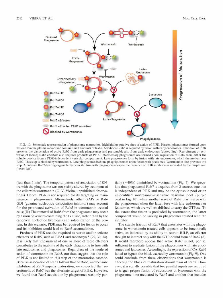
FIG. 10. Schematic representation of phagosome maturation, highlighting putative sites of action of PI3K. Nascent phagosomes formed uponfission from the plasma membrane contain small amounts of Rab5. Additional Rab5 is acquired by fusion with early endosomes. Inhibition of PI3Kprevents the dissociation of active Rab5 from early phagosomes and presumably also from early endosomes (dotted line). Recruitment or acti-vation of (some) Rab5 effectors also requires products of PI3K. Intermediary phagosomes are formed upon acquisition of Rab7 from either thesoluble pool or from a PI3K-independent vesicular compartment. Late phagosomes form by fusion with late endosomes, which themselves bearRab7. This step is blocked by wortmannin. Late phagosomes become phagolysosomes upon fusion with lysosomes. Wortmannin also prevents thisstep. A putative Rab7-bearing organelle that can still fuse with phagosomes despite the presence of PI3K inhibitors is indicated by the purple oval(lower left).
2512 VIEIRA ET AL. MOL. CELL. BIOL.

the PI3K-dependent event, likely the activation of Rab5 effec-tors, yet does not involve Rab7. Because RILP was recentlyshown to bridge between dynein and Rab7-containing cargo, itis possible that the Rab7 arm is involved in mechanically po-sitioning phagosomes in the vicinity of late endosomes/lyso-somes, while another, PI3K dependent step is required for theactual fusion event. In this context, it is noteworthy that, atleast in yeast, the association of SNAREs like Vam7 withmembranes of the endocytic pathway requires interaction withPI(3)P, a product of class III PI3K (9). Moreover, SNAREscan associate with activated Rab5 in mammalian systems (28).These concepts are synthesized in diagrammatic form in Fig.10. Nascent phagosomes are shown to contain some Rab5,present originally in the invaginating plasma membrane and/orrecruited there by activated Fc receptors. Additional Rab5 isacquired by fusion with early endosomes, but mostly from thesoluble pool, since experiments using Rab5-GFP showed rapidgain of phagosomal fluorescence that was not accountable byfusion with labeled vesicular compartments (31). Wortmanninis shown to interfere with the dissociation of the active (GTP-bound) form of Rab5 from early phagosomes and presumablyalso from early endosomes. Rab5 is shown in the diagram torecruit two types of effectors: some that are PI3K independentand others that require PI3K products for recruitment and/oractivation. Rab5-enriched early phagosomes then proceed tobecome intermediary phagosomes by shedding Rab5 and ac-quiring Rab7. We propose that the PI3K-independent effectorsof Rab5 take part in the recruitment of Rab7. Rab7 is recruitedinitially from the cytosol and/or from an unidentified vesicularcompartment distinct from late endosomes and lysosomes.Next, the Rab7-enriched phagosome fuses with late endo-somes by a 3-phosphoinositide-dependent process, perhapsinvolving Rab5 effectors, generating late phagosomes. These,in turn coalesce with lysosomes by a process that also requiresPI3K. It is not clear whether this is a distinct PI3K-dependentstep, or the manifestation of the inability of the phagosome toreach the late stage, which may be essential for fusion withlysosomes. Merger with late endosomes and lysosomes deliversto the phagosome additional Rab7 that is constitutively presentin these organelles. The latter component of Rab7 recruitmentis therefore wortmannin sensitive, accounting for the partialeffects of the inhibitor. The ultimate stage of the process is theformation of phagolysosomes, which have optimal microbicidalproperties. An essential feature of this model is the paralleloperation of two types of effectors, some that are PI3K depen-dent and others that are independent.
ACKNOWLEDGMENTS
We thank Pier Paolo di Fiore for his advice and for generouslysharing his reagents.
This work was supported by the Canadian Institutes of Health Re-search, the Canadian Arthritis Society and the National SanatoriumAssociation. O. V. Vieira is the recipient of a postdoctoral fellowshipfrom PRAXIS XX1 (BPD/22039/99). S.G. is supported in part by thePitblado Chair in Cell Biology.
REFERENCES
1. Aderem, A., and D. M. Underhill. 1999. Mechanisms of phagocytosis inmacrophages. Annu. Rev. Immunol. 17:593–623.
2. Alvarez-Dominguez, C., A. M. Barbieri, W. Beron, A. Wandinger-Ness, andP. D. Stahl. 1996. Phagocytosed live. Listeria monocytogenes influencesRab5-regulated in vitro phagosome-endosome fusion. J. Biol. Chem. 271:13834–13843.
3. Alvarez-Dominguez, C., and P. D. Stahl. 1998. Interferon-gamma selectivelyinduces Rab5a synthesis and processing in mononuclear cells. J. Biol. Chem.273:33901–33904.
4. Araki, N., M. T. Johnson, and J. A. Swanson. 1996. A role for phosphoino-sitide 3-kinase in the completion of macropinocytosis and phagocytosis bymacrophages. J. Cell Biol. 135:1249–1260.
5. Bucci, C., R. G. Parton, I. H. Mather, H. Stunnenberg, K. Simons, B.Hoflack, and M. Zerial. 1992. The small, GTPase rab5 functions as a regu-latory factor in the early endocytic pathway. Cell 70:715–728.
6. Bucci, C., P. Thomsen, P. Nicoziani, J. McCarthy, and B. van Deurs. 2000.Rab7: a key to lysosome biogenesis. Mol. Biol. Cell 11:467–480.
7. Callaghan, J., A. Simonsen, J. M. Gaullier, B. H. Toh, and H. Stenmark.1999. The endosome fusion regulator early-endosomal autoantigen 1 (EEA1)is a dimer. Biochem J. 338:539–543.
8. Cantalupo, G., P. Alifano, V. Roberti, C. B. Bruni, and C. Bucci. 2001.Rab-interacting lysosomal protein (RILP): the Rab7 effector required fortransport to lysosomes. EMBO J. 20:683–693.
9. Cheever, M. L., T. K. Sato, T. de Beer, T. G. Kutateladze, S. D. Emr, and M.Overduin. 2001. Phox domain interaction with PtdIns(3)P targets the Vam7t-SNARE to vacuole membranes. Nat. Cell Biol. 3:613–618.
10. Chen, X., and Z. Wang. 2001. Regulation of epidermal growth factor recep-tor endocytosis by wortmannin through activation of Rab5 rather than inhi-bition of phosphatidylinositol 3-kinase. EMBO Rep. 2:842–849.
10a.Collins, R. F., A. D. Schreiber, S. Grinstein, and W. S. Trimble. 2002.Syntaxins 13 and 7 function at distinct steps during phagocytosis. J. Immunol.169:3250–3256.
11. Cox, D., C. C. Tseng, G. Bjekic, and S. Greenberg. 1999. A requirement forphosphatidylinositol 3-kinase in pseudopod extension. J. Biol. Chem. 274:1240–1247.
12. Desjardins, M., J. E. Celis, G. van Meer, H. Dieplinger, A. Jahraus, G.Griffiths, and L. A. Huber. 1994. Molecular characterization of phagosomes.J. Biol. Chem. 269:32194–32200.
13. Desjardins, M., L. A. Huber, R. G. Parton, and G. Griffiths. 1994. Biogenesisof phagolysosomes proceeds through a sequential series of interactions withthe endocytic apparatus. J. Cell Biol. 124:677–688.
14. Downey, G. P., R. J. Botelho, J. R. Butler, Y. Moltyaner, P. Chien, A. D.Schreiber, and S. Grinstein. 1999. Phagosomal maturation, acidification, andinhibition of bacterial growth in nonphagocytic cells transfected withFc�RIIA receptors. J. Biol. Chem. 274:28436–28444.
15. Duclos, S., R. Diez, J. Garin, B. Papadopoulou, A. Descoteaux, H. Stenmark,and M. Desjardins. 2000. Rab5 regulates the kiss and run fusion betweenphagosomes and endosomes and the acquisition of phagosome leishmani-cidal properties in RAW 264.7 macrophages. J Cell Sci. 113:3531–3541.
16. Fratti, R. A., J. M. Backer, J. Gruenberg, S. Corvera, and V. Deretic. 2001.Role of phosphatidylinositol 3-kinase and Rab5 effectors in phagosomalbiogenesis and mycobacterial phagosome maturation arrest. J. Cell Biol.154:631–644.
17. Garin, J., R. Diez, S. Kieffer, J. F. Dermine, S. Duclos, E. Gagnon, R. Sadoul,C. Rondeau, and M. Desjardins. 2001. The phagosome proteome: insightinto phagosome functions. J. Cell Biol. 152:165–180.
18. Gorvel, J. P., P. Chavrier, M. Zerial, and J. Gruenberg. 1991. rab5 controlsearly endosome fusion in vitro. Cell 64:915–925.
19. Indik, Z. K., J. G. Park, S. Hunter, and A. D. Schreiber. 1995. The moleculardissection of Fc gamma receptor mediated phagocytosis. Blood 86:4389–4399.
20. Jahraus, A., T. E. Tjelle, T. Berg, A. Habermann, B. Storrie, O. Ullrich, andG. Griffiths. 1998. In vitro fusion of phagosomes with different endocyticorganelles from J774 macrophages. J. Biol. Chem. 273:30379–30390.
21. Jordens, I., M. Fernandez-Borja, M. Marsman, S. Dusseljee, L. Janssen, J.Calafat, H. Janssen, R. Wubbolts, and J. Neefjes. 2001. The Rab7 effectorprotein RILP controls lysosomal transport by inducing the recruitment ofdynein-dynactin motors. Curr. Biol. 11:1680–1685.
22. Kobayashi, T., F. Gu, and J. Gruenberg. 1998. Lipids, lipid domains andlipid-protein interactions in endocytic membrane traffic. Semin. Cell. Dev.Biol. 9:517–526.
23. Kobayashi, T., M. H. Beuchat, M. Lindsay, S. Frias, R. D. Palmiter, H.Sakuraba, R. G. Parton, and J. Gruenberg. 1999. Late endosomal mem-branes rich in lysobisphosphatidic acid regulate cholesterol transport. Nat.Cell Biol. 1:113–118.
24. Lanzetti, L., V. Rybin, M. G. Malabarba, S. Christoforidis, G. Scita, M.Zerial, and P. P. Di Fiore. 2000. The Eps8 protein coordinates EGF receptorsignalling through. Rac and trafficking through Rab5. Nature 408:374–377.
25. Li, G., C. D’Souza-Schorey, M. A. Barbieri, J. A. Cooper, and P. D. Stahl.1997. Uncoupling of membrane ruffling and pinocytosis during Ras signaltransduction. J. Biol. Chem. 272:10337–10340.
26. Li, G., M. A. Barbieri, M. I. Colombo, and P. D. Stahl. 1994. Structuralfeatures of the GTP-binding defective Rab5 mutants required for theirinhibitory activity on endocytosis. J. Biol. Chem. 269:14631–14635.
27. Liu, K., and G. Li. 1998. Catalytic domain of the p120 Ras GAP binds toRAb5 and stimulates its GTPase activity. J. Biol. Chem. 273:10087–10090.
28. McBride, H. M., V. Rybin, C. Murphy, A. Giner, R. Teasdale, and M. Zerial.1999. Oligomeric complexes link Rab5 effectors with NSF and drive mem-
VOL. 23, 2003 Rab5 AND Rab7 IN PHAGOSOMES 2513

brane fusion via interactions between EEA1 and syntaxin 13. Cell 98:377–386.
29. Nielsen, E., S. Christoforidis, S. Uttenweiler-Joseph, M. Miaczynska, F.Dewitte, M. Wilm, B. Hoflack, and M. Zerial. 2000. Rabenosyn-5, a novelRab5 effector, is complexed with hVPS45 and recruited to endosomesthrough a FYVE finger domain. J Cell Biol. 151:601–612.
30. Patki, V., J. Virbasius, W. S. Lane, B. H. Toh, H. S. Shpetner, and S.Corvera. 1997. Identification of an early endosomal protein regulated byphosphatidylinositol 3-kinase. Proc. Natl. Acad. Sci. USA 94:7326–7330.
31. Roberts, R. L., M. A. Barbieri, J. Ullrich, and P. D. Stahl. 2000. Dynamics ofrab5 activation in endocytosis and phagocytosis. J. Leukoc. Biol. 68:627–632.
32. Roberts, R. L., M. A. Barbieri, K. M. Pryse, M. Chua, J. H. Morisaki, andP. D. Stahl. 1999. Endosome fusion in living cells overexpressing GFP-rab5.J. Cell Sci. 112:3667–3675.
33. Scott, C. C., P. Cuellar-Mata, T. Matsuo, H. W. Davidson, and S. Grinstein.2002. Role of 3-phosphoinositides in the maturation of Salmonella-contain-ing vacuoles within host cells. J. Biol. Chem. 277:12770–12776.
34. Simonsen, A., R. Lippe, S. Christoforidis, J. M. Gaullier, A. Brech, J. Cal-
laghan, B. H. Toh, C. Murphy, M. Zerial, and H. Stenmark. 1998. EEA1links PI(3)K function to Rab5 regulation of endosome fusion. Nature 394:494–498.
35. Tall, G. G., M. A. Barbieri, P. D. Stahl, and B. F. Horazdovsky. 2001.Ras-activated endocytosis is mediated by the Rab5 guanine nucleotide ex-change activity of RIN1. Dev. Cell 1:73–82.
36. Underhill, D. M., and A. Ozinsky. 2002. Phagocytosis of microbes: complex-ity in action. Annu. Rev. Immunol. 20:825–852.
37. Vieira, O. V., R. J. Botelho, L. Rameh, S. M. Brachmann, T. Matsuo, H. W.Davidson, A. Schreiber, J. M. Backer, L. C. Cantley, and S. Grinstein. 2001.Distinct roles of class I and class III phosphatidylinositol 3-kinases in phago-some formation and maturation. J. Cell Biol. 155:19–25.
38. Xiao, G. H., F. Shoarinejad, F. Jin, E. A. Golemis, and R. S. Yeung. 1997.The tuberous sclerosis 2 gene product, tuberin, functions as a Rab5 GTPaseactivating protein (GAP) in modulating endocytosis. J. Biol. Chem. 272:6097–6100.
39. Zerial, M., and H. McBride. 2001. Rab proteins as membrane organizers.Nat. Rev. Mol. Cell. Biol. 2:107–117.
2514 VIEIRA ET AL. MOL. CELL. BIOL.