
Phosphatidylinositol 3-kinase (PI3K) signaling via glycogen synthase kinase-3 (Gsk-3) regulates DNA...
11
Phosphatidylinositol 3-Kinase (PI3K) Signaling via Glycogen Synthase Kinase-3 (Gsk-3) Regulates DNA Methylation of Imprinted Loci * □ S Received for publication, July 30, 2010, and in revised form, October 18, 2010 Published, JBC Papers in Press, November 3, 2010, DOI 10.1074/jbc.M110.170704 Anthony P. Popkie ‡ , Leigh C. Zeidner § , Ashley M. Albrecht § , Anthony D’Ippolito § , Sigrid Eckardt § , David E. Newsom ¶ , Joanna Groden , Bradley W. Doble**, Bruce Aronow ‡‡ , K. John McLaughlin § , Peter White ¶ , and Christopher J. Phiel ‡§1 From the ‡ Graduate Program in Molecular, Cellular and Developmental Biology, The Ohio State University, Columbus, Ohio 43210, the § Center for Molecular and Human Genetics and ¶ Center for Microbial Pathogenesis, The Research Institute at Nationwide Children’s Hospital, Columbus, Ohio 43205, the Department of Molecular Virology, Immunology, and Medical Genetics, The Ohio State University College of Medicine, Columbus, Ohio 43210, the **McMaster Stem Cell and Cancer Research Institute, McMaster University, Hamilton, Ontario L8N 3Z5, Canada, and the ‡‡ Division of Biomedical Informatics, Cincinnati Children’s Hospital, Cincinnati, Ohio 45229 Glycogen synthase kinase-3 (Gsk-3) isoforms, Gsk-3 and Gsk-3, are constitutively active, largely inhibitory kinases involved in signal transduction. Underscoring their biological significance, altered Gsk-3 activity has been implicated in dia- betes, Alzheimer disease, schizophrenia, and bipolar disorder. Here, we demonstrate that deletion of both Gsk-3 and Gsk-3 in mouse embryonic stem cells results in reduced ex- pression of the de novo DNA methyltransferase Dnmt3a2, causing misexpression of the imprinted genes Igf2, H19, and Igf2r and hypomethylation of their corresponding imprinted control regions. Treatment of wild-type embryonic stem cells and neural stem cells with the Gsk-3 inhibitor, lithium, pheno- copies the DNA hypomethylation at these imprinted loci. We show that inhibition of Gsk-3 by phosphatidylinositol 3-kinase (PI3K)-mediated activation of Akt also results in reduced DNA methylation at these imprinted loci. Finally, we find that N-Myc is a potent Gsk-3-dependent regulator of Dnmt3a2 ex- pression. In summary, we have identified a signal transduction pathway that is capable of altering the DNA methylation of imprinted loci. Gsk-3 2 is functionally defined as the aggregate activity of both Gsk-3 and Gsk-3 isoforms that are encoded at distinct genetic loci. These highly redundant kinases are constitutively active and generally play an inhibitory role in the signal trans- duction pathways that they regulate (1), such as insulin signal- ing and canonical Wnt signaling (2). In addition, Gsk-3 has a major role in regulating differentiation of embryonic stem cells (3) and neural progenitors (4). Both insulin and Wnt sig- naling have been implicated in the regulation of stem cell plu- ripotency (5–13). Insulin signaling modulates Gsk-3 activity via the activity of upstream effectors; insulin or insulin-like growth factor binds to the insulin receptor, resulting in the activation of PI3K, which in turn phosphorylates and activates Akt (also called protein kinase B; PKB) (14). Akt subsequently phosphorylates several substrates, including Gsk-3 isoforms, on N-terminal serine residues (Gsk-3 Ser-21/Gsk-3 Ser-9), resulting in the inhibition of Gsk-3 activity (15). Gsk-3 activity also represents a key regulatory step in the canonical Wnt signaling pathway. In the absence of ligand, a subset of cytoplasmic Gsk-3 is found in a protein complex that facilitates Gsk-3-mediated phosphorylation of -catenin, targeting the protein for ubiquitination and degradation via the 26 S proteasome and keeping the Wnt pathway repressed (16). Upon ligand binding to the Wnt co-receptors, Gsk-3 redistributes to the cell surface (17), rendering the -catenin destruction complex non-functional, causing the accumula- tion and subsequent nuclear translocation of -catenin pro- tein, which leads to the transcription of Wnt target genes. Although activated insulin and Wnt signaling both inhibit Gsk-3 activity, the mechanism of inhibition is distinct for each pathway; Wnt signaling does not affect insulin signaling, and insulin sig- naling does not activate Wnt target genes (18, 19). Gsk-3 function in the insulin and Wnt signaling pathways has been thoroughly investigated, and numerous substrates of Gsk-3 have been described (2), yet the downstream effects of such a widely influential enzyme likely extend beyond our current knowledge. Here, we present evidence of a novel role for Gsk-3 isoforms in the regulation of DNA methylation at imprinted loci in mouse embryonic stem cells (ESCs). The de novo DNA methyltransferase, Dnmt3a2, is down-regulated in Gsk-3 double knock-out (DKO) ESCs, resulting in reduced DNA methylation and altered expression of imprinted genes. * This work was supported, in whole or in part, by National Institutes of Health Grants P30 DK078392 and U54RR025216 (to B. A.), R01CA63517 (to J. G.), and R01AG031833 (to C. J. P.). □ S The on-line version of this article (available at http://www.jbc.org) con- tains supplemental Table 1 and Figs. 1 and 2. 1 To whom correspondence should be addressed: Center for Molecular and Human Genetics, The Research Institute at Nationwide Children’s Hospi- tal, 700 Children’s Dr. W432, Columbus, OH 43205. E-mail: phiel.1@osu. edu. 2 The abbreviations used are: Gsk-3, glycogen synthase kinase-3; Dnmt1, DNA methyltransferase 1; Dnmt3a, DNA methyltransferase 3a; Dnmt3a2, DNA methyltransferase 3a, isoform 2; ESC, embryonic stem cell; DMD, differentially methylated domain; DMR2, differentially methylated region 2; IAP, intracisternal A-particle; Igf2, insulin-like growth factor II; Igf2r, insulin-like growth factor II receptor; NSC, neural stem cells; qPCR, quan- titative polymerase chain reaction; DKO, double knock-out; Tricine, N-[2- hydroxy-1,1-bis(hydroxymethyl)ethyl]glycine. THE JOURNAL OF BIOLOGICAL CHEMISTRY VOL. 285, NO. 53, pp. 41337–41347, December 31, 2010 © 2010 by The American Society for Biochemistry and Molecular Biology, Inc. Printed in the U.S.A. DECEMBER 31, 2010 • VOLUME 285 • NUMBER 53 JOURNAL OF BIOLOGICAL CHEMISTRY 41337
-
Upload
independent -
Category
Documents
-
view
0 -
download
0
Transcript of Phosphatidylinositol 3-kinase (PI3K) signaling via glycogen synthase kinase-3 (Gsk-3) regulates DNA...

Phosphatidylinositol 3-Kinase (PI3K) Signaling via GlycogenSynthase Kinase-3 (Gsk-3) Regulates DNA Methylation ofImprinted Loci*□S
Received for publication, July 30, 2010, and in revised form, October 18, 2010 Published, JBC Papers in Press, November 3, 2010, DOI 10.1074/jbc.M110.170704
Anthony P. Popkie‡, Leigh C. Zeidner§, Ashley M. Albrecht§, Anthony D’Ippolito§, Sigrid Eckardt§,David E. Newsom¶, Joanna Groden�, Bradley W. Doble**, Bruce Aronow‡‡, K. John McLaughlin§, Peter White¶,and Christopher J. Phiel‡§1
From the ‡Graduate Program in Molecular, Cellular and Developmental Biology, The Ohio State University, Columbus, Ohio 43210,the §Center for Molecular and Human Genetics and ¶Center for Microbial Pathogenesis, The Research Institute at NationwideChildren’s Hospital, Columbus, Ohio 43205, the �Department of Molecular Virology, Immunology, and Medical Genetics, The OhioState University College of Medicine, Columbus, Ohio 43210, the **McMaster Stem Cell and Cancer Research Institute, McMasterUniversity, Hamilton, Ontario L8N 3Z5, Canada, and the ‡‡Division of Biomedical Informatics, Cincinnati Children’s Hospital,Cincinnati, Ohio 45229
Glycogen synthase kinase-3 (Gsk-3) isoforms, Gsk-3� andGsk-3�, are constitutively active, largely inhibitory kinasesinvolved in signal transduction. Underscoring their biologicalsignificance, altered Gsk-3 activity has been implicated in dia-betes, Alzheimer disease, schizophrenia, and bipolar disorder.Here, we demonstrate that deletion of both Gsk-3� andGsk-3� in mouse embryonic stem cells results in reduced ex-pression of the de novo DNAmethyltransferase Dnmt3a2,causing misexpression of the imprinted genes Igf2,H19, andIgf2r and hypomethylation of their corresponding imprintedcontrol regions. Treatment of wild-type embryonic stem cellsand neural stem cells with the Gsk-3 inhibitor, lithium, pheno-copies the DNA hypomethylation at these imprinted loci. Weshow that inhibition of Gsk-3 by phosphatidylinositol 3-kinase(PI3K)-mediated activation of Akt also results in reducedDNAmethylation at these imprinted loci. Finally, we find thatN-Myc is a potent Gsk-3-dependent regulator of Dnmt3a2 ex-pression. In summary, we have identified a signal transductionpathway that is capable of altering the DNAmethylation ofimprinted loci.
Gsk-32 is functionally defined as the aggregate activity ofboth Gsk-3� and Gsk-3� isoforms that are encoded at distinctgenetic loci. These highly redundant kinases are constitutively
active and generally play an inhibitory role in the signal trans-duction pathways that they regulate (1), such as insulin signal-ing and canonical Wnt signaling (2). In addition, Gsk-3 has amajor role in regulating differentiation of embryonic stemcells (3) and neural progenitors (4). Both insulin and Wnt sig-naling have been implicated in the regulation of stem cell plu-ripotency (5–13). Insulin signaling modulates Gsk-3 activityvia the activity of upstream effectors; insulin or insulin-likegrowth factor binds to the insulin receptor, resulting in theactivation of PI3K, which in turn phosphorylates and activatesAkt (also called protein kinase B; PKB) (14). Akt subsequentlyphosphorylates several substrates, including Gsk-3 isoforms,on N-terminal serine residues (Gsk-3� Ser-21/Gsk-3� Ser-9),resulting in the inhibition of Gsk-3 activity (15).Gsk-3 activity also represents a key regulatory step in the
canonical Wnt signaling pathway. In the absence of ligand, asubset of cytoplasmic Gsk-3 is found in a protein complexthat facilitates Gsk-3-mediated phosphorylation of �-catenin,targeting the protein for ubiquitination and degradation viathe 26 S proteasome and keeping the Wnt pathway repressed(16). Upon ligand binding to the Wnt co-receptors, Gsk-3redistributes to the cell surface (17), rendering the �-catenindestruction complex non-functional, causing the accumula-tion and subsequent nuclear translocation of �-catenin pro-tein, which leads to the transcription of Wnt target genes.Although activated insulin andWnt signaling both inhibit Gsk-3activity, the mechanism of inhibition is distinct for each pathway;Wnt signaling does not affect insulin signaling, and insulin sig-naling does not activateWnt target genes (18, 19).Gsk-3 function in the insulin and Wnt signaling pathways
has been thoroughly investigated, and numerous substrates ofGsk-3 have been described (2), yet the downstream effects ofsuch a widely influential enzyme likely extend beyond ourcurrent knowledge. Here, we present evidence of a novel rolefor Gsk-3 isoforms in the regulation of DNA methylation atimprinted loci in mouse embryonic stem cells (ESCs). The denovo DNAmethyltransferase, Dnmt3a2, is down-regulated inGsk-3 double knock-out (DKO) ESCs, resulting in reducedDNA methylation and altered expression of imprinted genes.
* This work was supported, in whole or in part, by National Institutes ofHealth Grants P30 DK078392 and U54RR025216 (to B. A.), R01CA63517(to J. G.), and R01AG031833 (to C. J. P.).
□S The on-line version of this article (available at http://www.jbc.org) con-tains supplemental Table 1 and Figs. 1 and 2.
1 To whom correspondence should be addressed: Center for Molecular andHuman Genetics, The Research Institute at Nationwide Children’s Hospi-tal, 700 Children’s Dr. W432, Columbus, OH 43205. E-mail: [email protected].
2 The abbreviations used are: Gsk-3, glycogen synthase kinase-3; Dnmt1,DNA methyltransferase 1; Dnmt3a, DNA methyltransferase 3a; Dnmt3a2,DNA methyltransferase 3a, isoform 2; ESC, embryonic stem cell; DMD,differentially methylated domain; DMR2, differentially methylated region2; IAP, intracisternal A-particle; Igf2, insulin-like growth factor II; Igf2r,insulin-like growth factor II receptor; NSC, neural stem cells; qPCR, quan-titative polymerase chain reaction; DKO, double knock-out; Tricine, N-[2-hydroxy-1,1-bis(hydroxymethyl)ethyl]glycine.
THE JOURNAL OF BIOLOGICAL CHEMISTRY VOL. 285, NO. 53, pp. 41337–41347, December 31, 2010© 2010 by The American Society for Biochemistry and Molecular Biology, Inc. Printed in the U.S.A.
DECEMBER 31, 2010 • VOLUME 285 • NUMBER 53 JOURNAL OF BIOLOGICAL CHEMISTRY 41337

Inhibition of Gsk-3 activity with lithium mimics the effects ofreducing DNA methylation in both wild-type ESCs and wild-type neural stem cells. Furthermore, inactivation of Gsk-3 viacomponents of the insulin signaling pathway results in re-duced DNA methylation at imprinted loci. Finally, microarraydata reveal that N-mycmRNA is down-regulated in Gsk-3DKO ESCs. We provide data that demonstrate that a highlyconserved N-Myc binding site in the Dnmt3a2 promoter isrequired for normal expression, and we demonstrate thatsiRNA knockdown of N-Myc results in a decrease inDnmt3a2 expression. Therefore, we have identified a novelfunction for Gsk-3 isoforms as key regulators of the epig-enome, and our results add a new perspective on the conse-quences of altering Gsk-3 activity.
EXPERIMENTAL PROCEDURES
Cell Culture—Feeder-free wild-type,Gsk-3��/�,Gsk-3��/�,and Gsk-3 DKO ESCs (3) were grown on gelatin-coated platesin high glucose DMEM (Invitrogen) supplemented with 15%fetal bovine serum (HyClone), 1� non-essential amino acids,1� sodium pyruvate, 2 mM L-glutamine, 1� penicillin/strep-tomycin (Invitrogen), 55 �M 2-mercaptoethanol, and 1000units/ml ESGRO (Millipore). Media was replenished everyother day. Neural stem cells were isolated from 12.5 dayspostcoitum embryos using NeuroCult neural stem cells (NSC)proliferation media (StemCell Technologies) following themanufacturer’s protocol.Microarray Analysis—Integrity of total RNA was evaluated
using capillary electrophoresis (Bioanalyzer 2100, Agilent)and quantified using a Nanodrop 1000 (Nanodrop, Wilming-ton, DE). Following confirmation of RNA quality, OvationTMbiotin RNA amplification and labeling system (NuGen Tech-nologies, Inc., San Carlos, CA) was used to prepare amplified,biotin-labeled cDNA from total RNA following manufactur-er’s instructions. Briefly, first strand cDNA was synthesizedfrom 25 ng of total RNA using a unique first strand DNA/RNA chimeric primer and reverse transcriptase. Followingdouble strand cDNA generation, amplification of cDNA wasachieved by utilizing an isothermal DNA amplification pro-cess that involves repeated SPIATM DNA/RNA primer bind-ing, DNA duplication, strand displacement, and RNA cleav-age. The amplified SPIATM cDNA was purified and subjectedto a two-step fragmentation and labeling process. The frag-mented/biotinylated cDNA content was measured in a ND-1000 spectrophotometer, and the quality was analyzed on anRNA 6000 Nano LabChip (Agilent) using an Agilent Bioana-lyzer 2100.For each array, 2.2 �g of cDNA was hybridized onto the
GeneChips� mouse genome 430 2.0 array (Affymetrix Inc.),which contains �39,000 transcripts. The sequences fromwhich these probe sets were derived were selected fromGenBankTM, dbEST, and RefSeq. The sequence clusters werecreated from the UniGene data base (Build 107, June 2002)and then refined by analysis and comparison with the publiclyavailable draft assembly of the mouse genome from theWhitehead Institute for Genome Research (Mouse GenomeSequencing Consortium (MGSC), April 2002). Hybridizationwas allowed to continue for 16 h at 45 °C followed by washing
and staining of microarrays in a Fluidics Station 450 (Af-fymetrix Inc.). GeneChip arrays were scanned in a GeneChipScanner 3000 (Affymetrix Inc.), and CEL files were generatedfrom DAT files using the GeneChip� operating software(GCOS) software (Affymetrix Inc.). The probe set signalswere generated using the RMA algorithm in ArrayAssist 3.4(Stratagene) and were used to determine differential gene ex-pression by pairwise comparisons. The genes that were alteredby 2-fold either way and had a false discovery rate of �10%weresorted and used for further interpretation of the microarray data.Microarray data have been deposited in GEO (www.ncbi.nlm.nih.gov/geo) under accession number GSE20015.Stable Expression of Dnmt3a2 and p110* in ESCs—Mouse
Dnmt3a2 cDNA was subcloned into a modified pCAGENplasmid (from Connie Cepko, Addgene plasmid 11160) with apreceding puromycin resistance gene and internal ribosomalentry site. The plasmid containing a constitutively active myr-istoylated mouse p110 subunit of PI3K (p110*) ((5); fromShinya Yamanaka, Addgene plasmid 15689) expressed a hy-gromycin resistance cassette. ES cells were transfected witheither the Dnmt3a2 or the p110* expression constructs withLipofectamine 2000 (Invitrogen) in serum-free Opti-MEM(Invitrogen). Cells were allowed to recover in non-selective EScell media for 24 h, after which puromycin-resistant(Dnmt3a2) or hygromycin-resistant (p110*) cells were grownin ES cell media containing 1 �g/ml puromycin or 200 �g/mlhygromycin, respectively. Puromycin/hygromycin-resistantcolonies were isolated after 14 days of selection and expandedin selective media.Dnmt3a2 Reporter Assays—The Dnmt3a2 promoter was
cloned by PCR amplification from bacterial artificial chromo-some clone 330c18 RPCI-24 library using primers describedin supplemental Table S1. Amplification products wereTOPO TA-cloned into pCR8GW (Invitrogen) and then trans-ferred by LR reaction to Gateway-modified pGL3-Basic, pGLF(from Glenn Maston, University of Massachusetts). Mutagen-esis of the N-Myc binding site was accomplished by using theQuikChange II site-directed mutagenesis kit (Stratagene)(supplemental Table S1). Plasmids containing the Dnmt3a2promoter constructs driving firefly luciferase were co-trans-fected into ESCs with pRL SV40 with Lipofectamine 2000(Invitrogen) in serum-free Opti-MEM (Invitrogen). Cells werereplated in a 24-well plate 24 h after transfection and thenlysed 48 h after transfection, at which time firefly and RenillaLuciferase assays were performed according to the manufac-turer’s protocol (Biotium) in a Veritas microplate luminome-ter (Turner Biosystems). Three replicates were performed foreach transfection.Antibodies and Protein Expression Analysis—Protein ex-
pression was assayed by Western blotting. Cells were resus-pended in lysis buffer (137 mM NaCl, 10 mM Tris, pH 7.4, 1%Nonidet P-40) containing protease inhibitor mixture (1:100Sigma) and sonicated for 10 30-s pulses at 4 °C in a Bioruptor(Diagenode) on the highest setting. Lysates were electro-phoresed (8–20 mg/lane) through 7.5% Tris/Tricine gels andtransferred onto nitrocellulose membrane (Whatman BA85)at 100 V for 1 h. Blots were blocked for 1 h with 5% milk/TBST (150 mM NaCl, 50 mM Tris, pH 7.4, 0.1% Tween) and
Regulation of Imprinted Genes via Gsk-3
41338 JOURNAL OF BIOLOGICAL CHEMISTRY VOLUME 285 • NUMBER 53 • DECEMBER 31, 2010

incubated in primary antibody diluted in 5% milk or 5% BSA(N-Myc and c-Myc antibodies)/TBST for 16–18 h at 4 °C.Antibodies were used under the following conditions. Anti-tubulin mouse mAb clone B-5-1-2 (Sigma) was diluted1:10,000; anti-Dnmt3a mouse mAb clone 64B1446(IMGENEX) was diluted 1:250; and anti-GSK-3�/� mousemAb clone 1H8 (Calbiochem), anti-phospho-GSK-3�/� (CellSignaling antibody 9331), anti-N-Myc (Cell Signaling anti-body 9405), anti-c-Myc D84C12 XP (Cell Signaling antibody5605), anti-PI3kinase p110� subunit C73F8 (Cell Signalingantibody 4249), anti-Akt1 2H10 (Cell Signaling antibody2967), and anti-phospho-Akt Ser-473 193H12 (Cell Signalingantibody 4058) were all used at a 1:1000 dilution. Blots wereincubated in anti-mouse or anti-rabbit IgG HRP secondaryantibody (GE Healthcare) diluted 1:5000 in 5% milk in TBSTfor 45 min. Proteins were visualized using ECL detection rea-gent (GE Healthcare). Blots were stripped in a buffer consist-ing of 2% SDS, 62.5 mM Tris HCl, pH 6.7, and 100 mM �-mer-captoethanol at 50 °C for 30 min followed by repeated rinsingwith TBST prior to reprobing with antibody.DNA Isolation—Highmolecular weight genomic DNAwas
isolated after ESCs were washed in PBS and resuspended in lysisbuffer (100mMNaCl, 10 mMTris, pH 8.0, 25 mM EDTA, pH 8.0,0.5% SDS) and lysed 16–18 h at 55 °C. An equal volume of phe-nol:chloroform:isoamyl alcohol was added and gently rotated atambient temperature for 2–3 h. DNAwas precipitated with 2volumes of ethanol and 0.1 volumes of sodium acetate, washed in70% ethanol, and resuspended in sterile water.Southern Blot—High molecular weight genomic DNA was
digested with methylation-sensitive HpaII or methylation-insensitive MspI isoschizomers (New England Biolabs). Di-gested DNA was separated on 0.6% agarose in 0.5� Tris-ace-tate-EDTA and transferred to charged nylon membrane(Osmonics Inc.). The blot was hybridized with a [�-32P]dCTPrandom primed labeled (Prime-It II, Stratagene) intracisternalA particle (IAP) probe (20) in FBI buffer (21) at 65 °C for 16 h,washed twice with 2� SSC � 0.1% SDS at 65 °C for 30 min,and imaged with a Typhoon 9400 PhosphorImager (GEHealthcare).RNA Isolation—Cells were resuspended in TRIzol (Invitro-
gen), and RNA was isolated following the manufacturer’s pro-tocol and then further purified with the RNeasy RNA cleanupprocedure (Qiagen).cDNA Synthesis and Quantitative RT-PCR—Complemen-
tary DNA was synthesized with the High Capacity cDNA re-verse transcription kit (Applied Biosystems) following themanufacturer’s protocol. Quantitative RT-PCR was done onan Applied Biosystems 7500 using TaqMan� master mix andone of the following TaqMan assays (Applied Biosystems):Dnmt3a2 (Mm00463987_m1), Igf2 (Mm00439564_m1),H19 (Mm01156721_g1), Igf2r (Mm01313554_m1), Airn(Mm03943369_s1), Snrpn (Mm02391920_g1), N-Myc(Mm00476449_m1), or c-Myc (Mm00487803_m1). Threebiological replicates and three technical replicates were usedfor each target analyzed. All threshold cycle (Ct) values werenormalized to a mouse GAPDH endogenous control (AppliedBiosystems), and relative quantitation was calculated from themedian Ct value.
Bisulfite Sequencing—High molecular weight genomicDNA was fragmented by rapid freezing on dry ice and thaw-ing at 42 °C for five repetitions. 500 ng of fragmented DNAwas bisulfite-converted with the methyl code bisulfite conver-sion kit (Invitrogen) per the manufacturer’s instructions. Igf2/H19 differentially methylated domain (DMD) and Igf2r differ-entially methylated region 2 (DMR2) were amplified frombisulfite-converted DNA with Platinum Taq (Invitrogen) us-ing primers shown in supplemental Table S1 (22) and the fol-lowing conditions: 94 °C for 10 min, 35 cycles of 94 °C for45 s, 54 °C for 45 s, and 72 °C for 60 s followed by 72 °C for 7min. PCR products were then TOPO TA-cloned intopCR8GW (Invitrogen). Plasmid clones were sequenced withM13 reverse primer (supplemental Table S1) using Big Dyeversion 3.1 chemistry with the following conditions: 96 °C for10 s, then 25 cycles of 96 °C for 10 s, 50 °C for 5 s, and 60 °Cfor 4 min, and reaction products were resolved on an AppliedBiosystems 3130XL genetic analyzer.siRNA Transfection—125 pmol of Nmyc siRNA (Thermo
SMARTpool L-058793-01-0005) or GFP siRNA (DharmaconD-1300-20) was combined with 0.25 ml of Opti-MEM (In-vitrogen). In parallel, 7.5 �l of Lipofectamine RNAiMAX (In-vitrogen) was combined with 0.25 ml of Opti-MEM per trans-fection by gentle mixing. siRNA/Opti-MEM and RNAiMAX/Opti-MEM were then combined for a total volume of �0.5 mland incubated for 20 min at room temperature. This mixturewas then used to resuspend a pellet of 1 million wild-typeESCs. The cell suspension was then plated into 2 ml of ESCmedia in an individual well of a gelatin-coated 6-well plate.Media was replaced with fresh ESC media the next day. At72 h, cells were harvested, and protein and RNA were isolatedas described above.
RESULTS
Expression of the de Novo DNAMethyltransferase Dnmt3a2Is Reduced in Gsk-3 DKO ESCs—In an effort to better under-stand the downstream targets of Gsk-3, we performed mi-croarray-based global gene expression analysis comparingwild-type (WT) mouse ESCs with those in which Gsk-3�,Gsk-3�, or both isoforms have been genetically deleted (Gsk-3��/�; Gsk-3��/� DKO) (3). Inspection of our array datarevealed a 6.2-fold down-regulation of the de novo DNAmethyltransferase Dnmt3a in Gsk-3 DKO ESCs but no effectin Gsk-3��/� or Gsk-3��/� ESCs (increased 1.1-fold in each).In addition to full-length Dnmt3a, there is a distinct smallerisoform, Dnmt3a2, which is transcribed from an alternativepromoter within the Dnmt3a locus. Dnmt3a2 lacks the ami-no-terminal 219 amino acids found in Dnmt3a, but the re-mainder of the protein, including the domain containingmethyltransferase activity, is identical between the isoforms(23). Importantly for this study, Dnmt3a2 is the isoform that ispredominantly expressed in ESCs (24). Themicroarray probesare unable to differentiate betweenDnmt3a isoforms, and themicroarray expression data represent a composite of the expres-sion of bothDnmt3a andDnmt3a2. Therefore, we evaluated theprotein expression of each individual isoform as the individualisoforms can be resolved by immunoblotting.We found thatDnmt3a2 protein expression is substantially reduced inGsk-3
Regulation of Imprinted Genes via Gsk-3
DECEMBER 31, 2010 • VOLUME 285 • NUMBER 53 JOURNAL OF BIOLOGICAL CHEMISTRY 41339
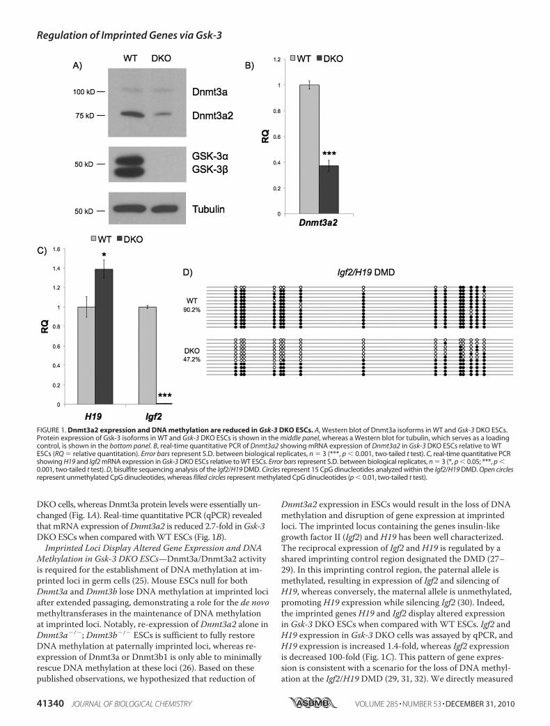
DKO cells, whereas Dnmt3a protein levels were essentially un-changed (Fig. 1A). Real-time quantitative PCR (qPCR) revealedthat mRNA expression ofDnmt3a2 is reduced 2.7-fold inGsk-3DKOESCs when compared withWT ESCs (Fig. 1B).Imprinted Loci Display Altered Gene Expression and DNA
Methylation in Gsk-3 DKO ESCs—Dnmt3a/Dnmt3a2 activityis required for the establishment of DNA methylation at im-printed loci in germ cells (25). Mouse ESCs null for bothDnmt3a and Dnmt3b lose DNA methylation at imprinted lociafter extended passaging, demonstrating a role for the de novomethyltransferases in the maintenance of DNA methylationat imprinted loci. Notably, re-expression of Dnmt3a2 alone inDnmt3a�/�; Dnmt3b�/� ESCs is sufficient to fully restoreDNA methylation at paternally imprinted loci, whereas re-expression of Dnmt3a or Dnmt3b1 is only able to minimallyrescue DNA methylation at these loci (26). Based on thesepublished observations, we hypothesized that reduction of
Dnmt3a2 expression in ESCs would result in the loss of DNAmethylation and disruption of gene expression at imprintedloci. The imprinted locus containing the genes insulin-likegrowth factor II (Igf2) and H19 has been well characterized.The reciprocal expression of Igf2 and H19 is regulated by ashared imprinting control region designated the DMD (27–29). In this imprinting control region, the paternal allele ismethylated, resulting in expression of Igf2 and silencing ofH19, whereas conversely, the maternal allele is unmethylated,promoting H19 expression while silencing Igf2 (30). Indeed,the imprinted genes H19 and Igf2 display altered expressionin Gsk-3 DKO ESCs when compared with WT ESCs. Igf2 andH19 expression in Gsk-3 DKO cells was assayed by qPCR, andH19 expression is increased 1.4-fold, whereas Igf2 expressionis decreased 100-fold (Fig. 1C). This pattern of gene expres-sion is consistent with a scenario for the loss of DNA methyl-ation at the Igf2/H19 DMD (29, 31, 32). We directly measured
FIGURE 1. Dnmt3a2 expression and DNA methylation are reduced in Gsk-3 DKO ESCs. A, Western blot of Dnmt3a isoforms in WT and Gsk-3 DKO ESCs.Protein expression of Gsk-3 isoforms in WT and Gsk-3 DKO ESCs is shown in the middle panel, whereas a Western blot for tubulin, which serves as a loadingcontrol, is shown in the bottom panel. B, real-time quantitative PCR of Dnmt3a2 showing mRNA expression of Dnmt3a2 in Gsk-3 DKO ESCs relative to WTESCs (RQ � relative quantitation). Error bars represent S.D. between biological replicates, n � 3 (***, p � 0.001, two-tailed t test). C, real-time quantitative PCRshowing H19 and Igf2 mRNA expression in Gsk-3 DKO ESCs relative to WT ESCs. Error bars represent S.D. between biological replicates, n � 3 (*, p � 0.05; ***, p �0.001, two-tailed t test). D, bisulfite sequencing analysis of the Igf2/H19 DMD. Circles represent 15 CpG dinucleotides analyzed within the Igf2/H19 DMD. Open circlesrepresent unmethylated CpG dinucleotides, whereas filled circles represent methylated CpG dinucleotides (p � 0.01, two-tailed t test).
Regulation of Imprinted Genes via Gsk-3
41340 JOURNAL OF BIOLOGICAL CHEMISTRY VOLUME 285 • NUMBER 53 • DECEMBER 31, 2010

DNAmethylation of the Igf2/H19 DMD by bisulfite sequenc-ing and found a 47% reduction of DNA methylation in Gsk-3DKO ESCs when compared with WT ESCs (Fig. 1D). Theseresults strongly support the hypothesis that the alterations inIgf2 and H19 expression are due to a loss of DNA methylationat the Igf2/H19 DMD.
Expression of Air, another imprinted gene that encodes fora long non-coding RNA (34), is increased in Gsk-3 DKOESCs. AirmRNA expression in Gsk-3 DKO ESCs was assayedby qPCR and is increased by 3.8-fold (Fig. 2A). We then deter-mined whether the change in expression of Air is due to a lossof DNA methylation. We performed bisulfite sequencing onDMR2 of Igf2r, from which Air is transcribed and known tobe methylated on the maternal allele (22, 35). DNA methyla-tion at DMR2 is reduced by 67% in Gsk-3 DKO ESCs (Fig.2B). These data suggest that the increase in Air expression islikely due to DNA hypomethylation within the Igf2r DMR2and demonstrate that the loss of Gsk-3 activity on DNAmethylation extends beyond the Igf2/H19 DMD, supporting apossible broader role for Gsk-3 in the regulation of imprintedgenes in the mouse.Global DNAMethylation Is Unchanged in Gsk-3 DKO ESCs—
We next determined whether the DNA hypomethylation weobserved at imprinted loci is due to a defect in global DNAmethylation. Because Dnmt1 is the primary enzyme responsi-ble for maintaining genome-wide DNA methylation (36), weexamined whether the loss of Gsk-3 activity affects Dnmt1function. Repetitive DNA sequences, such as IAP repeats, arehighly methylated in WT mouse ESCs (37) and largely un-
methylated in Dnmt1�/� and Dnmt1 hypomorph ESCs (38).IAP repeat methylation, although reduced, is largely retainedin ESCs deficient in de novo DNAmethyltransferase activity(Dnmt3a�/�; Dnmt3b�/�) (38, 39). Therefore, analyzingDNA methylation of IAP repeats provides an assay for Dnmt1activity. Southern blotting using methylation-sensitive restric-tion enzymes revealed that Gsk-3 DKO ESCs do not exhibit asignificant difference in DNA methylation of IAP repeatswhen compared with wild-type ESCs (Fig. 2C), suggestingthat there is not a general defect in DNA maintenance meth-ylation in Gsk-3 DKO cells. These data strengthen our hy-pothesis that the loss of DNA methylation observed at im-printed loci in Gsk-3 DKO ESCs is likely the result of specificdown-regulation of Dnmt3a2 expression.Ectopic Expression of Dnmt3a2 in Gsk-3 DKO ESCs Rescues
the Deficits in DNAMethylation—To evaluate whether exoge-nous expression of Dnmt3a2 could rescue the loss of DNAmethylation at imprinted loci in Gsk-3 DKO ESCs, we isolatedpuromycin-resistant Gsk-3 DKO ESCs stably expressingDnmt3a2 under the control of the chicken �-actin (CAG)promoter and evaluated DNA methylation at the Igf2/H19DMD by bisulfite sequencing. A puromycin-resistant clone inwhich Dnmt3a2 is stably overexpressed in DKO ESCs(Dnmt3a2) was selected for bisulfite sequencing analysis, anda puromycin-resistant clone that does not overexpressDnmt3a2 (Vector) served as a negative control (Fig. 3A). Bisul-fite sequencing of the Igf2/H19 DMD revealed that DNAmethylation is restored to levels previously observed in WTESCs in the overexpressing clone (92.7%; Fig. 3B). Rescue of
FIGURE 2. Effect on DNA methylation in Gsk-3 DKO ESCs extends to other imprinted genes but is not global. A, real-time quantitative PCR analysis ofAir mRNA expression in Gsk-3 DKO ESCs relative to WT ESCs (RQ � relative quantitation). Error bars represent S.D. between biological replicates, n � 3.B, bisulfite sequencing analysis of the Igf2r DMR2 in WT and Gsk-3 DKO ESCs. Circles represent 35 CpG dinucleotides analyzed within the Igf2r DMR2. Opencircles represent unmethylated CpG dinucleotides, whereas filled circles represent methylated CpG dinucleotides (p � 0.01, two-tailed t test). C, Southernblot analysis showing methylation of IAP in WT and Gsk-3 DKO ESCs. Genomic DNA from WT ESCs or Gsk-3 DKO ESCs was digested with MspI or HpaII. MspIis not sensitive to DNA methylation and cuts DNA regardless of methylation status. HpaII is an isoschizomer of MspI that does not cut methylated DNA.
Regulation of Imprinted Genes via Gsk-3
DECEMBER 31, 2010 • VOLUME 285 • NUMBER 53 JOURNAL OF BIOLOGICAL CHEMISTRY 41341

the Igf2/H19 DMDmethylation defect by stably expressingDnmt3a2 in Gsk-3 DKO ESCs strongly supports our hypothe-sis that reduced Dnmt3a2 expression in Gsk-3 DKO ESCs isthe cause of decreased DNA methylation at imprinted loci.Lithium Reduces DNAMethylation at Imprinted Loci—
Based on the data obtained from Gsk-3 DKO ESCs, we hy-pothesized that the Gsk-3 inhibitor lithium (40) should phe-nocopy the reduction in DNA methylation of imprintedgenes. It was previously reported that Dnmt3a�/�;Dnmt3b�/� ESCs required a high number of passages before
a loss of DNA methylation was detected at imprinted loci(26). In light of this observation, we grewWT ESCs in thepresence of lithium (40) for 4 weeks and then examined DNAmethylation at the Igf2/H19 DMD by bisulfite sequencing.Consistent with the reduction in DNAmethylation inGsk-3DKOESCs, treatment of wild-type ESCs with lithium reducesDNAmethylation at the Igf2/H19DMD in a dose-dependentmanner. DNAmethylation is reduced by 66% in wild-type ESCstreated with 20mM lithiumwhen compared withWT ESCs thathad been cultured in parallel without lithium treatment (Fig. 4A).
FIGURE 3. Reintroduction of Dnmt3a2 into Gsk-3 DKO ESCs rescues DNA methylation. A, Western blot of Dnmt3a2 expression in Gsk-3 DKO ESCs stablyexpressing Dnmt3a2 from the chicken �-actin (CAG) promoter (Dnmt3a2), DKO ESCs stably expressing the puromycin-resistant empty vector (Vector), andWT ESCs. The WT, Dnmt3a2, and Vector lanes are from the same blot, with intervening lanes removed. B, bisulfite sequencing of the Igf2/H19 DMD inDnmt3a2 rescued ESCs. Open circles represent unmethylated CpG dinucleotides, whereas filled circles represent methylated CpG dinucleotides.
FIGURE 4. Lithium inhibition of Gsk-3 results in hypomethylation. A, WT ESCs were continuously grown in the presence of either 10 mM or 20 mM LiCl for1 month. DNA methylation at CpG nucleotides within the Igf2/H19 DMD was assessed by bisulfite sequencing (WT versus 20 mM LiCl, p � 0.01, two-tailed ttest). B, DNA methylation of CpG nucleotides within the Igf2/H19 DMD in WT NSCs that were continuously grown in the presence of 5 mM LiCl for 1 month(p � 0.01, two-tailed t test). Open circles represent unmethylated CpG dinucleotides, whereas filled circles represent methylated CpG dinucleotides.
Regulation of Imprinted Genes via Gsk-3
41342 JOURNAL OF BIOLOGICAL CHEMISTRY VOLUME 285 • NUMBER 53 • DECEMBER 31, 2010

Next we examined the effect of Gsk-3 inhibitors on DNAmethylation in a cell type other than ESCs. Because the Gsk-3inhibitor lithium is used clinically to treat bipolar disorder, wechose to use cells from the neuronal lineage, specificallymouse NSC. After 4 weeks of growth in the presence of 5 mM
lithium, we observed a 35% reduction in DNA methylation atthe Igf2/H19 DMD (Fig. 4B). These results demonstrate thatthe effect of lithium on DNA methylation extends to neuralcells and expand the implications of our findings to other celltypes.Inactivation of Gsk-3 by PI3K results in Loss of DNAMeth-
ylation at Imprinted Loci—Activated insulin signaling nega-tively regulates Gsk-3 activity via inhibitory phosphorylationof an N-terminal serine by Akt (41). We investigated whetherinactivation of Gsk-3 by constitutively active insulin signalingpathway could affect DNA methylation of imprinted genes.Expression of a myristoylated p110 subunit of PI3K (p110*)has previously been demonstrated to constitutively activateinsulin signaling in ESCs (5). WT ESCs were stably trans-fected with p110*, and Western blots were performed to ver-ify activation of the insulin pathway. Stable expression ofp110* leads to robust phosphorylation of Akt on serine 473,which results in the activation of Akt. In addition, we foundincreased N-terminal phosphorylation of both Gsk-3� (serine21) and Gsk-3� (serine 9), suggesting that the cellular pool of
Gsk-3 regulated by Akt is inhibited (Fig. 5A). Bisulfite se-quencing of the H19/Igf2 DMD and Igf2r DMR2 demon-strated a substantial reduction of DNA methylation levels inp110* stable cells, similar to that seen in Gsk-3 DKO ESCs(Fig. 5, B and C). These data demonstrate that activation ofinsulin signaling by constitutively active PI3K reduces DNAmethylation at imprinted loci.N-Myc Is a Potent Gsk-3-dependent Regulator of Dnmt3a2
Gene Expression—Our data show that the reduced levels ofDNA methylation at imprinted loci in Gsk-3 DKO ESCs arelikely due to a decrease in Dnmt3a2 levels. To establish themechanism by which Gsk-3 activity regulates the expressionof Dnmt3a2, we considered that both Dnmt3a2 mRNA andprotein levels are decreased in Gsk-3 DKO ESCs, suggestingthat the effect is likely due to altered transcriptional regula-tion. A role for miRNAs was excluded because both Dnmt3aand Dnmt3a2 share a common 3�-untranslated region (UTR)yet are not equally affected by the loss of Gsk-3 isoforms.Therefore, we focused on understanding the mechanism con-trolling Dnmt3a2 transcription. A 1.9-kb fragment of theDnmt3a2 promoter was cloned (Fig. 6A), which is distinctfrom the regulatory regions of Dnmt3a and had previouslybeen shown to be sufficient for driving high levels of reporterexpression in ESCs (24). This 1.9-kb promoter fragment iscapable of yielding robust (�35-fold) expression of a lucifer-
FIGURE 5. Activation of components of the insulin signaling pathway reduces DNA methylation. A, Western blots showing that WT ESCs stably trans-fected with p110* constitutively activate insulin signaling. p-Akt, phospho-Akt; p-Gsk-3, phospho-glycogen synthase kinase-3. B, bisulfite sequencing analy-sis of the Igf2/H19 DMD in p110* ESCs when compared with WT ESCs (p � 0.01, two-tailed t test). C, bisulfite sequencing analysis of the Igf2r DMR2 in p110*ESCs when compared with WT ESCs. Open circles represent unmethylated CpG dinucleotides, whereas filled circles represent methylated CpG dinucleotides.
Regulation of Imprinted Genes via Gsk-3
DECEMBER 31, 2010 • VOLUME 285 • NUMBER 53 JOURNAL OF BIOLOGICAL CHEMISTRY 41343

Regulation of Imprinted Genes via Gsk-3
41344 JOURNAL OF BIOLOGICAL CHEMISTRY VOLUME 285 • NUMBER 53 • DECEMBER 31, 2010

ase reporter in WT ESCs (Fig. 6B). The reporter also recapitu-lates the effect of Gsk-3 deletion on Dnmt3a2mRNA, show-ing an �40% reduction in reporter activity when transfectedinto Gsk-3 DKO ESCs.In silico analysis of the Dnmt3a2 promoter revealed a re-
gion of highly conserved sequence just proximal to the exonunique to Dnmt3a2, which encodes the 5�-UTR. Within thisconserved region is a putative Myc binding site. Our microar-ray data showed a 2.8-fold decrease in N-Myc levels in Gsk-3DKO ESCs. Real-time qPCR revealed an 8.8-fold decrease inN-mycmRNA (Fig. 6C), and Western blot analysis confirmedthe corresponding decrease in N-Myc protein levels (Fig. 6D).To test the importance of the Myc binding site in theDnmt3a2 promoter, we performed site-directed mutagenesison the putative Myc binding site in the context of the 1.9-kbpromoter fragment and transfected this reporter into ESCs.We observed a complete loss of reporter activity, confirmingthe functional importance of this binding site (Fig. 6B). Fi-nally, transfection of WT ESCs with N-myc siRNA resulted ina 77% reduction in N-Myc levels (Fig. 6E) and a decrease inDnmt3a2 protein (Fig. 6F), confirming the importance of N-Myc on the regulation of Dnmt3a2 in ESCs.
DISCUSSION
Mechanisms to explain how extracellular environmentalsignals are transduced to the nucleus to affect epigeneticmodifications, such as DNA methylation, have been elusive(44). Here, we provide evidence for a novel role for Gsk-3 inthe epigenetic regulation of imprinted genes. In mouse ESCs,deletion of Gsk-3 isoforms or expression of myristoylatedPI3-kinase both lead to constitutive activation of downstreamcomponents of the insulin pathway, ultimately resulting inattenuated DNA methylation of imprinted loci. Although weprovide evidence that several imprinted genes are hypom-ethylated as a consequence of the loss of Gsk-3 activity, it re-mains to be determined whether DNA methylation is reducedat all known imprinted loci in Gsk-3 DKO ESCs or whetherthe effect on DNA methylation extends to non-imprintedgenes.Interestingly, we observed that �90% of CpG dinucleotides
analyzed in the Igf2/H19 DMD were methylated in WT ESCs.It is expected that the number of methylated CpG dinucleoti-des would be closer to 50% for this imprinted locus becausewe did not discriminate between alleles in our bisulfite se-quencing assay. The high amount of DNA methylation ob-served in WT ESCs is not likely due to PCR bias in our bisul-fite sequencing procedure as we observed 44% methylation ofthe Igf2/H19 DMD in NSC derived fromWTmice using the
same bisulfite sequencing assay (supplemental Fig. 1). It hasbeen reported that cultured ESC lines may exhibit almostcomplete methylation of the Igf2/H19 DMD on both parentalalleles (33). In light of these previously reported observations,it is likely that the high degree of DNA methylation we detectat the Igf2/H19 DMD in WT ESCs is due to biallelicmethylation.Our data show an increase in N-terminal phosphorylation
of Ser-9/Ser-21 on Gsk-3 isoforms upon ectopic expression ofPI3K, resulting in decreased DNA methylation at the Igf2/H19imprinting control region. Although our experimental ap-proach utilized constitutively active PI3-kinase as a means ofinducing phosphorylation on Gsk-3� Ser-21/Gsk-3� Ser-9,signal transduction of several pathways, such as protein ki-nase A (PKA) (45), also result in Gsk-3� Ser-21/Gsk-3� Ser-9phosphorylation. Therefore, our data predict that signalingpathways that lead to the phosphorylation and inhibition ofGsk-3 akin to insulin signaling would lead to decreased DNAmethylation at imprinted loci. We have not ruled out the pos-sibility that inactivation of Gsk-3 by Wnt signaling can lead tohypomethylation of Igf2/H19. Further investigation is neededto determine whether activation of the Wnt pathway has thesame effect on Dnmt3a2 expression.
The deficits we observe in DNA methylation are due toreduced levels of the de novo DNAmethyltransferaseDnmt3a2. Dnmt3a2 co-purifies with Dnmt3L in ES cells,whereas Dnmt3a does not (46, 47). Although Dnmt3L is cata-lytically inactive, it is required for establishment of DNAmethylation at imprinted loci (48, 49) and recruits Dnmt3a2to DNA sequences associated with unmethylated lysine 4 ofhistone H3 (47). Dnmt3L levels were not significantly changedin Gsk-3 DKO ESCs (decreased 1.3-fold). These findings sug-gest that Dnmt3a2 plays a prominent role in the maintenanceof DNA methylation at imprinted genes in early development.Heretofore, understanding the specific function of Dnmt3a2in vivo has been difficult because targeted disruptions of theDnmt3a locus in the mouse result in the simultaneous dele-tion of both Dnmt3a and Dnmt3a2 as the isoforms sharecommon 3� exons. Our identification of a signal transductionpathway capable of specifically reducing levels of Dnmt3a2provides a new approach for the study of this isoform in vivo.To better understand the mechanism by which Dnmt3a2
mRNA is regulated, we cloned and analyzed a 1.9-kb fragmentof mouse genomic DNA that contained a previously charac-terized promoter region. Computational analysis of the pro-moter revealed very little conservation of sequence acrossdifferent species, with the exception of an �200-bp region
FIGURE 6. N-Myc regulates Dnmt3a2 expression in a Gsk-3-dependent manner. A, schematic representation of the locus encoding Dnmt3a isoforms.Exons are shown as the vertical bars. The Dnmt3a2 promoter we used to drive the luciferase promoter is shown as the gray box. The arrow denotes the puta-tive N-Myc binding site that we mutated in our reporter construct. B, Dnmt3a2 promoter activity was assessed with a luciferase reporter. Reporters contain-ing WT 1.9-kb Dnmt3a2 promoter (WT) or promoter with two point mutations (bases underlined) in putative Myc binding site (CACGTG3 CAGCTG; muta-tion (mut)) were transfected into WT and Gsk-3 DKO ESCs. Bars represent -fold change over promoterless luciferase vector (pGLF). Error bars represent S.D.between replicate transfections, n � 3 (***, p � 0.001, two-tailed t test). C, real-time quantitative PCR analysis of N-myc mRNA expression in Gsk-3 DKO ESCsrelative to WT ESCs (RQ � relative quantitation). Error bars represent S.D. between biological replicates, n � 3 (***, p � 0.001, two-tailed t test). D, Westernblots showing N-Myc, Gsk-3�, Gsk-3�, and tubulin in WT and Gsk-3 DKO ESCs. E, real-time quantitative PCR analysis of N-myc mRNA expression in WT ESCstransfected with GFP siRNA and N-myc siRNA. Error bars represent S.D. between biological replicates, n � 3 (***, p � 0.001, two-tailed t test). F, Western blotfor Dnmt3a2 in WT cells transfected with GFP siRNA and N-myc siRNA. G, model depicting a pathway that leads from PI3K activation to changes in DNAmethylation in the nucleus.
Regulation of Imprinted Genes via Gsk-3
DECEMBER 31, 2010 • VOLUME 285 • NUMBER 53 JOURNAL OF BIOLOGICAL CHEMISTRY 41345

just upstream of the novel Dnmt3a2 exon. Within this region,we found a putative Myc binding site that is highly conserved,and a point mutation in this site completely impaired reporteractivation. Recent chromatin immunoprecipitation-sequenc-ing (ChIP-Seq) data showed that both c-Myc and N-Mycshare an identical DNA binding motif (50); however, we didnot observe a reduction in c-Myc protein in Gsk-3 DKO ESCs.Furthermore, ChIP-Seq data in mouse ESCs show that N-Myc, but not c-Myc, binds specifically to the Dnmt3a2 pro-moter (50). We have experimentally verified the importanceof N-Myc on regulating Dnmt3a2 expression by knockingdown N-myc via siRNA. Taken together with the decreasedlevels of N-Myc and unchanged levels of c-Myc (supplementalFig. 2, A and B), we conclude that N-Myc regulates Dnmt3a2transcription. Gsk-3-mediated phosphorylation and degrada-tion of c-Myc and N-Myc proteins by Gsk-3 have been re-ported in COS-7 cells (c-Myc) and cerebellar granule neuronprecursors (N-Myc) (42, 43, 51). We did not observe this reg-ulation of Myc proteins in our ESCs. In fact, we see an oppo-site effect of Gsk-3 on N-Myc expression and no effect onc-Myc expression. Our data likely reflect cell type-specificdifferences in the regulation of Myc proteins by Gsk-3. Fur-thermore, our real-time qPCR data indicate that N-Mycdown-regulation in Gsk-3 DKO ESCs is occurring at the levelof transcriptional regulation. The mechanism by which Gsk-3regulates N-myc transcription in ESCs remains to bedetermined.The ability of the Gsk-3 inhibitor lithium to phenocopy the
reduction in DNA methylation of imprinted genes seen inGsk-3 DKO ESCs is particularly intriguing. We have shownthat DNA methylation is reduced at the Igf2/H19 imprintingcontrol region in both ESCs and NSCs after prolonged expo-sure to lithium. The effectiveness of lithium in the treatmentof bipolar disorder was initially reported more than 60 yearsago (52). Lithium has been widely prescribed ever since, al-though the therapeutic mechanism of action has not beenfully defined. Growing evidence suggests that alterations inDNA methylation have a role in the development of bipolardisorder (53, 54). Our data raise the possibility that reducedDNA methylation of imprinted genes may be related to themechanism of lithium action in bipolar disorder. It is note-worthy that another drug widely used for the treatment ofbipolar disorder, valproic acid, also alters the epigenome byinhibiting histone deacetylase activity (55, 56). In the contextof our findings, it is tempting to speculate that lithium andvalproic acid are effective in treating bipolar disorder becausethey modify the expression of a common subset of genes viachanges in DNA methylation and histone acetylation,respectively.Our findings may also provide a new focus for the study of
neurological diseases. Gsk-3 activity has been linked to schiz-ophrenia (57), whereas alterations in the epigenome havebeen proposed to be a major factor in the development ofschizophrenia (53). Of the postnatal tissues that have beenexamined to date, imprinting is clearly predominant in thebrain, and alterations in these imprinting events have beenshown to affect behavior (58). One example in humans isPrader-Willi syndrome, which is caused by a deletion on
chromosome 15q11–13, an area that contains the imprintedgenes SNRPN and Necdin (NDN) (59). This raises the possi-bility that altered Gsk-3 activity, resulting in aberrant DNAmethylation of imprinted genes, could contribute to the de-velopment of schizophrenia or other behavioral disorders.In summary, we have discovered a novel role for Gsk-3 in
maintaining DNA methylation of imprinted loci in mouseESCs. Because Gsk-3 is a nexus for numerous signal transduc-tion pathways, the potential for Gsk-3 to regulate epigeneticchanges could have profound consequences for our under-standing of diverse human diseases.
Acknowledgments—We thank Drs. Jeff Kuret, Jing Yang, and ScottHarper for reading the manuscript and helpful discussions. We arealso grateful to Dr. Glenn Maston for the pGLF construct, Dr. Car-los Miranda for assistance in isolating neural stem cells, and Dr.Yulei Wang for assistance with c-Myc qPCR.
REFERENCES1. Force, T., and Woodgett, J. R. (2009) J. Biol. Chem. 284, 9643–96472. Kockeritz, L., Doble, B., Patel, S., and Woodgett, J. R. (2006) Curr. Drug.
Targets 7, 1377–13883. Doble, B. W., Patel, S., Wood, G. A., Kockeritz, L. K., and Woodgett, J. R.
(2007) Dev. Cell 12, 957–9714. Kim, W. Y., Wang, X., Wu, Y., Doble, B. W., Patel, S., Woodgett, J. R.,
and Snider, W. D. (2009) Nat. Neurosci. 12, 1390–13975. Takahashi, K., Mitsui, K., and Yamanaka, S. (2003) Nature 423, 541–5456. Sato, N., Meijer, L., Skaltsounis, L., Greengard, P., and Brivanlou, A. H.
(2004) Nat. Med. 10, 55–637. Paling, N. R., Wheadon, H., Bone, H. K., and Welham, M. J. (2004)
J. Biol. Chem. 279, 48063–480708. Watanabe, S., Umehara, H., Murayama, K., Okabe, M., Kimura, T., and
Nakano, T. (2006) Oncogene 25, 2697–27079. Lu, J., Hou, R., Booth, C. J., Yang, S. H., and Snyder, M. (2006) Proc.
Natl. Acad. Sci. U.S.A. 103, 5688–569310. Storm, M. P., Bone, H. K., Beck, C. G., Bourillot, P. Y., Schreiber, V.,
Damiano, T., Nelson, A., Savatier, P., and Welham, M. J. (2007) J. Biol.Chem. 282, 6265–6273
11. Miyabayashi, T., Teo, J. L., Yamamoto, M., McMillan, M., Nguyen, C.,and Kahn, M. (2007) Proc. Natl. Acad. Sci. U.S.A. 104, 5668–5673
12. Ying, Q. L., Wray, J., Nichols, J., Batlle-Morera, L., Doble, B., Woodgett,J., Cohen, P., and Smith, A. (2008) Nature 453, 519–523
13. Niwa, H., Ogawa, K., Shimosato, D., and Adachi, K. (2009) Nature 460,118–122
14. Cantley, L. C. (2002) Science 296, 1655–165715. Cross, D. A., Alessi, D. R., Cohen, P., Andjelkovich, M., and Hemmings,
B. A. (1995) Nature 378, 785–78916. MacDonald, B. T., Tamai, K., and He, X. (2009) Dev. Cell 17, 9–2617. Zeng, X., Tamai, K., Doble, B., Li, S., Huang, H., Habas, R., Okamura, H.,
Woodgett, J., and He, X. (2005) Nature 438, 873–87718. Ding, V. W., Chen, R. H., and McCormick, F. (2000) J. Biol. Chem. 275,
32475–3248119. Ng, S. S., Mahmoudi, T., Danenberg, E., Bejaoui, I., de Lau, W., Korswa-
gen, H. C., Schutte, M., and Clevers, H. (2009) J. Biol. Chem. 284,35308–35313
20. Dong, K. B., Maksakova, I. A., Mohn, F., Leung, D., Appanah, R., Lee, S.,Yang, H. W., Lam, L. L., Mager, D. L., Schubeler, D., Tachibana, M.,Shinkai, Y., and Lorincz, M. C. (2008) EMBO J. 27, 2691–2701
21. Budowle, B., and Baechtel, F. S. (1990) Appl. Theor. Electrophor. 1,181–187
22. Yamasaki, Y., Kayashima, T., Soejima, H., Kinoshita, A., Yoshiura, K.,Matsumoto, N., Ohta, T., Urano, T., Masuzaki, H., Ishimaru, T., Mukai,T., Niikawa, N., and Kishino, T. (2005) Hum. Mol. Genet. 14, 2511–2520
23. Okano, M., Xie, S., and Li, E. (1998) Nat. Genet. 19, 219–220
Regulation of Imprinted Genes via Gsk-3
41346 JOURNAL OF BIOLOGICAL CHEMISTRY VOLUME 285 • NUMBER 53 • DECEMBER 31, 2010

24. Chen, T., Ueda, Y., Xie, S., and Li, E. (2002) J. Biol. Chem. 277,38746–38754
25. Kaneda, M., Okano, M., Hata, K., Sado, T., Tsujimoto, N., Li, E., andSasaki, H. (2004) Nature 429, 900–903
26. Chen, T., Ueda, Y., Dodge, J. E., Wang, Z., and Li, E. (2003)Mol. Cell.Biol. 23, 5594–5605
27. DeChiara, T. M., Robertson, E. J., and Efstratiadis, A. (1991) Cell 64,849–859
28. Bartolomei, M. S., Webber, A. L., Brunkow, M. E., and Tilghman, S. M.(1993) Genes Dev. 7, 1663–1673
29. Tremblay, K. D., Duran, K. L., and Bartolomei, M. S. (1997)Mol. Cell.Biol. 17, 4322–4329
30. Ideraabdullah, F. Y., Vigneau, S., and Bartolomei, M. S. (2008)Mutat.Res. 647, 77–85
31. Jinno, Y., Sengoku, K., Nakao, M., Tamate, K., Miyamoto, T., Matsuzaka,T., Sutcliffe, J. S., Anan, T., Takuma, N., Nishiwaki, K., Ikeda, Y., Ishi-maru, T., Ishikawa, M., and Niikawa, N. (1996) Hum. Mol. Genet. 5,1155–1161
32. Thorvaldsen, J. L., Duran, K. L., and Bartolomei, M. S. (1998) Genes Dev.12, 3693–3702
33. Dean, W., Bowden, L., Aitchison, A., Klose, J., Moore, T., Meneses, J. J.,Reik, W., and Feil, R. (1998) Development 125, 2273–2282
34. Lyle, R., Watanabe, D., te Vruchte, D., Lerchner, W., Smrzka, O. W.,Wutz, A., Schageman, J., Hahner, L., Davies, C., and Barlow, D. P. (2000)Nat. Genet. 25, 19–21
35. Stoger, R., Kubicka, P., Liu, C. G., Kafri, T., Razin, A., Cedar, H., and Bar-low, D. P. (1993) Cell 73, 61–71
36. Li, E., Bestor, T. H., and Jaenisch, R. (1992) Cell 69, 915–92637. Howlett, S. K., and Reik, W. (1991) Development 113, 119–12738. Okano, M., Bell, D. W., Haber, D. A., and Li, E. (1999) Cell 99, 247–25739. Lei, H., Oh, S. P., Okano, M., Juttermann, R., Goss, K. A., Jaenisch, R.,
and Li, E. (1996) Development 122, 3195–320540. Klein, P. S., and Melton, D. A. (1996) Proc. Natl. Acad. Sci. U.S.A. 93,
8455–845941. Doble, B. W., and Woodgett, J. R. (2003) J. Cell Sci. 116, 1175–118642. Gregory, M. A., Qi, Y., and Hann, S. R. (2003) J. Biol. Chem. 278,
51606–5161243. Sjostrom, S. K., Finn, G., Hahn, W. C., Rowitch, D. H., and Kenney,
A. M. (2005) Dev. Cell 9, 327–33844. Berger, S. L., Kouzarides, T., Shiekhattar, R., and Shilatifard, A. (2009)
Genes Dev. 23, 781–78345. Li, M., Wang, X., Meintzer, M. K., Laessig, T., Birnbaum, M. J., and Hei-
denreich, K. A. (2000)Mol. Cell. Biol. 20, 9356–936346. Nimura, K., Ishida, C., Koriyama, H., Hata, K., Yamanaka, S., Li, E., Ura,
K., and Kaneda, Y. (2006) Genes Cells 11, 1225–123747. Ooi, S. K., Qiu, C., Bernstein, E., Li, K., Jia, D., Yang, Z., Erdjument-Bro-
mage, H., Tempst, P., Lin, S. P., Allis, C. D., Cheng, X., and Bestor, T. H.(2007) Nature 448, 714–717
48. Bourc’his, D., Xu, G. L., Lin, C. S., Bollman, B., and Bestor, T. H. (2001)Science 294, 2536–2539
49. Kato, Y., Kaneda, M., Hata, K., Kumaki, K., Hisano, M., Kohara, Y.,Okano, M., Li, E., Nozaki, M., and Sasaki, H. (2007) Hum. Mol. Genet.16, 2272–2280
50. Chen, X., Xu, H., Yuan, P., Fang, F., Huss, M., Vega, V. B., Wong, E.,Orlov, Y. L., Zhang, W., Jiang, J., Loh, Y. H., Yeo, H. C., Yeo, Z. X.,Narang, V., Govindarajan, K. R., Leong, B., Shahab, A., Ruan, Y.,Bourque, G., Sung, W. K., Clarke, N. D., Wei, C. L., and Ng, H. H. (2008)Cell 133, 1106–1117
51. Kenney, A. M., Cole, M. D., and Rowitch, D. H. (2003) Development130, 15–28
52. Cade, J. F. (1949)Med. J. Aust 2, 349–35253. Mill, J., Tang, T., Kaminsky, Z., Khare, T., Yazdanpanah, S., Bouchard,
L., Jia, P., Assadzadeh, A., Flanagan, J., Schumacher, A., Wang, S. C., andPetronis, A. (2008) Am. J. Hum. Genet. 82, 696–711
54. Kuratomi, G., Iwamoto, K., Bundo, M., Kusumi, I., Kato, N., Iwata, N.,Ozaki, N., and Kato, T. (2008)Mol. Psychiatry 13, 429–441
55. Phiel, C. J., Zhang, F., Huang, E. Y., Guenther, M. G., Lazar, M. A., andKlein, P. S. (2001) J. Biol. Chem. 276, 36734–36741
56. Gottlicher, M., Minucci, S., Zhu, P., Kramer, O. H., Schimpf, A., Giavara,S., Sleeman, J. P., Lo Coco, F., Nervi, C., Pelicci, P. G., and Heinzel, T.(2001) EMBO J. 20, 6969–6978
57. Mao, Y., Ge, X., Frank, C. L., Madison, J. M., Koehler, A. N., Doud,M. K., Tassa, C., Berry, E. M., Soda, T., Singh, K. K., Biechele, T., Petry-shen, T. L., Moon, R. T., Haggarty, S. J., and Tsai, L. H. (2009) Cell 136,1017–1031
58. Wilkinson, L. S., Davies, W., and Isles, A. R. (2007) Nat. Rev. Neurosci. 8,832–843
59. Horsthemke, B., and Wagstaff, J. (2008) Am. J. Med. Genet. A. 146A,2041–52
Regulation of Imprinted Genes via Gsk-3
DECEMBER 31, 2010 • VOLUME 285 • NUMBER 53 JOURNAL OF BIOLOGICAL CHEMISTRY 41347