Skeletal Muscle Uncoupling Proteins in Mice Models of Obesity
Upload
independentCategory
view
4download
0
A kinetic assessment of the C. elegansamyloid disaggregation activity enablesuncoupling of disassembly andproteolysis
Jan Bieschke,1† Ehud Cohen,2† Amber Murray,1 Andrew Dillin,2
and Jeffery W. Kelly1*
1Departments of Chemistry and Molecular and Experimental Medicine, The Skaggs Institute for Chemical Biology,
The Scripps Research Institute, La Jolla, California 920372The Howard Hughes Medical Institute and Molecular and Cell Biology Laboratory,
The Salk Institute for Biological Studies, La Jolla, California 92037
Received 27 May 2009; Accepted 13 August 2009DOI: 10.1002/pro.234
Published online 21 August 2009 proteinscience.org
Abstract: Protein aggregation is a common feature of late onset neurodegenerative disorders,including Alzheimer’s disease. In Alzheimer’s disease, misassembly of the Ab peptide is genetically
linked to proteotoxicity associated with disease etiology. A reduction in Ab proteotoxicity is
accomplished, in part, by the previously reported Ab disaggregation and proteolysis activities–underpartial control of heat shock factor 1, a transcription factor regulating proteostasis in the cytosol and
negatively regulated by insulin growth factor signaling. Herein, we report an improved in vitro assay
to quantify recombinant fibrillar Ab disaggregation kinetics accomplished by the exogenousapplication of C. elegans extracts. With this assay we demonstrate that the Ab disaggregation and
proteolysis activities of C. elegans are separable. The disaggregation activity found in C. elegans
preparations is more heat resistant than the proteolytic activity. Ab disaggregation in the absence ofproteolysis was found to be a reversible process. Future discovery of the molecular basis of the
disaggregation and proteolysis activities offers the promise of delaying the age-onset proteotoxicity
that leads to neurodegeneration in a spectrum of maladies.
Keywords: disaggregation; proteolysis; abeta; Alzheimer’s disease
Introduction
Protein aggregation is genetically and biochemically
linked to the development of human neurodegenera-
tive disorders, such as Alzheimer’s disease (AD), Par-
kinson’s disease (PD), Huntington’s disease (HD), the
familial amyloidoses, and the prion disorders.1–7
Aggregation of the 40- and 42-amino-acid amyloid
beta peptides (Ab40 and Ab42) putatively causes AD.8
Whether Ab proteotoxicity arises from within the neu-
ron, outside the cell, or both, how toxicity is effected
and what the structure(s) of the cytotoxic agent is, are
key unanswered questions. A prominent risk factor
common to all of these neurodegenerative diseases is
aging.9–14 Insulin/Insulin growth factor-1 signaling
(IIS) is perhaps the most prominent signaling pathway
Additional Supporting Information can be found in the onlineversion of this article.
yJan Bieschke and Ehud Cohen contributed equally to this work.
Jan Bieschke’s current address is Max-Delbruck-Center forMolecular Medicine, Robert-Roessle-Str. 10, Berlin-Buch 13125,Germany.
Ehud Cohen’s current address is Institute of Medical ResearchIsrael Canada (IMRIC), The Hebrew University of Jerusalem,Ein-Karem, Jerusalem 91121, Israel.
Grant sponsor: NGFNplus; Grant number: 01GS08132; Grantsponsor: NIH; Grant number: AG031097; Grant sponsors: TheBundy Foundation, the Helmholz Gemeinschaft, the SkaggsInstitute for Chemical Biology, and the Lita Annenberg HazenFoundation.
*Correspondence to: Jeffery W. Kelly, The Scripps ResearchInstitute, BCC 265, 10550 N. Torrey Pines Road, La Jolla, CA92037. E-mail: [email protected]
Published by Wiley-Blackwell. VC 2009 The Protein Society PROTEIN SCIENCE 2009 VOL 18:2231—2241 2231

regulating aging. Reduced IIS is linked to longevity in
worms,15 flies, mice,16 and humans.17,18 In worms, life-
span extension facilitated by IIS reduction (either by
mutating the sole insulin/IGF receptor, daf-2, or by
the application of RNAi directed against daf-2) is de-
pendent upon the activities of the transcription factors
DAF-16 and HSF-1.19,20
Why aging is the prominent risk factor in human
neurodegenerative diseases is not completely under-
stood. Some insight regarding the connection between
aging and neurodegeneration is provided by recent
studies demonstrating that delaying aging in organis-
mal models of HD and AD by IIS suppression amelio-
rates aggregation-associated proteotoxicity.9,12,19,21,22
The molecular basis for the link between the reduction
in IIS and diminished proteotoxicity is likely because
aging signaling pathways and their transcriptomes
have a strong influence on the protein homeostasis (or
proteostasis) network, that is, the biological pathways
that maintain our proteome and minimize aggregate
accumulation.9 For example, both the heat shock
response controlling proteostasis in the cytosol, and
longevity mediated by reduced IIS require the tran-
scription factor, HSF-1. As is the case for lifespan
extension, both the DAF-16 and HSF-1 transcriptomes
are required to ameliorate Ab42 proteotoxicity via IIS
suppression.22
Speculation23 that a specialized mammalian disag-
gregation activity, possibly involving chaperones, ena-
bles aggregate degradation has some experimental
support.22,24 Active and passive immunization studies
resulting in the clearance of Ab deposits from AD
animal models suggest that inducing degradation
pathways can be therapeutically beneficial.25,26 In one
potential model of the disaggregase activity, the disas-
sembly of fibrillar Ab aggregates is envisioned to be
driven by proteolysis of Ab monomers, which would
lead to the disassembly of Ab aggregates by depleting
the monomers below their critical concentration, caus-
ing dissociation of the fibrils to repopulate the disap-
pearing monomer population.27,28 Alternatively, disag-
gregation via a specific cellular pathway or utilizing a
macromolecular complex could take apart Ab fibrils,
independent of proteolysis. This scenario would enable
reaggregation of Ab into fibrils if Ab monomer proteol-
ysis was inhibited and the disaggregase activity was
depleted. Discovering the molecular underpinnings of
the C. elegans fibril disaggregase pathway(s)/activi-
ty(ies) and/or the degradation pathway(s)/activity(ies)
could reveal novel therapeutic strategies for human
neurodegenerative diseases.9,22 In this manuscript, we
focus primarily on the disaggregase activity.
The apparent importance of disaggregation and
degradation in countering Ab42 aggregation-associated
proteotoxicity and the linkage of this biological path-
way(s) or activity(ies) to aging-associated signaling
pathways22 motivated us to optimize and further scru-
tinize the Ab disaggregation assay to discern whether
the disaggregation and proteolysis activities are sepa-
rable. In this study, the Ab amyloid fibril-thioflavin T
(ThT) fluorophore complex prepared in vitro was
treated with C. elegans homogenate to discern the rate
of fibril disassembly and the degree of Ab proteolysis
in the presence or absence of selected protease inhibi-
tors. We utilized the decrease in ThT fluorescence to
follow disaggregation (verified by atomic force micros-
copy) and the decrease in immunoreactivity to Abantibodies and/or an HPLC assay to monitor the deg-
radation of Ab. These studies demonstrate that disag-
gregation does not require proteolytic degradation, as
these activities are separable.
Results
C. elegans homogenate possesses Ab40 fibrildisaggregation activity
To assess the disaggregation activity within the C. ele-
gans AD model, we prepared cellular extracts by ho-
mogenizing worms grown on bacteria to day one of
adulthood. The debris from Ab worm homogenization
was cleared by low speed centrifugation (as detailed in
the Methods section) and the resulting postdebris su-
pernatant (PDS) was used in subsequent experiments.
Since the disaggregation machinery might be induci-
ble, we used worms that express Ab42 in their smooth
body wall muscles under control of the unc54 pro-
moter that encodes the myosin heavy chain gene and
is expressed from embryogenesis through adulthood
(strain CL 200621), hereafter referred to as Ab worms.
We assumed that Ab42 overexpression in these worms
would be sufficient to induce this activity. These Abworms were grown on bacteria to day one of adult-
hood, at which stage no proteotoxicity is observed.21,22
Worms not expressing Ab42 (wild-type worms, strain
N2) were used as controls. Worm PDS was added to
preformed Ab40 (or Ab42) fibrils in vitro to assess its
disaggregation activity.
Ab fibrils were generated from monomerized Ab,prepared from Ab40 (or Ab42) peptide dissolved in
hexafluoroisopropanol (HFIP) overnight, then lyophi-
lized, dissolved at pH 10.5, and filtered through a low
molecular weight membrane filter (Microcon YM-10,
Millipore) to remove covalent and noncovalent aggre-
gates.29 Fibrillar aggregates were then formed by incu-
bating the initially monomeric Ab peptide (50 lM) at
37�C at pH 7.4 for 3 days, during which time the sam-
ple was agitated using an overhead rotary shaker (20
rpm). Amyloidogenesis was monitored by ThT fluores-
cence (Supporting information Fig. S1A). ThT is a fluo-
rophore that exhibits a higher fluorescence quantum
yield and a red shift upon binding to fibrils or spheri-
cal aggregates.30 Aggregates were characterized by cir-
cular dichroism spectroscopy to observe the expected
increase in b-sheet structure (Supporting information
Fig. S1B), and by atomic force microscopy (AFM) to
confirm the fibril morphology (Supporting information
2232 PROTEINSCIENCE.ORG Ab Disaggregation and Proteolysis by C. elegans

Fig. S1C). Fibrillar aggregates were sonicated for 30
min using a water bath sonicator to afford a uniform
(200–500 nm) length distribution of fibrils (Support-
ing information Fig. S1D). Disaggregation of the preso-
nicated fibrils was monitored in a fluorescence plate
reader by the decrease in ThT fluorescence over time,
with data being collected every 10 min following 5 s of
shaking.31 Disaggregation of Ab fibrils was confirmed
by AFM.22
The ability of worm PDS to disaggregate Ab40fibrils in phosphate buffer (150 mM NaCl, 50 mM Na-
phosphate, pH 7.4) at 37�C was scrutinized by the addi-
tion of Ab worm PDS at final total PDS protein concen-
trations ranging from 25 to 500 lg/mL [Fig. 1(A)]. ThT
(20 lM) fluorescence decreased in the presence of
worm PDS, whereas incubation with buffer alone (black
circles) did not significantly change the ThT fluores-
cence. Disaggregation time courses for all PDS reac-
tions were fitted by mono-exponential decay functions
[Fig. 1(A), lines]. Disaggregation time constants were
4.6 � 0.6, 3.4 � 0.3, 2.5 � 0.2, and 1.5 � 0.1 h for PDS
exhibiting a total protein concentration of 25, 50, 100,
Figure 1. Ab40 disaggregation activity A: Disaggregation activities of C. elegans PDS as a function of total protein concentration
(25–500 lg/mL). Lines show mono-exponential decay function fits to the data (open symbols). B: Disaggregation activities of
PDS (20 lg/mL) from Ab42-expressing (CL for CL2006) and wt (N2) C. elegans fed bacteria expressing hsf-1 RNAi or empty
vector controls (EV). Neither the disaggregation rate nor its reduction by hsf-1 downregulation depend on Ab42 expression. C:PDS-mediated Ab40 disaggregation is not discernibly inhibited when ATP in the PDS is hydrolyzed by pretreatment with apyrase
(AP, 0.1 and 0.5 U, 15 min, 30�C); average relative ThT signals of quintuplicate samples � SD. D: PDS-mediated (PDS, 25 lg/mL) Ab40 disaggregation kinetics in the presence of 0.025–0.2% SDS. ThT signals of detergents in buffer were subtracted from
the Ab signals. E: Relative ThT fluorescence signals after 10 h incubation with PDS in the presence of 0.025–0.2% SDS, Tween-
20, or NP-40. F: Separation of PDS-mediated disaggregation and proteolysis products of Ab40 by denaturing PAGE visualized
by Western blot analysis (mAb 6E10) of fibrillar Ab40 before and after PDS treatment. Ab samples were sonicated for 0, 15, 30, or
60 min before disaggregation. Arrows indicate apparent monomer (M), dimer (D), and trimer (T) bands as well as high-MW
aggregate (A) bands. Control: fibrillar Ab40 incubated for 4 d in the absence of PDS.
Bieschke et al. PROTEIN SCIENCE VOL 18:2231—2241 2233

and 500 lg/mL, respectively. While increasing PDS
concentrations accelerated disaggregation as expected,
increasing PDS concentrations also increased residual
ThT fluorescence, either as a consequence of fluores-
cence induced by ThT binding to worm protein(s) or as
a result of competing activities in the PDS, such as pro-
teolysis which could disable the disaggregase activity.
Next, we compared the disaggregation activities of
Ab worms to those of wt control (N2) worms to deter-
mine whether the C. elegans disaggregation activity or
its inhibition by hsf-1 downregulation depends on the
expression of the Ab peptide. PDS (25 lg/mL total
protein) prepared from Ab42-expressing (CL2006)
worms or N2(wt) worms grown on hsf-1 RNAi or on
noncoding (EV) RNAi was added to presonicated
fibrillar Ab40 and Ab disaggregation was monitored by
ThT fluorescence for 36 h [Fig. 1(B)]. Downregulation
of hsf-1 expression resulted in a reduced disaggrega-
tion activity in both wt and Ab worms (cf. open and
closed symbols). Though small differences between the
disaggregase activities could be observed in both worm
strains, it is clear that the disaggregase activity is con-
stitutive. That said, Ab expression could lead to more
HSF-1 activity and slightly enhanced activity.
We then proceeded to further characterize the dis-
aggregation activity by exploring the influence of NaCl
concentration and the length of time the Ab fibrils were
sonicated before being treated with Ab worm PDS.
NaCl concentration had little influence on the disaggre-
gase activity (Supporting information Fig. S2A),
whereas prolonged presonication of Ab fibrils hastened
disaggregation (Supporting information Fig. S2B).
While sonication is not required to achieve disaggrega-
tion (Supporting information Fig. S2B), the number of
accessible fibril ends is a significant factor in determin-
ing the disaggregation rates. On the other hand, NaCl,
which is known to promote fibril formation and would
be expected to electrostatically stabilize Ab amyloid
fibrils,32 does not have a measurable influence.
Disaggregation could be an ATP driven process.
To test this hypothesis, we preincubated Ab worm
PDS with apyrase (0.1 and 0.5 U) for 15 min at 30�C
before adding the treated PDS to Ab40 fibrils (15
lMmonomer) in PBS. Under these conditions, we have
demonstrated that the accessible ATP is hydrolyzed
(see Methods). No dependence of the disaggregation
activity on apyrase concentration was observed, as
ascertained by monitoring ThT fluorescence intensity
at 75 h (50% complete) and 120 h (80% complete)
into the disaggregation time course, [Fig. 1(C)]. Like-
wise, no dependence of the disaggregation activity on
ATP was observed in the presence of protease inhibi-
tor cocktail or upon addition of the competitive inhibi-
tor ATP-c-S. However, these experiments do not defin-
itively exclude a requirement for ATP, as vesicular
ATP could drive the process. Additional experiments
are ongoing to further scrutinize the ATP dependence
of the disaggregase activity.
Worm PDS was subjected to centrifugation to sep-
arate the soluble and membrane components. We
found that the majority of the disaggregation activity
could be pelleted at 100,000 � g (Supporting informa-
tion Fig. S2C), suggesting that the disaggregation ac-
tivity can associate with membranes. We therefore
assessed the impact of the nonionic detergents NP-40
and Tween-20, and the ionic detergent SDS [Fig.
1(D,E)] on the disaggregation activity of PDS at deter-
gent concentrations ranging from 0.025 to 0.2%. The
detergents SDS and NP-40 largely eliminate disaggre-
gation activity at concentrations �0.1% (w/v), whereas
Tween-20 induced a moderate reduction in disaggre-
gation activity over the whole concentration range
[Fig. 1(E)]. No clear correlation of the detergents’
effects with their respective critical micelle concentra-
tions could be inferred.
SDS accelerated disaggregation at concentrations
below 0.1%, suggesting that SDS may weaken Ab fibril
structures by partial denaturation and thus promote
disaggregation. Independent of the disaggregation ac-
tivity, SDS concentrations � 0.1% led to a gradual
increase in the Ab fibril fluorescence signal [Fig.
1(D,E)], suggesting that SDS may promote the accessi-
bility of Ab fibrils for the ThT dye. Background fluores-
cence signals resulting from ThT binding to detergent
micelles were subtracted from all ThT kinetic measure-
ments. Detergent-only ThT fluorescence remained con-
stant over the course of the experiment (Fig S3).
The ability of the detergents to inhibit the disag-
gregase activity at higher concentrations is compatible
with several interpretations, including the hypothesis
that the disaggregation activity is membrane associ-
ated and/or that the disaggregase has a quaternary
structure that is detergent sensitive. It is also possible
that detergents disrupt the interaction between the
disaggregation machinery and the Ab fibrils. How the
detergents influence the disaggregation activity is an
area of ongoing investigation.
The disaggregated Ab peptide is proteolyzedby worm PDS
To quantify the Ab40 concentration before and after
disaggregation by PDS, Ab40 fibrils were sonicated for
0, 15, 30, or 60 min and then subjected to disaggrega-
tion by PDS [Fig. 1(F)]. SDS soluble and insoluble
Ab40 was quantified by Western blot analysis using
two separate monoclonal Ab antibodies (clones 6E10
and 4G8) to visualize Ab before (t ¼ 0) and after being
subjected to worm PDS for 4 days [Fig. 1(F), 6E10
data shown, see Supporting information Fig. S2D for
4G8 data]. The amounts of monomeric (M), apparent
dimeric (D), and apparent trimeric (T) forms of SDS
soluble Ab40 were dramatically decreased after treating
the Ab40 fibrils with PDS for 4 days, implying that af-
ter disaggregation, Ab40 peptides were proteolyzed by
components of the worm PDS. Prolonged sonication
did not affect the amount of Ab40 detected before
2234 PROTEINSCIENCE.ORG Ab Disaggregation and Proteolysis by C. elegans

disaggregation, but reduced the residual monomer,
dimer, and trimer bands observed after treatment with
PDS, consistent with more complete disaggregation
and proteolysis when starting with smaller fibrils [Fig.
1(F), Supporting information Fig. S2B]. Note the
steeper decline in the ThT signal of Ab40 fibrils sub-
jected to prolonged sonication before treating them
with PDS (Supporting information Fig. S2B).
The C. elegans disaggregation activity isseparable from the proteolytic activity
As mentioned earlier, different models could explain
the PDS-mediated Ab40 disaggregation and proteoly-
sis activity observed. First, proteolysis of monomers
could drive disaggregation by lowering the monomer
concentration below the critical concentration for Abfibril formation.28 A second possibility is that direct
proteolytic digestion of fibrillar Ab could also result
in disaggregation. Since these models require proteol-
ysis for disaggregation, inhibiting proteolysis should
preclude disaggregation if these models apply. An al-
ternative model recognizes that disaggregation and
proteolysis could be separable activities and allows
for disaggregation in the absence of proteolysis. Both
activities could either be mediated by different com-
ponents or pathways of the C. elegans proteome or
by the same component(s) or pathway(s), having
distinct active sites.33
To distinguish between these two mechanistic cat-
egories, we tested whether disaggregation and proteol-
ysis could be uncoupled utilizing protease inhibitors.
Fibrillar aggregates of either synthetic Ab42 or Ab40peptides were treated with worm PDS in the absence
or presence of protease inhibitors. Sodium azide
(0.02%) was added to preclude potential proteolytic
activity caused by bacterial growth during disaggrega-
tion time courses of up to 75 h. Both Ab40 and Ab42fibrillar aggregates are readily detected by AFM in
ThT-positive samples before, but not after incubation
with PDS, either in the presence or absence of Roche
complete protease inhibitor cocktail (PIC) [Fig. 2(A)
and Ref. 22] demonstrating disaggregation. Very simi-
lar ThT-monitored disaggregation time courses were
observed in the absence and presence of PIC for Ab42fibrils [Fig. 2(B)], as well as for the first 24 h of Ab40disaggregation reaction [Fig. 4(A)], suggesting that
proteolysis is not required for disaggregation.
To test whether the addition of PIC prevented
proteolysis of Ab40 and Ab42 after PDS-mediated dis-
aggregation for 72 h, the Ab aggregates and monomers
were denatured and resolved by SDS-PAGE after PDS
treatment. Nearly complete proteolysis was observed
in PDS samples lacking PIC, whereas monomeric Ab40[Fig. 2(C)] or an equilibrium mixture of monomeric
(M) and tetrameric (T) Ab42 structures as well as SDS-
soluble aggregates (Asol) [Fig. 2(D)] was observed in
the presence of PIC. Collectively, these experiments
demonstrate that disaggregation can occur upon inhi-
bition of proteolysis.
To more accurately quantify the different Ab40species, especially aggregates, and to avoid possible
under-representation of SDS-insoluble Ab40 that might
not enter the gel or that might fail to be transferred,
we used membrane-based filter retardation assays and
dot blots as complementary assays.. Dot blots without
added SDS were used to quantify total Ab40, includingmonomeric, oligomeric, and aggregated species. In
parallel, Ab samples were denatured by boiling in 2%
SDS and filtered through a cellulose acetate mem-
brane (0.2 lm), where only large SDS-insoluble Abaggregates are retained on the membrane [Fig. 2(E)].5
Ab40 retained on the membrane was detected using
the 6E10 antibody and quantified from triplicate sam-
ples. Our results indicate that disaggregation by worm
PDS reduced SDS-insoluble Ab40 aggregates by 70–
80% [Fig. 2(E,F)]. However, proteolysis of Ab40 was
largely prevented by the addition of PIC or phenylme-
thylsulphonyl fluoride (PMSF) [Fig. 2(E,F)]. Fig. 2(E)
also demonstrates that heat inactivation of the PDS-
associated disaggregase activity (95�C, 10 min) pre-
vents disaggregation and proteolysis, as reported
previously.22
The disaggregation activity is more temperature
resistant than the protease activity
To evaluate the sensitivity of the disaggregation activ-
ity to thermal denaturation, we incubated PDS at 37,
70, 80, or 95�C for 10 min before its addition to fibril-
lar Ab40. Only a moderate decline in disaggregation ac-
tivity is observed upon treatment at 70 or 80�C [Fig.
3(A), cf. blue and green lines to black line], whereas
heating to 95�C substantially diminished disaggrega-
tion activity (orange line).
The relative amounts of total and SDS-resistant
Ab40 aggregates [Fig. 3(B,C), respectively] were quan-
tified by dot blot and filter retardation assays and nor-
malized to the untreated fibrillar Ab control. Heat
treatment at 80�C inactivated proteolysis [Fig. 3(B)]
but not the disaggregation activity, as evidenced by the
fact that the amount of SDS-resistant aggregates were
significantly reduced [Fig. 3(C)] when compared with
PDS heat treated at 95�C. Analogous results were inde-
pendently obtained for Ab42 [Fig. 3(D)] by quantifying
the soluble (M, T) and aggregate (A) bands from Ab42Western blots after disaggregation [cf. Fig. 2(D)] as a
complementary assay.
The higher heat resistance of the disaggregation
activity in comparison to the proteolytic activity
enabled us to test whether the disaggregation activity
itself can be compromised by proteolysis. Wild-type
worm PDS was pretreated without or with proteinase
K (100 lg/mL) for 1 h at 37�C before inactivation of
the proteinase K activity by heating to 80�C for 10
min (conditions that leave the disaggregase intact).
The capacity to disassemble ThT-binding Ab40 fibrils
Bieschke et al. PROTEIN SCIENCE VOL 18:2231—2241 2235

was greatly reduced in the protease- and heat-treated
PDS when compared with PDS that had been heat
treated but had not been subjected to proteinase K
treatment [Fig. 3(E)], demonstrating that a protein(s)
is largely responsible for the disaggregase activity(ies)
in C. elegans.
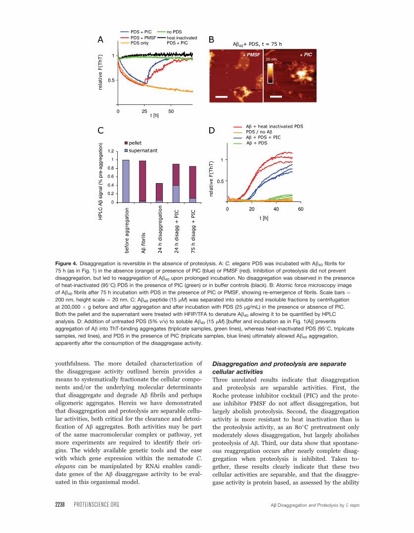
Disaggregation is reversible in the absenceof proteolysis upon exhaustion of the
disaggregase activity
If the disaggregation activity is enabled by a pathway
or a specific protein or protein complex that is separa-
ble from proteolysis, Ab monomers released by the
Figure 2. Disaggregation activity is distinct from proteolysis. A: Atomic force microscopy images of fibrillar Ab42 and Ab40incubated in the presence or absence of C. elegans PDS (25 lg/mL) and Roche complete protease inhibitor cocktail (PIC). Scale
bars ¼ 200 nm, height scale ¼ 20 nm. Incubation with PDS leads to a loss of readily detectable fibrillar structures both in the
presence and absence of PIC. B: Kinetic measurement of Thioflavin T fluorescence of Ab42 incubated for 75 h with PDS in the
presence or absence of PIC. C: Western blot of (mAb 6E10) of monomeric Ab40 peptide (non agg.), Ab40 aggregates (agg. control),and the disaggregation products resulting from PDS treatment (25 lg/mL) in the absence or presence of PIC. Arrows indicate
apparent monomer (M), dimer (D) and high-MW aggregate (A) bands. D: Coomassie staining reveals monomeric and tetrameric
bands of non-aggregated (non agg.) Ab42. Western blot of Ab42 aggregates (agg. control), and the disaggregation products
resulting from PDS treatment (25 lg/mL) in the absence or presence of PIC. Ab42 is degraded and proteolyzed after incubation
with C. elegans PDS for 72 h in the absence of PIC; bands labeled as in C, in addition a soluble aggregate band was observed
(Asol). E: Ab aliquots from disaggregation assays were analyzed by dot blots and filter retardation assays, respectively, after
incubation of Ab40 for 72 h to quantify the amounts of total Ab and SDS-resistant Ab aggregates. F: Quantification of filter
retardation assay and dot blot data. Error bars indicate standard deviations in three independent disaggregation experiments.
SDS-resistant, aggregated Ab is reduced after incubation with C. elegans PDS. Total amounts of Ab peptide remain constant after
incubation in the presence of PIC or phenylmethylsulfonylfluoride (PMSF), but decrease in the absence of protease inhibitors.
2236 PROTEINSCIENCE.ORG Ab Disaggregation and Proteolysis by C. elegans

disaggregase activity could reaggregate when proteases
are inhibited, provided that the disaggregase activity is
significantly reduced or extinguished. Spontaneous
reaggregation of Ab40 after 25 h was indeed observed
[Fig. 4(A)] in the presence of the PIC (blue) and
PMSF (red). AFM imaging of the Ab40 samples after
75 h of incubation with Ab worm PDS in the presence
of PIC or PMSF confirmed the re-emergence of fibril-
lar aggregates. Compare Fig. 4(B) with Fig. 2(A), bot-
tom right, Ab40 with PDS in the absence of PIC.
To verify the mass balance and the aggregation
state of the Ab peptide during the disaggregation and
reaggregation processes, the amounts of aggregated
and monomeric Ab40 peptide were quantified by
HPLC analysis. The Ab40 peptide (15 lM) was incu-
bated in PBS at 37�C for 75 h with rotary agitation (20
rpm) to form fibrillar aggregates and then incubated
for a further 24 or 75 h in the presence of PDS (1%),
either in the presence or absence of PIC. Aliquots of
the peptide were ultracentrifuged (200,000 � g, 20
min) at 24 or 75 h, supernatants were removed and
pellets were solubilized in HFIP/TFA overnight. Super-
natant and pellet fractions were then analyzed by
reverse phase HPLC and quantified by UV absorption
at 215 nm. Peptide fractions were quantified relative to
the initial amount of soluble Ab40 peptide [Fig. 4(C)].
While more than 95% of the Ab peptide was found in
the pellet fraction after aggregation, incubation with
PDS for 24 h reduced the quantity of Ab to �40% (no
PIC). While most of the soluble Ab was degraded by
proteolysis in the absence of PIC, addition of PIC
largely prevented proteolysis of soluble Ab. Interest-
ingly, �80% of Ab was again found in the insoluble
fraction after prolonged (75 h) incubation with PDS
and PIC, consistent with reaggregation.
To further verify that proteolysis prevents reaggre-
gation of Ab40 after incubation with worm PDS, heat-
inactivated PDS (5% v/v) was added to freshly mono-
merized Ab40 (15 lM) and aggregation was monitored
by ThT fluorescence [Fig. 4(D)]. The Ab40 peptide
aggregated [Fig. 4(D), red lines] in the presence of
heat-inactivated PDS (10 min, 95�C) or PDS with
added protease inhibitor cocktail [PIC, Fig. 4(D), blue
lines], on a similar time scale as in the absence of PDS
(Supporting information Fig. S1A). In contrast, proteo-
lytically active PDS largely prevented aggregation
(green lines). The ThT signal of PDS incubated with-
out Ab40 did not increase with time (orange lines).
Taken together, our results demonstrate that dis-
aggregation and proteolysis are distinct C. elegans
PDS activities that can be dissociated and monitored
separately.
Discussion
The C. elegans Ab disaggregation activity appears to
be a primary mechanism for the detoxification of Abaggregates in the worm AD model.22 The disaggregase
activity is partially upregulated by activation of the
HSF-1 transcription factor, which is negatively regu-
lated by the insulin growth factor-1 receptor signaling
pathway that also influences longevity and
Figure 3. Disaggregation activity is heat resistant A: Heat-
treated C. elegans PDS (25 lg/mL) incubated with fibrillar
Ab40 in the absence of protease inhibitor. PDS was heated to
37, 70, 80 or 95�C for 10 min before incubation with Ab40fibrils. B: Dot blot quantification of total Ab40 using mAB
6E10 after 72 h incubation. C: Ab40 SDS-resistant aggregatequantification after 72 h incubation (filter retardation, mAB
6E10). Bar graphs show normalized averaged dot blot
signals of three independent experiments, error bars indicate
standard deviations. Proteolytic activities are inactivated at
70�C, whereas 95�C is needed to inactivate the
disaggregation activity D: Fibrillar Ab42 was incubated with
heat-treated C. elegans PDS as in A in the presence or
absence of PIC. Disaggregation products were analyzed by
Western blotting as in Fig. 2(D). SDS-insoluble aggregate
bands (A) and 20 kDa tetramer bands (T) were analyzed
quantitatively. Bar graphs show intensities of aggregate
(white) and soluble (gray) Ab42 peptide bands relative to the
amounts of Ab before incubation (control) from four
independent experiments, * denotes P < 0.05; ** denotes P
< 0.01. E: C. elegans PDS was pretreated without (blue) or
with proteinase K (100 lg/mL) (red) for 1 h at 37�C before
inactivation of the proteinase K activity by heating to 80�C
for 10 min (conditions that leave the disaggregase intact) and
then added to fibrillar Ab40 (15 lM; Ab monomer).
Bieschke et al. PROTEIN SCIENCE VOL 18:2231—2241 2237
youthfulness. The more detailed characterization of
the disaggregase activity outlined herein provides a
means to systematically fractionate the cellular compo-
nents and/or the underlying molecular determinants
that disaggregate and degrade Ab fibrils and perhaps
oligomeric aggregates. Herein we have demonstrated
that disaggregation and proteolysis are separable cellu-
lar activities, both critical for the clearance and detoxi-
fication of Ab aggregates. Both activities may be part
of the same macromolecular complex or pathway, yet
more experiments are required to identify their ori-
gins. The widely available genetic tools and the ease
with which gene expression within the nematode C.
elegans can be manipulated by RNAi enables candi-
date genes of the Ab disaggregase activity to be eval-
uated in this organismal model.
Disaggregation and proteolysis are separate
cellular activitiesThree unrelated results indicate that disaggregation
and proteolysis are separable activities. First, the
Roche protease inhibitor cocktail (PIC) and the prote-
ase inhibitor PMSF do not affect disaggregation, but
largely abolish proteolysis. Second, the disaggregation
activity is more resistant to heat inactivation than is
the proteolysis activity, as an 80�C pretreatment only
moderately slows disaggregation, but largely abolishes
proteolysis of Ab. Third, our data show that spontane-
ous reaggregation occurs after nearly complete disag-
gregation when proteolysis is inhibited. Taken to-
gether, these results clearly indicate that these two
cellular activities are separable, and that the disaggre-
gase activity is protein based, as assessed by the ability
Figure 4. Disaggregation is reversible in the absence of proteolysis. A: C. elegans PDS was incubated with Ab40 fibrils for
75 h (as in Fig. 1) in the absence (orange) or presence of PIC (blue) or PMSF (red). Inhibition of proteolysis did not prevent
disaggregation, but led to reaggregation of Ab40 upon prolonged incubation. No disaggregation was observed in the presence
of heat-inactivated (95�C) PDS in the presence of PIC (green) or in buffer controls (black). B: Atomic force microscopy image
of Ab40 fibrils after 75 h incubation with PDS in the presence of PIC or PMSF, showing re-emergence of fibrils. Scale bars ¼200 nm, height scale ¼ 20 nm. C: Ab40 peptide (15 lM) was separated into soluble and insoluble fractions by centrifugation
at 200,000 � g before and after aggregation and after incubation with PDS (25 lg/mL) in the presence or absence of PIC.
Both the pellet and the supernatant were treated with HFIP/TFA to denature Ab40 allowing it to be quantified by HPLC
analysis. D: Addition of untreated PDS (5% v/v) to soluble Ab40 (15 lM) [buffer and incubation as in Fig. 1(A)] prevents
aggregation of Ab into ThT-binding aggregates (triplicate samples, green lines), whereas heat-inactivated PDS (95�C, triplicate
samples, red lines), and PDS in the presence of PIC (triplicate samples, blue lines) ultimately allowed Ab40 aggregation,
apparently after the consumption of the disaggregase activity.
2238 PROTEINSCIENCE.ORG Ab Disaggregation and Proteolysis by C. elegans

of proteinase K to disable the disaggregase activity.
Furthermore, our data suggest that proteolysis of amy-
loidogenic peptides after disaggregation may be
required for the detoxification of amyloid aggregates
to prevent their reaggregation into toxic structures.
Candidates to mediate disaggregationThe cellular components or pathway(s) that play a role
in the C. elegans disaggregation/proteolysis activities
are currently unknown. The thermal denaturation re-
sistance of the disaggregase activity is high when com-
pared with the protease activity. Nevertheless, the dis-
aggregase activity can be inactivated by proteolysis,
which suggests that it is mediated by a stable protein,
protein complex, or pathway, possibly involving mem-
branes or vesicle association. This hypothesis is sup-
ported by the finding that detergents compromise the
disaggregation activity even at concentrations below
those which would denature proteins.
In yeast, Hsp104/ClpB, a AAA ATPase family
member, is known to mediate disaggregation33,34 with
a dependence on Hsp70.35 Owing to the lack of an
obvious Hsp104 homolog in mammals, the identity of
the disaggregation-mediating protein, protein complex
or pathway in mammals remains unknown, and is the
subject of ongoing investigations.
The disaggregation assay described herein pro-
vides the means to identify the protein(s) and/or path-
ways responsible for disaggregation and/or proteolysis
of Ab fibrils by analyzing worm homogenate fractio-
nated by chromatographic and related strategies.
Using PDS from worms also enables investigation of
candidate genes using RNAi approaches that are
straightforward to apply in C. elegans proteotoxicity
models. The disaggregase/proteolysis assay reported
herein also enables small molecule screens to be con-
ducted to discover compounds that enhance or inhibit
these critical activities. Disaggregase enhancers may be
useful for ameliorating aging-associated intracellular
Ab aggregation and proteotoxicity thought to cause
neurodegeneration in Alzheimer’s disease.
MATERIALS AND METHODS
Materials
Protein concentration was determined using a BCA kit
(Pierce #23223). Complete protease inhibitor cocktail
(PIC, #1836170) was purchased from Roche (Basel,
Switzerland). Synthetic Ab40 and Ab42 peptides were
purchased from Synpep (Dublin, CA). Monoclonal
anti-Ab antibodies 4G8 and 6E10 were purchased
from Signet (Dedham, MA). All other materials were
purchased from Sigma.
C. elegans
CL2006 and control (wt) worms (N2)21 were obtained
from the Caenorhabditis Genetics Center (Minneapolis,
MN). The worms were grown at 20�C as previously
described.22
In vitro Ab disaggregation assay
C. elegans worms grown to day one of adulthood were
washed twice with M9 buffer (Sigma) and once more
with PBS at room temperature. The worms were then
resuspended in 300 lL ice cold PBS, transferred to a
tissue grinder (885482, Kontes, Vineland, NJ) and ho-
mogenized. Crude homogenates were centrifuged in a
desktop microfuge (Eppendorf 5810R, 3000 rpm, 3
min) to prepare post debris supernatants (PDS). PDS
were transferred to new tubes and total protein con-
centrations were measured with a BCA kit (Pierce,
Rockford, IL).
A stock solution of monomeric Ab40 peptide (200
lM) was prepared as described.29 Ab40 peptide (50
lM) was aggregated in phosphate buffer (300 mM
NaCl, 50 mM Na-phosphate, pH 7.4) at 37�C in a 1.5
mL reaction tube under constant agitation using a
rocking platform (20 cycles/min) (Fig. 1) or an over-
head shaker (20 rpm, Figs. 2–4) for 4 days. Ab40fibrils were sonicated for 30 min in a water bath soni-
cator (FS60, Fisher Scientific, Pittsburg, PA) and char-
acterized by far UV CD spectroscopy and atomic force
microscopy (Fig. S1). Ab40 peptide was then diluted to
a final concentration of 15 lM Ab and 150 mM NaCl
in phosphate buffer (50 mM Na-phosphate, pH 7.4)
containing ThT (20 lM) and PDS worm homogenate
(25 lg/mL) with or without protease inhibitors as
indicated. Synthetic Ab42 was pretreated by an analo-
gous procedure, but using a 30 kDa cut-off filter for
the second filtration step during monomerization. All
final Ab concentrations were 15 lM unless indicated
otherwise.
Three aliquots (100 lL) of each sample were incu-
bated at 37�C in low-binding, 96-well, clear-bottomed
plates (Corning). Samples were agitated for 5 s before
each reading and ThT fluorescence was measured
using a Gemini SpectraMax EM fluorescence plate
reader (Molecular Devices, Sunnyvale, CA) every 10
min. Fluorescence backgrounds from PDS and buffer
were subtracted from the ThT signal. Fluorescence
data were fit to single exponential decay functions
with a time constant s and a linear term allowing for a
sloped postdisaggregation baseline: F (t) ¼ mt þ b þA exp(-t/s). s-values are reported as averages þ/�standard deviations determined from at least three in-
dependent samples. ThT signals were normalized by
setting the fluorescence at t ¼ 0 h to one.
Roche complete protease inhibitor cocktail (PIC)
was diluted from aqueous stock solutions (25 x) to
final concentrations of 1 x dilution.
ATP hydrolysis
To hydrolyze ATP, PDS samples were preincubated for
15 min at 30�C with apyrase (New England Biolabs) at
a final concentration of 0.1 or 0.5 U before adding to
Bieschke et al. PROTEIN SCIENCE VOL 18:2231—2241 2239

the Ab aggregates. Under these conditions, 0.25 U
apyrase were sufficient to completely hydrolyze the
ATP present in the PDS as tested in a luciferase assay
(data not shown). Ab aggregates, PIC and azide
(0.02%) were added after hydrolysis as indicated.
Filter retardation assays and dot blottingEqual volumes of denaturation buffer (Tris buffered
saline (TBS) pH 8, 4% SDS) were added to Ab samples
(20 lL). Samples were boiled for 5 min and filtered
through a cellulose acetate membrane filter (Bio-Rad)
using a 96-well vacuum apparatus (Bio-Rad) as
described.5 For dot blots, Ab samples (20 lL) were
mixed with equal volumes of TBS and filtered through
a nitrocellulose membrane filter (Bio-Rad) using the
same vacuum apparatus.
Western blotting
SDS-PAGE was performed using 16.5% Tris-Tricine
buffered acrylamide gels and the peptides were then
blotted onto a PVDF membrane. Western blots, dot
blots and filter retardation blots were probed using
6E10 (1:2,000) and 4G8 (1:4,000) antibodies and
developed using an ECL system. For reprobing, PVDF
membranes were stripped by incubation in 300 mM
NaOH (5 min, RT), followed by neutralization by sev-
eral rinses in TBST (10 mM Tris-HCl pH 7.5, 150 mM
NaCl, 0.3% Tween-20). Ab monoclonal antibodies
clone 4G8 (#9220) and clone 6E10 (#9320) were pur-
chased from Signet (Dedham, MA), secondary antibod-
ies from Pierce.
Atomic force microscopy
Aliquots (20 lL) were removed from the Ab40 samples
and placed on freshly cleaved mica (1 � 1 cm) and
mounted onto a metal or glass sample holder. AFM
samples were prepared and imaged as described22
using a Nanoscope III (Veeco, Santa Barbara, CA) or
Nanowizard II AFM setup (JPK, Berlin, Germany).
HPLC quantification
Ab peptide aliquots (100 lL, 15 lM) were centrifuged
(200,000 � g 20 min), supernatants were removed
and stored at 4�C and pellets were solubilized over-
night in 50lL of hexafluoroisopropanol/trifluoroacetic
acid (HFIP/TFA 1:1, v/v) after sonicating for 5 min.
Water (50 lL) was added to the HFIP samples. Super-
natant and pellet fractions were then analyzed by
reverse phase HPLC (lRPC C2/C18SC 2.1/10, GE
Healthcare) in a water (0.1% TFA)/acetonitrile (0.1%
TFA) gradient and quantified by UV absorption at 215
nm. UV signals were normalized to the equivalent
amount of soluble Ab40 peptide.
Acknowledgments
The authors thank S. Kostka and G. Grelle for expert
technical support.
References
1. Kelly JW (1998) The alternative conformations of amyloi-dogenic proteins and their multi-step assembly pathways.Curr Opin Struct Biol 8:101–106.
2. Dobson CM (2003) Protein folding and misfolding. Na-ture 426:884–890.
3. Selkoe DJ (2004) Cell biology of protein misfolding: theexamples of Alzheimer’s and Parkinson’s diseases. NatCell Biol 6:1054–1061.
4. Sekijima Y, Wiseman RL, Matteson J, Hammarstrom P,Miller SR, Sawkar AR, Balch WE, Kelly JW (2005) Thebiological and chemical basis for tissue-selective amyloiddisease. Cell 121:73–85.
5. Scherzinger E, Lurz R, Turmaine M, Mangiarini L, Hol-lenbach B, Hasenbank R, Bates GP, Davies SW, LehrachH, Wanker EE (1997) Huntingtin-encoded polyglutamineexpansions form amyloid-like protein aggregates in vitroand in vivo. Cell 90:549–558.
6. Bates G (2003) Huntingtin aggregation and toxicity inHuntington’s disease. Lancet 361:1642–1644.
7. Prusiner SB (1998) The prion diseases. Brain Pathol 8:499–513.
8. Tanzi RE, Bertram L (2005) Twenty years of the Alzhei-mer’s disease amyloid hypothesis: a genetic perspective.Cell 120:545–555.
9. Balch WE, Morimoto RI, Dillin A, Kelly JW (2008)Adapting proteostasis for disease intervention. Science319:916–919.
10. Cohen E, Dillin A (2008) The insulin paradox: aging,proteotoxicity and neurodegeneration. Nat Rev Neurosci9:759–767.
11. Brignull HR, Morley JF, Morimoto RI (2007) The stressof misfolded proteins: C. elegans models for neurodege-nerative disease and aging. Adv Exp Med Biol 594:167–189.
12. Morley JF, Brignull HR, Weyers JJ, Morimoto RI (2002)The threshold for polyglutamine-expansion protein aggre-gation and cellular toxicity is dynamic and influenced byaging in Caenorhabditis elegans. Proc Natl Acad Sci USA99:10417–10422.
13. Dillin A, Crawford DK, Kenyon C (2002) Timing require-ments for insulin/IGF-1 signaling in C. elegans. Science298:830–834.
14. Amaducci L, Tesco G (1994) Aging as a major risk for de-generative diseases of the central nervous system. CurrOpin Neurol 7:283–286.
15. Kenyon C (2005) The plasticity of aging: insights fromlong-lived mutants. Cell 120:449–460.
16. Holzenberger M, Dupont J, Ducos B, Leneuve P, GeloenA, Even PC, Cervera P, Le Bouc Y (2003) IGF-1 receptorregulates lifespan and resistance to oxidative stress inmice. Nature 421:182–187.
17. Suh Y, Atzmon G, Cho MO, Hwang D, Liu B, Leahy DJ,Barzilai N, Cohen P (2008) Functionally significant insu-lin-like growth factor I receptor mutations in centenar-ians. Proc Natl Acad Sci USA 105:3438–3442.
18. Willcox BJ, Donlon TA, He Q, Chen R, Grove JS, Yano K,Masaki KH, Willcox DC, Rodriguez B, Curb JD (2008)FOXO3A genotype is strongly associated with human lon-gevity. Proc Natl Acad Sci USA 105:13987–13992.
19. Hsu AL, Murphy CT, Kenyon C (2003) Regulation ofaging and age-related disease by DAF-16 and heat-shockfactor. Science 300:1142–1145.
20. Morley JF, Morimoto RI (2004) Regulation of longevityin Caenorhabditis elegans by heat shock factor and mo-lecular chaperones. Mol Biol Cell 15:657–664.
21. Link CD (1995) Expression of human beta-amyloid pep-tide in transgenic Caenorhabditis elegans. Proc NatlAcad Sci USA 92:9368–9372.
2240 PROTEINSCIENCE.ORG Ab Disaggregation and Proteolysis by C. elegans

22. Cohen E, Bieschke J, Perciavalle RM, Kelly JW, Dillin A(2006) Opposing activities protect against age-onset pro-teotoxicity. Science 313:1604–1610.
23. Chaudhuri TK, Paul S (2006) Protein-misfolding diseasesand chaperone-based therapeutic approaches. FEBS J273:1331–1349.
24. Shorter J, Lindquist S (2004) Hsp104 catalyzes forma-tion and elimination of self-replicating Sup35 prion con-formers. Science 304:1793–1797.
25. Solomon B, Koppel R, Frankel D, Hanan-Aharon E(1997) Disaggregation of Alzheimer beta-amyloid bysite-directed mAb. Proc Natl Acad Sci USA 94:4109–4112.
26. Schenk D, Barbour R, Dunn W, Gordon G, Grajeda H,Guido T, Hu K, Huang J, Johnson-Wood K, Khan K,Kholodenko D, Lee M, Liao Z, Lieberburg I, Motter R,Mutter L, Soriano F, Shopp G, Vasquez N, Vandevert C,Walker S, Wogulis M, Yednock T, Games D, Seubert P(1999) Immunization with amyloid-beta attenuatesAlzheimer-disease-like pathology in the PDAPP mouse.Nature 400:173–177.
27. Leissring MA, Farris W, Chang AY, Walsh DM, Wu X,Sun X, Frosch MP, Selkoe DJ (2003) Enhanced proteoly-sis of beta-amyloid in APP transgenic mice prevents pla-que formation, secondary pathology, and prematuredeath. Neuron 40:1087–1093.
28. Powers ET, Powers DL (2006) The kinetics of nucleatedpolymerizations at high concentrations: amyloid fibril
formation near and above the ‘‘supercritical concentra-tion’’. Biophys J 91:122–132.
29. Bieschke J, Zhang Q, Powers ET, Lerner RA, Kelly JW(2005) Oxidative metabolites accelerate Alzheimer’s amy-loidogenesis by a two-step mechanism, eliminating therequirement for nucleation. Biochemistry 44:4977–4983.
30. Levine H (1999) Quantification of beta-sheet amyloidfibril structures with thioflavin T. Methods Enzymol 309:274–284.
31. Krebs MR, Bromley EH, Donald AM (2005) The bindingof thioflavin-T to amyloid fibrils: localisation and implica-tions. J Struct Biol 149:30–37.
32. Klement K, Wieligmann K, Meinhardt J, Hortschansky P,Richter W, Fandrich M (2007) Effect of different saltions on the propensity of aggregation and on the struc-ture of Alzheimer’s abeta(1–40) amyloid fibrils. J MolBiol 373:1321–1333.
33. Shorter J, Lindquist S (2006) Destruction or potentiationof different prions catalyzed by similar Hsp104 remodel-ing activities. Mol Cell 23:425–438.
34. Parsell DA, Kowal AS, Singer MA, Lindquist S (1994)Protein disaggregation mediated by heat-shock proteinHsp104. Nature 372:475–478.
35. Glover JR, Lindquist S (1998) Hsp104, Hsp70, andHsp40: a novel chaperone system that rescues previouslyaggregated proteins. Cell 94:73–82.
Bieschke et al. PROTEIN SCIENCE VOL 18:2231—2241 2241
Copyright © 2022 FDOKUMEN