Uncoupling Protein 3 (UCP3) Modulates the Activity of Sarco/Endoplasmic Reticulum Ca2+-ATPase...
9
Uncoupling Protein 3 (UCP3) Modulates the Activity of Sarco/Endoplasmic Reticulum Ca 2 -ATPase (SERCA) by Decreasing Mitochondrial ATP Production * □ S Received for publication, January 4, 2011, and in revised form, June 17, 2011 Published, JBC Papers in Press, July 20, 2011, DOI 10.1074/jbc.M110.216044 Umberto De Marchi, Cyril Castelbou, and Nicolas Demaurex 1 From the Department of Cell Physiology and Metabolism, University of Geneva, rue Michel-Servet, 1, CH-1211 Gene `ve, Switzerland The uncoupling proteins UCP2 and UCP3 have been postu- lated to catalyze Ca 2 entry across the inner membrane of mito- chondria, but this proposal is disputed, and other, unrelated proteins have since been identified as the mitochondrial Ca 2 uniporter. To clarify the role of UCPs in mitochondrial Ca 2 handling, we down-regulated the expression of the only uncou- pling protein of HeLa cells, UCP3, and measured Ca 2 and ATP levels in the cytosol and in organelles with genetically encoded probes. UCP3 silencing did not alter mitochondrial Ca 2 uptake in permeabilized cells. In intact cells, however, UCP3 depletion increased mitochondrial ATP production and strongly reduced the cytosolic and mitochondrial Ca 2 elevations evoked by his- tamine. The reduced Ca 2 elevations were due to inhibition of store-operated Ca 2 entry and reduced depletion of endoplas- mic reticulum (ER) Ca 2 stores. UCP3 depletion accelerated the ER Ca 2 refilling kinetics, indicating that the activity of sarco/ endoplasmic reticulum Ca 2 (SERCA) pumps was increased. Accordingly, SERCA inhibitors reversed the effects of UCP3 depletion on cytosolic, ER, and mitochondrial Ca 2 responses. Our results indicate that UCP3 is not a mitochondrial Ca 2 uniporter and that it instead negatively modulates the activity of SERCA by limiting mitochondrial ATP production. The effects of UCP3 on mitochondrial Ca 2 thus reflect metabolic altera- tions that impact on cellular Ca 2 homeostasis. The sensitivity of SERCA to mitochondrial ATP production suggests that mito- chondria control the local ATP availability at ER Ca 2 uptake and release sites. Mitochondria are multifunctional organelles that control the life and death of cells. Mitochondria are the site of oxidative phosphorylation and convert reducing equivalents into the ATP that cells use as energy source, release critical factors that induce the programmed cell death of apoptosis, and modulate cell signaling by capturing and subsequently releasing calcium ions. The ability of mitochondria to sequester Ca 2 ions released from the endoplasmic reticulum or entering across plasma membrane channels enables these organelles to shape cytosolic Ca 2 signals and to modulate the activity of mem- brane channels and transporters (1–3), whereas Ca 2 eleva- tions within the matrix of mitochondria activate three enzymes of the tricarboxylic cycle, thereby boosting oxidative phosphor- ylation and ATP production. Calcium uptake and release by mitochondria are therefore essential for the patterning of cyto- solic Ca 2 signals and for cells to decode Ca 2 signals as either metabolic or death signals. The importance of mitochondrial Ca 2 handling for cell physiology has fostered a long quest to identify the transport molecules that move Ca 2 in and out of mitochondria (4). This quest culminated in recent years with the report of several families of proteins that were proposed to participate in either mitochondrial Ca 2 uptake or mitochon- drial Ca 2 extrusion: the uncoupling proteins 2 and 3 (UCP2 2 and UCP3), the leucine zipper EF-hand-containing transmem- brane protein 1 (Letm1), the Na /Ca 2 exchanger (NCLX), the stomatin-like protein 2 (SLP-2), and the mitochondrial calcium uptake protein 1 (MICU1). Ca 2 enters mitochondria across a mitochondrial Ca 2 uniporter (MCU) that was characterized in 2004 at the electro- physiological level as a highly selective inward rectifying mito- chondrial inner membrane Ca 2 channel, inhibited by ruthe- nium red and Ru360 (5). In 2007, UCP2 and UCP3 were proposed to be fundamental for the MCU (6), based on the altered mitochondrial Ca 2 elevations ([Ca 2 ] mit ) of cells enriched or depleted of UCP2 or UCP3 and on the defective ruthenium red-sensitive Ca 2 uptake of liver mitochondria iso- lated from UCP2 / mice. This proposal was subsequently refuted by a study showing that purified liver mitochondria from UCP2 / and UCP3 / mice take up calcium normally (7). In 2009, Letm1, a protein previously shown to catalyze mitochondrial K /H exchange (8, 9), was shown to drive mitochondrial Ca 2 uptake by exchanging Ca 2 for H with a 1:1 stoichiometry (10). This stoichiometry, however, contra- dicts earlier studies in isolated mitochondria showing that Ca 2 enters mitochondria together with two positive charges and leaves the matrix in exchange for three protons (reviewed in Ref. 4). Letm1 is associated with Wolf-Hirschhorn syndrome, a severe human neurological disease characterized by mental retardation and seizures (11). In 2010, MICU1, an inner mito- * This work was supported by the Swiss National Foundation Grant 31-068317 (to N. D.). □ S The on-line version of this article (available at http://www.jbc.org) contains supplemental Figs. S1–S8. Author’s Choice—Final version full access. 1 To whom correspondence should be addressed. Tel.: 41-22-379-5399; Fax: 41-22-379-5338; E-mail: [email protected]. 2 The abbreviations used are: UCP, uncoupling protein; ER, endoplasmic reticu- lum; SERCA, sarco/endoplasmic reticulum Ca 2 -ATPase; MCU, mitochondrial Ca 2 uniporter; SOCE, store-operated Ca 2 entry; BHQ, 2,5-di-t-butyl-1,4-ben- zohydroquinone; [Ca 2 ] cyto , cytosolic [Ca 2 ]; [Ca 2 ] ER , endoplasmic retic- ulum [Ca 2 ]; [Ca 2 ] mit , mitochondrial [Ca 2 ]; TG, thapsigargin; YC, yellow cameleon; Ctrl, control; HEDTA, N-(2-hydroxyethyl)ethylenediaminetri- acetic acid. THE JOURNAL OF BIOLOGICAL CHEMISTRY VOL. 286, NO. 37, pp. 32533–32541, September 16, 2011 Author’s Choice © 2011 by The American Society for Biochemistry and Molecular Biology, Inc. Printed in the U.S.A. SEPTEMBER 16, 2011 • VOLUME 286 • NUMBER 37 JOURNAL OF BIOLOGICAL CHEMISTRY 32533 at Bibliotheque Faculte Medecine Geneve, on September 13, 2011 www.jbc.org Downloaded from http://www.jbc.org/content/suppl/2011/07/20/M110.216044.DC1.html Supplemental Material can be found at:
-
Upload
independent -
Category
Documents
-
view
0 -
download
0
Transcript of Uncoupling Protein 3 (UCP3) Modulates the Activity of Sarco/Endoplasmic Reticulum Ca2+-ATPase...
Uncoupling Protein 3 (UCP3) Modulates the Activity ofSarco/Endoplasmic Reticulum Ca2�-ATPase (SERCA) byDecreasing Mitochondrial ATP Production*□S
Received for publication, January 4, 2011, and in revised form, June 17, 2011 Published, JBC Papers in Press, July 20, 2011, DOI 10.1074/jbc.M110.216044
Umberto De Marchi, Cyril Castelbou, and Nicolas Demaurex1
From the Department of Cell Physiology and Metabolism, University of Geneva, rue Michel-Servet, 1, CH-1211 Geneve, Switzerland
The uncoupling proteins UCP2 and UCP3 have been postu-lated to catalyze Ca2� entry across the innermembrane ofmito-chondria, but this proposal is disputed, and other, unrelatedproteins have since been identified as the mitochondrial Ca2�
uniporter. To clarify the role of UCPs in mitochondrial Ca2�
handling, we down-regulated the expression of the only uncou-pling protein of HeLa cells, UCP3, andmeasured Ca2� andATPlevels in the cytosol and in organelles with genetically encodedprobes.UCP3 silencing did not altermitochondrial Ca2�uptakein permeabilized cells. In intact cells, however, UCP3 depletionincreased mitochondrial ATP production and strongly reducedthe cytosolic and mitochondrial Ca2� elevations evoked by his-tamine. The reduced Ca2� elevations were due to inhibition ofstore-operated Ca2� entry and reduced depletion of endoplas-mic reticulum (ER)Ca2� stores. UCP3 depletion accelerated theER Ca2� refilling kinetics, indicating that the activity of sarco/endoplasmic reticulum Ca2� (SERCA) pumps was increased.Accordingly, SERCA inhibitors reversed the effects of UCP3depletion on cytosolic, ER, and mitochondrial Ca2� responses.Our results indicate that UCP3 is not a mitochondrial Ca2�
uniporter and that it instead negativelymodulates the activity ofSERCA by limiting mitochondrial ATP production. The effectsof UCP3 on mitochondrial Ca2� thus reflect metabolic altera-tions that impact on cellular Ca2� homeostasis. The sensitivityof SERCA tomitochondrial ATPproduction suggests thatmito-chondria control the local ATP availability at ER Ca2� uptakeand release sites.
Mitochondria aremultifunctional organelles that control thelife and death of cells. Mitochondria are the site of oxidativephosphorylation and convert reducing equivalents into theATP that cells use as energy source, release critical factors thatinduce the programmed cell death of apoptosis, and modulatecell signaling by capturing and subsequently releasing calciumions. The ability of mitochondria to sequester Ca2� ionsreleased from the endoplasmic reticulum or entering acrossplasma membrane channels enables these organelles to shapecytosolic Ca2� signals and to modulate the activity of mem-
brane channels and transporters (1–3), whereas Ca2� eleva-tions within thematrix of mitochondria activate three enzymesof the tricarboxylic cycle, thereby boosting oxidative phosphor-ylation and ATP production. Calcium uptake and release bymitochondria are therefore essential for the patterning of cyto-solic Ca2� signals and for cells to decode Ca2� signals as eithermetabolic or death signals. The importance of mitochondrialCa2� handling for cell physiology has fostered a long quest toidentify the transport molecules that move Ca2� in and out ofmitochondria (4). This quest culminated in recent years withthe report of several families of proteins that were proposed toparticipate in either mitochondrial Ca2� uptake or mitochon-drial Ca2� extrusion: the uncoupling proteins 2 and 3 (UCP2 2
and UCP3), the leucine zipper EF-hand-containing transmem-brane protein 1 (Letm1), theNa�/Ca2� exchanger (NCLX), thestomatin-like protein 2 (SLP-2), and themitochondrial calciumuptake protein 1 (MICU1).Ca2� enters mitochondria across a mitochondrial Ca2�
uniporter (MCU) that was characterized in 2004 at the electro-physiological level as a highly selective inward rectifying mito-chondrial inner membrane Ca2� channel, inhibited by ruthe-nium red and Ru360 (5). In 2007, UCP2 and UCP3 wereproposed to be fundamental for the MCU (6), based on thealtered mitochondrial Ca2� elevations ([Ca2�]mit) of cellsenriched or depleted of UCP2 or UCP3 and on the defectiveruthenium red-sensitive Ca2� uptake of livermitochondria iso-lated from UCP2�/� mice. This proposal was subsequentlyrefuted by a study showing that purified liver mitochondriafrom UCP2�/� and UCP3�/� mice take up calcium normally(7). In 2009, Letm1, a protein previously shown to catalyzemitochondrial K�/H� exchange (8, 9), was shown to drivemitochondrial Ca2� uptake by exchanging Ca2� for H� with a1:1 stoichiometry (10). This stoichiometry, however, contra-dicts earlier studies in isolatedmitochondria showing thatCa2�
enters mitochondria together with two positive charges andleaves the matrix in exchange for three protons (reviewed inRef. 4). Letm1 is associated withWolf-Hirschhorn syndrome, asevere human neurological disease characterized by mentalretardation and seizures (11). In 2010, MICU1, an inner mito-
* This work was supported by the Swiss National Foundation Grant31-068317 (to N. D.).
□S The on-line version of this article (available at http://www.jbc.org) containssupplemental Figs. S1–S8.Author’s Choice—Final version full access.
1 To whom correspondence should be addressed. Tel.: 41-22-379-5399; Fax:41-22-379-5338; E-mail: [email protected].
2 The abbreviations used are: UCP, uncoupling protein; ER, endoplasmic reticu-lum; SERCA, sarco/endoplasmic reticulum Ca2� -ATPase; MCU, mitochondrialCa2� uniporter; SOCE, store-operated Ca2� entry; BHQ, 2,5-di-t-butyl-1,4-ben-zohydroquinone; [Ca2�]cyto, cytosolic [Ca2�]; [Ca2�]ER, endoplasmic retic-ulum [Ca2�]; [Ca2�]mit, mitochondrial [Ca2�]; TG, thapsigargin; YC, yellowcameleon; Ctrl, control; HEDTA, N-(2-hydroxyethyl)ethylenediaminetri-acetic acid.
THE JOURNAL OF BIOLOGICAL CHEMISTRY VOL. 286, NO. 37, pp. 32533–32541, September 16, 2011Author’s Choice © 2011 by The American Society for Biochemistry and Molecular Biology, Inc. Printed in the U.S.A.
SEPTEMBER 16, 2011 • VOLUME 286 • NUMBER 37 JOURNAL OF BIOLOGICAL CHEMISTRY 32533
at Bibliotheque F
aculte Medecine G
eneve, on Septem
ber 13, 2011w
ww
.jbc.orgD
ownloaded from
http://www.jbc.org/content/suppl/2011/07/20/M110.216044.DC1.html Supplemental Material can be found at:
chondrial membrane protein with two Ca2�-binding EF-handdomains, was shown to be required for high capacitymitochon-drial calcium uptake (12). MICU1 is a single-pass transmem-brane protein unlikely to form a channel pore and was pro-posed to be the calcium sensing regulatory subunit of theMCU because mutations of its EF-hand domains abrogatemitochondrial Ca2� uptake (13). Two proteins werereported to modulate Ca2� extrusion from mitochondria.NCLX/NCKX6 was shown to catalyze CGP-37157-sensitivemitochondrial Na�/Ca2� exchange (14), and SLP-2 wasshown to negatively modulate the activity of the mitochon-drial sodium-calcium exchanger (15).The successive reports of proteins essential for mitochon-
drial Ca2� uptake have generated some confusion about theidentity and mode of operation of the MCU. The first proteinsproposed to be fundamental for the MCU, UCP2 and UCP3(16), are mitochondrial inner membrane protein paralogues ofUCP1, the first UCP cloned. UCP1 is almost exclusivelyexpressed in brown adipose tissue, where it catalyzes adaptivethermogenesis by acting as a mitochondrial proton channel,thereby uncoupling oxidative phosphorylation from ATP syn-thesis (17). Unlike UCP1, UCP2 and UCP3 are expressed inseveral tissues and are present in ectothermic fishes and plantsthat do not require thermogenesis. The basal H� conductanceis unchanged in mitochondria isolated from UCP2 or UCP3null mice (18, 19), and the novel UCP homologues have beenproposed to mediate regulated proton leak (20), to modulateinsulin secretion (21), and to regulate the export of fatty acidsand fatty acid peroxides(22, 23). The ability of UCP2/3 to act asproton leak channels seems strictly related to the presence ofspecific activators such as alkenyls produced by the peroxida-tion ofmembrane phospholipids (24, 25). Thus, there is a broadconsensus that UCP2 and UCP3 do not mediate adaptive ther-mogenesis (25–27). Instead, the novel UCPs might act as milduncouplers and protect against oxidative damage by attenuat-ing the mitochondrial production of free radicals (28, 29).The report that the amplitude of [Ca2�]mit elevations in
intact cells directly correlates to the expression level of UCP2and UCP3 (6) might explain the plethoric effects attributed toUCPs because alterations in [Ca2�]mit signals are expected toimpact both on mitochondria bioenergetics and on cell signal-ing. Whether the altered [Ca2�]mit signals are due to defectivemitochondrial Ca2� uptake is unclear due to the conflictingreports obtained in isolated liver mitochondria from UCP2�/�
null mice (6, 7). These opposite datasets could be reconciled bypostulating a regulatory role for UCP2 and UCP3 in intact cellsthat would be lost in isolated mitochondria. Importantly, thealtered [Ca2�]mit signals reported by Trenker et al. (6) in intactcells depleted or enriched of UCP2/3 have not been confirmedor disproved. To clarify the physiological role of UCPs in Ca2�
homeostasis, we used RNA interference to down-regulateUCP3, the only UCP isoform of HeLa cells (6), and measuredthe impact of UCP3 depletion on the Ca2� and ATP levels indifferent cellular compartments.
EXPERIMENTAL PROCEDURES
Reagents—Minimal essential medium, fetal calf serum, pen-icillin, streptomycin, and Lipofectamine 2000 transfection rea-
gent were obtained from Invitrogen. Histamine, thapsigargin(TG), antimycin A, and oligomycin were obtained from Sigma.2,5-Di-t-butyl-1,4-benzohydroquinone (BHQ) were fromAldrich. Fura-2AMwas fromMolecular Probes. YC3.6cyto (30),4mtD3cpv (31), and D1ER (32) constructs were kindly providedby Drs. Amy Palmer and Roger Tsien (University of California,San Diego). Mitochondrial and cytosolic FRET-based ATPindicators (ATeam: adenosine 5�-triphosphate indicator basedon the � subunit for AnalyticalMeasurements) were kindly pro-vided by Drs. Hiromi Imamura (Japan Science and TechnologyAgency, Tokyo) and Hiroyuki Noji (Osaka University) (33).UCP2 and UCP3/mitochondria-targeted DsRed constructswere kindly provided by Dr.Wolfgang Graier (Medical Univer-sity of Graz).Cell Culture, Transfection, and RNA Interference—Culturing
of HeLa cells has been previously described (34). For all exper-iments, cells were plated on 25-mm-diameter glass coverslipsand co-transfected with the plasmid (1 �g/ml) coding for Ca2�
or pH probe and dsRNA using Lipofectamine 2000. All exper-iments were performed 2 days after transfection. For silencingUCP3 expression, commercial double-stranded RNAs fromQiagen were used (Hi PerFect-validated siRNA S102780771).For controls, cells were transfected with nonsilencing siRNA(AllStars negative control siRNA, Qiagen 1027281). Knock-down efficiency was verified by Western blots.Cell Lysis, Mitochondrial Isolation, and Western Blotting—
Whole cells were lysed for 30 min on ice in lysis buffer (25 mM
Tris-HCl, pH 7.6, 150 mM NaCl, 1% Nonidet P-40, 1% sodiumdeoxycholate, 0.1% SDS) supplemented with protease inhibi-tors (Roche Applied Science). The lysate was centrifuged at14,000 � g for 20 min, and the protein content of the superna-tant was determined using the BCA protein assay (Pierce). Themitochondrial fraction was obtained by differential centrifuga-tion as reported previously (35). 50�g of total protein (from celllysate or isolated mitochondria) was loaded per lane of SDS-PAGE. For immunoblotting, proteins were transferred ontonitrocellulose membrane and probed with the following anti-bodies: anti-UCP3 (Santa Cruz Biotechnology, sc-7756) andanti-Tom20 (Santa Cruz Biotechnology, sc-11415), anti-SERCA2 (ThermoScientificMA3-919), and anti-actin (Chemi-con). Horseradish peroxidase-conjugated secondary antibodies(Amersham Biosciences) were used followed by detection bychemiluminescence (Amersham Biosciences).Mitochondrial, Cytosolic, and Endoplasmic Reticulum Ca2�
Measurements—Experiments were performed inHEPES buffersolution containing (inmM): 140NaCl, 5 KCl, 1MgCl2, 2 CaCl2,20 Hepes, 10 glucose, pH 7.4, with NaOH at 37 °C. Ca2�-freesolution contained 1 mM EGTA instead of CaCl2. Glass cover-slips were inserted in a thermostatic chamber (Harvard Appa-ratus, Holliston, MA), and solutions were changed by hand.Cells were imaged on an Axiovert s100 TV using a�40, 1.3 NAoil immersion objective (Carl Zeiss AG, Feldbach, Switzerland)and a cooled, 16-bit CCD back-illuminated frame transferMicroMax camera (Roper Scientific, Trenton, NJ). CytosolicCa2� was measured with fura-2 (2 �M, 0.2% dimethyl sulfoxide(DMSO), 0.01% Pluronic F127, Invitrogen) followed by 20 minof de-esterification before 10min of equilibration on the heatedstage or with YC3.6cyto. Mitochondrial and endoplasmic retic-
UCP3 Modulates Activity of SERCA
32534 JOURNAL OF BIOLOGICAL CHEMISTRY VOLUME 286 • NUMBER 37 • SEPTEMBER 16, 2011
at Bibliotheque F
aculte Medecine G
eneve, on Septem
ber 13, 2011w
ww
.jbc.orgD
ownloaded from

ulum Ca2� levels were measured with 4mtD3cpv and D1ER,respectively. For dual emission imaging of cameleon constructs(4mtD3cpv, YC3.6cyto, and D1ER), cells were excited at 430 nmthrough a 455DRLP dichroic and alternately imaged with480AF30 and 535DF25 emission filters (Omega Optical).Fura-2 was measured simultaneously with 4mtD3cpv and wasexcited alternately at 340 and 380 nm through a 455DRLPdichroic and 535DF25 emission filter. Images were acquiredevery 2 s. Fluorescence ratios were calculated in MetaFluor 6.3(Universal Imaging) and analyzed in Excel (Microsoft) andGraphPad Prism 4 (GraphPad). [Ca]ER was calculated fromD1ER ratios using the equation
R � Rmin �[Rmax � Rmin]
1 � 10(LogK�d � Log[Ca2�]ER)h (Eq. 1)
where Rmin and Rmax are the minimal and maximal ratioobtained at pCa �8 and �2, respectively, K�d is the apparentdissociation constant, and h is the Hill coefficient derived fromthe in situ Ca2� titration of the D1ER probe in semipermeabi-lized cells, as described previously (36).Mitochondrial and Cytosolic [ATP] Measurements—ATP
imaging was performed on the epifluorescence systemdescribed above, with the genetically encoded ATP sensorsATeammito and ATeamcyto, for mitochondrial and cytosolicATP, respectively. For the dual emission imaging of these twocameleon-based constructs, cells were excited and imaged asdescribed previously for mitochondrial Ca2� imaging. Imageswere acquired every 2 s. Fluorescence ratios were normalizedon the minimum of fluorescence (Rmin), obtained after inhibi-tion of glycolysis with 10 mM 2-deoxyglucose and in the pres-ence of the inhibitor of mitochondrial ATP synthesis oligomy-cin A (10 �g/ml). Data were analyzed as described previously.Permeabilized Cells—Cells were washed with high K� intra-
cellular buffer, containing (in mM) 110 KCl, 10 NaCl, 0.5K2HPO4, 5 succinate, 10 mM HEPES (pH 7.0 at 37 °C), supple-mented with 5 HEDTA or 1 EGTA. For permeabilization, 100�M digitonin was added for 1 min, and the cells were thenallowed to recover in intracellular buffer before the addition ofCaCl2. Free [Ca2�] was calculated with the Maxchelatorprogram.Statistics—The significance of differences between means
was established using the Student’s t test for unpaired samples(*, p � 0.05; **, p � 0.01; ***, p � 0.001).
RESULTS
Effect of UCP3 Depletion on Mitochondrial and CytosolicCa2� Elevations—To clarify the role of UCPs in mitochondrialCa2� handling, wemeasured Ca2� responses in the cytosol andinmitochondria inHeLa cells depleted or not of UCP3, the onlynovel UCP isoform expressed in this cell type (6). Consistentwith earlier results (6), this treatment decreased UCP3 proteinlevels by 50% (supplemental Fig. S1). Cells were then trans-fected with a cameleon Ca2� probe targeted to mitochondria(Fig. 1A) or to the cytosol (Fig. 1B) to enable [Ca2�]mit and[Ca2�]cyt recordings. As shown in Fig. 1A, UCP3 depletionstrongly reduced the [Ca2�]mit elevations evoked by histamine,the peak [Ca2�]mit response being reduced by 57% and the inte-
grated response being reduced by 62%. To exclude possible off-target effects linked to siRNA transfection, we overexpressedUCP3 protein and, in another set of experiments, attempted torescue the defect caused byUCP3 siRNAby expressing anotheruncoupling protein, UCP2. As shown in supplemental Fig. S2A,UCP3 overexpression significantly increased the peak and inte-grated [Ca2�]mit responses evoked by histamine, mirroring theeffects of UCP3 depletion, whereas expression of UCP2restored normal [Ca2�]mit responses in cells depleted of UCP3(supplemental Fig. S3A). These data indicate that the alteredmitochondrial Ca2� signals are indeed causally related to thechanges in UCP3 levels. The UCP3-dependent Ca2� defect wasnot restricted to mitochondria, however, because the cytosolicCa2� elevations were also reduced in cells depleted of UCP3,the peak and integrated [Ca2�]cyt responses being decreased by25 and 44%, respectively (Fig. 1B). To confirm that the altera-
FIGURE 1. Effect of UCP3 knockdown on mitochondrial and cytosolic Ca2�
elevations. A and B, HeLa cells were transiently co-transfected with the mito-chondrial Ca2� probe 4mtD3cpv (A) or the cytosolic Ca2� probe YC3.6cyto (B)and with either scrambled siRNA (control siRNA) or UCP3-specific siRNA (UCP3siRNA) for 48 h. A, left, averaged [Ca2�]mit recordings in HeLa cells stimulatedwith 100 �M histamine. Right, statistical evaluation of the UCP3 siRNA effectson the amplitude of the [Ca2�]mit signal evoked by histamine (upper panel)and on the integrated [Ca2�]mit response (lower panel). Bars are mean � S.E. of80 (n � 7) and 101 cells (n � 7) for Ctrl and UCP3 siRNA, respectively. AUC, areaunder the curve. B, left, averaged [Ca2�]cyto recordings in HeLa cells stimu-lated with 100 �M histamine. Right, statistical evaluation of the UCP3 siRNAeffects on the amplitude of the [Ca2�]cyto signal evoked by histamine (upperpanel) and on the integrated [Ca2�]cyto response (lower panel). Bars aremean � S.E. of 40 (n � 3) and 37 cells (n � 3) for Ctrl and UCP3 siRNA, respec-tively. ***, p � 0.001.
UCP3 Modulates Activity of SERCA
SEPTEMBER 16, 2011 • VOLUME 286 • NUMBER 37 JOURNAL OF BIOLOGICAL CHEMISTRY 32535
at Bibliotheque F
aculte Medecine G
eneve, on Septem
ber 13, 2011w
ww
.jbc.orgD
ownloaded from

tion in Ca2� handling affected both compartments, we mea-sured [Ca2�]cyt and [Ca2�]mit simultaneously instead of sepa-rately. In these conditions, both the cytosolic and the mito-chondrial Ca2� responses were blunted in cells depleted ofUCP3 (supplemental Fig. S4). These data indicate that UCP3knockdown has a global effect and not only blunts mitochon-drial Ca2� elevations but also reduces cytosolic Ca2�
elevations.Effect ofUCP3Depletion onAgonist-evokedCa2�Release and
Influx—The novel UCPs have been recently proposed to spe-cifically mediate the mitochondrial uptake of Ca2� releasedfrom the ER, but not of the Ca2� entering across SOCE chan-nels (37). To verify this possibility, we separated the agonist-evoked Ca2� response into its release and influx components.Cells were stimulated with histamine in Ca2�-free conditionstomobilize Ca2� from stores, andCa2�was subsequently read-mitted to promote Ca2� entry. As shown in Fig. 2, the [Ca2�]cytand [Ca2�]mit elevations caused by Ca2� release from storeswere slightly but significantly decreased. This inhibition, how-ever, was much smaller than the inhibition observed in Ca2�-containing medium (Fig. 1). Unexpectedly, the [Ca2�]cyt eleva-tions evoked byCa2� readmissionwere severely blunted in cells
depleted of UCP3, the integrated [Ca2�]cyt response beingreduced by 82% (Fig. 2A). An opposite effect was obtained byoverexpressing UCP3, the [Ca2�]cyt responses increasingslightly (14%) during Ca2� release from stores and markedly(75%) during Ca2� influx (supplemental Fig. S2B). Moreover,UCP2 expression restored a normal Ca2� influx in UCP3-de-pleted cells (supplemental Fig. S3B), confirming that this defectwas due to the depletion of an uncoupling protein. As reportedpreviously (38), the [Ca2�]mit elevations evoked by this Ca2�
readmission protocol were barely detectable and were notaltered by UCP3 depletion (Fig. 2B).These observations indicate that UCP3 depletion slightly
impairs Ca2� release from stores and, surprisingly, stronglyinhibits the cytosolic Ca2� elevations caused by Ca2� readmis-sion to cells with depleted Ca2� stores. The primary effect ofUCP3 depletion thus appears to be a reduced entry of Ca2�
across SOCE channels.Effect of UCP3 Depletion on ER Ca2� Release and Refilling—
SOCE channels are activated by the depletion of ER Ca2�
stores, which induces the translocation of the ER-resident Ca2�
sensor STIM1 to the plasma membrane, where it binds andactivates the Orai and Transient Receptor Potential channels(39). The reduced Ca2� entry of UCP3 knockdown cells sug-gested that the SOCEmachinerywas inhibited, prompting us tostudy the filling state of ERCa2� stores.We thereforemeasured[Ca2�]ER at rest and during store depletion with the ER-tar-geted cameleonCa2� indicatorD1ER.As shown in Fig. 3A, D1ERbasal ratio fluorescence levels were identical in control andUCP3-depleted cells, the basal [Ca2�]ER levels averaging 520�M in both conditions (supplemental Fig. S5, A and B). Uponstimulation with histamine, however, [Ca2�]ER decreasedmuch more slowly in cells depleted of UCP3, the half-timeincreasing from 72 to 269 s (Fig. 3B, inset). This four timesslower [Ca2�]ER decrease was surprising considering thatUCP3 knockdown cells released Ca2� efficiently when stimu-lated with agonists (Fig. 2A). Reduced depletion of ER Ca2�
stores could reflect either a reduced passive permeability toCa2� or an increasedCa2� pumping into the ER. To distinguishbetween these possibilities, we inhibited SERCA pumps withTG to reveal the intrinsic Ca2� leak rates of the ER. As shown inFig. 3C, the addition of TG elicited identical [Ca2�]ER decreasesin control and UCP3 knockdown cells, indicating that the pas-sive Ca2� permeability of the ER was not affected by the deple-tion of UCP3. The addition of TG together with histamineaccelerated the kinetics of [Ca2�]ER decrease by 2-fold inUCP3-depleted cells, but these cells still released Ca2� moreslowly than control cells (supplemental Fig. S5C). Because[Ca2�]ER decreased significantly 40 s after TG addition (sup-plemental Fig. S5D), we then addedTG40 s before histamine toensure full inhibition of SERCA. In these conditions, the kinet-ics of [Ca2�]ER decrease evoked by histamine were similar incontrol and UCP3-depleted cells (Fig. 3D). These data indicatethat the both the passive and the active (i.e. InsP3R-mediated)Ca2� permeability of the ER are not affected by the depletion ofUCP3. Similar effects were observedwith the reversible SERCAinhibitor BHQ (not shown), confirming the involvement ofSERCA. Of note, BHQ required more time than TG to com-pletely inhibit SERCApumps (supplemental Fig. S5E; see under
FIGURE 2. Effect of UCP3 knockdown on Ca2� release and influx. A and B,HeLa cells were transiently co-transfected with the cytosolic Ca2� probeYC3.6cyto (A) or the mitochondrial Ca2� probe 4mtD3cpv (B) and with Ctrl(scramble) siRNA or the UCP3 siRNA for 48 h, washed, and stimulated with 100�M histamine in Ca2�-free medium to deplete intracellular Ca2� stores. ThenCa2� was readmitted to monitor Ca2� influx from plasma membrane. A, aver-age of cytosolic Ca2� responses (left) elicited by histamine and by Ca2� read-mission. Right, statistical evaluation of the UCP3 siRNA on the integrated[Ca2�]cyto response during release from ER and during Ca2� readmission. Barsare mean � S.E. of 84 (n � 9) and 86 cells (n � 8) for Ctrl and UCP3 siRNA,respectively. AUC, area under the curve. B, average of mitochondrialCa2�responses elicited by histamine and Ca2� readmission (left) and relatedstatistical evaluation (right) on integrated [Ca2�]cyto response during Ca2�
release and influx. Bars are mean � S.E. of 77 (n � 8) and 59 cells(n � 7) for Ctrland UCP3 siRNA, respectively. *, p � 0.05; ***, p � 0.001. NS, not significant.
UCP3 Modulates Activity of SERCA
32536 JOURNAL OF BIOLOGICAL CHEMISTRY VOLUME 286 • NUMBER 37 • SEPTEMBER 16, 2011
at Bibliotheque F
aculte Medecine G
eneve, on Septem
ber 13, 2011w
ww
.jbc.orgD
ownloaded from

“Discussion” to compare our results with recent data of theGraiergroup (37)). The restoration of normal ER Ca2� release in cellsdepleted of UCP3 by SERCA inhibitors suggested that the activityof SERCA was increased in these cells. The expression levels ofSERCA2b assessed byWestern blot (Fig. 4A) and real-time quan-titative PCR (not shown) were not influenced by UCP3 depletion.To directly estimate the activity of SERCA, intact cells were tran-siently treated with the reversible inhibitor BHQ to deplete ERCa2� stores, andCa2�was thenreadmitted topromotestore refill-ing. As shown in Fig. 4B, the kinetics of ER refilling were faster incells depleted of UCP3, the time required to reach half-maximalrefilling being increased by 14 s, confirming that SERCA pumpsweremoreactive in these cells.Thisdifferencewasnotobserved inpermeabilized cells, however, the kinetics of ER refilling beingidentical in this condition (Fig. 4C). Our [Ca2�]ER measurementsthus indicate that the activity of SERCA pumps is increased inintact cells depleted of UCP3.Effect of SERCA Inhibition onMitochondrial Ca2� Uptake in
UCP3-depleted Cells—The effects of SERCA inhibitors on[Ca2�]ER handling prompted us to investigate whether SERCA
inhibition could normalize mitochondrial Ca2� uptake in cellsdepleted of UCP3. To test this possibility, we measured the[Ca2�]mit responses evoked by Ca2� release and by Ca2� influxin cells treated with TG. As shown in Fig. 5A, the [Ca2�]mitresponses evoked by Ca2� release from stores and by SOCEwere identical in control and UCP3-depleted cells treated withthe SERCA inhibitor. Parallel [Ca2�]cyto measurements con-firmed that both the release and the influx components werenormal in UCP3-depleted cells treated with TG (supplementalFig. S6), whereas the SOCE component remained blunted afterwashout of the reversible SERCA inhibitor BHQ (supplementalFig. S7). These results confirm that UCP3 modulates cellularCa2� signals by interfering with the activity of SERCA.
To verify that UCP3 depletion did not alter mitochondrialCa2� uptake, we measured [Ca2�]mit responses evoked by theaddition ofCa2� to permeabilized cells. As shown in Fig. 5B, theaddition of 3.5 �M free Ca2� to permeabilized cells evokedrobust [Ca2�]mit elevations that were observed irrespective ofthe depletion of UCP3. The [Ca2�]mit elevations were pre-vented by the MCU-inhibitor Ru360 both in control and inUCP3-depleted cells, and the Ru360-sensitive mitochondrialCa2� uptake was not significantly different between the twoconditions (Fig. 5B, right panel). These data indicate that UCP3does not contribute to mitochondrial Ca2� uptake.Effect of UCP3 Depletion on Mitochondrial ATP Production—
BecauseUCPs have been proposed to uncouple oxidative phos-
FIGURE 3. Effect of UCP3 knockdown on ER Ca2� release. HeLa cells weretransiently co-transfected with the ER Ca2� probe D1ER and with the Ctrl (scram-ble) siRNA or the UCP3 siRNA for 48 h and washed, and Ca2� responses weremeasured. A, resting D1ER ratio values of 73 (n�8) and 77 (n�8) cells, transfectedwith Ctrl and UCP3 siRNA, respectively. NS, not stimulated. B, averaged [Ca2�]ERrecordings of HeLa cells stimulated with 100 �M histamine in Ca2�-free mediumand related statistical evaluation (inset) of the UCP3 siRNA effects on the kineticsof Ca2� release. For the latter statistics, the D1ER responses were fitted with aone-phase exponential decay function to extract the half-time. Bars are mean �S.E. of 73 (n � 8) and 77 cells (n � 8) for Ctrl and UCP3 siRNA, respectively. ***, p �0.001. C, the same as in B, but stimulating the cells with 1 �M TG. Bars are mean �S.E. of 69 (n � 8) and 74 cells (n � 9) for Ctrl and UCP3 siRNA, respectively. D, thesame as in B, but preincubating the cells with 1 �M TG 40 s before histaminestimulation. Bars are mean � S.E. of 59 (n � 5) and 54 cells (n � 5) for Ctrl andUCP3 siRNA, respectively.
FIGURE 4. Effect of UCP3 knockdown on ER Ca2� refilling in intact andpermeabilized cells. A, SERCA2 immunoblot of HeLa cells transfected withCtrl or UCP3 siRNA for 48 h. 50 �g/lane of protein from cell extracts wasanalyzed, using actin as loading control. B, left, averaged [Ca2�]ER recordingsof intact HeLa cells during ER Ca2� refilling. After 15 �M BHQ induced Ca2�
release in Ca2�-free medium, cells were washed, and Ca2� was then added toassess the kinetics of store refilling. Data were fitted with the sigmoidal equa-tion to extract the EC50 (right), and they are mean � S.E. of 40 (n � 5) and 30cells (n � 4) for Ctrl and UCP3 siRNA, respectively. **, p � 0.01. C, left, averaged[Ca2�]ER recordings during ER Ca2� release and refilling in permeabilizedcells. The K�-rich intracellular buffer was supplemented with 1 mM Mg-ATPand 1 mM MgCl2 to allow ER Ca2� refilling. Where indicated, 100 �M digitonin(dig), 15 �M BHQ, and 100 nM free Ca2� were added. Right, statistical evalua-tion of the ER Ca2� refilling kinetics. EC50 was determined as described for B.Data are mean � S.E. of 47 (n � 3) and 33 cells (n � 3) for Ctrl and UCP3 siRNA,respectively. NS, not stimulated.
UCP3 Modulates Activity of SERCA
SEPTEMBER 16, 2011 • VOLUME 286 • NUMBER 37 JOURNAL OF BIOLOGICAL CHEMISTRY 32537
at Bibliotheque F
aculte Medecine G
eneve, on Septem
ber 13, 2011w
ww
.jbc.orgD
ownloaded from

phorylation from ATP production, we postulated that mito-chondria could generatemore ATP at lowUCP3 levels, therebyfavoring the activity of nearby SERCA. Such a local control ofSERCA pumps by mitochondrial ATP production was previ-ously proposed (40), and the interaction between SERCA andmitochondria was postulated from the local competitionbetween SERCA andmitochondria for the uptake of Ca2� (41).To test whether mitochondria produced more ATP in UCP3-depleted cells, we preventedmitochondrial ATP production byinhibiting the ATP synthase with oligomycin or the respiratorychain with antimycin A and measured the [Ca2�]cyto influxcomponent evoked by readmission of Ca2� to cells stimulatedwith agonists, as in Fig. 2A. We used this experimental para-digm as agonist-evoked Ca2� entry was the component mostsensitive to UCP3 depletion. Unfortunately, inhibition of mito-chondrial ATP production severely decreased agonist-evokedCa2� entry, an effect that had been previously reported (38).Interestingly, the effects of UCP3 depletion disappeared in the
presence of the inhibitors (supplemental Fig. S8), consistentwith a role of mitochondrial ATP production in the alterationsin Ca2� handling.
To confirm that the effects of UCP3 depletion are mediatedby the availability of ATP, we measured changes in ATP levelsoccurring in individual living cells with the genetically encodedATP-sensitive probes ATeam (33). These FRET-based probeswere efficiently targeted to the cytosol and the mitochondrialmatrix (Fig. 6B), enabling real-time monitoring of changes in[ATP]cyt and [ATP]mit by ratiometricmeasurements. As shownin Fig. 6A, basal [ATP]mit levels were slightly elevated in cellsdepleted of UCP3 and increased further following exposure tohistamine (filled circles). In contrast, the addition of histaminehad little effect on [ATP]mit in control cells (open circles). Wethen sequentially added oligomycin and 2-deoxyglucose toassess the contribution of the mitochondrial ATP synthase andof glycolysis to the [ATP]mit and[ATP]cyt signal. Oligomycinhad a relatively modest effect on [ATP]cyt levels, indicating that
FIGURE 5. Effect of SERCA inhibition or cell permeabilization on mitochondrial Ca2� elevation. A, the same protocols and conditions as in Fig. 2 were usedto deplete Ca2� stores and to monitor Ca2� release and then the influx component in mitochondria, but in the presence of the SERCA pump inhibitor 1 �M TG.Histamine was 100 �M. Left, average of [Ca2�]mit recordings from Ctrl (scramble) siRNA or the UCP3 siRNA cells. Right, statistical evaluation of the UCP3knockdown on the integrated [Ca2�]mit responses evoked by histamine or after Ca2� readmission, from data shown in the left panel. Bars are mean � S.E. of 51(n � 5) and 45 cells (n � 4) for Ctrl and UCP3 siRNA, respectively. NS, not stimulated. AUC, area under the curve. B, Ru360-sensitive mitochondrial Ca2�-uptakein permeabilized cells, in ATP-depleted medium. HeLa cells were transiently co-transfected with the mitochondrial calcium probe 4mtD3cpv and the indicatedsiRNAs. After permeabilization with digitonin, [Ca2�]mit was measured in intracellular buffer. Left, original [Ca2�]mit recordings of permeabilized HeLa cellsduring the addition of 3.5 �M free Ca2� in the presence of or after wash-out of the mitochondrial Ca2� uniporter inhibitor Ru360 (10 �M). Right, statisticalevaluation of UCP3 siRNA effects on the slope of Ca2� elevation. For the latter statistics, the Ca2� responses for each trace were fitted with a linear function, andthe Ru360-dependent slope was subtracted from the following one in the absence of the inhibitor. Bars are mean � S.E. of 79 (n � 6) and 67 (n � 6) cells for Ctrland UCP3 siRNA, respectively.
UCP3 Modulates Activity of SERCA
32538 JOURNAL OF BIOLOGICAL CHEMISTRY VOLUME 286 • NUMBER 37 • SEPTEMBER 16, 2011
at Bibliotheque F
aculte Medecine G
eneve, on Septem
ber 13, 2011w
ww
.jbc.orgD
ownloaded from
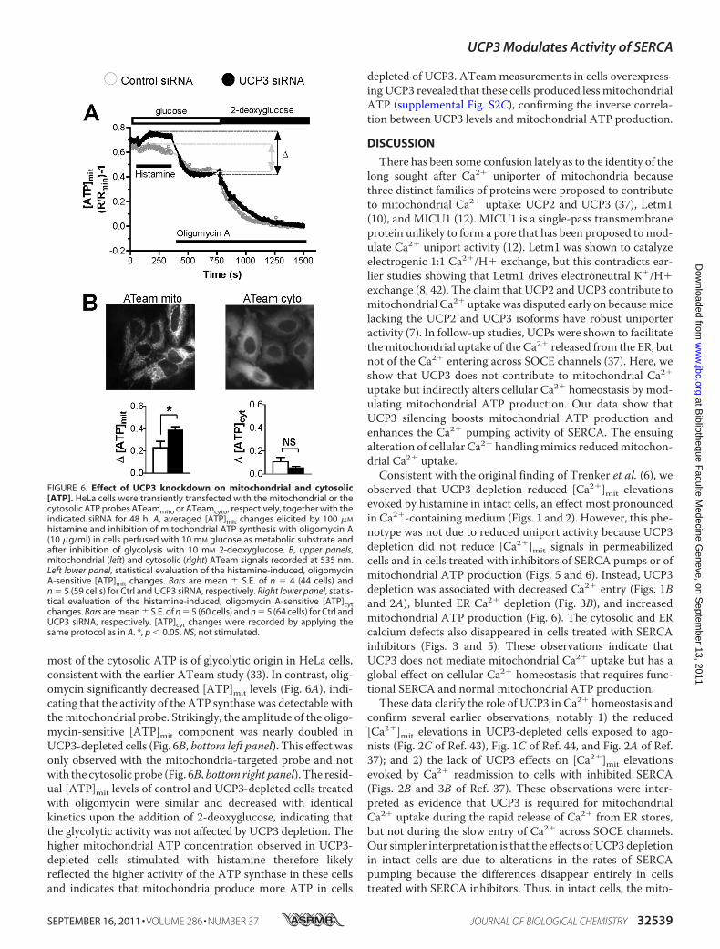
most of the cytosolic ATP is of glycolytic origin in HeLa cells,consistent with the earlier ATeam study (33). In contrast, olig-omycin significantly decreased [ATP]mit levels (Fig. 6A), indi-cating that the activity of the ATP synthase was detectable withthemitochondrial probe. Strikingly, the amplitude of the oligo-mycin-sensitive [ATP]mit component was nearly doubled inUCP3-depleted cells (Fig. 6B, bottom left panel). This effect wasonly observed with the mitochondria-targeted probe and notwith the cytosolic probe (Fig. 6B, bottom right panel). The resid-ual [ATP]mit levels of control and UCP3-depleted cells treatedwith oligomycin were similar and decreased with identicalkinetics upon the addition of 2-deoxyglucose, indicating thatthe glycolytic activity was not affected by UCP3 depletion. Thehigher mitochondrial ATP concentration observed in UCP3-depleted cells stimulated with histamine therefore likelyreflected the higher activity of the ATP synthase in these cellsand indicates that mitochondria produce more ATP in cells
depleted of UCP3. ATeam measurements in cells overexpress-ing UCP3 revealed that these cells produced less mitochondrialATP (supplemental Fig. S2C), confirming the inverse correla-tion between UCP3 levels and mitochondrial ATP production.
DISCUSSION
There has been some confusion lately as to the identity of thelong sought after Ca2� uniporter of mitochondria becausethree distinct families of proteins were proposed to contributeto mitochondrial Ca2� uptake: UCP2 and UCP3 (37), Letm1(10), and MICU1 (12). MICU1 is a single-pass transmembraneprotein unlikely to form a pore that has been proposed tomod-ulate Ca2� uniport activity (12). Letm1 was shown to catalyzeelectrogenic 1:1 Ca2�/H� exchange, but this contradicts ear-lier studies showing that Letm1 drives electroneutral K�/H�exchange (8, 42). The claim that UCP2 and UCP3 contribute tomitochondrial Ca2� uptakewas disputed early on becausemicelacking the UCP2 and UCP3 isoforms have robust uniporteractivity (7). In follow-up studies, UCPs were shown to facilitatethemitochondrial uptake of the Ca2� released from the ER, butnot of the Ca2� entering across SOCE channels (37). Here, weshow that UCP3 does not contribute to mitochondrial Ca2�
uptake but indirectly alters cellular Ca2� homeostasis by mod-ulating mitochondrial ATP production. Our data show thatUCP3 silencing boosts mitochondrial ATP production andenhances the Ca2� pumping activity of SERCA. The ensuingalteration of cellular Ca2� handlingmimics reducedmitochon-drial Ca2� uptake.
Consistent with the original finding of Trenker et al. (6), weobserved that UCP3 depletion reduced [Ca2�]mit elevationsevoked by histamine in intact cells, an effect most pronouncedin Ca2�-containing medium (Figs. 1 and 2). However, this phe-notype was not due to reduced uniport activity because UCP3depletion did not reduce [Ca2�]mit signals in permeabilizedcells and in cells treated with inhibitors of SERCA pumps or ofmitochondrial ATP production (Figs. 5 and 6). Instead, UCP3depletion was associated with decreased Ca2� entry (Figs. 1Band 2A), blunted ER Ca2� depletion (Fig. 3B), and increasedmitochondrial ATP production (Fig. 6). The cytosolic and ERcalcium defects also disappeared in cells treated with SERCAinhibitors (Figs. 3 and 5). These observations indicate thatUCP3 does not mediate mitochondrial Ca2� uptake but has aglobal effect on cellular Ca2� homeostasis that requires func-tional SERCA and normal mitochondrial ATP production.These data clarify the role of UCP3 in Ca2� homeostasis and
confirm several earlier observations, notably 1) the reduced[Ca2�]mit elevations in UCP3-depleted cells exposed to ago-nists (Fig. 2C of Ref. 43), Fig. 1C of Ref. 44, and Fig. 2A of Ref.37); and 2) the lack of UCP3 effects on [Ca2�]mit elevationsevoked by Ca2� readmission to cells with inhibited SERCA(Figs. 2B and 3B of Ref. 37). These observations were inter-preted as evidence that UCP3 is required for mitochondrialCa2� uptake during the rapid release of Ca2� from ER stores,but not during the slow entry of Ca2� across SOCE channels.Our simpler interpretation is that the effects of UCP3 depletionin intact cells are due to alterations in the rates of SERCApumping because the differences disappear entirely in cellstreated with SERCA inhibitors. Thus, in intact cells, the mito-
FIGURE 6. Effect of UCP3 knockdown on mitochondrial and cytosolic[ATP]. HeLa cells were transiently transfected with the mitochondrial or thecytosolic ATP probes ATeammito or ATeamcyto, respectively, together with theindicated siRNA for 48 h. A, averaged [ATP]mit changes elicited by 100 �M
histamine and inhibition of mitochondrial ATP synthesis with oligomycin A(10 �g/ml) in cells perfused with 10 mM glucose as metabolic substrate andafter inhibition of glycolysis with 10 mM 2-deoxyglucose. B, upper panels,mitochondrial (left) and cytosolic (right) ATeam signals recorded at 535 nm.Left lower panel, statistical evaluation of the histamine-induced, oligomycinA-sensitive [ATP]mit changes. Bars are mean � S.E. of n � 4 (44 cells) andn � 5 (59 cells) for Ctrl and UCP3 siRNA, respectively. Right lower panel, statis-tical evaluation of the histamine-induced, oligomycin A-sensitive [ATP]cytchanges. Bars are mean � S.E. of n � 5 (60 cells) and n � 5 (64 cells) for Ctrl andUCP3 siRNA, respectively. [ATP]cyt changes were recorded by applying thesame protocol as in A. *, p � 0.05. NS, not stimulated.
UCP3 Modulates Activity of SERCA
SEPTEMBER 16, 2011 • VOLUME 286 • NUMBER 37 JOURNAL OF BIOLOGICAL CHEMISTRY 32539
at Bibliotheque F
aculte Medecine G
eneve, on Septem
ber 13, 2011w
ww
.jbc.orgD
ownloaded from

chondrial Ca2� uptake rates indeed correlate with the expres-sion levels of UCP2/3, as reported previously (6), but this effectis indirect and mediated by SERCA.The requirement for active SERCA appears at odds with the
blunted [Ca2�]mit responses reported in cells co-stimulatedwith histamine and the SERCA inhibitor BHQ (Figs. 2A, 3A,and 4A of Ref. 37). However, our data show that effectiveSERCA inhibition requires at least 30 s of preincubation withTG and that BHQ releases Ca2� more slowly than TG (supple-mental Fig. S3). Thus, SERCApumpswere likely not fully inhib-ited in these experiments, and the UCP3 effects could stillreflect alterations in the rates of SERCA pumping. Our datafrom permeabilized cells also diverge from the data of Trenkeret al. 6, who reported a reduced Ca2� uptake in isolated livermitochondria from UCP2�/� mice (Fig. 3 of Ref. 6). In ourhands, the Ru360-sensitive component of mitochondrial Ca2�
uptake was not affected by UCP3 depletion in permeabilizedHeLa cells (Fig. 5B). These data are consistent with the normalmitochondrial Ca2� uptake reported by Brookes et al. (7) inpurified heart and liver mitochondria treated with UCP inhib-itors and in skeletal muscle mitochondria isolated fromUCP2�/� and UCP3�/� mice (Figs. 1 and 2 of Ref. 44). Toaccount for these discrepancies, Trenker et al. (44) argued thatthe failure of Brookes et al. (7) to detect UCP3 effects in per-meabilized cells was due to the harsher conditions of their puri-fication procedure that used differential centrifugation insteadof density gradients. Size distribution analysis revealed thatmitochondria isolated by density gradient had a larger diameter(0.75/1.00 �m versus 0.25/0.50 �m for differential centrifuga-tion, supplemental Fig. S1 of Ref. 44). This increase in mito-chondrial size could reflect a different amount of mitochon-dria-associated ER membranes, ER-derived structures thatco-purify with mitochondria during isolation (45). Mitochon-dria-associated ERmembranes are enriched in ERp57, a proteinthat directly interacts with SERCA2b andmodulates its activity(46). The presence of functional SERCAs on mitochondria-as-sociated ERmembranes co-purifiedwithmitochondria isolatedby density gradient might explain the effects of UCP2 ablationin this mitochondrial preparation. Finally, another discrepancyis the normal rates of SERCA pumping reported in cells over-expressingUCP2orUCP3 in the original study byTrenker et al.(6) (supplemental Fig. S1g of Ref. 9) and in a more recent pub-lication (Fig. 2C of Ref. 37). In our study, increased SERCApumping was evident in cells depleted of UCP3 (Fig. 4A), andthe rates of [Ca2�]ER decrease were severely reduced duringhistamine stimulation (Fig. 3B). The increased activity ofSERCA can account for the reduced influx of Ca2� observed incells depleted of UCP3 and treated with agonists (Fig. 2A), aphenotype that was not reported previously. Trenker et al. (6)did not attempt to measure [Ca2�]ER changes in cells depletedof UCP2 or UCP3; thus, our data cannot be readily comparedwith theirs. However, based on the effects of UCP3 depletion,we would predict that UCP3 overexpression would decreaseSERCA pumping.Several mechanisms could explain the sensitivity of SERCA
to changes in UCP3 levels. First, mitochondria could producemore reactive oxygen species in response to UCP3 depletionbecause an increase in UCP3 levels is known to lower reactive
oxygen species production (47). Because increased reactiveoxygen species levels decrease SERCA activity, however (48,49), UCP3 knockdown should decrease, rather than increase,SERCAactivity. Alterations in reactive oxygen species levels arethus unlikely to account for the UCP3 effects that we reporthere. Second, UCP3 depletion could increase the number ofER-mitochondria contact sites or reduce the distance betweenmitochondria and the ER, enabling mitochondria to supplymore ATP to nearby SERCA. Increased ER-mitochondria con-tact, however, should increase, rather than decrease, the effi-ciency of ER-mitochondria Ca2� transmission. Finally, mito-chondria could produce more ATP at low UCP3 levels, therebyfueling the activity of SERCA more efficiently. Our data indi-cate that this last mechanism is likely to occur because 1) themitochondrial ATP production, measured with a geneticallyencoded ATP-sensitive indicator, was significantly increasedfollowing stimulation of cells with Ca2�-mobilizing agonists;and 2) the UCP3 effects disappeared in cells treated with inhib-itors of mitochondrial respiration and of the ATP synthase.UCP2/3 are mitochondrial inner membrane proteins whosefirst postulated function is to uncouple oxidative phosphoryla-tion from ATP production (20). Although this uncouplingfunction is also disputed (16), the simplest explanation for theincreased production ofmitochondrialATPobserved inUCP3-depleted cells is that the energy stored in the proton-motiveforce is usedmore efficiently by the ATP synthase. Thus, amilduncoupling function of UCP3 could account for the whole phe-notype that we report here in UCP3-depleted cells, including 1)the increased production of mitochondrial ATP, 2) increasedactivity of SERCA, 3) decreased store-operatedCa2� entry (dueto reduced store Ca2� depletion), and finally, 4) reduced mito-chondrial Ca2� uptake due to sequestration of the releasedCa2� by SERCA and (predominantly) to the blunted SOCE.
Our data highlight the pitfalls of interpreting alterations in[Ca2�]mit signals occurring in intact cells. In intact cells, mito-chondria are embedded in the ER, and the changes in matrix[Ca2�] are influenced by the activity of numerous Ca2� trans-port proteins, including Ca2� release and influx channels(InsP3R, Orai), Ca2� pumps (SERCA, secretory pathwayCa2�-ATPase (SPCA), and plasma membrane Ca2�-ATPase(PMCA)), and Ca2� buffering proteins, which are often over-looked. The simplistic interpretation that a reducedmitochon-drial Ca2� signal is due to reduced mitochondrial Ca2� uptakedisregards the complexity of the Ca2� signaling circuitry inintact cells, where Ca2� release and influx occur concomitantlywith Ca2� uptake catalyzed by SERCA and Ca2� extrusion cat-alyzed by plasmamembrane Ca2�-ATPase. Our data show thata phenotype typical of defectivemitochondrial Ca2�uptake canin fact be explained by a metabolic alteration that changes theactivity of Ca2� pumps.
Acknowledgments—We thank Drs. R. Y. Tsien and A. Palmer for pro-viding the cameleon constructs, Drs. H. Imamura and H. Noji forproviding the ATeam probes, Dr. Wolfgang Graier for UCPs con-structs, and Ariane Widmer for expert technical assistance.
Note Added in Proof—Aproteinwith all characteristic features of theCa2� uniporter of mitochondria was recently identified by two inde-
UCP3 Modulates Activity of SERCA
32540 JOURNAL OF BIOLOGICAL CHEMISTRY VOLUME 286 • NUMBER 37 • SEPTEMBER 16, 2011
at Bibliotheque F
aculte Medecine G
eneve, on Septem
ber 13, 2011w
ww
.jbc.orgD
ownloaded from

pendent groups (50, 51). The purified MCU protein exhibited Ca2�
channel activity in lipid bilayers (50), confirming that UCPs are notessential for mitochondrial Ca2� uptake. The effects of UCPs onmitochondrial Ca2� handling that were previously reported there-fore likely reflect the metabolic alterations described in this study.
REFERENCES1. Szabadkai, G., and Duchen, M. R. (2008) Physiology 23, 84–942. Rimessi, A., Giorgi, C., Pinton, P., and Rizzuto, R. (2008) Biochim. Biophys.
Acta 1777, 808–8163. Demaurex,N., Poburko,D., and Frieden,M. (2009)Biochim. Biophys. Acta
1787, 1383–13944. Bernardi, P. (1999) Physiol. Rev. 79, 1127–11555. Kirichok, Y., Krapivinsky, G., and Clapham, D. E. (2004) Nature 427,
360–3646. Trenker, M., Malli, R., Fertschai, I., Levak-Frank, S., and Graier, W. F.
(2007) Nat. Cell Biol. 9, 445–4527. Brookes, P. S., Parker, N., Buckingham, J. A., Vidal-Puig, A., Halestrap,
A. P., Gunter, T. E., Nicholls, D.G., Bernardi, P., Lemasters, J. J., andBrand,M. D. (2008) Nat. Cell Biol. 10, 1235–1237; author reply 1237–1240
8. Nowikovsky, K., Froschauer, E. M., Zsurka, G., Samaj, J., Reipert, S.,Kolisek, M., Wiesenberger, G., and Schweyen, R. J. (2004) J. Biol. Chem.279, 30307–30315
9. Dimmer, K. S., Navoni, F., Casarin, A., Trevisson, E., Endele, S., Winter-pacht, A., Salviati, L., and Scorrano, L. (2008) Hum. Mol. Genet. 17,201–214
10. Jiang, D., Zhao, L., and Clapham, D. E. (2009) Science 326, 144–14711. Endele, S., Fuhry, M., Pak, S. J., Zabel, B. U., and Winterpacht, A. (1999)
Genomics 60, 218–22512. Perocchi, F., Gohil, V.M., Girgis, H. S., Bao, X. R., McCombs, J. E., Palmer,
A. E., and Mootha, V. K. (2010) Nature 467, 291–29613. Hajnoczky, G., and Csordas, G. (2010) Curr. Biol. 20, R888–89114. Palty, R., Silverman, W. F., Hershfinkel, M., Caporale, T., Sensi, S. L.,
Parnis, J., Nolte, C., Fishman, D., Shoshan-Barmatz, V., Herrmann, S.,Khananshvili, D., and Sekler, I. (2010) Proc. Natl. Acad. Sci. U.S.A. 107,436–441
15. Da Cruz, S., DeMarchi, U., Frieden,M., Parone, P. A., Martinou, J. C., andDemaurex, N. (2010) Cell Calcium 47, 11–18
16. Azzu, V., and Brand, M. D. (2010) Trends Biochem. Sci. 35, 298–30717. Cannon, B., and Nedergaard, J. (2004) Physiol. Rev. 84, 277–35918. Cadenas, S., Echtay, K. S., Harper, J. A., Jekabsons, M. B., Buckingham,
J. A., Grau, E., Abuin, A., Chapman, H., Clapham, J. C., and Brand, M. D.(2002) J. Biol. Chem. 277, 2773–2778
19. Couplan, E., del Mar Gonzalez-Barroso, M., Alves-Guerra, M. C., Ric-quier, D., Goubern, M., and Bouillaud, F. (2002) J. Biol. Chem. 277,26268–26275
20. Krauss, S., Zhang, C. Y., and Lowell, B. B. (2005)Nat. Rev.Mol. Cell Biol. 6,248–261
21. Chan, C. B., MacDonald, P. E., Saleh, M. C., Johns, D. C., Marban, E., andWheeler, M. B. (1999) Diabetes 48, 1482–1486
22. Himms-Hagen, J., and Harper, M. E. (2001) Exp. Biol. Med. (Maywood)226, 78–84
23. Goglia, F., and Skulachev, V. P. (2003) FASEB J. 17, 1585–159124. Echtay, K. S., Esteves, T. C., Pakay, J. L., Jekabsons, M. B., Lambert, A. J.,
Portero-Otín, M., Pamplona, R., Vidal-Puig, A. J., Wang, S., Roebuck, S. J.,and Brand, M. D. (2003) EMBO J. 22, 4103–4110
25. Brand, M. D., and Esteves, T. C. (2005) Cell Metab. 2, 85–9326. Cioffi, F., Senese, R., de Lange, P., Goglia, F., Lanni, A., and Lombardi, A.
(2009) Biofactors 35, 417–42827. Bezaire, V., Seifert, E. L., and Harper, M. E. (2007) FASEB J. 21, 312–32428. Arsenijevic, D., Onuma, H., Pecqueur, C., Raimbault, S., Manning, B. S.,
Miroux, B., Couplan, E., Alves-Guerra, M. C., Goubern, M., Surwit, R.,Bouillaud, F., Richard, D., Collins, S., and Ricquier, D. (2000) Nat. Genet.26, 435–439
29. Brand, M. D., Affourtit, C., Esteves, T. C., Green, K., Lambert, A. J., Miwa,S., Pakay, J. L., and Parker, N. (2004) Free Radic. Biol. Med. 37, 755–767
30. Nagai, T., Yamada, S., Tominaga, T., Ichikawa, M., and Miyawaki, A.(2004) Proc. Natl. Acad. Sci. U.S.A. 101, 10554–10559
31. Palmer, A. E., Giacomello, M., Kortemme, T., Hires, S. A., Lev-Ram, V.,Baker, D., and Tsien, R. Y. (2006) Chem. Biol. 13, 521–530
32. Palmer, A. E., Jin, C., Reed, J. C., and Tsien, R. Y. (2004) Proc. Natl. Acad.Sci. U.S.A. 101, 17404–17409
33. Imamura, H., Nhat, K. P., Togawa, H., Saito, K., Iino, R., Kato-Yamada, Y.,Nagai, T., and Noji, H. (2009) Proc. Natl. Acad. Sci. U.S.A. 106,15651–15656
34. Jousset, H., Malli, R., Girardin, N., Graier, W. F., Demaurex, N., and Frie-den, M. (2008) Cell Calcium 43, 83–94
35. De Marchi, U., Campello, S., Szabo, I., Tombola, F., Martinou, J. C., andZoratti, M. (2004) J. Biol. Chem. 279, 37415–37422
36. Poburko, D., Liao, C. H., van Breemen, C., and Demaurex, N. (2009) Circ.Res. 104, 104–112
37. Waldeck-Weiermair, M., Malli, R., Naghdi, S., Trenker, M., Kahn, M. J.,and Graier, W. F. (2010) Cell Calcium 47, 433–440
38. Frieden, M., James, D., Castelbou, C., Danckaert, A., Martinou, J. C., andDemaurex, N. (2004) J. Biol. Chem. 279, 22704–22714
39. Prakriya, M. (2009) Immunol. Rev. 231, 88–9840. Landolfi, B., Curci, S., Debellis, L., Pozzan, T., and Hofer, A. M. (1998)
J. Cell Biol. 142, 1235–124341. Csordas, G., and Hajnoczky, G. (2001) Cell Calcium 29, 249–26242. Zotova, L., Aleschko, M., Sponder, G., Baumgartner, R., Reipert, S., Prinz,
M., Schweyen, R. J., and Nowikovsky, K. (2010) J. Biol. Chem. 285,14399–14414
43. Waldeck-Weiermair, M., Duan, X., Naghdi, S., Khan, M. J., Trenker, M.,Malli, R., and Graier, W. F. (2010) Cell Calcium 48, 288–301
44. Trenker,M., Fertschai, I., Malli, R., and Graier,W. F. (2008)Nat. Cell Biol.10, 1237–1240
45. Hayashi, T., Rizzuto, R., Hajnoczky, G., and Su, T. P. (2009) Trends CellBiol. 19, 81–88
46. Li, Y., and Camacho, P. (2004) J. Cell Biol. 164, 35–4647. Toime, L. J., and Brand, M. D. (2010) Free Radic. Biol. Med. 49, 606–61148. Rosado, J. A., Redondo, P. C., Salido, G.M., and Pariente, J. A. (2006)Mini
Rev. Med. Chem. 6, 409–41549. Ermak, G., and Davies, K. J. (2002)Mol. Immunol. 38, 713–72150. De Stefani, D., Raffaello, A., Teardo, E., Szabo, I., and Rizzuto, R. (2011)
Nature, in press51. Baughman, J. M., Perocchi, F., Girgis, H. S., Plovanich, M., Belcher-
Timme, C. A., Sancak, Y., Bao, X. R., Strittmatter, L., Goldberger, O.,Bogorad, R. L., Koteliansky, V., and Mootha, V. K. (2011)Nature, in press
UCP3 Modulates Activity of SERCA
SEPTEMBER 16, 2011 • VOLUME 286 • NUMBER 37 JOURNAL OF BIOLOGICAL CHEMISTRY 32541
at Bibliotheque F
aculte Medecine G
eneve, on Septem
ber 13, 2011w
ww
.jbc.orgD
ownloaded from