Increased myocardial SERCA expression in early type 2 diabetes mellitus is insulin dependent: In...
11
ORIGINAL INVESTIGATION Open Access Increased myocardial SERCA expression in early type 2 diabetes mellitus is insulin dependent: In vivo and in vitro data Sabine Fredersdorf 1,6* , Christian Thumann 1 , Wolfram H Zimmermann 2 , Roland Vetter 3 , Tobias Graf 4 , Andreas Luchner 1 , Günter AJ Riegger 1 , Heribert Schunkert 4 , Thomas Eschenhagen 5 and Joachim Weil 4 Abstract Background: Calcium (Ca2+) handling proteins are known to play a pivotal role in the pathophysiology of cardiomyopathy. However little is known about early changes in the diabetic heart and the impact of insulin treatment (Ins). Methods: Zucker Diabetic Fatty rats treated with or without insulin (ZDF ± Ins, n = 13) and lean littermates (controls, n = 7) were sacrificed at the age of 19 weeks. ZDF + Ins (n = 6) were treated with insulin for the last 6 weeks of life. Gene expression of Ca2+ ATPase in the cardiac sarcoplasmatic reticulum (SERCA2a, further abbreviated as SERCA) and phospholamban (PLB) were determined by northern blotting. Ca2+ transport of the sarcoplasmatic reticulum (SR) was assessed by oxalate-facilitated 45Ca-uptake in left ventricular homogenates. In addition, isolated neonatal cardiomyocytes were stimulated in cell culture with insulin, glucose or triiodthyronine (T3, positive control). mRNA expression of SERCA and PLB were measured by Taqman PCR. Furthermore, effects of insulin treatment on force of contraction and relaxation were evaluated by cardiomyocytes grown in a three-dimensional collagen matrix (engineered heart tissue, EHT) stimulated for 5 days by insulin. By western blot phosphorylations status of Akt was determed and the influence of wortmannin. Results: SERCA levels increased in both ZDF and ZDF + Ins compared to control (control 100 ± 6.2 vs. ZDF 152 ± 26.6* vs. ZDF + Ins 212 ± 18.5*# % of control, *p < 0.05 vs. control, #p < 0.05 vs. ZDF) whereas PLB was significantly decreased in ZDF and ZDF + Ins (control 100 ± 2.8 vs. ZDF 76.3 ± 13.5* vs. ZDF + Ins 79.4 ± 12.9* % of control, *p < 0.05 vs control). The increase in the SERCA/PLB ratio in ZDF and ZDF ± Ins was accompanied by enhanced Ca2+ uptake to the SR (control 1.58 ± 0.1 vs. ZDF 1.85 ± 0.06* vs. ZDF + Ins 2.03 ± 0.1* μg/mg/min, *p < 0.05 vs. control). Interestingly, there was a significant correlation between Ca2+ uptake and SERCA2a expression. As shown by in-vitro experiments, the effect of insulin on SERCA2a mRNA expression seemed to have a direct effect on cardiomyocytes. Furthermore, long-term treatment of engineered heart tissue with insulin increased the SERCA/PLB ratio and accelerated relaxation time. Akt was significantly phosphorylated by insulin. This effect could be abolished by wortmannin. * Correspondence: [email protected] 1 Klinik und Poliklinik für Innere Medizin II, Universität Regensburg, Regensburg, Germany 6 Klinik und Poliklinik für Innere Medizin II des Universitätsklinikums Regensburg, 93042 Regensburg, Germany Full list of author information is available at the end of the article CARDIO VASCULAR DIABETOLOGY © 2012 Fredersdorf et al.; licensee BioMed Central Ltd. This is an Open Access article distributed under the terms of the Creative Commons Attribution License (http://creativecommons.org/licenses/by/2.0), which permits unrestricted use, distribution, and reproduction in any medium, provided the original work is properly cited. Fredersdorf et al. Cardiovascular Diabetology 2012, 11:57 http://www.cardiab.com/content/11/1/57
-
Upload
independent -
Category
Documents
-
view
4 -
download
0
Transcript of Increased myocardial SERCA expression in early type 2 diabetes mellitus is insulin dependent: In...

CARDIOVASCULAR DIABETOLOGY
Fredersdorf et al. Cardiovascular Diabetology 2012, 11:57http://www.cardiab.com/content/11/1/57
ORIGINAL INVESTIGATION Open Access
Increased myocardial SERCA expression in earlytype 2 diabetes mellitus is insulin dependent: Invivo and in vitro dataSabine Fredersdorf1,6*, Christian Thumann1, Wolfram H Zimmermann2, Roland Vetter3, Tobias Graf4,Andreas Luchner1, Günter AJ Riegger1, Heribert Schunkert4, Thomas Eschenhagen5 and Joachim Weil4
Abstract
Background: Calcium (Ca2+) handling proteins are known to play a pivotal role in the pathophysiology ofcardiomyopathy. However little is known about early changes in the diabetic heart and the impact of insulintreatment (Ins).
Methods: Zucker Diabetic Fatty rats treated with or without insulin (ZDF ± Ins, n = 13) and lean littermates (controls,n = 7) were sacrificed at the age of 19 weeks. ZDF + Ins (n = 6) were treated with insulin for the last 6 weeks of life.Gene expression of Ca2+ ATPase in the cardiac sarcoplasmatic reticulum (SERCA2a, further abbreviated as SERCA)and phospholamban (PLB) were determined by northern blotting. Ca2+ transport of the sarcoplasmatic reticulum(SR) was assessed by oxalate-facilitated 45Ca-uptake in left ventricular homogenates. In addition, isolated neonatalcardiomyocytes were stimulated in cell culture with insulin, glucose or triiodthyronine (T3, positive control). mRNAexpression of SERCA and PLB were measured by Taqman PCR. Furthermore, effects of insulin treatment on force ofcontraction and relaxation were evaluated by cardiomyocytes grown in a three-dimensional collagen matrix(engineered heart tissue, EHT) stimulated for 5 days by insulin. By western blot phosphorylations status of Akt wasdetermed and the influence of wortmannin.
Results: SERCA levels increased in both ZDF and ZDF + Ins compared to control (control 100 ± 6.2 vs. ZDF152 ± 26.6* vs. ZDF + Ins 212 ± 18.5*# % of control, *p < 0.05 vs. control, #p < 0.05 vs. ZDF) whereas PLB wassignificantly decreased in ZDF and ZDF + Ins (control 100 ± 2.8 vs. ZDF 76.3 ± 13.5* vs. ZDF + Ins 79.4 ± 12.9* % ofcontrol, *p < 0.05 vs control). The increase in the SERCA/PLB ratio in ZDF and ZDF ± Ins was accompanied byenhanced Ca2+ uptake to the SR (control 1.58 ± 0.1 vs. ZDF 1.85 ± 0.06* vs. ZDF + Ins 2.03 ± 0.1* μg/mg/min,*p < 0.05 vs. control). Interestingly, there was a significant correlation between Ca2+ uptake and SERCA2aexpression. As shown by in-vitro experiments, the effect of insulin on SERCA2a mRNA expression seemed to have adirect effect on cardiomyocytes. Furthermore, long-term treatment of engineered heart tissue with insulin increasedthe SERCA/PLB ratio and accelerated relaxation time. Akt was significantly phosphorylated by insulin. This effectcould be abolished by wortmannin.
* Correspondence: [email protected] und Poliklinik für Innere Medizin II, Universität Regensburg,Regensburg, Germany6Klinik und Poliklinik für Innere Medizin II des UniversitätsklinikumsRegensburg, 93042 Regensburg, GermanyFull list of author information is available at the end of the article
© 2012 Fredersdorf et al.; licensee BioMed Central Ltd. This is an Open Access article distributed under the terms of theCreative Commons Attribution License (http://creativecommons.org/licenses/by/2.0), which permits unrestricted use,distribution, and reproduction in any medium, provided the original work is properly cited.

Fredersdorf et al. Cardiovascular Diabetology 2012, 11:57 Page 2 of 11http://www.cardiab.com/content/11/1/57
Conclusion: The current data demonstrate that early type 2 diabetes is associated with an increase in the SERCA/PLB ratio and that insulin directly stimulates SERCA expression and relaxation velocity. These results underline theimportant role of insulin and calcium handling proteins in the cardiac adaptation process of type 2 diabetesmellitus contributing to cardiac remodeling and show the important role of PI3-kinase-Akt-SERCA2a signalingcascade.
Keywords: Diabetic heart, Insulin, SERCA expression, Relaxation velocity
IntroductionThe sarcoplasmatic reticulum (SR) plays a pivotal role inthe contraction and relaxation cycle of the heart byvirtue of its ability to tightly regulate intracellular cal-cium (Ca2+) concentration. Myocardial contraction isinitiated by Ca2+ entry through Ca2+ channels of theplasma membrane (L-type Ca2+ channel) [1] triggeringthe Ca2+ release from the SR through the ryanodine re-ceptor. During diastole Ca2+ is transported from thecytoplasma into the SR by the SR Ca2 +ATPase(SERCA). Recent cloning analysis revealed three distinctgenes encoding for SR Ca2 +ATPases (SERCA 1–3), ofwhich the SERCA2a is predominately expressed in car-diac tissue [2]. SERCA2a activity depends on the amountof SERCA2a protein and is further regulated by its in-hibitory protein phospholamban (PLB) [3]. SERCA2a isthe major determinant of the beat-to-beat regulation ofcardiac contraction, and overexpression of this proteinin mice has been shown to enhance myocardial relax-ation [4]. Unphosphorylated PLB inhibits the Ca2+ up-take of SERCA2a and phosphorylation of the proteindisrupts the inhibitory interaction resulting in increasedCa2+ transport towards the SR. Recent studies onPLB knockout mice have underlined the importanceof PLB as a key regulator of cardiac contraction andrelaxation [5].While diabetes leads to cardiomyopathy in later stages
with hypocontractility and reduced SERCA activity [6],we have observed increased contractility in earlier stagesof type 2 diabetes mellitus [7]. Various animal models ofdiabetes have been used to study the causes and under-lying subcellular events of diabetic cardiomyopathy.These studies suggest a dysfunctional sarcoplasmicreticulum (SR), leading to altered intracellular calciumhandling in cardiac myocytes. This mechanism might beinvolved in the development of diabetic cardiomyopathy[6,8]. A reduced sequestration of calcium into the SRcould readily explain the prolonged cardiac relaxationobserved in diabetic cardiomyopathy. As a consequence,the SR calcium content declines, leading to a reducedsystolic calcium release and therefore a weaker cardiaccontraction. However, these studies were mainly carriedout in models resembling insulin-dependent diabetes.Previous studies with diabetes type 1 models have showna down-regulation of SERCA2a in the heart associated
with a decrease in systolic and diastolic function. Thesefunctional alterations can be reversed by insulin treat-ment or SERCA2a overexpression [4,9]. So far, there islittle information available on myocardial SERCA2a andPLB changes in the early stages of type 2 diabetes whichis characterized by rather high insulin levels [8,10,11].The Zucker Diabetic Fatty rat (ZDF/Drt-fa) is charac-
terized by early onset of hyperglycemia, hyperphagia,hyperinsulinemia, adiposity and hyperlipidemia, therebyresembling the clinical features of human type 2 diabetes[12]. In an earlier study, we were able to demonstratethat animals in transition from insulin resistance to type2 diabetes but not lean control rats develop significantmyocardial hypertrophy associated with increased sys-tolic function as evaluated by echocardiography [7].These findings raised the possibility that one or moreproteins regulating intracellular calcium homeostasismay be altered in these animals. Therefore, the presentstudy was designed to determine whether the cardiacphenotype of ZDF rats is associated with alterations incardiac SR function and expression of SERCA2a and itsregulating protein PLB. Since insulin treatment is widelyused as a therapeutic option for patients with poorlycontrolled type 2 diabetes, its impact on the aforemen-tioned proteins was studied in isolated cell preparationsof the heart. Furthermore, we investigated the functionalconsequences of high insulin levels in an innovativeengineered heart tissue model.
Materials and methodsAnimal modelMale Zucker Diabetic Fatty rats (body weight (BW)range: 106–158 g, n = 13) and male Zucker lean rats(BW 85–118 g, n = 7) were obtained at the age of fiveweeks from Genetic Models (Indianapolis, USA). Ani-mals were maintained on RMH-B rat chow from HopeFarms (Woerden, Netherlands) with water ad libitum.All animals were individually housed in a 12 h dark/lightcycle controlled room. The protocol had been approvedby the local committee on animal research and conformsto the Guide for the Care and Use of Laboratory Ani-mals, published by the US National Institutes of Health(NIH Publication No. 85–23; revised 1985). At the ageof 13 weeks, one week after developing hyperglycemia,the animals were divided into three groups: (1) Zucker
Fredersdorf et al. Cardiovascular Diabetology 2012, 11:57 Page 3 of 11http://www.cardiab.com/content/11/1/57
lean rats (control group, n = 7), (2) Zucker Diabetic Fattyrats without insulin treatment (ZDF; n = 7), and (3)Zucker Diabetic Fatty rats treated with insulin (ZDF+Ins; n = 6). Insulin treatment (Actrapid HM U500,Novo Nordisk, Mainz, Germany) was initiated at a doseof 25.0.U/kg/day with subcutaneously implanted Alzet os-
motic minipumps (Model 2ML2 and 2ML4, CharlesRiver Wiga, Sulzfeld, Germany). Pumps were changedafter 2 weeks and the insulin dose was adaptedto normalize blood glucose levels. Body weight wasdetermined every week, and blood glucose levels every2–3 weeks (Accu-Chek Plus Roche, Mannheim,Germany). At the age of 18 weeks, systolic blood pres-sure and heart rate were measured by indirect tail-cuffmethod as described [13] using an automated cuffinflator-pulse detection system (W+W electronic AG,BP recorder No. 8005, Basel, Switzerland). After 6 weeksof insulin treatment, at the age of 19 weeks, the animalswere killed.
Tissue preparationHearts were rapidly excised, rinsed with saline and blot-ted dry. The whole heart weight was determined. Theheart was dissected free from the atria, cut into rightand left ventricular tissue, frozen in liquid nitrogenwithin 3 minutes and stored at −80 C until analyzed, asdescribed earlier [7].
Neonatal rat cardiac myocytesRat cardiac myocytes were isolated from 1- to 3-day-oldneonatal Wistar rats (University of Regensburg breed,from Charles River, Sulzfeld, Germany) as describedearlier [14]. Briefly, hearts from 50–70 pups wereminced and subjected to serial trypsin digestion to re-lease single cells. After the final digestion, cells werewashed and pre-plated for 1–2 h in complete culturemedium (MEM supplemented with 10% fetal calf serumand 1% penicillin/streptomycin). Unattached cells werepelleted and suspended in culture medium containing0.1 mmol/l 5´-bromo-2´-desoxyuridine (BrdU) to sup-press overgrowth of non- myoctes. Cells were then pla-ted on culture dishes at a density of 150,000 cells/cm2and incubated for 5 days at 37°C before being stimulatedwith insulin (0.1-3.0 μmol/l), wortmannin (300 and 1000nM) or triiodothyronine (3.0 nmol/l) as a positive con-trol for up-regulation of SERCA2a mRNA expression[15].
Engineered heart tissuesEngineered heart tissues (EHT) were prepared asdescribed previously [16]. Briefly, circular EHT were pre-pared by mixing freshly isolated cardiac myocytes fromneonatal rats with collagen type 1 prepared from rat
tails, a basement membrane mixture (Matrigel, tebu,Offenbach, Heidelberg, Germany), and concentratedserum containing culture medium (2xDMEM, 20% horseserum, 4% chicken embryo extract, 200 U/ml penicillinand 200 μg/ml streptomycin); pH was neutralized by ti-tration with NaOH (0,1 N). The reconstitution mix waspipetted into circular casting molds and incubated for30 to 45 min at 37oC and 5% CO2 to allow hardening ofthe reconstitution mix. Thereafter, 5 ml serum-containing culture medium (DMEM, 10% horse serum,2% chicken embryo extract, 100 U/ml penicillin and100 μg/ml streptomycin) was added to each dish. Cul-ture was performed as described earlier [16]. After 7 daysin culture, EHTs were transferred to a stretch device andsubjected to phasic stretch (to 110% of their originallength) for 5 days. Culture medium was changed 12hours after EHT casting and then every other day. Aftertransfer to the stretch device, the culture medium waschanged every day and supplemented with insulin at ahigh physiological concentration (0.1 μg/ml; Sigma-Aldrich, Taufkirchen, Germany).
Force measurementAfter 12 days (7 days in casting molds and 5 days ofstretching), the EHTs were transferred to thermostatedorgan baths containing gassed Tyrode´s solution andsubjected to isometric force measurement as describedelsewhere [16]. Briefly, electrically stimulated EHTs(2 Hz) were stretched to the length at which force ofcontraction was maximal and inotropic and lusitropicresponses to cumulative concentrations of isoprenaline(0.1-1000 nM) in the presence of 0.2 mM calcium wererecorded. Contractile activity was evaluated with a PC-assisted system (BMON2, Ingenieurbüro Jäckel, Hanau,Germany).
RNA analysisTotal RNA from left ventricles or cultured cardiacmyocytes was isolated with TrizolW (Canadian LifeTechnologies Inc., Burlington, Ontario, Canada)according to the manufacturer’s instructions. The con-centration was determined photometrically at 260 nm.Total RNA was stored at 80°C. For Northern blot ana-lysis 20 μg of total RNA were denatured, size-fractionated by electrophoresis on 1% agarose gelsunder denaturing conditions, transferred to nylonmembranes (Gene Screen Plus, NEN, Dreieich, Ger-many) and immobilized by ultraviolet irradiation. Blotswere prehybridized and hybridized using standard pro-tocols as described previously [17]. Hybridized filterswere washed and exposed at −80 C° to x-ray films(XAR-5, Eastman Kodak, N.Y., USA) by using intensify-ing screens. Different exposures of all autoradiogramswere obtained to ensure that laser scanning (Personal

Fredersdorf et al. Cardiovascular Diabetology 2012, 11:57 Page 4 of 11http://www.cardiab.com/content/11/1/57
Densitometer No. 50301, Molecular Dynamics) wasperformed within the linear range of densitometry. Forhybridization cDNA probes for rat SERCA, PLB (kindlygifted by K.R. Boeheler) and GAPDH were radiolabelledwith α32-P dCTP (specific activity 3000 Ci/mmol,Amersham, Dreieich, Germany) for Northern blot ana-lysis. Values were normalized to these house-keepinggene GAPDH. The rat cDNA of GAPDH was clonedby reverse transcriptase PCR using the following pri-mers: forward 5´-CTTCACCACCATGGAGAAGG-3´;and reverse 5´-ATTGAGAGCAATGCCAGCC-3´.For quantitative RT-PCR (qRT-PCR), total RNA was
transcribed with SuperScriptII RT (Invitrogen, CA,USA). Individual samples of 20 ng cDNA were amplifiedwith AmpliTaqGold Polymerase (Applied Biosystems,CA, USA) utilizing gene specific primers and fluorogenicprobes (5’ FAM and 3’ TAMRA; see below for completeprimer/probe sequence information) in an ABI PRISMW
7900HT Sequence Detection System (Applied Biosys-tems). Probes were designed to cross exon/intronboundaries with primer annealing sites being located inthe adjacent exons to eliminate the possibility of gen-omic DNA amplification. . Standard curves were per-formed in duplicate with serially diluted cDNA fromneonatal rat heart tissue (1.5 - 50 ng) to determine PCRefficiency, which was similar in all groups SERCA andPhospholamban expression were evaluated as SERCA/PLB ratio and correlated to EHT twitch tension and re-laxation time (T2). Quantification was performed by thestandard curve and 2-ΔΔCt methods [18].SERCA2a: forward primer 5´- AGT GGC TGA TGG
TGC TGA AA-3´.reverse primer 5´- GCA CCC GAA CAC CCT TAC
AT-3.probe 5´ FAM- TTA CTC CAG TAT TGC AGG CTC
CAG GTA -TAMRA 3´.PLB: forward primer 5´- GCA GCT GAG CTC CCA
GAC TT-3.reverse primer 5´- TTT CCA TGA TGC CAG GAA
GAC-3´.probe 5´ FAM- CAC AGA AGC CAA GGC CTC
CTA AAA GGA G -TAMRA 3´.We checked 18 S, GAPDH, and CSQ2 (data not
shown). However, corrections are not necessary becausewe have determined PLB and SERCA from the samecDNA samples.
Western blot analysis20 μl of cell suspension of the cardiomyocyte cell culturewere separated on 10% SDS-polyacrylamide gels. Gelswere run andseparated proteins were transferred to nitro-cellulose membranes in 50 mM sodium phosphate buffer,pH 7.4, for 20 h at 300 mA, and 4°C. Nitrocellulose sheetswere incubated with a rabbit polyclonal anti-human
antisera (Sigma) at a 1:2000 dilution. Phospho-Akt andAkt antibodies (1:1000, from New England Biolabs) werevisualized colorimetrically by using horseradishperoxidase- (HRP) conjugated goat anti-rabbit immuno-globulin G at a 1:1000 dilution. After phospho-Akt blot-ting, the blot was stripped for 30 min at 50oC and thenblotted for Akt, serving also as loading control. Apparentmolecular weights were determined by using a prestainedstandard (kaleidoscope prestained standard, Biorad, USA).
Oxalate-supported Ca2+ uptakeOxalate-supported SR Ca2+ uptake was measured in leftventricular homogenates as described previously [19].Briefly, the Ca2+ uptake medium of 0.2 ml contained40 mmol/l imidazole (pH 7.0), 100 mmol/l KCl,5 mmol/l MgCl2, 5 mmol/l TrisATP, 6 mmol/l phospho-creatine, 10 mmol/l K + -oxalate, 10 mmol/l NaN3,10 μM synthetic protein kinase A-inhibitor peptide [PKI(6–22)amide; GIBCO-BRL, Eggenstein, Germany],0.2 mmol/l EGTA, and 0.08 or 0.250 mmol/l 45CaCl2corresponding to 0.34 or 3.68 μmol/l free Ca2+, respect-ively After 2 min of preincubation at 37°C, the measure-ment was started by addition of homogenate (30 μgprotein) and 2 min later a 0.15-ml sample was filteredthrough 0.45- μm Millipore filters using a vacuumpump. The filter was immediately washed twice with3 ml ice-cold solution containing 100 mM KCl, 2 mMEGTA, and 40 mM imidazole (pH 7.0). Radioactivitybound to dry filters was determined by liquid scintilla-tion counting. All measurements were done in duplicateCa uptake was measured within the linear range of thereaction. Calculated Ca2+ uptake values were expressedas nmoles of Ca2+ per mg of protein per min or μmolesof Ca2+ per g wet LV wt min.
StatisticsStatistical analysis was performed using GraphPadPRIZM 5.0. Results are expressed as mean ± SEM.Comparisons between multiple groups were assessed byone-way analysis ANOVA-test and post-hoc analysis byBonferroni. The strength of the relationship betweentwo variables was assessed by calculating the product–moment correlation coefficient r. Statistical significancewas accepted at p < 0.05.
ResultsDiabetic animalsThe ZDF rats developed a manifest diabetes at the ageof 12 weeks (Capillary Glucose control group78 ± 1.8 mg/dl vs. ZDF 252 ± 39* mg/dl vs. ZDF + Ins292 ± 33* g/dl*, p < 0.05 vs. control). Body weight in ZDFrats increased steadily over time compared to non-diabetic lean animals. Treatment with insulin led to afurther increase in body weight three weeks after
A SERCA PLB
200
300SERCAPLB *
*
of
cont
rol)
d to
GA
PD
H
B
Fredersdorf et al. Cardiovascular Diabetology 2012, 11:57 Page 5 of 11http://www.cardiab.com/content/11/1/57
beginning treatment (see Table 1). Absolute heart weightwas significantly higher in the treatment group, but notin non-treated ZDF rats compared to age- matched ZDFrats (see Table 1). As expected, treatment with insulindecreased blood glucose level towards normoglycemicvalues in diabetic animals (see Table 1). Plasma C-peptide-levels were markedly elevated in diabetic ZDFrats indicating severe hyperinsulinemia. Interestingly,heart rate was significantly lower in all diabetic animalsand blood pressure was the same in the non-treatedZDF group and even reduced in the insulin-treated ZDFgroup (at 19 weeks of age: control 484± min-1 vs. ZDF416 ± 13* min-1 vs. ZDF+ Ins 421 ± 18* min-1, *p < 0.05vs. control).
Control ZDF ZDF+Ins0
100 * *
mR
NA
(%
nor
mal
ize
Control ZDF ZDF+Ins0
1
2
3
*
#*
SE
RC
A/P
LB m
RN
A r
atio
C
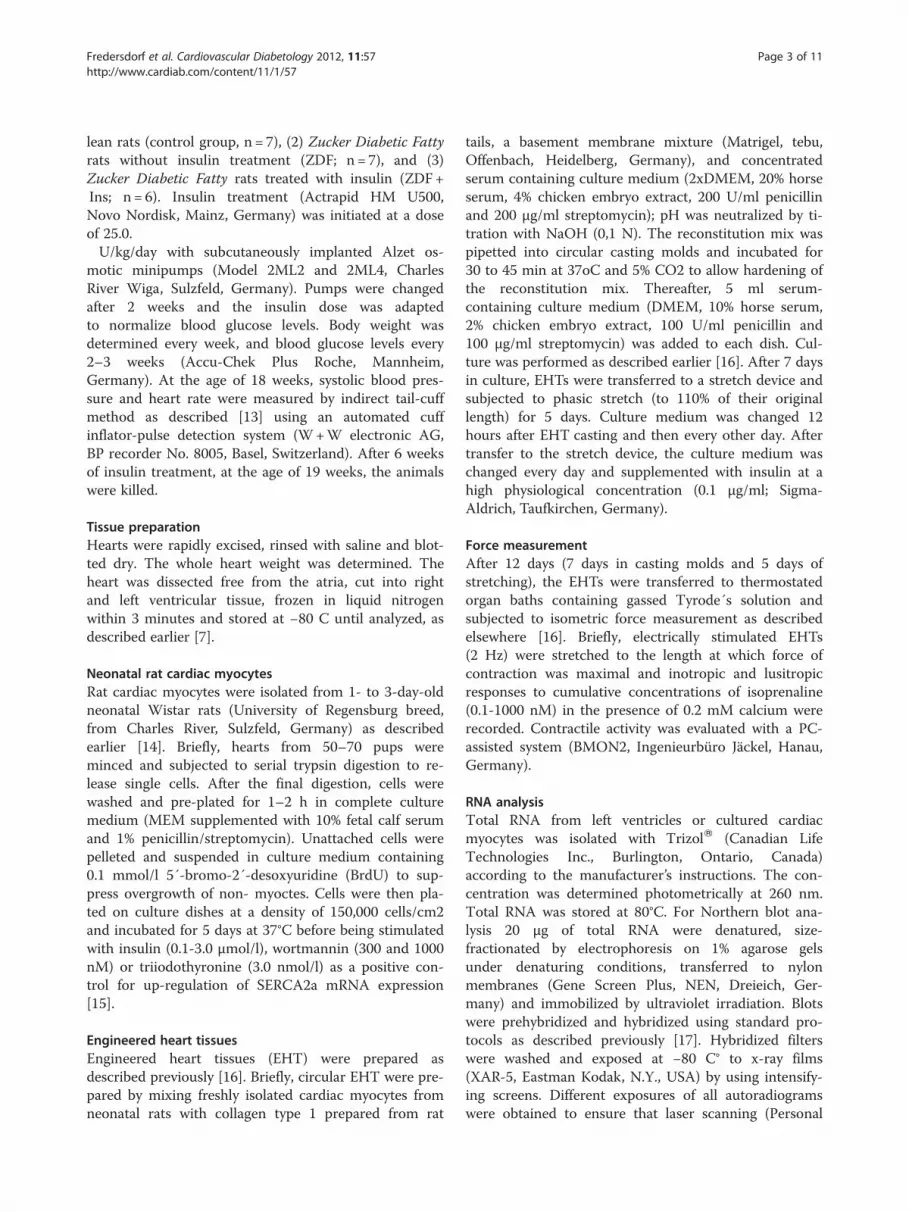
Figure 1 In vivo data: Expression of SERCA 2a and PLB in leftventricular tissue from non-diabetic control rats (n = 7), ZuckerDiabetes Fatty (ZDF) rats (n = 7) and ZDF (n = 6) rats treatedwith insulin. (A) Shows a representative Northern blot of SERCAand phospholamban (lane 1 = left ventricular tissue from control rats,lane 2= left ventricular tissue from diabetic ZDF rats; lane 3 = leftventricular tissue from ZDF rats treated with insulin). Arrow depictsthe position of the 28 S ribosomal RNA. (B) quantitative analysis and(C) SERCA2a/PLB ratio. Values are given in mean± SEM. *p < 0.05 vs.control; #p < 0.05 vs. ZDF.
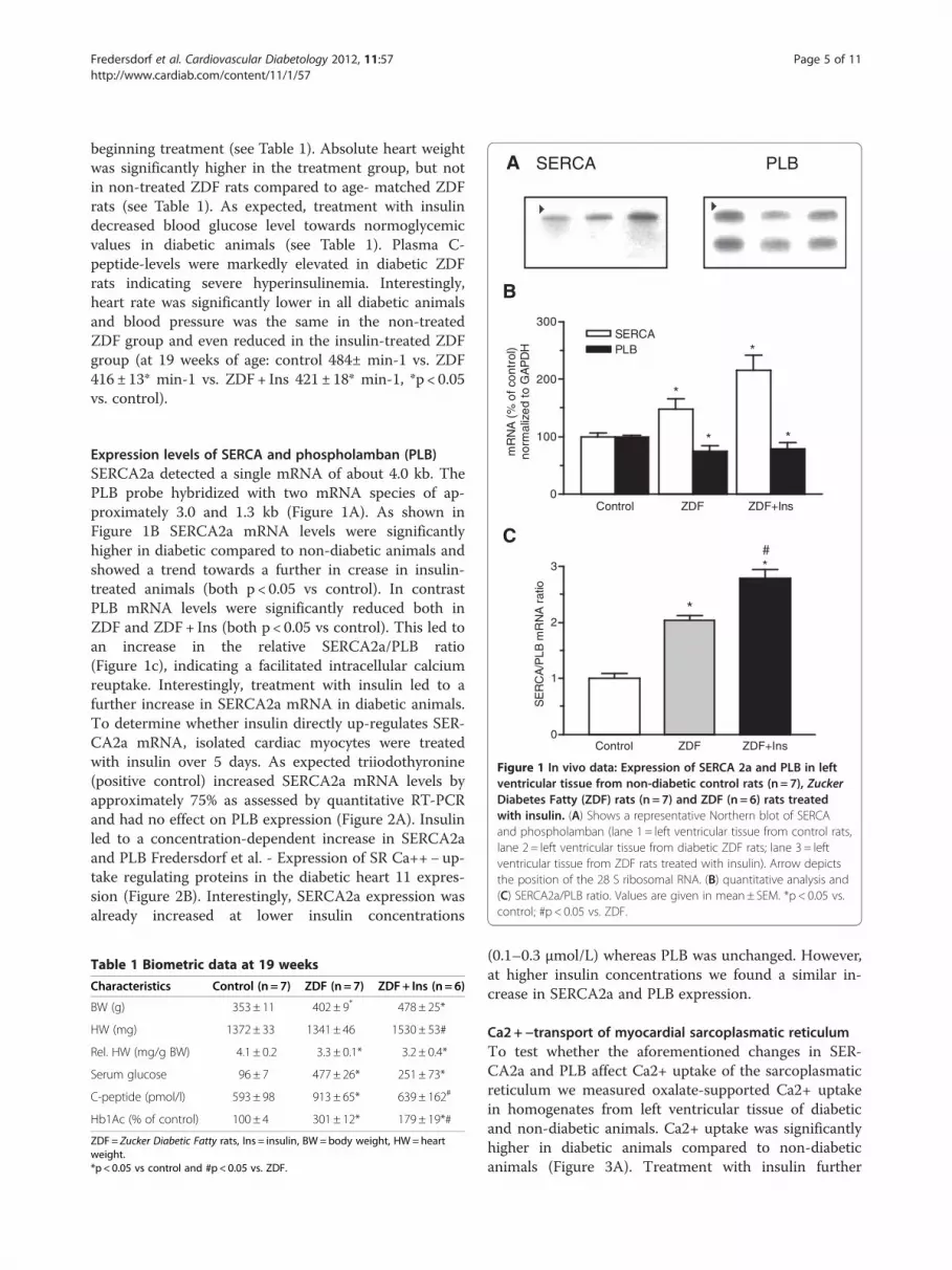
Expression levels of SERCA and phospholamban (PLB)SERCA2a detected a single mRNA of about 4.0 kb. ThePLB probe hybridized with two mRNA species of ap-proximately 3.0 and 1.3 kb (Figure 1A). As shown inFigure 1B SERCA2a mRNA levels were significantlyhigher in diabetic compared to non-diabetic animals andshowed a trend towards a further in crease in insulin-treated animals (both p < 0.05 vs control). In contrastPLB mRNA levels were significantly reduced both inZDF and ZDF+ Ins (both p < 0.05 vs control). This led toan increase in the relative SERCA2a/PLB ratio(Figure 1c), indicating a facilitated intracellular calciumreuptake. Interestingly, treatment with insulin led to afurther increase in SERCA2a mRNA in diabetic animals.To determine whether insulin directly up-regulates SER-CA2a mRNA, isolated cardiac myocytes were treatedwith insulin over 5 days. As expected triiodothyronine(positive control) increased SERCA2a mRNA levels byapproximately 75% as assessed by quantitative RT-PCRand had no effect on PLB expression (Figure 2A). Insulinled to a concentration-dependent increase in SERCA2aand PLB Fredersdorf et al. - Expression of SR Ca++− up-take regulating proteins in the diabetic heart 11 expres-sion (Figure 2B). Interestingly, SERCA2a expression wasalready increased at lower insulin concentrations
Table 1 Biometric data at 19 weeks
Characteristics Control (n = 7) ZDF (n= 7) ZDF+ Ins (n = 6)
BW (g) 353± 11 402± 9* 478± 25*
HW (mg) 1372 ± 33 1341± 46 1530± 53#
Rel. HW (mg/g BW) 4.1 ± 0.2 3.3 ± 0.1* 3.2 ± 0.4*
Serum glucose 96 ± 7 477± 26* 251± 73*
C-peptide (pmol/l) 593 ± 98 913± 65* 639± 162#
Hb1Ac (% of control) 100 ± 4 301± 12* 179± 19*#
ZDF= Zucker Diabetic Fatty rats, Ins = insulin, BW=body weight, HW=heartweight.*p < 0.05 vs control and #p < 0.05 vs. ZDF.
(0.1–0.3 μmol/L) whereas PLB was unchanged. However,at higher insulin concentrations we found a similar in-crease in SERCA2a and PLB expression.
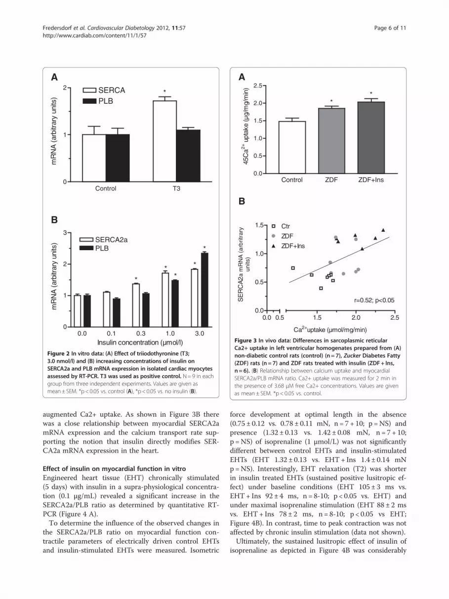
Ca2+−transport of myocardial sarcoplasmatic reticulumTo test whether the aforementioned changes in SER-CA2a and PLB affect Ca2+ uptake of the sarcoplasmaticreticulum we measured oxalate-supported Ca2+ uptakein homogenates from left ventricular tissue of diabeticand non-diabetic animals. Ca2+ uptake was significantlyhigher in diabetic animals compared to non-diabeticanimals (Figure 3A). Treatment with insulin further
A
B
Control T30
1
2 SERCAPLB
*
mR
NA
(ar
bitr
ary
units
)
0.0 0.1 0.3 1.0 3.00
1
2
3SERCA2aPLB
*
* *
*
*
Insulin concentration (µmol/l)
mR
NA
(ar
bitra
ry u
nits
)
Figure 2 In vitro data: (A) Effect of triiodothyronine (T3;3.0 nmol/l) and (B) increasing concentrations of insulin onSERCA2a and PLB mRNA expression in isolated cardiac myocytesassessed by RT-PCR. T3 was used as positive control. N=9 in eachgroup from three independent experiments. Values are given asmean± SEM. *p<0.05 vs. control (A), *p< 0.05 vs. no insulin (B).
A
B
0.0 0.50.0
0.5
1.0
1.5
1.5 2.0 2.5
r=0.52; p<0.05
Ctr
ZDF
ZDF+Ins
Ca2+uptake (µmol/mg/min)
SE
RC
A2a
mR
NA
(a
rbrit
rary
units
)
Control ZDF ZDF+Ins0.0
0.5
1.0
1.5
2.0
2.5
**
45C
a2+ u
ptak
e (µ
g/m
g/m
in)
Figure 3 In vivo data: Differences in sarcoplasmic reticularCa2+ uptake in left ventricular homogenates prepared from (A)non-diabetic control rats (control) (n = 7), Zucker Diabetes Fatty(ZDF) rats (n = 7) and ZDF rats treated with insulin (ZDF+ Ins,n = 6). (B) Relationship between calcium uptake and myocardialSERCA2a/PLB mRNA ratio. Ca2+ uptake was measured for 2 min inthe presence of 3.68 μM free Ca2+ concentrations. Values are givenas mean± SEM. *p < 0.05 vs. control.
Fredersdorf et al. Cardiovascular Diabetology 2012, 11:57 Page 6 of 11http://www.cardiab.com/content/11/1/57
augmented Ca2+ uptake. As shown in Figure 3B therewas a close relationship between myocardial SERCA2amRNA expression and the calcium transport rate sup-porting the notion that insulin directly modifies SER-CA2a mRNA expression in the heart.
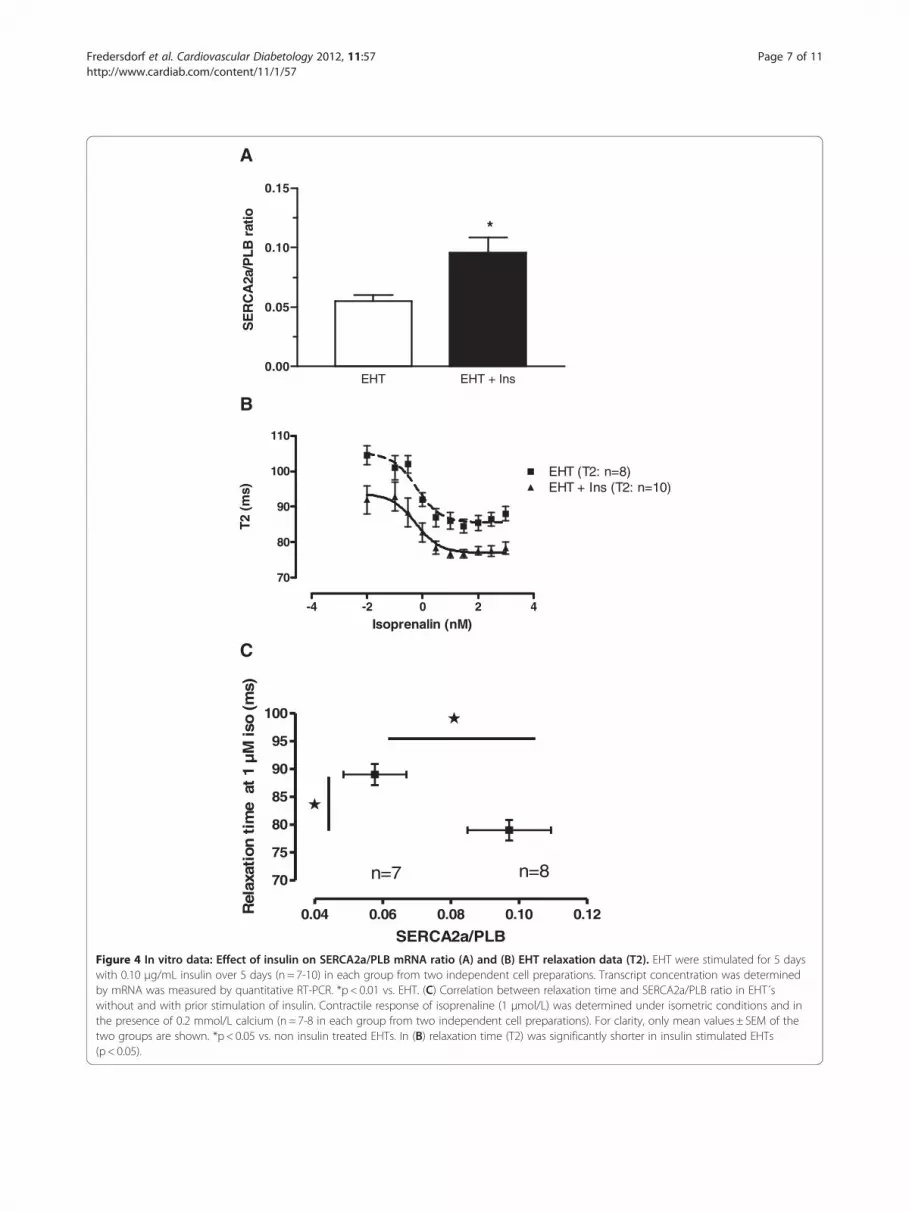
Effect of insulin on myocardial function in vitroEngineered heart tissue (EHT) chronically stimulated(5 days) with insulin in a supra-physiological concentra-tion (0.1 μg/mL) revealed a significant increase in theSERCA2a/PLB ratio as determined by quantitative RT-PCR (Figure 4 A).To determine the influence of the observed changes in
the SERCA2a/PLB ratio on myocardial function con-tractile parameters of electrically driven control EHTsand insulin-stimulated EHTs were measured. Isometric
force development at optimal length in the absence(0.75 ± 0.12 vs. 0.78 ± 0.11 mN, n = 7+ 10; p =NS) andpresence (1.32 ± 0.13 vs. 1.42 ± 0.08 mN, n = 7+ 10;p =NS) of isoprenaline (1 μmol/L) was not significantlydifferent between control EHTs and insulin-stimulatedEHTs (EHT 1.32 ± 0.13 vs. EHT+ Ins 1.4 ± 0.14 mNp=NS). Interestingly, EHT relaxation (T2) was shorterin insulin treated EHTs (sustained positive lusitropic ef-fect) under baseline conditions (EHT 105 ± 3 ms vs.EHT+ Ins 92 ± 4 ms, n = 8-10; p < 0.05 vs. EHT) andunder maximal isoprenaline stimulation (EHT 88 ± 2 msvs. EHT+ Ins 78 ± 2 ms, n = 8-10; p < 0.05 vs EHT;Figure 4B). In contrast, time to peak contraction was notaffected by chronic insulin stimulation (data not shown).Ultimately, the sustained lusitropic effect of insulin of
isoprenaline as depicted in Figure 4B was considerably
EHT EHT + Ins0.00
0.05
0.10
0.15
*S
ER
CA
2a/P
LB r
atio
-4 -2 0 2 4
70
80
90
100
110
EHT (T2: n=8)EHT + Ins (T2: n=10)
Isoprenalin (nM)
T2 (
ms)
A
B
C
0.04 0.06 0.08 0.10 0.12
70
75
80
85
90
95
100
n=7 n=8
SERCA2a/PLB
Rel
axat
ion
tim
e a
t 1
µM
iso
(ms)
Figure 4 In vitro data: Effect of insulin on SERCA2a/PLB mRNA ratio (A) and (B) EHT relaxation data (T2). EHT were stimulated for 5 dayswith 0.10 μg/mL insulin over 5 days (n = 7-10) in each group from two independent cell preparations. Transcript concentration was determinedby mRNA was measured by quantitative RT-PCR. *p < 0.01 vs. EHT. (C) Correlation between relaxation time and SERCA2a/PLB ratio in EHT´swithout and with prior stimulation of insulin. Contractile response of isoprenaline (1 μmol/L) was determined under isometric conditions and inthe presence of 0.2 mmol/L calcium (n = 7-8 in each group from two independent cell preparations). For clarity, only mean values ± SEM of thetwo groups are shown. *p < 0.05 vs. non insulin treated EHTs. In (B) relaxation time (T2) was significantly shorter in insulin stimulated EHTs(p < 0.05).
Fredersdorf et al. Cardiovascular Diabetology 2012, 11:57 Page 7 of 11http://www.cardiab.com/content/11/1/57

Fredersdorf et al. Cardiovascular Diabetology 2012, 11:57 Page 8 of 11http://www.cardiab.com/content/11/1/57
enhanced in EHTs treated with insulin compared to con-trol EHTs. Interestingly, there was a significant correl-ation between the isoprenaline induced relaxation andthe higher SERCA2a/PLB ratio (Figure 4C). The latterwas the result of a higher SERCA2a concentration andat the same time lower PLB transcript concentrationNotably, absolute PLB transcript abundance was mark-edly lower in insulin treated EHTs when compared tocontrol EHTs.
Effect of insulin on aktAs shown by Figure 5, Akt was significantly phosphory-lated by insulin (B) in cardiomyocytes. Unphosphory-lated Akt blot served as control (A). This effect could beabolished by wortmannin (C) at higher wortmannin con-centrations (1000 nM) for every insulin stimulation, atlower wortmannin concentration (300 nM) only forlower insulin concentrations.
DiscussionThe key findings of this study are that in the early stagesof type 2 diabetes, myocardial expression of SERCA2a ismarkedly elevated, whereas the expression of PLBmRNA is reduced. These changes go along with a sig-nificant increase in sarcoplasmatic Ca2+ uptake. Inter-estingly, the SERCA/PLB ratio in diabetic animals wasfurther increased by insulin treatment. In vitro, we wereable to demonstrate that insulin treatment of isolated
197 kDa
126 kDa
81 kDa
40 kDa
A
Insulin (pmol/L)
Wortmannin (nmol/L)
Insulin (pmol/L)
C
170 1.7×10⁻⁴ 1.7×10⁻⁶ 0
1000
0 170 1.7×10⁻⁴ 1.7×
Figure 5 Effect of increasing concentrations of insulin on the phosphblot incubated with a specific antibody directed to total Akt. (B) The identiphospho-Akt (Ser473). The insulin- mediated increase of phosphorylated Akconcentration of the PI3K-Inhibitor Wortmaninn, where Wortmaninn per se
cardiac myocytes led to a concentration-dependent in-crease in SERCA2a expression. This effect was also seenin a more complex model of engineered heart tissue andcorrelated positively with cardiac relaxation in vitro. . Inaddition, Akt was significantly stimulated by insulin incardiomyocyte cell culture. This approach allows us todirectly determine contractile alterations caused by insu-lin treatment without considering insulin-related sys-temic changes which might also affect the cardiovascularsystem. However we must consider that in vitro data cannot totally be compared to in vivo data. Neverthelessthey give essential additional information. Together, thissuggests that insulin, which is particularly elevated inpremature type 2 diabetes mellitus, may be involved inthe induction of SERCA2a mRNA expression and maybe an early step in the pathogenesis of diabeticcardiomyopathy.
Alterations of SERCA, PLB and ca uptake in diabetesmellitusDiastolic relaxation of the heart is mediated to a largeextent by the uptake of Ca2+ into the sarcoplasmicreticulum. Several studies have shown that diabetic car-diomyopathy is associated with decreased contractilityand impaired relaxation [19]. These changes have beenattributed to a reduced ability to sequester calcium intothe SR, which primarily determines the speed of cardiacrelaxation. Consecutive experiments have shown that
40 kDa
81 kDa
126 kDa
197 kDa
B
83 kDa
39 kDa
300
170 1.7×10⁻⁴ 1.7×10⁻⁶
10-6 0 170 1.7×10⁻⁴ 1.7×10⁻⁶
orylation status of Akt in isolated cardiomyocytes. (A) Westerncal Western blot incubated with a specific antibody directed tot was abolished by the administration of an intermediatedhad no effect on phospho-Akt (C).

Fredersdorf et al. Cardiovascular Diabetology 2012, 11:57 Page 9 of 11http://www.cardiab.com/content/11/1/57
SERCA2a mRNA expression, protein level and activity isdown-regulated in streptozotocin-induced (STZ) type 1diabetes mellitus [10,11]. In another model Wold andco-workers demonstrated in cardiomyocytes of rats withsucrose-induced insulin resistance that impaired SERCAactivity with normal protein content contributes to car-diomyocyte dysfunction, whereas NCX function and ex-pression are normal [20]. The authors concluded thatsubtle changes in Ca2+ regulation which occur prior toovert ventricular dysfunction and/or failure, may becommon to early stages of a number of disorders involv-ing insulin resistance. Furthermore, it has been demon-strated that reduced Ca2+ signaling in vascular smoothmuscle cells from diabetic animals is related to a declineand/or redistribution in the IP3R Ca2+ channels andSERCA proteins. These changes could be repeated incell culture experiments with high glucose levels [21].The mechanism of alteration of SR proteins in STZ-induced diabetes, however, is unclear at present. Thesame is true for the Otsuka-Long-Evans Tokushima Fattyrats (OLETF), a type 2 diabetes model. Here in latestages of diabetes, after additional sucrose feeding, theventricular relaxation rate was significantly slower andwas associated with reduced SERCA2a level [22]. Theseexperiments are therefore in apparent contrast to ourresults. This discrepancy is most likely due to the differ-ent models used and the rather advanced duration of thediabetes and older age (60 weeks in the latter experi-ments as compared to 19-week-old diabetic rats in ourstudy). OLETF rats present a milder form of diabetesmellitus with later onset and milder hyperglycemia atthe beginning [23]. We examined ZDF rats at earlier dia-betic stages, and our previous experiments have shownthat C-peptide levels are high at this age [7].
Insulin directly up-regulates SERCA and preserves cardiacfunctionMost of the previous studies used a model with type 1diabetes which differs fundamentally in pathophysiology.Whereas type 1 diabetes mellitus results from selectivedestruction of the insulin- producing beta cells of thepancreas, type 2 diabetes is primarily characterized byinsulin resistance followed by progressive beta-cell dys-function, resulting in low insulin levels in the long term.As recently shown, obese ZDF rats are insulin-resistantand have basal hyperinsulinemia that is due mainly tohypersecretion of insulin, as indicated by their elevatedbasal C-peptide levels [7,24]. Obese pre- diabetic anddiabetic rats also show a reduction in insulin clearance,as indicated by their lower C- peptide/insulin ratio.Interestingly, the decrease in SERCA2a activity in STZ-treated rats can be reversed by insulin treatment [9,11],suggesting a direct stimulatory effect of insulin on SER-CA2a. This hypothesis is further supported by other
experiments demonstrating an up-regulation of SERCA1in skeletal muscle after stimulation with insulin [1].These observations demonstrate a possible link betweeninsulin and expression of SR calcium ATPase, which isfurther confirmed by the present in vitro studies show-ing a direct effect of insulin on SERCA2a transcriptionin isolated cardiac myocytes. The important role of insu-lin for heart function is further supported by Kim et al.,who showed that insulin preserves heart function instreptozotocin-induced diabetic heart failure with andwithout transplantation of smooth muscle cells [25].In this context it has been shown that transgenic (TG)
mice with cardiac-specific overexpression of active Aktnot only exhibit hypertrophy and enhanced left ventricu-lar function but also show a 6.6-fold increase in SER-CA2a protein levels, which could be recapitulatedin vitro by adenovirus-mediated overexpression of Aktin cultured adult ventricular myocytes [26]. We demon-strated on isolated cardiac myocytes a strong and rapidphosphorylation of Akt after stimulation with insulin.Conversely, inhibiting SERCA2a with either ryanodineor thapsigargin affected myocyte contraction and relax-ation and Ca2+ channel kinetics more in TG than inWT. Thus, myocytes from mice with overexpressed Aktdemonstrated enhanced contractility and relaxation,Fura-2 Ca2+ transients, and Ca2+ channel currents [26].Furthermore, increased protein expression of SERCA2aplays an important role in mediating enhanced leftventricular function by Akt. Interestingly, insulinstimulation led to a significant increase in SERCA2a,co-immunoprecipitated with insulin receptor substrateproteins (IRS-1 and IRS-1) in isolated cardiac muscledemonstrating a link between insulin, insulin receptorand SERCA2a [27] in cardiac tissue.Recent experiments showed that insulin-like growth
factor 1 (IGF-1) activates multiple signaling pathways,which involve the activation of the phosphatidylinositol(PI)3-kinase and Akt [28]. It is well known that the PI3-kinase-Akt cascade modulates diverse cellular functions.Furthermore, it has been shown that that IGF-I causedincreases in myocyte contraction and relaxation func-tion, increases in intracellular Ca2+ transients, and anupregulation of SERCA2a [29]. The same group demon-strated that transgenic mice with cardiac-specific overex-pression of Akt showed an enhanced left ventricularfunction, associated with an increased expression ofSERCA2a [30]. These data are in accord with the studiesof von Lewinski and co-workers [31](4), who suggestedthat Akt contribute to the acute inotropic effect of IGF-Iin myocytes from human failing hearts.In the present study, the insulin induced increase in
phosphorylated Akt in isolated cardiomyocytes was abol-ished by the PI3-kinase inhibitor wortmannin, whichprovided evidence for a role of PI3-kinase. This finding

Fredersdorf et al. Cardiovascular Diabetology 2012, 11:57 Page 10 of 11http://www.cardiab.com/content/11/1/57
suggests that the underlying cellular mechanism for upregulation of SERCA2a is mediated by the PI3-kinase-Akt-SERCA2a signaling cascade.From a pathophysiological point of view, insulin-
induced up-regulation of myocardial SERCA2a may beseen as a feedback mechanism in handling the volumeoverload caused by high glucose levels in the early phaseof type 2 diabetes, when insulin levels are high. Withprogression of the disease and decreasing levels of insu-lin the expression of SERCA2a in the heart becomesimpaired. The reduction of SERCA2a, as typically seenin the late phase of type 2 diabetes, is a major cause ofreduced diastolic and systolic function of the heart. Thishypothesis is reinforced by the findings of Sakata et al.who demonstrated that cardiac SERCA2a gene transferrestores systolic and diastolic function to normal in dia-betic rats [8]. Furthermore, in-vitro experiments provideevidence that high glucose levels also impair cytosolicCa2+ removal involving slowed SR Ca2+ uptake. It hasbeen speculated that slowed SR Ca2+ uptake resultsfrom depressed protein kinase A (PKA) down-regulatingSERCA2a, rather than through depressed SERCAexpression. Both expression and function of the Na-Ca-exchanger (NCX) appear to be normal in these experi-ments [16]. In summary, the up-regulation of SERCA2ain the early phase of type 2 diabetes is an importantphysiological adaptation of the heart allowing it tohandle volume overload caused by high glucose levels.
Competing interestsThe authors declare that they have no conflict of interest.
Authors’ contributionsSF carried out the molecular experiments and drafted the main parts of themanuscript. CT performed the work with the rats and northern blotting,WHZ supported us with the engineered heart tissue, RV performed thecalcium uptake experiments, TG helped to draft the manuscript, ALparticipated in the design of the study and of the manuscript, GAJparticipated in its design and coordination, HS helped to draft themanuscript, TH coordinated the molecular experiments and the engineeredheart tissue model, JW conceived the study, performed the statistical analysisand drafted parts of the manuscript. All authors read and approved the finalmanuscript.
AcknowledgmentsThe authors wish to thank Sabine Laberer and Josef Simon for excellenttechnical assistance. This project was supported in part by a grant from theUniversity of Regensburg to J. Weil and S. Fredersdorf (ReformA).
Author details1Klinik und Poliklinik für Innere Medizin II, Universität Regensburg,Regensburg, Germany. 2Institut für Pharmakologie, Universitätsmedizin,Georg-August Universität Göttingen, Göttingen, Germany. 3Institut fürKlinische Pharmakologie und Toxikologie, Universitätsmedizin - Berlin, Berlin,Germany. 4Medizinische Klinik II, Universitätsklinikum Schleswig-Holstein,Campus Lübeck, Lübeck, Germany. 5Institut für Klinische und ExperimentellePharmakologie und Toxikologie, Universität Hamburg, Hamburg, Germany.6Klinik und Poliklinik für Innere Medizin II des UniversitätsklinikumsRegensburg, 93042 Regensburg, Germany.
Received: 20 December 2011 Accepted: 2 May 2012Published: 23 May 2012
References1. Dibb KM, Graham HK, Venetucci LA, Eisner DA, Trafford AW: Analysis of
cellular calcium fluxes in cardiac muscle to understand calciumhomeostasis in the heart. Cell Calcium 2007, 42:503–512.
2. Frank KF, Bolck B, Erdmann E, Schwinger RH: Sarcoplasmatic reticulumCa2+−ATPase modulates cardiac contraction and relaxation. CardiovascRes 2003, 57:20–27.
3. Periasamy M, Bhupathy P, Babu GJ: Regulation of sarcoplasmic reticulumCa2+ ATPase pump expression and its relevance to cardiac musclephysiology and pathology. Cardiovasc Res 2008, 77:265–273.
4. Trost SU, Belke DD, Bluhm WF, Meyer M, Swanson E, Dillmann WH:Overexpression of the sarcoplasmic reticulum Ca(2+)-ATPase improvesmyocardial contractility in diabetic cardiomyopathy. Diabetes 2002,51:1166–1171.
5. Slack JP, Grupp IL, Dash R, Holder D, Schmidt A, Gerst MJ, Tamura T,Tilgmann C, James PF, Johnson R, Gerdes AM, Kranias EG: The enhancedcontractility of the phospholamban- deficient mouse heart persists withaging. J Mol Cell Cardiol 2001, 33:1031–1040.
6. Ligeti L, Szenczi O, Prestia CM, Szabó C, Horváth K, Marcsek ZL, van StiphoutRG, van Riel NA, Op den Buijs J, Van der Vusse GJ, Ivanics T: Alteredcalcium handling is an early sign of streptozotocin-induced diabeticcardiomyopathy. Int J Mol Med 2006, 17:1035–1043.
7. Fredersdorf S, Thumann C, Ulucan C, Griese DP, Luchner A, Riegger GAJ,Kromer EP, Weil J: Myocardial Hypertrophy and Enhanced Left VentricularContractility in Zucker Diabetic Fatty Rats. Cardiovasc Pathol 2004,13:11–19.
8. Sakata S, Lebeche D, Sakata Y, Sakata N, Chemaly ER, Liang LF,Padmanabhan P, Konishi N, Takaki M, del Monte F, Hajjar RJ: Mechanicaland metabolic rescue in a type II diabetes model of cardiomyopathy bytargeted gene transfer. Mol Ther 2006, 13:987–996.
9. Golfman L, Dixon IM, Takeda N, Chapman D, Dhalla NS: Differentialchanges in cardiac myofibrillar and sarcoplasmatic reticular geneexpression in alloxan-induced diabetes. Mol Cell Biochem 1999, 200:15–25.
10. Zarain-Herzberg A, Yano K, Elimban V, Dhalla NS: Cardiac sarcoplasmaticreticulum Ca2+− ATPase expression in streptozotocin-induced diabeticrat. Biochem Biophys Res Comm 1994, 203:113–120.
11. Zhong Y, Ahmed S, Grupp IL, Matlib MA: Altered SR protein expressionassociated with contractile dysfunction in diabetic rat heart. Am J PhysiolHeart Circ Physiol 2001, 281:H1137–H1147.
12. Reed MJ, Scribner KA: In-vivo and in-vitro models of type 2 diabetes inpharmaceutical drug discovery. Diabetes, Obesity and Metabolism 1999,1:75–86.
13. Peterson RG, Shaw WN, Neel MA, Little LA, Eichberg J: Zucker diabetic fattyrat as a model for non-insulin-dependent diabetes mellitus. ILAR News1990, 32:16–19.
14. Weil J, Benndorf R, Fredersdorf S, Griese DP, Eschenhagen T:Norepinephrine upregulates vascular endothelial growth factor in ratcardiac myocytes by a paracrine mechanism. Angiogenesis 2003,6:303–309.
15. Muller A, Zuidwijk MJ, Simonides WS, van Hardeveld C: Modulation ofSERCA2 expression by thyroid hormone and norepinephrine incardiocytes: role of contractility. Am J Physiol 1997, 272:H1876–H1885.
16. Zimmermann WH, Schneiderbanger K, Schubert P, Didié M, Münzel F,Heubach JF, Kostin S, Neuhuber WL, Eschenhagen T: Tissue engineering ofa differentiated cardiac muscle construct. Circ Res 2002, 90:223–230.
17. Weil J, Eschenhagen T, Fleige G, Mittmann C, Orthey E, Scholz H:Localization of preproenkephalin mRNA in rat heart: selective geneexpression in left ventricular myocardium. Am J Physiol 1998,275:H378–H384.
18. Livak KJ, Schmittgen TD: Analysis of relative gene expression data usingreal-time quantitative PCR and the 2(−ΔCt)method. Methods 2001,25:402–408.
19. Belke DD, Dillmann WH: Altered cardiac calcium handling in diabetes.Curr Hypertens Rep 2004, 6:424–429.
20. Wold LE, Dutta K, Mason MM, Ren J, Cala SE, Schwanke ML, Davidoff AJ:Impaired SERCA function contributes to cardiomyocyte dysfunction ininsulin resistant rats. J Mol Cell Cardiol 2005, 39:297–307.
21. Searls YM, Loganathan R, Smirnova IV, Stehno-Bittel L: Intracellular Ca2+regulating proteins in vascular smooth muscle cells are altered with type1 diabetes due to the direct effects of hyperglycemia. Cardiovasc Diabetol2010, 9:8.

Fredersdorf et al. Cardiovascular Diabetology 2012, 11:57 Page 11 of 11http://www.cardiab.com/content/11/1/57
22. Abe T, Ohga Y, Tabayashi N, Kobayashi S, Sakata S, Misawa H, Tsuji T,Kohzuki H, Suga H, Tanigucchi S, Takaki M: Left ventricular diastolicdysfunction in type II diabetes mellitus model rats. Am J Heart Circ Physiol2002, 282:H139–H148.
23. Yagi K, Kim S, Wanibuchi H, Yamashita T, Yamamura Y, Iwao H:Characteristics of diabetes, blood pressure, and cardiac and renalcomplications in Otsuka Long-Evans Tokushima Fatty rats. Hypertension1997, 29:728–735.
24. Goh TT, Mason TM, Gupta N, So A, Lam TK, Lam L, Lewis GF, Mari A, GiaccaA: Lipid-induced beta-cell dysfunction in vivo in models of progressivebeta-cell failure. Am J Physiol Endocrinol Metab 2007, 292:E549–E560.
25. Kim BO, Verma S, Weisel RD, Fazel S, Jia ZQ, Mizuno T, Li RK: Preservationof heart function in diabetic rats by the combined effects of muscle cellimplantation and insulin therapy. Eur J Heart Fail 2008, 10:14–21.
26. Kim YK, Kim SJ, Yatani A, Huang Y, Castelli G, Vatner DE, Liu J, Zhang Q, DiazG, Zieba R, Thaisz J, Drusco A, Croce C, Sadoshima J, Condorelli G, Vatner SF:Mechanism of enhanced cardiac function in mice with hypertrophyinduced by overexpressed Akt. Biol Chem 2003, 278:47622–47628.
27. Algenstaedt P, Antonetti DA, Yaffe MB, Kahn CR: Insulin receptor substrateproteins create a link between the tyrosine phosphorylation cascadeand the Ca2+−ATPases in muscle and heart. J Biol Chem 1997,272:23696–23702.
28. Ren J, Samson WK, Sowers JR: Insulin-like growth factor I as a cardiachormone: physiological and pathophysiological implications in heartdisease. J Mol Cell Cardiol 1999, 31:2049–2061.
29. Kim SJ, Abdellatif M, Koul S, Crystal GJ: Chronic treatment with insulin-likegrowth factor I enhances myocyte contraction by upregulation of Akt-SERCA2a signaling pathway. Am J Physiol Heart Circ Physiol 2008,295:H130–H135.
30. Kim YK, Kim SJ, Yatani A, Huang Y, Castelli G, Vatner DE, Liu J, Zhang Q, DiazG, Zieba R, Thaisz J, Drusco A, Croce C, Sadoshima J, Condorelli G, Vatner SF:Mechanism of enhanced cardiac function in mice with hypertrophyinduced by overexpressed Akt. J Biol Chem 2003, 278:47622–47628.
31. Von Lewinski D, Voss K, Hulsmann S, Kogler H, Pieske B: Insulin-like growthfactor-1 exerts Ca2+−dependent positive inotropic effects in failinghuman myocardium. Circ Res 2003, 92:169–176.
doi:10.1186/1475-2840-11-57Cite this article as: Fredersdorf et al.: Increased myocardial SERCAexpression in early type 2 diabetes mellitus is insulin dependent: In vivoand in vitro data. Cardiovascular Diabetology 2012 11:57.
Submit your next manuscript to BioMed Centraland take full advantage of:
• Convenient online submission
• Thorough peer review
• No space constraints or color figure charges
• Immediate publication on acceptance
• Inclusion in PubMed, CAS, Scopus and Google Scholar
• Research which is freely available for redistribution
Submit your manuscript at www.biomedcentral.com/submit