Validating Type 1 and Type 2 Diabetes Mellitus in the Mini ...
Upload
khangminh22Category
view
0download
0
STUDIES ON CHILDHOOD DIABETES MELLITUS.

STUDIES ON CHILDHOOD DIABETES MELLITUS.
(STUDIES OVER DIABETES MELLITUS BIJ KINDEREN)
PROEFSCHRIFT
TER VERKRIJGING VAN DE GRAAD VAN DOCTOR IN DE
GENEESKUNDE
AAN DE ERASMUS UNIVERSITEIT ROTTERDAM
OP GEZAG VAN DE RECTOR MAGNIFICUS
PROF. DR. M.W. VAN HOP
EN VOLGENS BESLUIT VAN RET COLLEGE VAN DEKANEN.
DE OPENBARE VERDEDIGING ZAL PLAATSVINDEN OP
VRIJDAG 6 JULI 1984 TE 15.45 UUR
DOOR
GERBRAND JAN BRUINING
GEBOREN TE EINDHOVEN

BEGELEIDINGSCOMMISSIE
PROMOTOREN
OVERIGE LEDEN'
PROF. DR. H.K.A. VISSER
PROF. DR. J.J. VAN ROOD
PROF. DR. R. BENNER
PROF. DR. J.C. BIRKENHAGER
PROF. I.R. COHEN M.D., Ph.D.
PROF. DR. N. MASUREL
PROF. DR. W.H.H. TEGELAERS
PROF. DR. H.A. VALKENBURG
The work was supported by grants from the Sophia Foundation for
Medical Research, Stichting Diabetes Research Fonds, Novo In
dustries, the Nederlands Komi t€ voor Kinderpostzegels, Nordisk
Insulin Laboratories and Boehringer-Mannheim Netherlands.

Aan Wanda, Iris en Hilgo.


CONTENTS PAGE
INTRODUCTION 11
CHAPTER I ETIOLOGY
§1.
§2.
§3.
§4.
§5.
Background
Pathology and insulin secretion
Scope of the studies
Addendum
reprints of papers
17
22
27
32
"HLA and GM in insulin-dependent 32 diabetes in the Netherlands: report on a combined multiplex family and population study".
"Neonatal onset permanent diabetes 62 mellitus and incomplete fetal alco-hol syndrome: cause or coincidence"?
"Prediction of type-1-diabetes mellitus: 70 a report on three cases".
"Clinical time-course and characteristics of islet-cell cytoplasmatic antibodies in childhood diabetes".
85
"Autoantibodies to the insulin re- 102 ceptor in juvenile onset insulin-dependent diabetes".
Discussion 110
1. HLA-antigens as genetic markers for 111 the susceptibility to childhood diabetes.
2. Islet-cell antibodies as markers for 117 childhood pre-diabetes.
3. Insulin antigenicity. 125

4. Immune-intervention in childhood
diabetes.
§ 6 . S uromary
References to Chapter I
CHAPTER II EPIDEMIOLOGY
§1.
§2.
Introduction
Addendum
reprint of paper
136
139
140
150
154
"The incidence of childhood diabetes 154 in the Netherlands. A decrease from north to south over north-western Europe"?
§3. Discussion 165
1. Prevalence estimated from incidence. 165
2. Viruses as exogenous factors. 168
§4. Summary 175
References to Chapter II 177
CHAPTER III TREATMENT
§1.
§2.
§3.
An inventory of past and present
trends
Design of the home care program
Addendum
reprints of papers
183
193
195
"Home care for children with 195 diabetes mellitus in the Netherlands".

§4,
§5.
§6,
"Limitations to the use of glycosylated hemoglobins as a parameter of glycemic control in childhood diabetes".
Additional findings with horne care
Discussion
Summary
References to Chapter III.
Summary and conclusions (in the english language)
Summary and conclusions (in the dutch language)
Acknowledgements (in the dutch language)
Curriculum vitae
203
221
240
242
243
247
252
258
260

11
Introduction
This thesis consists of a number of collaborative studies aimed
at the improvement of the diagnosis and care of children with
diabetes mellitus. For the reader, who is not familiar with me
dical problems, a brief account is given of the clinical "be
havior" of the disease ( 1) . It is perhaps clarifying to describe
a disease as an entity which may display a behavior as if it were
a living being. For non-medical people it sheds some light on the
magic doctors seem to operate with.
The many variables by which diseases may manifest their behavior
imply that doctors, in caring for patients, constantly perform
experiments. During a single week of active practice with a com
plex disease as diabetes, the clinician conducts more experiments
than most of his laboratory collegues do in a year.
The urge of some adolescents with diabetes mellitus to perform
even more experiments at home, without their doctors knowing it,
generates an even more lively behavior of this disease.
In a psychologic or social sense, the word "behavior" refers to a
person's attitude in various situations in life. In a more gene
ral sense, the concept of behavior applies to the interaction of
an entity to its environment. With this concept, we can contem
plate the behavior of a disease during its interaction with the
human host (patient) , who provides the environment in which the
disease conducts its "life".
The disease can behave morphologically, to alter structures in
the host's body. It can behave biochemically or biophysically to
affect functions of the body. It behaves clinically, to produce
symptoms typical for the host's illness, and psychologically as
the host will counteract the impact the disease will exert on the
host's life. This classification allows a summary of basic pro
blems of childhood diabetes.
Morphologically, the disease is characterized by the destruction
of specialized cells, producing a crucial hormone for the metabo
lism of energy sources and other nutrients, insulin {2). The host
is unable to repair this damage and will need insulin substitu-

12
tion (injections) for the rest of her or his life. The mechanisms
leading to this destruction are presently inaccessible for pre
ventive intervention.
Biochemically, failure to completely normalize the metabolism of
energy sources and other nutrients is a hallmark of the disease,
despite the availability of highly purified insulin preparations.
A spectre of chronic metabolic disregulations is still found, of
ten with remarkably few symptoms (3) .
After 10 or 20 years the average patient will display an array of
structural and functional abnormalities. These are called the
chronic complications of diabetes. They have shortened the life
expectancy of children with diabetes mellitus by 1/3 in years.
Clinically, there are presently no means to prevent or even stop
the irreversible damage to cells producing insulin.
A classic controversy in medicine (4) has been whether meticulous
insulin-administration and diet-keeping could alter the behavior
of the disease in its tendency to develop the incapacitating com
plications. One of the reasons for this controversy is the lack
of practical biochemical parameters, to monitor the quality of
the metabolic regulation as a result of such therapeutic regimens
(5).
Psychologically, the child has to grow up with one or two daily
shots of insulin, dietary measures, to experience regular hospi
tal-visits, including admissions for unforeseen severe disregula
tion of the diabetes (6)
The parents are usually aware of the possibility of chronic com
plications and have to live with the idea their child's inconspi
cuous disease is at least partially inherited. The uncertainty
whether strict following of medical desirabilities, interfering
with the child's upbringing, would improve the long-term progno
sis or not, is an embarrassment to health-care professionals as
well as patients.
The ramifications of these problems of childhood diabetes provide
a daunting prospect, if the behavior of this disease cannot be
modified.
Although the understanding of the disease is far from complete,

13
advances in knowledge have been made steadily since the discovery
of insulin in 1921. In the pre-insulin-era diabetes mellitus in
children meant "melting" away in a couple of weeks or months with
certain death. The impact of the discovery of Banting and Best,
that their pancreatic extract saved the lives of these children
since 1922 was great. The dramatic effect of injected insulin on
the blood glucose of patients with hyperglycemia undoubtedly led
to the belief that hyperglycemia, and hence diabetes mellitus,
was always due to insulin lack, and the more profound the insulin
deficiency, the higher the glucose concentration. It was not un
til 1959 that the painstaking studies of Somogyi demonstrated
that excess insulin could in fact exacerbate the diabetic state
(7). Just thereafter, in 1960, Berson and Yalow published their
historic finding that insulin-concentrations could be measured in
biologic fluids by radio-immuno-assay. This finding was preceded
by their observation that many patients with diabetes had antibo
dies against the injected insulin in their circulation (8).
Early in the 1930's blindness caused by longstanding diabetes,
treated with insulin, was virtually unheard of. Now it is appre
ciated that diabetes is the leading cause of new cases of blind
ness {9) • The rediscovery of an old medical file in the cellars
of the Hospital for Sick Children in Toronto exemplifies this
point: it contained an autopsy performed in 1932 on a man who had
been one of the very first patients ever to receive insulin. A
full description of abnormalities classic for chronic complica
tions was in the report, but it was stated publicly the young man
had died from a motor-cycle accident (10).
The past decade has delivered an explosion of data on insulin de
pendent diabetes, as a comparison between the proceedings of
large meetings of 10 years ago and today would demonstrate: immu
nology, virology, epidemiology ( 11) , pharmacokinetics ( 12) and
biochemistry (13) have made steady contributions.
With the echo of these explosions of knowledge ringing in the
ears, the clinician selects those topics that would benefit the
patient most, either directly or indirectly.
This thesis is the result of the selection and subsequent work on
such topics between 1978 and 1984.
14
It consists of three chapters, each divided into introductory pa
ragraphs, a selection of papers relevant to that chapter and a
discussion of its contents, concluded with a summary. Figures and
tables are numbered by their sequence throughout each chapter,
excepting illustrations of the reprints of papers. Also, the
literature references cited throughout each chapter are only gi
ven at the end of that chapter and do not refer to literature re
ferences given in the reprints of papers.
References to the introduction
1. Feinstein, A.R. "Clinical Judgement"
Williams and Williams Baltimore, 1967, p. 141.
2. Gepts, W. in "The diabetic Pancreas",
Volk, B.W. and Wellmann, K.F. Eds.,
Plenum Press, N.Y. 1977, p. 325-365.
15
3. Alberti, K.G.M.M. "Metabolic abnormalities in juvenile
diabetes mellitus",
Second Nordisk Symposium, Oslo 1967, p. 125-138.
4. Ingelfinger, F .J. "Debates on diabetes"
New England Journal of Medicine 296: 1228-1230, 1977
5. Malone, J.I., Hellrung, J.M., Malphus, E.W. et al.
"Good diabetic control- a study in mass delusion",
Journal of Pediatrics 88: 943, 1976.
6. Schiff, I.J, "Emotional problems
and their parents",
Psychosomatics~: 362, 1964.
of diabetic children
7. Somogyi, M. "Exacerbation of diabetes by excess insulin",
American Journal of Medicine~: 169, 1959,
8. Berson, S.A., Yalow, R.S., Bauman, A., Rothchild, M.A.,
Newerly, K.
"Insulin - 131 I metabolism in human subjects: demonstration
of insulin binding globulin in the circulation of insulin
treated subjects.",
Journal of Clinical Investigation 35: 170-190, 1956.
16
9. Newell, F.W., 11 The problem of diabetic retinopathy"
In: "Vascular complications of Diabetes Mellitus 11,
Kimura, S.J., Caygill, W.M. Eds., C,V, Mosby, St. Louis,
1967, p. 35-39.
10. Burrow, G.M., Hazlett, B.E., Phillips, M.J.
"A case of diabetes mellitus 11
New England Journal of Medicine 306: 340-343, 1982.
11. Notkins, A.L. "The causes of Diabetes 11,
Scientific American, November 1979, p. 56-57.
12. Binder, C., "State of the art lecture"
EASD Meetings, Amsterdam 1981.
13. Randle, P.J. "New perspectives on the metabolic aspects
of insulin-deficiency in "Etiology and Pathogenesis of
insulin-dependent Diabetes Mellitus 11
Martin, J.M., Ehrlich, R.M. and Holland, F.J. Eds.,
Raven Press New York 1981, p. 21-37.

CHAPTER I ETIOLOGY
§1. Background
For I dipt into the future,
far as human eye could see,
Saw the vision of the world,
17
and all the wonder that would be.
Alfred, Lord Tennyson, 1809-1892.
{From: Locksley Hall 1.119)
The recognition of thirst and honey-sweet urine in massive
amounts as signs of the disease is old. In ancient India descrip
tions have been found in texts on Hindu medicine ( 1) . Diabetes
mellitus as a term is derived from the greek diabainein (to fall
through) and mellos (honey-sweet) (2) •
Today the notion that a variety of genetic and evironmental fac
tors may lead to diabetic states and sequelae thereof, prompted
attempts to associate these factors with clinical classifica
tions. However, etiologic factors may be expressed in several
clinical forms and putative clinical presentations may involve
several etiologic factors. Associations are only valid in a "sta
tistical" manner. What has been learned from such associations?
A first fundamental point is that insulin-requiring or insulin
dependent diabetes mellitus (also known as juvenile-onset, keto
sis prone or labile diabetes) is a distinct disorder from non
insulin-requiring diabetes mellitus (maturity-onset, non-ketosis
prone or stable diabetes) . A new classification separating these
broad categories has been put forward (3,4), distinguishing type-
1-diabetes {insulin-dependent diabetes mellitus IDDM) from
type-2-diabetes (non-insulin-dependent diabetes mellitus
NIDDM). This classification has subsequently be refined (5).

Table 1.
Type 1
1 .a.
l.b.
Type 2
2. a.
2.b.
*
Classification of diabetes mellitus (5)
insulin-dependent diabetes
genetic susceptibility known
predominantly young onset
slight male excess
transient autoimmune phenomena
probably initiated by viruses *
18
primary association with both HLA -DR3 and HLA-DR4
predominantly middle age onset - often insiduous
striking female preponderance
tendency for persistent autoimmune phenomena
strong family history for autoimmune disease
strong association with HLA-DR3
non-insulin-dependent diabetes
genetic susceptibility unknown
strong familial segregation
associated with obesity
inheritance of inappropriate metabolic genotype?
abnormality of centrally mediated control of blood
glucose?
abnormalities of insulin receptor concentration and
affinity on target cells?
HLA - human leukocyte antigens.
The relevance of this later classification is, that it distin
guishes childhood diabetes mellitus in its clinically classic
form as type-1a-diabetes from other diabetic syndromes with insu
lin dependency. Diabetes mellitus other than type-la is not the
19
subject of this thesis. There are many unusual, but genetically
well characterized, childhood syndromes associated with glucose
intolerance, such as dystrophia myotonica, Werner's syndrome,
Down 1 s syndrome and lipodystrophies. These syndromes, together
with the less rare maturity-onset-type-diabetes of the young and
diabetes associated with cystic fibrosis, have been reviewed by
Rimoin (6). These unusual syndromes comprise less than 5 percent
of all diabetic syndromes of childhood, 95 percent or more of the
children using insulin are considered to have type-la-diabetes
mellitus. None of the patients described in this chapter had any
of these syndromes, except the patient reported in the second pa
per. Therefore all others are considered to have type-la-diabe
tes.
The characteristics of type-la-diabetes indicate that the disease
is possibly related to an environmentally initiated, perhaps vi
ral (7) perturbation, that causes destruction of the child's own
pancreatic beta-cells {8) , which normally produce insulin.
A second fundamental point raised in table 1 is that type-1-dia
betes per se is not inherited, but it is the susceptibility to
develop the disease which is transmitted (9). This susceptibility
is associated with genes of the major histocomptability complex,
in particular expressed by HLA-DR3 and HLA-DR4. It is unknown how
these genes cause this susceptibility.
Table 1 also indicates that the disease is characterized by tran
sient autoimmune phenomena as a reflection of such involvement of
the pancreatic beta-cells. These autoimmune phenomena, albeit
transient, were found to be relatively common in adult patients
(10). Irvine then developed the following model of the etiology
of type-1-diabetes, visualized in 1978.

Fig. 1
Autommunity
+++
+
Q
rare
+
b
common
'fon!l in!Kiioo
+++
c
rare
20
Fig. 1. The possible interaction between autoinmunity and pancreatotropic
viral infection in the pathogenesis of type-1-diabetes, based on genetic
susceptibility to either or to both. Source: Irvine, 1978, Lancet, i, 638.
Reprinted with peDmission.
The ultimate goal of any study into the etiology of chronic or
irreparable disease is its prevention. The Irvine-model combined
the features related to the etiology of childhood diabetes effec
tively. It should be noted that Irvine already suspected that
acute viral infections rarely caused insulin dependent diabetes.
The primary goal of the studies of this chapter was to identify
children at risk for the development of insulin dependency, pa
ving the way for the consideration of future (immuno-)preventive
measures.
What sort of preventive measures might be considered is discussed
at the end of this chapter (paragraph 5).
The basis for the studies reprinted in this chapter was the fol
lowing interpretation of the Irvine-model of the natural course
of diabetes type-la: As the nature of the viruses and hence their
mode of action is unknown (Chapter II - paragraph 3), interac
tions of viral infections with autoimmunity leading to pancreatic
beta-cell destruction may be gradual and repetitive. The disease
process may evolve insidiously over years in contrast to the im-

21
pression the clinician obtains from a history of some weeks of
diabetic symptoms on admission of a child to the hospital for
initial treatment.
The grounds for the idea, that the disease process of childhood
diabetes might be insidious and asymptomatic, were found in the
literature on the pathology and the dynamics of insulin secre
tion, summarized in the next paragraph.

22
§2, Pathology and insulin secretion
The hallmark of the pathology of childhood diabetes is a selecti
ve loss of beta-cells of the islets of Langerhans normally produ
cing insulin ( 11) .
What loss of beta-cells can be sustained without deterioration of
glucose homeostasis? Or, conversely, what is the minimum amount
of beta-cells necessary to prevent diabetic symptoms?
The studies by Martin and Lacy in 1963, using experimental surge
ry, indicated that diabetic symptoms do not develop as long as 20
to 30 percent of the pancreas remains in situ (12). They also
found that a decrease in total insulin reserve required a paral
lel decrease in body weight to maintain glucose homeostasis. This
explains the clinical experience that weight loss may precede
marked polyuria and polydipsia in children developing diabetic
symptoms over weeks or months. In children where this sequence of
events can be documented using previous weights and heights, a
gradual loss of pancreatic beta-cells is likely, whereas almost
simultaneous loss of weight with the advent of diabetic symptoms
would suggest a more acute loss of beta-cells.
Accordingly, injection of mice with compounds specifically toxic
to beta-cells, such as streptozotocin, produce graded losses of
beta-cells. Those studies too revealed that fasting hyperglycemia
can be prevented until 70-80 percent of the islet-cell mass is
lost (13).
The studies by Martin and Lacy (12) indicated in addition that
remaining beta-cells after major losses may become functionally
more active, leading to increased synthesis and release of insu
lin compared to normal beta-cells. That finding is in keeping
with clinical studies in humans indicating that many newly diag
nosed children (and adults) have some residual beta-cell-function
and that improvement of the metabolic state will enhance the
remnant capacity of the diabetic pancreas to secrete insulin
(14' 15) .
An important question then becomes, to what extent beta-cells
have the ability to regenerate and recover from initial lesions.
This is essentially unknown. By radio-labelling of islet-cells,

23
including beta-cells, in vivo in animals and in vitro in tissue
culture, a slow turn-over of intact new islet-cells replacing old
islets has been documented. It is now generally accepted that
islet-cells may regenerate, but whether new beta-cells can be
formed once they are destructed, is unknown {16). To answer this
question in human patients, serial pancreatic biopsies would be
required, which is obviously ethically prohibited. Also such
biopsies may well show different results, depending on the part
of the pancreas they were taken from (17).
The islets of Langerhans encornprise about 2 percent of the total
weight of the pancreas and consist of 65 percent beta-cells, pro
ducing insulin, C-peptide and pro-insulin, 17 percent alpha
cells, producing glucagon, 9 percent delta-cells, containing so
matostatin, 9 percent P.P.-cells, producing pancreatic polypepti
de (P.P.) (16). In childhood diabetes, the loss of endocrine
cells is highly selective for beta-cells, whereas alpha-cells,
delta-cells and P.P.-cells initially remain in amounts similar to
or higher than those in the normal pancreas. From the few pancre
atic specimens available from deceased recently diagnosed diabe
tic patients, it appeared some beta-cells remained and some even
proliferated (11). Ultimately, however, all endocrine pancreatic
tissue is lost and replaced by fibrous tissue. The apparently
temporal proliferation was erratic and had little resemblance to
the normal architecture of islets of Langerhans: few small
strands along pancreatic ductuli were found, consisting of mainly
P. P. -cells. This very partial regeneration does not remind of
stages of the embryologic development of islets, as one might
think.
During normal human embryologic development, the first islet-cell
to be identified is the more numerous alpha-cell (A2) at 9 weeks,
shortly followed, at 10~ weeks by beta-, delta- (Al) and P.P.
cells (16). The alpha- and delta-cells continue to be more nume
rous than beta-cells throughout fetal life. In the rat, rapid ex
pansion of beta-cells takes place about mid-gestational, whether
or when this occurs in humans is unknown (18).
An important pathologic hallmark of childhood diabetes, was the
old finding of mononuclear cell infiltrates around the islets of

24
diabetic children (19), designated as "insulitis", later con
firmed by Gepts and his associates (20).
About two-thirds of the pancreases of children dying following
the acute onset of diabetes will have infiltration of granulocy
tes and monocytes, in particular around the islets. Similar
changes are almost never seen in adult diabetics ( 20) . A few
cases of insulitis with an infiltrate consisting of lymphocytes
have been reported in non-diabetics, all in very young children
(18) •
About one-third of the pancreases of diabetic children examined
shortly after diagnosis showed "hydropic degeneration" accompa
nied by lymphocytic infiltration. This picture suggests a more
chronic inflammation (20) .
Acute inflammation, involving other polymorphonuclear cells, has also been noted in diabetic children expiring very shortly after
an acute onset of diabetes. By contrast, insulitis of the newborn
child of a diabetic mother will usually show a predominance of
eosinophils (16). The rather constant finding of lymphocytes in
and around islets of diabetic children, evidence for cell-media
ted autoimmune mechanisms, the association with other autoimmune
endocrinopathies and the presence of auto-antibodies against the
islets of Langerhans has led to the "autoimmune hypothesis" of
childhood diabetes (figure 1). Whether such involvement is re
versible or whether regeneration of islets before the stage of
clinically manifest diabetes may occur is unknown.
Can the pre-diabetic state of childhood diabetes be detected by
partially impaired insulin secretion? There is a lack of long
term studies of cohorts of children with such impairment, in the
absence of overt diabetic symptoms. Furthermore there are few da
ta on standardized insulin responses after oral glucose loading
with the exception of the classic Rosenbloom-studies (21). Their
group traced lOS of 140 siblings of children with insulin-depen
dent diabetes, who had had an oral glucose tolerance test 10-12
years earlier. Six of these 105 children had developed insulin
deficiency in the meantime. Nineteen of these 105 children had
shown 7.8 mMol/L glucose or more, 2 hours after glucose loading
orally. Five of the six siblings that later developed diabetes,

25
belonged to those 19 children (22). Obviously such data may not
be reversed: it has been estimated only 0-10 percent of children
with such defined "chemical diabetes" do develop diabetes (23).
The simultaneous measurement of insulin or 4 more glucose-values
up to 4 hours after the ingestion of glucose did not improve the
predictability of these "prediabetic" siblings. The data implied
poor sensitivity and poor predictability of oral glucose tole
rance tests to foretell clinically overt diabetes mellitus, but
suggested a long latency of diabetic symptoms in some cases.
The (remnant) endogenous capacity of the pancreatic beta-cells to
secrete insulin may be estimated in patients already treated with
insulin by the measurement of "connecting-peptide" (C-peptide).
Insulin is formed from pro-insulin, which consists of insulin and
C-peptide. C-peptide is secreted in equimolar amounts to insulin
by the pancreatic beta-cells into the circulation after cleavage
of the pro-insulin. C-peptide will not react significantly with
circulating (exogenous) insulin in suitable radioirnrnunoassays and
may thus be measured separately. This is mostly done after admi
nistering one of several suitable stimuli, so that the maximal
(remnant) capacity of the pancreatic beta-cell to endogenously
secrete insulin (as C-peptide) is assessed (24) •
Partial, temporary, remissions of endogenous insulin secretion
are frequently seen in recently diagnosed children. This episode
is called the "temporary remission phase" or the "honeymoon-pha
se" during which low doses or even no insulin substitution may be
necessary over weeks or months after the initial treatment, re
quiring higher dosages again thereafter (23). This apparent tem
porary recovery of the function of pancreatic beta-cells may be
documented by the measurement of C-peptide, despite the circula
tion of exogenous insulin (14) .
Taken together, the literature data suggest that clinical symp
toms may not develop until some 70-80 percent of endocrine tissue
of the pancreas is lost. This destruction is accompanied by mono
nuclear cell infiltrates and selective loss of the pancreatic be
ta-cells, without signs of organized regeneration of the islets
of Langerhans.
Insidious deterioration of glucose-tolerance in sibs of diabetic

26
children, that later developed the disease themselves, did occur,
but this testing had poor sensitivity in predicting such cases.
None of these data spoke against a symptom-free, perhaps years
long, prediabetic phase in diabetes-type-la.
27
§3. Scope of the studies
When the studies reprinted in paragraph 4 were initiated, the as
sociation of the susceptibility to the disease and genes of the
major histocompatibility complex (MHC) appeared the only tangible
marker. Therefore a detailed analysis of these genes was made,
described in the first paper of this chapter. The relation be
tween HLA-antigens and disease in general is reviewed in the the
sis of B.M. de Jongh (Leiden, 1983).
It had been found that the relationship between two specific liLA
antigens (HLA-DR3 and HLA-DR4) and insulin dependency was parti
cularly strong: some 90 percent of children with diabetes-type-la
had either of these haplotypes (5). Thus, the absence of either
haplotype in children with proven total insulin-deficiency may
point to unusual pathogenetic mechanisms leading to the disease.
This application is exemplified in the case report given in the
second paper of this chapter.
Another purpose of the study on the genetics of HLA in diabetes
was to answer the question to what extend HLA-identity carries a
risk to siblings of diabetic children. In other words: what are
the chances of a child that is HLA-identical to a diabetic sib to
become diabetic itself over a given period of time, versus the
chances of a sibling that is only haplo-HLA-identical or non-HLA
identical? From the data in the reprint of paper 1 the recurrence
risk for HLA-identical siblings was calculated at 11.6 percent.
Thus, only 1 in 8 HLA-identical siblings would actually develop
the disease.
This was a figure of little predictive value if children at risk
were to be identified, with the consideration of preventive mea
sures in mind. Therefore additional associated factors were exa
mined: the transient autoimmune phenomena.
The various methods for the detection of such islet-cell antibo
dies, lacked a definition of their accuracy (25).
This premise was confirmed in 1981 by comparative studies using
fresh frozen sections of pancreatic tissue: assay precision and
reproducibility of islet-cell cytoplasmatic fluorescence were
such, that a multicentre study revealed only 50 to 60 percent

28
concordant scores of diabetic sera between four different labora
tories (A. Drash, 1981, at the annual meeting of the Internatio
nal Study Group of Diabetes in Youth, Les Collons, Switzerland).
This was not surprising as the control of technical details, such
as the quality of fluorescent antibodies, microscope lamp and
filters, was not previously established and the expression of re
sults in titres rather than "scores" was exception rather than
rule. A comparison of methods had to be made for this particular
method.
In a collaborative study we examined (26) the sensitivity and
specificity of the assay for islet cell cytoplasmatic antibodies
in human serum using cryostat sections from freshly frozen pan
creas. The specificity of the assay was close to 100% while the
sensitivity was 40%-98% depending on the pancreas used. Inter-ob
server variation was 12-27%, End-point titres of islet-cell anti
bodies varied with the sensitivity of each pancreas. End-point
titration of the antibodies in two different laboratories using
the same pancreas was significantly correlated (Spearman's test
p<O.OOl). We concluded that a reliable determination of islet
cell antibody titres in human serum requires careful characteri
zation of the sensitivity and specificity of each pancreas used
as a source of frozen sections, in the indirect immunofluorescen
ce assay. Given these findings we selected one of the particular
ly suitable pancreatic specimens (designated "HP" - kindly provi
ded by the Department of Surgery of the Dijkzigt Hospital, Rot
terdam) for further studies.
The rationale for the study of islet-cell antibodies was that
these were seen at diagnosis, but disappeared thereafter in a few
years (27) . If one is seeking a marker of children at risk for
the development of diabetes, it is conceivable that the islet
cell antibodies might be present before the disease becomes actu
ally manifest.
Given an assay-system with known sensitivity and precision for
the detection of islet-cell antibodies, the accuracy of the pre
diction of childhood diabetes was explored retrospectively, des
cribed in the third paper of this chapter.
In the meantime the late dr. Andrew G. Cudworth and his collabo-

29
rators in London reported that one particular property of islet
cell cytoplasmatic antibodies, complement fixation, appeared to
be present in circulating antibodies of children that later deve
loped diabetes. It was suggested that this particular property
was associated with ~-diabetic stages, as it disappeared rapid
ly after diagnosis (28).
Subclasses of IgG fix complement in the order IgG3 > IgG1 >
IgG2 > IgG4 •
This suggested that subclasses of IgG - namely IgG1 and IgG3 -
might be preferentially involved in the children's autoimmunity
towards islet-cells before the disease becomes clinically mani
fest. Restrictions to certain subclasses of IgG had been found in
the following diseases: two cases of myasthenia gravis had ex
clusively IgG3 antibodies to the acetylcholine-receptor; comple
ment fixing antibodies to epidermal basement membrane in herpes
gestationis were mainly IgG1 ; in bullous pemphigoid non-comple
ment fixing antibodies contained only IgG 4 (29). Also preferen
tially IgG3 was seen in human viral antibody activity (30). As
the IgG-subclasses by which children • s auto-antibodies reacted
were unknown, the above suggestion of possible restriction of the
IgG-subclasses involved was important for two reasons:
1. IgG-subclasses are isotypes of the heavy chains of immunoglo
bulins that are coded for by chromosome number 14. The isotypes
(IgG1 , IgG 2 , IgG 3 , IgG4 ) coded for by the Gm-locus comprise
different allotypes. Restriction to certain IgG-subclasses might
suggest the involvement of non-HLA-related genes, which explain
the inherited susceptibility only partially. Thus the finding of
additional susceptibility-genes for the detection of children at
risk might have considerable importance. Even more so, since Gm
genes were demonstrated to be involved in the susceptibility to
Graves disease, next to HLA-linked genes (31).
2. The isolation of IgG-subclasses specifically involved in is
let-cell autoimmunity might be used to detect subcellular frac
tions of islet-cells serving as antigens to these auto-antibo
dies.
The complement-fixation and IgG-subclasses of children's islet-

30
cell antibodies were therefore examined. This is described in the
fourth paper of this chapter.
So far the studies were aimed at the identification of children
in prediabetic-stages, for which stages future techniques for in
tervention might be considered, that might stall ongoing pancrea
tic beta-cell destruction.
It should be mentioned here that the children and their parents
participating in the investigations into prediabetic stages gave
their consent if these consisted of single blood-drawings at
home, which was done outside working hours. It was agreed upon
beforehand the results of the measurements would not be disclosed
to them.
At this point in our studies, we turned to another stage in the
course of childhood diabetes, namely the first year of treatment
of the manifest disease.
After initial treatment, about 2/3 of the children experience a
temporary remission in their insulin requirements, also named the
honeymoon-phase, as explained in paragraph 2. This 50 percent or
larger reduction in insulin dosages, coincides with an apparent
recuperation of the endogenous insulin secretion ( 3 2) . However,
the reason for this phenomenon has never been fully elucidated.
If this partial regeneration of the capacity to secrete insulin
could be maintained, this might have important consequences for
the children. It had been suggested type-1-diabetic patients with
even remnant capacity to secrete insulin had less retinopathy af
ter 15 years of disease-treatment (33), but this important asso
ciation has been questioned by others (34). Also, even partial
endogenous insulin secretion protected against the formation of
ketone bodies, when insulin treatment was withdrawn (35).
During a retreat of the Department of Immunohematology of Leiden
University aboard the "Eendracht", this curious remission in in
sulin requirements after the initiation of insulin injections was
discussed.
With regard to this phenomenon, Prof. I.R. Cohen of the depart
ment of Cell Biology of the Weizmann Institute referred to their
finding {36) of the generation of insulin-anti-idiotypes in mice
as a consequence of the vaccination with insulin. These anti-

31
idiotypes were probably antibodies to the insulin-antibodies as a
part of an idiotype-anti-idiotype regulatory network, put forward
by Prof. N.K. Jerne in 1974 (37). Prof. Cohen supposed that such
idiotypic antibodies might also be raised in children by the in
evitable insulin-substitution.
Further it was reasoned that some of these anti-idiotypes might
be internal-image-specific, meaning they would 11 mirror" the
structure of the original antigen, insulin. In other words: if
"classic" antibodies to insulin were raised, the anti-idiotypes
raised against these "classic" antibodies might in turn have the
structure of the antigen they were originally derived from. Such
anti-idiotypes, depending on the specific quantities that emer
ged, might be expected to have effects at the level of the insu
lin receptor (36).
The nature of insulin receptor antibodies found in children is
described in the fifth paper of this chapter.
The bearings of the findings, reprinted as papers in the next pa
ragraph, to the consideration of possible preventive intervention
will be discussed in paragraph 5.
32
§4. Addendum
Paper 1
HLA and GM in insulin-dependent diabetes in the Netherlands:
report on a combined multiplex family and population study
1 . . 2 B.M. de Jongh G.J. Bru1n1ng G.M.Th. 1 3
R.K.B. Schuurman , J.K. Radder , E. van 5 6
P. Meera Khan , G. Hauptmann , J.J. van
Schreuder1 , 4 Loghem ,
Rood1
1 Dept. of Immunohaematology and Bloodbank, University Hospital
Leiden, Leiden, The Netherlands
2 Dept, of Pediatrics, Endocrine Unit, Erasmus University and
University Hospital Rotterdam, Sophia Children's Hospital,
Rotterdam, The Netherlands
3 Dept. of Endocrinology, University Hospital Leiden, The
Netherlands
4 Central Laboratory of The Netherlands Red Cross Blood Trans
fusion Service and Laboratory for Experimental and Clinical
Immunology, University of Amsterdam, Amsterdam, The Nether
lands
5 Dept. Human Genetics, State University Leiden, The Netherlands
6 Centre de Transfusion de Strasbourgr Strasbourg, France
ABBREVIATIONS from: AR = Autosomal recessive; EF = Etiologic
fraction; GM = Gamma markers, allotypes of immunoglobulin G heavy
chain; HLA = The MHC of man; HLA-A,-B,-C-0/DR = Loci of the HLA
system; H-W equilibrium = Hardy-Weinberg equilibrium; IDD = Insu
lin-dependent diabetes mellitus; MHC = Major histocompatibility
complex; NIDD = Non-insulin dependent diabetes mellitus; OR
Odd's ratio; RAD = Rare autosomal dominant; RR = Relative Risk
Reprinted from: Human Immunology {1984), in press, with permis
sion.

33
Summary
This report deals with the genetic factors involved in insulin
dependent diabetes mellitus (IDD) in The Netherlands.
Twenty-two Dutch multiplex families with IDD were typed for
HLA-A, -B, -C and -DR antigens, for BF, C2, C4 and GLO polymor
phisms as well as for GM allotypes of immunoglobulins. In addi
tion, 53 unrelated IDD children and 31 unrelated patients with
adult onset IDD were typed for HLA-A, -B, -C and -DR antigens. A
significant heterogeneity for the frequency of HLA-DR4 related to
age of onset was observed. A significant deviation of the Hardy
Weinberg equilibrium was observed for the HLA-DR locus with an
excess in patients of heterozygotes HLA-DR3, -DR4. HLA-BB and
HLA-BlS were not only secondary associated, but constituted with
HLA-DR3 and -DR4, respectively, a haplotype in association with
IDD.
Non-random segregation of HLA-haplotypes was observed in multi
plex families exemplified by an excess of HLA-identical affected
sibpairs. Cross-overs between HLA-DR and GLO identified the HLA
DR segment as mainly involved in the association with IDD. Three
diabetic haplotypes were confirmed to occur frequently among af
fected sibs:
a) Al, BB, BFS, C2.l,C4AQO,C4Bl, DR3,GL02
b) Aw30,Cw5,Bl8,BFFl,C2.l,C4A3, C4BQ0,DR3,GL02
c) A2,Cw3, BlS,BFS, C2.1,C4A3, C4B3, DR4,GL01
The segregation of GM allotypes to affected sibpairs was not sig
nificantly different from random segregation.
The main conclusions from this study are that significant hetero
geneity for age of onset exists and that the data are not compa
tible with simple genetic models including dominant, recessive
and intermediate models of inheritance. The data do require more
complex models, involving two different HLA-linked (sets) of sus
ceptibility genes.
34
Introduction
The reported association of genes of the Major Histocompatibility
Complex (MHC) with insulin-dependent diabetes mellitus (IDD, type
1) and not with non-insulin dependent diabetes mellitus {NIDD,
type 2) , has enabled considerable progress in studies of the ge
netics of IDD.
Singal & Blajchman (l) were the first in showing an increase in
frequency of HLA-BlS among patients as compared to normal con
trols. Other investigations confirmed the HLA-BlS association and
revealed an association with HLA-BB as well (2). Later on, the
HLA-D and -DR alleles Dw3, Dw4 and DR3, DR4, respectively, were
shown to have an even stronger association with IDD. Furthermore,
DR2 was significantly decreased among patients (3,4). Studies of
families with more than one affected sibling revealed an excess
of HLA-identical affected sibpairs (5) .
These data indicated that the susceptibility for IDD was related
to the HLA system (6) which led to a substantiation of various
genetic models for inheritance of IDD. These models encompassed a
dominant (7), recessive (B), overdominant (9) and intermediate
mode of inheritance (10).
This variety of models can partly be explained by the fact that
environmental factors are at least as important as genetic fac
tors in the development of IDD, as may be inferred from the ob
servation that less than 50 percent of monozygotic twins are con
cordant for the disease (11). Our studies focussed on the genetic
factors involved in IDD in The Netherlands. Three aspects will be
considered in more detail:
Genetic heterogeneity in IDD, genetic factors of IDD related to
the HLA system, and non-HLA genetic factors involved in IDD.
Regarding the first aspect, several factors involved in genetic
heterogeneity of IDD have been proposed. An association of IDD
with other autoimmune diseases, such as thyroiditis and Addison's
disease, is related to HLA-BB, -DR3 with persistance of islet
cell antibodies (ICA) , whereas IDD without other autoimmune dis
eases is characterized by a transient presence of ICA and an as
sociation with either HLA-B15, -DR4 or HLA-B8, -DR3 (12). Stu-
35
dies on genetic heterogeneity associated with the age of onset
are conflicting. Sibpair analysis in multiplex families showed a
lack of concordance for age of onset (13). However, HLA-DR4 and
HLA-BlS are particularly increased in patients with a young age
of onset (14,15). Thus it appeared worthwhile to investigate the
association of HLA and IDD with regard to age of onset of the
disease.
The second aspect refers to the characterization of HLA related
markers associated with IDD. To this purpose we investigated 75
unrelated IDD patients with onset of the disease before the age
of 17. Among the different tests, to detect associations, we em
ployed a recently described method to detect haplotype associa
tions (16). In addition, 22 multiplex families were genotyped for
HLA-A, -B, -C, -DR, complement factors (BF, C2, C4) and GLO, and
analysed for HLA haplotype segregation and occurence of specific
haplotypes related to IDD. The detection of HLA-DR-GLO cross
overs and a clustering of "diabetic" haplotypes allowed an analy
sis of the chromosomal segments associated with the susceptibili
ty for IDD.
Regarding the third aspect, we investigated whether non-HLA genes
are involved in IDD. This question was prompted by the observa
tion of a discrepance in concordance rate for IDD of 50 percent
among monozygotic twins (11), as compared to 12-14 percent in
HLA-identical affected siblings (17,18).
Grn, the allotype of immunoglobulin heavy chains was studied be
cause this marker was reported to be associated with other HLA
related immunopathogenic diseases (19,20).
Subjects and methods
I. Subjects
All patients included in this study were diagnosed as having in
sulin-dependent diabetes, according to criteria of the National
Diabetes Data Group (21) and were of Dutch Caucasoid extraction.

36
A. Unrelated patients
The unrelated patients were divided into three groups:
1. Unrelated patients with age of onset of IDD after 17 years
In = 31) .
2. Unrelated patients with age of onset of IDD before 17 years
In = 53) .
3. Propositi of multiplex IDD families with age of onset before
!?years (n=-22).
Group 2 and 3 (n = 75) were combined in the population analysis.
ad. 1 The first group comprised 31 unrelated patients with re
cently diagnosed IDD and age of onset after 17 years, who conse
cutively entered the adult-diabetic out-patient clinic of the
University Hospital in Leiden. Of 31 patients, nine were female
and 22 male (ratio= 2 5)~ the mean age of onset was 30.34
years, ranging from 17.5 to 63.5 years.
ad. 2 Group two comprised 53 unrelated children with IDD and age
of onset before 17 years, attending the out-patient clinic for
diabetes at the Sophia Children's Hospital in Rotterdam. Of these
53 patients, 24 were female and 29 male (ratio 4 : 5); the mean
age of onset was 6.94 years, ranging from 1.25 to 16.75 years.
B. Multiplex families
Of 22 Dutch Caucasoid multiplex families, 19 responded to a call
for families with more than one diabetic patient, at a yearly
meeting of the Dutch Diabetic Patient Union (DVN) . The remaining
three families were known at the Sophia Children's Hospital. The
families originated mainly from the Western part of the Nether
lands. With the exception of one healthy child the families were
completely investigated, and genotyped for HLA-A, -B, -C and -DR.
In addition, most families were typed for BF, C2, C4, GLO and GM.
In 12 of the 22 families, only two children were affected and in
six families one parent and two children. In two families, three
children and in one familiy, four children were affected.
In one family both members of a monozygotic twin were affected,
and this family was not included in the haplotype segregation
37
analysis. Arbitrarily, in each family the eldest affected child
was assigned as the index patient (propositus). In total, we
found 22 propositi (12 female, 10 male, mean age of onset 7.0
years, range from 1.3 to 16.8) and 32 secondary patients {12 fe
male, 20 male, mean age of onset 10.6 years, range from 1.3 to
44) •
Marker phenotyping
The HLA typing was performed by the standard microcytotoxicity
assay, using peripheral blood lymphocytes. 16 HLA-A, 40 HLA-B and
6 HLA-C antigens were determined, using a set of 120 well-defined
alloantisera (22). With the two-colour fluorescence assay 10 HLA
DR antigens were investigated, using 80 different alloantisera
(23) •
Properdin factor B (BF), complement factor 2 (C2), and complement
factor 4 (C4A and C4B) were determined by electrophoretic techni
ques (24,25).
Electrophoretic phenotypes of glyoxalase-1 (GLOl) were determined
using cellogel techniques (26).
Allotypes of immunoglobulins (GM, AM and KM) were typed by hae
magglutination-inhibition assay (27).
Genetic analysis
As normal controls, a panel of 1018 local blood donors was used
for the HLA-A, -B and -C antigens and a panel of 201 randomly se
lected Dutch Caucasoid individuals for the HLA-DR antigens. The
HLA antigen frequencies in patients and controls were compared
using the method given by Woolf as modified by Haldane (28) . If
mentioned, the P values were corrected for the number of compari
sons made (29).
Heterogeneity between estimates of relative risks was analysed
with a chi-square test (28). Heterogeneity in relative risk for
HLA-DR3 and HLA-DR4 was investigated among IDD patients with on
set of the disease before the age of eight, between the age of
eight and 17, and after the age of 17. The age of 17 was chosen
because at that age no overlap occurred between the patients from
the adult Leiden clinic and the childhood patients.

38
As another parameter to determine the strength of an association,
the etiologic fraction (EF) (30) was estimated for a selected
group of antigen combinations. Generally speaking, the EF value
indicates how much of an association is "due to 11 the disease as
sociated factor, and under certain conditions the EF provides in
formation about the degree of linkage disequilibrium between a
marker gene and an assumed disease susceptibility gene. Gene fre
quencies, used for the Hardy-Weinberg (H-W) expectations were
calculated by gene counting in the patients and estimated with
the square-root method in the controls. In comparing subgroups of
allelic combinations, we employed also the odd's ratio (OR) , as
defined by Svejgaard and Ryder (31) with the subgroup of pheno
type (HLA-non DR3, non-DR4 (= HLA-Drx, x)) as reference.
A striking feature of the HLA system is the degree of linkage
disequilibrium among alleles of closely linked loci. For this
reason an association with an allele of one locus often leads to
a secondary association with an allele of a locus in linkage dis
equilibrium with the first locus. It is, however, important to
distinguish whether an association is secondary, due to linkage
disequilibrium with a primarily associated antigen, or whether
the association is due to a real haplotype association. In the
latter case, a third order linkage disequilibrium is implicated
between an assumed disease locus and two test loci. Recently,
Porta & McHugh (16) have developed a method to calculate a delta
value for a third order linkage disequilibrium from population
data. Two extreme genetic models, rare autosomal dominant (RAD),
and autosomal recessive (AR) are investigated, based on the as
sumption of a disease locus with a normal and a disease allele.
We will refer to the delta-value for a third order linkage dis
equilibrium as delta-haplotype.
The occurence of HLA-DR and GLO cross-overs has been analysed in
affected sibpairs, one of which had a cross-over. The question
was addressed whether the sibs shared the HLA-DR, or the GLO seg
ment, thus delineating the "diabetic" segment (32) .
The haplotypes, determined in the genotyped families, were divi
ded in three categories, according to a suggestion of J .H. Ed
wards (personal corrununication) :

39
l. Haplotypes in patients and shared with the index patients
("hot" diabetic haplotypes), 2. haplotypes in patients but not
shared with the index patients ("warm" diabetic haplotypes), and
3. haplotypes only present in normal individuals (non-diabetic
haplotypes). The "hot" and "warm" diabetic haplotypes were com
bined and clustered according to HLA specificity (starting with
HLA-DR) . Analysis of the clusters allows to characterize among
unrelated "diabetic" haplotypes, those segments which are parti
cularly conserved in evolution and associated with susceptibility
for diabetes. Since only those haplotypes are selected that are
associated with IDD, it is now possible to learn from cross-overs
that occurred in history: cross-overs and mutations that do not
alter the predisposing capacity of the chromosome are present in
the "diabetic" haplotypes and thus may help to delineate the seg
ment where the factor (s) associated with susceptibility for IDD
is present.
Non-random segregation of parental haplotypes to the children was
analysed with the method of haplotype sharing as given by Thomson
& Bodmer (8) and with the method given by De Vries et al. (33).
Because the latter may not be generally known, we will describe
this method of segregation analysis to affected or normal sib
lings.
When the paternal haplotypes are a and b, and the maternal haplo
types are c and d, D is the absolute value of the number of sibs
with a minus the number with b (or: the number with c minus the
number with d). The expected value of D is d, and can be calcula
ted for each sibship-size and so can the variance of d = ( ad2).
The sum of the D values (I D) of the various sibships should be
supposed to be equal to the sum of the d values (l d}. When, how
ever, an association or linkage would exist I. D would have a
higher value than that of L d, The difference between the obser
ved values of I D and of ld can be tested for its statistical
significance in the following way:
I D - Id - 0. 5 and the value of P, found in a x2
table,
is twice the suitable P value.

40
Results
We will first describe the results of the analysis of heterogene
ity related to age of onset. Secondly, we present the data of a
population analysis of patients with age of onset before 17 years
and thirdly we report the findings in the multiplex families.
Age of onset
Table 1 shows the relation between the age of onset of IDD and
the frequencies of HLA-DR3 and -DR4. Three categories were com
pared: onset of IDD before the age of eight, between the age of
eight and 17, and after 17 respectively.
Table 1. Frequencies of HLA-DR3 and HLA-DR4 in IDD patients,
related to age of onset in years.
Age HLA-DR3 Rel.Risk x2 x2 P'
of Hetero-
onset pas. neg. geneity
Controls 58 143
0- 8 31 18 4.2 18.96
Patients 8-17 15 11 3.3 8.43 0.20* NS
>17 13 18 1. 8 2.29 2.87** NS
HLA-DR4
Controls 49 152
0- 8 39 10 11.6 42.14
Patients 8-17 17 9 5. 7 16.55 1.57* NS
>17 15 16 2.9 7.64 6.64** 0.036
* X (1) 2 of heterogeneity between RR (0-8) and RR (8-17)
** X (2) 2
of heterogeneity be""-"'en RR (0-8), RR (8-17) and RR 1>17) 'P value of the x2 for heterogeneity

41
The frequencies of HLA-DR3 and -DR4 were the highest in the youn
gest age category and showed a gradual decrease in frequency in
the middle and oldest age category. The differences among the re
lative risks for HLA-DR3 were not significant. However, inclusion
of the oldest age category resulted in significant heterogeneity
of the relative risks for HLA-DR4 (p = 0.036). Because of the
latter heterogeneity, we restricted the further analysis in this
report, to the patients with onset of IDD before the age of 17.
Population analysis
7 5 Unrelated patient.s with onset before 17 years were analysed
for HLA, and compared to the results of normal controls. The re
sults are shown in table 2.
Table 2. HLA antigen distribution in IDD patients (n 75) and
normal controls.
Phenotype Patients Normal Rel. x2 P value Etiologic
(n ~ 75) controls Risk fraction
(n ~ 1018)
HLA! pos. (%) pas. (%)
B7 9 (12) 282(27.7) 0.4 8.142 0.005
B8 33 (33) 133 (24.0) 1.4 14.426 0.000* 0.118
B12 11 (15) 260 (25. 6) 0. 5 3.950 0.044
B15 23 (31) 173(17.0) 2.2 8.934 0.003 0.169
B18 12 (16) 73 (7. 2) 2. 5 8.059 0.005 0.095
Bw22 0 (0) 48 (4. 7) 0.1 3.952 0.044
Cw3 36 (48) 351 (34. 7) 1.7 5.460 0.018 0.200
Cw4 7 (9) 215 (21. 2) 0.4 5.425 0.019
(n ~ 201)
DR1 10 (13) 40(19.9) 0. 6 1. 466 0.224
DR2 2 (3) 56(27.9) 0.1 15.785 0.000*
DR3 46 (61) 58(28.8) 3. 9 23.297 0.000* 0.457

42
Table 2 continued. HLA antigen distribution in IDD patients
(n = 75) and normal controls.
Phenotype Patients Normal Rel.
Risk
HLA!
DR4
DRS
DRw6
DR7
DRw8
DRw9
DRwlO
(n = 75) controls
(n = 1018)
pas.(%)
56 (75)
3 (4)
11(15)
4 (5)
2 (3)
2 (3)
0 (O)
pos. (%)
49(24.4) 8.9 50.933
35(17.4) 0.2 7.417
59(29.4)
50(24.9)
5 (2. 5)
5 (2. 5)
10 (5. 0)
0.4 6.827
0.2 11.575
1.2 0.073
1.2 0.069
0.1 3.661
P value
0.000*
0.007
0.009
0.001*
0. 777
0.782
0.053
Etiologic
fraction
0.665
HIA-A, -B, and -c antigens included if significant before cor
rection for number of comparisons
* Significant after correction for 40 comparisons
The frequencies of HLA-DR3 and -DR4 were significantly increased
in patients with a relative risk of 3.9 and 8.9, respectively.
The corresponding values of the etiologic fraction confirmed the
strong positive association with HLA-DR3 and especially with HLA
DR4, and indicated a weaker association with B locus antigens,
-88, -BlS and -Bl8. HLA-DR2, -DRS, -DR7 and HLA-B7 were decreased
among the patients. Taking into account the associations with DR3
and DR4, only DR2 appeared to be decreased significantly (data
not shown) .
Table 3 shows the analysis of H-W equilibrium among patients and
controls. It must be noted that the patients sample is not a true
population in the genetical sense, and that within the present
patient .group heterogeneity might still exist, as some patients
come from simplex and others from multiplex families. That this
ascertainment difference is not critical as far as the HLA-DR

43
frequencies are concerned, may be seen from the separate figures
shown in table 3.
Table 3. Analysis of Hardy-Weinberg equilibrium in IDD patients
(n = 75) and controls (n 201)
Patients Controls
(0-E) 2 (0-E) 2
OBS. EXP. E OBS. EXP. E
Mult.Simpl. Total
DR3/x (0+ 8) 8 12.48 1. 61 48 45.06 0.19
DR3/3 (3+ 3) 6 9.01 1. 01 5 4.98 0.00
DR3/4 (9+23) 32 21.49 5.41 5 8.25 1. 28
DR4/4 (2+ 4) 6 12.81 3.61 3 3.41 0.05
DR4/x (5+13) 18 14.87 0.66 41 37.33 0.36
DRx/x (3+ 2) 5 4.32 010 99 101.96 0.09
(22+53) 75 74.98 11.95 201 200.99 1. 97
X(3) 2 11.95 X(3) 2 1. 97
p 0.0075 p NS
Gene frequency patients Gene frequency controls
DR3: 52/150 0.3467 DR3 0.1574
DR4: 62/150 0.4133 DR4 0.1304
DRx:36/150 0.2400 DRx 0. 7122
The gene frequencies of HLA-DR3 and -DR4 did not differ between
simplex and rnul tip lex propositi (maximal difference 2 percent) ,
and also the frequency of HLA-DR3/4 propositi is remarkably simi
lar (43 and 41 percent, respectively) for these two categories.
Therefore, we pooled the propositi for analysis of H-W equilibri-

44
urn. Whereas the control population did not deviate significantly
from H-W equilibrium, the distribution of the allelic combina
tions in patients was significantly different from the expecta
tion of H-W equilibrium (X2 = 11.95, p = 0.008). The homozygous
DR3 I 3 and DR4/4 combinations were relatively decreased in fre
quency. The heterozygote DR3/4 combination was increased in fre
quency and contributed with 5.41 most to the chi-square value. It
must be noted that these calculations are conservative, because
in the homozygous DR3/3 and DR4/4 subgroups, the phenotypes with
a possible blank allele were included.
Table 4 presents a comparison of HLA-DR phenotypes in patients
and controls. A significant increase was found in relative risk
for DR3/4 heterozygotes (RR = 26.7) and for DR3 and DR4 homozygo
tes (RR ~ 3.3 and RR = 5.3, respectively). The highest odd ratio
(OR), as defined by Svejgaard and Ryder (31) , among the possible
allelic combinations was observed for heterozygote DR3/4.
Table 4. HLA-DR allelic combinations in IDD (n 75)
patients and controls (n = 201)
Phenotype Patients Controls Rel.Risk x2 Etiologic Odd's #
DR3/x*
DR3/3
DR3/4
DR4/4
DR4/x
DRx/x
(75) (201)
pos. (%) pos. (%)
8 (11)
6 (8)
32 (43)
6 (B)
18(24)
5 (7)
48(23.8) 0.4
5 (2.5) 3.3
5 (2.5) 26.7
3 (1.5) 5.3
41(20.4) 1.2
99(49.3) 0.08
75 201
x* denotes non-DR3 , non-DR4
5.54
4.42
47.99
6.75
0.48
31.71
fraction
0.057
0.412
0.066
0.045
# Cdd's ratio, calculated according to Svejgaard (31), using
DRx/x as reference group
ratio
3.3
23.8
126.7
39.6
8.7
1.

45
In comparing the different OR's, the OR (DR3/4) vs OR (DR4/4) can
be calculated as (32 x 3)/(5 x 6) = 3.2 (exact P = 0.17). Simi
larly, the OR (DR3/4) vs OR (DR3/3) equaled 5.3 (exact P
0.036). In addition, the OR (DR3/3) vs (DR3/x) was 7.2 and OR
(DR4/4) vs (DR4/x) was 4.6. The latter two values differed signi
ficantly from unity with P exact values of 0.01 and 0.04, respec
tively.
A strong association with DR3 and DR4 and a lower, but signifi
cant association with B8, B18 and BlS had led us to consider the
question whether the B locus association was part of a r·eal hap
lotype association, or was rather an association secondary to
HLA-DR, due to linkage disequilibrium. To investigate the pre
sence of a haplotype association, we employed the method des
cribed by Porta and McHugh (16).
Table 5. Test of HLA-B8-DR3 and HLA-Bl5-DR4 haplotype association
with IDD
Haplotype Genetic Estimate
model Delta =I=
haplotype
B8-DR3 RAD 0.1485
AR 0.0775
Bl5-DR4 RAD 0.1782
AR 0.1054
RAD = Rare autosanal dcminant
AR = Autosomal recessive
of S.E. 0 Z*
0.0587 2.5310
0.0336 2.3065
0.0513 3.4756
0.0205 3.7016
# = Delta refers to value of third order linkage disequilibrium 0 SE = Standard error
*Z = Denotes the value on the abscis of a nonnal distribution
P value
0.02
0.02
0.01
0.01

46
We selected two antigen combinations with a positive association
in IDD and known to occur in linkage disequilibrium, namely HLA
BB, -DR3 and HLA-Bl5, -DR4. Both HLA-B8, -DR3 and HLA-BlS, -DR4
haplotypes revealed a significant haplotype association in IDD
patients, tested in the RAD and the AR model (table 5).
Multiplex families
Twenty-two multiplex families were genotyped for HLA-A, -B, -c,
-DR and most families for BF, C2, C4, GLO and GM. 21 Informative
families were investigated for segregation analysis of HLA haplo
types.
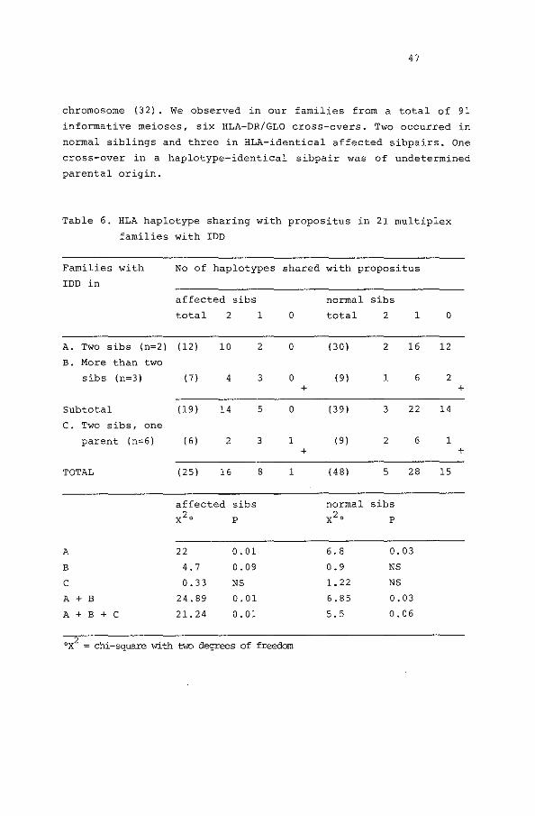
Table 6 presents the HLA segregation data and it compares the
number of HLA haplotypes of the index patient shared in affected
and normal siblings. Assuming random segregation, the expected
Mendelian ratio is 1 : 2 : 1 for sibs sharing both one or no hap
lotype with the propositus. A departure of the observed from the
expected ratio can be analysed with a chi-square test with two
degrees of freedom.
The families were divided in three groups: group A: families with
two affected siblings; group B: families with more than two af
fected siblings; group C: families with two affected siblings and
one affected parent.
Affected siblings in group A shared both haplotypes with the pro
positus significantly more often than expected (X2 22). In
group B the relative proportion of sharing one haplotype was in
creased (X2
= 4.7) and in group C the observed ratio was not sig
nificantly different from the expected {X 2 = 0.33). The normal
sibs in group A showed also a significant departure in the other
direction from the 1 : 2 : 1 ratio in sharing haplotypes with the
propositus {X2 = 6.8; P = 0,034).
Another observation relates to the occurrence of cross-overs be
tween HLA-DR and the GLO locus, which is located centromeric of
HLA-DR at 4 percent of recombination in males (34). Cross-overs
in one of an affected sibpair suggest that the gene segment pre
disposing for IDD is located to the telomeric, HLA-DR side of the
47
chromosome (32). We observed in our families from a total of 91
informative meioses, six HLA-DR/GLO cross-overs. Two occurred in
normal siblings and three in HLA-identical affected sibpairs. One
cross-over in a haplotype-identical sibpair was of undetermined
parental origin.
Table 6. HLA haplotype sharing with propositus in 21 multiplex
families with IDD
Families with No of haplotypes shared with propositus
IDD in
affected sibs normal sibs
total 2 1 0 total 2 1 0
A. Two sibs (n=2) (12) 10 2 0 (30) 2 16 12
B. More than two
sibs (n=3) 171 4 3 0 (9) 1 6 2 + +
Subtotal (19) 14 5 0 (39) 3 22 14
c. Two sibs, one
parent (n=6) I 61 2 3 1 (9) 2 6 1 + +
TOTAL (25) 16 8 1 (48) 5 28 15
affected sibs normal sibs X2' p X2' p
A 22 0.01 6. 8 0.03
B 4. 7 0.09 0.9 NS
c 0.33 NS 1. 22 NS
A + B 24.89 0.01 6.85 0.03
A + B + c 21.24 0.01 5. 5 0.06
ox2 = chi-square with tvJo degrees of freedom

48
The fact that three affected sibpairs were HLA-identicalt but
different for GLO, confirmed that the "hot" segment for IDD was
located towards the HLA-DR side of the chromosome.
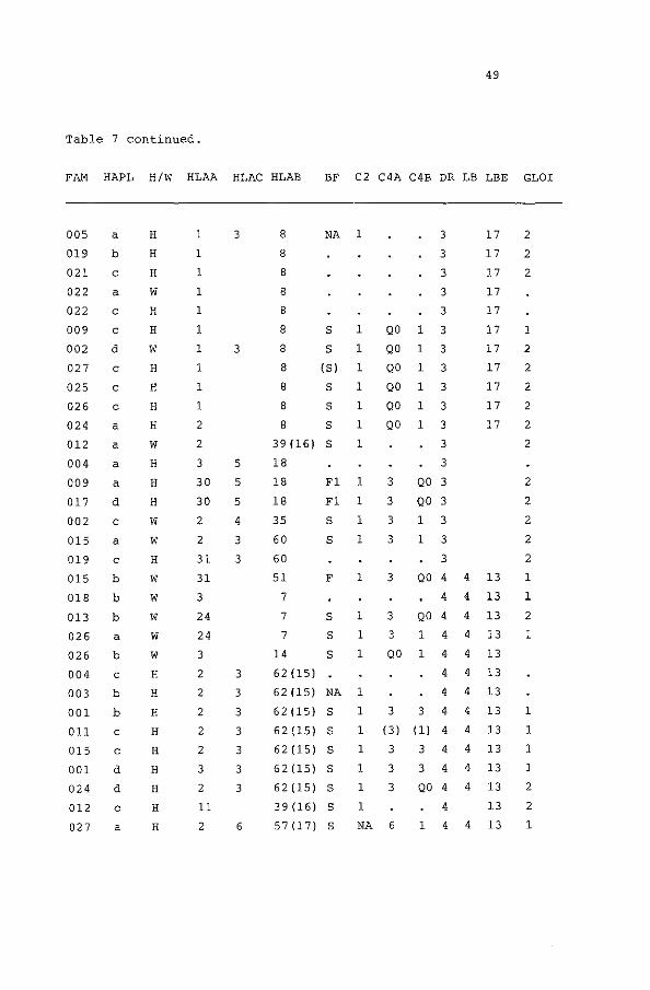
Another way of characterizing the HLA-haplotypes associated with
IDD is shown in table 7. The unrelated haplotypes encountered in
patients {"diabetic" haplotypes) are clustered according to their
HLA specificities. In the meantime the same approach has been re
ported in families from France (35). Three haplotypes were con
firmed to occur frequently among the IDD patients.
a} Al, BS, BPS, C2,1, C4AQ0, C4Bl, DR3, GL02
b) Aw30, Cw5, Bl8, BFFl, C2.1, C4A3, C4BQ0, DR3, GL02
c) A2, Cw3, BlS, BFS, C2.1, C4A3, C4B3, DR4, GLOl
In addition to this observation, the haplotype clusters showed
that "historical 1' cross-overs at the GLO side and the HLA-A side
of the haplotypes occurred. Apparently these cross-overs did not
change the diabetogenic properties of the haplotypes and inspec
tion of the clusters seems to confine the diabetogenic segment of
the B-DR segment.
The gene frequencies of HLA-DR3 and -DR4 (counted from table 7)
were lower in the 11 warm" haplotypes than those in the "hot" hap
lotypes, but these differences were not significant.
Table 7. Diabetic ("hot" and "warm") haplotypes, in 22 multiplex
families, with IDD in two or more IDD patients
FAM HAPL H/W HLAA HLAC HLAB BF C2 C4A C4B DR LB LBE GLOI
011 a w 33 14 s 1 2 2 1 12 2
003 d H 24 39 NA 1 1 12
025 a H 2 39 F 1 3 QO 1 12 2
012 b w 24 4 35 F 1 1 12 2
013 c H 2 7 s l (3) (1) 2 12 2
013 a w 2 44 (12) s 1 3 1 2 12 2
010 c w 3 4 35 s 1 3 1 2 12 1
49
Table 7 continued.
FAM HAPL H/W HLAA HLAC HLAB BF C2 C4A C4B DR LB LEE GLOI
005
019
021
022
022
009
002
027
025
026
024
012
004
009
017
002
015
019
015
018
013
026
026
004
003
001
011
015
001
024
012
027
a
b
c
a
c
c
d
c
c
c
a
a
a
a
d
c
a
c
b
b
b
a
b
c
b
b
c
c
d
d
c
a
H
H
H
w H
H
w H
H
H
H
w H
H
H
w w H
w w w w w H
H
H
H
H
H
H
H
H
1
1
1
1
1
1
1
1
1
1
2
2
3
30
30
2
2
31
31
3
24
24
3
2
2
2
2
2
3
2
11
2
3
3
5
5
5
4
3
3
3
3
3
3
3
3
3
6
8
8
8
8
8
8
8
8
8
8
NA 1
s 1
s 1
(S) 1
s 1
s 1
8 s 1
1 39 (16) s 18
18 Fl
18 Fl
35
60
60
51
7
7
7
14
62(15)
s s
F
s s s
1
1
1
1
1
1
1
1
62 (15) NA 1
62(15) s 1
62(15) s 1
62(15) s 1
62(15) s 1
62(15) s 1
1
3
3
3
3
3
QO 1 3
QO 1 3
QO 1 3
QO 1 3
QO 1 3
QO 1 3
3
3
3 QO 3
3 QO 3
3
3
1 3
1 3
3
17
17
17
17
17
17
17
17
17
17
17
3 QO 4 4 13
4 4 13
3 QO 4 4 13
3 1 4 4 13
QO 1 4 4 13
4
4
3 3 4
(3) (1) 4
3 3 4
3 3 4
3 QO 4
4
4
4
4
4
4
4
13
13
13
13
13
13
13
4 13 39 (16) s 57(17) s NA 6 1 4 4 13
2
2
2
1
2
2
2
2
2
2
2
2
2
2
2
1
1
2
1
1
1
1
1
2
2
1

50
Table 7 continued.
FAM HAPL H/W HLAA HLAC HLAB BF C2 C4A C4B DR LB LEE GLOI
017 a H 25 18 s 1 3 1 4 4 13 1
006 b H 2 2 27 4 4 13 1.2
022 b H 2 2 27 4 4 13
021 a H 3 2 27 4 4 13 1
002 b H 3 6 37 F 1 3 1 4 4 13 1
005 c H 24 6 37 NA NA 4 4 13 2
023 d H 31 3 60 (40) s 1 3 1 4 4 13 1
023 a H 2 44 (12) s 1 3 1 5 5 13 1
001 a w 24 18 s 1 3 1 5 5 13 2
018 c w 2 3 62 (15) 6 12 1
018 a w 33 3 58 (17) 6 12 1
011 b H 24 3 55 (22) s 2 4 5 6 12 2
010 a H 11 4 35 s 2 3 1 6 12 1
024 c w 2 3 60 (40) s 1 QO 2 6 12 2
006 c H 2 3 60 (40) 6 12 1.2
018 d w 2 6 50 (5) 7 7 17 1
010 d H 3 13 s 1 3 1 7 7 17 1
Legend to table 7
HAPL = haplotype LBE =defined by serum MO which
H/W = "hot" and "wann" recognizes MB2 + DR3
LB = subtypes of HLA-DR. See for definition of LB and
LB4 and LBlO are LBE reference 44.
defined as a split NA = no activity
of DR4, LB5 and LB58 = not tested
of DRS and LB7 and QO = Quantity zero = null
LBll of DR7 allele
51
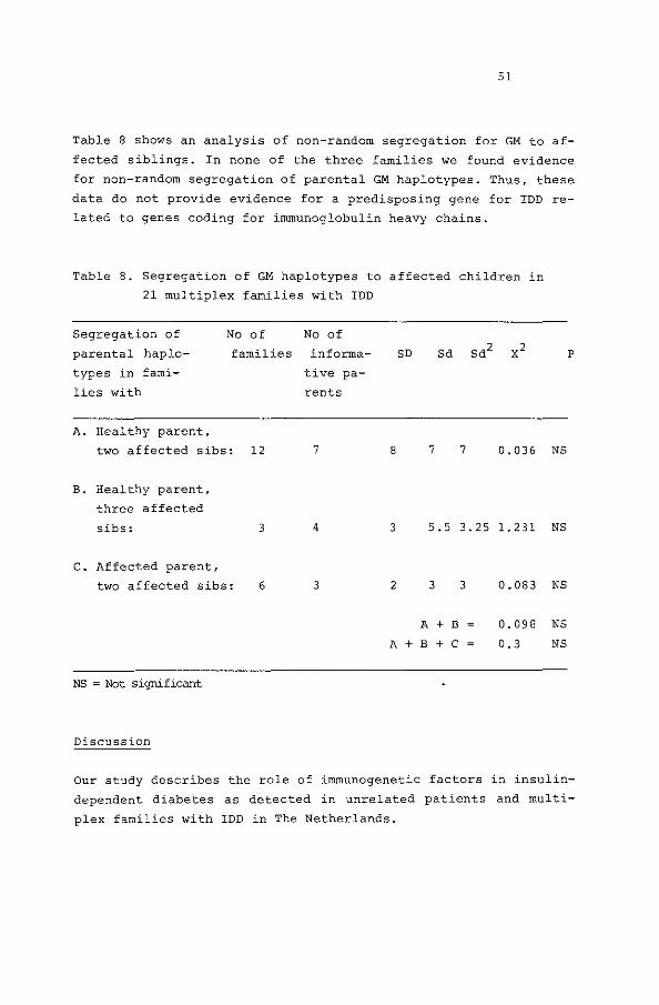
Table 8 shows an analysis of non-random segregation for GM to af
fected siblings. In none of the three families we found evidence
for non-random segregation of parental GM haplotypes. Thus, these
data do not provide evidence for a predisposing gene for IDD re
lated to genes coding for immunoglobulin heavy chains.
Table 8. Segregation of GM haplotypes to affected children in
21 multiplex families with IDD
Segregation of
parental haplo
types in fami
lies with
A. Healthy parent,
No of No of
families informa-
tive pa
rents
two affected sibs: 12 7
B. Healthy parent,
three affected
sibs: 3 4
c. Affected parent,
two affected sibs: 6 3
NS = Not significant
Discussion
SD
8 7 7 0.036 NS
3 5.5 3.25 1.231 NS
2 3
A + B
A + B + C
3 0.083 NS
0.098 NS
0.3 NS
p
Our study describes the role of immunogenetic factors in insulin
dependent diabetes as detected in unrelated patients and multi
plex families with IDD in The Netherlands.

52
We will consider the following aspects in more detail.
1. Genetic heterogeneity related to age of onset of IDD.
2. Associations in unrelated patients.
3. Genetics in multiplex families with regard to liLA-segregation,
occurrence of cross-overs and haplotypes.
4. Segregation of GM.
Age of onset.
1. We have investigated the frequencies of HLA-DR3 and HLA-DR4 in
IDD patients with regard to age of onset. Comparing three age
groups, we found that the frequencies of both HLA-DR3 and -DR4
decreased with older age of onset and that the relative risk
of HLA-DR4 was significantly heterogenous. Whereas in patients
with onset at adult age, IDD may be diluted with NIDD, we fa
vour another explanation for the observed heterogeneity.
Because IDD is a disease caused by a mixture of genetic and
environmental factors, one might expect that at younger age,
where the exposure time to environmental factors is shorter,
the genetic factors are more prominent.
Evidence for this hypothesis was recently reported in a study,
which showed an increased recurrence of IDD in sibs of propo
siti with onset of IDD before the age of ten as compared with
that in sibs of probands with onset after the age of ten {36).
To make a genetic analysis more meaningful, we excluded for
further study the patients with onset of the disease after the
age of 17. Although the cut-off point of 17 may be arbitrari
ly, it coincided with excluding patients from the adult cli
nic.
2. The patient and control sample were investigated for the pre
sence of H-W equilibrium and the data of the patients were
compared with those expected under various genetic models. As
stated before, this analysis requires the sample to be chosen
randomly from a large random mating population. A priori this
assumption may not be fulfilled.
However, we considered our patient and control population to
be fairly homogeneous with regard to ethnic background, be
cause only individuals of Dutch Caucasoid extraction were in-

53
eluded. Moreover, we reduced heterogeneity within the patients
by confining the analysis to patients with onset of the dis
ease before the age of 17. Although the ascertainment was not
the same for all the patients, it appeared that the observed
gene frequencies and allelic combinations of HLA-DR were very
similar in the population and multiplex family sample.
Under the assumption of a single recessive susceptibility gene
in linkage disequilibrium with HLA-DR3 and -DR4, the expected
proportions of allelic combinations are in H-W equilibrium
(37). However, the observed proportions deviated significant
ly from expected, providing evidence against a single reces
sive model.
Moreover, the excess of observed over expected HLA-DR3/4 he
terozygotes is incompatible with a single dominant or inter
mediate model {37). In a different approach we compared the
odd's ratio's (OR) of the different allelic combinations, em
ploying the method of Svejgaard and Ryder (31). They showed
that the OR of the heterozygote DR3/4 would fall between the
two homozygote OR's, if the disease were due to a single sus
ceptibility gene acting as recessive, dominant or intermediate
gene.
The observed excess of the OR (DR3/4) over OR {DR3/3), provi
ded in another way evidence against a simple recessive, inter
mediate or dominant model.
Information in determining the mode of inheritance can be also
derived from the haplotype segregation in families.
3. Without considering the allelic combination of HLA-haplotypes,
the multiplex families demonstrate an excess of sharing both
HLA-haplotypes among pairs of affected sibs. The observation
that the families with an affected parent less often demon
strate HLA identity among affected sibs than families with two
normal parents, can be explained by an increased conditional
probability of observing susceptible genotypes in the children
of affected parents, as compared to genotypes segregating from
normal parents (32). Thus, also the high frequency of sharing
both haplotypes in affected sibpairs indicates that a dominant
model of inheritance can be ruled out. Moreover, the combined

54
data of the allelic combinations in unrelated patients and of
the haplotype segregation in multiplex families, do not fit a
simple recessive or intermediate mode of inheritance. There
fore, more complex genetic models seem to be required to des
cribe the relationship between the HLA system and IDD ade
quately.
We considered the possibility that HLA-haplotypes rather than
a particular antigen were associated with IDD. Based on popu
lation data we found evidence that HLA-B8, -DR3 and HLA-BlS,
-DR4 haplotypes manifested a third order linkage disequili
brium under a RAD and AR model. This observation seems compa
tible with a susceptibility gene between HLA-B and -DR or with
a set of genes with an additive or interactive effect. Al
though the conclusion of a haplotype association should be ta
ken with some caution, because the patient population is not a
true population in genetical sense and the assumptions of the
mode of inheritance are only partially valid, the difference
of linkage disequilibrium of these haplotypes in patients as
compared to normal controls, seems worthwhile in considering
the genetics of IDD.
The occurrence of cross-overs between HLA-DR and GLO in one of
an affected sibpair enabled us to localize the diabetic seg
ment towards the HLA-DR segment of the chromosome. We cannot,
however, determine at present what the distance is of the 11 hot" diabetic segment to GLO. Markers between GLO and HLA-DR
would allow to answer this question, and the newly discovered
SB locus (38) may turn out to be a very good candidate for re
solving this question {39) .
Another way of further characterizing the chromosomal segment
associated, is the analysis of haplotypes occurring in affec
ted sibs as shown in table 7.
Sorting of diabetic haplotypes reveals clusters of chromosomal
segments selected for being diabetogenic. Inspection of these
clusters indicates that "historical" cross-overs have occurred
both on the GLO and the HLA-A side of the haplotypes, and that
the identity of the segments concentrates around the HLA-B-DR
region. Three clusters have been delineated, that previously

55
were reported to occur in IDD patients (35) . Considering that
these haplotypes are unrelated, it is striking to see that in
these three clusters the identity even seems to extend from
HLA-A to GLO. Because, at present we have no appropriate data
on control genotypes, the significance of the latter observa
tion remains to be determined.
However, our population data suggest that indeed an increased
linkage disequilibrium occurred in patients as compared to
normal controls, with regard to the haplotype HLA-B8-DR3 and
HLA-Bl5-DR4.
4. In a search for genetic factors in IDD, not related to HLA, we
investigated GM as a marker system, because GM has been impli
cated in other HLA-associated diseases such as chronic active
hepatitis and myasthenia gravis {19,20). In the present fami
lies we found no evidence for non-random segregation of GM
haplotypes.
This observation may not be generalized to all families with
IDD, because a recent study demonstrated that non-HLA factors
may only be prominent in families with a propositus negative
for HLA-DR3 and DR4 ( 40) .
Taken together, the results of this study suggest that the HLA
system is associated with a major susceptibility effect for IDD.
Moreover HLA-DR4, or a susceptibility gene in linkage disequili
brium with it, is associated with age of onset. In addition, the
data provide strong evidence against a single recessive, inter
mediate or dominant mode of inheritance of the susceptibility
genes. Therefore more complex models are required to account for
the data (40).
With regard to more complex models, it is relevant to consider
some molecular aspects of the MHC system. The HLA-DR or class II
molecules consist of an alpha and beta chain that are encoded by
two closely linked loci (41). In the MHC of the mouse it has been
shown that these class II molecules are the products of the immu
ne response genes {42). These alpha and beta loci act in comple
mentation and hybrid alpha-beta molecules of both cis- and trans
complementation can be formed.
In addition to the creation of "new" hybrid molecules as the re-

56
sult of transcomplementation, it has been shown that preference
for cis- or transcomplementation is dependent on the allelic com
bination of haplotypes (43). This preference is reflected in an
immune response gene effect (43).
By analogy, one could suggest that in humans both HLA-DR3 and
-DR4 are associated with susceptibility for IDD, and that in a
heterozygote DR3/4 a preferential transcomplementation leads to
an even increased susceptibility for IDD. However, because this
possibility does not easily explain the observed B-DR haplotype
association, one should also consider the possibility, that a
gene interaction between alleles coding for class I and class II
(and perhaps even class III) HLA molecules might be involved in
the susceptibility for IDD.
Thus recognition of fine specificities of the HLA-region has also
implications for the study of the molecular mechanisms underlying
the susceptibility of IDD.
Acknowledgements
We thank the patients and the families for their co-operation and
the Diabetes Vereniging Nederland (DVN) for their support. We are
indebted to Dr. J. Molenaar (SSDZ, Delft) for determining the
presence of islet cell antibodies, to Dr. J. D'Amaro for computer
support, to Dr. R.R.P. de Vries for his valuable suggestions and
to Mrs J. Pesant-Plaizier for typing the manuscript.
In part supported by the Dutch Foundation for Medical Research
(FUNGO) which is subsidized by the Dutch Organization for the Ad
vancement of Basic Research (ZWO), the J.A. Cohen Institute for
Radiopathology and Radiation Protection (IRS) , the Sophia Founda
tion for Medical Research to G.J .B., and INSERM (CRL 821015) to
G.H.

57
References
1. Singal, D.P., Blajchman, M.A.: Histocompatibility (HL-A)anti
gens, lymphocytotoxic antibodies and tissue antibodies in
patients with diabetes mellitus. Diabetes 22: 429, 1973.
2. Nerup, J., Platz, P., Andersen, 0.0., Christy, M., Lyngsoe,
J., Poulsen, J.E., Ryder, L.P., Thomsen, M., Nielsen, L.S.,
Svejgaard, A.: HL-A antigens and diabetes mellitus. The
Lancet ii: 864, 1974.
3. Thomsen, M., Pla.tz, P., Andersen, o.o., Christy, M., Lyngsoe,
J., Nerup, J., Rasmussen, K., Ryder, L.P., Nielsen, L.S.,
Svejgaard, A.: MLC typing in juvenile diabetes mellitus and
idiopathic Addison's disease. Transpl. Rev. 22: 125, 1975.
4. Batchelor, J.R., Morris, P.J.: HLA and disease. In: W.F. Bod
mer, J.R. Batchelor, J.G. Bodmer, H. Festenstein, P.J. Mor
ris, eds. Histocompatibility Testing 1977. Copenhagen,
Munksgaard, 1977, p. 205.
5. Cudworth, A.G., Woodrow, J.C.: Evidence for HL-A linked genes
in "juvenile" diabetes mellitus. Brit. Med, J. 2: 133,
1975.
6. Svejgaard, A., Platz, P., Ryder, L.P.: Insulin-dependent dia
betes mellitus. In: P.I. Terasaki, ed. Histocompatibility
Testing 1980, Los Angeles, The UCLA Tissue Typing Labora
tory, 1980, p. 638.
7. Spielman, R.S., Baker, L., Zmijewski, C.M.: Inheritance of
susceptibility to juvenile onset diabetes. In: C.F. Sin,
M. Skolmick, eds. Genetic analysis of common diseases:
applications to predictive factors in coronary disease. New
York, Alan R. Liss, 1979, p. 567.
8. Thomson, G., Bodmer, W.F.: The genetic analysis of HLA and
disease associations. In: J. Dausset, A. Svejgaard, eds.
HLA and disease. Copenhagen, Munksgaard, 1977, p. 84.
9. Svejgaard, A., Platz, P., Ryder, L.P., Staub Nielsen, L.,
Thomsen, M.: HL-A and disease associations- a survey.
Transpl. Proc. 22: 3, 1975.
58
10. Spielman, R.S., Baker, L., Zmijewski, C.M.: Gene dosage and
susceptibility to insulin-dependent diabetes. Ann. Hum.
Genet. London 44: 135, 1980,
11. Barnett, A.H., Eff, C., Leslie, R.D.G., Pyke, D.A.: Diabetes
in identical twins: a study of 200 pairs. Diabetologia 20:
87, 1981.
12. Irvine, W.J.: Classification of idiopathic diabetes. The Lan
cet i: 638, 1977.
13. Gamble, D.R.: An epidemiological study of childhood diabetes
affecting two or more siblings. Diabetologia 19: 341, 1980.
14. Dausset, J.: Clinical implications (nosology, diagnosis, prog
nosis, and preventive therapy). In: J. Dausset, A. Svej
gaard, eds. HLA and disease 1977, Copenhagen, Munksgaard,
1977, p. 296,
15. Cudworth, A.G.: Type I diabetes. Diabetologia 14: 281, 1978.
16. Porta, J., McHugh, R.: Detection of HLA haplotype associations
with disease. Tissue Antigens 15: 337, 1980.
17. Platz, P., Jacobsen, B.K., Marling, N., Ryder, L.P., Svej
gaard, A., Thomsen, M., Christy, M., Kromann, H., Benn, J.,
Nerup, J., Green, A., Hauge, M.: HLA-D and -DR antigens in
genetic analysis of insulin-dependent diabetes mellitus.
Diabetologia 21: 108, 1981.
18. Gorsuch, A.N., Spencer, K.M., Lister, J., Wolf, E., Bottazzo,
G.F., Cudworth, A.G.: Can future type I diabetes be predic
ted? A study in families of affected children. Diabetes 31:
862, 1982.
19. Whittingham, S., Mathews, J.D., Schansfield, M.S., Tait, B.D.,
MacKay, I.R.: Interaction of HLA and GM in autoimmune chro
nic active hepatitis. Clin. Exp. Immunol. 43, 1981.
20. Nakao, Y. Matsumoto, H., Miyazaki, T., Nishitani, H., Takatsu
ki, K., Kasukawa, R,, Nakayama, S., Izumi, S., Fujita, T.,
Tsuji, K.: IgG heavy-chain allotypes (GM) in autoimmune
diseases. Clin. Exp. Immunol. 42{1): 20, 1980.
21. National Diabetes Data Group: Classification and diagnosis of
diabetes mellitus and other categories of glucose intole
rance, Diabetes 28: 1039, 1979.

59
22. Van Rood, J.J.: Microlymphocytotoxicity Method. In: J.G. Ray,
ed. NIAID Manual of Tissue Typing Techniques, Maryland, NIH
no. 80-545, 1979.
23. Van Leeuwen, A., Van Rood, J.J.: Description of the B cell
methods. In: P.I. Terasaki ed. Histocompatibility 1980. Los
Angeles, The UCLA Tissue Typing Laboratory, 1980, 278.
24. Alper, C.A., Boenisch, T., Watson, L.: Genetic polymorphism in
human glycine-rich betaglycoprotein. J. Exp. Med. 135: 68,
1972.
25. Awdeh, z., Alper, C.A.: Inherited structural polymorphisms of
the fourth component of human complement. Proc. Natl. Acad.
Sci. USA 77(6): 3576, 1980.
26. Meera Khan, P., Doppert, B.A.: Rapid detection of glyoxalase I
(GLO) on cellulose-acetate gel and the distribution of GLO
variants in a Dutch population. Hum. Genet. 34: 53, 1976.
27. Van Loghem, E. Genetic studies on human immunoglobulins. In:
D.M. Weir, ed. Handbook of Experimental immunology vol. 1.
"Immunochemistry". Oxford, Blackwell Sci. Publ., 1978.
28. Svejgaard, A., Jershild, C., Nielsen, L.S., Bodmer, W.F.: HL-A
antigens and disease: Statistical and genetic considera
tions. Tissue Antigens 4: 95, 1974.
29. Edwards, J.H.: HL-A and disease. The detection of associa
tions. J. Irnmunogenet. 1: 249, 1974.
30. Bengtsson, B.O., Thomson, G.: Measuring the strength of asso
ciations between HLA antigens and diseases. Tissue Antigens
18, 356, 1981.
31. Svejgaard, A., Ryder, L.P.: HLA genotype distribution and ge
netic models of insulin-dependent diabetes mellitus. Ann.
Hum. Genet. 45: 293, 1981.
32. Rubinstein, P., Ginsberg-Fellner, F., Falk, C.T.: Genetics of
type I diabetes mellitus; a single, recessive predisposi
tion gene mapping between HLA-B and GLO. Amer. J. Hum. Ge
net. 33: 865, 1981.
33, De Vries, R.R.P., Lai-A-Fat, R.F.M., Nijenhuis, L.E., Van
Rood, J.J.: HLA-linked genetic control of host response to
mycobacterium leprae. The Lancet ii: 1328, 1976.

60
34. Weitkamp, L.R., Lamm, L.U.! Report of the committee on the ge
netic constitution of chromosome 6. Cytogenet. Cell Genet.
32: 130-143, 1982.
35. Cantu, L., Deschamps, I., Lestradet, H., Hors, J., Schmid, M.,
Bussen, M., Benajam, A., Marcelli-Barge, A., Dausset, J.:
HLA haplotype study of 53 juvenile insulin-dependent dia
betic (I.D.D.) families. Tissue Antigens 20: 123, 1982.
36. Chern, M.M., Anderson, V.E., Barbosa, J.: Empirical risk for
insulin-dependent diabetes (IDD) in sibs. Further defini
tion of genetic heterogeneity. Diabetes 31: 1115, 1982.
37. Falk, C.T., Mendell, N.R., Rubinstein, P.: Effect of popula
tion associations and reduced penetrance on observed and
expected genotype frequencies in a simple genetic model:
application to HLA and insulin-dependent diabetes mellitus.
Ann. Hum. Genet. 47: 161, 1983.
38. Shaw, S., Duquesnoy, R.J., Smith, P.L.: Population studies of
the HLA-linked SB antigens. Immunogenetics 14: 153, 1981.
39. DeJongh, B.M., Termijtelen, A., Bruining, G.J., De Vries,
R.R.P., Van Rood, J.J.: Relation of insulin-dependent dia
betes mellitus (IDD) and the HLA-linked SB system. Tissue
Antigens, 1984 (in press).
40. Green, A., Morton, N.E., Iselius, L., Svejgaard, A., Platz,
P., Ryder, L.P., Hauge, M.: Genetic studies of insulin-de
pendent diabetes mellitus: segregation and linkage analy
ses. Tissue Antigens 19: 213, 1982.
41. Winchester, R.J., Kunkel, H.G.: The human Ia system. Adv.
Immunol. 28: 221, 1979.
42. Nagy, Z.A., Baxevanis, C.N., Ishii, N., Klein, J.: Ia antigens
as restriction molecules in Ir-gene controlled T-cell pro
liferation. Immunol. Rev. 60: 59, 1981.
43. Conrad, P.J., Lerner, E.A., Murphy, B.V., Jones, P.P., Jane
way, C.A.: Differential expression of Ia glycoprotein com
plexes in F1 hybrid mice detected with alloreactive cloned
T cell lines. J. Immunol. 129(6): 2616, 1982.

61
44. Schreuder, G.M.Th., Van Leeuwen, A., Termijtelen, A., Parle-
vliet, J., D 1 Amaro,
for
J., Van Rood, J.J.: Cell membrane poly
in the HLA-D/DR region. I. Relation be-morphisms coded
tween D and DR. Human Imrnunol. 4: 343, 1982.

62
Paper 2
Neonatal onset permanent diabetes mellitus and incomplete fetal
alcohol syndrome: cause or coincidence?
G.J. Bruining, R.S.R. Aarsen, S.L.S. Drop, H.K.A. Visser
Department of Pediatrics, subdivision Endocrinology, Erasmus Uni
versity and University Hospital Rotterdam/Sophia Children's Hos
pital, Rotterdam, the Netherlands.
Submitted for publication.
Summary
Neonatal onset permanent diabetes mellitus is extremely rare and
pathogenetic mechanisms are suspected to differ from those des
cribed in childhood diabetes developing at later ages. The pre
sent child had other HLA-types than mostly seen in childhood dia
betes, lacked islet-cell auto-immunity but did have features of
the fetal alcohol syndrome. Other (genetic) syndromes associated
with diabetes at very young ages were excluded.
Introduction
Since the fetal alcohol syndrome was rediscovered in 1973 (1) a
variety of associated anomalies have been reported, including ex
trahepatic biliary atresia, and renal anomalies (2). We report
ketotic diabetes mellitus starting the seventh day of life in a
child with features of the fetal alcohol syndrome.

63
Case report
A 2200 g boy was born in another hospital to a 29 year old mother
after an unknown duration of gestation. Although length and head
circumference were not recorded at birth, a "small-for-date ap
pearance" was noted. The pregnancy was complicated by the daily
ingestion of unknown quantities of beer and wine, and by the in
gestion of half a litre or more of dutch gin per day intermit
tendly in the third month of pregnancy, during a summer vacation.
Previously two healthy children had been born. The mother thought
she had taken less alcohol during those pregnancies. Two years
before the mother had an oral glucose tolerance test with normal
result. This was only done because her mother had developed glu
cose intolerance at age 60 and the mother herself had complaints
of thirst at that time. In the last trimester of this and previ
ous pregnancies no glucose was found in the urine. Except for am
pillicin for a urinary tract infection no medication was taken by
the mother, as evidenced from her pharmacy 1 s record. She smoked
20-40 cigarettes daily. Lues was excluded. The placenta weighed
400g. Microscopy revealed the partial presence of a single layer
of trophoblast-cells, but no other abnormalities.
At birth fetal alcohol syndrome was not suspected. Maximal bili
rubin was 65 jlMol/1 at the fourth day of life. On the seventh
day of life mild glycosuria was detected, without ketonuria. This
gradually increased up to 20 g per 24 hrs and the child was re
ferred to our hospital at the age of 13 weeks.
At that time weight was 3400 g (lOth percentile), length 53 em
( < -2 SO), headcircumference 34.2 em ( < -2 SD). On physical exa
mination features of the fetal alcohol syndrome were found {1).
These are tabulated in table l.

64
Table 1
SYMPTOMS IN INDEX CASE COMPARED TO THE FETAL
ALCOHOL SYNDROME (FAS)
SYMPTOM:
GROWTH:
prenatal length
postnatal length
adipose tissue
CNS-DYSFUNCTION:
mental deficiency,
mild-moderate
microcephaly
poor coordination/
hypotonia
irritability
hyperactivity
FACIAL/HEAD:
~plO
~plO
short palpebral fissures
short nose
hypoplastic philtrum
maxillar hypoplasia
thin upper vermillion
retrognathia
ptosis/strabismus
eye abnormality
ear malposition
prominent palatal ridges
small teeth
CARDIAC:
defects
INDEX CASE
?
+
+
+
+
+
+
+
+
+
+
**
+I-+
+
+
% PAS-PATIENTS
SHOWING SYMPTOMS
80
80
50
80
80
50
80
50
80
50
80
50
80
50
26-50
26-50
26-50
26-50
1-25
26-50
*

Table 1 continued.
URINARY TRACT:
hypospadia's
small kidneys,
hydronephrosis
CUTANEOUS:
* **
hemangioma's
hirsutism
data from reference
+ is present; - is absent
+
65
1-25
1-25
26-50
1-25
Initial laboratory results included: an oral glucose tolerance
test (1.75 g/kg) with glucose rising from 8 to 25.4 mMol/1, all
insulin values below 1 mU/1, glucagon 36-98 ng/1 (normal up to 80
ng/1), growth hormone elevated ranging from 10 to over 40 mU/1.
Pre-treatment random blood-glucoses varied from 9-38 mMol, urina
ry ketones were only present in minor amounts. A 24 hrs urine
analysis for amino-acids and oligosaccharides were normal, except
for glucose. The diurnal rhythm for cortisol was normal. Chromo
some-analysis using trypsin-Giemsa banding was normal male ( 4 6,
XY) • Epiphyseal dysplasia, cystic fibrosis, exocrine pancreas
dysfunction, and coeliac disease were excluded. Thyroid function
was low normal, TSH never elevated. Tests for liver and kidney
function, blood gases, electrolytes and hematology were unremark
able.
Insulin substitution was initiated at the age of 15 weeks in
single or multiple dosages not exceeding 0.7 U/kg, using mixtures
of short- and intermediate-acting insulin. Despite the careful
administration of these little amounts of insulin, blood glucoses
bounced from 1.8 up to 25 mMol/1 in an erratic fashion. Glycosy-

66
lated hemoglobins varied from 10-14% (6-8% in normal children)
and urinary ketones (2+ or more) were found twice weekly on the
average. Frank hypoglycemia occurred two times, the child quickly
recovering on oral glucose administration. Keto-acidosis
(pH< 7 .2) was first seen at the age of 6 months during an upper
respiratory tract infection. Respiratory tract infections recur
red, requiring adenotonsillectomy, ear-tubes for chronic otitis
and antibiotics for Haemophilus and Proteus bacteria. Keto-acido
sis requiring admission occurred two times thereafter. Recovery
with intravenous insulin and electrolyte solutions was normal.
Diaper-dermatitis was present, requiring almost chronic local
treatment mostly for Candida. Staphylococcus folliculitis was
seen four times. Immunoglobulins, including secretory IgA and
complement-factors were normal for age. Islet-cell cytoplasmatic
antibodies, thyroid antibodies, adrenal antibodies and gastric
antibodies were first tested at 8 months, subsequently with year
ly intervals. No positive results were obtained, neither in the
family. The patients' HLA-D-genotype was HLA-DRl and HLA-DRS. Hy
perrea_cti vi ty and irritability were almost constantly present.
Fine-motor seizures and athetotic movements lasting 5-10 minutes
became apparent during the second year of life. Electroencephalo
graphy revealed aspecific epileptic activity, for which phenobar
bital and phenytoin were prescribed. At present the child is 4
years and 10 months old, has severe growth and mental retarda
tion, and stays in an institution for handicapped children.
Comment
Diabetes mellitus has not been recognized as a symptom of the fe
tal alcohol syndrome; however, the present case suggests the pos
sibility of such an association.
Permanent insulin dependent diabetes developing before the age of
six months is extremely rare. In a recent nation-wide incidence
study over three years time only 3/1271 newly diagnosed children
from 0-19 years of age became permanently insulin dependent be
fore the age of six months (3). One was the present patient, the
other two had multiple congenital malformations, other than seen

67
in the fetal alcohol syndrome. The term congenital diabetes has
been used to indicate failure of fetal development of islets of
Langerhans (hypoplasia or aplasia of islets) . Only one well docu
mented case is known to have been delivered alive, and it is be
lieved that congenital diabetes is essentially non-existent (4).
As glucose-intolerance developed gradually some days after birth
in the present case, it may be presumed the neonatal pancreatic
response was sufficient during the first week of life to prevent
diabetic symptoms. Insulin dependency was documented by hypergly
cemia, ketonuria, absolute insulin deficiency, relative hyperglu
cagonemia and elevated growth hormone levels. These endocrine
findings are compatible with selective insulin deficiency. No au
toantibodies to islet-cells were found within one year of the
clinical onset of insulin deficiency in this patient and his HLA
DR-type was neither HLA-DR3 nor HLA-DR4. These immunologic fin
dings speak against the autoimmune involvement of the islets seen
in childhood diabetes: 80% of the newly diagnosed children at ol
der ages have such antibodies at diagnosis and 60% after one year
of treatment (5).
Almost all children with an age of onset below 8 years studied in
the Netherlands had HLA-DR3 and/or HLA-DR4 haplotypes as well as
90% or more of children studied elsewhere with older ages of on
set (6, 7). The present patient's HLA-genotype comprised neither
of these haplotypes. Other etiologic factors may be supposed in
this highly unusual presentation of childhood diabetes, but the
known genetic syndromes in which diabetes mellitus concurs were
excluded.
The amounts of alcohol ingested during gestation were probably
not as high as those reported in cases of children with the fetal
alcohol syndrome in which cases permanent alcohol consumption
throughout gestation is believed to occur (2). It has been sug
gested that 90 ml of absolute alcohol per day (equivalent to some
six hard drinks) constitutes a major risk to the fetus (7). The
present history contained an intermittend alcohol consumption,
far exceeding this quantity during the third month of pregnancy
by history and there appears enough descriptive evidence (table
1) to at least suppose fetal alcohol effects.

68
The time sequence as well as the quantities of the gestational
alcohol ingestion may be related to the partial expression of the
fetal alcohol syndrome of the present case. Hitherto, insulin de
pendency has never been seen in the full syndrome. This does not
preclude interference with the development of the functions of
islet-cells during fetal life to a lesser extent than leading to
frank insulin dependency: insulin levels in the full fetal alco
hol syndrome have not been reported although it is known many of
such children brought in at later ages were referred for "being
too skinny" and there are no follow-up data with regard to this
clinical feature (2).
Acknowledgements
We thank the Department of Immunohematology (Prof. Dr. J.J. van
Rood) of Leiden University for the HLA-typing; the Department of
Clinical Immunology (Dr. J .L. Molenaar) of the Stichting Samen
werking Delftse Ziekenhuizen for assaying auto-antibodies and the
Department of Cell Biology and Genetics (Prof. Dr. M.F. Nier
meyer) of the Erasmus University, Rotterdam, for the chromosomal
analysis and his valuable advise.
References
1. Jones, K.L., Smith, B.W., Ulleland, C.N., Streissgath, A.P.
(1973)
Pattern of malformation in affirming of chronic alcoholic
mothers.
The Lancet i: 1267-1271.
2. Clarren, S.K., Smith, D.W. (1978)
The fetal alcohol syndrome.
New Engl. J. Med. 298: 1063-1067.
69
3. Vaandrager, G.J., Bruining, G.J., Veenhof, F.J., Drayer,
N.M.H. (1984).
The incidence of childhood diabetes (0-19 years) in the Ne
therlands - a decrease from north to south over north-western
Europe?
Diabetologia, in press.
4. Bloomsworth, J.M.B., Greider, M.H.: The endocrine pancreas and
diabetes mellitus.
In: Endocrine pathology, 2d Edition, Bloomsworth, J.M.B. Ed.,
Williams and Williams, Baltimore, U.S.A. (1982), page 556-586.
5. Bruining, G.J., Molenaar, L.J., Tuk, C.W., Lindeman, J., Brui
ning, H.A., Marner, B. (1984).
Clinical time-course and characteristics of islet cell cyto
plasmatic antibodies in childhood diabetes
Diabetologia (1984) 26: 24-29.
6. Cudworth, A.G., Wolff, E. (1982).
The genetic susceptibility to type I (Insulin-dependent) Dia
betes Mellitus
In: Clinics in Endocrinology and Metabolism Vol. 11 nr. 2
pp 389-408.
7. de Jongh, B.M., Bruining, G.J., Schreuder, G.M.Th., Schuurman,
R.K.B., Radder, J.K., van Loghem, E., Meera Khan, P., Haupt
mann, G., van Rood, J .J. (1984)
HLA and GM in insulin-dependent diabetes in the Netherlands:
report on a combined multiplex family and population study.
Human Immunology, in press.
B. National Institute of alcohol abuse and alcoholism: critical
review of the fetal alcohol syndrome (1977) •
Alcohol, drug abuse and mental health, Rockville, U.S.A.

70
Paper 3
Prediction of type-1-diabetes mellitus: a report on three cases.
G.J. 1
sen
. . 1 1 2 Bru~nlng , J.L. Mo enaar ,
and H.K.A. Visser1
B.M. de R.S.R. Aar-
Department Pediatrics, subdivision Endocrinology, Erasmus Univer
sity and University Hospital Rotterdam I Sophia Children's Hospi
tal1,
Stichting Samenwerking Delftse Ziekenhuizen, Delft2
Department of Immunohematology, University of Leiden,
Medical School, Leiden, the Netherlands 3 .
Accepted for publication by the European Journal of Pediatrics
(1984). Reprinted with permission.
Summary In three children (patients 1, 2 and 3) insulin-dependency was
predicted 28, 32 and 4 months before the disease became clinical
ly manifest, respectively, by the finding of islet-cell antibo
dies at that time.
These retrospective findings confirm the evidence for a long pre
diabetic phase in childhood diabetes, marked by the presence of
islet-cell antibodies, as well as the linkage of HLA-antigens to
the susceptibility to this disease.
The possibility to detect pre-diabetic states in children before
the endogenous insulin secretion decreases to the point of clini
cal symptoms, supports efforts by basic scientists to develop
techniques for immunologic intervention early in the course of
this disease.

71
Introduction
It is well known that diabetes mellitus shows a familial tenden
cy. However, attempts to assess modes of inheritance failed, be
fore it was realized type-1 (insulin-dependent) diabetes mellitus
(IDDM) is a distinct disorder from type-2 (non-insulin-dependent)
diabetes mellitus (NIDDM) ( 1) .
From the Pediatric Register of the British Diabetic Association
it has been estimated that 5. 7% of the siblings of diabetic
children will develop insulin dependency themselves by the age of
16 years (2) . In the Pittsburgh studies (3) the recurrence risks
for siblings in families already having a type-1-diabetic child
by the age of 20 years was 3.3% to 6.0%, depending on the calcu
lation methods.
In the past decade two factors have been associated with the re
currence risk for siblings of diabetic children: one is HLA iden
tity, on the basis of the fact that the genetic susceptibility to
the disease is strongly HLA-linked (4); the other is the presence
of islet-cell cytoplasmatic antibodies (ICCA), found in 80% of
the newly diagnosed children with IDDM (S) •
The question to what extent these two factors might predict fu
ture type-1-diabetes in families of affected children has been
investigated in the Barts-Windsor Family study (6) .
They found that sibs HLA-identical to the already diabetic child,
would have a 100 times greater risk of developing the disease
themselves, whereas non-HLA-identical sibs would have the same
risk as the general population. In that prospective study each of
the six siblings that did develop diabetes in the course of their
follow-up (up to 4 years at the time of the report) did have ICCA
from the moment they entered the study, whereas in a control po
pulation 1% positives for the presence of ICCA were found. The
study indicated that type-1-diabetes in children has a long pre
diabetic period and carries implications for future research into
preventive measures before the disease becomes clinically mani-

72
fest.
The present report describes three children retrospectively (pa
tients 1, 2 and 3), in which diabetes mellitus was predicted 28,
32 and 4 months before the disease became clinically manifest,
based on the finding of islet-cell antibodies.
Patients
Patient 1
Patient 1 participated in a study on genetic markers in families
with two or more children having type-1-diabetes, described in
detail elsewhere (7) . Blood was also drawn for the determination
of ICCA. This study included 22 families, selected from all over
the Netherlands on the basis of their willingness to cooperation. It was agreed upon beforehand with each of the 22 families that
single blood drawings would be done and that the results of the
tests would not be disclosed. The 22 families had 111 children in
total, aged 2-20 years {average 11.6 years), 51 of the children
were already treated for insulin-dependent diabetes, 60 sibs had
no diabetes. Of these 60 sibs patient 1 was the only child, that
did have ICCA (titre 1:64), without any sign of clinical diabe
tes: a random bloodglucose at the moment he participated in the
study {fig. l) was 5.4 mmol/1, his percent glycosylated hemoglo
bin (HbA 1 %) was 6.3% {normal range 6-8%). His disease became cli
nically manifest 28 months later at age 19 years, with a -retro
spective- history of increasing lassitude, thirst and hunger of
three months. He was then admitted to another hospital with mild
keto-acidosis (blood pH 7.2, urinary ketones 2+, blood glucose 25
mmol/1).
The HLA-segregation of his family is depicted in figure 1, inclu
ding the titres of (complement-fixing-) islet-cell cytoplasmatic
antibodies {CF-ICCA). Islet cell surface antibodies (ICSA) were
not measured in this family. The ICCA found in patient l, indica
ted by a half-open square, fixed complement in a titre of 8. His
already diabetic brother, born in 1964, had a remnant ICCA-titre.

73
The HLA (-A-B-C-DR) haplotypes are indicated by A1
/B1
for the
father and c1;n
1 for the mother. Patient 1 is haplo-identical
(shared c 1 only) to his older sister and younger brother, alrea
dy having IDDM, and HLA-identical (sharing both A1
c1
) to his
brother born in 1961, who did not develop IDDM to date and did
not have ICCA positivity either.
With regard to the family-history of patient 1, no second degree
relatives had IDDM, but his father had undergone an operation for
hyperthyroid thyroidism 15 years earlier.
Fig. 1 liLA-segregation, islet-cell antibodies and glycosylated
hemoglobins found in the family of patient 1 in January
1980 (legend see text).
A1/B1 Cl/01
D 0
I I I I I I I • D D .. .. 0
year of 1958 1961 1962 1963 1961.J 1965
birth
year of 1960 1982 1978
lOOM
HLA- A1Cl B1/C1 Bl/01 B1/C1 A1/C1 BtfDt antigens
ICCA- 0 0 0 64 4 0 titre
CF-ICCA 0 0 0 B 0 0 titre
HbA1 % 12.9 6.1 7.4 6.3 15.1 6.8
,. patient 1
• female lOOM
.. male lOOM

74
Legend to figure 1.
IliA
A B c DR
~ 2 8 3
Bl 11 W35 W4 1
cl 2 Wl5.2 W3 4
Dl 2 W40.1 W3 W6
None of the other 59 non-diabetic siblings of this multiplex fa
mily study~ all being negative for (CF-)ICCA, developed IDDM over
a three year time span, from the moment each child entered the
study. The bloodsarnple of patient 1, obtained 28 months prior to
his insulin dependency and another sample obtained one month
thereafter, showed no significant difference in virus-titres for
mumps, measles, german measles, cytomegalia, adenoviruses, reovi
ruses, no tests were done for titres against coxsackie-virus.
Patient 2
Patient 2 is a girl of 14 years old when IDDM developed clinical
ly. She is the cousin of two IDDM children that took part in the
multiplex family study aforementioned. However, neither she her
self nor her first degree relatives had diabetes, but it so hap
pened her father's sister was the mother and her mother's brother
was the father of her two IDDM cousins (figure 2). Because of
this relationship it was decided to draw blood of her family too,
the HLA-haplotypes found in these two families are in table 2.
Patient 2 had no (CF-)ICCA in March 1979 (fig. 2) but 32 months
later, at the clinical onset of her disease, an ICCA-titre of 8
was found, that fixed complement with a titre of 2. Because of
the absence of ICCA in March 1979 this serum was assayed for is
let-cell surface antibodies (ICSA) on beta-cells obtained by
cell-sorting techniques from rat-islets (8). Islet cell surface
antibodies (ICSA) were 3+, in the absence of cytoplasmatic anti
bodies (ICCA) . Two years after treatment the ICCA, present in a

75
titre of 8 at diagnosis, had increased to 16. Her HLA-identical
cousin, born in 1961 had "persisting-ICCA", not usually seen af
ter a duration of IDDM for ten years (5) .
As indicated in figure 2 her uncle also had IDDM. He developed
the disease at age 29, but no other more distant relatives were
insulin-dependent nor had other endocrine autoimmune diseases,
been reported. The same virus-titres as in patient 1 were ob
tained, but the results were unremarkable.
Fig. 2. HLA-genotypes, islet-cell antibodies and glycosylated
year of birth
year of lOOM
HLA-genotype5
ICCAtitre
CF-ICCA titre
ICSA
HbA1 %
hemoglobins, found in patient 2 and her relatives in
March 1979 (legend, see text) .
Cz/P A2/Q A2/82 GztD2
; l 0 D
I ; l l 1961 1966 1967 1963 1965 1966
1972 1969 1982
A2/C2 C2/Q A2/C2 A2/02 B2/C2 A2/C2
16 0 0 0 0 0 II :Bl
4 0 0 0 0 0 11 :2]
+
13.2 6.0 11.3 5.6 5.7 6.0

76
Legend to figure 2.
HLA-haplotvpes found in patient 2 and her relatives
HLA. types
A B c DR
~ 1 8 W3 3
B2 11 44 W2 3
c2 2 27 W2 4
02 29 18.4 W3 5
p 2 8 W2 3
Q 3 7 W2 2
Patient 3
Patient 3 is a boy, aged 3 3/12 years, when insulin was first
administered. He is the only son of a type-1-diabetic mother who
had developed the disease herself at age 27 years. This patient
came to our attention because of glucosuria and hyperglycemia
( ~13 mmol/1) without ketonuria, seen only during corticosteroid
treatment for severe bouts of asthma. For this he received 2
mg/kg prednisone per day as well as 20 mg/kg aminophylline per
day. Such bouts lasted 4-7 days, after withdrawal of the cortico
steroid treatment he became normoglycemic.
Three days after the first episode an oral glucose tolerance test
{OGTT) was done, yielding a peak glucose of 9.7 mmol/1, peak in
suline of 13 mU/L and C-peptide increasing from 0.2 nmol/1 to>
2 nmol/1. Glucagon and cortisol were 9-14 ng/1, respectievely
0.27-0.41 iJ.mol/1. Five non-diabetic children between 3 and 5
years of age had 6.3-7.8 mmol/1 peak glucose, 10-24 mU/1 peak in
sulin and> 2 nmol/1 C-peptide on OGTT, also with 1.75 g glucose/
kg as the glucose load, with a maximum of 50 g.
Three months later (with the aminophylline treatment continued at
5 mg/kg/day, without corticoste:-oid treatment in between) the
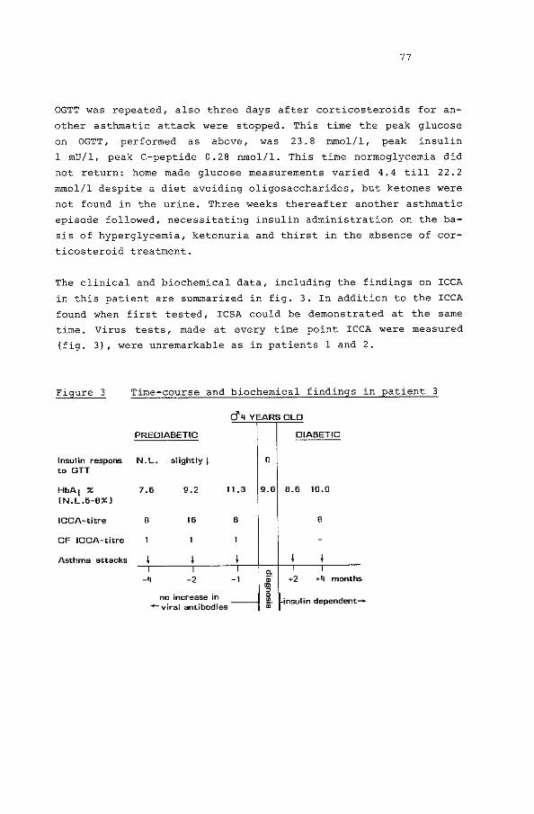
77
OGTT was repeated, also three days after corticosteroids for an
other asthmatic attack were stopped. This time the peak glucose
on OGTT, performed as above, was 23.8 mmol/1, peak insulin
1 mU/1, peak C-peptide 0.28 nmol/1. This time normoglycernia did
not return: home made glucose measurements varied 4.4 till 22.2
mmol/1 despite a diet avoiding oligosaccharides, but ketones were
not found in the urine, Three weeks thereafter another asthmatic
episode followed, necessitating insulin administration on the ba
sis of hyperglycemia, ketonuria and thirst in the absence of cor
ticosteroid treatment.
The clinical and biochemical data, including the findings on ICCA
in this patient are summarized in fig. 3. In addition to the ICCA
found when first tested, ICSA could be demonstrated at the same
time. Virus tests, made at every time point ICCA were measured
(fig. 3), were unremarkable as in patients 1 and 2.
Figure 3 Time-course and biochemical findings in patient 3
PREDIABETIC
cJ" 4 YEARS OLD
DIABETIC
Insulin respons N. L. slightly 1 0 to GTT
HbA1 % (N.L.fi-6%)
ICCA-titre
CF ICCA-titre
Asthma attacks
7.6 9.2 11.3 9.0 6.6 10.0
B 16
-l.J -2
no increase in -viral antibodies
B 6
-I +2 +4 months
insulin dependent-

78
As patient 3 is an only child, his HLA-type cannot be compared to
that of sibs. His mother had HLA-DR3/DR3, his father HLA-DR2/DR4,
patient 3 himself had HLA-DR3/DR4.
Apart from his mother's IDDM since the age of 27, his father had
slight asthmatic symptoms as a youth, declining when he became an
adult. There were no other family-members with known (auto-immu
ne) endocrine disease.
Methods
The HLA-typing was performed by the standard microcytotoxici ty
assay, using periferal lymphocytes: 16 HLA-A, 40 HLA-B and 6
HLA-C antigens were determined, using a set of 120 well defined
antisera (9). With the two-coloured fluorescence assay 10 HLA-DR
antigens were investigated, using 80 different allosera (10).
Intra-HLA cross-overs were not observed,
Islet-cell cytoplasmatic antibodies (ICCA) were measured by indi
rect fluorescence on fresh frozen human pancreas sections (11),
complement fixation of ICCA was determined in addition in each
sample (5). Islet-cell surface antibodies (ICSA) were estimated
on dispersed islet-cells from rodents, as described by Lernmark
et al. (12). In patient 2 this assay was performed after separa
ting the pancreatic beta-cells of rodents by cell sorting techni
ques (8).
Virus-titres were estimated by standard serologic technique, glu
cose by specific enzymatic reaction (GOD) , hormones by radio-im
muno-assay, (stable) glycosylated hemoglobins (HbA1 ) by a modifi
cation of the microcolumn (IsolabR -Akron (Ohio) USA) chromato
graphy, drawing the blood in excess buffer prior to assay, the
normal range is 6-8% HbA1
.
Discussion
Patient 1 illustrates that recurrence of IDDM may occur in haplo
identical sibs and that the other healthy sib to which this pa
tient was HLA-identical in turn, did not have islet cell antibo
dies. Further, patient 1 was the only healthy participant in the

79
multiplex family study that did have ICCA and did develop IDDM.
Thus standardized ICCA ( 11) may be a good screening agent for
prediabetic states of IDDM (12) .
Patient 2 had no ICCA 32 months before she came down with IDDM,
but she did have ICSA. When her disease became manifest she had
low titres of (CF-)ICCA, suggesting that ICSA developed previous
to ICCA. This is consistent with the view (14) that cytoplasmatic
antibodies (ICCA) would not react with intact pancreatic islets,
but only after they have been previously damaged by surface anti
bodies ( ICSA) . To the best of our knowledge, this is the first
patient in which this supposed sequence of events has actually
been documented. The opportunity to study the HLA-segregation in
her family, with her parents married to each others brother and
sister, demonstrates the association between the susceptibility
to IDDM in childhood and HLA-antigens in another way than by con
currence of HLA-antigens in affected siblings. Yet, only 12% of
the healthy sibs that are HLA-identical to affected children in
the same family, will develop IDDM before the age of 20 (15).
Patient 3 indicates that ICCA may be used in clinical instances
where the development of IDDM is suspected. It has been suggested
before that corticosteroid-treatment may precipitate the develop
ment of IDDM ( 16) , which is in contrast to reports indicating
that corticosteroid-treatment, initiated as soon as the diagnosis
is made, may have a preservative effect on the residual capacity
of pancreatic beta cells to secrete insulin (17).
This contrast exemplifies the necessity to elucidate first patho
genetic mechanisms leading to IDDM, before immunosuppressive the
rapies are considered in individual patients. Alternatively, the
maintenance aminophylline treatment of this patient (5 mg/kg/day)
may have stimulated his declining capacity to secrete insulin to
some extent: theophylline, by in vitro studies on islet-cells in
culture, is a known pancreatic beta-cell secretagogue.

80
The reports of these children confirm that pre-diabetic states
can be detected by the presence of islet-cell antibodies and that
the susceptibility to the disease is HLA associated. However, the
pathogenetic significance of neither association has been estab
lished.
Recently two protein-fractions have been isolated from human is
let-cells, reacting with immuno-reactive serum components of dia
betic children {18). These protein-fractions may be used to deve
lop more sensitive radio-immune-assays for the detection of
islet- cell antibodies. Also hybridoma techniques, after dissec
ting the DNA of the HLA-DR4 region with suitable endonucleases,
yielded DNA-sequences which may lead to a better understanding of
the role of the HLA-genes in the pathogenesis of IDDM in child
hood (19).
With these novel approaches the nature of islet-cell antibodies
and the genetic mechanisms of the susceptibility may be further
elucidated. If so, prospective studies in sibs of diabetic child
ren may be considered exploring possibilities for preventive im
munologic measures, on the basis of defined abnormalities to be
corrected. In particular, the recently described techniques to
vaccinate against animal-disease models for encephalitis, rheuma
toid arthritis and thyroiditis should be explored with respect to
IDDM (20).
References
1. National Diabetes Data Group (1979)
Classification and diagnosis of diabetes mellitus and other
categories of glucose intolerance.
Diabetes 28: 1039-1057
2. Gamble, D.R. (1980)
An epidemiologic study of childhood diabetes affecting two
or more siblings.
Diabetologia 19: 341-344

81
3, Wagener, D.K., Sacks, J.M., La Porte, R.E. and MacGregor, J.M.
(1982)
Risk for diabetes among relatives of IDDM.
Diabetes 31: 136-144
4. Cudworth, A.G. and Wolf, E. (1982)
The genetic susceptibility to type I (insulin-dependent) dia
betes mellitus. In: Clinics in Endocrinology and Metabolism
D.G. Johnson and K.G.M.M. Alberti, Eds., W.B. Saunders London,
11: 389-409
5. Bruining, G.J., Molenaar, J.L., Tuk, c.w., Bruining, H.A.,
Lindeman, J. and Marner, B. {1984)
Clinical time-course and characteristics of islet cell cyto
plasmatic antibodies in childhood diabetes.
Diabetologia 26: 24-30
6. Gorsuch, A.M., Spencer, K.M., Lister, J., Wolf, E., Bottazzo,
G.F. and Cudworth A.G. (1982)
Can future types diabetes be predicted? A study in families
of affected children.
Diabetes 31: 862-867
7. de Jongh, B.M., Bruining, G.J., Schreuder, G.M.Th, Schuurman,
R.K.B., Radder, J.K., van Loghem, E., Meera Kahn, P., Haupt
mann, G. and van Rood, J.J. (1984)
HLA and GM in insulin dependent diabetes in the Netherlands:
report on a combined multiplex family and population study.
Human Immunology - in press
8. Van de Winkel, M. and Pipeleers, D. (1983)
Autofluorescence activated cell sorting of pancreatic islet
cells: purification of insulin producing beta cells according
to glucose induced changes in cellular redox state.
Biochem. Biophys. Res. Comm. 114: 835-842
82
9. Van Rood, J.J. (1979)
Microlymphocytotoxicity method. In: NIH Manual of tissue ty
ping techniques.
J.G. Ray, Ed. NIH no. 80-545, Maryland
10. Van Leeuwen, A. and Van Rood, J.J. (1980)
Description of B. cell methods, In: Histocompatibility 1980
P. I. Terasaki, Ed. The UCLA Tissue Typing Laboratory Los
Angeles, 278
11. Marner, B., Lernmark, A., Nerup, J., Molenaar, J.L., Tuk, C.W.
and Bruining, G.J. {1983)
Quantitative analysis of islet cell antibodies on sections of
human pancreas. Diabetologia 25: 93-97
12. Lernmark, A., Freedman, Z.R., Hofmann, C., Rubenstein, A.H.,
Steiner, D.F., Jackson, R.L., Winter, R.J. and Traisman, H.S.
(1978)
Islet cell surface antibodies in juvenile diabetes mellitus.
New Engl. J. Med. 229: 375-380
13. Bottazzo, G.F., Dean, B.M., Gorsuch, A.M., Cudworth, A.G.
and Doniach, D. (1980)
Complement-fixing islet-cell antibodies in type-1-diabetes:
possible monitors of active B. cell damage.
Lancet i: 668-672
14. Brogren, C.H. and Lernmark, A. (1982)
Islet cell antibodies. In: Clinics in Endocrinology and Meta
bolism, D.G. Johnston and K.G.M.M. Alberti, Eds., W.B. Saun
ders, London 11: 409-431
15. Platz, P., Jacobsen, B.K., Morling, M., Ryden, L.P., Svej
gaard, A., Thomsen, M., Christy, M., Kromann, H., Benn, J.,
Nerup, J., Grean, A. and Hauge, M. (1981)
83
HLA-D and -DR antigens in genetic analysis of insulin depen
dent diabetes mellitus.
Diabetologia 20: 108-115
16. Perlman, K. and Ehrlich, E.M. {1982)
Steroid diabetes in childhood.
Am. J, Dis. Child. 136: 64-69
17. Elliot, R.B., Crossley, J.R., Berryman, G.C. and James, A.G.
(1981)
Partial preservation of pancreatic beta cell function in chil
dren with diabetes.
Lancet ii: 1-7
18. Baekkeskov, S., H~iriis Nielsen, J., Marner, B., Bilde, T.,
Ludvigson, J. and Lerrunark, A. (1982)
Auto-antibodies in newly diagnosed diabetic children immune
precipitate human pancreatic islet cell proteins.
Nature 303: 815-817
19. Owerbach, D., Lernmark, A., Platz, P., Ryder, L.P., Raskr L.,
Peterson, P.A. and Ludvigsson, J. (1983)
HLA-D region beta-chain DNA endonuclease fragments differ be
tween HLA-DR identical healthy and insulin-dependent diabetic
individuals.
Nature 303: 815-817
20. Cohen, I.R., Ben-Nun, A., Holoshitz, J., Maron, R. and Zeru
bavel, R. (1983)
Vaccination against auto-immune disease with lines of auto
immune T lymphocytes.
Immunology Today 4: 1-4

84
Acknowledgements
We are much indebted to Prof. D. Pipeleers and his associates
(Free University at Brussels, Belgium) for measuring ICSA in pa
tient 2.
We thank Dr. R.P.R. de Vries and Dr. H. Przyrembel for their re
view of the paper. Part of the work was supported by the Sophia
Children 1 s Foundation for Medical Research.
Mrs. E. Berkouwer and Mrs. A. de Reus typed the manuscript,

Paper 4
Clinical time-course and characteristics
cytoplasmatic antibodies in childhood diabetes.
G.J.
H.A.
Bruining1 , J,
Bruining3 and B.
Molenaar2 ,
Marner4 c.w. 2 Tuk ,
85
of islet-cell
J. Lindeman2 ,
1 Department of Pediatrics, Erasmus University and University
Hospital Rotterdam/Sophia Children's Hospital, Rotterdam; 2 Department of Clinical Immunology, Stichting Samenwerking Delft-
se Ziekenhuizen, Delft; 3 Department of Surgery, Erasmus University and University
Hospital, Rotterdam, The Netherlands; and 4 Hagedorn Research Laboratory, Gentofte, Denmark
Reprinted from: Diabetologia (1984) 26: 24-29, with permission
Summary Circulating islet-cell antibodies (ICA) were present in high
frequency (80%) early after diagnosis and decreased in the time
course of childhood diabetes mellitus. The complement fixing
ability of islet-cell antibodies (CF-ICA) in the course of the
disease appeared· to depend on the titre of ICA: the coefficient
of correlation between ICA and CF-ICA titres was 0. 79 and all
ICA's with a titre over 16 were complement-fixing. Incubating
fresh-frozen human pancreatic sections thrice rather than once
with the children's sera, increased the detectability of comple-

86
ment fixation by a factor 1.4 in all ICA-positive sera. Thus tes
ted, the detection of complement fixation per se did not appear
to have a separate pathogenic significance, as the fraction of
complement fixing ICA's was almost constant throughout the clini
cal course. The presence of ICA-IgG subclasses also was dependent
on the ICA titre: above a titre of 16 mostly all four subclasses
could be detected. Incubating the pancreatic tissue thrice rather
than once with rCA-positive sera resulted in enhanced detectabi
lity of ICA-IgG1
. Early in the course of childhood diabetes, in
cluding two prediabetic children, most of the IgG subclasses
could be detected in ICA, but after a duration of one year IgG 1 alone was mainly seen. In two other children, having a family
history of insulin-dependency, restriction to the IgG2 subclass
was found.
Introduction
It has been demonstrated that complement-fixing islet-cell anti
bodies CF-ICA) relate more closely to the clinical onset of
childhood diabetes mellitus than non-complement fixing islet-cell
antibodies (ICA) , Importantly, CF-ICA have been found in siblings
of diabetic children, up to 4 years before their disease became
manifest (1). There is evidence that CF-ICA encomprise beta-cell
cytotoxic antibodies (2), thereby explaining the closer relation
ship of CF-ICA, rather than ICA, with the actual onset of the
disease, Also, CF-ICA tend to disappear more quickly than ICA in
the course of the disease (3) However, the ICA-IgG from sera not
fixing complement may react with fewer epitopes in the islet
cells, so that the IgG bound would be too far apart to fix suffi
cient complement components, especially c 3 . To test this hypo
thesis, fresh-frozen human pancreatic sections were incubated
once or thrice with the patient's sera, to augment the amount of
IgG-antibodies reacting with antigens in the islet-cells. Subse
quently, the ability to fix complement as well as the presence of
IgG subclasses in the bound ICA was tested after single or triple
serum incubation.

87
Previously, we compared our methods of detecting ICA between two
laboratories (4) ,
We now report our findings on the occurence of complement fixa
tion and IgG subclasses during childhood diabetes, including fin
dings in two children who had CF-ICA-28 and 4 months before the
clinical onset of diabetes.
Patients and sera
Sera were obtained from consecutive referrals to the Sophia Chil
dren 1 s Hospital of newly diagnosed children (1980-1982) and in
cluded follow-up samples (Tables 1, 2 and 4). Figure 1 and Table
3 include samples of recently diagnosed children from other pae
diatric units (1982). All samples were spun immediately after
clotting, frozen at -20°C and assayed within 72 h for ICA. For
all other studies, sera were stored up to 3 years before assay.
None of the children had thyroid or adrenal disease.
The 25 newly diagnosed children (Tables 1 and 2) had an average
age of 9.2 years (range 3-17 years), the male:female ratio was
1:1. On first referral, stable glycosylated haemoglobin levels
(HbA1 ) averaged 14% (range 10%-17%); 17 children had ketonuria
and three had ketoacidosis.
Two patients (1, 2, Table 4) were CF-ICA positive 28 and 4 months
respectively before the diagnosis of clinically overt diabetes,
necessitating the administration of insulin. The data from these
patients are omitted from Tables 1, 2, 3.
Patient 1 (aged 17 years) participated in a study on genetic mar
kers, involving 22 families with two or more children (in total
51) with treated diabetes. These families also had 60 siblings
without diabetes. This patient was the only sibling with CF-ICA
without any sign of diabetes, a random blood glucose was 5.4
mmol/1, HbA1 6.3% (normal range 6%-8%), the disease becoming ma
nifest only 28 months later. None of the other healthy siblings
developed diabetes over a 3-year time span.
Patient 2 is a boy, aged 3 years 3 months when insulin was first
administered. He is the only son of an insulin-dependent diabetic
mother, coming to our attention because of hyperglycaemia
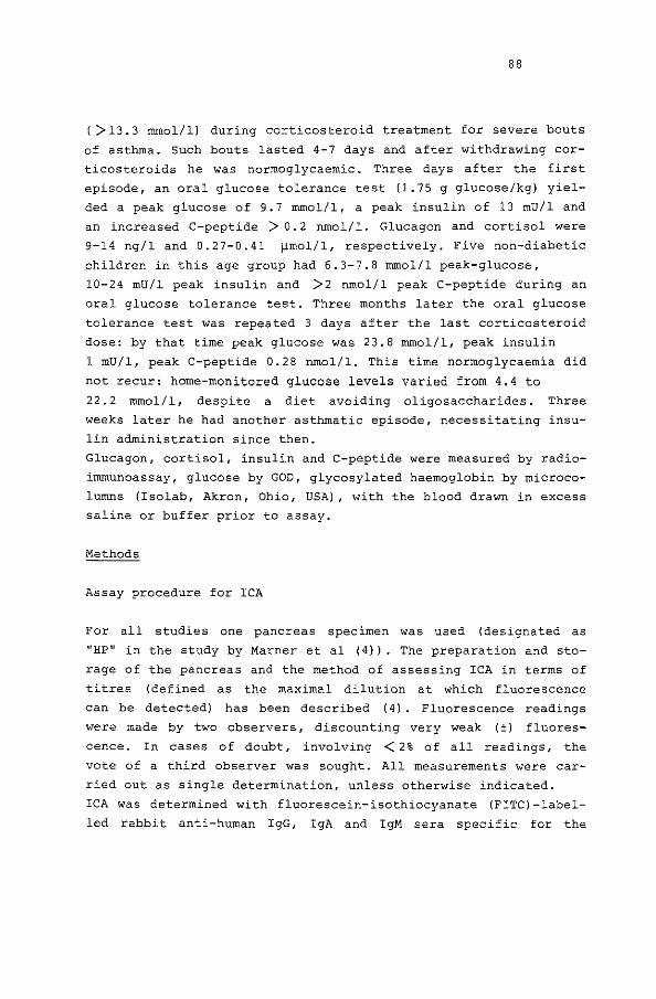
88
( >13.3 mrnol/1) during corticosteroid treatment for severe bouts
of asthma. Such bouts lasted 4-7 days and after withdrawing cor
ticosteroids he was normoglycaemic. Three days after the first
episode, an oral glucose tolerance test (1.75 g glucose/kg) yiel
ded a peak glucose of 9.7 mmol/1, a peak insulin of 13 mU/1 and
an increased C-peptide > 0. 2 nmol/ 1. Glucagon and cortisol were
9-14 ng/1 and 0.27-0.41 ~mol/1, respectively. Five non-diabetic
children in this age group had 6.3-7.8 rnmol/1 peak-glucose,
10-24 mU/1 peak insulin and >2 nmol/1 peak C-peptide during an
oral glucose tolerance test. Three months later the oral glucose
tolerance test was repeated 3 days after the last corticosteroid
dose: by that time peak glucose was 23.8 mmol/1, peak insulin
1 mU/1, peak C-peptide 0.28 nrnol/1. This time normoglycaemia did
not recur: home-monitored glucose levels varied from 4.4 to
22.2 rnmol/1, despite a diet avoiding oligosaccharides. Three
weeks later he had another asthmatic episode, necessitating insu
lin administration since then.
Glucagon, cortisol, insulin and C-peptide were measured by radio
immunoassay, glucose by GOD, glycosylated haemoglobin by microco
lumns (Isolab, Akron, Ohio, USA), with the blood drawn in excess
saline or buffer prior to assay.
Methods
Assay procedure for ICA
For all studies one pancreas specimen was used (designated as
"HP" in the study by Marner et al (4)). The preparation and sto
rage of the pancreas and the method of assessing ICA in terms of
titres (defined as the maximal dilution at which fluorescence
can be detected) has been described (4). Fluorescence readings
were made by two observers, discounting very weak ( ±) fluores
cence. In cases of doubt, involving < 2% of all readings, the
vote of a third observer was sought. All measurements were car
ried out as single determination, unless otherwise indicated.
ICA was determined with fluorescein-isothiocyanate (FITC) -label
led rabbit anti-human IgG, IgA and IgM sera specific for the

89
heavy chain, purchased from the Central Laboratory of the Blood
transfusion Service, Amsterdam, the Netherlands (anti-IgG, batch
number: KH-16-105-FOl, anti-IgA, batch number: KH-14-17-FOl, an
ti-IgM, batch number: KH-15-21-FOl) . A Leitz Orthoplan microscope
(Wetzlar, FRG) with epi-illumination (100 Watt lamp) and dichroic
mirror RKP 510 with filter LP 515 (Wetzlar, FRG) were used.
In two out of 46 samples IgG-rheumatoid factors were found (5) •
Those samples were excluded from the study.
Complement-fixing ICA
Since complement binding to IgG is dependent on the close proxi
mity of IgG molecules, the occurrence of CF-ICA was tested under
two different conditions, i.e. pancreas sections were incubated
once (complement-fixing-titre) and thrice (complement-fixing-pro
perty) with preheated (30 min, 56°C) patient sera (50 Ill) for
30 min. The sections were washed with phosphate buffered saline
(PBS, pH=7. 2) and incubated with human serum as a complement
source (fresh serum, blood group AB, stored -80°C, diluted 1:5).
After another washing procedure the presence of c3
was demonstra
ted by incubation with FITC-conjugated rabbit anti-human c 3c
(1:80 dilution, Dakopatts A/s, Copenhagen). Fluorescence was eva
luated as decribed above.
The conjugate did not react with human IgG, nor could fluorescen
ce be demonstrated under the above conditions, if ICA-positive
samples (titres 8-64) were incubated thrice with tissue in the
absence of c3.
ICA-IgG subclass detection
For the determination of IgG subclasses in ICA, specific rabbit
anti-human IgG subclass sera were used. The principle of the pre
paration of thes·e sera has been described (6): they were made
specific in agglutination and subsequently in indirect immuno
fluorescence by adsorption with a panel of relevant isolated IgG
paraproteins and tested against IgG subclass-paraproteins bound
to erythrocytes by tannin. When applied to bone marrow smears ob-

90
tained from multiple myeloma patients, these subclass antisera
appeared suitable for use by indirect immunofluorescence. The
sensitivity and specificity of these anti-sera (anti-IgG1 ,
batch number: KH 161-02-A1
; anti-IgG 2 , KH 162-19-A6 ; anti-IgG3 ,
KH 163-0l-A1
; anti-IgG4
, KH 164-05-A3 , obtained from the Central
Laboratory of the Blood Transfusion Service, Amsterdam) was found
adequate in immunofluorescence, as well as in an enzyme-linked
immunosorbent assay (Elisa) in two other studies (7,8).
To demonstrate the occurrence of a particular IgG-subclass, rCA
positive sera were incubated once or thrice with pancreatic tis
sue and subsequently tested for the subclasses. Anti-IgG subclass
sera were used at 1:20 dilution (excepting anti-IgG2 , 1:40) and
were applied in a double layer method in conjunction with FITC
labelled swine anti -rabbit Ig ( 1:8 0 dilution, F205, Dakopatts,
Copenhagen). The latter did not cross-react with human IgG, as
demonstrated by the absence of fluorescence when tested directly
on bone marrow cells of 10 multiple myeloma patients or when tes
ted on pancreas sections bound with ICA. Finally, extensive ab
sorption of the conjugate with human IgG did not affect in IgG
subclass detection.
Results
Table 1 indicates the prevalence of ICA and CF-ICA during child
hood diabetes. Each sample assayed came from a different child.
At the onset of disease the prevalence of ICA was 80%, but after
a duration of 4 years or more, only 11% of the children had cir
culating ICA. All positive sera contained ICA of the IgG class.
In addition, two sera contained ICA-IgA. ICA-IgM were not found.
If the sera were incubated once with pancreatic tissue, comple
ment fixation was found in 46% (19/41) of the samples (Table 1),
but triple incubation of the same sera increased the percentage
fixing complement by a factor 1.4 (to 27/41). Of 100 healthy
adult blood donors one sample was weakly positive for ICA and
none for CF-ICA.
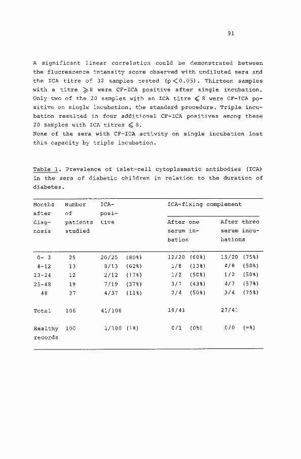
91
A significant linear correlation could be demonstrated between
the fluorescence intensity score observed with undiluted sera and
the ICA titre of 32 samples tested (p<O.OS). Thirteen samples
with a titre ¢8 were CF-ICA positive after single incubation.
Only two of the 20 samples with an ICA titre ( 8 were CF-ICA po
sitive on single incubation, the standard procedure. Triple incu
bation resulted in four additional CF-ICA positives among these
20 samples with ICA titres ~B.
None of the sera with CF-ICA activity on single incubation lost
this capacity by triple incubation.
Table 1. Prevalence of islet-cell cytoplasmatic antibodies (ICA)
in the sera of diabetic children in relation to the duration of
diabetes.
Months Number ICA- ICA-fixing complement
after of posi-
diag- patients tive After one After three
nos is studied serum in- serum incu-
bat ion bations
0- 3 25 20/25 (80%) 12/20 (60%) 15/20 (75%)
4-12 13 8/13 (62%) 1/8 (13%) 4/8 (50%)
13-24 12 2/12 (17%) 1/2 (50%) 1/2 (50%)
25-48 19 7/19 (37%) 3/7 (43%) 4/7 (57%)
48 37 4/37 (11%) 2/4 (50%) 3/4 (75%)
Total 106 41/106 19/41 27/41
Healthy 100 1/100 (1%) 0/1 (0%) DID (=%)
records

92
Table 2. Follow up of islet-cell antibody (ICA) and complement
fixing islet-cell antibody (CF-ICA) titres 0-12 months after
diagnosis.
Patient a no.
ICA/CF-ICAb titres
0 months 0-5 months 5-10 months 8-12 months
1
2
3
4
5
6
7
8
a Eight
Table 1);
32/210) 32/413)
2/0 10) 2/0 12)
8/010) 0 (0 12)
64/210) 64/412)
8/110) 8/112)
32/210) 16/213)
16/415)
16/413) 16/213)
64/1617)
0/0 15)
16/1 15)
16/4 17)
8/1110)
16/8 18)
8/2 16)
2/0 Ill)
0(0 18)
16/1 19)
8/0 112)
8/4111)
patients (randanly selected fran the 25 (0-3
b CF-ICA titres obtained after single incubation of the
months)
sera follow the corresponding ICA-IgG titre and the exact number
of months after diagnosis is shown in parentheses
of
Of ten samples that lost their complement fixation by dilution
1:3, the complement fixing property reappeared in nine of them
after triple incubation. In the sera of 25 healthy adult blood
donors no complement fixation could be detected after triple in
cubation with the pancreatic tissue.
The sequential titres of Table 2 show a gradual decrease of ICA
as well as CF-ICA titres during the first year of diabetes treat
ment. A strong correlation was found between ICA and CF-ICA tit
res obtained after single incubation (Spearman's rank correlation
gave a rho of 0. 79, p < 0. 0001). The CF-ICA titres were mostly
much lower than those of ICA-IgG.
Table 3 shows the relationship between ICA titres and IgG sub
class detection. The first samples of 44 recently diagnosed
diabetic children (0-12 months after diagnosis) were tested for

93
ICA-IgG subclasses after single or triple incubation of the tis
sue with the serum.
Firstly, we considered samples that fixed complement after single
incubation and in which ICA-IgG subclasses were determined by
single incubation. In this category samples with an ICA-IgG tit
re >16 had most IgG subclasses detectable. Secondly, of the 27
samples with ICA-IgG titres ~ 8, the number of IgG subclasses
detected exceeded two in only three samples, all of which fixed
complement on single. incubation.
Finally, the 17 samples that either did not fix complement at all
or only after triple incubation all had an ICA titre of ~ 16 and
in six of these sera no IgG subclass was detectable, regardless
of the incubation procedure.
The above findings raise the question whether the detectability
of specific IgG subclasses may be enhanced or suppressed by sin
gle or triple serum incubation. IgG subclasses were detected in
37 of the 44 ICA-positive samples of Table 3, after single or
triple incubation. Of those 37 sera, 34 contained IgG 1 , 17 IgG2 ,
18 IgG3
, 10 IgG4 •
ICA-IgG1
was detected in 9/27 sera with ICA-IgG titre ~ 8,
while triple incubation resulted in the detection of 20/27 ICA
IgG1. Only in one sample of this category ICA-IgG1 was lost by
triple incubation. The chi square test for paired observations
gave p < 0.05 for this comparison. Thus, IgG 1 detectability was
enhanced by triple incubation in CF-ICA. In CF-ICA negative sam
ples, however, ICA-IgG1
detectabili ty appeared in 3 I 10 samples
by triple incubation and disappeared in 2/10.
With the other IgG subclasses no significant suppression or en
hancement of detectability was found by single or triple incuba
tion.
Seven patients were followed up from before their diagnosis up to
20 months thereafter (Table 4) . Despite variability in the detec
tion of IgG subclasses caused by single or triple incubations,
consistency of IgG subclasses was found in parts of the clinical
course. Changes in subclasses were seen in patients 1, 2 and 5
where a 16-fo1d decrease in ICA-IgG titre was found.
In all patients, excepting patient 6, IgG 1 was the predominant

94
ICA-IgG subclass detected in the time course of their disease.
This result is in agreement with the quantitative distribution of
IgG subclasses found in the sera of 50 normal children, aged 8-12
years, using the same antisera (IgG 1 :IgG2 :IgG3 :IgG 4 = 70:20:8:4).
Ten randomly selected children with treated diabetes had a simi
lar IgG subclass distribution to that found in the sera of normal
children {data not shown) .
Patient 6 (encircled in Table 4) with ICA-IgG 2 alone and another
patient (encircled in Table 3) having ICA-IgG2
only on single as
well as triple serum incubation, had family histories of insulin
dependency. The maternal grandfather of the first patient was in
sulin-dependent at age 27 years and his maternal uncle developed
diabetes at age 10 years. The other patient had two maternal
grand-uncles with insulin-dependency as of ages 36 and 49 years.
The sera of these two patients, however, contained normal quanti
ties of IgG subclasses.
Table 3 The distribution of IgG subclasses (1, 2, 3, 4) in rela
tion to ICA-IgG titre and complement fixation in sera of newly
diagnosed diabetic children.
Number
of in-
cuba-
tions
ICA-IgG
titre
256
128
64
CF-ICA nega-
tive
One Three
Overall
result
CF-ICA positive
on triple incu-
bat ion only
One Three
Overall
result
CF-ICA positive on
single incubation
One Three
Overall
result
4 1+2+4 1+2+4
1+3 1+3

95
Table 3 continued.
Number CF- ICA nega- CF-ICA positive CF- ICA positive on
of in- tive on triple incu- single incubation
cuba- bation only
tions
One Three One Three One Three
ICA- IgG Overall Overall Overall
titre result result result
32 0 0 0 2+3 1+2+3+4 1+2+3+4
1+2+3+4 1+2+3+4 1+2+3+4
1+2+3+4 1+2+3+4 1+2+3+4
1+2+3+4 1+3+4 1+2+3+4
1+2+3+4 1+3+4 1+2+3+4
16 1 1 1 1+2+3 1 1+2+3
2 1+2 1 +2 1+3 1 1+3
1+2 1 1+2
1+2+3+4 1+2+3 1+2+3+4
1+3 1+2+3 1+2+3
1+4 1 1+4
1 1
8 1 1 1 1
1 1
1+4 1+4
4

Legend to Table 4
nd = not detectable
98
a Five patients (3-7) were randomly selected from the 25 of
Table
fore
1 and children 1, 2 were those
the disease became clinical! y
in whan ICA
manifest; b was rreasured be
IgG subclasses
are given as the overall result of their detection by single and
triple incubation; c titres are those obtained after single incu-
bation of the sera with the tissue, All measurements were done in
triplicate
was always
only
Discussion
and the
within one
average
titre
titre reported. The reproducibility
difference; d after triple incubation
The complement fixing nature of ICA may be particularly relevant
for the pathogenesis of childhood diabetes as this variety of ICA
has been detected in children before the disease becomes clini
cally manifest (1). This association is confirmed by two more ca
ses in the present study. In clinically overt childhood diabetes,
no complement fixation was found if the ICA-IgG titre was 4 or
lower, whereas the correlation between ICA-IgG and CF-ICA titres
was high, suggesting a limited sensitivity of our system for the
detection of CF-ICA titres.
The fixation of complement depends on the proximity of IgG-anti
bodies bound to the islet cells of the pancreatic sections. Trip
le incubation resulted in the detection of 1.4 times as many com
plement fixing ICA as single incubation (Table 1) , primarily be
cause samples with ICA-IgG titres of 4 or lower appeared to fix
complement by this procedure (Fig. 1). If CF-ICA-positive samples
were diluted until the complement fixing detectability disappea
red and were subsequently incubated thrice at that dilution, the
complement fixing property reappeared. This finding raises the
question whether the complement fixing nature of ICA has any sig
nificance other than that it is more likely to be seen in samples
with higher ICA-IgG titres.
As in other studies, ICA decreased with the duration of the dia
betes. However, our fraction of ICA's fixing complement remained

99
50%-75% (Table 1). Therefore, we do not feel the detection of
complement fixation per se has a separate significance.
Dean et al. (9) recently investigated ICA-IgG subclasses and
found 50% of the ICA-positive sera showed a restricted response,
with 12% reacting only with the IgG 2 subclass. We too found an
uneven distribution of IgG subclasses, however some apparent res
trictions were clearly dependent on the ICA-IgG titre on which in
turn the CF-ICA titre depended. Dean et al. found the restric
tions to be independent of CF-ICA titres, although their reported
CF-ICA titres were much higher than those in the present study.
We used triple incubation of the pancreatic sections to enhance
the detectability of complement fixation and if samples demon
strating all four ICA-IgG subclasses were diluted, IgG1
was the
last subclass to disappear. Conversely, triple incubation signi
ficantly enhanced the detectability of IgG1 . In addition, the
IgG1 subclass appeared predominant in 13 rcA-positive sera not
fixing complement, which suggests again that the IgG1
subclass
was relatively more abundant in ICA, than in whole serum. The ab
sence of detection of any IgG-subclass in seven sera of lower
ICA-IgG titre could be due to the limited number of epitopes pre
sent in ICA which are specific for the relevant subclasses. The
finding that ICA with lower titres did not fix complement may be
due to the low density of ICA bound from those sera. The discre
pancies indicated may be resolved with the availability of mono
clonal IgG subclass-specific antisera and sensitive enzyme-la
belled or radiolabelled assays (10) , now that two human ICA-reac
tive islet-cell protein fractions have been separated (11).
In conclusion, indirect immunofluorescence carried out on one
single fresh-frozen human pancreas, with a previously established
specificity and sensitivity for the detection of ICA (4) , yielded
variable results for the detection of complement fixation and IgG
subclasses, depending on ICA-IgG titres and incubation procedu
res. The present data suggest the occurrence of a non-restricted
IgG subclass response early in the course of childhood diabetes.
However, IgG2 subclass restriction in ICA was seen in two child
ren having a family history of insulin-dependency.

100
Acknowledgements
We thank Professor Dr. H.J. Degenhart for statistical advice and
Professor Dr. R. Benner, Department of Immunology, for his criti
cal review of this manuscript. The secretarial assistance of Ms.
E. Berkouwer and Ms. A. de Reus is gratefully acknowledged. The
work was supported in part by the Sophia Foundation for Medical
Research.
References
1. Gorsuch AN, Spencer KM, Lister J, Wolf E, Bottazzo GF, Cud
worth AG (1982) Can future Type 1 diabetes be predicted?
A study of families of affected children. Diabetes 31: 862-867
2. Van de Winkel M, Smets G, Gepts W, Pipeleers D (1982) Islet
cell surface antibodies from insulin-dependent diabetics bind
specifically to pancreatic B cells. J Clin Invest 70: 41-49
3. Lernrnark A, HagglOf B, Freedman ZR, Irvine WJ, Ludvigsson J,
Holmgren G (1981) A prospective analysis of antibodies reac
tive with pancreatic islet cells in insulin-dependent diabetic
children. Diabetologia 20: 471-474
4. Marner B, Lernmark A, Nerup J, Molenaar JL, Tuk c.w., Bruining
GJ (1983) Analysis of islet cell antibodies on sections of hu
man pancreas. Diabetologia 25: 93-96
5. Estes D, Atra E, Peltier A (1973) An immunofluorescent method
for the detection of antigamma-globulin antibodies. Arthritis
Rheum 16: 1-9
6. Van der Giessen M, Freyee W, Rossouw E, van Loghem E (1973)
Qualitative and quantitative studies on IgG2
globulins in in
dividual human sera with an antiserum capable of differentia
ting between Gm (n+) and Gm (n-) proteins. Clin Exp Immunol
14: 127-132
7. Von dem Borne AEGK, Helmerhorst FM, van Leeuwen EF, Pegels HG,
van Riesz E, Engelfriet CP (1980) Autoimmune thrombocytopenia:
detection of platelet antibodies with the suspension immuno
fluorescence test. Br J Haematol 45: 319-327
101
B. Horai S, Claass FHJ, van Rood JJ (1981) Detection of platelet
antibodies by enzyme linked immunosorbent assay (Elisa) . Com
parative studies with the indirect immunofluorescence assay.
Immunol Lett 3: 155-158
9. Dean BM, Bottazzo GF, Curdworth AG {1983) IgG-subclass distri
bution in organ specific autoantibodies. The relationship to
complement-fixing ability. Clin Exp Immunol 52: 61-66
10. Huen AHJ, Haneda M, Freedman Z, Lernmark A, Rubenstein AH
(1983) Quantitative determination of islet cell surface anti
bodies using 125
r-protein A. Diabetes 32: 460-465
11. Baekesskov s, Nielsen JH, Marner B, Bilde T, Ludvigsson J,
Lernmark A {1982) Autoantibodies in newly diagnosed diabetic
children immunoprecipitate human pancreatic islet cell pro
teins. Nature 298: 167-169

Paper 5
Autoantibodies to the insulin receptor in
juvenile onset insulin-dependent diabetes.
102
R. Maron1 , D. Elias1 2 , B.M. de Jongh3 , G.J. Bruining
4
J.J. van Rood 3 , Y. Shechter 2 & I.R. Cohen1
Department of 1cell Biology and 2 Hormone Research, The Weizmann
Institute of Science, Rehovot, Israel 3oepartment of Immunohematology, University of Leiden, Leiden,
The Netherlands 4 Department of Endocrinology, Sophia Children's Hospital, Rotter-
dam, The Netherlands
Reprinted from: Nature (1983) 303: 817-818, with permission.
Summary
Insulin-dependent diabetes mellitus
childhood or early adulthood, and its
(IDDM) usually begins in
aetiology is thought to in-
valve autoimmune damage to the islet-cells that secrete insulin
(1). To investigate an additional target of autoimmunity in IDDM
we examined sera for antibodies to insulin receptors. Such anti
bodies were defined by their ability to compete with insulin for
binding to insulin receptors and by their capacity to behave like
insulin in activating lipogenesis in adipocytes. We now report
the occurrence of anti-insulin receptor antibodies of the IgM
class in the sera of 10 of 22 IDDM patients obtained before their
treatment with exogenous insulin. Furthermore, two of five IDDM
patients who were initially negative developed anti-insulin re
ceptor antibodies during treatment with human or pork insulin.
These findings suggest that autoimmunity to the insulin receptor
may contribute to the pathophysiology of IDDM.

103
Results and discussion
The population of IDDM patients we examined comprised children
who were referred consecutively to the Department of Endocrinolo
gy of the Sophia Children's Hospital. Sera were obtained from 22
patients before treatment and from 5 of them also after they be
gan to receive insulin. Control sera were obtained from healthy
volunteer blood donors at the Blood Bank of the Leiden University
Hospital and from the investigators. We surveyed the sera by as
saying 0.5-5 ~l for insulin-like activity in stimulating lipoge
nesis in rat adipocytes measured by incorporation of radiolabel
led glucose into lipid (2,3).
Table 1 shows that the insulin-like activity in a representative
serum could be identified as anti-insulin receptor antibody of
the IgM class by fractionating the serum and testing the various
fractions for lipogenic activity. For example, 75% of the lipoge
nic activity found in the whole serum could be eluted from an an
ti- ~-column that specifically binds IgM. The effluent which con
tained the non-IgM material had relatively little lipogenic acti
vity. Filtration through Sephacryl 300 to separate IgM from IgG
confirmed that the lipogenic activity was a property of the frac
tion containing IgM, while the fraction containing IgG was inac
tive. Dissociation of possible insulin-insulin antibody complexes
by acidification and separation of serum fractions on Sephadex
G-100 showed that the lipogenic activity remained intact and re
sided in the insulin-free, high-molecular weight fraction (2) .
The low-molecular weight fraction did not contain sufficient in
sulin to activate lipogenesis. The lipogenic activity of the po
sitive sera was not inhibited by adding antibodies to insulin,
illustrating by another method that the lipogenic activity was
not due to insulin itself. In contrast, goat antibodies to human
IgM inhibited about 95% of the lipogenic activity in the positive
sera. Lipogenic IgM did not bind to immobilized insulin (not
shown). These results taken together indicate that the lipogenic
activity could be assigned to IgM molecules and not to free insu
lin or insulin-insulin antibody complexes. The lipogenic IgM was

104
identified as anti-receptor antibody as it specifically competed
with radiolabelled insulin for binding to insulin receptors.
Table 2 illustrates that increasing amounts of IgM purified on an
anti- ~-column increasingly inhibited the binding of insulin to
the insulin receptors of adipocytes, Control IgM isolated from
the serum of an IDDM patient without lipogenic activity had no
effect on the binding of insulin to its receptor.
Normal human IgG stimulates lipogenesis in rat adipocytes in vi
tro (4). Although the IgM anti-receptor antibodies described here
also caused lipogenesis, the two lipogenic effects differ with
regard to both ligand and receptor. The stimulatory effect of IgG
is exerted through the non-variable Fe portion of the molecule
(4,5) and normal IgG does not compete with insulin for binding to
the insulin receptor (P. Dandona, personal communication).
Table 1. Anti-insulin receptor antibodies in serum of IDDM pa
tient are IgM
Treatment of serum
None
Anti- j..l-column
Sephacryl 300
Dissociation of
complexes,
Sephadex G-100
Antibodies to
insulin*
Antibodies to
IgM!
Serum fraction
Whole
Effluent (not IgM)
Eluate (IgM)
Large (IgM)
Small (IgM)
Large (Ig)
Small (insulin)
Whole
Whole
% Relative lipogenesis
of adipocytes
100
15
75
100
0
90
0
100
5

105
Legend Table 1.
Sera were fractionated (2) using columns of goat antibodies to
human 1.1.-chain (Dako Im:nunoglobulins), bound to Sepharose (Pharrra
cia (1)), Sephacryl 300 (Pharrnacia (12)) or Sephadex G-100 (Phar
macia). Antigen-antibody complexes were dissociated by acidifica
tion (0.01 M HCl, pH 2. 7) of serum (2) before gel filtration.
Control serum or fractions for each procedure were obtained fran
healthy donors and from an IDDM patient whose whole serum was ne-
gative for
was canputed
serum (100%).
pad of male
anti-receptor antibody activity.
relative to that found in 1 ~l
Adipocytes were obtained from
Lipogenic activity
14 D(U- C)glucose
as described
equivalent to
insulin (10 ng
ne::l without added
Wistar rats {90-120 g)
(4-7 nCi mol-1 ; NEN")
previously (2,3). Max:inal
and
into
of the unfractionated
the epididymal fat
the incorporation of
lipid was measured
lipogenesis (100%) was
that produced by incubation of the adipocytes with
rnl-11 and was 300% of control lipogenesis obtai-
insulin.
* Guinea pig anti-insulin antisennn (Miles-Yeda; titre 10-S)
was added (2 !Jl) to the lipogenic assay.
Goat antiserum to IgM (Miles-Yeda) was adde::l (30
lipjgenic assay.
1111 to the
Table 2. Purified IgM receptor antibody competes with insulin for
binding to insulin receptors on rat adipocytes
Affinity-purified
IgM ( 11gl
5
10
20
% Inhibition insulin binding to adipocytes
Test serum
29
48
72
Origin of IgM
Control serum
9
4
0
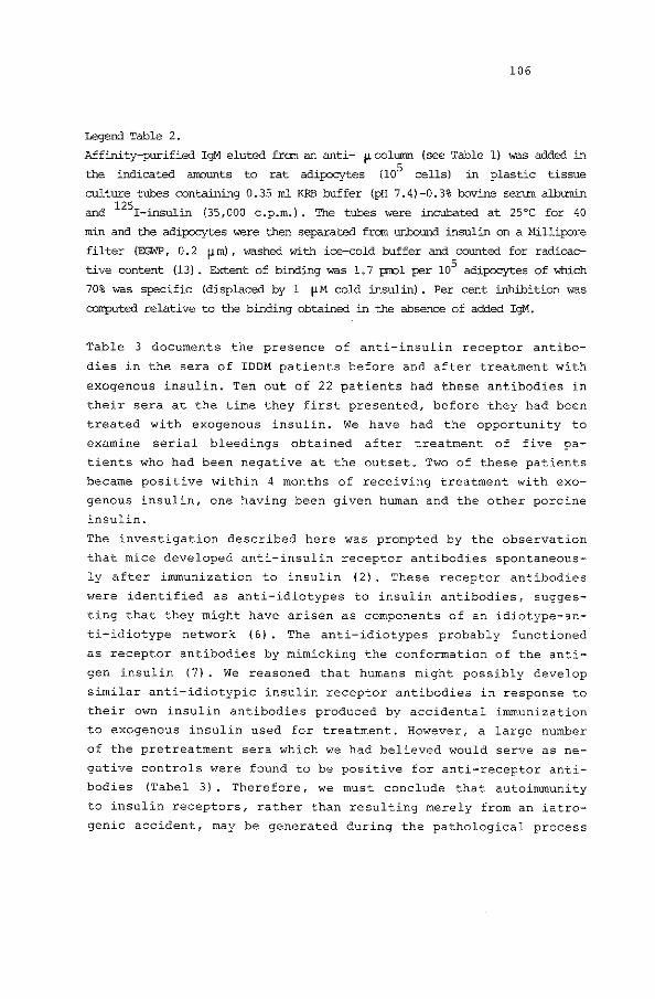
106
Legend Table 2.
Affinity-purified IgM eluted fran an anti- j..L column (see Table 1) was added in
the indicated arrounts to rat adipocytes (105 cells) in plastic tissue
culture tubes containing 0.35 ml KRB buffer (pH 7.4)-0.3% bovine serum albumin
and 125r-insulin (35,000 c.p.m.). The tubes were incubated at 25°C for 40
min and the adipocytes were then separated from unbound insulin on a Millipore
filter (EGWP, 0.2 j..Lffi), washed with ice-cold buffer and counted for radioac
tive content (13). Extent of binding was 1.7 pmol per 105 adipocytes of Which
70% was specific (displaced by 1 !lM cold insulin) . Per cent inhibition was
computed relative to the binding obtained in the absence of added IgM.
Table 3 documents the presence of anti-insulin receptor antibo
dies in the sera of IDDM patients before and after treatment with
exogenous insulin. Ten out of 22 patients had these antibodies in their sera at the time they first presented, before they had been
treated with exogenous insulin. We have had the opportunity to
examine serial bleedings obtained after treatment of five pa
tients who had been negative at the outset. Two of these patients
became positive within 4 months of receiving treatment with exo
genous insulin, one having been given human and the other porcine
insulin.
The investigation described here was prompted by the observation
that mice developed anti-insulin receptor antibodies spontaneous
ly after immunization to insulin (2). These receptor antibodies
were identified as anti-idiotypes to insulin antibodies, sugges
ting that they might have arisen as components of an idiotype-an
ti-idiotype network {6). The anti-idiotypes probably functioned
as receptor antibodies by mimicking the conformation of the anti
gen insulin (7). We reasoned that humans might possibly develop
similar anti-idiotypic insulin receptor antibodies in response to
their own insulin antibodies produced by accidental immunization
to exogenous insulin used for treatment. However, a large number
of the pretreatment sera which we had believed would serve as ne
gative controls were found to be positive for anti-receptor anti
bodies (Tabel 3). Therefore, we must conclude that autoimmunity
to insulin receptors, rather than resulting merely from an iatro
genic accident, may be generated during the pathological process
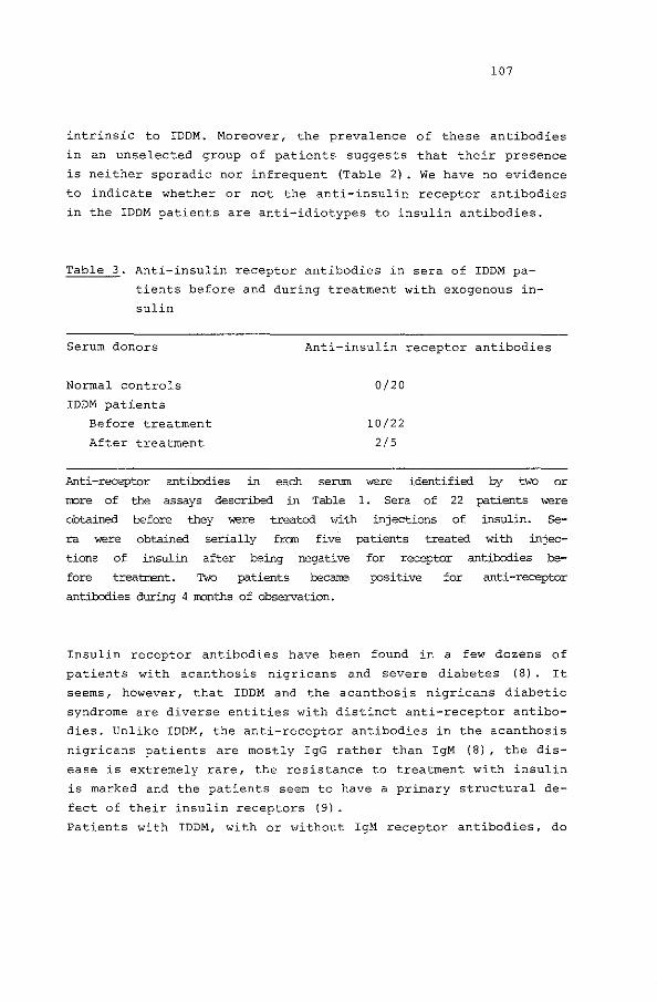
107
intrinsic to IDDM. Moreover, the prevalence of these antibodies
in an unselected group of patients suggests that their presence
is neither sporadic nor infrequent (Table 2). We have no evidence
to indicate whether or not the anti-insulin receptor antibodies
in the IDDM patients are anti-idiotypes to insulin antibodies.
Table 3. Anti-insulin receptor antibodies in sera of IDDM pa
tients before and during treatment with exogenous in
sulin
Serum donors Anti-insulin receptor antibodies
Normal controls
IDDM patients
Before treatment
After treatment
0/20
10/22
2/5
Anti -receptor antibodies in each serum were identified by two or
rrore of the assays described in Table 1 . Sera of 22 patients were
obtained before they were treated >vith injections of insulin. Se
ra were obtained serially fran five patients treated with injec
tions of insulin after being negative for receptor antibodies be-
fore treatrrent. Two patients becarre positive for anti-receptor
antibodies during 4 rronths of observation.
Insulin receptor antibodies have been found in a few dozens of
patients with acanthosis nigricans and severe diabetes (8). It
seems, however, that IDDM and the acanthosis nigricans diabetic
syndrome are diverse entities with distinct anti-receptor antibo
dies. Unlike IDDM, the anti-receptor antibodies in the acanthosis
nigricans patients are mostly IgG rather than IgM (8), the dis
ease is extremely rare, the resistance to treatment with insulin
is marked and the patients seem to have a primary structural de
fect of their insulin receptors (9).
Patients with IDDM, with or without IgM receptor antibodies, do
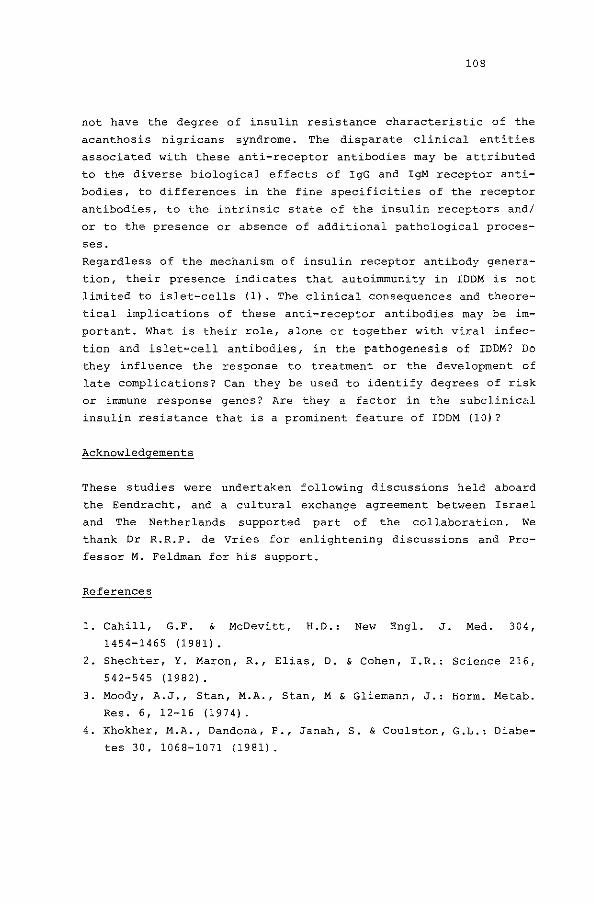
108
not have the degree of insulin resistance characteristic of the
acanthosis nigricans syndrome. The disparate clinical entities
associated with these anti-receptor antibodies may be attributed
to the diverse biological effects of IgG and IgM receptor anti
bodies, to differences in the fine specificities of the receptor
antibodies, to the intrinsic state of the insulin receptors and/
or to the presence or absence of additional pathological proces
ses.
Regardless of the mechanism of insulin receptor antihody genera
tion, their presence indicates that autoimmunity in IDDM is not
limited to islet-cells (1). The clinical consequences and theore
tical implications of these anti-receptor antibodies may be im
portant. What is their role, alone or together with viral infec
tion and islet-cell antibodies, in the pathogenesis of IDDM? Do
they influence the response to treatment or the development of
late complications? Can they be used to identify degrees of risk
or immune response genes? Are they a factor in the subclinical
insulin resistance that is a prominent feature of IDDM (10)?
Acknowledgements
These studies were undertaken following discussions held aboard
the Eendracht, and a cultural exchange agreement between Israel
and The Netherlands supported part of the collaboration. We
thank Dr R.R.P. de Vries for enlightening discussions and Pro
fessor M. Feldman for his support.
References
1. Cahill, G.F. & McDevitt, H.D.: New Engl. J. Med. 304,
1454-1465 (1981).
2. Shechter, Y. Maron, R., Elias, D. & Cohen, I.R.: Science 216,
542-545 (1982).
3. Moody, A.J., Stan, M.A., Stan, M & Gliemann, J.: Harm. Metab.
Res. 6, 12-16 (1974).
4. Khokher, M.A. 1 Dandona, P., Janah, s. & Coulston, G.L.: Diabe
tes 30, 1068-1071 (1981).
109
5. Khokher, M.A. & Dandona, P.: Diabetologia 23, 180 (1982).
6. Jerne, N.K.: Annls Immun. Inst. Pasteur, Paris 125C, 373-389
(1974) .
7. Nisonoff, A. & Lamoyi, A.: Clin. Immun. Immunopath. 21,
397-406 (1981).
8. Flier, J.S., Kahn, C.R., Jarrett, D.B. & Roth, J.: J. Clin.
Invest. 58, 1442-1449 (1976).
9. Podskalny, J.M. & Kahn, C.R.: J. Clin. Endocr. Metab. 54,
261-268 (1982).
10. De Fronzo, R.A., Hendler, R. & Simonson, D.: Diabetes 31,
795-801 (1982).
11. March, S.C., Parkish, I. & Cuatrecasas, P.: Analyt. Biochem.
60, 149-152 (1974).
12. Johansson, I. & Lundgren, H.: J. Biochem. Biophys. Meth. 1,
37-44 (1979)' 47' 378-389 (1979).
13. Cuatrecasas, P.: Proc. Natn. Acad. Sci. U.S.A. 68, 1264-1268
(1971) .

110
§5. Discussion
The etiology of childhood diabetes can be represented as pancrea
tic beta-cell destruction as a function of time {figure 2) .
Fig. 2 Pancreatic beta-cell destruction
Pancreatic beta cell destruction
Percent of the beta cell mass left l 00 +------, ,-------,------------------------------ --
I
1 I?
II
onset of symptoms
m
The gradual destruction of pancreatic beta-cell with ti.rre,
clinical symptoms not developing before 70-80 percent of the
original beta-cell mass has been destructed (paragraph 2) .
Question marks indicate putative phases of beta-cell regeneration.
Raman numbers on the horizontal axis indicate time spans of:
I. genetic susceptibility (HLA-related)
II. subclinical expression of the disease (islet-cell antibodies)
III. the clinical! y overt state (when insulin is administered) .
Time
The papers presented in the previous paragraph can be assigned
each to one the three phases of the time-course of childhood dia
betes depicted in figure 2. The purpose of the following discus
sion is to present the findings in as much as they contribute to

111
the consideration of possible immune-intervention of childhood
diabetes.
This fascinating new field of clinical research has very recently
received much attention, but only preliminary data are available
(to be discussed in the final section of this paragraph).
To clarify the connection of the studies presented to this new
field of research it must be understood that all investigations
hitherto into immunosuppression of childhood diabetes dealt with
possible prolongation of the remission-phase {paragraph 2) , once
the disease became clinically manifest. In other words: immuno
suppressive measures were only tried in phase III of figure 2.
This discussion attempts to demonstrate that present findings in
the other phases of the clinical course of childhood diabetes
have consequences for the interpretation of the possible finding
of prolongation of the remission-phase by virtue of immuno-sup
pressive measures, after insulin treatment is initiated. Further
more new approaches are mentioned that will contribute to an im
proved understanding of each of the phases of the clinical course
of childhood diabetes depicted in figure 2.
1. HLA-antiqens as genetic markers for the susceptibility to
childhood diabetes.
From twin studies it is known identical twins have a concordance
rate of 50% or less (38,39). Gm-markers were not associated with
diabetes-type-1a generally (paper 1) , but they may still be in a
small subgroup of children (paper 3) , as far as the IgG-subclas
ses of their islet-cel1-cytoplasmatic-antibodies are concerned.
Limiting this discussion to HLA:
From the multiplex families of paper 1 it was calculated that in
our multiplex type-la-diabetic families HLA-identity with an af
fected sib would carry a 11.6% risk for that sib to become diabe
tic himself. This is the same figure (12%) as calculated by Platz
et al., who studied 51 multiplex young onset diabetic families
and 212 monoplex families, meaning that only one sib had treated
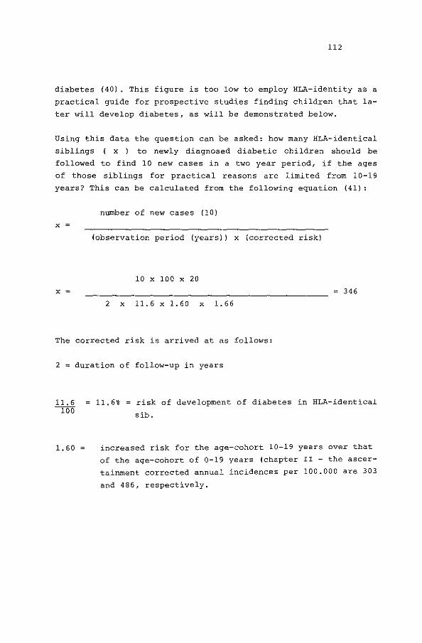
112
diabetes (40). This figure is too low to employ HLA-identity as a
practical guide for prospective studies finding children that la
ter will develop diabetes, as will be demonstrated below.
Using this data the question can be asked: how many HLA-identical
siblings ( x ) to newly diagnosed diabetic children should be
followed to find 10 new cases in a two year period, if the ages
of those siblings for practical reasons are limited from 10-19
years? This can be calculated from the following equation (41) :
number of new cases (10)
X
(observation period (years)) x (corrected risk)
10 X 100 X 20
X 346
2 X 11.6 X 1.60 X 1.66
The corrected risk is arrived at as follows:
2 duration of follow-up in years
1. 60
11.6% risk of development of diabetes in HLA-identical
sib.
increased risk for the age-cohort 10-19 years over that
of the age-cohort of 0-19 years (chapter II - the ascer
tainment corrected annual incidences per 100.000 are 303
and 486, respectively.

1. 66
20
113
correction for the finding that one third of all sib
lings to diabetics become diabetic themselves within two
years after their already diabetic sibling, according to
a large study in England (42).
correction to annual rates for the use of risks for the
age-cohort of 0-19 years.
More than 300 HLA identical sibs would have to be identified to
be followed prospectively for two years in order to detect 10
children of 0-19 years becoming clinically diabetic in that
time-span. The ascertainment corrected incidence per year for
that age-span in the Netherlands is 486 (Chapter II) and only one
in four sibs will be HLA-identical as there is no evidence of se
lection against or for HLA-combinations in families with type-1-
diabetes ( 5) .
Thus, 346 x 4 = 1384 healthy siblings would have to be HLA-typed,
which would involve 659 (1384 : 2.1 (the average dutch kindred))
families, which represents a formidable task against the back
ground of the dutch total annual incidence.
In what way did the study of HLA by serology contribute to the
understanding of the mechanism of the genetic susceptibility?
The mode of inheritance of susceptibility is complex and the "two
or more gene concept", linked to HLA-DR3 and/or HLA-DR4 seems to
fit the data best. This is in agreement with other studies, sum
marized by the late Dr. Andrew G. Cudworth et al. (5). They also
described haplotype-clusters, involving genes over the whole of
the major histocompatibility complex in association with suscep
tibility to type~l-diabetes. This finding suggests common ances
tor haplotype-clusters among the patients, but, more importantly,
lends support to the hypothesis of sets of interacting genes to
act in concert within these clusters.

114
In another study (42), we found that the SB-locus, situated be
tween the HLA-D locus and GLO (hence centromeric from HLA-D),
shared no allotypes with type-la-diabetes.
The cross-overs observed in our family-study, suggested that the
HLA-A locus contributed more likely by linkage-disequilibrium to
diabetic clusters than by truly being part of them. Thus, two lo
ci situated between HLA-A and SB, were selected for their possi
ble involvement, namely HLA-B and HLA-DR. Two haplotype-combina
tions, HLA-B8/HLA-DR3 and HLA-B15/HLA-DR4 were tested for third
order disequilibrium with type-la-diabetes. The indication found
of the genes of two loci acting in concert, involving different
classes of gene-products, namely HLA-B antigens (class I histo
compatibility antigens) and HLA-D antigens (class II histocompa
tibility antigens) 1 may have important consequences for the un
derstanding of the immunologic pathogenesis and hence the consi
deration of future immunologic interventions. What these would be
is at present unclear, as the interactions of the multiple anti
gens coded for by genes of the major histocompatibility complex
(MHC) are only beginning to become elucidated.
The merit of the described suggestion of third order linkage-dis
equilibrium, arrived at by "classic" genetic analysis, is, that
it can be tested by novel techniques, known as "fine genetics".
This implies the use of DNA-probes on segments of chromosomes
dissected by endonucleases.
Recently, D. Owerbach and associates at the Hagedorn Institute
(43), described two such fragments, relevant for the association
between HLA-DR4, the commonest haplotype in our childhood diabe
tic population, and insulin dependency. It is known class !!-an
tigens (HLA-D related) show a marked genetic polymorphism asso
ciated with the beta-chains of these molecules (44). This poly
morphism appears to be associated with several MHC genes, eviden
ced from hybridization studies (45). Thus, differences in hybri
dization pattern between DNA from healthy individuals and diabe
tic patients, using the beta-chain c DNA probe (p DR- p -1) after
digestion with certain restriction endonucleases may be highly
relevant. They found an increased frequency of one fragment among
HLA-DR4 I HLA-DRx individuals as well as among HLA-DR4 I HLA-DR3
115
individuals, that were insulin dependent. They also described an
other fragment, occurring in 30-40% of controls, but in only 0-2%
of the diabetics. Other probes, suitable for the examination of
the HLA-B locus could yield similar results, perhaps supporting
the proposed "two or more loci concept".
Should relevant DNA-fragments be found, monoclonal antibodies to
their gene-products {membrane antigens) may be generated, facili
tating the detection of cells carrying such antigens.
For the detection of children with a genetic risk for type-la
diabetes, the accuracy using the above techniques could be higher
than presently possible with serologic typing.
In what further way may the study of HLA contribute to better un
derstanding of childhood diabetes?
The MHC-complex in humans (as well as genes of the H-2-complex in
mice) encomprises a large variety of functions pertinent to pre
sentations of type-1-diabetes.
With regard to insulin-dependent-diabetes it is relevant to men
tion biologic functions that have so far been associated with
HLA-genes or their products: the HLA-antigens on the cell-membra
ne. Table 2 summarizes such functions, in mice (of the H-2-com
plex) and in humans (of the HLA-complex) , contrasted to their
possible relevance for all stages of childhood diabetes.

Table 2.
Function
1. Production of humoral antibodies
2. Production of killer Tcells and helper T-cells
116
Relevance for diabetes-type-!
1. Islet-cell antibodies, thyroid, adrenal, parietal cell and other prevailing auto-antibodies (46). Antibodies to injected insulin.
2. Pancreatic beta-cell cytotoxicity (47) Impaired suppressor T-cell function (48).
3. Regulation of immune response 3. Ibidem including mode of expression of autoimmunities.
4. Biosynthesis of some comple- 4. ment components
5. Regulation of embryogenesis 5.
6. Regulation of synthesis of 6. some steroid hormones
7. Regulation of ligand-membrane 7. interactions of some protein hormones
Ibidem
Unknown
Unknown
Insulin action (49) also modified by insulin antigenicity.
With present serologic HLA-typing sub-groups of type-la-diabetic
patients cannot be separated easily in view of the already men
tioned haplotype-clusters that contain genes in linkage to each
other rather than to disease-states. Fine-genetics, referred to
above, may detect and hence predict highly relevant entities
within type-la-diabetic patients. Such entities might include:
1. differences of islet-cell antibody occurrence in particular in
pre-diabetic-states;
2. differences in velocity of loss of remnant endogenous insulin
secretion;
3. differences in sensitivity to injected insulin.
117
2. Islet-cell antibodies as markers for childhood pre-diabetes
The second tool investigated for the evaluation of the predicta
bility of diabetes-type-la were antibodies to the islets of Lan
gerhans (ICA). One human pancreatic specimen with previously es
tablished specificity and sensitivity for the assay of ICA was
used for this purpose. The prevalence of ICA in newly diagnosed
children was 80% with this system (paper 4), in the same order of
magnitude of other studies, using fresh-frozen, rather than Bouin
fixed pancreatic specimens. In controls, only 1% was weakly posi
tive, indicating a better specificity in this respect than found
in other studies.
Subsequently, the question was asked to what extent these ICA
might be present before the diagnosis is made.
Two years before the study reprinted as paper 5 was started, the
London group lead by the late Dr. A.G. Cudworth and Dr. G.F. Bot
tazzo set up a prospective study into the predictability of
childhood diabetes by ICA. This study became known as the Barts
Windsor study. It involved 154 healthy brothers, 134 healthy sis
ters from 160 families having a diabetic child. ICA were assayed
every 1-4 months with a mean follow-up of 2 (1.1-2.5) years at
the time point the study reprinted as paper 4 was initiated. In
their preliminary report (SO) it was indicated six subjects of
these families had developed insulin-dependency. Four of these
six subjects had been ICA-positive from the moment they entered
the study and had remained positive on repetitive testing until
the disease became clinically manifest. The two other subjects
had become positive during their follow-up. Of these six subjects
two were HLA-identical sibs, two were haplo-identical sibs and
two were parents.
In total, that study involved 473 healthy siblings and parents.
In their final report (51) seven developed diabetes in two years
and 54 of these subjects had ICA positivity without the develop
ment of clinical diabetes. If one assumes all these subjects
(children and parents) would become diabetic eventually, the re
currence risk would be 54/473 = 11.2 %.
It is difficult, to compare this figure with the recurrence risk

118
derived from epidemiologic studies. Very few epidemiologic sur
veys have taken the recurrence risk in parents into account and
only one corrected for incomplete ascertainment, the study from
the Pittsburgh group (52). Another difficulty with the Barts
Windsor data is that several pancreas specimens were used for the
assay of ICA and that the fluorescence was not expressed in
titres.
Provided pancreatic tissue with defined sensitivity and precision
for the detection of ICA would be used, this tool appears to have
a high accuracy screening for pre-diabetic states in childhood
(papers 3 and 4). A practical advantage of this method as an en
try to identify pre-diabetes of childhood is that minute quanti
ties of serum can be used. In positive cases, only 50 microliters
of serum is needed for a reliable assay. As demonstrated in paper
3 and 4 the titres of prediabetic children as well as of newly
diagnosed children were usually 4 or higher, if positive. Such
quantities would be obtainable by fingerprick, a technique most
families with diabetic children are used to, so that the partici
pation of children younger than 10 years of age may be conside
red.
It should be noted that 20% of the newly diagnosed children did
not have ICA. The one patient (paper 3) that developed ICA only
after diagnosis did have 3+ islet-cell surface antibodies (ICSA)
only, three years before her disease became manifest. This data
and the sequence of the islet-cell antibodies developing in this
particular patient, suggests that next to ICA, ICSA should be
used as screening tools for prediabetes of childhood. For this
method 1 milliliter of serum is presently required and the fluor
escence assay involved is much more difficult.
At the moment we initiated our own survey of rCA-positivity among
healthy sibs and parents of 22 multiplex families, we only knew
of 4 children that had become diabetic in the Barts-Windsor study
forementioned, with ICA positivity from the moment they entered
that study. We agreed with our participants we would make the
HLA-data available only to their family physicians, but would not
disclose the - to us at that point uncertain - meaning of rcA-po
sitivity in a healthy family member. We decided to look for ICA

119
in sibs and parents on the basis of single bleedings (with the
above preliminary findings of the Barts-Windsor study on the per
sistence of ICA in mind). The 22 multiplex families, only 3 co
ming from our own clinic, participated voluntarily, in full know
ledge of our own uncertainties to the meaning of HLA and/or ICA
with respect to their future health.
With the data on the accuracy of predicting childhood diabetes by
ICA (described in paper 3) , one might feel less insecure about
the prospect of a sib of a diabetic child having ICA without any
signs o'r symptoms and this represents major problems. It was re
ported (the late Dr A.G. Cudworth, personal communication) that
some participants of the Barts-Windsor study did resent their
non-diabetic child becoming diabetic during those prospective
studies. This experience holds an important warning if such stu
dies are to be repeated. Participation of children not becoming
diabetic may generate an undue awareness of the child and its
family that the child may still become diabetic after an unknown
number of years. The burden of this "partially inherited" life
long disorder cannot be overestimated and causes numerous inter
actions in the families of children with treated diabetes (Chap
ter III - paragraph 1) • Similar interactions may be projected by
parents on healthy sibs by their mere participation in a screen
ing program of childhood diabetes, that has no therapeutical con
sequences. Possible interference with the upbringing of these
sibs cautions for a repeat of such studies on ethical grounds.
Before considering any screening program for pre-diabetes of
childhood, more needs to be known on the nature of islet-cell-an
tibodies as predictors of childhood diabetes. Both islet-cell cy
toplasmatic - and surface fluorescence-techniques are qualitative
in nature, and the following three principle shortcomings (a-c)
remain:
a.children with mumps infection (but not with Coxsackie, Measles,
Rubella or Influenza) may have temporary ICA, not persisting for
more than 2-4 months and without developing insulin-dependency
subsequently (53). Beside this problem there are other limits to
the immunologic specificity of ICA.

120
Islet-cell cytoplasmatic antibodies, usually not reacting with
intact cells, are measured as islet-cell flurorescence on pancre
atic slices. The antigenicity of the whole pancreatic beta-cell
is assessed and the antigens of the whole islets they react with
have not been specified to date. The complement fixing ICA appea
red a fairly good screening agent for the pre-diabetic state of
children. In view of the absent definition of their immunologic
functions and biochemical nature they have been compared to "the
crowd gathering around the accident". However, it happens with
criminal accidents that "the criminals hide within this crowd to
secretely enjoy the effects of their act" and the problem to
identify them remains. Even the number of protein-isolates acting
as antigens of the pancreatic islets was undefined until recent
ly. S. Baekkeskov et al. (54) isolated two protein-fractions from
cultured pancreatic islets, that reacted with a panel of ICA po
sitive samples obtained from several institutions, including our
own. They used detergent-solubilized radiolabelled islet-cell an
tigens from cultured human and rodent pancreatic islets, com
plexed these to antibodies isolated from the sera of diabetic
children (obtained by plasmapheresis) and detected the two reac
ting protein-fractions by gel electrophoresis after specific ab
sorption to protein A-sepharose. Their findings that ICA reacted
with at least a limited number of (two, thus detected) protein
fractions raises hopes to the further elucidation of the nature
of these fractions.
Further, it is unknown where the relevant antigens reside within
the islets. Studies with human-human hybridoma systems, where pe
riferal lymphocytes of a diabetic child were fused with a human
myeloma line, yielded one clone reactive with human pancreatic
islets by fluorescence. Further analytical studies indicated that
the target antigen was possibly situated in the cytosol of gluca
gon-, rather than insulin-producing cells of these islets (25).
The significance of this experiment (the first to employ this no
vel technique with regard to childhood diabetes) remains unclear.
ICA react with the cytoplasmatic contents of islet-cells as mea
sured on fresh-frozen pancreatic sections. One would expect that
cytotoxicity to intact islet-cells is mediated by islet-cell-sur-

121
face-antibodies ( ICSA) . These have been found to react with is
let-cells of rodents. ICSA appeared to be organ-specific, but not
species-specific. ICSA react mainly with pancreatic beta-cells
and they can mediate a complement-dependent cytotoxic reaction
(55) . One would expect ICSA to react first with pancreatic beta
cells, destructing their membrane. Consequently the contents of
the pancreatic beta-cells are exposed, which subsequently results
in the generation of ICA. As forementioned, this sequence of
events was actually documented in patient 2 of paper 3. In this
view ICSA would be "the criminal" and ICA "his accomplice".
b. A second enigma, with possible immunogenetic implications, is
exemplified by the detection of IgG-subclass restriction of ICA
in a few diabetic children as described in paper 4. Early after
diagnosis as well as in two pre-diabetic children (patients 1 and
2 of paper 4) mostly all four IgG-subclasses were present and no
IgM-ICA was found. The absence of IgM-ICA suggests that auto-im
munity towards the islets must have been going on for some time,
because at the onset of auto-immune reactions antibodies of the
IgM-class generally predominate. The presence of mostly all four
IgG-subclasses early in the course of childhood diabetes suggests
that there was no restriction of B-lymphocyte clones producing
these multiple classes of antibodies. Of course, this absence of
restriction does not exclude switching of such B-lymphocyte
clones. Two children were found to have IgG 2-rcA restriction,
despite relatively high (complement-fixing} ICA-titres. Admitted
ly, the demonstration of the IgG2-subclass appeared to be more
difficult than that of other IgG-subclasses. Hence, a dilution of
1:40 was used to demonstrate this subclass in ICA, as opposed to
the dilution of 1:20 for the demonstration of the other subclas
ses, IgG1 , IgG3
and IgG 4 (paper 4). These two children had 11 Ver
tical transmission" of insulin dependency in their families. Al
though the family histories of these two children have not been
compared to those of the other 42 studied with regard to IgG-ICA
subclasses, such family history is exception rather than rule in
childhood diabetes. Although IgG-ICA-subclass restriction appears
"a characteristic of an accomplice", this feature hints to a par
ticular kind of 11 criminal act 11 in the comparison used before.

122
c. A third problem with ICA that needs to be considered is that
ICA may encomprise idiotypic as well as anti-idiotypic antibodies
against human islets. The relevance of this feature has been re
viewed elegantly by C.H. Brogren and A. Lernmark {25): "The hy
bridoma technology provides a novel approach for investigating
the network theory of Jerne, which implies that the antibody res
ponse is regulated through negative and positive feed-back mecha
nisms. Let us imagine that a monoclonal antibody able to induce
diabetes has been isolated. According to Jerne's theory these an
tibodies, injected into syngeneic recipients will induce the for
mation of anti-antibodies (or so called anti-idiotype antibo
dies) . We speculate that these anti-idiotype antibodies have a
specific immunosuppressive effect, when administered to the dia
betic recipient. A suppressive effect may be achieved by those
anti-idiotypic antibodies depicting the antigenic determinants as
the target molecules (internal image) which are expressed on is
let-beta-cells. Anti-idiotype antibodies reactive with acetylcho
line receptor antibodies have been produced after syngeneic mouse
spleen cells were "educated 11 with acetylcholine receptors. Mono
clonal anti-idiotype antibodies may thus prove to be useful re
agents in studies of immune response regulation".
Human-human and human-mouse hybridomas have been produced from
patients with auto-immune polyendocrine disease, including diabe
tes-type-lb (paragraph 1 of this chapter), thyreoiditis and some
times adrenalitis (55). These hybridomas synthesize auto-antibo
dies that react with multiple normal human tissues, pituitary,
gastric mucosa, endocrine pancreas and thyroid, at least partial
ly explaining the multiorgan autoimmunity both in animals (25)
and humans. The availability of large quantities of such monoclo
nal antibodies will aid in raising anti-idiotypic antibodies for
the abovementioned purpose.
This specific example is relevant for "diabetes-type-1a" (para
graph 1) as well. In our own clinic (with presently 136 children
of 0-19 years followed annually for 0-14 years, 4 years averaged)
10%-20% had antithyroid-, 4% anti-gastric-mucosa- and 1% anti
adrenal-antibodies, next to other varieties of auto-antibodies.
If the organs involved in these cases could be demonstrated to be
123
antigenic by the sharing of common antigens to the pancreatic be
ta- cell, the generation of monoclonal.anti-idiotypes may be use
ful to elucidate immune response regulation. The nature of this
supposed common auto-antigen is still unknown. "Heterogeneity" of
monoclonal antibodies, however, may still ensue, as these may re
act with still other constituents of the body, only sharing mole
cular substructures with relevant antigens.
A different problem with the interpretation of the significance
of islet-cell antibodies deals not so much with their nature or
specificity (a-c) but with their possible effects: it has been
suggested immunoglobulins of diabetic children, that react with
islet-cells, interfere with the islet's capacity to secrete insu
lin. In the presence of complement, the children's antibodies
blocked the insulin release of rat islets (56) . Cytotoxic antibo
dies markedly slowed down the insulin release of human islets in
a system where the cultured islets were peri-fused (57). This ef
fect casts doubt on tests assessing the secretory capacity of is
let-cells as a measure of their (remnant) quantity: in the pre
sence of islet-cell antibodies, interfering with insulin release,
the C-peptide stimulation by for instance glucagon may well be
hampered. During the first years of insulin treatment, when is
let-cell antibodies prevail (paper 4) , C-peptide reserve testing
may "underestimate" the number of islets with functional poten
tial, as this may be blocked by the potential of islet-cell anti
bodies abovementioned. This potential of islet-cell antibodies
may seriously hamper the interpretion of beta-cell saving immuno
suppressive therapies, reviewed in the last section of this para
graph.
Taken together, islet-cell antibodies in the course of childhood
diabetes may become more heterogeneous as the disease-state pro
gresses. Auto-antibodies towards islet-cells may or may not com
prise reactivities with antigens that served as recipients for
initial lesions of pancreatic beta-cells along the course of
childhood diabetes: the heterogeneity of auto-antibodies in
childhood diabetes is further expressed by the generation of an-

124
tibodies to insulin and to its receptor, discussed in the next
section.

125
3. Insulin antigenicity
The one antigen that may be involved in the immunopathology of
childhood diabetes and that has a known structure is insulin
(paper 5). The concept that insulin antigenicity may be part of
the disease process, evidenced by insulin-receptor-antibodies
perhaps emerging as insulin-anti-idiotypes, suggests that the
hormonal contents of damaged pancreatic beta-cells may also
become involved in the auto-immunity of childhood diabetes.
Dr. W. G. Reeves very concisely summarized present knowledge on
insulin antigenicity (58} as follows:
"The insulin molecule is of modest seize: 5600 daltons, the num
ber of antigenic sites is limited, accordingly sera of treated
patients have insulin-containing immune complexes of modest sei
ze, which suggests that only one or at most two antibodies are
able to combine with a single insulin molecule in these circum
stances. Larger, precipitating complexes can be produced when bo
vine insulin is injected into guinea pigs: the insulins of these
two species have 18 different sequences.
Insulins hitherto used for the treatment of human diabetics (bo
vine, porcine) show up to three amino-acid sequence differences,
but the antibody produced in response to these injection is usu
ally not specific for these variant residues, but reacts with de
terminants shared by the endogenous insulin molecule, Similar
findings have been demonstrated in other species. Very little is
known about the topography of these antigenic determinants on the
surface of the molecule, but it is likely that they occur in suf
ficient proximity to each other to cause steric hindrance between
the respective antibodies".
Next to species differences, the source of insulin preparations
may be important: very highly purified porcine insulins and semi
synthetic human insulins are derived from pancreas extractions,
r-DNA-insulins from E-coli pools. Minute but repetitive contami
nations from any of these preparations with molecular forms other
than native circulating insulin may still contribute to antibody
formation after subcutaneous injection. Further it is of note
that all insulin preparations for conventional treatment contain
126
complexed forms of native insulin, to slow down subcutaneous ab
sorption. Complexed insulins may have different irnmunogenicity
from crystalline insulin.
Many investigations have been done into the significance of anti
bodies to insulin in treated patients. Generally, these antibo
dies do not cause clinical insulin-resistance, but they can alter
the molecule's pharmacokinetics (59).
Importantly, their presence may modify the duration of the remis
sion-phase (60), further complicating the interpretation of pro
longations thereof by virtue of immunosuppressive measures.
The insulin-receptor antibodies of the IgM-class described in pa
per 5, may or may not be related to the sub-clinical insulin re
sistance that is a prominent feature of treated insulin-dependent
patients. It has been known for some time (61) that insulin-re
ceptor antibodies have a paradoxical insulin-like effect on the
metabolism of adipose tissue, namely lipogenesis. Therefore the
bio-activity of the IgM insulin-receptor antibodies described was
tested both in terms of lipogenic activity in the rat epidydimis
fat-pad assay and in terms of specific displacement of radio-la
belled insulin from isolated adipocytes (see paper 5 for methodo
logy) . It appeared relevant to search for clinical expressions of
the IgM insulin-receptor antibodies in the two patients in which
these IgM insulin-receptor antibodies emerged after the initia
tion of insulin therapy, compared to three patients in which
these were not found. With regard to the ten of twenty-two child
ren in which IgM lipogenic activity was found before insulin
therapy: the specificity for insulin-receptors of their IgM has
not been tested yet conclusively in insulin radioreceptor assays.
The two patients referred to above, in which this specificity was
documented (paper 5), are described next and are called patient 1
and patient 2.
Patient 1 is of negroid extraction, born in the Netherlands to
parents from the Cape Verde-isles. At age 12.4, she was admitted
with a severe keto-acidosis and dehydration with a history of
thirst, hunger, weight loss of three weeks. She had been mildly
obese as of age 4, her weight 2 kg above the 90th percentile for
weight for height on the dutch growth chart (fig. la). Her health

127
history was otherwise unremarkable.
Before intravenous therapy was initiated her blood glucose was
32.2 mMol/1, glycosylated hemoglobin 18.6% (the presence of Hb-S
and Hb-F were excluded) , insulin ( 1 mU/L. IgG-antibodies
against the islet-cells were positive {titre 32) and fixed com
plement. Also titres of antibodies against mitochondria and
smooth muscle were present but not against thyroid, adrenal or
parietal cells. Her family history revealed no diabetes mellitus,
both her parents had normal weights for height. Her HLA-D-geno
type was HLA/DR3, homozygous.
During the first four months of treatment (fig. 2a) she used 0.7
- 0.9 U human semisynthetic human insulin/kg after initial stabi
lization also with human insulin (Monotard HMR in two daily dosa
ges) . Between 3 and 6 months glucosuria did not exceed 30 grams
per day, stable glycosylated hemoglobin varied from 7 - 9 %. A
diet of 1500 cal. per day was prescribed, her weight stabilized
during that same period at the 90th percentile (57 - 58.4 kg
weight) • Her chief complaint remained hunger and her weight in
creased until 70 kg two years later.
Patient 2 is of caucasoid extraction. She was 10 years old when
her general practioner found glucosuria by coincidence. She was
obese (like her mother) . Between age 3 and 10 her weight had been
4 to 6 kg above the 90th percentile for weight for height on the
dutch growth chart (fig. 1b). Her mother was also obese, but not
her father. A 1000 cal. diet was prescribed without any other me
dication as her general practioner did not think of childhood
diabetes. During the five months thereafter she lost 13 kg of
weight and in the sixth month she developed mild complaints typi
cal for insulin deficiency. Except for obesity, her health histo
ry was otherwise unremarkable.
A month later glucosuria increased until 75 gm/day, ketones ap
peared, and porcine- insulin was administered (MixtardR in twice
daily dosages) on referral to our hospital. During the second
year of treatment her daily dosage was increased from 36 U to 44
u (fig. 2b).
Her family history revealed diabetes to be present in members of
her fathers side: his mother used "tablets" as of age 69, her

128
sister was insulin-dependent as of age 40, the son of this sister
became diabetic at age 9. On the side of the mother the family
history was unremarkable, except for obesity.
The patient•s HLA-D-genotype was HLA-DR3/HLA-DR4, islet-cell an
tibodies were present (titre 8) without other relevant auto-anti
body titres.
She was classified as a "slow-onset" diabetes-type-la. In a se
ries of 44 children she was the only child that had IgA-islet
cell antibodies next to IgG at the clinical onset of diabetes
(paper 4) • During the first eight months of treatment 0. 4 U of
insulin/kg was used, glucosuria did not exceed 30 gm per day and
glycosylated hemoglobin did not exceed 10.2% (fig. 2b). No hypo
glycemia was documented with a diet of 1500 - 2000 cal. per day
and her weight increased from 45 to presently (44 months after
the beginning of insulin therapy) 81 kg. Also her chief complaint
remained hunger.
After one year of treatment islet-cell antibodies were still de
tectable (titre 4) , after two years not any more.
Three other children, that had no significant (paper 5) lipogenic
activities either before or up to one year after the initiation
of insulin therapy, were two girls and one boy. They had no obe
sity beyond the 90th percentile for weight/height of figures la/
lb, neither before or after the initiation of insulin therapy.
(data not comprised in fig. la or fig. lb).

129
FIGURE 1A groeldlagram 1-20jaar melsjes
.. _N_I "" LEN(lTE OUOE~S
VAOER' 180 em
' S? _____ :,~~=~~ AGE
"'OEOER, 170 em
- -16-<19
' ' I
' ' ·,
'
patient 1 dx-diagnosis
' ""
h t-- I ! ~~::
' /II :
/,, _ FA ERS
' \ - HEIGHT
/ I.
' HEIGHT
·EEf k
·~ .
' r= ., I ,,
"' rtf .// c ' t1- ! I . ljl
~~ - ' H 1--
t : - : .. i : ld,x I ; • : I , I, : J I.~~~;, ~~EH PYLE) : !
' I '
: : ' ; . :1. I :IiI I : 1-- j-- _, I
tH ' '9' weight height
' date year month kg em
1 ..
- : t : __ c ' 6 21 !12
rc j " ' c--- 6 5 36 121 !1 10 57 150 1 ..
f:t: I· I : dx 12 ' 51 157
! 12 B " 158 IN ~~-- J :
13 ' 59 158 : 13 7 " 160 - .. I ' '" I 1
; LE1~FT~~~ IN ~~AREN "'""'"" o•" "'"'''" "" '<•"''"'""'" """"" 000EIV">C"01.,.0 '"0'"'""'"' "" n10 "'"'"'

FIGURE 18
" "
..
..
..
80 70 00 90 1M
r= "
-
---- :i -, __
T
' '
. , .. , -------13-<16 AGE
' '
,_
HEIGHT FOR AGE
: f ; I! I ' . ' '
OE0'5f"O o• OE 0<0"'"' "' J C • '""""'"
,,.-'
dx 10 0 10 6 10 7 12 0 14 0
' " ' ,. " " "
,. "
"'""'' ''''"'""""""'"' ""'"""""' ''" "'' '"''"'
130
gro•idlagram 1-2ojaar melsjes
~~; LENOTE OUOEAS
VAOEA 177 em
MOEOfR 168 em
patient 2 dx= diagnosis
LEEFTIJO IN JA~EN
' " " "' ; ! I om
I I
' ' i •
"' ' I
"' e.,
•,.:.!: ~
' _ .. , -~-
. _,_ -,;,.-
57 47
" 50 72
"
'· e,
height em ---
90 101 113
150 152 154 160 170
" " " LEEFTIJO IN JAREN
7 _, ~
"'
"' , ..
..
..
..
..
FATHERS HEIGHT
MOTHERS HEIGHT

131
The time-course of patient 1 and patient 2 is depicted in figure
2a and figure 2b, respectively. Apart from the lipogenic activity
of the IgM insulin-receptor antibodies, the lipogenic bio-activi
ty of the IgG isolated from their sera was plotted. One reason
for doing so was the recent observation of Khoker and Dandona
(personal communication) that human IgG has a powerful lipogenic
effect by itself (also see paper 5),
Patient 1 developed high lipogenic activities, first IgM, subse
quently IgG before high affinity "classic" insulin antibodies had
been detected, kindly measured by Prof. Dr. J.H.H. Thijssen of
the Endocrine Chemistry Department of the Utrecht University
School of Medicine. In that time period her insulin dosage (36 U
per day) was lower than before and after and also her glycosyla
ted hemoglobin levels were lowest then. Her urinary C-peptide
excretion as well as her C-peptide reserve suggested remnant en
dogenous insulin secretion, synchronous with a temporary reduc
tion in insulin requirements.
Patient 2 developed also high lipogenic activities, but in her
these consisted primarily of IgM up to a year, before high affi
nity insulin antibodies were detected. In this time period her
insulin was gradually increased until 50 U per day, but, in con
trast to patient 1, her stable glycosylated hemoglobins were ne
ver lower than 11.2%. Her weight increased from 45.5 until 74.6
kg, presently (5 years after the beginning of insulin therapy)
81 kg. Her urinary C-peptide was lower from the onset of insulin
treatment, compatible with the absence of a temporary remission
in insulin requirements.
As illustrated in figure 1a and figure lb both patient 1 and 2
had previous obesity prior to the onset of type-1-diabetes, in
contrast to five other type-1-diabetic children of the same age,
not depicted in these figures.

Figure 2a
lipogenic activity
100%
50~
TIME-COURSE PATIENT 1
132
till !gM IIIII !gG
0~ :---~--- J----=-------urinary C. peptide ·
per gram creatine
HbA 1 ~
U.ins/day
160
0.27 0.80 0. 42 0.26 0.28
2. 4 11. 7 9. 9 1. 9 2. 1
14.1 10. 2 8.4 6.8 8. 7 10. 7 10.1 12. 3 10. 6
Insulin antibodies high affinity ( 1. 4 n moi/L)
~ 36U
:J 40~~~~
0 9 10 11 12
weight 50. 56.0 57. 58. 58.4 57. 57.0 58.5 66.0
height 157. 7 161 162
n moi/L
n mol/gr
months
kg
em

Figure 2B
lipogenic activity
100:1::
50%
urinary C.peptide
per gram creatine
HbA1%
U.inslday
60
" 20
0
weight
height
30%
§
9.9
" 153.3
TIME- COURSE PATIENT 2
~
""' -lgM Ill ·lgG
·············~--··--·~·--L--~----·1 0.07
0.2
12.5 13.1
" 'lll.ll '17.5 '16
0.03
0.1
11.3 11.2 8.9 8.0 8.8
insulin antibodies high+ low affinity (15 n moi/L)
,, 30
53.6 61.5 7'1.6
163.0 169.0
133
n moi/L
n moi/QI"
months
'9
om

134
These preliminary observations have clinical and pathophysiologi
cal implications with respect to the role of immunoglobulins in
the regulation of the metabolism of adipose tissue.
Clinically, these activities may be related to the experience
that degrees of obesity in treated childhood diabetes are not a
simple function of insulin dosages, diet and/or physica·l exerci
se, but that body weight attained under treatment may also be me
diated by these immunoglobulins. This is in keeping with the cli
nical experience that it may be very difficult in very obese ado
lescent girls {as patients 1 and 2) to loose weight by changes in
the usual therapeutic regimen of their diabetes. Pathophysiologi
cally, the significance of the IgM insulin-receptor antibodies
was discussed in paper 5, The lipogenic activity of IgG, that ap
peared to predominate in patient 1 over that of IgM, may be rela
ted to the non-suppressible insulin-like-activity-P, described by
Froesch and colleagues to account for 90% of the insulin-like ac
tivity of human serum. NSILA-P has a molecular weight of about
150,000 Daltons like human IgG, whereas IgM is a pentamer, with a
molecular weight of about 900,000 Daltons. The lipogenic effect
of human IgG appeared to be mediated by the (non-variable) F c
part of the IgG molecule and was not exerted via the insulin re
ceptors of fat-cells (62). On a recent symposium (63) Khoker,
Taylor and Dandona expressed the need for a critical reassessment
of the observations that 1) insulin-receptor antibodies induce a
paradoxical stimulation of adipocytes in vitro; 2) their finding
that human IgG stimulated adipocytes in vitro; 3) the findings of
paper 5, that IgM isolated from newly diagnosed diabetic children
contained specific insulin receptor antibodies, that might be an
ti-idiotypic antibodies to the insulin injected,
The present observation of clinically expressed obesity in pa
tients 1 and 2 implies that the sera of treated diabetic children
may be very useful for such critical reassessment.
The findings further exemplify the complexity of functions of
(auto-)antibodies involved in early stages of childhood diabetes,
either emerging spontaneously or as a consequence of insulin in
jections, depicted in figure 3.
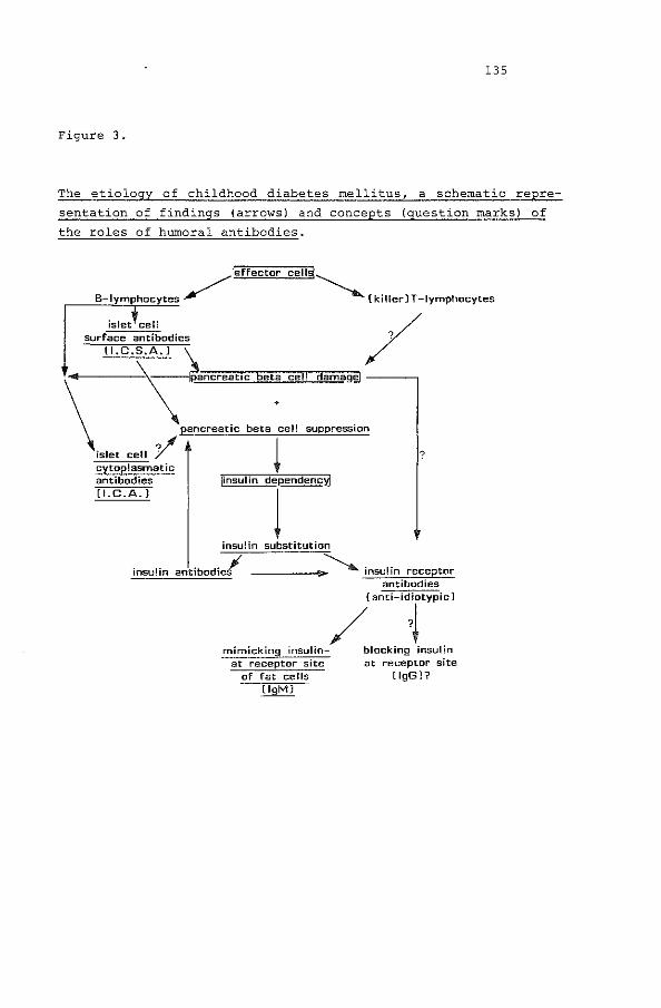
135
Figure 3.
The etiology of childhood diabetes mellitus, a schematic repre
sentation of findings (arrows) and concepts (question marks) of
the roles of humoral antibodies.
~le'fFffe~c£i;to~cuc~e~1[1@~~
B-lymphocytes / (killer]T-Iymphocytes
islet cell surface antibodies
{I.C.S.A.] / \ ~e~a~n~c!c~e~aito~c;:]~e!]t~a!::£c~eill:J!a~millia~g~e ------,
pancreatic beta cell suppression \ ~s~l~e~t~c~e~ll~~ cytoplasmatic antibodies (I.C.A.)
insulin
~ linsulin de~endencYJ
1 insulin substitution
. . jl anttbod1es ------~ insulin receptor
antibodies
/[anti id;rypicl
mimicking insulin- blocking insulin at receptor site
of fat cells [lgM)
at receptor site (lgGl?
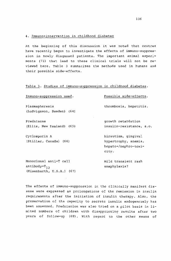
136
4. Immune-intervention in childhood diabetes
At the beginning of this discussion it was noted that centres
have recently begun to investigate the effects of immuno-suppres
sion in newly diagnosed patients. The important animal experi
ments (71) that lead to these clinical trials will not be re
viewed here. Table 3 summarizes the methods used in humans and
their possible side-effects.
Table 3. Studies of immuno-suppression in childhood diabetes.
Immuno-suppression used.
Plasmapheresis
(Ludvigsson, Sweden) (64)
Prednisone
(Ellis, New Zealand) {65)
Cyclosporin A
(Stiller, Canada) (6 6)
Monoclonal anti-T cell
antibody-T12 (Eisenbarth, U.S.A.) (67)
Possible side-effects.
thrombosis, hepatitis.
growth retardation
insulin-resistance, a.o.
hirsutism, gingival
hypertrophy, anemia,
hepato-/nephro-toxi
city.
mild transient rash
anaphylaxis?
The effects of immuno-suppression in the clinically manifest dis
ease were expressed as prolongations of the remission in insulin
requirements after the initiation of insulin therapy. Also, the
preservation of the capacity to secrete insulin endogenously has
been assessed. Prednisolon was also tried on a pilot basis in li
mited numbers of children with disappointing results after two
years of follow-up (68). With regard to the other means of

137
immuno-suppression abovementioned, formal studies are underway
and the results mentioned here are preliminary. It should also be
mentioned most trials involve older children and young adults.
Stiller et al (66) used cyclosporin in a dosage of 10 mg per kg
per day, adjusted to serum levels ranging from 100-200 ng per rnl
and found considerable side-effects. At the time of last observa
tion {48-114 days, the cyclosporin initiated within 6 weeks after
diagnosis) three of seven patients were receiving no insulin.
In another group of patients, initiated on cyclosporin two or
more months after diagnosis, none had a reduction in insulin do
sage of 50% or more and there was no significant increase in
C-peptide levels.
Eisenbarth et al. (67) using monoclonal T12 antiserum in a simi
lar study, concluded that none of the pilot studies on immunothe
rapy of type-1-diabetes so far gave clear beneficial results.
In other studies (without applying immunosuppression) intensi
fied conventional treatment during the first year of disease was
associated with prolongation of remission-phase (14), although
not as markedly as seen in the patients referred to above.
At present, the preliminary findings on the effects of immuno
suppression raise the following questions:
1. why did some patients appear to react much more favorably than
others?
2. to what extend was the prolongation of the remission-phase due
to the immunosuppression applied?
3. what relevant humoral antibodies were suppressed?
Some pathophysiological difficulties interpreting the clinical
phenomenon of a remission-phase were discussed in the previous
sections.
It seems to the author these important questions can only be
answered clinically by previous stratification of patients, with
regard to already known characteristics of remission-phases in
sub-groups of patients. Such stratification would include con
trolling for the following factors:
1. the age of the patients as young children {below age 5 in par
ticular) as these very young children have shorter remission
phases (if any) compared to their older peers on conventional
138
treatment (69);
2. the quality of metabolic control during the follow-up of
trials;
3, velocity and ferocity of pancreatic beta-cell loss prior to
diagnosis.
The weight of the first two variables, with respect to their in
fluence on remission-phases, may be established by age selection
and by choosing optimal treatment-modalities in defined age
groups. Having done so, it may be less difficult to evaluate ef
fects on remission-phases by virtue of immunosuppression,
The third factor is dictated by circumstances prevailing on re
ferral of a newly diagnosed child and can only be controlled for
by screening programs of pre-diabetic states of childhood diabe
tes. One might envisage the identification of children at risk by
"refined" HLA-typing 1 defined islet-cell antibodies 1 followed by
intensive treatment alone or in combination with immunosuppres
sive therapy in a randomized prospective study, as soon as "early
phase" glucose-intolerance (70) developed. The ethical legitimi
city of such a trial, that has potential therapeutic consequen
ces, should be discussed with platforms of parties involved, no
tably ethical committees of medical faculties and patient orga
nizations.
In view of the daunting prospects of children suffering from the
disease, it is hoped for that such trials can be considered in
the not too distant future: along with expected progress in
screening methodology, improved specificity of immune-interven
tion and defined means to assess its effects.
139
6. Summary
The findings reported in this chapter may be summarized as fol
lows:
1. The strong association between childhood diabetes and HLA-DR3/
HLA-DR4 is confirmed.
2. The recurrence risk for a sib of a diabetic child being HLA
identical to that child is about 12%.
3. The susceptibility to childhood diabetes may be mediated by
two or more loci, one associated with HLA-B8/HLA-DR3, the
other with HLA-Bl5/HLA-DR4, in third order linkage disequili
brium.
4. Generally, childhood diabetes is not associated with Gm-mar
kers.
5, Absence of cytoplasrnatic islet-cell antibodies at the clinical
onset of the disease occurs in 20% and absence of either
HLA-DR3 or HLA-DR4 in less than 10%. Absence of these immuno
logic parameters may be found in unusual syndromes with child
hood insulin-dependency.
6. The accuracy of islet-cell antibodies, measured by immunoflu
orescence, to detect pre-diabetic states in childhood is pro
bably high, although their pathogenetic significance is un
clear.
7. Restriction to single IgG-subclasses in islet-cell fluores
cence suggests linkage to Gm-markers for the small subgroup of
diabetic children in which this was found, with regard to the
expression of this auto-antibody.
8. Besides islet-cell autoimmunity, autoimmunity towards fat-cell
insulin receptors may develop spontaneously.
9. IgM insulin-receptor antibodies arising after treatment with
insulin may be associated with obesity.
10. Before the consideration of "immunoprevention" of childhood
diabetes, more needs to be known about the pathophysiology and
the immunology of the disease and disease states in which im
mune-intervention is applied.
140
References to Chapter I
1. Frank, L.L.
Diabetes mellitus in the texts of old Hindu medicine.
Am. J, Gasteroenterol. ~. 76, 1957,
2, Papaspyoros, M.S.
The history of diabetes mellitus.
Thieme, Stuttgart, 1964.
3. Keen, H., Jarrett, R.J., Alberti, K.G.M.M.
Diabetes mellitus a new look at diagnostic criteria.
Diabetologia ~. 283-285, 1979.
4. Rosenbloom, A.L., Kohrman, H., Sperling, M.
Classification and diagnosis of diabetes mellitus in children
and adolescents.
J. of Paed. ~, 320-323, 1981.
5. Cudworth, A.G., Wolf, E.
The genetic susceptibility to type 1 (insulin dependent) dia
betes mellitus in Clinics of Endocrinology and Metabolism,
Johnston D.G., Alberti, K.G.M.M., Eds. W.B. Saunders, London
ll· 389-409, 1982.
6. Rimoin, D.L.
Genetic syndromes associated with glucose intolerance. In: The
genetics of diabetes mellitus, Creutzfeld, W., KObberling, J.,
Neel, J.V., Eds., Springer, New York, il, 64, 1976.
7. Craighead, J.E.
The role of viruses in the pathogenesis of pancreatic disease
and diabetes mellitus.
Prog. Med. Virol. ~, 161-170, 1975.
141
8. Notkins, A.L.
The causes of diabetes.
Scientific American 56-67, nov. 1979.
9. Cahill, G.F., McDivitt, H.O.
Insulin-dependent diabetes mellitus: the initial lesion.
New Engl. J. Med. 304, 1454-1465, 1981.
10. Irvine, W.J., Gray, R.S., MacCallum, C.J.
Pancreatic-islet cell antibody as a marker for as~ptomatic
and latent diabetes and prediabetes.
Lancet l• 1097-1102, 1976.
11. Gepts, W. and Le Compte, P.M.
The pancreatic islets in diabetes.
Am. J. Medicine 70: 101-115, 1981.
12. Martin, J.M. and Lacy, P.E.
The prediabetic period in partially pancreatictomized rats.
Diabetes ll• 238-242, 1963.
13. Bonnevie-Nielsen, V., Streffes, M.W. and Lernrnark, A.
A major loss in islet cell mass and beta-cell function pre
cedes hyperglycemia in mice given multiple low doses of strep
tozotocin.
Diabetes~, 424-429, 1981.
14. Ludvigsson, J., Reding, L.G.
C-peptide in diabetic children after stimulation with glucagon
compared with fasting C-peptide levels in non-diabetic child
ren. Acta Endocrinol. ~, 364-367, 1977.
15. Madsbad, S.
Effect of strict blood-glucose control on residual beta-cell
function in insulin-dependent diabetics.
Diabetologia ~, 530-534, 1981.

16. Endocrine pathology, general and structural.
J.M.B. Bloomsworth, Ed., p. 557-586.
2nd Ed., Williams and Wilkins, Baltimore, 1982.
17. Orci, L.
Macro- and micro-domains in the endocrine pancreas.
Banting Lecture 1981, Diabetes l!' 538-566, 1981.
18. Rahur, J., Wallen, J. and Henquin, J.C.
142
Cell populations in the endocrine pancreas of human neonates
and infants.
Diabetologia ~, 540-546, 1981.
19. Von Meyenburg, H.
Ueber "insulitis" bei diabetes.
Schweiz. Med. Wochenschrift ~. 554-557, 1940.
20. Gepts, w. In: The Diabetic Pancreas.
Volk, B.W., Wellman, K.F., Eds.
Plenum Press, N.Y., 1977, p. 325-365.
21. Rosenbloom, A.L., Wheeler, L., Bianchi, K., Chiu, F.T.,
Tiwary, C.M. and Crgic, A.
Age adjusted analysis of insulin responses during normal and
abnormal oral glucose tolerance tests in children and adoles
cents.
Diabetes~~ 820-828, 1975.
22. Rosenbloom, A.L., Hunt, s.s., Rosenbloom, E.K. and McLaren,
N.K.
Ten year prognosis of impaired glucose tolerance in siblings
of patients with insulin-dependent diabetes.
Diabetes ll• 385-387, 1982.

143
23. Drash, A.
Childhood Diabetes. In: "Clinical Diabetes: Modern Manage
ment".
S. Podolsky Ed., Appleton-Century-Crofts, New York 1980,
p. 361-402, 1980.
24. Hoekstra, J.B.L.
C-peptide, clinical and biochemical studies.
Thesis, Utrecht, 1982.
25. Brogr:en, C.H., Lernmark, A.
Islet cell antibodies. In: Clinics of Endocrinology and Meta
bolism, D.G. Johnston, K.G.M.M. Alberti Eds.,
W.B. Saunders, London l!, 409-431, 1982.
26. Marner, B., Lernmark, A., Nerup, J., Molenaar, J.L., Tuk,
C.W., Bruining, G.J.
Analysis of islet cell antibodies on sections of human pan
creas.
Diabetologia 25: 93-96, 1983.
27. Lernmark, Haggl6f, B., Freedman, Z.R., Irvine, W.J., Ludvigs
son, J. and Holmgren, G.
A prospective analysis of antibodies reactive with pancreatic
islet cells in insulin dependent diabetic children.
Diabetologia ~, 471-474, 1981.
28. Gorsuch, A.N., Spencer, K.M., Lister, J., McNally, J.M., Dean,
B.M., Bottazzo, G.F., Cudworth, A.G.
Evidence for a long prediabetic period in type 1 diabetes mel
litus.
Lancet~, 1363-1365, 1981.
29. Dean, B.M., Bottazzo, G.F., Cudworth, A.G.
IgG subclass distribution in organ specific auto-antibodies.
The relationship to complement fixing ability.
Clin. Immunol. ~~ 61-66, 1983.

144
30. Beek, O.E.
Distribution of virus antibody activity among human IgG sub
classes.
Clin. Exp. Immunol. !l, 626-632, 1981.
31. Uno, H., Sasazuki, T., Tarnai, H. and Matsumoto, H.
32.
Two major genes, linked to HLA and Gm, control susceptibility
to Graves' disease.
Nature 292, 768-770, 1981.
Dr ash, A.L., Weber, B.' Zick, R.' Sires, J.M., Gillet, P.'
Krzisnic, c. Remission in juvenile diabetes.
Bulletin of the International Study Group on Diabetes in
Children and Adolescents. 4' 4-17, 1980.
33. Deckert, T.
The influence of supervision and endogenous insulin secretion
on the course of insulin dependent diabetes mellitus.
Acta Endocr. (Kbh) 2_! (suppl. 238): 31-38: 1980.
34. Lutterman, J.
Labiele en stabiele insuline afhankelijke (type 1) diabetes
mellitus.
Thesis (Nijmegen) 1982.
35. Madsbad s., Alberti, K.G.M.M., Burrin J.M., Faber, O.K.,
Krarup, T., Regur, L.
Role of residual secretion in protecting against keto-acidosis
in insulin dependent diabetes.
Br. Med. J. 2: 1257-1259: 1979.
36. Shechter, Y., Maron, R., Elias, D., Cohen, I.R.
Autoantibodies to the insulin receptor develop as anti-idio
types in mice immunized with insulin.
Science 216, 542-545, 1982.

37. Jerne, N.K.
Towards a network theory of the immune system.
Annales d'Immunologie 12SC, 373-389, 1974.
38. Tattersall, R.B. and Pyke, D.A.
Diabetes in twins.
Lancet~. 1120-1124, 1972.
39. Pyke, D.A.
Diabetes: the genetic connections.
Diabetologia l2• 333-343, 1979,
145
40. Platz, P., Jacobsen, B.K., Marling, M., Nerup, J. et al.
HLA-D and -DR antigens in genetic analysis of insulin
dependent diabetes mellitus.
Diabetologia ~. 108-115, 1981.
41. Nerup, J.
Etiology and pathogenesis of insulin dependent diabetes melli
tus: present view and future developments. In: Etiology and
pathogenesis of insulin-dependent diabetes mellitus.
J.M. Martin, R.M, Ehrlich, F.J. Holland, Eds. Raven Press,
1981, page 282.
42. de Jongh, B.M., Termijtelen, A., Bruining, G.J., de Vries,
R.R.P., van Rood, J.J.
Relation of insulin dependent diabetes mellitus (IDD) and the
HLA-linked SB-system.
Tissue Antigens 1984, in press.
43. Owerbach, D., Lernmark, A., Platz, P. Ryder, L.P., Rask, L.,
Peterson, P.A., Ludvigsson, J.
HLA-D region beta-chain DNA endonuclease fragments differ be
tween HLA-DR identical healthy and insulin-dependent diabetic
individuals.
Nature 303, 815-817, 1983.

146
44. Wake, T.C., Long, E.O., Strubin, M., Cross, M., Accolla,
R .. S., Carrel, S, Mach, B.
Isolation of eDNA clones encoding HLA-DR Alpha-chains.
Proc. Natl. Ac. Sci. (USA) ~, 6979-6983, 1982.
45. Accolla, R.S., Cross, M., Carrel, S., Corte, E.
Distinct forms of both Alpha-subunit and Beta-subunit are
present in the human Ia molecular pool.
Prod. Natl. Ac. Sci (USA) ~, 4549-4551, 1981.
46. Steinberg, A.D.
Aetiology of autoimmunity.
Nature 304, 20, 1983.
47. Lernmark, A., Freedman, Z.R., Hofman, C., Rubenstein, A.H.,
Steiner, D.F., Jackson, R.L., Winter, R.J. and Traisman, H.S.
Islet cell surface antibodies in juvenile diabetes mellitus.
N, Engl. J. Med. 299, 375-380, 1978.
48. Pozzilli, P., Zuccarini, 0., Iavicoli, M., Andreani, D., Sen
si, M., Spencer, K.M., Bottazzo, G.F. et al.
Monoclonal antibodies defined abnormalities of T-lymphocytes
in type 1 (insulin-dependent) diabetes.
Diabetes 32: 91-95: 1983,
49. Olsson, L., Honsik, C.J.
Specific binding of human insulin to a determinant associated
with, or identical to, a monomeric part of HLA-A, -B, -C.
Science, 1984: in press
50. Gorsuch, A.N., Spencer, K.M., Lister, J., McNally, J.M., Dean,
B.M., Bottazzo, G.F., Cudworth, A.G.
Evidence for a long prediabetic period in type-1-diabetes
mellitus.
Lancet ~~ 1363-1365, 1981.

147
51. Gorsuch, A.N., Spencer, K.M., Lister, J., Wolf, E., Bottazzo,
G.F., Cudworth, A.G.
Can future type 1 diabetes be predicted?
Diabetes l!, 862-866, 1982.
52. Wagener, O.K., Sacks, J.M., Laporte, R.E., MacGregor, J.M.
The Pittsburgh study of insulin dependent diabetes mellitus:
risk among relatives of IDDM.
Diabetes l!• 136-144, 1982.
53, Helmke, A., Otten, A., Willems, W.
Islet cell antibodies in children with mumps infection.
Lancet~. 211-212, 1980.
54. Baekkeskov, S. , H!hriis Nilsen, J. , Marner, B., Bilde, T. ,
Ludvigsson, J., Lernrnark, A.
Autoantibodies in newly diagnosed diabetic
immunoprecipitate pancreatic islet cell proteins.
Nature 298, 167-169, 1982.
children
55. Haspel, M.V., Onodera, T., Prabhakar, B.S., McClintock, P.R.,
Essani, K., Ray, U.R., Yagihashi, S., Notkins, A.L.
Multiple organ-reactive monoclonal autoantibodies.
Nature 304, 73-76, 1983.
56. Kanatsuna, T., Freedman, Z.R., Rubenstein, A.H., Steiner, D.F.
Block in insulin release from column perifused pancreatic B
cells induced by surface antibodies and complement.
Diabetes iQ, 231-234, 1981.
57. Kanatsuna, T., Baekkeskov, S., Lernmark, A., Ludvigsson, J.
Immunoglobulin from insulin dependent children inhibits
glucose induced insulin release.
Diabetes ~. 520-525, 1983,
148
58. Reeves, W.G.
Insulin antibody determination: theoretical and practical
considerations.
Diabetologia ~. 399-403, 1983.
59. Roy B., Chou, M.C.Y., Field, J.B.
Time-action characteristics of regular and MPH insulin in
insulin treated diabetes
J. Clin. Endocr. Met. 50: 475-479: 1980.
60. Ludvigsson, J.
Insulin antibodies in diabetic children treated with mono
component porcine insulin from the onset: relationship to
B-cell function and partial remission. Diabetologia ~: 138-142: 1984.
61. Kahn, C.R., Baird 1 K., Flier, J.S.
Effects of autoantibodies to the insulin receptor on isolated
adipocytes. Studies of insulin binding and insulin action.
J. Clin. Invest. 60: 1094-1106: 1977.
62. Khoker, M.A., Dandona, P., Janah, S., Coulston, G.L.
Insulin-like stimulatory effect of human immunoglobulin G on
adipocyte lipogenesis.
Diabetes 30: 1068-1071: 1981.
63. Khoker, M.A., Taylor, A.M., Dandona, P.
Insulin-like effect of human immunoglobulins on adipocyte tis
sue in immunology in Diabetes 84, M. Iavicoli Ed., Novo Farma
ceutici Italia, 1984, page 62.
64. Ludvigsson, J.
Partial preservation of beta-cell function by plasrnaferesis.
International Bulletin of the I.S.G.D. 5: 22-23: 1981.

149
65. Elliot, R.B., Crossley, J.R., Berryman, G.C., James, A.G.
Partial preservation of pancreatic B-cell function in children
with diabetes.
Lancet 2: 1-4: 1981.
66. Stiller, C.R., Laupauris, A., Dupre, J. et al.
Cyclosporin for treatment of early Type 1 diabetes: prelimi
nary results.
New Engl. J. Med. 308, 1255-1256, 1983.
67. Eisenbarth, G.S.
Immunotherapy of type 1 diabetes.
Diabetes Care 6: 521-522: 1983.
68. Ludvigsson, J.
Prednisolon in the initial treatment of insulin dependent dia
betes mellitus in Immunology in Diabetes 84, M. Iavicoli Ed.,
Novo Far.maceutici Italia, 1984, page 69.
69. Drash, A., Laporte, R., Becker, D.J., Singh, B., Fisbein, H.,
Goldstein, D.
The natural history of diabetes mellitus in children! insulin
requirements during the initial two years.
Bulletin of the I.S.G.D. 4: 4-7: 1980.
70. Srikanka, S., Ganda, O.P., Eisenbarth G.S., Soeldner, J.S.
Islet cell antibodies and beta cell function in monozygotic
triplets and twins initially discordant for type 1 diabetes.
New Engl. J. Med. 308: 322-325: 1983.
71. Rossini, A.A., Mordes, J.P., Like, A.A.
Animal models of insulin-dependent diabetes mellitus in Immu
nology of Diabetes, D. Andreani, U. di Mario, K.F. Federlin,
L.G. Heding Eds., Kimpton Medical Publ. 1984, 35-44.

150
CHAPTER II EPIDEMIOLOGY
"for Art and Science cannot exist
§1. Introduction
but in minutely organized Particulars"
(Blake)
The fourth year medical student lectures at the Erasmus Universi
ty, taught by Prof. Dr. H.A. Valkenburg and his associates contain
a quotation from Francis (1954):
"Epidemiology provides a third dimension to the understanding of
disease by creating awareness of the nature of the environment in
which disability arises, of the factors in the community which
contribute to its causation and, in turn, its effect upon the
community".
The retrospective investigation of the incidence of childhood dia
betes in the Netherlands (paragraph 2), was the result of a colla
borative effort of the "Working Committee on the Epidemiology of
childhood diabetes" established in 1978 through the initiative of
Prof. Dr, W. H. H, Tegelaers, chairman of the Department of Pedia
trics at the University of Amsterdam Medical School.*
With regard to the methods of epidemiologic studies it is interes
ting to review the minutes of the committee: first, the testing
for urinary glucose in healthy school children was considered as a
method to detect children in which insulin-deficiency might be de~
veloping. A pilot-study was carried out by two school-physicians
M, Wirix-Nagelsmit and J.M. van der Ros, demonstrating nil positi
ves among 450 healthy children.
* The members to the working cannittee, other than the authors of
the paper reprinted, in alphabetic order, were:
Prof. Dr. J.L. van den Brande, Dr. H.F. Dankrreyer, Dr. M.H.
Gons, Drs. M. Jansen, Prof. Dr. G.A. de Jonge, Prof. Dr.
W.H.H. Tegelaers, Dr. H.P. Verbrugge, Drs. A.A.M. Vloemans
and Drs. F. Wafelbakker.

151
This approach was prompted by the clinical observation, impres
sion rather, that relatively more children, compared to former
years, presented with asymptomatic glucosuria, when first seen
clinically, than with keto-acidosis. Indeed, in our own hospital
we found in retrospect 12 (10-20) percent of 55 newly diagnosed
children with diabetes mellitus presented with keto-acidosis
(blood pH< 7.2.) between 1975-1980, whereas 36 (30-40) percent
of 71 newly diagnosed children did so between 1970-1975 (1}.
The rejection of urinary glucose as marker for beginning diabetes
of childhood coincided with the formal acceptance (1979) of dia
betes-type-2 being different from diabetes-type-! by authorities
as the NIH and the WHO: in diabetes-type-2 glucosuria may be a
marker by virtue of its slow onset; in diabetes-type-1 (childhood
diabetes} the clinical onset is much more sudden, so that glycos
uria would only be present weeks or months at most before its on
set (Chapter I - paragraph 1).
Subsequently this approach was abandoned and a retrospective ana
lysis of newly diagnosed children was considered by the committee
of which the author of this thesis had the honour to become
chairman.
The first question raised was what criteria should be used for
diabetes-type-1 in childhood.
In the epidemiologic sense, it is an advantage of diabetes in
childhood that patients are either insulin dependent or not. Tem
porary use of insulin is occasionally seen in neonates (2), but
such cases can be excluded by checking on reported cases of neo
natal diabetes, expected to be rare. In contrast to type-2-diabe
tes, the incidental administration of insulin, for example around
operations in elderly patients, does not occur in youth.
Mild glucose intolerance, usually not accompanied by glucosuria,
may be seen in obese (pre-) adolescents, but these children never
require insulin (3) , nor do most of the genetically determined
mild cases referred to in paragraph 1 of chapter I.
It is of course possible that children with mild glucose-intole
rance are inadvertently treated with insulin. In such case one
would suppose that either hypoglycemia would develop or that such
low dosages were needed that specialists - who invariably treat
152
children with insulin dependency in the Netherlands - would doubt
the necessity thereof. In incidence studies, assessing newly
diagnosed children retrospectively only, the contribution of such
cases was felt to be low, if any.
One exception should be made: the insulin-dependency that concurs
in up to 8 percent of the patients with cystic fibrosis (4). In
terestingly, the HLA-DR3 and HLA-DR4 haplotypes predominated mar
kedly in the cystic fibrosis patients on insulin over cystic fi
brosis patients without fasting hyperglycemia (5). Up to 8 per
cent of all living cystic fibrosis patients under the age of 19
years in the Netherlands would represent 80 or less cases (Dr.
L.P. ten Kate, Institute for Anthropogenetics, University of Gro
ningen, personal communication). Compared to an expected preva
lence of 4,000 for all minors under age 19 with insulin-dependen
cy this subgroup of children with cystic fibrosis would represent
too small a fraction to be considered separately.
With these considerations in mind it was felt "insulin-dependen
cy" as a criterion would encomprise "classic" type-1-diabetic
children. The committee decided to conduct a retrospective survey
of the incidence of childhood diabetes under age 19 all over the
Netherlands, using this simple, but practical criterion, by a
questionnaire to all dutch pediatricians and internists.
The second question raised was how to ascertain the data obtained
from a retrospective survey among dutch pediatricians and inter
nists.
Methods used in Scandinavian countries (drug-registry or hospi
tal-file checking) were either impossible or very difficult in
the dutch health-care system. On the other hand the Netherlands
are unique in having the largest relative membership of the Dutch
Diabetes Association (Diabetes Vereniging Nederland- D.V.N.) in
the world, with 35.000 members in a country of 14.3 million inha
bitants. With the help of the medical advisor to the D.V.N., Dr.
H.F. Dankmeyer, it was decided to separately conduct a retrospec
tive survey on the incidence of childhood diabetes by a question
naire to the members of the D.V.N., as an ascertainment procedu
re.
The report of the investigation, conducted by the Netherlands In-

153
stitute for Preventive Health Care under direction of Dr. G.J.
Vaandrager, is reprinted in the next paragraph.
Other data forthcoming of this epidemiologic study were published
in a monograph in the dutch language ("De incidentie van insuli
ne-afhankelijke diabetes mellitus bij 0-19 jarigen in Nederland
1978-1980" - G.J. Vaandrager, F.J. Veenhof, M.M. van der Klaauw
published by the Nederlands Insti tuut voor Preventieve Gezond
heidszorg (NIPG-TNO) Leiden, 1984).

154
§2, Addendum
The incidence of childhood diabetes in the Netherlands.
A decrease from north to south over north-western Europe?
1 . . 2 1 3 G.J. Vaandrager G.J. Bru1n1ng F.J. Veenhof , N.M. Drayer
Netherlands Institute for Preventive Healthcare/ TNO, Leiden1
,
Dept. of Pediatrics, Division of Endocrinology, Erasmus Universi
ty and University Hospital Rotterdam/Sophia Children's Hospital,
Rotterdam2
, Dept. of Pediatrics, Endocrine Unit, State Universi
ty of Groningen, the Netherlands 3 •
Accepted for publication by Diabetologia (1984), reproduced with
permission.
Introduction
In the past decade incidence-rates of childhood diabetes (0-14 or
0-19 years of age) were determined in all Scandinavian countries
(1-4) and parts of the United Kingdom (S). In these studies high
percentages of ascertainment were possible by virtue of the
health-care system: drug-registry and hospital files could be
used. In the Netherlands there is no nationalized health-care
system, therefore such methods for ascertainment cannot be used.
However there is an effective registration system. We decided to
conduct a retrospective survey on the incidence of insulin depen
dency among all diabetic children in the Netherlands from 0-19
years of age, during 1978, 1979 and 1980.
The Netherlands national register counted 4.46 million youngsters
from 0-19 years at that time, of whom 6.1 percent were born to
first generation immigrants. In the health-care system only pae
diatricians and internists would treat insulin dependency in mi
nors and both types of specialists act as registered consultants.

155
Methods
A questionnaire was sent to all consultant paediatricians and
consultant internists to obtain data on newly diagnosed children
between 1978 and 1981. The questionnaire contained only questions
regarding the initials of the child's name, sex, age, date of the
first insulin injection and residence at that time (no attempt
was made to identify children of immigrants or of non-caucasoid
background). The same information was obtained separately from
the Dutch Diabetes Association, to ascertain the data obtained
from the questionnaire sent to the specialists.
The questionnaire was sent to the 660 consultant internists and
the 263 consultant paediatricians in the 181 general hospitals
and to a contact person in each of the 8 internal medicine and 7
paediatric university clinicsf including residential care cen
ters. After one or two calls 100 percent of the paediatricians
and residential care centres responded between February and Sep
tember 1981.
Patient-members of the Diabetes Association were requested to
submit the same information after a call to do so in their month
ly periodical, sent to all 35.000 members, 2.44 per 1000 inhabi
tants, with a range from 1.87-3.26 per thousand over the eleven
provinces in the Netherlands. The membership of the Diabetes As
sociation is high as members can obtain treatment materials for
retail prices at local offices of the Association. The request to
the members was published at a time point that 80 percent of the
specialist responses had been obtained. In addition to the above
mentioned questions the members were also asked who was their
child's doctor: all answers contained either a consultant paedia
trician or a consultant internist, confirming the premise that
all diabetic children were treated by either one of these consul
tant specialists. Also, all treating doctors thus reported were
present on our mailing lists.
The paediatricians reported 856 children, the internists 415, to
gether 1271 youngsters from 0-19 years of age in whom insulin
substitution was initiated and continued in 1978-1980.

156
From the Diabetes Association 305 complete responses were obtai
ned of which 268 could be found also in the specialist's respon
ses. This implied a reporting percentage of 21 from the Diabetes
Association and an ascertainment percentage of 88 of the total
number of patients found by the questionnaire among the specia
lists. For 1978 the ascertainment percentage was 85 percent, for
1979 84 percent and for 1980 93 percent, not statistically diffe
rent. The method used for the estimation of the corrected number
of new patients and its confidence limites {expressed as s 0 in
table 2) was based on the capture-recapture census described by
Bishop et al. (6) and by Sekar and Deming (7). A premise with
this method is that the response of the patients/members of the
Diabetes Association was independent of that of the specialists.
The homogeneity of each file (physicians and patient/members of
the Diabetes Association) was tested by comparing ascertainment
percentages found for answers with regard to age, sex, kind of
specialist, grade of urbanization (8) of living place, month of
the first insulin injection and province of living at that time.
The two-sided chi-square test was used, no differences were found
below the 5 percent level except for "age" and "kind of specia
list". If however a distinction was made between paediatricians
and internists this relation was no longer present: both the de
gree of completeness of the files and the age of the patients
were related to the factor "kind of specialist". The ascertain
ment of the paediatricians 1 data (94 percent) was considerably
higher than that of the internists (75 percent) (table 1). For
the age-groups 0-4, 5-9, 10-14 and 15-19 the paediatricians as
certainment percentages were respectively 98, 90, 97 and 100 per
cent, of the internists -, 67, 70 and 77 percent. The material
was split-up according to the two subgroups of specialists. The
incidence rate was then calculated: the two sets of data were
multiplied by the reciproce of their ascertainment percentages
and subsequently added. Statistical calculations for geographic
distributions and for degree of urbanization were done by the
two-sided chi-square test, deviations from uniform distribution
over the months of the year were tested by a modification of the

157
Kolmogorov/Smirnov test (9). The level of significance chosen was
p < o.os.
Figure 1
annual incidence per 100.000
20
16
12
B
0
0
Results
... · .... ... ..
2 6 B
········· ... cJ' n-682
·. · .. ·
9 n-589
10 12 16 16 20 age at first insulin injection
Figure 1 depicts the incidence by age at the first insulin injec
tion, separated for the two sexes. The male to female sex ratio's
in the different age groups were: 0-4 years: 1.18, S-9: 1.18,
10-14: 0.94, 15-19: 1.34, for 0-14: 1.05 and 0-19: 1.12.

158
Table 1 shows the ascertainment percentages were 94 percent for
paediatricians and 75 percent for internists, while this percen
tage decreased also with age: a significant (p ~0.02) progressi
ve decrease was found between five year intervals.
Table 1
Insulin dependency reported by type of specialist, patient/mem
bers of the Diabetes Association, age in years and ascertainment
percentage.
Specialist Patient/ Both Ascertainment
members files percentage
Paediatrician 856 206 194 947p<0.001
Internist 415 99 74 75
0- 4 179 50 49 987p<0.02
5- 9 323 90 80 89
10-14 463 99 87 887
p<O .02
15-19 306 66 52 79
Table 2 indicates the incidence rates found with a distinction
between sex and age, and the standard deviation (SD) of the me
thod of estimation. The ascertainment corrected incidence was
10.95/100.000 per year for 0-19 years and 11.10/100.000 per year
for 0-14 years. The 95 percent confidence intervals were
10.47-11.43 and 10.64-11.56 respectively for these age groups.

159
Table 2. The three-year incidence and the incidence per 100.000
inhabitants per year according to sex and to age group
(1978-1980); after correction for ascertainment.
sex
age
(years)
boys
0- 4
5- 9
10-14
15-19
0-19
girls
0- 4
5- 9
10-14
15-19
0-19
number s0
102
202
261
228
790
82
164
266
163
676
3.4
11.9
11.2
16.5
22.8
(-) 1
5.9
15.9
16.8
24.3
1 Ascertainment 100 %
mean population incidence per
per year (Central 100.000/yr
Bureau for Sta-
tistics, 1979-1982)
456.327
564.636
625.878
633.133
2.279.974
434.746
539.960
597.359
605.746
2.177.811
7.45
11.91
13.88
11.98
11.55
6.29
10.11
14.84
8.95
10.35
0.25
0.70
0,60
0.87
0,33
(-)1
0.36
0.89
0.92
0.37
The range of the differences between the Netherlands provinces
was 8.9-13.7/100.000 children from 0-19 years of age. These dif
ferences were not statistically significant.
"Countryside", "urbanized municipalities" and "large municipali
ties" had incidence rates of respectively 11.61, 10.52 and
11.14/100.000 per year, not significantly different.
The distribution of the months of the year in which the first in
sulin injection was administered revealed no statistically signi
ficant differences for 0-4 nor for 5-9 year old children. In the

160
age groups 10-14 and 15-19 years a seasonal influence was present
with the incidence rates higher in the winter months than in the
summer months (p < 0. 05 respectively p < 0. 01, for these age
groups) • When the seasonal varation was studied for the three
years separately, these differences were significant for 1978 and
1980, but not for 1979.
Discussion
The annual incidence found in the present study over the whole of
the Netherlands (14.3 million inhabitants) was lower than that
found in any other ascertained survey in western Europe conducted
in the same time period. The trends in age of initiation of insu
lin treatment and the distribution over the sexes were very similar to those found in other studies. The present survey found no
seasonal variation in the onset of clinical diabetes in children
younger than 10 years of age. No significant differences in inci
dence were found with regard to the distribution over the provin
ces of the Netherlands, neither in the degree of urbanization.
This latter finding is perhaps not surprizing as truely rural
areas are rare in the densely populated Netherlands.
Given the limits of the Netherlands health-care system to employ
drug-registry for ascertaining the data, the capture-recapture
census method was chosen to calculate the confidence limits of
the incidence found by two separate questionnaires. The advantage
being that the largest childhood population so far could be exa
mined relatively simply, the disadvantage being that an ascer
tainment percentage of only 79 percent was found for the children
aged 15-19 years. For children younger than 14 years of age al
most all responses were obtained before the survey among members
of the Dutch Diabetes Association was initiated an the ascertain
ment percentage for that age-cohort was 94 percent. It is likely
that the difference in ascertainment percentages found between
paediatricians and internists was associated with the much higher
"density" of newly diagnosed children seen by paediatricians than
by internists. Over the three years of the study 3.25 cases were
per reporting paediatrician (856/263) and 0. 62 cases per inter-

161
nist (415/660).
It was found that 837 newly diagnosed children from 0-14 years
were reported by consultant paediatricians (94 percent) and 287
children from 14-19 years by consultant internists (87 percent).
These percentages indicate a referral pattern sharply distinct by
the age of the newly diagnosed diabetic children in the dutch
health-care system.
The incidence found for children 0-14 years of age was almost
threefold lower than that found in Finland (1) almost two-fold
than in Sweden (2) or Norway {3) and also lower than that repor
ted from Denmark, excepting children from 0-5 years old {4), As
emphasized by Christau et al. (10) it is very difficult to compa
re incidence rates even with high percentages of ascertainment as
long as different methods were used. Yet, it is hard to conceive
the magnitude of these differences would be only due to differen
ces in ascertainment methods. Genetic differences are not a like
ly explanation as childhood diabetes was associated with the same
genotypes of the major histocompatibility complex in the Nether
lands as in other north-western European countries (11). Over
time, by comparing yearly incidence rates, remarkable changes in
incidence rates have been noticed as well as two-fold differences
between regions of the same country have been reported (1-5).
However the numbers of newly diagnosed children involved in such
breakdowns were small (10).
In north-western European countries the incidence of childhood
diabetes (0-14 years) appears to have increased over the past de
cades (1, 5, 12). In the Netherlands the prevalence of diabetes
among army conscripts at age 18 increased gradually from 0.99 per
thousand in 1960 to 1.72 per thousand in 1980, indicating a simi
lar trend. The incidence found in the present survey adds another
suggestion: that of a possible north-south gradient over north
western Europe. This suggestion is by no means proven but rein
forces the importance of exogenous factors involved in the clini
cal onset of childhood diabetes.

162
Acknowledgements
We thank the members of the Dutch Diabetes Association (DVN) for
their enthousiastic cooperation and the paediatricians and the
internists of the Netherlands for their time and help. The Minis
try of Health (WVC) gave valuable advise and the division for me
dical examinations of army conscripts of the Ministry of Defense
for their data 1960-1979. The support of the Stichting Diabetes
Research Fonds and of Nordisk Nederland, part of Nordisk Insulin
Laboratories, Denmark, is gratefully acknowledged.
Literature
1. Reunanen A., Akerblom H.K., Kaar M.L. (1982)
Prevalence and ten-year (1970-1979) incidence of insulin-de
pendent diabetes mellitus in children and adolescents in Fin
land.
Acta Paediatr. Scand. 71: 893-899.
2. Dahlquist G., Gustavsson K.H., Holmgren G., Hagglof B., Lars
son Y., Nilsson K.O., Samuelsson G., Sterky G., Thalme B.,
Wall s. (1982)
The incidence of diabetes mellitus in Swedish children 0-14
years of age: a prospective study 1977-1980.
Acta Paediatr. Scand. 71: 7-14.
3. Joner G., SaSvik 0. (1981)
Incidence, age at onset and seasonal variations of diabetes
mellitus in Norwegian children, 1973-1977.
Acta Paediatr. Scand. 70: 329-335.
4. Christau B., Kromann H., Christy M., Andersen o.o., Nerup J.
(1979)
Incidence of insulin-dependent diabetes mellitus (0-29 years
at onset) in Denmark.
Acta Med. Scand. suppl. 624: 54-60.
163
5. Patterson D.C., Thorogood 1>1., Smith P.G., Heasman M.A., Clarke
J.A., Mann J.I. (1983)
Epidemiology of Type 1 (Insulin Dependent) Diabetes in Scot
land 1968-1976: Evidence of an increasing incidence.
Diabetologia 24: 238-243.
6, Bishop Y.M.M., Fienberg S.E., Holland P.W. (1978).
Estimating the size of a closed population. In: Discrete mul
tivariate analysis; 5th ed.
Cambridge Mass., MIT, Chapter 6: 229-256.
7. Sekar C.C., Deming W.E. (1949)
On a method of estimating birth and death rates and the extent
of registration.
Am. Statist. Ass. J. 44: 101-115.
8. Typologie van de Nederlandse gemeenten naar urbanisatie-graad,
Centraal Bureau voor de Statistiek. (1964)
W. de Haan Publ. Co, Zeist, the Netherlands.
9. Freedman L.S. (1979)
The use of a Kolmogorov-Smirnov type statistic in testing hy
potheses about seasonal variation.
J. Epidemiol. Cornrn. Hlth. 33: 223-228.
10. Christau B., Akerblom H., Joner G., Dahlquist G., Ludvigsson
J., Nerup J. (1981)
Incidence of childhood insulin-dependent diabetes mellitus in
Denmark, Finland, Norway and Sweden: a workshop report.
Acta Endocr. (kbh) suppl. 245: 68-80.
11. DeJongh B.M., Bruining G.J., Schreuder G.M.Th., Schuurman
R.K.B., Radder J.K., van Loghem E., Meera Khan P., Hauptmann
G., van Rood J.J. (1984)
HLA and GM in insulin-dependent diabetes in the Netherlands:
report on a combined multiplex family and population study.
Human Immunology, in press.

164
12. Ustvedt H.J., Olsen E. (1977)
Incidence of diabetes mellitus in Oslo, Norway, 1956-1965.
Brit. J. prev. soc. Med. 31: 251-257.

165
§3. Discussion
The purpose of this discussion is two-fold:
1. to give an estimate of the prevalence from the incidence data
and to compare these data to findings elsewhere;
2. to review data on viruses as exogenous factors in the etiology
of childhood diabetes, as epidemiologic data have often been
used as circumstantial evidence in this respect.
1. Prevalence estimated from incidence.
With certain provisos, one might attempt to estimate a prevalence
from the incidence data, reprinted in the previous paragraph. If
it is assumed the incidence of childhood diabetes did not change
between 1959 and 1981 (the years in which the children encompri
sed in the incidence study were born) and if corrections for
changes in mortality and migration are omitted, the calculated
incidences per year cohort may be added. This summation repre
sents cumulated yearly incidences rather than a true prevalence,
and includes the abovementioned provisos. The results of this
calculation (6) are given in the next table.
Table 1 Prevalence estimated from incidence rates, according to
age group
Estimated annual pre-
Annual incidence valence in 1980
Age in years per 100.000 (1978-1980) !! per 1000
o- 4 6. 8 142 0. 2.
5- 9 11.0 598 0.6
10-14 14.4 1346 1.1
15-19 10.5 2149 1.7
0-14 11.1 2086 0.7
0-19 11.0 4235 1.0

166
An increase in the incidence?
The thus calculated annual prevalence of 18 year old boys for
1980 was 1.96 per thousand, the observed annual prevalence in ar
my conscripts between 1978 and 1981 was 1.72 per thousand. The
difference between this expected prevalence (projected from inci
dences observed 1978-1980) of 1.96 °100 at age 18 and the obser
ved prevalence at age 18 1978-1980 of 1. 72 °/00 for the male
sex represents an educated guess because of the provisos made. It
would suggest that a further rise in the prevalence of childhood
diabetes incidence might be expected during the next decade in
the Netherlands. A rise in the incidence of childhood diabetes
during the past decades was however found in other north-western
European countries as indicated in the previous paragraph.
Comparison with other countries
Table 2A shows the magnitude of the differences in incidences
found between 1970-1980 for children 0-14 years old in north-wes
tern European countries, performed on a nation-wide scale. Table
2B shows the methods used and the ascertainment-percentages.
Table 2A
Country
Finland
Sweden
Norway
Denmark
Netherlands
Studies of the incidence of diabetes mellitus in children up to 14 years old over north-western Europe (1970-1980).
Population - Time of Registration
seize study
1. 03 X 10 6 1970-1979 hospital-files
(retrospective)
1. 68 X 106
1977-1980 central registry
(prospective)
0.95 X 10 6 1973-1977 hospital-files
(retrospective)
0.51 X 10 6 1970-1975 hospital-files
(retrospective)
3.25 X 10⁶ 1978-1980 doctor's ques-
tionnaire
(retrospective)

167
Table 2B. Ascertainment-procedures and incidence-rates found.
Country
Finland
Sweden
Norway
Denmark
Netherlands
Ascertainment
drug-registry
hospital-files
national insurance
doctor's question
naire
death certificate
army conscript data
patient's question
naire
* after correction for ascertainment
Percentage
94
93
91; 93
88;98
90
Incidence
per 100.000
28.6
22.7
17.6
14.0
11.1 *
To illustrate that the design of these epidemiological surveys
may have profound influences on the findings: in Sweden (7) a
retrospective study (1970-1975) was conducted (foregoing the
prospective study in this country comprised in table 2A and 2B)
in 7 different hospital areas with a total population of about
331.000 showing a mean yearly incidence of diabetes in children
(0-14 years) of 19.6 per thousand. The prospective study (1977-
1980) over the whole Sweden found an annual incidence of 22.7 per
thousand. More importantly, differences were seen in the varia
bles studied: notably there was a significant increase of boys
under age 5 developing the disease compared to the former study
(8). In both the retrospective and the prospective survey there
was no seasonal variation in the onset of diabetes in children
below age 5.
In the other studies of table 2 seasonal variation was also lack
ing in these very young children. In the dutch survey, reprinted
168
in paragraph 2, this association was absent until an age of onset
of 10 years.
2. Viruses as exogenous factors
Peak-incidences in the autumn and winter months have ofteh been
taken to support the idea that viral infections might trigger the
susceptibility to develop childhood diabetes. What is the eviden
ce for this hypothesis from viral studies?
Several studies have proposed a relationship between insulin-de
pendent diabetes and Coxsackie virus (9), Rubella (10) and Mumps
(12), next to Epstein-Barr virus, cytomegalia and poliomyelitis
(11) . The evidence for Coxsackie virus (type B) is strongest and
will therefore be considered in some detail. A two-year prospec
tive study in the Montreal area (13) found no differences in an
tibody titres to any of these single viruses in newly diagnosed
diabetic children, compared to changes in antibody titres found
in their healthy· siblings. It is of note though, a fourfold rise
in antibody-titre to one or more Coxsackie (B1, B3, B4, B5) vi
ruses was found in 30 of the 109 diabetics studied as compared to
7 of the 72 healthy siblings. None of these diabetic children, as
a subgroup, differed from the others, with regard to age or sea
son of onset. This speaks against a common viral denominator as a
single causative event of diabetes in childhood, despite the
seemingly uniform clinical presentation of these children at
diagnosis. Obviously, increases in viral antibody titres between
0 weeks (at diagnosis) and 4 weeks later, provide circumstantial
evidence only for viruses as causative agents. An interesting ob
servation has been that two Mumps-epidemics were associated with
two peaks in the incidence of childhood diabetes with an interval
of about three years (12).
Ji Won-Yoon et al. (9) reported direct evidence for viral invol
vement as the cause for a first diabetic ketoacidosis, implica
ting a variant of Coxsackie virus B4. They were able to isolate
this virus from the pancreas of a 10 year old boy who died from
this ketoacidosis, 7 days later. This, by itself, is a rare event
in a clinic with much expertise in the treatment of that condi
tion. It may therefore be that the sequence of events in this
169
particular patient was exceptional. Nevertheless, inoculation of
mice with this human isolate produced hyperglycemia, inflammatory
cells in the islets of Langerhans and beta-cell necrosis. In 1979
for the first time, these authors were able to implicate a virus
as the cause of diabetes in a child, fullfilling Koch's four pos
tulates. However, the best evidence that viruses have a causative
role in insulin-dependent diabetes has come from animal studies.
In mice, the M variant of encephalomyocardi tis (EMC) virus can
infect pancreatic beta-cells and produce a diabetes-like syndrome
in certain inbred strains (14). In addition, variants of two vi
ruses of human origin, reovirus type 3 and Coxsackie virus B4,
could infect pancreatic beta-cells and produce a diabetes-like
syndrome in, again, certain strains of mice (15,16).
How do these virologic data fit together? Concentrating on Cox
sackie B4 for the moment, diabetes seems unlikely to be a common
sequela of this infection, as antibodies to this virus occur in
about half the population (17,18), whereas the prevalence of dia
betes mellitus before age 19 is much lower. Antisera prepared to
prototype strains of Coxsackie virus sometimes produce little, if
any, neutralization of newly isolated strains (19). Differences
in the degree of neutralization also occur among subpopulations
of Coxsackie virus particles isolated from the same virus pool
(20). Finally, continuous passage of Coxsackie viruses in animals
as well as in cell culture can alter their antigenicity (19).
Preliminary studies with monoclonal antibodies prepared against
Coxsackie B4 indicate that this serotype does not represent a
single virus, but many variants which differ antigenically (21).
This implies that Coxsackie-viruses may at present be difficult
to detect serologically as a cause of childhood diabetes, but
this certainly does not exclude the possibility thereof. Epide
miologic evidence in England (22), the United States (14) and In
dia (23), next to the prospective Montreal study as foremen
tioned, support this view. Occasional reports of diabetes with
rapid onset, during infections with Coxsackie B1 and B5 (24), B2
(25), B4 (26), B5 (28) in addition to the evidence provided by Ji
Won-Yoon (9) generate a compelling issue.

170
Animal experiments supporting the evidence are reviewed briefly
next.
In Ji Won-Yoon's case the isolated virus readily induced a diabe
tic condition in certain strains of mice, neutralization tests
indicated a close relationship between the virus isolated from
the childs pancreas and another strain of Coxsackie developed in
the laboratory with the purpose of inducing diabetes in these
mice. Strains of viruses generated to induce diabetes in labora
tory animals are developed by enriching pancreatropic viral sub
populations through passaging the virus colonies on cultured be
ta-cells, obtained from those laboratory animals, mostly mice
(29, 21). To such diabetogenic viral populations not all labora
tory animals (mice) were susceptible, For example, the mouse
strains DBA/2J, SJL/J and Swiss were susceptible to such virus
populations, with the exception that DBA/2J were resistant to
Coxsackie B4. BALB/c and C3H failed, in any case, to develop vi
rus induced diabetes (15).
These experiments suggested the presence of a genetically con
trolled common receptor on the surface of beta-cells, shared by
different viruses. In the case of encephalomyelitis and Coxsackie
B4 virus induced diabetes, not all mice reacted within the sus
ceptible inbred strains, although mice of the same age and sex
were used. This suggests that intrinsic, ~-genetic factors
(e.g. metabolic factors) did have an effect on the extent of the
virus induced diabetes. Successful attempts have been made to
render mice more susceptible to virus-induced diabetes with
treatments of subdiabetogenic doses of streptozotocin and gluco
corticoids (32) . The fact that glucocorticoids rendered these mi
ce more susceptable to virus induced diabetes, may be related to
the clinical history of patient 3 {Chapter I - paper 4).
Further evidence for non-genetic susceptibility to diabetes in
duced by streptozotocin comes from the observation that the like
lihood of diabetes-induction is inhibited by oestrogens, but po
tentiated by androgens in BALB/C and C57BL/6 mice {31).
This is of interest in relation to the decline of the incidence
of diabetes during puberty observed in girls, preceded by a peak
at the onset of their puberty, whereas in males a gradual but

171
higher incidence was observed throughout puberty (paragraph 2) .
Mice have been shown to develop insulin-autoantibodies after vi
ral infections (34). This raises another pathogenetic possibility
resulting in a modification of the presentation of diabetic syn
dromes, involving the hormone itself rather than the organ pro
ducing it. The question than must be raised whether viruses in
ducing diabetes in these laboratory animals are "pancreo-tropic"
and/or "insulino-tropic". In the past 5 years we have noted the
spontaneous occurrence of "classic" insulin-antibodies in 1 out
of 59 (1978-1982) newly diagnosed diabetic children, i.e. prior
to the administration of insulin. This occurred in 1978 in a four
year old girl, but her sample was not further analyzed at the
time, nor was it used for any of the studies of this thesis.
Another aspect of viral involvement in the genesis of experimen
tal diabetes should be mentioned: some beta-cell cytotropic reo
viruses can infect the anterior pituitary and produce hypophysi
tis (35) • This finding may be related to the fact that 16 percent
of newly diagnosed children had antibodies reacting with diffe
rent cells of the hypophysis (36) . The question whether viruses
can be implicated in the cause of multiple endocrinopaties of
which human diabetes mellitus takes part (adrenalitis, thyreoidi
tis, pernicious anemia, e.g. diabetes-type-lb - table 1, Chapter
I) 1 is at present unanswered.
Taken together, viruses may be pancreo-tropic, insulino-tropic
and they may also be implicated in the autoimmunities towards
other endocrine organs. Variants of known experimental viruses
could cause this spectre of autoimmunities in suitable animals.
Such variants may well go undetected by the serologic techniques
used for the detection of the species the variants are derived
from. Furthermore 1 it has been suggested the susceptibility of
the infected animals to develop insulin dependency may be enhan
ced by sex-hormones and corticosteroids.
The difficulty to identify the nature of diabetogenic viruses re
mains, but again novel techniques may elucidate some of the pro
blems. Dr. A.L. Notkins and his associates, at the National In
stitute for Dental Research in Bethesda, discussed some of their

172
latest results using RNA-fingerprints to identify variants of the
EMC-virus (encephalomyelitis-virus) at the IVth International
Workshop on immunology of diabetes, held in London in 1983 (37).
They suggested that there are two variants of EMC; variant D
which causes diabetes in some strains of mice and variant B which
does not. Biologically they are very different, but serologically
not. Only RNA fingerprints show the difference between these vi
ruses. At least one spot, spot 7A, is present in the D variant,
but not in the B variant. Mengo virus causes also diabetes in
mice, but it rarely affects humans. It causes diabetes in animals
much more rapidly than EMC virus. The difference between Mengo
and EMC does not show up serologically, but there are obvious
differences in the nucleotide sequence of these viruses. There
may be two types of receptors in the cells of susceptible hosts,
one for Menge virus, one for EMC virus.
To investigate receptor susceptibility to infection they studied
the binding of EMC virus and Mengo virus to cells. Each virus was
internally labelled through radiation and added to a suspension
of pancreatic cells in which the binding of the virus was measu
red. The rate of binding was rapid and (as expected) Menge virus
was found to bind faster than EMC virus. In other tissues of the
body, such as the thymus and spleen, the binding rate was very
slow. Other tissues were therefore considered negative and were
not affected by the viruses (37).
These experiments show different affinities of viruses with a
known chemical structure to specific organs e.g. the islets of
Langerhans. The same group showed that the spleen cells of these
mice could be stimulated with these viruses, indicating the viral
receptors were in a dynamic state and could be modulated or in
duced. This provides strong evidence for heterogeneity in viral
receptiveness. It may be that such receptor heterogeneity is as
sociated with the clinical variability of the expression of is
let-cell damage: children at toddler's ages seem to suffer from a
more rapid devastation of beta-cell function of the pancreatic
islets than older children (1).
The role of viruses in the etiology of childhood diabetes has
further been underlined in the literature by the finding that one

173
third of the second cases in families already having a diabetic
child would develop the disease clinically within three years of
the first case (Chapter I - paragraph 5). In our own 22 multiplex
families (Chapter I - paper 1) this was not the case. The time
span between the clinical onset of insulin-dependency between the
first and the second child (before the age of 19 years) varied
from 3 months to 13 years. Only 3 of 22 such sibpairs became in
sulin dependent within three years from each other. The develop
ment of the disease in parents, if present, was even further
apart in time.
Such sequence of events questions the role of viruses in the sen
se of viral infections in man. What other pathogenetic mechanism
apart from infection may be suspected from viruses in the etiolo
gy of type-1-diabetes?
Retroviruses are the only group of RNA viruses that replicate
through a DNA intermediate. Some members produce tumors or non
neoplastic disease, others are not noticeably pathogenic. Retro
viral genomes are essentially ubiquitous in vertebrate species
and are maintained in nature predominantly as proviruses integra
ted in the host chromosomes, thus being transmissable to the pro
geny, as Mendelian genetic traits (endogenous retroviruses) • The
activation of viral genes does not result necessarily in the pro
duction of complete viral particles. Most often, the viral genome
is only partially expressed (38). Products of endogenous retrovi
ruses may stimulate specific humoral and cell-mediated immune
responses in wild and laboratory animals. Natural antibodies to
murine retroviruses are present in most mouse strains (39). Most
recently, it has been demonstrated retrovirus genomes may be ex
pressed at the pancreatic beta-cells of mice both in vitro and in
vivo (38). It might be worthwhile investigating whether this ten
dency is associated with an immune response to retroviral anti
gens. As the expression of endogenous retroviruses is regulated
by viral and host mechanisms and may be influenced by genetic and
epigenetic factors, this hypothesis will be very difficult to
test in man. Indirect evidence for this hypothesis may however be
obtained by the finding of the integration of retroviruses to
specific locations on chromosomes, in association with the ex-

174
pression of type-1-diabetes, analogous to the expression of some
forms of leukemia and mouse mammary tumours by viral integration
and activation of oncogenes.
Such mechanism of the involvement of virus particles would better
fit the erratic and very partial inheritance of type-1-diabetes.
With this concept of genetic susceptibility and viral particles
acting in concert to cause the disease (Chapter I) , epidemiologi
cal surveys may "create an awareness of the nature of the envi
ronment in which disability arises, of the factors in the commu
nity which contribute to its causation and, in turn, its effect
upon the community." The incidence in male toddler • s may show
other alterations over time than the incidence in, for instance,
girls well into puberty. To monitor such alterations, ongoing
prospective incidence studies are necessary. These prevail in
Scandinavian countries. From Finland and Sweden a quite unexpec
ted drop of 30 and 20 percent in the overall incidence of child
hood diabetes (0-14 years) , respectively, has been noted begin
ning in 1980 (Dr. J. Nerup, personal communication).
Another major advantage of prospective surveillance of new cases
of childhood diabetes is that it would provide a structure for
the detection of sibs at risk for the development of the disease
from the moment the first family member developed the disease.
Family members are the prime population for the consideration of
immune-preventive measures (Chapter I - paragraph 5).
With this perspective, the study reprinted in paragraph 2 might
have served as a pilot-study for the feasibility of ongoing pros
pective studies in the dutch health-care system.
Should it not be possible to establish ascertained ongoing pros
pective surveys in the Netherlands, the presently identified re
latively large group of diabetic children may be willing to co
operate with retrospective investigations into environmental cir
cumstances. These circumstances might include the influence of
the vaccination status (40), of parental age (41) and of nursing
habits (42).

175
§4. Summary
The findings of Chapter II may be summarized as follows:
1. The incidence (newly diagnosed children per year) of childhood
diabetes in the Netherlands under the age of 19 years is 10.9
per 100,000, under the age of 14 years 11.1 per 100,000,
2. There is indirect evidence for a rising prevalence with time
in the Netherlands.
3. As in other surveys, young male children (0-4; 5-9 years) ap
pear to be more susceptible than young female children, girls
well into puberty have a lower incidence than pubertal boys.
4. A seasonal variation is not seen in the age-cohort from 0-9
years, for 10-19 year olds the incidence is higher in the au
tumn and winter than it is in the summer months.
5. No pronounced differences in incidence were found in the Ne
therlands between provinces nor with different degrees of ur
banization of the living place at the time the first insulin
injection was administered.
6. There appears to be a north-south gradient in the incidence of
childhood diabetes over north-western Europe: the incidences
reported are three times higher in Finland, two times higher
in Norway and Sweden and slightly higher in Denmark, compared
to the present rates in the Netherlands.
7. Viral studies in newly diagnosed children have only occasio
nally provided evidence for viruses as causative agents.
8. Classic serologic techniques to detect viruses as causative
agents are likely to fall short to do so.
176
9, The mechanism by which viral particles induce insulin depen
dence in man may be examined by searching for integrations of
retroviruses, known to be expressed on pancreatic beta-cells
of mice.
10. For a better understanding of factors associated with the cli
nical expression of childhood diabetes ongoing, prospective,
incidence studies are recommended.

177
References to chapter II
1. Bruining, G.J.
Diabetes mellitus op de kinderleeftijd.
Ned. Tijdschr. v. Geneesk. 52: 2371-2374, 1983.
2. Schiff, D., Colle, E. and Stern, L.
Metabolic and growth patterns in transient neonatal diabetes.
N. Engl. J. Med. 287: 119124, 1972.
3. Drash, A.
The management of the diabetic child. In: s. Podolsky, Ed.
Diabetes mellitus, modern management.
Appleton Century Crofts, New York, pages 431-463, 1981.
4. Rodman, H.M., Matthews, L.W.
Hyperglycemia in cystic fibrosis: a review of the literature
and our own patient experience. In: 1000 years of cystic fi
brosis, W.J. Warwick Ed., University of Minnesota Continuing
Medical Education, pages 67-77, 1981.
5. Rodman, H.M., Stern, R., Matthews, L.W.
Secondary diabetes: does it exist?
Diabetes 31 (suppl. 2): 44, 1982.
6. Vaandrager, G.J., Bruining, G.J., veenhof, F.J., Drayer, N.M.
De incidentie van diabetes mellitus bij 0-19 jarigen in Neder
land (1978-1980).
Ned. Tijdschr. v. Geneesk. 52: 2355-2361, 1983.
7. Sterky, G., Holmgren, G., Gustavsson, K.H. et al.
The incidence of diabetes mellitus in Swedish children
1970-1975.
Acta Paed. Scand. 67: 139-143, 1978.

178
8. Dahlquist, G., Gustavsson, K.H., Holmgren! G., HagglOf, B.,
Larsson, Y. et al.
The incidence of diabetes mellitus in Swedish children.
Acta Paed. Scand, 71: 7-14, 1982.
9. Ji-Won Yoon, Austin, M. and Onodera, T.
Virus-induced diabetes mellitus: Isolation of a virus from the
pancreas of a child with diabetic ketoacidosis.
N. Engl. J. Med. 300: 1173-1179, 1979.
10. Craighead, J.E.
Workshop on viral infection and diabetes mellitus in man.
J. Infect. Dis. 125: 568-570, 1972.
11. Gamble, D.R.
The epidemiology of insulin dependent diabetes with particular
reference to the relationship of virus infection to its etio
logy.
Epid. Rev. 2: 49-70, 1980.
12. Sultz, H.A., Hart, B.A., Zielezny, M. and Schlesinger, E.R.
Is mumps virus an etiologic factor in juvenile diabetes melli
tus?
J. Paediatr. 86: 645-656, 1975.
13. Belmonte, M.M., Colle, E. and West, R.
Greater Montreal survey of insulin dependent diabetes in pa
tients less than 17 years old, 1971-1979: an epidemiologic
study. In: Etiology and pathogenesis of insulin-dependent dia
betes mellitus.
Martin, J.M., Ehrlich, R.M. and Holland, J.F., Eds.
Raven Press New York, 189-201, 1981.
14. Craighead, J.E.
The role of viruses in the pathogenesis of pancreatic disease
and diabetes mellitus.
Frog. Med. Virol. 19: 161-214, 1975.

179
15. Onodera, T., Jenson, A.B. and Ji-Won Yoon
Virus induced diabetes mellitus: reovirus infection of pan
creatic beta-cells in mice.
Science 201: 529-531, 1978.
16. Ji-Won Yoon, Onodera, T. and Notkins, A.L.
Virus induced diabetes mellitus: XV beta-cell damage and insu
lin-dependent hyperglycemia in mice infected with Coxsackie
virus B4.
J. Exp. Med. 148: 1068-1080, 1978.
17. Melnick, J.L.
Enteroviruses. In: Viral infections in humans: epidemiology
and control.
A.S. Evans, Ed., Plenum Medical New York, 1976, p. 163-207.
18. Hierholzer, J.C., Farris, W.A.
Follow-up of children infected in a Cocksackie B3 and B4 out
break: no evidence of diabetes mellitus.
J. Infect. Dis. 129: 741-746, 1974.
19. Wigand, R. and Sabin, A.B.
Intratypic antigenic heterogeneity of Coxsackie B-viruses.
Arch. Ges. Virusforschung 12: 29-41, 1962.
20. Choppin, P.W. and Eggers, H.J.
Heterogeneity of Coxsackie B4 virus: two kinds of particles,
which differ in antibody sensitivity, growth rate and plaque
size.
Virology 18: 470-476, 1962.
21. Toniolo, A., Onodera, T., Jordan, G., Ji-Won Yoon and Notkins,
A.L.
Glucose abnormalities produced in mice by six members of the
Coxsackie B-virus group.
Diabetes 31: 496-499, 1982.

22. Gamble~ D.R. and Taylor, K.W.
Coxsackie B-virus and diabetes.
Br. Med. J. 1: 286, 1973.
180
23, Hazra, O.K., Singh, R., Wahal, P.K., Lahivi, V., Gupta, M.K.,
Jain, N.K. and Echence, B.R.
Coxsackie antibodies in young Asian diabetics.
Lancet 1: 877, 1980.
24. Nelson, P.G., Arthur, L.J.H., Gurling, K.J., Gamble, O.K. and
Taylor, K.W.
Familial juvenile onset diabetes.
Br. Med. J. 2: 1126, 1977.
25. Wilson, c., Conolly, J.H., Thompson, D.
Coxsackie B2-virus, infection and acute onset diabetes in a
child.
Br. Med. J. 1: 1008, 1977.
26. Huff, J.C., Hierholzer, J.C. and Farris, W.A.
An outbreak of juvenile diabetes mellitus: consideration of a
viral etiology.
Am. J. Epidemiol. 160: 277, 1974.
27. Gladish, R., Hotrnann, W. and Waldhen, R.
Myocarditis und insulinitis nach Coxsackie virus infekt.
Zeitschr. Kardiol. 65: 837, 1976.
28. Charnpsaur, H.F., Bottazzo, G.F., Bertrarns, J., Assan, R. and
Bach, C.
Virologic, immunologic and genetic factors in insulin-depen
dent diabetes mellitus.
J. Paediatr. 100: 15-21, 1982.

181
29. Ross, M.E., Onodera, T., Brown, K.S. and Notkins, A.L.
Virus induced diabetes mellitus IV: Genetic and environmental
factors influencing the development of diabetes after infec
tion with the M-variant of encephalomyocarditis virus.
Diabetes 25: 190, 1976.
30. Boucher, D.W., Hayaski, K., Rosenthal, J. and Notkins, A.L.
Virus induced Diabetes Mellitus III: Influence of the sex and
strain of the host.
J. Infect. Dis. 131: 462, 1975.
31. Paik, S.G., Michelis, M.A., Kim, Y.T. and Skin, S.
Induction of insulin-dependent diabetes by streptozotocin: in
hibition by estrogens and potentiation by androgens.
Diabetes 31: 724-729, 1982.
32. Teniola, A., Onodera, T., Ji-Won Yoon and Notkins, A.L.
Induction of diabetes by cumulative environmental insults from
virus and chemicals.
Nature 288: 383, 1980.
33. LeCompte, P.M., Gepts, w. In:
The diabetic pancreas.
B.W. Volk, K.F. Wellman, Eds., Balliere Tindall,
London 1977, p. 325-365.
34. Onodera, T., Roy, U.R., Melez, K.A., Suzuki, H., Toniolo, A.
and Notkins, A.L.
Virus-induced diabetes mellitus: autoimmunity and polyendo
crine disease prevented by immunosuppression
Nature 297: 66-68, 1982.
35. Onodera, T., Tonolio, A., Ray, U.R., Janson, A.B., Knazek, A.
and Notkins, A.L.
Virus induced diabetes mellitus XX: Polyendocrinopathy and au
toimmunity.
J. Exp. Med. 153: 1457-1473, 1981.

182
36. Mirakian, R., Cudworth, A.G., Bottazzo, G.F., Richardson, C.A.
and Doniach, D.
Autoimmunity to anterior pituitary cells and the pathogenesis
of type 1 (insulin-dependent) diabetes mellitus.
Lancet 1: 755-759, 1982.
37. Notkins, A.L.
Receptor roles in susceptibility.
Diabetes Dialogue 2: 1-2, 1983.
38. Toniolo, A. and Onadera, T.
Viruses and Diabetes in Immunology and Diabetes,
D. Andreani, K.F. Federlin, U. di Mario, L. Reding Eds.,
Kimpton Medical Publications, London, 1984, p. 71-94.
39. Inle, J.N., Enjuanes, L., Lee, J.C., Keller, J.
The immune-response to C-type viruses and its potential role
in leukemogenesis.
Curr. Top. Microbial. Irnmunol. 101: 31-49: 1982.
40. North, A.F., Gorwitz, K. Sultz, H.A.
A secular increase in the incidence of childhood diabetes.
J. Pediatr. 91: 706-710, 1977.
41. Wagener, O.K., La Porte, R.E., Orchard, T.J. et al.
The Pittsburgh Diabetes Mellitus Study 3: An increase preva
lence with older maternal age.
Diabetologia 25: 82-86, 1983.
42. Borch-Johnsen, K., Mandrup-Poulsen, T., Christy, M. et al.
Are nursing habits related to insulin-dependent diabetic
aetiology?
Diabetologia 25: 142, 1983.

183
CHAPTER III TREATMENT
Primum nil nocere
(Galenus)
§l, An inventory of past and present trends
Table 1 is a schematic representation of factors involved in
the treatment of any chronic disorder of childhood, defined as
lifelong illness. Factors are arranged at three levels of inter
action (A, B and C) . Only those factors are included that under
went most change during the past ten years with regard to the
treatment of childhood diabetes. The purpose of this arrangement
into three levels of interaction is to demonstrate that until
some ten years ago two levels of table 1 appeared to belong to a
separate realm of the literature. Studies on "attitudes" (table 1
- A) were found almost exclusively in the psychosocial or public
health literature. The instruments of the treatment proper (table
1 - C) were the domain of the classic medical journals. In the
past ten years medical units - pediatric and adult - have started
programs to study the effects of modes of therapeutic interven
tion at several levels of interaction. The determinants of thera
peutic intervention (table 1 - C) are placed in the middle and
are recurrent themes throughout the literature to date.
Table 1. A schematic representation of the dynamics of the stra
tegy of therapeutic intervention in childhood diabetes.
A. Attitudes involved in therapeutic intervention.
The family The child The Therapist
B. Determinants of therapeutic intervention.
State of Disease Aims Location
C. Instruments of therapeutic intervention.
Practical means Guidance Education

184
Before that time, the scope of studies was usually limited to in
teractions within the levels indicated in table 1. For example,
the need for patient-education has been recognized as long as in
sulin-substitution exists (1) .
At pediatric ages the increased awareness of all three levels of
table 1 interacting in the treatment of childhood diabetes came
also from experiences with summer camps for diabetic children
{2). As a consequence, hospital-units started adding social wor
kers and psychologists to their teams mostly consisting of a nur
se, a dietrician and a physician. With these facilities, the
scene was set to study different levels of interaction in the
same group of children, pioneered by Prof. Z. Laron in Israel,
Prof. D. Etzweiler in the U.S.A. among other investigators.
It is of interest to note that countries, such as the Nether
landsT that had no summer-camps for diabetic children, were late
in establishing such teams. There was a well-known home for dia
betic children with an experienced (para-)medical staff (Bosch en
Duin 1972 - 1976). This however functioned as a residential care
centre, despite efforts to have it function as an educational
centre (3).
In this inventory of past and present trends the most significant
changes in concept over the past ten years will now be mentioned
or briefly reviewed:
A. Attitudes involved in therapeutic intervention .
..!_. The family.
The powerful role of the family in the management of childhood
diabetes has been a recurrent theme throughout the literature.
However, the studies used to be based on a linear model of family
functioning. Interpersonal influences between parent (usually the
mother) and child were seen as extending exclusively from parent
to child. Because of its limits this approach has been seriously
challenged {4). The focus of research has shifted to the broader
family milieu, with an emphasis on patterns of cooperation and
conflict among all members of the child 1 s family in implementing

185
the diabetic regimen. Major factors affecting the familial impact
were the time spent in the home managing the disease and the
amount of change in family life-style that had to be made (5).
The above-mentioned shift in research on family-interactions to
all family members does not imply a denial of the mother as the
person most intimately bound to the repercussions of the disease.
Earlier studies indicated that the following 11 aspects of care
were of concern to mothers of diabetic children: injections,
diet, control, hypoglycemic episodes, urine testing, finances,
amount of help and support available, time demands, independence
of the child, feelings that the disease is a stigma and fears
about the future (6,7).
A recent study explored how age, sex of the child, age at diagno
sis and socio-economic status were related to the mother's per
ception of problematic aspects of care {8). It was found that,
for example, the younger the child was at diagnosis, the more the
mother worried about hypoglycemic reactions; a short duration of
the disease wa_s related to concern about insulin injections.
This recent study described interactions between the mother's
concerns and states of the disease, whereas earlier studies ex
plored mother's fears as such.
2. The child.
At all ages, the occurrence of diabetes super-imposes new tasks
and coping demands. It poses significant challenges to the normal
developmental processes of childhood. Actual consequences of this
fact have not been drawn systematically however. This is hardly
surprising as most health-care professionals, patients and fami
lies had no model that clarified the nature of such challenges in
order to provide defined principles of effective treatment and
preventive intervention.
Only recently multivariate models for coping with predictable
crises have been developed. The model referred to (5) is based on
a construct of crisis, coping and social support as well as on a
developmental and life course perspective. The essence of this
model is that a time of crisis is not only one of demand and dis-

186
tress, but also one of opportunity for a child that has most of
its living time still to go. In predictable crises anticipatory
coping can provide a basis for knowledgeable, competent health
care behaviour and for mastery and personal growth in the face of
adversity.
Children show widely different levels of functional ability and
personal satisfaction, independent of the degree of illness (9).
Adopting a "wellness"- or a "sick"-stance is only very partially
related to the state of the disease, as the perception of such
stances during childhood will depend on family member interac
tions, as forementioned. The medical desirability of metabolic
control in childhood is unlikely to become achieved unless the
diabetic life is tolerated by the child as a routine in the back
ground of otherwise normal development.
What are examples of predictable crises in childhood diabetes?
1. disease-specific emotionally distressing symptoms
{nycturia, failure to grow, early chronic complications);
2. hospitalizations or increased frequency of hospital visits
(fear for separation, impending separation, guilt-feelings);
3. response to minor acute complications
{fear for injections, low bloodglucose without symptoms);
4. confrontation with therapeutic choices
(seeing yet another specialist, advise of more insulin-injec
tions per day) ;
5. failure to obtain a desired therapeutic response
(high bloodglucose despite diet-keeping);
6. threat of coma
(stepping up insulin-dosage, intercurrent illnesses)~
7. jealousy or resentment by sibs
(extra attention devoted to patient, having to share limita
tions of diseased sibling) ;
B. fear of loss of esteem by peers
(not being invited on parties or with the engagement of roman
tic relations) .
Readers familiar with the daily management of insulin dependency

187
will recognize each of such predictable crises and will be able
to think of many more, challenging the emotional development of
diabetic children.
3. The therapist.
In psychotherapy the attitude of the therapist is viewed as part
of the medication. In somatic medicine there is the threshold of
the power and magic physicians appear to operate with.
Even to date, systematic investigations into the characteristics
of attitudes that are associated with positive patient-physician
interaction, desired by patients, are relatively rare in the me
dical literature. Di Matteo et al (10) define the desired atti
tudes (not knowledge or ability) of physicians by patients suffe
ring from chronic disorders as follows:
1. adopts a "person" rather than an "illness" orientation and is
aware of developmental, psychological and socio-cultural fac
tors;
2. establishes a relationship of trust, shows respect for family
members and has a non-authoritan, responsive mode of communi
cating;
3. works out mutual agreement with the patient On treatment goals
and takes both medical and psychosocial needs into account;
4. establishes a therapeutic team that includes the family, ad
junct medical staff and physician as leader;
5. emphasizes patient education, self-motivation and personal
responsibility for implementing the medical regimen;
6. is aware of predictable crises in the course of illness and
provides anticipatory guidance, to minimize the impact and
gives extra support during crises;
188
7. is aware of indications for counceling or psychiatric refer
raL
B. Determinants of the strategy of therapeutic intervention.
1. The state of the disease
A first requirement of knowledge on the nature and state of any
disease is its frequency (Chapter II) • Ascertained studies on the
incidence of childhood diabetes in large populations are not ol
der than ten years, with a few exceptions (11).
A second requirement of knowledge to the design of therapeutic
strategy regards the "natural" course of childhood diabetes. This
involves temporal courses - initial keto-acidosis, temporary remissions and the propensity of chronic complications - courses at
different ages - toddlers - schoolchildren and adolescents - next
to incidental courses - with infections, stress, excercise and
with emotional upheaval - all of which go along with different
requirements of insulin, hence strategies of therapeutic inter
vention. The delineation of these temporal, age-related and inci
dental courses of childhood diabetes has become increasingly more
important throughout the medical literature of the past decades:
the numbers of pages devoted to these aspects of the therapy of
childhood diabetes in pediatric or diabetes textbooks has about
doubled in the past ten years. The one characteristic of the dis
ease that dominates all others with regard to the aims of treat
ment is the relation between the quality of the metabolic control
of the treated disease and the propensity of a great variety of
chronic complications. The past ten years have not resolved this
issue. Conflicting editorials in reknown medical journals and nu
merous publications on the subject illustrated this. The recent
initiative of the National Institutes of Health to reexamine this
controversy by a multi-million dollar study is indicative of the
persisting uncertainty. Both objectively quantifying degrees of
vascular disease (12) and the accurate assessment of states of
metabolic control (13) constitute major problems in resolving
this classic dispute of internal medicine. In both fields signi-

189
ficant advances have been made. To mention two examples: elegant
techniques to evaluate early states of retinopathy (14) and neu
rophysiologic studies on functional neuropathy (15). To illustra
te the complexity of pathogenetic mechanisms underlying the con
troversy: it was recently suggested that glycosylation of colla
gen may result in the recognition of fibrous tissues as "foreign"
by the immune-system (16).
Aims
Of course the aims of therapeutic intervention are in the centre
of table 1. Although hotly disputed, they have changed little in
the past ten years. Most pediatricians experienced in the treat
ment of childhood diabetes would agree with the following four
aims:
1. normal physical development;
2. normal emotional development;
3. absence of overt acute symptoms of diabetes mellitus;
4. metabolic control as good as acceptable by the child.
Location
A marked change in the past ten years came with the introduction
of methods for self-monitoring of bloodglucoses (17). Management
of diabetes, unlike that of many other disorders, depends on ac
tive participation by the patient. This implies that diabetic
control will be achieved by the patient at home and requires the
establishment of a partnership between doctor and patient. Until
the introduction of self-monitoring of bloodglucoses, it was cus
tomary to employ urine testing as an index of metabolic control.
The limitations to the usefulness therof are presently widely ac
cepted (17), excepting the use of urinary ketones to detect immi
nent metabolic disregulation at home (18).

190
C. Instruments of therapeutic intervention.
Education
The need for education of patients in treating their disease is
as old as insulin itself and already Joslin indicated its para
mount importance in 1924. In the past ten years separate sections
of professional societies for diabetes education have started
flourishing, in keeping with the notion that medical doctors are
generally not trained to become teachers to their patients. Sys
tematic studies evaluating various methods of teaching to pa
tients or to patient groups have been published recently (19).
Such studies comprised some quarter of the contents of diabetes
journals as "Diabetes Care", established in 1976 through the initiative of the American Diabetes Association, to be followed by
"Diabetic Medicine" in Europe in April 1984.
Guidance
Guidance is a terrible word as it pretends a lot while it may
mean little by its passive significance. It is still used in the
present context to indicate that scholarly knowledge and capabi
lity of patients to manage their diabetes with the means provi
ded, does not imply patients will thereby do so. The weight of
individual circumstances, prevailing in the homes of children
with diabetes, cannot be overestimated. Adolescents may resent
their diabetes to the point of consequent denial and mothers of
diabetic toddler's may be so entrenched in their fear for noctur
nal hypoglycemia, that it may become difficult to have them cut
down on the number of fingerpricks to measure bloodglucose in
their child. Individual circumstances require the development of
adapted strategies for such and other sub-groups of diabetic mi
nors. The development of such strategies has only recently begun,
in particular for adolescents (20).

191
Means of treatment
Apart from the introduction of means for self-monitoring of
bloodglucose, the most significant change of the past ten years
was insight into the pharmaco-kinetics of various insulin prepa
rations. The introduction of human insulins may represent pro
gress to a lesser degree, as the immediate effectiveness thereof,
compared to highly purified porcine insulins, differs hardly
(21) . It is now clear (22) that 60 percent of al~ variation in
bloodglucose-profiles of treated patients is associated with va
riation in resorption from the subcutaneous depot, highly puri
fied porcine- and human-insulin alike. It is perhaps surprising
that it took more than half a century of insulin usage to arrive
at this explanation. However, until recently, mono-iodinated in
sulin tracers suitable for the study of the subcutaneous fate of
injected insulins, were not available. It should be noted that
the above figure of 60 percent was derived under metabolic ward
conditions, with standardized injection techniques. It may well
be that 60 percent is an underestimate in every day life, espe
cially in children resisting the injections, resulting in even
larger variation in bloodglucoses.
Another new tool in the treatment deals with the evaluation of
metabolic control: glycosylated hemoglobins. Although recently
applauded in an Editorial of the New England of Medicine (13) its
merits as an index of control in labile forms of childhood diabe
tes have been insufficiently assessed. In view of the great po
tential value of an independent measure of glycemic control in
labile childhood diabetes, the clinical usefulness of different
components of glycosylated hemoglobins was investigated in the
second paper reprinted in this chapter.
The first paper reprinted is a comparative study on modes of
health-care delivery to diabetic children. Many of the factors of
the strategy of therapeutic intervention aforementioned were in
volved in this comparison. Present trends in therapeutic inter
vention were incorporated, that were either emerging or ongoing
at the moment the home-care study was initiated. It should be em
phasized this was possible as all participants of the team for

192
the home-care had extensive experience with the treatment of
childhood diabetes before. An important aspect of this study, its
experimental design, is reported on first in the next paragraph.

193
§2. Design of the home care program
The aim of the home-care-study 1 reprinted in the following para
graph, was to investigate whether "home-care" might be a better
and cheaper treatment modality for childhood diabetes than the
current "hospital-based" systems in the Netherlands.
The terms "home-care" and "hospital-based" suggest that the loca
tion is the prime difference between these two systems.
As will be demonstrated in the reprint, also the amount and type
of education offered to the families differed, next to the avai
lability of materials for the self-measurement of bloodglucoses
at home, of a 24 hours telephone service and of regular advice by
child-psychiatrists to the team treating the children.
The keyperson in the execution of the home-care-program was and
is the nurse practioner visiting the homes of the children.
First, a prospective random assignment of all children attending
the diabetes clinic to either home-care or continuation of hospi
tal-based-care was considered, In the original proposal to the
Ethical Committee of the Sophia Children's Hospital early in
1978, a comparison between the results obtained in 18 months time
in each group was planned. One of the provisos made by the Ethi
cal Committee, worded by Dr. E. BenGershOm, was that this ap
proach needed to be interrupted, as soon as it became obvious
that children >vith home-care were better of in terms of admis
sion-rates for metabolic disregulation or emergency visits to the
out-patient department.
The nurse practioner engaged in this study, Sister J.J. de Vis
ser, started her work in September 1978. Her excellent training
and subsequent experience in the ongoing treatment of children
with diabetes from her previous position as nursing director of
"Bosch en Duin" {home for diabetic children - Dr. S. G. Th. Hulst,
medical director) soon paid off: in three months time, before
Christmas 1978, the abovementioned proviso of the Ethical Commit
tee appeared prophetic. Six children assigned to the "home-care
group", known with a variety of problems with regard to their
diabetes, improved considerably. It was obvious that children
suffering from similar problems, but assigned to the other study-
194
group, could morally not be withheld from the home-care for one
and half a year for scientific purposes only.
The randomized prospective design of the investigation comparing
"hospital-based" versus "home-care" was then changed into a
longitudinal follow-up comparing relevant parameters before home
care was introduced (September 1977-September 1978) to changes
found in a three year follow-up (September 1978 September
1981) offering all children attending the clinic home-care at the
same time.

195
§3. Addendum
Paper 1
Home Care for children with diabetes mellitus in the Netherlands
G.J. Bruining, J.J. de Visser, A.E. Hart, M.S.Ph. Zwakhals,
R.S.R. Aarsen
Department of Pediatrics, subdivision of endocrinology, Universi
ty Hospital/ Sophia Children's Hospital, Erasmus University, Rot
terdam, the Netherlands
Reprinted from: Recent Progress in Medico-Social Problems in Ju
venile Diabetics. Z. Laron and A. Galatzer Eds. Pediat. Adolesc.
Endocr. val. 11, pp. 81-85 (Karger, New York 1983), with permis
sion.
Introduction
A three-year longitudinal study was done to compare effects of
(traditional) "hospital-based-care" versus "home-care" (table I) .
As parameters of effect three variables were chosen: (1) Admis
sion rates to the hospital for newly diagnosed diabetes and pre
existing diabetes, because of metabolic disregulation. (2) "Acu
te" (i.e. non-planned) outpatient clinic visits, because of meta
bolic disregulation. (3) The quality of metabolic control as evi
denced by glycosylated hemoglobin.
The crucial role of the nurse-practioner can be appreciated from
her 5-fold task: (a) To evaluate knowledge and ability of diabe
tic self-management as taught in five evening sessions, concluded
by an examination. (b) To evaluate the execution of clinical ad
vice at home and modulate it in timing and to individual family
circumstances. (c) To visit homes in case of threatened disregu
lation. {d) To ensure medical and social adjustment at school
(half of the children gave a talk in the classroom. "Is diabetes
contagious?" was a common question). (e) To signal long-term psy
chosocial complications. These were discussed by the diabetes
team including a child psychiatrist once monthly.

196
Table I. Sophia Children's Hospital - Diabetes Clinic
Team
A
Hospital-based care
2 pediatricians
3 dieticians
(no nurse-practitioner)
Outpatient 3 - 4 times per year
visits
Telephone
services
Metabolic
control
Patient
education
(classes)
Patient
education
(indivi
dually)
Child
psychia
try
to pediatric
resident-on-call
only in emergencies
fractionated 24-hour
urines collected at
home every 3 weeks
on Sundays
1 evening a year on
mixed topics
no structured program
at clinic visits by
dietician and doc
tor 2 - 3 times a
year
only in emergencies
B
Home care
2 pediatricians {the same
3 dieticians as in A)
1 nurse-practitioner
3 - 4 times per year
to nurse-practitioner or
pediatrician, 24 hours a
day, encouraged
glycosylated hemoglobins
at each clinical visit
self-monitoring by urinary
and blood strips
5 evenings a year
structured program direc
ted at self-care
at home (5 - 15 1-hour vi
sits to each patient)
at clinic visits by dieti
cian, nurse and doctor,
3 - 4 times a year
round-table meeting with
diabetes-team-members
every 4-8 weeks, 1 hour
197
Subjects and Methods
Table II shows the design of the study and characteristics of the
41 children out of 78 selected for the longitudinal study compa
ring the (traditional) hospital-based system and the home-care
system. All children who had diabetes for 1.5 years or longer by
September 1978, using 0.6 units of insulin or more per kilogram
body weight per day, which excludes children with protracted re
mission phases, were included. Newly diagnosed patients were all
excluded for the longitudinal comparison, as well as referrals of
children with treated diabetes. From September 1977 to September
1981, 11 patients were referred to internists because of their
age {15.6 - 18.1 years). Their average HbA1 was 12.1%.
Temperature-controlled "Isolab" minicolumns were used to measure
HbA1 . Bloods were drawn on ice at each clinic visit. Samples
from patients to serve as internal standards were chosen, accor
ding to suspect quality of control, yielding high, medium and low
diabetic HbA1 percentages. Standards were stored for 2 12
months and measured every week. The results are expressed as
means ± 1 SE.
High HbA1 ( > 13%) 18.0 ± 0.4% n 10
Medium HbA 1 (10 - 13%) 12.3 ± 0.3% n 9
Low HbA1 << 10%) 7.4 ± 0.2% n 8
This implies an average coefficient of variation between assays,
using the micro-column (Isolab) system, of 10 - 15% or, in terms
of percentage HbA1X ± 1.0% (95% confidence limits).*
* Total-glycosylated hemoglobins were assayed, e.g. without dis
carding the fast-glycosylating component (paper 2) .

Table II. Sophia Children 1 s Hospital - Diabetes Clinic
Number of
children
Average
age
Age range,
years
Mean dia
betes du-
ration,
years
Duration
range,
years
Mean
HbA1
, %
Range
HbAl, %
1 Not done
Sept. 1977
to Sept.
1978
41
9. 9
4.0-13.8
5.3
1. 6-9. 9
1
1
hospital
based
Sept. 1978
to Sept.
1979
41
10.9
5.0-14.8
6.3
2.6-10.9
12.9
10.2-19.3
home care
Sept. 197 9
to Sept.
1980
41
11.9
6.0-15.8
7.3
3.6-11.9
13.2
9.2-18.1
198
Sept. 1980
to Sept.
1981
41
12.9
7.0-16.8
8.3
4.6-12.9
12.7
9.1-18.7

199
Table III. Hospital admissions at Sophia Children's Hospital
Diabetes Clinic
Number
of child-
ren seen
Admis-
sions
days
Metabolic
disregula-
tion (ex-
eluding
newly diag-
nosed)
"acute"
visits
Newly
diag-
nosed
Admis-
sian
days
Sept. 1977 Sept. 1978 Sept. 1979 Sept. 1980 Diffe-
to to to to renee
Sept. 1978 Sept. 1979 Sept. 1980 Sept. 1981 factor
1977-
1980
78 88 99 106
160 (2. 4) * 65 (0. 8) * 38 (0. 4) * 8 (0. 08) * - 30x
24 (0. 3) * 10 (0. 09) * 3 (0. 02) * 1 (0. 01) * - 30x
11 10 12 8
209 (19) 141 (14) 40 (3. 3) 26 (3. 2) - 6x
hospital
based
home care
* the numbers between parentheses represent averages per child in
the years indicated

200
Results
Table III lists the hospital admission rates before and during
the home-care system. Also the number of "acute" (i.e. non-plan
ned) clinic visits are shown. The numbers between parentheses in
dicate the number of hospital days and acute visits per child.
Finally the numbers of hospital days for newly diagnosed children
(initial treatment) are given. With the home care the number of
hospital days, because of metabolic disregulation, excluding ini
tial treatments, decreased 30 times, as was the decrease in acute
visits.
The first HbA1 value during the period September 1978 to Septem
ber 1979 in the 41 children was 14.2% compared to an average va
lue of 12.9% for the whole year, suggesting an improvement asso
ciated with the change in medical regimen. Girls in puberty did
much worse than boys, their average HbA 1 over the 3 years fol
lowed was 14.3 versus 12.2% in boys. Whereas boys were able to
maintain the same level of control before and in puberty, the pu
bertal girls did not.
Discussion
As early as 1924 Joslin (1) recognized the paramount importance
of diabetes education to adults and children alike. Since diabe
tic regimens involve diet, insulin injections, testing of urine
or blood, diabetes treatment became teamwork, including dieti
cians, nurses and doctors (2). Centres treating childhood diabe
tes have added social workers and psychologists or child psychia
trists to these teams, as it became obvious that psychological
stability of the families of these children was a major determi
nant for the outcome in terms of hospitalization and quality of
metabolic control (3).
Approaches such as these have indeed led to fewer hospitaliza
tions and better metabolic control, as evidenced by patient's
urinary test results (4). From this achievement on, pediatric
centres have focused on behavioral or psychologic issues, such as
coping styles (5) , compliance (6) , maternal emotional adjustment

201
(7). More recently the focus of this sort of research has shifted
to the broader family milieu (8) and it became apparent that all
family members are active and interdependent contributors to the
effectiveness of diabetes management at home. If one accepts this
viewpoint it is obvious that the home of the children deserves
more attention. The 96% acceptance rate of the families receiving
the nurse-practioner in their homes 2 - 15 times per year illus
trates that she was not felt to be an intruder into family inti
macies nor to impose unintelligible or impossible medical desir
abilities on the families.
In a country with small distances and with an unbridgeable gap
between the hospital and the homes, as all internists and pedia
tricians act as consultants, the visiting experienced nurse-prac
tioner is a crucial and economic adjunct to the care for diabetic
children.
Acknowledgements
This study was supported by grants from Novo Industries and from
the Dutch Children's Stamp Foundation (Nederlands Komit§ voor
Kinderpostzegels).
References
1. Podolsky, S.; White, P.; Winterbottom, L.: Elliot P. Joslin's
approach to home care of diabetes mellitus in the pre- and
early insulin era; in Laron, Psychological aspects of balance
of diabetes in juveniles. Pediat. adolesc. Endocr. vol. 3,
pp. 68-76 (Karger, Basel 1977).
2. Laron, Z.; Karp, M.; Greenberg, D.; Averbuch, Z.; Nitzan, D.:
Intensive and comprehensive care of the juvenile diabetic in
his surroundings; in Laron, Habilitation and rehabilitation of
juvenile diabetics, pp. 154-158 (Stenfert Kroese, Leiden
197 o I .
3. Laron, Z.; Karp, M.; Frankel, J.J.: A study of the rehabilita
tion of juvenile and adolescent diabetics in the central re
gion of Israel (Kupat Holim Press. Petah Tikva 1972).
202
4. Laron, Z.; Galatzer, A.; Amir, S.; Gil, R.; Karp, M.; Mimouni,
M.: A multidisciplinary, comprehensive ambulatory treatment
scheme for diabetes mellitus in children. Diabetes Care 2:
342-348 (1979).
5. Gath, A.; Smith, M.A.; Baum, J.D.: Emotional behavioural and
educational disorders in diabetic children. Archs. Dis.
Childh. 55, 371-375 (1980).
6. Belmonte, M.M.; Gunn, T.; Gonthier, M.: The problem of chea
ting in the diabetic child and adolescent. Diabetes Care 4:
116-120 (1980).
7. Swift, C .F.; Seidman, F. ; Stein, H. : Adjustment problems in
juvenile diabetics. Psychosom. Med. 29: 555-571 (1967).
8. Anderson, B.J.; Auslander, W.F.: Research on diabetes manage
ment and the family: a critique. Diabetes Care 3: 696-701
(1980) .

203
Paper 2
Limitations to the use of glycosylated hemoglobins as
a parameter of glycemic control in childhood diabetes
Aanstootl' . . 1 H.J. G.J. BruJ.nJ.ng S. Zwakhals1
, A. van Rhijn2
From the Department of Pediatrics, Subdivision of Endocrinology,
Erasmus University and University Hospital/Sophia Children's
Hospital, Rotterdam1 and Kinabu, Soesterberg2 , the Netherlands
Submitted for publication.
Abbreviations used:
HbA1
(%)
t-HbAl (%)=
glycosylated hemoglobins, percent of total hemoglobin
total-glycosylated hemoglobins, percent of total he
moglobin
f-HbA1
(%)= fast-glycosylated hemoglobins, percent of total hemo-
globin
s-HbA1
(%)= stable-glycosylated hemoglobins, percent of total
hemoglobin
IEF
bgl
Summarv
iso-electric-focussing
bloodglucose concentration in roMol/L.
The clinical utility of glycosylated hemoglobins to interprete
degrees of hyperglycemia in childhood diabetes was investigated
by in vitro and irr vivo studies. Two components of glycosylated
hemoglobins (fast- and slow-glycosylating) were measured by stan
dardized micro-column-cationexchange chromatography. Stable-gly
cosylated hemoglobin assayed by this method correlated well with
iso-electric-focussing (r = 0.92).
Red-cells of normal subjects and diabetic children were incubated
with physiologic and supraphysiologic (25 mMol/L) glucose to stu-
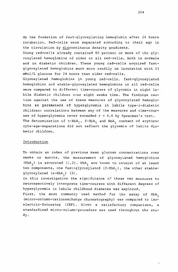
204
dy the formation of fast-glycosylating hemoglobin after 24 hours
incubation. Red-cells were separated according to their age in
the circulation by discontinuous density gradients.
Young red-cells already contained 60 percent or more of the gly
cosylated hemoglobins of older or all red-cells, both in normals
and in diabetic children. These young red-cells acquired fast
glycosylated hemoglobins much more readily on incubation with 25
mMol/L glucose for 24 hours than older red-cells.
Glycosylated hemoglobins in young red-cells, fast-glycosylated
hemoglobins and stable-glycosylated hemoglobins in all red-cells
were compared to different time-courses of glycemia in eight la
bile diabetic children over eight weeks time. The findings cau
tion against the use of these measures of glycosylated hemoglo
bins as parameters of hyperglycemia in labile type-1-diabetic
children: correlations between any of the measures and time-cour
ses of hyperglycemia never exceeded r = 0.6 by Spearman's test.
The determination of t-HbA1 , f-HbA1
and HbA1
content of erythro
cyte-age-separations did not reflect the glycemia of labile dia
betic children.
Introduction
To obtain an index of previous mean glucose concentrations over
weeks or months, the measurement of glycosylated hemoglobins
(HbA 1 ) is advocated {1,2). HbA1
are known to consist of at least
two components, one fast-glycosylated {f-HbA1
J, the other stable
glycosylated (s-HbA1
J (3).
In this investigation the significance of these two measures to
retrospectively interprete time-courses with different degrees of
hyperglycemia in labile childhood diabetes was explored.
First, the most commonly used method for the assay of HbA1
{micro-column-cationexchange chromatography) was compared to iso
electric-focussing (IEF). Given a satisfactory comparison, a
standardized micro-column-procedure was used throughout the stu
dy.
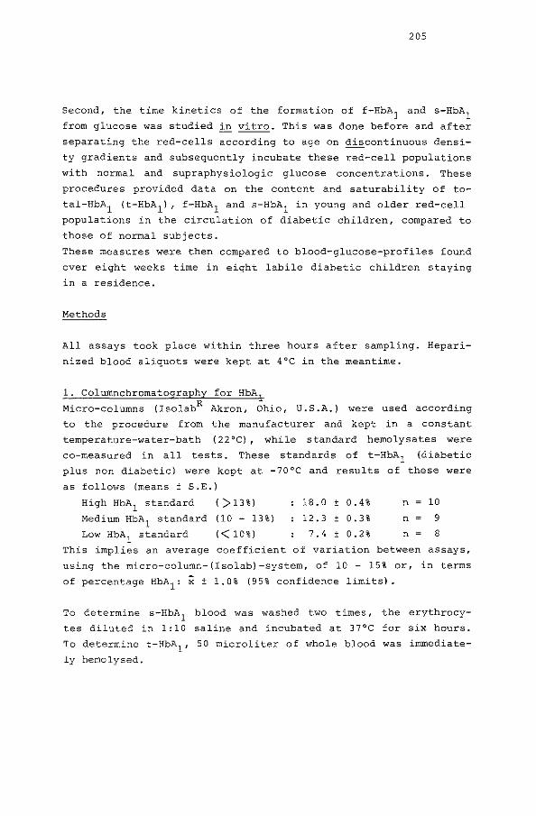
205
Second, the time kinetics of the formation of f-HbA1
and s-HbA1
from glucose was studied in vitro. This was done before and after
separating the red-cells according to age on discontinuous densi
ty gradients and subsequently incubate these red-cell populations
with normal and supraphysiologic glucose concentrations. These
procedures provided data on the content and saturability of to
tal-HbA1 (t-HbA1 ) , f-HbA 1 and s-HbA1 in young and older red-cell
populations in the circulation of diabetic children, compared to
those of normal subjects.
These measures were then compared to blood-glucose-profiles found
over eight weeks time in eight labile diabetic children staying
in a residence.
Methods
All assays took place within three hours after sampling. Hepari
nized blood aliquots were kept at 4°C in the meantime.
1. Columnchromatography for HbA1 Micro-columns (IsolabR Akron, Ohio, U.S.A.) were used according
to the procedure from the manufacturer and kept in a constant
temperature-water-bath (22°C), while standard hemolysates were
co-measured in all tests. These standards of t-HbA1 (diabetic
plus non diabetic) were kept at -70°C and results of these were
as follows (means ± S.E.)
High HbA 1 standard ( > 13%)
Medium HbAl standard (10 - 13%)
Low HbA 1 standard << 10%)
18.0 ± 0.4%
12.3 ± 0.3%
7.4 ± 0.2%
n
n
n
=
=
10
9
8
This implies an average coefficient of variation between assays,
using the micro-column-(Isolab)-system, of 10 - 15% or, in terms
of percentage HbA1
: ¥ ± 1.0% (95% confidence limits).
To determine s-HbA1 blood was washed two times, the erythrocy
tes diluted in 1:10 saline and incubated at 37°C for six hours.
To determine t-HbA 1 , 50 microliter of whole blood was immediate
ly hemolysed.

206
2. !so-electric-focussing (I.E.F.)
This method was originally described and modified by Jeppson (4).
LKB AmpholineR FAG-plates (LKB-Produkter AB, Sweden) were used.
The coefficient of correlation (Spearman's test) gave a rho of
0.92 for IEF versus s-HbA1 % by micro-columns chromatography for
45 samples over a s-HbA1 range of 7 - 16 percent.
3. Incubations of erythrocytes
Blood was taken by venapuncture from six diabetic and four non
diabetic subjects. Red-cells were washed three times in saline.
t-HbA1 was determined before the red-cells were incubated for
six hours in saline to determine s-HbA1 . After this the cells
were washed and diluted to a hematocrite (Ht) of 0.50. A sample
was then taken to determine s-HbA1 .
Incubations were in basal medium Eagle (BME) with Hanks salts
(GibcoR biocult) with added adenine, bicarbonate and glucose to
physiologic concentrations. A 37°C incubator with 5 percent co 2 was used. Cells were suspended in the medium and put in Petri
dishes. Dilution was at an hematocrite of 0.20 1/1, glucose con
centrations used for incubation were 5 and 25 mMol/1. HbA 1 was
determined every four hours until a maximum of 24 hours. After
harvesting the erythrocytes, a sample of the medium was taken to
exclude hemolysis. The cells were then washed three times and
glycosylation measured as f-HbA1
and s-HbA1
. Young cells and
unfractionated erythrocytes (see below) were incubated, one in
5mMol/l glucose, the other in 25 mMol/1 glucose. Diabetic as well
as non-diabetic erythrocytes in sterile conditions were studied.
4. Density separation of erythrocytes according to age
A modification of the method described by Rennie et al (5) was
used: discontinuous gradients were prepared to harvest a greater
and quantifiable amount of erythrocytes of a different age. This
was done by taking different mixtures of BSA-PercollR (Pharmacia,
Sweden) and the BSA-water solutions and carefully layering these
on top of each other by means of threechannel-peristaltic pumps.
First testruns were made with 74 - 85 percent BSA-PercollR >vith
steps of 2 percent. For each layer 1. 5 ml was used. After the

207
testruns a suitable gradient was chosen with narrower ranges of
percent BSA-Percoll R, to yield five different layers (red-cell
age groups). Ten tubes were used for each person.
After washing in saline, 1.5 ml of suspended erythrocytes
{Ht = 50 percent) were layered on the top of these gradients.
Centrifugation was according to Rennie et al (5).
A small amount of clumped cells was exclusively found in the bot
tom layer and data from this layer were excluded from further
analyses. The method separated young cells from old cells: no re
ticulocytes were found in layers lower than the top one. For
cell-age control various tests were used immediately after sepa
ration. 2,3-diphosphoglycerate (2,3-DPG, mMol/1 ery) (6), gluco
se-6-phosphate dehydrogenase (G-6-PD, mU/10 9 ery's) (7), pyruva
tekinase (PK, mU/ 10 9 ery's) (7), reticulocytes (0
/00) and mean
cellular volume (MCV, fl) ( 6) were decreased in older erythrocy
te fractions, as expected. These parameters were determined in
young (layer 1), medium (layer 3) and old erythrocytes (layer 4).
Hematological indices were calculated after determination of Hb,
Ht and erythrocytes number in a Coulter counter. Enzymes and
2, 3-DPG were tested with BoehringerR Testki ts (Boehringer-Mann
heim, FRG). All determinations were in duplo. The results are in
table l.
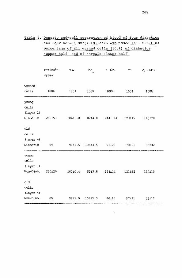
208
Table 1. Density red-cell separation of blood of four diabetics
and four normal subjects: data expressed (± 1 S.D.) as
percentage of all washed cells (100%) of diabetics
(upper half) and of normals (lower half)
washed
cells
young
cells
(layer 1)
Diabetic
old
cells
(layer 4)
Diabetic
young
cells
(layer 1)
Non-Diab.
old
cells
(layer 4)
Non-Diab.
reticulo- MCV
cytes
100% 100% 100%
266±53 104±3.0 82±4.0
0% 98±1.5 106±3.5
250±20 101±0.4 85±3.8
0% 98±2.0 109±5.0
G-6PD PK 2,3-DPG
100% 100% 100%
244±114 223±45 140±20
97±20 70±21 80±32
198±12 131±12 110±10
86±11 57±21 45±12
209
Patients
An 8 weeks' study with eight diabetic children was conducted at
the Kinabu medical residence (consultant pediatrician A.v.R.), to
which the children were admitted because of inadequate metabolic
control, with or without psychosocial problems. The eight child
ren (mean age 13. 6 ± 2. 2 years, range: 10-16) and their parents
gave their written consent to have bloodglucose-determinations by
fingerprick, urinary glucose and ketones twice daily in addition
to 24 hours-bloodglucose-profiles once a week, during 8 weeks.
This "home"-monitoring was under day and night supervision of the
health-care-personnel as was the administration of insulin. Body
weight was determined once a week. Changes in diet and insulin
were done by one of us (A.v.R.), who was kept unaware of the HbA1 results. Four children and parents also agreed to have venapunc
tures once a week to determine additional HbA1 measures, inclu
ding erythrocyte-age-separation. Daily records of each child were
kept of daily activities, symptomatic hypo/hyperglycemias and in
tercurrent illnesses,
All children were well beyond the remission-phase of their diabe
tes and were on combinations of long and short acting highly pu
rified porcine insulins twice daily. The findings on HbA1 % and
on insulin dosages and diets used are in table 2.

210
Table 2. Total and stable glycosylated hemoglobins (± 150) in 8
labile diabetic children over 8 weeks time with
averaged insulin dosages and caloric intake.
patient rrean rrean s-~ change in rrean insu- rrean
t-HbA1 s-HbA1
1in/U/kg/ Kcallkg/
weekl-8 day day
1. 14.4 ± 0.8 12.9 ± 0.7 + 1.1 0.8 25
2. 12.8 ± 0.8 11.9 ± 0.6 - 0.1 0.4 55
3. 15.7 ± 0.5 13.9 ± 0.6 - 0.5 0.6 28
4. 11.6 ± 0.8 10.4 ± 0.4 + 0.7 o. 7 41
5. 12.9 ± 0. 7 12.1 ± 0.8 - 0.4 0.6 52
6. 14.9 ± 0.6 13.8 ± 0.8 + 1.9 0.8 39
7. 12.3 ± 0.8 11.1 ± 0.5 - 0.4 1.2 49
8. 10.8 ± 2.1 9.4 ± 0.2 + o. 7 0.9 68
rreans 13.2 ± 1.7 11.9 ± 1.6
Changes in s-HbA1 (95 percent confidence limits - 1% s-flli11_) between the
beginning of the study (~k 0) and the end ('week 8). There was no correlation
(p { 0. OS) between the averaged insulin dosage and the averaged calories taken
per day. Patients 1 and 3 used rrost insulin ccmpared to their caloric intake.
Bloodqlucose and urine analysis
done with Ames-glucometersR
which correlated well capil-
Bloodglucose-determinations were
(Ames Netherlands B.V., Mijdrecht)
lairy GOD-determinations (r = 0.93).
M-values were calculated as the index of glycemic excursions for
the whole period according to Schlichtkrull et al. (8). All
children at the residence had M-values above 30, confirming the
lability of their diabetes.
Urine analysis was done with Ames-GlucosticksR and -KetosticksR
twice daily.
The findings on bloodglucose-profiles and M-values are in table
3.

211
Table 3. Glucose-Erofiles in 8 labile diabetic children over 8
weeks time.
patient age mean blcxxl- mean bql. du- mean fas- mean of mean
in glucose ring first 2 ting bgl. 24-hours M-
years in rrMol/L weeks minus profiles value
mean bgl. du- of bgl. (see
ring last 2 text)
weeks
l. 16 16.5 ± 2.4 + 2.5 15.4 ± 2.3 15.7 ± 3.0 68
2. 12 10.5 ± 3. 7 + 4.2 9.9 ± 3.1 10.5 ± 3.7 32
3. 15 10.2 ± 6.9 - 6.4 9.5 ± 6.3 9.9 ± 4.5 67
4. 16 11.0 ± 5.5 + 1.0 10.0 ± 3.9 9.2 ± 3.8 50
5. 10 11.7 ± 4.3 - 0.6 10.0 ± 3.9 12.1 ± 4.4 45
6. 12 12.4 ± 5.8 + 6.2 9.9 ± 4.8 11.3 ± 2.8 61
7. 15 9.6 ± 3.8 + 0.3 8.3 ± 1.7 11.3 ± 2.3 33
B. 11 12.4 ± 5.0 + o.s 12.1 ± 4.9 11.3 ± 2.8 55
means: 12.4 11.8 ± 2.2 10.8 ± 2.2 11.4 ± 2.0 51
Patients 1 and 3 were selected for a visual comparison between bloodglucoses
and glycosylating components of~ (figure 2).
Results
1. In vitro studies: incubation of red-cells of diabetic children
and normal subjects.
Table 4A contains the findings by incubating red-cells at 5
rnMol/L glucose (normal concentration) and 25 mMol/L glucose (su
praphysiologic concentration) in young and all red-cells of four
normal subjects. Table 4B contains the same data on red-cells ob
tained from six diabetic children.
By these short-term incubations the s-HbA1 content did not
change and the difference between "starting s-HbA1 " and "final
t-HbA1
11 was due only to increment of f-HbA 1 , as indicated next

212
in figure 4A. Young red-cells acquired f-HbA1 significantly fas
ter on incubation in 25 mMol/1 glucose, both from diabetics and
from normals. Calculated slopes in increment were respectively
0. 20 and 0.17 for these categories, whereas all red-cells had
slopes of 0.06 and 0.09 for diabetics and for normals. The fin
dings demonstrate different kinetics for the formation of
f-HbA 1 in young cells as a fraction of all red-cells.
Furthermore, young red-cells before incubation contained already
60% or more of the HbA1 contents of all red-cells. This relation-
ship was also found in 30 other
in figures 4A and 4B. In these
content (t-HbA1 0.5-3.0%.
s-HbA1 ) was
diabetic samples, not comprised
30 samples the average f-HbA1 1.7% HbA1 with a range of
Table 4A. Increment in f-HbA1 by the incubation of red-cells
in two glucose-concentration over 24 hours in young
red-cells and all red-cells of four normal subjects
- means and ranges.
All red-cells
in 5 Itt>bl/L
in 25 Itt>bl/L
Young red-cells
in 5 Itt>b1/L
in 25 Itt>bl/L
Starting
s-~%
7.6
7.6
5.6
5.6
(6.5-8.1)
(4.9-6.3)
Final
t-~%
7.3 (5.5.-9.1)
9.1 (8.6-9.5)
7.9 (7.5-8.3)
8.9 (8.6-9.2)
Calculated
slope of
increment
0
0.09
0.12
0.20

213
Table 4B. The same experiment as in table 4A, for red-cells of
six diabetic children.
All red-cells
in 5 nMol/L
in 25 nMol/L
Young red-cells
in 5 rrMol/L
in 25 rrMo1/L
Starting
s-HbA1%
13·8
{13.5-14. 7) 13.8
not done
7.7 (6.7-8.9)
Final
t-HbA1%
14.0 {13.3-14.9)
15.4 (13.3-16. 7)
11.1 (10. 7-12.1)
Calculated
slope of
increrrent
0.01
0.06
0.17
2. Clinical studies: Comparison between bloodglucose-profiles and
HbA 1
All measures of glycosylated hemoglobins (f-HbA1 , t-HbA 1 , s-HbA1 of all red-cells and t-HbA
1 of young red-cells) were compared to
glucose-profiles of the eight children studied over 8 weeks (glu
cose in the bloodsarnple itself, 24 hours bloodglucoses on the
previous day, the same day, the previous 1 - 8 weeks and all 8
weeks). Glucosuria was also compared to these measures of HbA1
.
The linear regression coefficient was never greater than 0.6 ex
cepting for a comparison between:
the bloodglucose of the sample itself and t-HbA1 {r = 0.96) and s-HbA1 (r = 0.91);
mean of 24 hours bloodglucoses the same day and t-HbA1 (r ~ 0.60)
It was to be expected that the mean of these widely fluctuating
bloodglucoses over the 8 week period correlated only weakly to
final glycosylated Hb-values (t-HbA1
: r 0.34, s-HbA1 : r
0. 20) •
To illustrate the lack of correlation between components of
214
HbA 1 and episodes of glycemia in these labile patients visually,
the clinical time-course of two children is represented in figure
2.

Figure 2.
Pa~ient I
(glue) 20.0 mmoiiL
10.0
'-'
X glycosylated
l<t.O he~ "-' l
12.0
10.0
.... /_,.,~~.,...------..., ...... ___ '
I
17.0
111.0
15.0
1'0.0
13.11
X glycooylated hemoglobins
~
• TIME[ weeks]
Patient 3
215
We!!kly means -- of 2'lhrn profiles
weekly means o--<:l of gluc..,.,conc
__.. old cells .,. _ _.,young cells
~ ' ' 'y'
-- t-HbA1 (%) "s-HbAJ [%)
• , TIME( weeks)

216
In the upper panel averaged bloodglucoses of 24 hours profiles
(closed circles) and means of daily bloodglucoses (open circles)
were plotted against time. In patient 1 high, but stable blood
glucoses were seen, whereas patient 3 had {symptomatic) hypogly
cemia during week 2 and 3, despite the temporary decrease of her
insulin dosages.
In the middle the t-HbA1 % of both young and old cells failed to
show a significant decrease up to five weeks after this hypogly
cemic episode in patient 3, whereas patient 1 showed a decrease
in t-HbA1 % of young cells during the first three weeks, not re
flected by the expected closely time-related changes in bloodglu-
cases.
Discussion
Although the existence of fast- and stable- glycosylating compo
nents of HbA1 has been well recognized (9,10,11), the time-kine
tics of the formation of these two components have not been re
solved {10). Yet the separate measurement of these two components
might be useful if fast-glycosylation reflected glycemic control
over shorter periods and stable-glycosylation over longer periods
of time.
In the present study fast-glycosylation was examined by the sepa
rate assay of f-HbA1 (t-HbA1 minus s-HbA1 ) and by the HbA1 con
tents of young erythrocytes, that have had shorter exposure times
to the glucose of the plasma than older red-cells.
The precision of the micro-column system used was 1 percent of
HbA 1 (95 percent confidence limits), s-HbA1 thus measured compa
red well to the IEF-method.
In individual samples f-:HbA1 did not correlate with either t-HbA1 or s-HbA1 , endorsing the premise that f-HbA1 might separately re
flect short episodes of hyperglycemia. This however could not be
demonstrated from the correlation with glycemia over short pe
riods of time in eight labile diabetic children.
Also, the HbA 1 content of young erythrocytes failed to do so,
which could be explained by the finding that these young red
cells contained olready 60 percent or more of the total HbA1 con-
217
tent of all red-cells, despite their shorter living time in the
circulation. Furthermore, young red-cells (of normals as well as
of diabetic children) acquired HbA1
faster than all red-cells on
incubation in supraphysiologic glucose-concentrations.
These findings suggest that shorter episodes of glycemia cannot
be detected by either f-HbA1
or by the HbA1
content of young red
cells. The red-cell separation method was validated by the fin
ding of the expected differences in hematologic as well as bio
chemical parameters for red-cell age. Red-cells from layer 4 in
the present discontinuous density separation experiments correla
ted with age parameters for all red-cells, including HbA1 , indi
cating this separate represented average red-cells. This is ex
plicable by the fact that the young cells thus separated compri
sed only 10 percent of the red-cells (6,10). In keeping with this
study Spicer et al (12) found that incubation of red-cells with
different glucose-concentrations showed a linear increment of the
HbA1-content with time, in red-cells of normals and of diabetics.
As in this study, this increment was due to f-HbA 1 and not to
s-HbA1 . Gillery et al (13) reported that young cells are capable
of actively transferring glucose to hemoglobin by means of a red
cell membrane fraction. Kinetics might therefore differ in young
cells. Differences in passive glucose permeability between red
cells were found by Higgins et al {14) . Furthermore glycosylated
products with other time-kinetics may be assayed by the present
methods as recently suggested by Garlick et al (15). These au
thors found that by rechomatographing "HbAlc" (from Biorex-70
columns) on a Glycogel B boronate affinity resin gave a 70 per
cent adherence indicating that less than 30 percent of the
"HbAlc" consisted of hemoglobin with beta-chain terminal valine
bound glucose, The remainder of this "HbA1c" was glycosylated
HbA0 , with 14 percent alpha-chain N-terminal valine-bound gluco
se, 40 percent alpha-chain lysine- and 46 percent beta-chain ly
sine-bound glucose. The kinetics of these compounds are diffe
rent, compared to each other. This may be connected to the pre
sent finding, that even s-HbA1 correlated with glucose of the
sample itself.
To circumvene the co-chromatography of glucose-adjuncts to hemo-

218
globin other than at the beta-chain terminal valine (HbA1c) ,
the thiobarbituric-acid-method has been propagated. One of the
problems with this method however is that the colour-development
differs with the incubation conditions of the hemolysate and with
the use of acetic acid instead of oxalic acid in the incubation
mixture (16).
In this study there was no relation between the formation of com
ponents of HbA1
and the glycemic control in labile diabetes of
childhood. The determination of fast glycosylated products or the
HbA1
content of young erythrocytes did not reflect shorter epi
sodes of glycemia, studied over 8 weeks time.
Acknowledgements
This investigation was supported by Novo Industries; the work at
Kinabu was supported by Nordisk Netherlands. H.J.A. worked on a
4th year student-fellowship of the Erasmus University Medical
School. We are indebted to Mrs and Mr van Royen, psychologist and
general director of Kinabu for their overnight and weekend super
vision of bloodglucose-measurements.
References
1. Mayer, T.K., Freedman, Z.R.
Protein glycosylation in diabetes mellitus: a review of
laboratory measurements and their clinical utility.
Clin. Chim. Acta 1983; 127: 147-184.
2. Jovanovic, L., Peterson, C.M.
The clinical utility of glycosylated hemoglobin.
Am. J. Med. 1981; 70: ·331-338.
3. Ukena, T., Merrill, E., Morgan, C.
An analysis of the importance of the "labile" fraction of gly
cosylated hemoglobin as determined by a rnicrocolumn method.
Am. J. Clin. Path. 1982; 78: 724-728.

219
4. Jeppson, J.O., Franzen, B., Gaal, A.B.
Simplified determination of hemoglobin Ale in diabetic pa
tients by use of electrofocusing.
In: Radola, B.J. ed. Electrophoresis 1979. New York, NY: de
Gruyter 1980: 655-661.
5. Rennie, C.M., Thompson, s., Parker, A.C., Maddy, A.
Human erythrocyte fractionation in "Percell" density gra
dients.
Clin. Chim. Acta 1979; 98: 119-125.
6. Harris, J.W., Kellerroeyer, R.W.
Red Cell metabolism and Methemoglobinemia.
In: The red cell
Cambridge, Mass. Harvard Univ. Press.: 1970; 447-513.
7. Bartos, H.R., Desforges, J.F.
Enzymes as erythrocyt age reference standards.
Am. J. Med. Sci. 1967; 254: 862.
8. Schlichtkrull, J., Munck, o., Jersild, M.
TheM-value, an index of blood-sugar control in diabetics.
Acta Med. Scan. 1965; 177: 95-102.
9. van Essen, L.H.
Geglycosyleerd haemoglobine: Een nieuwe parameter voor diabe
tesregulatie?
Thesis, 1982, State University Groningen.
10. Svendsen, P.Aa., Christiansen, J.A., S¢egaard, u., Nerup, J.
Synthesis of glycosylated haemoglobin in vivo.
Diabetologia 1981; 549-553.
11. Svendsen, P.Aa., Lauritzen, T., S¢egaard, U., Nerup, J.
Glycosylated Haemoglobin and steady-state mean blood glucose
concentration in type 1 (Insulin-Dependent) diabetes.
Diabetologia 1982; 23: 403-405.

220
12. Spicer, K.M., Allen, R.C., Hallett, D., Buse, M.G.
Synthesis of Hemoglobin Ale and related minor hemoglobins by
erythrocytes.
J. Clin. Invest. 1979; 64: 40-48.
13. Gillery, P., Maquart, F.X., Gattegno, L. et al.
A glucose-containing fraction extracted from the young ery
throcyte membrane is capable of transferring glucose to hemo
globin in vitro.
Diabetes 1982; 31: 371-374,
14. Higgins, P.J., Garlick, R.L., Bunn, H.F.
Glycosylated Hemoglobin in Human and animal red cells: Role of
glucose permeability.
Diabetes 1982; 31: 743-748.
15. Garlick, R.L., Mazer, J.S., Higgins, P.J., Bunn, H.F.
Characterization of glycosylated hemoglobins.
J. Clin. Invest. 1983; 71: 1062-1072.
16. Kloosterboer, H.J., Bruining, G.J., van Dongen-Brand, F.C.M.,
Zwakhals, M.S.Ph.
A modified method for the estimation of glycosylated plasma
proteins.
Submitted for publication.
221
§4, Additional findings with home care
The purpose of this paragraph is to communicate additional fin
dings with the home-care project. These findings relate to:
a. Patient-selection.
b. Nature of admissions and acute visits.
c. Patient education.
d. Self-monitoring.
e. Metabolic control.
f. Economy of home-care.
a. Patient-selection.
The deviation from a randomized prospective design with the home
care study (described in paragraph 2 of this chapter) necessitates
a more detailed account of the selection of patients that
participated in the comparison between "hospital-based" and
"home-care". This comparison dealt also with HbA1 % before and af
ter the introduction of the home-care system (table II of paper 1
- page 198) and involved 41 children.
In the year before the home-care system was introduced 78 child
ren (table III of paper 1 - page 199) visited the diabetes clinic,
from which 78 - 41 = 37 were excluded by the following criteria:
having diabetes for 1.5 years or less by September 1977 25
newly diagnosed between September 1977 and September 1978 11
referred by pediatrician and back to pediatrician 1
37.
Newly diagnosed patients and children with a duration of 1. 5
years or less may have (remnant) endogenous insulin secretion,
which facilitates the achievement of good metabolic control. Such
achievement would not be due to the home-care system employed.

222
The changes in admission-days and acute visits (table III of pa
per 1 - page 199) before and after home-care was introduced were
plotted against the number of all children attending the qiabetes
clinic. Table 4.1 of the present discussion indicates the numbers
of children entering or leaving the diabetes clinic, during the
follow-up.
Table 4.1. Flux of patients seen at the Sophia Children•s Diabe
tes Clinic.
All children
seen
Newly
diagnosed
Referred
with trea
ted diabe
tes by pe
diatri
cian {n=19)
Referred back
to -pediatrician (n=lO)
Referred to
internist (n=ll)
Moved out
of area (n=2) 11Drop-outs"
{n=2)
September 1977 September 1978 September 1979 September 1980
September 1978 September 1979 September 1980 September 1981
78 88 99 106
11 10 12 8 I in
1 7 7 4 l 0 2 l 7 1
0 4 4 3
out
l 0 0 1 1
2 0 0 0
"hospital-based" "hare-care"

223
Children leaving the diabetes clinic ("out" in the table above)
comprised:
10 children referred back to the pediatrician elsewhere. These
10 children all came from the 19 referred to the clinic be
tween September 1977 and September 1981 by pediatricians else
where ("in" in the table above). None of these 10 children re
ferred back was admitted to the hospital, nor did they visit
the clinic in emergency situations.
11 children were referred to the internist because of their
age. Between September 1977 and September 1978 their averaged
number of admission-days was 2.0 days, less than the average
of 2. 4 days for the whole patient-population in that time
span. The same applied for acute visits. Between September
1978 and September 1981 none of these children was admitted
nor did they visit the clinic acutely because of metabolic
disregulation. Their HbA1
were 12. 2% on the average (with a
range of 10 - 16.3%) not different from the HbA1 % of the 41
children of table II of paper 1 - page 198.
2 children moved out of the area, without being admitted or
visiting the clinic acutely, their averaged HbA1
being 11.8%
and 12.0%.
1 patient was excluded from these tabulations: she was refer
red in November 1979 with an extremely brittle diabetes, ne
cessitating the use of subcutaneous continuous insulin infu
sion started in July 1980. She was admitted for more than 100
days and her case history has been reported elsewhere (23) .
Taken together, there is no evidence for referral bias with re
gard to admission-days, acute visits or HbA1 % in the conventio
nally treated children with diabetes mellitus tabulated in table
III of paper 1 - page 199. The same reductions in admissions or
acute visits for metabolic disregulation were seen with the in
troduction of the home-care system in the 41 children that were
selected for the follow-up of metabolic control, as in all child
ren attending the diabetes clinic.

224
b. Nature of admissions and acute visits.
The next tabulation (table 4. 2 of this discussion) summarizes
numbers of children, the nature of their admissions, acute visits
before and after home-care, including the number of newly diag
nosed children that received initial treatment without being ad
mitted to the hospital.
Table 4.2. Nature of metabolic disregulation before and after
horne-care.
Number of ad
mission-days Number of
children ad
mitted
Hyperglyce
mia/ketosis
Hypoglycemia
Number of
acute visits
Hyperglyce
mia/ketosis
Hypoglycemia
Number of new
ly diagnosed
children
Number of new
ly diagnosed
children not
admitted for
initial treat
ment
September 1977 September 1978 September 1979 September 1980
September 1978 September 1979 September 1980 September 1981
160
13
9
4
24
10
14
11
0
65
5
3
0
10
5
5
10
0
38
3
2
1
2
0
2
12
5
8
1
1
0
1
0
1
8
6

225
After the introduction of home-care the principal reason for ad
mission was decreased consciousness. In cases of hyperglycemia/
ketosis, this coincided with a blood Ph of 7.25 or less including
more than 5 percent dehydration by weight. In cases of hypoglyce
mia, this involved failure of self-administered glucagon and sub
sequent carbohydrate intake to normalize consciousness. In the
first year of the home-care project (September 1978 - September
1979) three of the five acute visits occurred because of hypogly
cemia in families that had not yet been trained in the use of
glucagon. A similar situation led to admission in two of the
three admissions for hyperglycemia/ketosis in that year: these
two families called the help of the nurse practioner only 48
hours after the initiation of symptoms.
Not given in the above table are the numerous visits by the nurse
practioner, especially during the first year of the horne-care, to
assist families in actually treating hypoglycemia or hyperglyce
mia/ketosis at home: the nature and severity of such metabolic
disregulations varied too much to allow a classification thereof.
c. Patient-education.
Teaching to patients was both to individual families at home and
in "class-rooms" in the hospital.
Home-teaching was mainly on insulin-injections (all patients ha
ving diabetes, except the two "drop-outs", were put on insulin
injections twice daily once the "honey-moon"-phase was over),
self-monitoring and adaptation thereof in individual circumstan
ces (sports, parties etc.).
Class-room-teaching was in five evening sessions held once a year
for parents of whom the children were first seen during that
year. By this sequence class-room teaching reinforced the indivi
dual lessons taught at home. The evening sessions contained an
intermission of of half an hour, during which parents got to know
each other. In the first two years of home-care separate evenings
were held for the parents of the other children, that were not
newly diagnosed during that year.
The program was:
1st evening
2nd evening
3rd evening
4th evening
5th evening
226
a. causes of diabetes;
b. insulins, insulin action.
a. self-monitoring (also practical);
b. special situations to use self-monitoring.
a. diet and dietary allowances;
b. insulin, self-monitoring and diet.
a. inheritance, chronic complications,
choice of profession;
b. pregnancy, anti-conception,
future developments.
a. multiple choice examination;
b. results of examination, final discussion.
Part a. (before the intermission) took three quarters of an hour,
part b. (after the intermission) also, including one quarter for
questions and discussion. The setting was informal, not involving
more than 35 parents per class. The attendance-rate was 70 - 80
percent of the parents invited. Care was taken not to expose pa
rents of very recently diagnosed children to this program, but to
advise them to participate in the next years class.
This examination is reprinted next.
Exam for parents of children having diabetes mellitus
All questions are multiple choice. You are kindly requested to
darken the circle left of the right answer.

227
Example:
Diabetes of childhood is caused by taking too much sweets.
a. 0 partially correct
b. e not correct
c. 0 correct
b right answer.
1. A "hyper" (high bloodsugar) is caused by ... a. 0 too little insulin-effect
b. 0 too much insulin-effect
c. 0 too little food
d. 0 a + b
2. Glucose (the main energy source of the body), in case of too
little insulin-effect, is ..•
a. 0 not taken up sufficiently by body-cells and subsequently
utilized to yield energy
b. 0 converted into ketones
c. 0 taken up rapidly by body cells and subsequently utilized
to yield energy
3. Ketones (acetone) appears in the urine, if a. 0 too much fat is broken down, because there is too much
insulin-effect
b. 0 too much fat is broken down, because there is too little
insulin-effect is.
c. 0 none of these (a + b)

228
4. A child with diabetes may have high bloodsugars (or urinesu
gars) also if the child eats little or nothing. This is be
cause
a. 0 the body will produce glucose from the liver, if there
is too little insulin-effect
b. 0 food is not carbohydrates only and the body can make
glucose from other nutrients
c. 0 both answers (a + b) are correct
5. The following values of a home-made bloodglucose (day + night)
profile are within the "diabetes-norm 11:
a. 0 2. 2
b. 0 4. 4
c. 0 4. 4
22.2
10.0
13.3
6. Reading the Hemoglucotest (Chemstrip) you will note by prefe
rence?
a. 0 2.2 4.4
b. 0 2. 2
c. 0 4. 4
7. The strip for urinary ketones is called?
a. 0 Diabur
b. 0 Hemoglucotest (Chemstrip)
c. 0 Ketur
8. The new strip for urinary glucose can read?
a. 0 until 5 grams per 100 ml urine
b. 0 until 2 grams per 100 ml urine
c. 0 until 10 grams per 100 ml urine
9. Using Hemoglucotest (Chemstrip) the use of a stopwatch is
a. 0 desirable
b. 0 necessary
c. 0 cumbersome

229
lO.Glucagon-injections are used in case of ...
a. 0 low bloodsugar
b. 0 low bloodsugar if the child cannot or will not take su
gar
c. 0 with vomiting
ll.Glucagon is injected
a. 0 like insulin
b. 0 in the muscle
c. 0 both answers (a + b) are correct
12.Glucagon will ...
a. 0 raise the bloodsugar
b. 0 raise the blood sugar within 10 minutes
c. 0 raise the blood sugar within 10 minutes, but the child
must eat subsequently and have bloodsugars measured
13.Insuline Actrapid works ...
a. 0 12 hours
b. 0 6 hours
c. 0 24 hours
14.Insuline Mixtard is ... a. 0 1/3 short acting, 2/3 long acting
b. 0 1/2 short acting, 1/2 long acting
c. 0 2/3 short acting, 1/3 long acting
15.The most common cause of swinging bloodsugars is
a. 0 eating too much
b. 0 irregular resorption from the injection site
c. 0 irregular meal-times
16.The most common cause of fat-pads is ...
a. 0 injecting too superficially
b. 0 injecting at one spot
c. 0 both answers {a + b) are correct

230
17.A five year old child wakes up and is drowsy, nauseated, re
fuses breakfast. What would you do?
a. 0 take the body-temperature, weight and test for urinary
ketones and call the hospital
b. 0 give the usual insulin-dose and do as under a
c. 0 give sugarwater and do as under b + a
d. 0 take bloodsugar and do as under a
18.This child has diabetes for one half year and takes 18 U of
insulin per day in the morning only. The bloodsugar is
13.3-22.2 and urinary ketones are 2+. No fever. The doctor/
nurse advises to give 4 U Actrapid insulin extra. What would
you do next?
a. 0 check every urine for glucose and ketones
b. 0 check bloodsugar and urinary ketones some 4 hours later
and call back.
c. 0 go to the hospital
19.Another child, 10 years old, wants to participate in an eve
ning running-match over 5 miles. She takes 24 U of insulin in
the morning and 8 U in the evening. What would you do?
a. 0 give extra food before the match
b. 0 as a, but give 4 U instead of 8 U
c. 0 as a + b and check bloodsugars before and after the
match
20.The following week she has a barbecue-party at 20.00 hours.
What would you do?
a. 0 give 12 U instead of 8 U at her usual dinner time
(± 18.00 hours) and have her take her usual dinner
b. 0 give the usual 8 U at ± 19.30 hours and tell her to take
as much food at the party as she would at dinner time
c. 0 give her 12 U at ± 19.30 hours if she wants to eat more
at the party than at dinner, after explaining her how to
do this

231
2l.This same girl is on summer-vacation in Southern-France. It is
warm and she runs, swims the whole day. What do you suspect?
a. 0 that less insulin will be needed
b. 0 as a and try this by taking morning and late afternoon
bloodsugars
During the intermission of the last evening the answers were
checked, the number of correct answers indicated and the forms
returned to the parents. The average percent of acceptable an
swers was 85 percent, with an annual range from 70 - 92 percent.
Answers to questions on materials and their use (insulin, blood
glucosestrips) were almost invariably correct. Mostly also on the
(difficult) clinical questions at the end of the examination. In
terestingly, most errors were in answers on questions on insulin
action, apparently by their abstract nature.
d. Self-monitoring.
The primary aim of self-monitoring was avoidance of hypoglycemia
and symptomatic hyperglycemia. The secondary aim of self-monito
ring was to get an impression of bloodglucoses during a given
day-and-night, taken four times a year. Children and/ or parents
preferring not to do the latter, got treatment advices on the ba
sis of clinic visits once every 6 - 8 weeks. This included histo
ry-taking and the measurement of HbA1
%. This latter category con
sisted entirely of children of 12 years or older. They constitu
ted about half of the population over that age seen in the first
year of home-care. Presently (1983) 24 of 71 of the children of
12 years or older prefer not to note and report their bloodgluco
ses to the clinic, except under pressing circumstances, such as
intercurrent illnesses with impending ketosis. Of these 4 will
not visit the clinic at the recommended 3 - 4 monthly intervals.
It should be mentioned here that dutch health-care insurance com
panies, with few exceptions, cover the costs, for 80 - 100 per
cent, of 25 bloodglucose-strips per month since 1983. Before that
time the costs were covered by the home-care project budget and
by the separate support of the local national insurance institu-

232
tion (Stichting Rotterdamse Ziekenfondsen) mediated by its medi
cal advisor Mrs. dr. H. Boers.
An important aspect of self-monitoring of bloodglucose is quality
control. Measured in a glucose-reflectometer the linear correla
tion coefficient of these strips with the laboratory GOD-method
on 100 diabetic samples was r = 0.96.
The form used by the families to note self-measured bloodglucoses
is reprinted next.

233
Bloodsuqar day-and-night curve
Name ••••• , • , . • • • • • • . . • . . . . . . • . . . . . . Doctor's notes •..........
Day and date
Insulin dose and sort ......•••.•• +
1 hr after before 1 hr af- before
n.p.o. breakfast lunch ter lunch supper 24 hrs 3 hrs
n.p.o.
Tirre
(marine
times)
Blood-
sugar
Urine-
test
Extra
food
Extra
exer-
cise
Hyp:::>'s?
Hyper's?
Note: it is assumed taking the rreal takes not rrore than 20 minutes.

234
The day-and-night curves of 15 families, were time-labelled by
the families and stored in the container of the bloodglucose
strips, that has an exsiccator. In the hospital these were read
again within 48 hours and compared to the values reported by the
families. This involved 105 bloodglucose-strips in total. The
bloodglucose-strips used (Hemoglucotest 20/BOOR, Boehringer,
Mannheim, F.G.R.) are read in ranges: 1.1 - 2.2 mMol/L, 2.3 - 4.4
mMol/L, 4.5- 6.6 mMol/L, 6.7- 10.0 mMol/L, 10.1- 13.3 rnMol/L,
13.4 - 22.2 mMol/L glucose. The fifteen families were selected in
case of doubt of the quality of their bloodglucose-assessments.
The following deviations were noted with these 105 strips:
failure to note two rather than one glucose
value as the estimate of bloodglucose, but
otherwise agreement
hospital reading off one range of estimate
by patient (all at 3 A.M.)
not readable
hospital reading off more than one range
of estimate by patient
skipped time-points of day-and-night curve
(6x at 3 A.M.)
£• Metabolic control.
25 X
3 X
4 X
0 X
30 X
During the hospital-based period the assay of glycosylated hemo
globin (HbA1 ) was not available, hence a comparison between gly
cemic control achieved by hospital-based and by home-care with
this parameter is not possible.
During the three years of the home-care project total glycosyla
ted hemoglobins (t-HbA1 %) were measured (table II of paper 1 -
page 198). The findings of paper 2 however question the signifi-

235
cance of this measurement. Therefore no statistic was used to
examine possible changes in t-HbA1 % during the horne-care.
With the limitations of HbA1
% as an index of glycemia in mind,
the self-measured bloodglucoses of the 41 children (table II of
paper 1 - page 19 8) were examined. This method was introduced
with the home-care system, hence only changes during home-care
were evaluated.
The following scoring-system was designed, using the bloodgluco
se-strips described earlier.
Result of self-measurement
glucose mMol/L
< 1.1
1.2 - 2.2
2. 3 - 4.4
4.5 - 6. 6
6.7 - 10.0
10.1 - 13.3
13.4- 22.2
>22.2
Points
3
2
1
0
0
1
2
3
As indicated earlier almost half of all children seen of 12 years
and older preferred not to produce bloodglucose day-and-night
profiles at each 3-4 monthly visit.
38 of the 41 children chosen for the follow-up (that were much
younger, see table II of paper 1 - page 198) had home-profiles
made at the frequency intended throughout the three years of the
follow-up of home-care. Only if the 3 A.M. value was included and
contained seven or more time-points measured in total, their pro
file was used to calculate "points" according tot he above sco
ring-system. The parents were instructed to avoid days with in
tercurrent infections, and unusual physical exercise. The fami
lies produced three or more of such profiles during each year of
the home-care. The "points" of each child's profile were counted
and its summation averaged per child per year of home-care. This
is represented in the following tabulation.
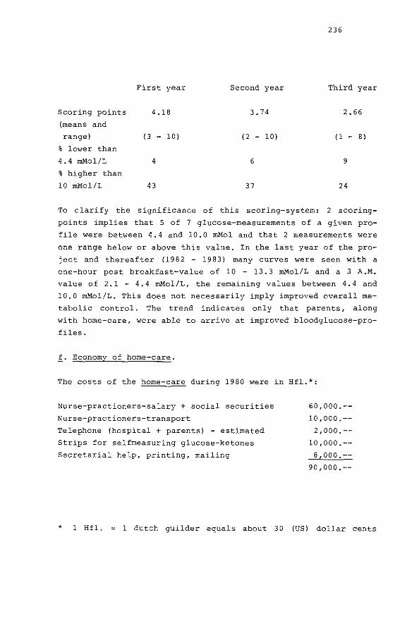
236
First year Second year Third year
Scoring points 4.18 3. 74 2.66
(means and
range) (3 - 1 0) (2 - 10) (1 - B)
% lower than
4.4 mMol/L 4 6 9
% higher than
10 mMol/L 43 37 24
To clarify the significance of this scoring-system: 2 scoring
points implies that 5 of 7 glucose-measurements of a given pro
file were between 4.4 and 10.0 mMol and that 2 measurements were
one range below or above this value. In the last year of the pro
ject and thereafter (1982 - 1983) many curves were seen with a
one-hour post breakfast-value of 10 - 13.3 mMol/L and a 3 A.M.
value of 2.1 - 4.4 mMol/L, the remaining values between 4.4 and
10.0 mMol/L. This does not necessarily imply improved overall me
tabolic control. The trend indicates only that parents, along
with home-care, were able to arrive at improved bloodglucose-pro
files.
f. Economy of home-care.
The costs of the home-care during 1980 were in Hfl.*:
Nurse-practioners-salary + social securities
Nurse-practioners-transport
Telephone (hospital + parents) - estimated
Strips for selfmeasuring glucose-ketones
Secretarial help, printing, mailing
60,000.--
10,000.--
2,000.--
10,000.--
8,000.--
90,000.--
* 1 Hfl. 1 dutch guilder equals about 30 (US) dollar cents

237
The hospital-costs during home-care in 1980 were in Hfl.:
1 acute visit (hospital + parents) - estimated
8 admission-days for treated diabetes
(1 hospital-day in 1980 casted 400)
26 admission-days for 8 newly diagnosed dia
betic children (26 x 400 = 10,400)
400.--
3,200.--
10,400.--
14,000.--
In 1980 106 children attended the diabetes clinic and a total of
Hfl. 104,000.-- was spent or Hfl. 980.-- per child.
The costs of the hospital-based-care during 1977 were in Hfl.:
Telephone (hospital + parents) - estimated 1,000.--
Fractioned 24 hrs urinary glucose- and ketone
determinations n = 42
a. transport by parents - estimated
b. laboratory costs
5,000.--
10,000.--
16,000.--
The hospital costs during hospital-based-care, based on 1980-
norms were in 1977 in Hfl.:
24 acute visits (hospital +parents) - estimated 9,600.--
160 admission-days for treated diabetes 64,000.--
209 admission-days for 11 newly diagnosed dia-
betic children corrected to 152 days for 8
(the number of newly diagnosed children in
1980) 60,800.--
134,400.--
In 1977 78 children attended the diabetes clinic and a total of
Hfl. 158,400 was spent or Hfl. 2,030.-- per child. In conclusion,
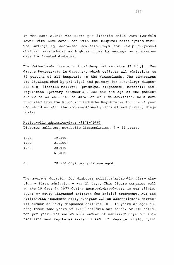
238
in the same clinic the costs per diabetic child were two-fold
lower with home-care than with the hospital-based-system-care.
The savings by decreased admission-days for newly diagnosed
children were almost as high as those by savings on admission
days for treated diabetes.
The Netherlands have a national hospital registry (Stichting Me
dische Registratie in Utrecht) , which collects all admissions to
95 percent of all hospitals in the Netherlands. The admissions
are distinguished by principal and primary (or secondary) diagno
ses e.g. diabetes mellitus (principal diagnosis), metabolic dis
regulation (primary diagnosis) . The sex and age of the patient
are noted as well as the duration of each admission. Data were
purchased from the Stichting Medische Registratie for 0 - 16 year
old children with the abovementioned principal and primary diag
nosis:
Nation-wide admission-days (1978-1980)
Diabetes mellitus, metabolic disregulation, 0 - 16 years.
1978
1979
1980
or
19,800
21,100
20,900
61,800
20,600 days per year averaged.
The average duration for diabetes mellitus/metabolic disregula
tion - first admission - was 21 days. This figure compares well
to the 19 days in 1977 during hospital-based-care in our clinic,
spent by newly diagnosed children for initial treatment. For the
nation-wide incidence study (Chapter II) an ascertainment correc
ted number of newly diagnosed children (0 - 16 years of age) du
ring those same years of 1,320 children was found, or 440 child
ren per year. The nation-wide number of admission-days for ini
tial treatment may be estimated at 440 x 21 days per child: 9,240
239
days. By substraction of this number from the total number of ad
mission-days (20,600) the annual number of admission-days for me
tabolic disregulation after initial treatment may be estimated:
20,600 - 9,240 = 11,460 days.
With an estimated prevalence for diabetes (0 - 16 years) of 3,000
(Chaper II) it follows that each diabetic child would be admitted
for metabolic disregulation after initial treatment for almost 4
days per year on the average! This is even higher than the 2.4
days per diabetic child found during hospital-based-care in the
Sophia Children's Hospital in 1977 (table III of paper 1 - page
17).
With these nation-wide data on all admission-rates, the costs
were 20,600 x 400 = Hfl. 8,36 million in 1980. The savings by a
(hypothetical) nation-wide introduction of the home-care-system
would comprise Hfl. 4 million if only the savings observed in the
Sophia Children 1 s Hospital were extrapolated. From this amount of
money alone, some 40 home-care-projects could be maintained, each
costing Hfl. 100,000.-- per year.
By assigning 100 children to each project, the total prevalence
of childhood diabetes in the Netherlands (0 - 19 years) would be
covered.

240
§5. Discussion
Large reductions in admission-rates of diabetic patients after
reorganization of care to these patients was first reported in
1972 from a large U.S. county hospital (24), followed by other
centres for adult patients (25) and children (26). The efficacy
of our present home-care system was assessed by comparing admis
sion-rates before and after the introduction of the home-care.
Also nation-wide admission-rates were used for this purpose. Na
tion-wide admission-rates were collected from a central hospital
registry, marked by "diabetes mellitus - primary diagnosis, meta
bolic disregulation - secundary diagnosis". This should exclude
for example "diabetes mellitus, appendicitis", but the data pur
chased from the central registry were not ascertained.
In the Funen County of Denmark (450,000 inhabitants) hospitali
zations of all previously ascertained type-1-diabetic patients
were followed for eight and a half years by record linkage, but
the data were also scrutinized from the medical records them
selves by the investigators (27). Age, sex and nature of admis
sions were recorded. Boys from 1 - 14 years spent 3.62 days per
child per year in the hospital for metabolic disregulation of
their diabetes, girls 5.90 days. These admission-rates from the
Funen County were virtually identical to the admission-rates seen
in the Sophia Children's Hospital before the introduction of
home-care: 160 days for 78 children with treated diabetes and 209
days for 11 newly diagnosed children, totalling 4.19 days per
diabetic child per year (table III -paper 1 -page 17).
The health-care system employed in the Funen County resembled the
hospital-based system of the Sophia Children's Hospital very clo
sely (27) and suppo;ted the use of the hospital-based data as a
reference for the efficacy of home-care indirectly.
Again in Denmark, but in another area, a home-care study very si
milar to the present one was initiated at exactly the same time
as the present one (28). This study was a randomized prospective
investigation involving newly diagnosed patients (mostly adults)
to be followed over two years. A 2. 4-fold drop in readmission
days was found in patients receiving home-care compared to the

241
group of patients, not visited at home.
At the same time stable glycosylated hemoglobins in these newly
diagnosed patients were significantly better in the home-care
group, but not the profiles of home-bloodglucose or -urinary glu
cose measurements. In the present study an improvement in scores
of home-glucose-profiles was found, but glycosylated hemoglobins
were found unreliable in children, that were all well beyond
their remission-phase.
It should be emphasized however the improvement in bloodglucose
profiles may not have been due to home-care as a system, but ra
ther to the fact that almost all patients were put on a twice
daily insulin regimen with the introduction of horne-care. Also
improved bloodglucose-profiles do not necessarily implicate bet
ter overall metabolic control, but they indicate better patient
education and ability to arrive at such improvement by patients.
No firm conclusions can be drawn with respect to changes in the
quality of metabolic control by virtue of the present home-care.
The potential economic savings by home-care, extrapolated from
the savings found by reduced admission-rates in the Sophia Child
ren's Hospital appeared large enough to justify wider application
of home-care for diabetic children. Yet the calculations used may
be invalid. Citing an economist: "You did not save Hfl. 400. per
admission-day reduced as hospitals won't close down beds. You
saved mainly hospital-meals and -laundry". On another occasion
with tenured health-care politicians: " .... nice study .... , but
this system affronts present health-care policies attempting to
have health-care provided at home by family physicians and local
general nurses, away from hospitals". The author wishes to ac
knowledge the economic, medical and nursing directory of the So
phia Children's Hospital for their offering the nurse practioner
of this study a position in their nursing staff to continue home
care. Their decision to do so was effectuated as soon as the
funds for the home-care project, kindly donated by the Dutch
Children's Stamp Fund and Novo Industri B.V., expired in 1981.
Perhaps home-care is a model for other complex chronic disorders
of childhood.

242
§6. Summary
The findings of Chapter III may be summarized as follows:
1, Home-care as a means of health-care delivery to children with
diabetes mellitus leads to sustained marked reductions in ad
mission-rates and acute out-patient visits.
2. The commitment of an expert nurse-practioner, willing to work
also outside regular hours, was crucial to the home-care-sys
tem.
3. Commonly used methods to measure glycosylated hemoglobins ap
peared of very limited practical usefulness in the assessment
of glycemic control of long-standing childhood diabetes with
poor control.
4. Glucose-determinations obtained by home-monitoring, reflecting
the educational status of parents of pre-adolescent diabetic
children, improved during home-care.
5. The home-care-system was at least twice less costly as the
hospital-based system and deserves wider application in the
Netherlands.

243
References to Chapter III
1. Podolsky S., White, P., Winterbottom, L.
Elliot P. Joslin's approach to home care of diabetes mellitus
in the pre- and early insulin era in Psychologic aspects of
balance of diabetes in juveniles.
Z. Laron Ed., Paediat. Adolesc. Endocr. l, 68-76, 1977.
2. McCraw, R.K. and Travis, L.B.
Psychological effects of a special summer camp on juvenile
diabetes.
Diabetes~, 275, 1973.
3. de Visser, J.J.
Extramurale begeleiding van kinderen met diabetes mellitus.
Scriptie voor de opleiding kader-verpleegkunde, maart 1977.
4. Anderson, B.J. and Auslander, W.F.
Research on diabetes management and the family: a critique.
Diabetes Care 3: 696-701, 1980.
5. Hamburg, B.A. and Inoff, G.E.
Coping with predictable crises of diabetes.
Diabetes Care 4: 409-416, 1983.
6. Etzweiler, D.D. and Sines, L.K.
Juvenile diabetes and its management: family, social and aca
demic implications.
J.A.M.A. 181• 304-308, 1962.
7. Simpsom, O.W. and Smith, W.A.
Lightening the load for parents of children with diabetes.
J. Mat. Child. Nurs. 4: 293-296, 1979.
8. Banion, C.R., Miles, M.S., Carter, M.L.
Problems of mothers in the management of children with diabe
tes.

244
Diabetes Care 6: 548-551, 1983.
9. Mattson, A.
Long-term physical illness in childhood: a challenge to psy
chosocial adaptation.
Pediatrics 50: 801-811, 1972.
10. DiMatteo, M.R., Prince, L.M., Tavanta, A.
Patients' perceptions of physicians' behaviour: determinants
of patient commitment to the therapeutic relationship.
J. Community Health±: 280-290, 1979.
11. Epidemiological Studies in Etiology and Pathogenesis of
insuline-dependent diabetes mellitus.
J. Martin, R.M. Ehrlich, F.J. Holland Eds., Raven Press, New
York, 1981, pages 179-238.
12. Colwell, J.A.
Pathogenesis of diabetic vascular disease: new concepts in
Clinical diabetes: modern management.
S. Podolsky Ed., Appleton-Century-Crofts, New York, 1981,
pages 363-373.
13. Goldstein, D.E.
Is glycosylated hemoglobin clinically useful?
New. Engl. J. Med. 310: 384-386, 1984.
14. Kohner, E.M. and Barry, P.J.
Prevention of blindness in diabetic retinopathy.
Diabetologia 26: 173-179, 1984.
15. Hogenhuis, L.A.H. and Clifford Rose, F.
The neurological complications from diabetes mellitus.
Raven Press, New York, 1984, in press.
16. Kennedy, L. and Baynes, J.W.
Non-enz:ymatic glycosylation and the chronic complications of

245
diabetes: an overvie\.;.
Diabetologia 26: 93-98, 1984.
17. Peacock, I. and Tutterall, R.
Self monitoring of diabetic control in Clinics in Endocrinolo
gy and Metabolism.
D.G. Johnston and K.G.M.M. Alberti Eds., 11: 485-501, 1982.
18. Baum, J.D.
Home monitoring of diabetic control.
Arch. Dis. Child. 56: 97-99, 1981.
19. Diabetes Educat1on: how to improve patient education.
J.P. Assal, M. Berger, N. Gay, J. Canivet Eds.
Excerpta Medica, Amsterdam, 1983,
20. Ahlfield, J.E., Soler, N.G., Marcus, S.D.
Adolescent diabetes mellitus: parent/child perspectives of the
effect of the disease on family and social interactions.
Diabetes Care 6: 393-398, 1983.
21. Symposium on human insulin of r-ONA origin.
J.S. Skyler Ed. Diabetes Care (suppl. 2) ~: 1-186, 1982.
22. Binder, c.
The pharmacokinetics of injected insulin.
Diabetes (1984) - in press.
23. Bruining, G.J. and van Ballegooie, E.
Problemen bij de indicatiestelling voor continue subcutane in
suline infusie (C.S.I.I.) bij twee minderjarige patienten met
diabetes mellitus.
Tijdschr. v. Kindergeneesk. 6: 223-226, 1983.
24. Miller, L.V. and Goldstein J.
More efficient care of diabetic patients in a county-hospital
setting.

N. Engl. J. Med. 286• 1388-1391, 1972.
25. Moffitt, P., Fowler, J., Bather, G.
Bed occupating by diabetic patients.
Med. J. Aust. 1: 244-245, 1979.
26. Hardi, J., McPherson, K., Baum, J.D.
Hospital admission rates of diabetic children.
Diabetologia 16: 225-228, 1979.
27. Green, A. and Selander, F.
246
Epidemiological studies of diabetes mellitus in Denmark:
Use of hospital services by insulin-treated diabetic patients.
Diabetologia 26: 195-198, 1984.
28. Nerup, V.A. and Larsen, D.F.
Home education and instruction of newly diagnosed insulin-de
pendent diabetes mellitus patients. In Recent Progress in Me
dic-Social Problems in Juvenile Diabetics.
Z. Laron and A. Galatzer Eds., Karger, New York, 1983, pages
71-80.

247
Summary and conclusions
This thesis contains investigations into three aspects of child
hood diabetes: etiology (chapter I) , epidemiology (chapter II)
and therapy {chapter III) . The cause (etiology) of the disease is
unknown, the incidence (epidemiology) has not been investigated
systematically in the Netherlands, the treatment (therapy) is un
satisfactory: the burden for the child and its family is large.
The long-term prognosis is not good. The so-called chronic com
plications emerge after some 20 years of treatment and shorten
the life-expectancy by one-third in years.
It has not been established to what extend meticulous treatment
will postpone the chronic complications. Apart from the mortali
ty, the morbidity by the chronic complications is considerable.
Diabetes mellitus is the leading cause for new cases of blind
ness; terminal renal-insufficiency is seen llx more frequent than
in the normal population; ischemic necrosis with amputation of
bodylimbs 9x; myocardial infarction 7x. All of this next to an
array of other invalidating abnormalities.
It is hoped for the studies of chapter I will contribute to a
future immune-prevention of this disease. In the first paper the
role of immunogenetic factors, taking part of the major histo
compatibility complex (MHC-complex), is investigated in a com
bined population- and family-study. The association between HLA
and insulin dependent diabetes mellitus is confirmed. The asso
ciation with HLA-DR3 and HLA-DR4 is particularly strong in child
ren. A predominance of HLA-DR4 is found in children younger than
8 years of age. From the family-data the susceptibility to the
disease appears to be connected to genes that are in "linkage
disequilibrium" with HLA-haplotypes. The data disagree with ei
ther simple recessive, intermediate or dominant Mendelian inhe
ritance. Assuming HLA-haplotypes themselves are linked to the
susceptibility to the disease, several forms of segregation can
be tested. With this approach the haplotype combinations HLA
Bl5/-DR4 and HLA-B8/-DR3 appear to contribute separately to the
susceptibility to the disease. According to this model, in com
bination with other findings in the study, the susceptibility to

248
diabetes is best explained by the effects of one or more genes
situated between the HLA-B- and HLA-DR-loci. Also the possible
linkage between non-HLA immunogenetic factors and childhood dia
betes is examined: the Gm-markers investigated are however not
linked to the disease.
The second paper describes a diagnostic application of HLA-sero
logy. A patient with diabetes in the first week of life is pre
sented, which is extraordinary rare. This child has nor HLA-DR4
or HLA-DR3 and lacks antibodies towards the islets of Langerhans,
confirming the premise of an unusual pathogenesis of its diabe
tes. Further examination reveals this child has features of the
fetal alcohol syndrome. An association between insulin dependent
diabetes and fetal alcohol syndrome has hitherto not been repor
ted,
The third paper explores another hallmark of childhood diabetes~
the transient occurence of antibodies against the islets of
Langerhans, that normally produce the insulin. A previously es
tablished method to estimate cytoplasmatic islet-cell fluorescen
ce predicts insulin dependency in children, before any clinical
or biochemical sign of diabetes is present. These antibodies were
present 24 and 4 months in two children before symptoms develop.
A third child has islet-cell membrane antibodies 32 months before
the disease becomes manifest and has cytoplasmatic antibodies
thereafter.
The fourth paper examines the nature of cytoplasmatic islet-cell
antibodies before and after the disease becomes manifest. Their
complement fixing capacity is studied, as other investigators
suggest this capacity prevails before the disease becomes mani
fest and disappears thereafter. This sequence of events is not
confirmed. Rather t~e assessment of complement fixing appears to
depend on the sensitivity of the technique employed. Also, the
demonstration of IgG-subclasses involved depends on the tech
nique: the higher the titre, the more IgG-subclasses are detec
ted. Two children however have high ti tres and IgG2
only and
also have a family history of diabetes.
So far the autoimmunity is examined mostly before the manifest
disease prevails and insulin treatment is necessary. A pecularity

249
of insulin dependent diabetes is the so-called "temporary remiss
ion": just after the initiation of insulin therapy the doses re
quired often diminish in association with a temporary recovery of
the islets of Langerhans to produce insulin. It is not clear why
this remission in insulin requirements is temporary or why subse
quently higher doses of insulin are needed. It is possible the
insulin injections raise antibodies to the insulin receptor. In
mice the injection {vaccination) with insulin causes the genera
tion of antibodies towards the receptor for insulin.
In the fifth paper the occurrence of insulin receptor antibodies
in man is confirmed. Unexpectedly, these antibodies are also
found before the initiation of insulin therapy. This suggests the
insulin itself may be part of the disease process. The original
hypothesis, the emergence of insulin receptor antibodies as the
result of insulin injections holds for two of five children in
vestigated. The effects of their insulin receptor antibodies are
tested on rat-fat-cells. Their (paradoxical) lipogenic effect is
confirmed. It is of note that these two children develop obesity,
not easily explained from their insulin doses nor from their
diets prescribed. The complexity of humoral antibodies relevant
to childhood diabetes is elaborated in the discussion to chapter
I. At the end of this discussion the significance of this com
plexity is explored with regard to the findings of preliminary
studies with immunosuppression in newly diagnosed diabetes of
childhood.
Chapter II contains an incidence study of diabetes mellitus among
children aged 0-19 years, over all the Netherlands during 1978-
1981. The data are obtained retrospectively by questionnaires
among all consultant dutch pediatricians and internists and as
certained by gathering the same information among (parent-)mem
bers of the patient-organisation (the Diabetes Vereniging Neder
land). After correction for ascertainment-percentages, almost 500
new cases are diagnosed each year and the number of prevailing
0-19 year olds with diabetes is estimated at more than 4000.
These data are used in the discussion of chapter I to calculate
the logistics of a possible screening for childhood diabetes be
fore the disease is clinically manifest in sibs of children that

250
already have the disease.
The data are also used in the discussion of chapter III to calcu
late the economy of "home-care" for diabetic children. The seaso
nal variation in the incidence of childhood diabetes is used in
the literature to suggest a role for viral infections with the
clinical onset of the disease. In this dutch epidemiologic survey
this variation is hardly seen; only in children that are older
than 10 years of age when insulin is first administered, the in
cidence is higher in the autumn and winter months, than it is in
the spring and summer months.
In the discussion the literature on the role of viral infections
as causative agents is explored. A pathogenetic mechanism other
than viral infections is supposed.
Chapter III concerns the treatment of childhood diabetes. First,
past and present trends in the literature over the past ten years
are reviewed. Many of these changing concepts are incorporated in
a new approach to the health-care delivery to the children, named
"home-care". This home-care resulted in a rapid decrease of ad
mission-days for metabolic disregulation of the treated diabetes
as well as in emergency visits. Because of this preliminary fin
ding, the original randomized prospective design to study the ef
fects of home-care is interrupted. A follow-up study is reported
instead, comparing admission-days and emergency-visits the year
before home-care is introduced to three years of home-care there
after.
The first paper of this chapter briefly reports effects of the
home-care: a thirty-fold decrease in both admission-days and
emergency-visits is found. Also a marked reduction in admission
days for the institution of insulin-therapy in newly diagnosed
children is noted.
The second paper examines the significance of various components
of glycosylated hemoglobins as parameters of the quality of the
metabolic control of the treated disease. This parameter is also
followed during home-care. However, these components of glycosy
lated hemoglobins appear not to be a suitable parameter in labile
forms of childhood diabetes.

251
Subsequently the shift in patients during home-care is described.
The techniques used by families at home to self-measure bloodglu
cose are evaluated and the patient-teaching program are des
cribed. It is calculated home-care is a much cheaper system than
prevailing hospital-based health-care delivery: from the savings
by home-care 40 projects may be run. If 100 children, aged 0-19
years, are assigned to each project, the total prevalence in the
Netherlands is covered. In the discussion the findings with home
care are compared to those in other countries.
252
Samenvatting en conclusies
Dit proefschrift beschrijft onderzoek over drie aspecten van dia
betes mellitus bij het kind: de etiologie (hoofdstuk I), de epi
demiologie (hoofdstuk II) en de therapie (hoofdstuk III). De oor
zaak (etiologie) van deze ziekte is niet bekend. Het v66rkomen
(epidemiologie) is in Nederland nag niet systematisch onderzocht.
De behandeling {therapie) is onbevredigend: de belasting voor het
kind en het gezin zijn groat. Op langere termijn is de prognose
niet goed. De zogenaamde chronische complicaties treden op na ge
middeld 20 jaar behandeling. Deze chronische complicaties bekor
ten de levensverwachting met een derde in levensjaren. Het is on
zeker in hoeverre nauwgezette behandeling deze lange termijn
prognose kan beinvloeden. Afgezien van de mortaliteit, is de mor
biditeit door de chronische complicaties aanzienlijk. Diabetes
mellitus is thans de meest voorkomende oorzaak van blindheid; op
den duur terminale nierinsuffici§ntie komt llx vaker voor dan bij
de vergelijkbare normale bevolking; gangreen met amputatie van
een ledemaat 9x; hart-infarct 7x. Dit alles naast tal van andere
invaliderende afwijkingen.
De onderzoeken van Hoofdstuk I dragen hopelijk bij tot een toe
komstige preventieve behandeling van de ziekte.
In het eerste artikel is de rol van genen die deel uitmaken van
het major histocompatibility complex (MHC-complex) onderzocht,
door middel van een gecombineerd populatie- en familie-onderzoek.
De associatie van HLA met insuline afhankelijke diabetes mellitus
wordt bevestigd. Vooral wanneer de ziekte zich bij het kind open
baart, is de associatie met HLA-DR3 en/of -DR4 sterk. Bij kinde
ren jonger dan 8 jaar lijkt HLA-DR4 te overwegen. Uit het fami
lie-onderzoek blijkt dat de gevoeligheid voor het krijgen van de
ziekte verbonden is met genen, die in "linkage disequilibrium"
verkeren met HLA-haplotypen. De familiegegevens pleiten tegen en
kelvoudige recessieve, intermediaire of dominante Mendelse over
erving.
Wanneer wordt verondersteld dat HLA-haplotypen zelve met gevoe
ligheid voor diabetes mellitus zijn geassocieerd, kunnen diverse

253
vor.men van overerving worden getoetst. Met deze benadering lijken
de haplotype-combinaties HLA-B8/-DR3 en HLA-Bl5/-DR4 elk een a
parte bijdrage tot de predispositie voor diabetes met zich mee te
brengen. Volgens dit model en een aantal andere bevindingen uit
dit artikel wordt de predispositie voor diabetes, voor zover ge
associeerd met het MHC-complex, het beste verklaard door gen
effecten, gelegen tussen het HLA-B en het HLA-DR locus. Naast de
immunogenetische factoren van het MHC-complex is de rol van Gm
allotypen bij het ontstaan van diabetes bestudeerd. Deze allo
typen blijken niet geassocieerd te zijn met het v66rkomen van
diabetes in families.
In het tweede artikel wordt een diagnostische toepassing van HLA
serologie beschreven. Het gaat om een patient, die reeds een week
na de geboorte diabetes krijgt, hetgeen extreem zelden gebeurt.
Dit kind heeft noch HLA-DR3 noch HLA-DR4 waardoor het vermoeden
op een afwijkende pathogenese wordt gesterkt. Oak ontbreken anti
lichamen tegen de eilandjes van Langerhans. Bij nader onderzoek
blijkt dit kind verschijnselen van het foetale alcohol syndroom
te hebben. Een samengaan van diabetes mellitus en foetaal alcohol
syndroom is tot dusverre niet beschreven.
In het derde artikel is een ander hoofdkenmerk van diabetes on
derzocht: het v66rkomen van antistoffen tegen de eilandjes van
Langerhans, waar insuline normaliter wordt geproduceerd. Een te
voren gestandaardiseerde methode om cytoplasmatische fluorescen
tie aan te tonen, lijkt diabetes in-statu-nascendi bij kinderen
te kunnen vaststellen; dat wil zeggen, v66rdat enig klinisch of
biochemisch verschijnsel van diabetes aanwezig is. Bij twee kin
deren kon worden aangetoond.dat deze antistoffen aanwezig waren
24 en 4 maanden voordat de ziekte zich openbaarde; een derde kind
had 32 maanden voordien alleen antistoffen gericht tegen de mem
braan van eilandjes van Langerhans en vervolgens cytoplasmatische
antistoffen, vanaf het moment dat de ziekte klinisch manifest
was.
In het vierde artikel is de aard van deze antistoffen onderzocht,
zoals die voorkomen bij kinderen bij wie de ziekte zich klinisch
heeft geopenbaard; in het bijzonder zijn de complement-fixerende
eigenschappen van cytoplasmatische antistoffen onderzocht r di t
254
orndat andere onderzoekers suggereerden dat deze eigenschap zou
verdwijnen bij het klinisch manifest worden van de ziekte. Een
dergelijke samenloop bij het klinisch manifest worden van de
ziekte wordt niet bevestigd. Daarentegen blijkt dat het vaststel
len van complement-fixatie afhankelijk is van de gevoeligheid van
de daartoe gebruikte techniek. Oak het aantonen van de IgG-sub
klasse van deze antilichamen is van de gebruikte techniek afhan
kelijk: hoe hager de titer, des te meer IgG-subklassen worden ge
vonden. Bij twee kinderen met hoge titers van deze antilichamen
werden alleen IgG2-antilichamen aangetroffen, waarbij opviel dat
insuline afhankelijkheid vaker in hun farnilie voorkomt dan mees
tal het geval is.
Tot hier toe is het onderzoek gericht op de auto-irnmuniteitsas
pecten van diabetes, v66rdat de ziekte klinisch manifest wordt en
met insuline behandeling moet worden begonnen. Een bijzonderheid
in het beloop van de behandeling met insuline is, dat de benodig
de dosis na het begin van de behandeling vaak - tijdelijk - zeer
veel minder wordt. Di t be rust op een hers tel van het insuline
producerend vermogen van de eilandjes van Langerhans en wordt de
"tijdelijke remissie-fase" genoemd. Het is onduidelijk waarom de
ze remissie tijdelijk is en daarna weer hogere doses insuline no
dig zijn. E§n van de mogelijkheden is dat de insuline injecties
aanleiding geven tot de vorming van antistoffen tegen de receptor
voor insuline. Daardoor zou het lichaam minder gevoelig kunnen
worden voor eigen of toegediend insuline, waaruit een hogere in
suline behoefte voortvloeit. Deze veronderstelling stoelt op ex
perimenten bij muizen, waarbij de inspuiting (vaccinatie) met in
suline antistoffen tegen de insuline receptor opwekt.
In het vijfde artikel is het v66rkomen van dergelijke antistoffen
bij de mens beschreven. Een onverwachte vondst bij dit onderzoek
was dat zulke antistoffen ook worden aangetroffen bij kinderen,
v66rdat insuline wordt geinjecteerd. Daarmede wordt de mogelijk
heid geopperd dat insuline zelf bij de auto-irnmuniteit betrokken
kan zijn, als oorzakelijke factoren in het ziekte-proces. De oor
spronkelijke veronderstelling, het v66rkomen van antistoffen te
gen de insulin~-receptor volgend op insuline-therapie, is bewaar
heid bij 2 van de 5 daarop onderzochte kinderen. Deze receptor

255
antistoffen worden wat hun effect betreft getest op rattevetcel
len en blijken een {paradoxaal) lipogeen effect te hebben. Ret is
opmerkelijk dat juist deze twee kinderen heiden vetzucht ontwik
kelden, die niet goed kan worden verklaard ui t de door hen ge
bruikte insuline-doses, noch uit het voorgeschreven dieet.
In de discussie bij hoofdstuk I wordt de complexiteit van de rol
van hurnorale antistoffen bij diabetes toegelicht, naast de te
kortkomingen van de thans beschikbare bepalingstechnieken.
Tenslotte wordt ingegaan op de betekenis van elders lopende on
derzoeken met immuno-interventie bij pas ontdekte diabetes, tegen
de achtergrond van bovengenoemde complexiteit.
In Hoofdstuk II is een incidentie-bepaling (het v66rkomen van
nieuwe gevallen) beschreven van diabetes mellitus bij kinderen
van 0 - 19 jaar, over geheel Nederland gedurende de jaren 1978 -
1981. De gebruikte gegevens zijn verkregen door middel van een
enqu@te die werd gehouden onder alle klinisch werkzame internis
ten en kinderartsen in het land. De betrouwbaarheid van dit re
trospectieve onderzoek is onderbouwd door het apart opvragen van
dezelfde gegevens bij leden van de patienten-vereniging, de Dia
betes Vereniging Nederland. Wanneer met de onbetrouwbaarheid van
de gegevens uit de specialisten-enqu€te wordt rekening gehouden,
blijken gemiddeld bijna 500 nieuwe gevallen per j aar te zijn
vastgesteld. Ret aantal bestaande gevallen van diabetes mellitus
onder kinderen van 0-19 jaar is op ruim 4.000 geschat.
In de discussie van Hoofdstuk II zijn deze incidentie-gegevens
gebruikt om logistieke facetten van een eventuele screening op
voorstadia van diabetes mellitus bij broers en zusters van kinde
ren, die de ziekte al hebben, te berekenen. In de discussie van
Hoofdstuk III zijn de gegevens gebruikt om economische aspecten
van "thuisbehandeling" te belichten.
Ret seizoengebonden v66rkomen van nieuwe gevallen van diabetes
bij kinderen wordt in de literatuur gebruikt als aanwijzing voor
een rol van virale infecties bij het manifest worden van de ziek
te. In di t Nederlandse onderzoek is deze tendens maar zeer ge
deeltelijk waarneembaar; namelijk alleen bij kinderen die ouder
dan 10 jaar zijn, wanneer hun eerste insuline-injectie wordt toe
gediend.

256
In de discussie werd de literatuur over de rol van virale infec
ties bij het ontstaan van de ziekte nader onderzocht. Een ander
pathogenetisch mechanisme dan virale infecties wordt geopperd.
Hoofdstuk III gaat over de behandeling van kinderen met diabetes
mellitus. Eerst wordt omschreven op welke marrier de opvattingen
hierover zijn veranderd gedurende de afgelopen tien jaar. Veel
van deze veranderde opvattingen zijn terug te vinden in een nieu
we vorm van behandeling, die thuisbehandeling wordt genoemd. Deze
aanpak leidde tot een zeer snelle vermindering van het aantal op
name-dagen en het aantal acute polikliniek-bezoeken wegens ontre
geling van de diabetes. Om die reden rnoest worden afgezien van de
oorspronkelijke opzet van onderzoek naar de resultaten van thuis
behandeling. Deze oorspronkelijke opzet betrof een vergelijkend
onderzoek gedurende anderhalf jaar tussen thuisbehandeling ener~
zijds en de gebruikelijke behandelingswijze anderzijds. In plaats
daarvan is een vervolg-onderzoek gedaan naar effecten van thuis
behandeling.
In het eerste artikel van dit hoofdstuk wordt in het kart be
schreven in hoeverre het aantal opnarne-dagen en acute polikli
niek-bezoeken vermindert gedurende drie j aar thuisbehandeling;
dit wordt vergeleken met het aantal opname-dagen en acute poli
kliniek-bezoeken gedurende het laatste jaar voordat met de thuis
behandeling voor alle kinderen tegelijk is begonnen. Na drie jaar
thuisbehandeling is er een 30-voudige teruggang opgetreden in het
aantal opname-dagen wegens metabole ontregeling en in acuut poli
kliniek-bezoek. Ook de daling in het aantal opname-dagen nodig
voor het instellen van pas ontdekte diabetes is aanzienlijk.
In het tweede artikel wordt de betekenis van diverse componenten
van geglycosyleerd hemoglobine, als parameter van de metabole in
stelling van de diabetes onderzocht. Deze componenten weerspiege
len kortere perioden van glycemie echter niet.
Vervolgens wordt toegelicht hoe de in- en uit-stroom van patien
ten voor en gedurende de thuisbehandeling is geweest. De door de
gezinnen thuis gebruikte technieken om zelf bloedsuikers te meten
en het opleidingsprogramma voor de ouders van kinderen zijn nader
uiteengezet. Ook is het verloop van het patroon in thuis gemeten
bloedsuikers gedurende drie jaar beschreven.

257
Thuisbehandeling leidt niet alleen tot veel minder opnames en
acute polikliniek-bezoeken, maar is oak aanzienlijk goedkoper: de
besparingen uit thuis-behandeling kunnen de kosten van 40 derge
lijke projecten dekken. Wanneer ieder project 100 kinderen van
0-19 jaar oud bestrijkt, is de totale prevalentie in Nederland
omvat.
In de discussie van hoofdstuk III worden de bevindingen met
thuisbehandeling naast die in het buitenland geplaatst.

258
Nawoord
Dit proefschrift kwam voort uit samenwerkingsverbanden, zoals
blijkt uit het auteurschap van de opgenomen artikelen.
Ik wil hier mijn dankbaarheid betuigen aan vele anderen die hun
bijdrage hebben geleverd. Ik beperk mij echter tot de direct bij
het werk betrokkenen, wetend daarmee anderen, die daartoe bij
droegen, te kart te doen. Ik hoop dat zij mij in de gelegenheid
willen stellen hen op een andere wijze te bedanken.
Prof. Dr. H.K.A. Visser, mijn opleider en promotor, heeft de om
geving, of beter, het geestelijk klimaat geschapen, waarin di t
proefschrift kon worden bewerkt. Dit lijkt een vanzelfsprekend
heid. Dat is het niet wanneer men zich bedenkt, hoe complex de
eindverantwoordelijkheid is voor optimale behandeling, onderzoek
en onderwijs met name ten aanzien van zieke kinderen. Dit geldt
zeker in een tijd waarin de mogelijkheden daartoe snel verande
ren.
Prof. Dr. J.J. van Rood, mijn co-promotor, dank ik voor de diep
gaande belangstelling welke hij heeft voor de ziekte, waarover
dit proefschrift gaat. Het vertrouwen dat hij ons samenwerkings
verband schenkt vormt een zeer bijzondere stimulans.
Prof. Dr. R. Benner en Prof. Dr. H.A. Valkenburg ben ik erkente
lijk voor hun bereidwilligheid tussentijds advies te geven en
voor de bijzondere aandacht welke zij aan het manuscript hebben
be steed.
I would like to acknowledge Dr. G.B. Forbes of the Rochester Me
dical School, who lead my first steps in the area of childhood
diabetes.
Prof. Or. J .L. van den Brande bracht mij de beginselen van de
kinder-endocrinologi~ bij en maakte mij vertrouwd met laborato
riumtechnieken.
Een proefschrift bewerken gaat altijd ten koste van dagelijkse
werkzaamheden, in het bijzonder die van de diabetes-polikliniek.
Deze polikliniek is een team-gebeuren en dit functioneert alleen
wanneer de daarbij horende geest aanwezig is. Deze geest werd op
onnavolgbare wijze vertegenwoordigd door:

259
Mw. Zr. J.J. de Visser, verpleegkundige, Mw, A.E. Hart, dietiste,
Mw. E. Berkhouwer, secretaresse, Mw. C.E. de Beaufort en Mw.
C.M.G.J. Houtzagers, artsen, Mw. Dr. F.M.E. Slijper, psychologe,
R.S.R. Aarsen, kinderarts, H.J. Aanstoot, arts, R.B. Minderaa en
F.C. Verhulst, kinderpsychiaters. Hen is veel dank verschuldigd
voor hun aanhoudende steun.
De diabetes-polikliniek rnaakt deel uit van de polikliniek voor
endocrinologie en groeistoornissen. Veel is oak te danken aan de
stimulans van Dr. S.L.S. Drop. Prof. Dr. H.J. Degenhart gaf waar
devolle adviezen en beheerde het laboratoriurn. De Heer J.B.C. van
Veen, directeur van de medische afdeling van Novo Industri B.V.
te Amsterdam ondersteunde het onderzoek met literatuurgegevens.
Mw. A. Aanstoot-Brandsma typte het gehele manuscript, nadat
Mw. M. de Bruyne daarmee een aanvang had gemaakt.
De beide paranymfen, Dr. C. Pries en Dr. R.J. in 't Veld met hun
echtgenoten en kinderen zijn door jarenlange vriendschap verbon
den met de verdediging van het proefschrift en bijbehorende stel
lingen.
Mijn ouders dank ik voor de opleiding waartoe zij mij in staat
hebben gesteld.
Mijn vrouw Wanda en onze kinderen Iris en Hilgo voltooiden deze
periode met wijsheid. Ik verheug mij op onze vakantie.
260
Curriculum vitae
De schrijver van dit proefschrift werd op 15 juli 1941 te Eindho
ven geboren. Nadat hij in 1960 aan het Lorentz-lyceum te Eindho
ven het diploma gymnasium b@ta behaalde, werd begonnen met de
studie van de geneeskunde te Leiden met een beurs van het Phi
lips-van der Willigen fonds,
De stage pathologische anatornie werd aan de Royal Infirmary te
Edinburgh gevolgd; een maand lang was hij werkzaam aan het H6pi
tal des Maladies Contagueuses te Parijs. Als lid van het studen
tenfaculteitsbestuur voerde hij in 1965 een orienterend onderzoek
uit naar de appreciatie door studenten van het geboden candi
daatsonderwijs, gecoached door wijlen Prof. Dr. C. Vervoort, on
derwijssocioloog te Leiden.
In 1968 studeerde hij af en werd hij als districtspsychiater toe
gevoegd aan het Territoriaal Bevel Zuid van de Koninklijke Land
macht onder leiding van Dr. L. Somers en Dr. D. Mulder. Tevens
werkte hij op de afdeling Anthropogenetica te Leiden (Prof. Dr.
M, Siniscalco) .
In 1969 werd hij aangenomen als assistent-kinderarts in opleiding
bi j Prof. Dr. H. K .A. Visser, in het Sophia Kinderziekenhuis te
Rotterdam.
In 1973 werd een vrije stage in de kinderendocrinologie gevolgd,
geleid door Prof. Dr. J.L. van den Brande.
Van eind 1973 tot begin 1977 was hij werkzaam als NIH-research
fellow onder Prof. G.B. Forbes aan de University of Rochester Me
dical School, Rochester (N.Y.), USA, het laatste jaar toegevoegd
aan de staf als instructor of pediatrics, In die jaren werkte hij
met Prof. I.B. Pless over diverse aspecten van diabetes mellitus
bij kinderen als model voor chronische ziekte.
In 1976 werd hem de Rochester Diabetes Award (van de American
Diabetes Association) overhandigd door wijlen Prof. A. Lazarow.
In 1977 keerde hij terug naar het Sophia Kinderziekenhuis en was
hij werkzaam bij Prof. Dr. J.L. van den Brande.
In 1978 werd een aanvang gemaakt met de in dit proefschrift be
schreven onderzoeken.
Intussen kreeg hij zitting in de medische subcommissie van de

261
Diabetes Vereniging Nederland (DVN) , werd hij lid van de Werk
groep Diabetes Mellitus bij kinderen van de Nederlandse Vereni
ging voor Kindergeneeskunde en was hij formeel oprichter van de
Diabetes Education Study Group-Netherlands.
De schrijver is gehuwd met Wanda Hartland. Zij hebben twee kin
deren, Iris en Hilgo.


Copyright © 2022 FDOKUMEN