nanosistemas para el tratamiento de la diabetes mellitus por ...
262
-
Upload
khangminh22 -
Category
Documents
-
view
0 -
download
0
Transcript of nanosistemas para el tratamiento de la diabetes mellitus por ...


A mi mejor amigo,
Pedro Valle


UNIVERSIDADE DE SANTIAGO DE COMPOSTELA
FACULTADE DE FARMACIA
Departamento de Farmacia e Tecnoloxía Farmacéutica
NANOSISTEMAS PARA EL TRATAMIENTO DE LA
DIABETES MELLITUS POR VIA TRANSMUCOSAL.
Angela Valle Gallego
Santiago de Compostela, 2010


“Es de bien nacido ser agradecido”
Esta tesis no se habría podido llevar a cabo sin la colaboración de
diferentes personas y organizaciones a quienes les quiero expresar mi
agradecimiento.
En primer lugar, a mi director Francisco Goycoolea, quien me ha
acompañado con calidez y compañerismo en cada paso,
compartiendo inquietudes, éxitos y fracasos durante la realización de
la tesis. Gracias por el altruismo, el apoyo, el aliento, por la constante
dedicación y la pasión por la investigación. A María José Alonso por
ofrecerme la oportunidad y los medios para dedicarme a la
investigación, sin ella esta tesis no habría sido posible. Agradecer
también sus valiosos, prácticos y experimentados consejos, y el rigor
de su trabajo. A Federico Mallo, por sus puntos de vista, acertadas
sugerencias y apoyo financiero.
A Lucas González por sus ideas, explicaciones y aportaciones en este
trabajo. A Josefina Oliver y Encarnación de Miguel por sus
colaboraciones en los estudios de histología. A Cristina Taboada por
el material prestado y sus recomendaciones.
A las entidades financiadoras de este trabajo, el proyecto europeo
“Nanobiosaccharides”, a la Red Gallega para el estudio de la
Obesidad, al proyecto “Desenvolvemento de novos sistemas de
liberación de fármacos e material xenético " y al Instituto Nacional de
Empleo (INEM).
A todos mis compañeros, excompañeros y profesores del
Departamento de Farmacia y Tecnología Farmacéutica de la
Universidad de Santiago de Compostela, especialmente al grupo

Nanobiofar. Agradecer sus consejos y compañía: Yolanda P., Edison
S., Ana G., Teresa G., Ivana D., Cecilia P., Bruno S., Pablo H.,
Manuela R., Jenny P., Patrizia P., Sara V., Felipe O., Celina V., Manuel
S., Victoria L., Giovanna L., Sonia A., Gustavo R., Giovanni K., José
Vicente G., Ester P., Rita G., Desiré T., María A., Marcos G., Noemi C.,
Carmen R., Dolores T., Begoña S., Alejandro S. También al resto de
compañeros del departamento, Felipe, Manolo, Nano, Lourdes, Laura,
Álvaro, Ángel, Carmen, Fran, Mariana, Ramón… Quiero resaltar la
labor facilitada por Rafael R. durante los experimentos in vivo.
A mis compañeros del Laboratorio de Endocrinología de la Universidad
de Vigo, especialmente destacar a dos grandes amigos Mayte C. y
Manuel G. por su paciencia y ayuda para transmitirme todos sus
conocimientos sobre manipulación animal. A Soledad F., Marina R.,
Eva V., Darío A., Verónica C., Vanesa y Alba. También mencionar a los
compañeros del laboratorio de Inmunología, especialmente a Daniel P.
En los agradecimientos personales quiero destacar a mi familia por
apoyarme siempre a pesar de la incomprensión. Especialmente quiero
mencionar a mi madre, mi tía Isolina y mi sobrino David. A todos mis
amigos que se han interesado por mi, me han escuchado
pacientemente durante estos años. Gracias por vuestro apoy: Isabel
L., Fran L., Guillermo E., Ana S., Pedro B., Marta P., Ana F., Alicia C.,
Minia A. y en general a la “tropa de Café Sur”. Por último, pero no por
ello menos importante, a mis compañeros de Tai Chi con los que
tanto me río: Carmen T., Mercedes C. y Manchi.


Resumen
A pesar de los avances científicos en la búsqueda de
nuevas formas farmaceuticas, el desafío sobre la administración
de péptidos terapéuticos por vías no parenterales continua
presente. Por ello, el principal objetivo de esta tesis ha sido el
desarrollo de sistemas biocompatibles de tamaño nanométrico
capaces de vehiculizar insulina y exendina-4 a través de la
mucosa oral y/o nasal, para el tratamiento de la diabetes mellitus.
Con este fin, se investigaron sistemas específicos para cada
molécula. Para la administración de insulina por vía oral, los
nanocomplejos de lecitina-insulina lograron reducir los niveles de
glucosa en rata diabética. Aunque el mayor efecto sobre la
glucemia se alcanzó con la incorporación de quitosano al sistema,
originando nanopartículas de lecitina-quitosano. Por otra parte,
las nanocápsulas a base de lecitina-quitosano para la
vehiculización de exendina-4, demostraron ser una herramienta
eficaz para la administración del péptido por vía nasal


INDICE PAG
INTRODUCCIÓN
Diabetes mellitus: definición y clasificación 3
Tratamiento farmacológico 5
Limitaciones de la terapia actual 16
Nuevas dianas farmacológicas 17
Interés de la lecitina y el quitosano 31
PARTE I: Desarrollo de nanosistemas a base de lecitina para la administración de insulina por vía oral para el tratamiento de la diabetes mellitus tipo 1
37
Antecedentes, hipótesis y objetivos 41
Trabajo experimental
Artículo 1: Lecithin-based nanocarriers for oral delivery of insulin in diabetic rodents
47
Discusión parte I 89
PARTE II: Aplicación de las nanocápsulas de lecitina-quitosano para la vehiculización transmucosal de exendina-4 para el tratamiento de la diabetes mellitus tipo 2
108
Antecedentes, hipótesis y objetivos 111
Trabajo experimental
Artículo 2: Chitosan–coated nanocapsules: Physical characterization, capsaicin encapsulation efficiency and stability in biological media
117
Artículo 3: Chitosan-based nanocapsules for transmucosal delivery of exendin-4
155
Discusión parte II 185

CONCLUSIONES 199
REFERENCIAS 205
ANEXOS 219
Necropsia e histología hepática 223
Artículo 4: Chitooligosaccharides (COS) do not modify the short-term glucose plasma levels of healthy and streptozotocin (STZ)-induced diabetic rats
229


INTRODUCCIÓN


3
Introducción
1. DIABETES MELLITUS: Definición y clasificación Según la Organización Mundial de la Salud (OMS) “la diabetes es una
enfermedad crónica que aparece cuando el páncreas no produce
suficiente insulina o cuando el organismo no utiliza eficazmente la
insulina que produce (resistencia insulínica). El efecto de la diabetes
no controlada es el aumento de los niveles de glucosa en sangre
(hiperglucemia)”.
La Diabetes Mellitus (DM) es una enfermedad metabólica crónica,
encuadrada dentro de las enfermedades crónicas no transmisibles
que son las responsables de la pérdida de la mayor cantidad de años
potenciales de vida. Esta patología se considera responsable de
generar la mayor discapacidad y mortalidad mundial (5 %), ocupando
gran parte de los recursos sanitarios de todos los países. Por esta
razón, el 80% de la mortalidad se producen en países con ingresos
bajos o medios. La OMS estima que en el mundo hay más de 220
millones de personas con diabetes, causada por la evolución del estilo
de vida, sedentarismo e incremento de grasas/azucares en la dieta.
Probablemente, de no mediar intervención alguna, para el año 2030
esta cifra se habrá duplicado.
El diagnostico de la enfermedad es un punto clave para su evolución.
Según la Asociación Americana de Diabetes (ADA, año 2000), los
criterios para el diagnóstico son:
1. Síntomas de diabetes mellitus (polidipsia, poliuria, polifagia,
pérdida de peso) + glucemia casual ≥ 200 mg/dl (11.1mmol/dl).
Casual es definido como en cualquier momento del día sin respetar el
tiempo desde la última ingesta.

4
Introducción
2. Glucemia en ayunas (8 h) ≥ 116 mg/dl (7 mmol/l). El ayuno es
definido como la no ingesta calórica de por lo menos 8 horas.
3. Glucemia a las 2 h de sobrecarga oral de glucosa (75 g de glucosa
anhidra disuelta en agua) ≥ 200 mg/dl (11.1 mmol/l).
DM de tipo 1 (juvenil o de inicio en la infancia). Se caracteriza por
una producción muy deficiente o ausencia de insulina debido a la
destrucción de las células β del páncreas generalmente por un
mecanismo autoinmune o idiopático.
• Sus síntomas consisten, entre otros, en excreción
excesiva de orina (poliuria), sed (polidipsia), hambre constante
(polifagia), pérdida de peso, trastornos visuales, cansancio y
tendencia a producir cuerpos cetónicos.
• Se produce en individuos genéticamente predispuestos
que presentan 90% HLA DR3 o HLA DR4 asociado a un factor
ambiental.
DM de tipo 2 (de inicio en la edad adulta). Se debe a una utilización
ineficaz de la insulina (resistencia) o a una producción insuficiente.
Este tipo representa el 90% de los casos mundiales y se debe en gran
medida a sobrepeso y a la inactividad física, asociado a un factor
genético.
• Los síntomas pueden ser similares a los de la diabetes
de tipo 1, pero a menudo menos intensos. En consecuencia, la
enfermedad puede diagnosticarse sólo cuando ya tiene varios
años de evolución y han aparecido complicaciones.
• La mayor incidencia es en adultos, sin embargo, en la
actualidad también se está incrementando en adolescentes e
incluso preadolescentes con obesidad.

5
Introducción
• Existe una relación entre la hiperinsulinemia y el
síndrome X o síndrome de metabólico que incluye
hipertensión, dislipemia e hipercoagulación [1]
Como consecuencia de los elevados niveles de glucosa prolongados en
el tiempo, se producen complicaciones crónicas que se dividen en tres
categorías: microangiopatía, macroangiopatía y neuropatía,
responsables de enfermedad cardiovascular, retinopatía diabética,
nefropatía y mal perforante plantar (pie diabético). En líneas
generales, la diabetes es una de las principales causas de ceguera
adquirida a nivel mundial y la mayor causa de transplante renal y
amputaciones no-traumáticas. El 75% de los pacientes diabéticos
fallecen en un episodio cardiovascular [2].
2. TRATAMIENTO FARMACOLÓGICO DE LA DIABETES MELLITUS La historia de la diabetes mellitus cambió radicalmente tras el
descubrimiento de la insulina. En 1921, Banting y Best descubrieron
que tras la administración intravenosa de extractos pancreáticos de
perro se producía una disminución en los niveles de glucosa.
Actualmente, la terapia de la DM tipo 1 continua basándose en la
administración exógena de insulina. Sin embargo, en general el
tratamiento farmacológico para la DM podría resumirse de la
siguiente forma:
1. Aporte exógeno de insulina (humana o animal)
2. Aumento de la sensibilidad a la insulina endógena, bien con
biguanidas o tiazolidinas.
3. Aumento de la secreción/liberación de insulina endógena:
sulfonilureas, metiglinidas y análogos, o bien con incretino-

6
Introducción
miméticos (análogos del GLP-1 y los inhibidores de la DPP-4):
Exenatida
4. Reducción de la resistencia periférica a la acción de la
insulina: metformina.
5. Reducción de la absorción digestiva de glucosa, mediante
inhibidores de la α-glucosidasas o fibras vegetales y derivados
[1].
2.1. Insulina
Es una hormona polipeptídica de 51 residuos de aminoácidos (masa
molecular ≈ 5807.69 Da) en su forma activa, promueve de forma
directa la incorporación de monosacáridos a las células y
consecuentemente, el almacenamiento y metabolismo de
carbohidratos, proteínas y grasas en la mayoría de los tejidos. La
insulina es un dímero formado por dos cadenas, cadena A (21aa) y
cadena B (30aa), unidas por dos puentes de disulfuro (Figura 1).
Figura 1: estructura de la insulina humana [3]
La secreción de la insulina se produce en el páncreas, más
concretamente en las células β de los islotes de Langerhans, en forma
de precursor inactivo llamado proinsulina. En el aparato de Golgi, se

7
Introducción
almacena en forma de gránulos y es liberada por medio de un proceso
de exocitosis. Ahí, la proinsulina pierde el péptido C,
transformándose en insulina activa (Figura 2).
Figura 2. Proceso de formación de la insulina endógena
Las cadenas de insulina humana han sido preservadas de cambios
evolutivos substanciales, presentando similitudes con insulinas de
otras especies. Dentro de los vertebrados, la insulina bovina difiere
únicamente en 3 residuos de aminoácidos, mientras que la insulina
porcina presenta diferencia en uno (ver tabla 1). El cambio de
aminoácidos provoca cambios de hidrofilia/hidrofobia causando
variaciones en la solubilidad de la insulina.
Tabla 1: Aminoácidos de insulina humana, porcina y bovina [3]
Insulina A8 A10 B30
Humana Treonina Isoleucina Treonina
Porcina Treonina Isoleucina Alanina
Bovina
Alanina
Valina
Alanina
Bovina

8
Introducción
A pesar de las diferencias en los residuos aminoacídicos de las
diferentes especies, la actividad biológica de la insulina depende de la
estructura helicoidal formada entre los aminoácidos A12-18, que se
mantuvo entre las diferentes especies, mostrando prácticamente la
misma actividad biológica. Por esta razón, durante años las insulinas
de origen animal se emplearon como tratamiento de la DM. Sin
embargo, actualmente las insulinas de origen bovino y porcino han
desaparecido del mercado, desplazadas por las insulinas humanas
recombinantes obtenidas por ingeniería genética [4].
Es importante conocer la conformación de la insulina porque es la
responsable de la distribución del péptido en sangre y tejidos tras la
administración, por tanto, se puede modular la duración del efecto
variando la estructura tridimensional de la insulina. Los monómeros
y los dímeros son estructuras que difunden fácilmente logrando un
efecto rápido, mientras que los hexámeros tienen que disociarse
previamente, logrando un comienzo de la acción más lento y
prolongado.
En el caso de la insulina, los cambios de pH (p.I ~5.4) [5] además de
modificar la carga neta, alteran la conformación del péptido. Del
mismo modo que la concentración de la insulina también condiciona
su disposición en el espacio. Por ello, cuando el pH está próximo a
7.4 o las concentraciones son muy elevadas, o bien, en presencia de
iones Zn, la estructura de la insulina es hexamérica. Mientras que
con valores de pH 3 la conformación es tetramérica, y en forma de
dímero para valores muy ácidos (pH 1.6). Sin embargo, la estructura
dimérica se mantiene para valores de pH entre 2-8 si la concentración

9
Introducción
es ≤ 1.5 mg/dl, aunque para concentraciones > 1.5mg/dl la
estructura es tetramérica [6, 7].
La semivida plasmática (t1/2) del monómero de insulina varía entre 5-
6 minutos. Su metabolización se produce en el hígado, riñón y
músculo, aunque la mayor cantidad se lleva a cabo en el hígado
(50%) y su excreción es renal. Sin embargo, la t1/2 del péptido C es
notablemente más larga (30 min). Ambos son secretados en
concentraciones equimolares, por tanto, se puede inferir la cantidad
de insulina liberada por el páncreas determinando la concentración
de péptido C.
La corta vida plasmática de la insulina obligó al desarrollo de
estrategias capaces de prolongar su acción. Entre ellas, unir la
insulina a proteínas (p.e. protamina) o conseguir una cristalización
controlada mediante adición de Zn (estructura hexamérica) y/o
control del pH. Por el contrario, para conseguir insulinas de acción
rápida se intercambian la posición de 2 aminoácidos que son claves
para la formación de hexámeros obteniendo monómeros de insulina
de acción rápida.
A pesar de no existir un acuerdo consensuado, las insulinas se
clasifican según el comienzo y la duración del efecto en varios tipos
[8] (Figura 3).
• Acción rápida son soluciones para administración
intravenosa, mientras que las suspensiones son
exclusivamente de aplicación subcutánea. El comienzo de
la acción se produce a los 15 min y el efecto dura entre 3-
5 h.

10
Introducción
• Acción corta (glulisina, lispro, normal, aspart): el
comienzo de la acción oscila entre 0.5-1 h con una
duración entre 5- 8 h.
• Acción intermedias (lispro-protamina y NPH): la acción
comienza entre 1-3 h y la duración será entre 18-24h.
• Acción lenta (detenir y glargina): la duración oscila
entre 18-24 h pero el comienzo de la acción varía respecto
a las insulinas de acción intermedia (6-8 h). Estas
insulinas destacan porque producen una meseta en el
perfil de liberación [3]. Este tipo de insulina reproduce la
secreción basal de insulina del páncreas. Actualmente, se
están llevando a cabo los estudios clínicos de fase 1 de la
nueva generación de insulinas, que emplea un sistemas de
nanopartículas formadas con poliaminoácidos auto-
ensamblados, denominadas Medusa®. Los resultados
ofrecen una substancial mejora sobre el control de la
glucosa [9].
Con el fin de conseguir una acción prolongada con comienzo rápido,
se desarrollaron las insulinas bifásicas. Se preparan mezclando dos
tipos de insulinas, una de acción rápida (30%) con otra de acción
intermedia (70%).

11
Introducción
Figura 3: Clasificación de las insulinas
Para mantener adecuados niveles de glucosa es necesario que el
aporte exógeno de insulina mimetice la secreción pancreática. Para
ello, suspensiones de insulina con diferente acción se administran
por vía subcutánea varias veces al día mediante bolígrafos de
insulina. Estas jeringuillas especiales en forma de pluma
estilográfica son el método más utilizado. Habitualmente se
administra una insulina de acción intermedia o larga que intenta
mimetizar la secreción basal del páncreas, combinada con la
administración de insulinas de acción rápida o corta junto a las
comidas principales (Figura 4a). Se estima que un paciente diabético
se aplica ~80.000 inyecciones a lo largo de su vida (75 años). Otra
forma de administración subcutánea menos común son las bombas
de insulina que administran de forma continua una pequeña dosis
de insulina de acción rápida (infusión basal) imitando la función del
páncreas. Tras la ingesta de alimentos, la bomba se programa para
infundir una dosis extra de insulina. Esta forma de infusión continua
de insulina reduce el riesgo de hipoglucemia en un 70% (Figura 4b).
0 3 8 24h 5
Acción Rápida
1
Acción Corta
Acción Intermedia
Acción Lenta
6 12 18

12
Introducción
a)
b)
Figura 4: Perfiles de insulina suministrados por a) repetidas inyecciones subcutáneas b) el páncreas en individuos no diabéticos o por la bomba de insulina
Sin embargo, el tratamiento con bomba de insulina se asocia a
numerosos inconvenientes: riesgo de infecciones, problemas en la
higiene personal, problemas asociados al equipo y el coste del equipo,
entre otros. Por ello, no está considerado el tratamiento de elección,
y su uso se restringe a pacientes muy motivados (Figura 5).
Figura 5. Dispositivo de una bomba de inyección.

13
Introducción
2.1.1. Detección de insulina
Para determinar la concentración de insulina in vitro se emplean
métodos directos cromatográficos (HPLC) [10] o colorimétricos (mBCA)
[11]. Sin embargo, la detección de insulina en muestras de plasma
puede realizarse a través de métodos directos o indirectos. Con la
determinación directa se deduce la cantidad total de insulina
(endógena y/o exógena) mediante radio inmuno ensayo (RIA) [12].
Mientras que con los métodos indirectos se pueden determinar
variaciones en la concentración de péptido C (procedimiento válido
exclusivamente para determinaciones de insulina endógena, RIA)
[13].
2.2. Exenatida (Byetta®) / Exendina-4
La Exenatida, producto sintético (AC2993) derivado de la Exendina-4
(Ex4), recientemente aprobada en Europa (2007) y USA (2005) para el
tratamiento de la diabetes tipo 2 por vía subcutánea. Se administra
en combinación con otros antidiabéticos orales (metformina o
sulfonilureas) en pacientes en los que la terapia convencional no
consigue normalizar la glicemia [14]. Generalmente, la dosis de
exenatida para el tratamiento de la diabetes varía entre 5-10µg/12h
por vía subcutánea [15]. Su biodisponibilidad subcutánea oscila entre
65-75% [16]. La Ex4 es el péptido natural, originalmente extraído de
las secreciones salivares del lagarto Heloderma suspectum,
comúnmente llamado monstruo de Gila (Figura 6).
Figura 6: Heloderma suspectum

14
Introducción
La Ex4 es una cadena peptídica de 39 amino ácidos (4186.6 Da) [17]
posee 53% de homología estructural con la hormona humana
endógena GLP-1 (glucagon like peptide-1) (Figura 7), por tanto, actúa
como agonista de los receptores pancreáticos del GLP-1, mimetizando
sus acciones insulinotrópicas.
Figura 7: Representación de las secuencias de aminoácidos del GLP-1 y exendina-4. Los residuos comunes aparecen resaltados en rojo.
Entre sus diversas acciones, la Ex4 estimula la secreción de insulina
e inhibe la secreción de glucagón (principal hormona
hiperglucemiante), ambos efectos son dependientes de los niveles de
glucosa. Retrasa el vaciamiento gástrico que paradójicamente está
incrementado en pacientes diabéticos y reduce la ingesta. Además,
suprime la apoptosis y promueve la proliferación y neogénesis de las
células β pancreáticas en modelos in vitro e in vivo [18].
Recientemente, un estudio en ratones ob/ob se ha demostrado que
Ex4 previene la hipertensión arterial [19]. A diferencia de la insulina,
presenta un riesgo de hipoglucemia muy reducido ya que sus
acciones principales dependen de los niveles circulantes de glucosa.
El riesgo de hipoglucemia se asocia con frecuencia a la co-
administración con sulfonilureas [20].
No existen suficientes estudios sobre la biodisponibilidad oral de la
Ex4 (B = 0.01%) [21], por ello se cree que el efecto de primer paso es
el mismo que el GLP-1, sufriendo una reducción del 25% en el paso
hepático [20]. Del mismo modo que el GLP-1, la eliminación se
GLP-1: H A E G T F T S D V S S Y L E G Q A A K E F I A W L V K G R
EX-4: H G E G T F T S D L S K Q M E E E A V R L F I E W L K N G G P S S G A P P P S

15
Introducción
produce por vía renal hacia el filtrado glomerular [16]. Sin embargo,
la semivida plasmática de la Ex4 es notablemente mayor que la
semivida del GLP-1 (t ½ < 2 min), gracias a la diferencia estructural
que le aporta mayor resistencia frente a la hidrólisis enzimática por
Di-Peptidil-Peptidasa-IV (DPP-IV), prolongando su semivida entre 1 y
3 horas.
2.2.1. Detección de exendina-4
Hasta el momento la cuantificación in vitro mediante cromatografía
(HPLC) presentaba un inconveniente debido a su escasa sensibilidad
(concentración mínima ~100µg) [17]. Para mejorar la sensibilidad del
método se empleó un detector de fluorescencia (FLD, Agilent
Technologies, Alemania), basándose en la presencia del aminoácido
triptófano en la cadena de Ex4. Con ellos se alcanzaron valores en la
detección de ~5µg.
La determinación in vivo puede ser directa, cuantificando los niveles
de Ex4 en sangre por RIA, o bien indirecta, a través de la variación en
los niveles de insulina, péptido C o glucosa. Preferiblemente se
emplea la determinación de glucosa por ser la más rápida y
económica. Sin embargo, se debe tener en cuenta que la Ex4 en
función de los niveles de glucosa del organismo produce 2 efectos
opuestos sobre la glicemia:
- Modelo diabético (niveles de glucosa basal elevados): como
consecuencia de la activación del receptor GLP-1 pancreático, la
Ex4 normaliza los niveles de glucosa pre- y postprandial en
humanos y animales diabéticos a largo plazo. Reduce la
hemoglobina glicosilada (HbA1) en 0.8-1.0 y el peso corporal 1-
3kg [18].

16
Introducción
- Modelo no diabético (niveles de glucosa basal normales):
incremento a corto plazo de la glucemia debido a la activación
de un receptor diferente al receptor del GLP-1, que estimula el
sistema nervioso simpático y la glándula adrenal. Este aumento
se desarrolla tras 20 minutos y se prolonga durante 2 h tras
administración intravenosa o intraperitoneal. Hasta el
momento, este efecto se ha comprobado únicamente en ratas y
puede ser utilizado como parámetro de respuesta de la acción
sistémica de la Ex4 [22].
3. LIMITACIONES DE LA TERAPIA ACTUAL CON INSULINA Y
EXENATIDA
La administración de insulina exógena es el tratamiento clave para
diabetes tipo 1 y, para algunos casos de diabetes tipo 2. Sin embargo,
el estrecho margen terapéutico y su corta semivida no permiten
reproducir fielmente la secreción endógena mediante administración
intravenosa y/o subcutánea, provocando riesgo de hipoglucemia,
hiperinsulinemia periférica y ganancia de peso.
Otro inconveniente de las insulinas subcutáneas de efecto retardado
es que presentan variabilidad en el perfil de acción entre diferentes
personas, o incluso, dentro de la misma persona en distintos
momentos [23]. A estas desventajas se suma la disconformidad del
paciente por el uso de múltiples inyecciones molestas y dolorosas
[24], riesgos de infección, inhabilidad del manejo de insulinas y
deposiciones locales de insulina que provocan deposición de grasa e
hipertrofia local que reduce la sensibilidad [25]

17
Introducción
A diferencia de la insulina, con la administración subcutánea
exenatida (exclusivamente para tratar la diabetes tipo 2) se reduce el
número de efectos secundarios, gracias a que el mecanismo de acción
de la exenatida resulta dependiente de los niveles de glucosa. Sin
embargo, mantiene todos los inconvenientes asociados al uso de
inyecciones repetidas.
4. NUEVAS DIANAS FARMACOLÓGICAS PARA EL TRATAMIENTO
LA DIABETES MELLITUS.
Pese al avance científico en la búsqueda de nuevas terapias, no existe
todavía en el mercado un tratamiento que consiga reproducir los
niveles fisiológicos de insulina en paciente diabéticos. La
investigación progresa en esta dirección gracias a la evaluación de los
estudios en animales de experimentación, en los cuales se emplean
modelos sanos, o bien, diabéticos. En general, los nuevos
tratamientos con insulina emplean animales diabéticos para evaluar
la eficacia. Al igual que en humanos, los animales pueden desarrollar
la DM tipo 1 o tipo 2. Principalmente, nos centraremos en el
desarrollo de modelos de DM tipo 1 caracterizada por la ausencia de
insulina y, por tanto, los descensos en los niveles de glucosa son
atribuidos a la administración de insulina exógena. Dentro de los
biomodelos diabéticos tipo 1 se pueden clasificar según su
mecanismo de desarrollo de la enfermedad en prototipos espontáneos
o inducidos experimentalmente.
Los biomodelos espontáneos son animales que desarrollan la
enfermedad por factores genéticos e inmunológicos. Sin embargo,
estos modelos no son lo suficientemente comparables para el estudio
de la diabetes tipo 1 en seres humanos.

18
Introducción
Los biomodelos inducidos se consiguen a través de diversos métodos,
entre los cuales está la manipulación genética (genes knock-out), el
empleo de agentes infecciosos (virus), la extracción quirúrgica
(animales pancreatectomizados) o la administración de sustancias
químicas. La técnica más empleada es la administración agentes
químicos como la streptozotocina (STZ). La STZ es un antibiótico de
amplio espectro citotóxico (aprobado para el cáncer de páncreas) que
administrado en dosis altas simples (50-70 mg/kg) por la vía
intravenosa o intraperitoneal, causa la muerte de las células β en un
plazo de 24h. También puede administrarse mediante dosis bajas
subdiabetogénicas (5mg/kg) repetidas. Se administra a ratas,
ratones, perros, hamsters, ovejas y monos. Además de la STZ, se
emplean otras sustancias β destructoras como alloxan, clorotozin,
vacor y la ciproheptadina, pero con un efecto menor [26].
4.1. Administración de insulina por vía transmucosal
Los investigadores, desde el descubrimiento de la insulina en 1921,
han dedicado numerosos esfuerzos a buscar una forma de
administración fácil, indolora y efectiva. En 2006, la comercialización
de Exubera® (insulina humana inhalada) abrió una puerta en la
mejora del tratamiento de los enfermos diabéticos insulino-
dependientes, aunque debía ser co-administrada con insulinas
subcutáneas u otros antidiabéticos orales. Sin embargo, 2 años más
tarde, su posterior retirada del mercado puso de manifiesto los
múltiples inconvenientes que se deben superar para conseguir un
tratamiento eficaz de insulina por vías no invasivas. Actualmente,
varias compañías están realizando estudios clínicos para lanzar al
mercado nuevas propuestas para la administración de insulina. Entre
ellas destacan, comprimidos de insulina conjugada con oligómeros
anfifílicos para la absorción entérica (H1M2, Nobex Corporation),

19
Introducción
insulina líquida en spray para nebulización bucal Oral-lyn® (Generex
Corporation) y un parche adhesivo que libera insulina basal (Altea
Development Corporation) [27].
El éxito de una formulación alternativa a la insulina subcutánea
requiere, entre otros atributos, la plena optimización de un sistema
de liberación, el cual debe poseer buena eficacia de encapsulación,
prevenir de la degradación enzimática, conseguir permeabilidad
epitelial, controlar la liberación del péptido y conservar la bioactividad
durante la formulación y la liberación de la insulina en el lugar diana.
Como alternativa a la administración de insulina por vías invasivas,
se han desarrollado sistemas que capaces transportar la insulina a
través de la mucosa pulmonar [28, 29], bucal [30], ocular [31] o
transdermal [32] demostrando ser eficaces para disminuir los niveles
de glucosa. Asimismo, se han testado otros sistemas por vías menos
convencionales; colónica [33, 34], rectal [35, 36], vaginal [37, 38] y
uterina [39]. Sin embargo, la vía nasal y oral son las que gozan de
mayor aceptación por el paciente, asegurando un mayor
cumplimiento de la terapia. Entre las posibles estrategias para
obtener una formulación con las características anteriores, las
nanopartículas son potenciales candidatos proporcionando un medio
estable y biocompatible a la insulina que garantiza su actividad
biológica. En comparación con otros transportadores, como las micro-
y nano- emulsiones, liposomas o micropartículas, las nanopartículas
poseen mayor estabilidad en fluidos biológicos.
4.1.1. Nasal
En la actualidad, prácticamente todas las vías de administración han
sido exploradas. Sin embargo, la vía oral y nasal son las que han

20
Introducción
demostrado poseer gran potencial para el tratamiento de la diabetes.
Múltiples estudios publicados por vía oral consiguieron disminuir los
niveles de glucosa, incluso durante largos periodos de tiempo, como
veremos en detalle más adelante. En cambio, el activo aclaramiento
mucociliar de la vía nasal, evita el contacto prolongado del fármaco
con el epitelio de absorción, impidiendo el mantenimiento de la
concentración plasmática de insulina durante períodos prolongados.
En general, los estudios con insulina por vía nasal presentan un
perfil similar al obtenido tras la administración de insulina por vía
intravenosa (acción rápida). Se caracterizan por una marcada
reducción en los niveles de glucosa tras la administración que se
recupera de forma rápida en las horas siguientes. Particularmente, en
los estudios desarrollados con micro- y nanopartículas para
liberación intranasal de insulina el descenso máximo de glucosa se
produce entre 0.5-2 h y en ningún caso se mantiene después de 4-5
h de la administración (ver tabla 2). Consecuentemente, las insulinas
diseñadas para administración nasal solo podrían sustituir a las
insulinas de acción rápida, siendo su tratamiento complementado
con otra insulina de acción más prolongada por vía subcutánea. Por
esta razón y otras que se discuten más adelante, nos centraremos en
la vía oral como objetivo para desarrollar un sistema de liberación de
insulina capaz de simular la secreción endógena.

21
Introducción
Tabla 2: Principales características de las propuestas de las formulaciones de nanopartículas para la administración por vía nasal de insulina testadas en animalesa.
Sistemab Modelo
animal n Ins
IU/kg
Tmin (h)
Cmin (% glucosa basal)
Efecto-tiempo
Ref.
CS/TPP/Alg NP
Conejo sano
6
5
1
70
≤ 80%-0.5-2h
[40]
CS-NAC/TPP NP
Rata sana anestesiad
3-4
10
0.5
60
≤ 80%-0.25-2h
[41]
CS/CD/TPP NP
Conejo sano
6
5
1
65
≤ 80%-0.7-1.5
[42]
PEG-g-
CS/TPP NP
Conejo sano
6
5 ~1
3
PEG3gCS64:~40 PEG7gCS64;~40
≤ 75%-0.5-4h
≤ 75%-0.5-4h [43]
EE-NP Rata
diabética
5
10
1
30
≤ 60%-0.5-2h
[44]
CS-TBA MP
Rata Sana
3
~40
0.5
30
≤ 60%-0.5-2h
[10]
CS-Gl/TPP NP
Rata (anestesiada )Oveja
5 6
~2 ~1.6
~2 0.5
~50 ~70
-
[45]
CS/TPP NP Conejo
sano
6
5
1
50
≤ 80%-0.5-2h
[11]
aDatos actualizados hasta Abril 2010 (orden cronológico) bAbreviaturas: CS: Quitosano; TPP: Tripolifosfato; Alg: alginato; NP: nanopartículas, CD: ciclodextrina; NAC: N-acetil.l-Cisteina; PEG: polietilenglicol; EE: Epiclorohidrin emulsión; CS-TBA: quitosano tiolado; MP: Micropartículas; CS-Gl: glutamato de quitosano
4.1.2. Oral
Convencionalmente, la vía oral es la mejor aceptada por el paciente
asegurando una mejora en el cumplimiento de la terapia, y
consecuentemente, una mayor eficacia del tratamiento. Entre las
ventajas que supondría la administración oral de insulina, la mayor
está relacionada con su acción, ya que sería la única vía que
conseguiría mimetizar la liberación de insulina tras la ingesta de
alimentos de forma fisiológica y mejorar la homeostasis. La
administración de insulina por vía oral teóricamente podría
22
Introducción
reproducir la respuesta pancreática más fielmente que la
administración parenteral. Tras la absorción intestinal, la insulina
accedería directamente por la vena porta hepática suprimiendo la
producción de glucosa hepática, y consecuentemente, reduciría los
niveles de glucosa. Por tanto, supondría alcanzar directamente
mayores concentraciones de insulina en el hígado reduciendo la
insulinemia periférica. Con la terapia actual por vía subcutánea, sólo
el 20% de la insulina administrada alcanza el hígado, demandando
mayores cantidades de insulina para generar un efecto biológico que
podrían provocar hiperinsulinemia periférica y el riesgo de
hipoglucemia [7]. Otra ventaja estaría relacionado con el tiempo vida
medio del péptido, ya que la degradación de la insulina parece
reducirse tras la administración oral [3]
Sin embargo, el tracto gastrointestinal posee múltiples barreras
fisiológicas y morfológicas para la vehiculización eficaz de insulina.
Cuando la insulina atraviesa el estómago debe superar el medio ácido
con la acción enzimática de la pepsina, posteriormente en el lumen
del duodeno están las enzimas pancreáticas: tripsina, quimotripsina
y carbopeptidasas que unidas con las endopeptidasas del borde del
cepillo constituyen una barrera eficaz frente proteínas y péptidos. A
esta barrera enzimática hay que sumarle, las bacterias de la flora
intestinal, la capa de mucus y las células del epitelio intestinal.
Aunque la insulina consiguiera alcanzar el lado apical y/o lateral de
los enterocitos y se absorbiera a través de la mucosa, alcanzaría el
medio intracelular de los enterocitos donde debe resistir la
degradación de los lisosomas y la enzima degradadora de insulina
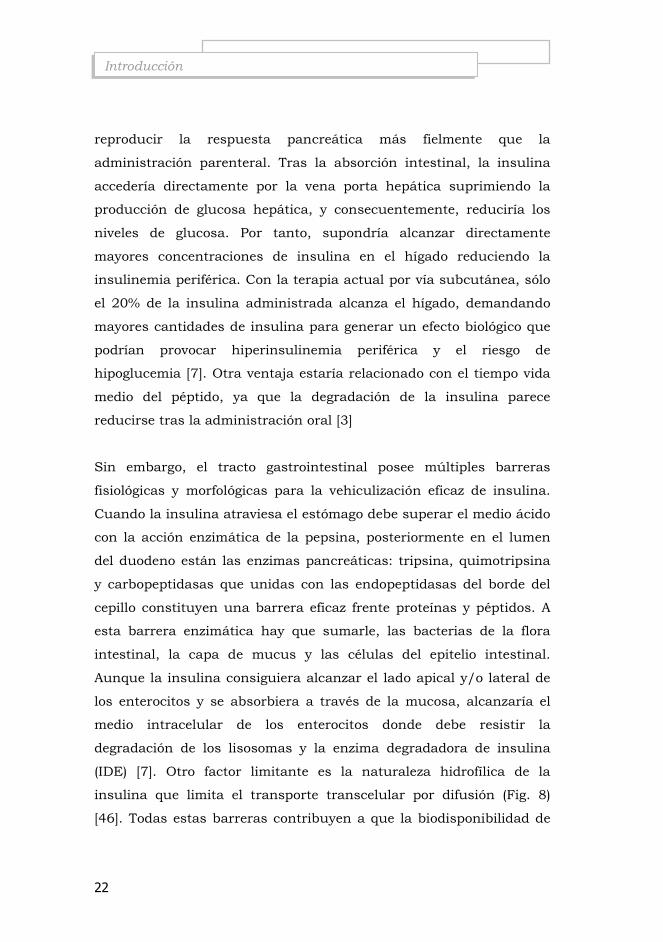
(IDE) [7]. Otro factor limitante es la naturaleza hidrofílica de la
insulina que limita el transporte transcelular por difusión (Fig. 8)
[46]. Todas estas barreras contribuyen a que la biodisponibilidad de

23
Introducción
la insulina por vía oral sea muy limitada (1-2 %) [47]. Por ello, con el
fin de mejorar la biodisponibilidad se han propuesto numerosas
estrategias capaces de proteger el péptido de la degradación
Figura 8: Mecanismos de transporte de sustancias a través del epitelio intestinal[46].
i.-Promotores de la absorción incrementan el transporte paracelular
[48] y transcelular a través de diferentes mecanismos, incluyendo
cambios en la fluidez de la membrana, disminuyen la viscosidad del
mucus y abriendo las uniones íntimas. Los ejemplos comunes de
promotores no específicos son las sales biliares [49, 50], n-lauryl-β-D-
maltopyranoside [51], EDTA [51] or EPA and DHA [52].

24
Introducción
ii.-Inhibidores enzimáticos disminuyen la velocidad de degradación
de la insulina por trypsina, α-quimotrypsina, y elastasa, y en menor
grado por enzimas de la membrana. Los inhibidores de polímeros
conjugados [53, 54] y los derivados de ovomucoides [55]
iii.- Sistemas poliméricos mucoadhesivos incrementan el tiempo de
residencia en el lugar de absorción, evitando la dilución o la
degradación en el fluido del lumen. Con polímeros hidrofílicos, como
poliacrilatos [56] derivados de celulosa [57] y quitosano [10, 58] se
han desarrollado sistemas de liberación para aumentar la absorción
del fármaco. Los polímeros tiolados (tiómeros) [59] catiónicos [60] y
aniónicos [61] mediante enlaces intra- e intermoleculares prolongan
el tiempo de descomposición controlando la liberación del péptido.
iv.- Sistemas de transporte nanoparticulados
Se han desarrollado numerosos sistemas coloidales para mejorar la
liberación intestinal de péptidos: nano- y micropartículas poliméricas,
nanocápsulas, microemulsiones, liposomas y micelas. La absorción
por vía oral de péptidos asociadas a sistemas particulados puede ser
mediante transporte paracelular, transcelular (endocitosis), o bien, a
través de las células M, tejido linfoide asociado al intestino (GALT) (<
10 µm) (Fig. 8) [48].
Para este trabajo nos centraremos en sistemas nanométricos testados
in vivo en distintos modelos animales para liberación de insulina por
vía oral. Los estudios han sido divididos en varios grupos; polímeros
naturales, polímeros sintéticos y liposomas. La Tabla 3 muestra los
estudios que utilizan un polímero naturale, quitosano (CS). El CS se
utilizó por su efecto mucoadhesivo como estrategia para incrementar
el tiempo de residencia del sistema en el epitelio intestinal,

25
Introducción
intensificando el contacto con el mucus y por tanto, reduciendo la
degradación entre la liberación del fármaco y la absorción a través del
epitelio. Por otra parte, el CS favorece la separación temporal de las
uniones íntimas entre células (tight juntions) del epitelio intestinal,
promueviendo el paso de sustancias a través de la membrana. Por
estas razones principalmente, el CS fue seleccionado como base para
el desarrollo de numerosas formulaciones de administración oral. En
general, los nanosistemas de CS muestran perfiles de liberación de
insulina que mantienen la reducción de los niveles plasmáticos de
glucosa (≤ 50%) durante periodos superiores a 5 h (tiempo máximo
alcanzado por vía nasal). Varios de los sistemas testados alcanzaron
reducciones en la glucemia durante 24h. Entre ellos destaca, los
nanocomplejos de liposoma-CS que decrecen los niveles de glucosa
un 35% respecto del valor basal durante 2-24h postadministración en
la rata [62] y las nanopartículas de CS/TPP (tripolifosfato) con 0.5-
10% poloxamer 188 [70c] reducen los niveles durante 30 h en ratas
diabéticas. Mientras que las nanopartículas que combinan dos
polímeros naturales, alginato o dextrano con el quitosano [63, 64],
necesitaron mayor dosis de insulina para reducir la glucemia durante
≤ 24 h. Por otra parte, los estudios que ensayaron nanopartículas con
CS modificados [65, 66], mostraron reducciones de la glucemia de
duración inferior a 12 h. Al igual que los complejos insulina-CS [67] o
las nanopartículas de CS combinadas con ácido glutámico [68] o con
metacrilatos [69].

26
Introducción
Tabla 3: Sistemas nanométricos a base de un polímero natural, quitosano (CS), para la liberación de insulina por vía orala
Sistemab Modelo
animal n Ins
UI/kg Tmin (h)
Cminc
Efecto-duración
Ref.
L/CS complexes Rata 7 10 24 55 ≤ 65%-2-24h [62]
TMC-Cys NP TMC NP
Rata
4
50 3
4 65 78
≤ 85% de 2-8h ≤ 85% de 2-5h
[66]
LSC/TPP NP Rata
diabética 4 60 6 65 ≤ 80% de 3-7h [65]
Ins/CS complex Rata
diabética
10
50
5
60
≤ 80% de 4-12h
[67]
Alginate/CS NP Rata
diabética
6 50
100
14
>60
≤ 80% de 2-18h
[64]
DS/CS NP Rata
diabética
6
100
14
65
< 80% de 4-24h
[63]
CS/γ-PGA NP
Rata diabética
6
30
5-10
50
≤ 50% de 5-10h
[68]
CS reduced gold NP Rata
diabética
6
50
2
70
≤ 80% de 1-3h
[70a]
CS/TMAEMC NP Rata 6 100 8 65% ≤ 80% de 6-12h [69]
CS/TPP NP 0.1% Pol 188
Rata diabética
8 14
21
10 60
40 ≤50% de 10-24 ≤ 70% de 10-24
[70b]
CS/TPP NP 0.5-10% Pol 188
Rata diabética
8
10
15
40
≤50% de 15-32
[70c]
aDatos actualizados hasta Abril 2010 (orden cronológico) bAbreviaturas: Ins:Insulina; NP:nanopartículas; NC:nanocápsulas; CS:quitosano; L/CS:nanocomplejos liposoma-quitosano; TMC:Trimetilquitosano; Cys Cisteina; LSC: Lauril Succinil Quitosano; DS:Dextrano Sulfato; γγγγ-PGA:Poli(γ-glutamic acid); TMAEMC:N-trimetilaminoetil metacrilato cloridrato; TPP: Tripolifosfato; Pol 188:poloxámero 188 cConcentración mínima de glucosa respecto del valor basal
Sin embargo, las nanopartículas de dextrano (DS) cubiertas con vitamina B12 (Tabla 4), utilizan el transportador de vitaminas para la absorción a través del tracto gastrointestinal prolongando la reducción de la glucemia durante 54 h [71, 72].

27
Introducción
Tabla 4: Sistemas nanométricos a base de un polímero natural, dextrano (DS), para la liberación de insulina por vía orala
Sistemab Modelo
animal
n Ins
UI/kg Tmin (h)
Cmin (%)c
Efecto-duración
Ref.
VB12/DS-NP
conjugates
Rata
diabética
6
20
M31: 5 M41:3-4 M42: 12
>30 30 >30
<70% de 24-54h <60% de 10-54h <60% de 24-54h
(2ª fase)
[72]
VB12/DS NP
conjugates
Rata
diabética
4
20 5 >30
60% de 24-54h
(2ªfase)
[71]
a Datos actualizados hasta Abril 2010
b Abreviaturas:VB12: vitamina B12; c Concentración mínima de glucosa respecto del valor basal
En la tabla 5 se resumen las principales características de los
nanosistemas formados con polímeros sintéticos para liberación de
insulina por vía oral. En general, los sistemas presentan tiempos de
hipoglucemia superiores a 5 h, excepto un sistema con PEG
(polietilenglicol) [73] y las nanopartículas de PLGA vehiculizadas con
baja dosis de insulina [74b] revelaron descensos de glucemia durante
tiempos inferiores a 5 h. Mientras que las partículas de PLGA con
mayor carga de insulina [75] o combinadas con lípidos [75b]
incrementaron el tiempo hasta las 24 h. Por otra parte, los
nanosistemas formados con acrilatos mostraron perfiles superiores a
12 h [76, 77], alcanzando en algunos casos las 36 h [78] y días [77,
81]. De entre todos los polímeros sintéticos estudiados destaca el
sistema formado con plurónico y ácido poliláctico que reduce los
niveles de glucosa entre 5-24 h postadministración hasta un 75%
respecto al valor basal [79].

28
Introducción
Tabla 5: Sistemas nanométricos a base de polímeros sintéticos para la liberación de insulina por vía orala.
Sistemab Modelo
animal n Ins
IU/kg Tmin (h)
Cmin (% glucosa basal)
Efecto-tiempo Ref.
PECA NP Rata
diabética 6 100 12-36 60 ≤80% de 10-36 [78]
Tris/PC/PEG NP Ratón
diabética
10 29
72
3
55
45
≤75% de 3-4h
≤75% de 2-5h
[73]
PLGA NP PLGA/Hp55 NP
Rata diabética 6 20 2 20 ≤80% de 2-8h [73
b] PLA-pluronic-PLA
vesicles Ratón
diabético 5 50 6 25 ≤25% de 5-23h [79]
PLGA NP Rata 5 50 10 60 ≤75% de 4-24h [75]
Ins-SPC complex /PLGA NP
Rata diabética 6 20 12 50 ≤80% de 2-24 [75
b]
PBA NP Rata
diabética 5 10 <0.5 30 ≤80% 1h [50]
P(AA-g-PEG) NP Rata
diabética 4 50 8 80 - [74]
PBCA NC Rata
diabética 5 50 7 70 ≤80% de 6-16h [76]
Ins-transferrin conjugates
Rata diabética
3-4 80 11 25 ≤70% de 7-11h [80]
PLGA NP Rata 5 20 1 60 ≤70%de 1-4h [74b]
PACA NC Rata
diabética 8 100 2 50 ≤80% de 2-13d [77]
PACA NC Rata
diabética 8 50 2 >50 ≤50% de 2-13d [81]
aDatos actualizados a diciembre 2009 (orden cronológico). bAbreviaturas: Ins: Insulina; NP: nanopartículas; NC: nanocápsulas PLGA: poly(lactide-co-glycolide); PECA: Polietilcianoacrilato; Tris/PC/PEG: Tristearina/ Fosfatydilcolina/ Polietilenglicol; SO: Sodio Oleato; M3,M4:Amino alquil derivados de la vitamina B12; Hp-55: hypromelosa Ftalato, HPMCP-55; SPC: Fosfatidilcolina de soja PBA: poli-isobutil-acrilato; P(AA-g-PEG): Poli (acril acid-g-polietilenglicol); PBCA: poli (iso-butil cianoacrilato); PBCA: Poli(isobutilcianoacrilato); PACA: Polialquilcianoacrilato En general, la mayoría de los sistemas de liberación de insulina por
vía oral propuestos hasta el momento presentan niveles de reducción
de la glucemia durante periodos prolongados (≥ 12 h) [62- 64, 67, 69,

29
Introducción
70b-72, 75-78, 81], incluso en casos alcanzando varios días [71, 72,
77, 81]. Sin embargo, el mayor descenso de glucosa producido por
estos sistemas se observa entre 5-14 horas tras la administración,
que induciría un alto riesgo de hipoglucemia si este descenso no
coincide con el horario de la ingesta de alimento.
La Tabla 6 muestra los ensayos de liposomas por vía oral en
diferentes modelos animales. En general, los porcentajes de
reducción de la glucemia son bastante limitados y no superan las 6h
como sucede en los estudios por vía nasal.
Tabla 6: Sistemas liposomiales y micelares nanométricos para la liberación
de insulina por vía orala .
Sistema Modelo
animal n Ins
IU/kg Tmin (h)
Cmin
(%)b Efecto-tiempo Ref.
ARMc Rata
diabética 5 25 4 70 ≤80% de 4-24h
[83]
liposomasd Rata
diabética 7 12 1.7-5 ~50 50% de 1.7-5h
[84]
Mucin- & PEG-liposomas
Rata
4
100
3 90
95
2-4h
[85]
liposomase Conejo diabético 6 100 2-4 40 <60% de
1-6h [86]
liposomasf Rata
diabética - 100 2 50 4h↑ [87]
liposomasg Rata
diabética 5 30 70 3
60 40 - [88]
a Datos actualizados hasta Abril 2010 b Concentración mínima de glucosa respecto del valor basal c Anhydrous reverse Michelle (micela reversa anhidra) dfosfatiletanolamina elecitina-colesterol ffosfatidilcolina-colesterol g fosfatidilcolina-colesterol-dicetil fosfato

30
Introducción
4.2.- Administración de Exenatida/Exendina-4.
A diferencia de otros fármacos, la biodisponibilidad por diferentes
vías de administración de la exenatida es muy baja. Por vía
subcutánea está aproximadamente entre 65-75 %, mientras que por
vía intranasal e intraduodenal 1.68 % y ~0.005 %, respectivamente
[6]. Aunque hasta el momento pocos estudios se han llevado a cabo
para mejorar su administración. Hargrove et al sustituyeron un
aminoácido de la cadena peptídica de la exenatida para incrementar
su estabilidad plasmática in vitro [89], sin embargo Son et al
consiguieron desarrollar un derivado de la exendina-4 de larga
duración sin pérdida de actividad, ensamblando ácidos biliares en la
cadena [90]. Únicamente, Jin et al desarrollaron un prototipo para vía
oral. Ensamblaron vitamina H (biotina) en la secuencia aminoacídica
del péptido, aumentando su estabilidad gastrointestinal, su
biodisponibilidad (~4 %) y su efecto hipoglucemiante en ratones
diabéticos [21]. Hasta el momento, no se ha desarrollado ningún
sistema de liberación efectivo que permita la administración de Ex4
por vías no invasivas (p.e. oral o nasal).
A diferencia de la insulina, la Ex4 no requiere niveles constantes en
sangre para mantener su acción. Por tanto, la administración de Ex4
por vía nasal se presenta como una alternativa adecuada por sus
numerosos beneficios. El epitelio nasal presenta una gran superficie
de absorción al estar cubierto de microvellosidades, está altamente
vascularizado y los fármacos administrados pasan directamente a
sangre evitando el efecto de primer paso por el filtro hepático.
Además, permite la administración de bajas dosis de fármaco, el
comienzo de la acción es muy rápido y es una vía fácilmente
accesible.

31
Introducción
A pesar del potencial de la vía nasal para la administración de
fármacos, son numerosos los factores que limitan la absorción nasal.
El aclaramiento mucociliar, la actividad enzimática y la barrera que
constituyen el epitelio y la capa de mucus frente a moléculas de alto
peso molecular e hidrofílicas. Por tanto, los grandes péptidos y
proteínas consiguen atravesar el epitelio mediante endocitosis en
bajas cantidades. Para solventar estas limitaciones de la vía nasal e
incrementar la biodisponibilidad de péptidos como la insulina se han
empleado múltiples estrategias: promotores de la absorción [91, 92],
fosfolípidos [93], inhibidores enzimáticos [94], mucoadhesivos [45] y
sistemas de liberación en polvo seco [95].
5. INTERÉS DE LA LECITINA Y EL QUITOSANO PARA LA
FORMACIÓN DE NANOSISTEMAS.
Desde hace décadas, la lecitina y el quitosano han mostrado un
mayor número de aplicaciones en biotecnología. El gran interés que
suscitan ambos compuestos como vehículos para el transporte de
moléculas activas proviene de sus propiedades. Ambas son de origen
natural, biocompatibles y biodegradables y carecen de toxicidad. De
aquí, que se hayan propuesto varios sistemas de transporte que
combinan ambas moléculas usando diferentes métodos de
preparación: micropartículas [96], nanopartículas [96b],
nanocápsulas [97], complejos [67] y liposomas recubiertos de
quitosano [98].
La LECITINA (LEC), según la United States Pharmacopeia (USP),
es un agente tensoactivo que está formado por una mezcla
compleja de fosfolípidos insolubles en acetona, formada
principalmente por fosfatidilcolina, fosfatidiletanolamina,

32
Introducción
fosfatidilserina y fosfatidilinusitol (Fig. 9), combinadas en varias
cantidades con otras sustancias, como los triglicéridos, ácidos
grasos y en algunos casos con carbohidratos. Su composición varía
dependiendo de la fuente y del grado de purificación,
consecuentemente alterando también sus propiedades físicas. La
lecitina es soluble en ácidos minerales y en hidrocarburos alifáticos
y aromáticos. Sin embargo, es parcialmente soluble en alcoholes
alifáticos y prácticamente insoluble en acetona y agua. Aunque la
LEC es una sustancia lipófila, debido a la presencia de ácidos
grasos esterificados de longitud variable, a su vez contiene un
grupo fosfatidil de carácter polar. De ello, resulta su carácter
anfifílico y su utilización extendida como emulsificante [99].
Figura 9: Estructura química de los principales componentes de la lecitina, donde R1 y R2 son cadenas de ácidos grasos.
Forma parte de todas las membranas celulares del organismo.
Además, este compuesto tiene una gran variedad de aplicaciones

33
Introducción
farmacéuticas (bases de supositorios; emulsificador, dispersante, y
estabilizante en inyectables y en la nutrición parenteral; protector
hepático en cirrosis alcohólicas), también se ha utilizado en
cosmética y en productos alimentarios. Además, estudios recientes
demuestran que la lisofosfatidilcolina de la LEC incrementa la
captura de carotenoides en cultivo de células humanas Caco-2 y
mejora la absorción oral de luteína en estudios in vivo [100, 101].
El QUITOSANO (CS), es un polisacárido pseudo-natural sintetizado
mediante un proceso de desacetilación en medio básico a partir de
la quitina, la cual está presente en el exoesqueleto de los
crustáceos, insectos y algunos hongos. El CS es un co-polímero
formado por una cadena lineal de unidades monoméricas de N-
acetil-glucosamina y N-glucosamina distribuidas aleatoriamente. El
grado de polimerización (G.P.) depende del número de residuos
monoméricos que formen la cadena. El número de subunidades
para clasificar el G.P en alto, medio o bajo es relativo. Por otra
parte, el grado de acetilación (G.A) está definido por el porcentaje
relativo de unidades de N-acetil-glucosamina presentes en el
polímero. El G.A del CS puede oscilar entre ~1-~70 % (Figura 10).
Sus condiciones de estabilidad dependen del G.A y son
especialmente importantes para la producción de soluciones
estables de quitosano, nanopartículas, macro- y microgeles y su
administración.

34
Introducción
Figura 10: Estructura química del quitosano (CS)
A diferencia de la quitina, el quitosano forma sales con ácidos
orgánicos (p.e. ácido glutámico) e inorgánicos, por tanto en
disolución, los grupos amino están protonados y el polímero
soluble está cargado positivamente. Las propiedades del CS, como
su pK0 y solubilidad, se pueden modificar variando su grado de
acetilación y factores como el pH y la fuerza iónica [102].
El CS es un biomaterial especialmente interesante gracias a la
combinación de sus propiedades mucoadhesiva y promotora de la
absorción. Su adherencia, viene definida por el carácter catiónico
de los residuos de glucosamina que le permiten interaccionar con
los residuos negativos de ácido siálico presentes en el mucus.
Además, a diferencia de los promotores de la absorción clásicos,
permite la apertura de las uniones íntimas intercelulares (“tight
juntions”) de forma temporal, facilitando la absorción de
numerosas moléculas a través de transporte paracelular y evitando
lesiones en el epitelio [103, 104].
Habida cuenta de sus propiedades, el CS es un biopolímero muy
útil para el desarrollo de materiales con diversas aplicaciones
biomédicas. Se han propuesto múltiples sistemas para la liberación
de fármacos compuestos por CS, entre ellos destacan las
nanopartículas, los complejos preparados por diferentes técnicas, o

35
Introducción
bien los sistemas coloidales recubiertos con CS (nanocápsulas o
liposomas)
El CS puede ser degradado por numerosas enzimas quitosanasas,
quitinasas, lisozima, o peptidasas [105-108], descomponiéndose en
unidades de menor peso molecular como CS con menor G.P. y/u
oligosacáridos de CS (COS, < 10kDa). Los oligosacáridos han sido
ampliamente utilizados en alimentación y cosmética [21]. Recientes
estudios demuestran que son capaces de reducir los niveles de
glucosa tras administraciones prolongadas y en determinadas
condiciones [109-112].
En este trabajo se desarrollarán sistemas nanométricos para la
administración transmucosal de insulina y exendina-4 a base de
lecitina y quitosano. El uso combinado de ambos materiales ha
permitido aprovechar las ventajas que ofrecen desde el punto de
vista físico-químico y biofarmacéutico.

36

PARTE I
Desarrollo de nanosistemas a base de lecitina para la administración de insulina por vía oral para el tratamiento de la diabetes mellitus tipo 1.


Antecedentes, hipótesis y objetivos
Artículo 1
Discusión
PARTE I


41
Antecedentes, hipótesis y objetivos Parte 1
“Desarrollo de nanosistemas a base de lecitina para la
administración de insulina por vía oral para el tratamiento de la
diabetes tipo 1”.
Antecedentes
1. El aporte exógeno de insulina por vía subcutánea a través de
inyecciones combinadas, que constituye la base de la terapia
actual para el tratamiento de la diabetes tipo 1, no consigue
reproducir fielmente el perfil fisiológico requerido, provocando
numerosos efectos secundarios. Actualmente, los
nanosistemas propuestos para la liberación oral de insulina
no presentan un perfil adecuado de liberación que permitan
sustituir la administración subcutánea de las insulinas de
acción corta.
2. Las interacciones electrostáticas (iónicas) entre ciertos
componentes con cargas opuestas dan lugar a la formación de
nanosistemas [113]. La insulina posee capacidad de la
interacciona con fosfolípidos presentes en la lecitina [96, 114].
3. Sistemas coloidales desarrollados a partir lecitina [62, 75b,
85, 88] o quitosano [63, 64, 66] han permitido mejorar la
absorción sistémica de insulina a través de la mucosa oral.
Ambas moléculas unidas por interacción iónica forman, entre
otros, nanopartículas que han demostrado encapsular
eficazmente diferentes tipos de fármacos, entre ellos proteínas,
Albúmina Sérica Bovina (BSA) [115] e incrementar la
absorción de melatonina [116] y tamoxifeno [116b] en
cultivos celulares de Caco-2.

42
Antecedentes, hipótesis y objetivos Parte 1
Hipótesis
1. La administración oral podría reproducir la liberación de
insulina producida por el páncreas más fielmente que la
administración subcutánea. La insulina accederá al hígado
directamente a través de la vena porta, suprimiendo la
producción de glucosa hepática, reduciendo los riesgos de
hiperinsulinemia periférica e hipoglucemia.
2. La interacción iónica entre cargas opuestas de la insulina y la
lecitina producirá una estructura nanoscópica que potenciará
la absorción de la insulina por vía oral.
3. La adicción de quitosano al sistema conferirá a la superficie
de las nanopartículas la capacidad de interaccionar con la
mucosa intestinal, a través de los residuos de ácido siálico
presentes en la mucina. Esta interacción específica con las
células epiteliales, promoverá la apertura de las uniones
íntimas mejorando la absorción por vía oral
Objetivos
Teniendo en cuenta lo anterior, el objetivo general de la primera parte
de este trabajo ha sido el desarrollo de nanosistemas capaces de
incrementar la absorción de insulina tras administración por vía oral.
El trabajo experimental llevado a cabo para alcanzar el objetivo
global, se ha recogido en los apartados que se detallan a
continuación:

43
Antecedentes, hipótesis y objetivos Parte 1
1. Desarrollo y evaluación del comportamiento in vitro de las
nanocomplejos de lecitina-insulina para la administración de
insulina por vía oral. En este objetivo se desarrollará la
optimización del proceso de preparación de los
nanocomplejos, evaluando diferentes variables de formulación
tecnológicas sobre las características finales de los sistemas.
2. Desarrollo y evaluación del comportamiento in vitro de las
nanopartículas de lecitina y quitosano para la administración
de insulina por vía oral. Se evaluará específicamente la
incorporación del péptido al sistema, utilizando la posibilidad
que ofrecen las proteínas de variar su carga según el pH del
medio de disolución.
3. Estudio de los efectos in vivo de los nanocomplejos y las
nanopartículas cargadas con insulina mediante
administración oral en diferentes modelos experimentales
diabéticos. Se pretende evaluar si los nanosistemas de lecitina
pueden favorecer la absorción sistémica de insulina
administrada por vía oral. Se analizarán la influencia de la
presencia o ausencia de quitosano en el sistema sobre la
respuesta in vivo en dos modelos de roedores diabéticos.

44

Antecedentes, hipótesis y objetivos
Artículo 1
Discusión
PARTE I


47
ARTÍCULO 1
Lecithin-based nanocarriers for oral delivery of insulin
in diabetic rodents
Valle-Gallego A1,2., Goycoolea F.M1., Mallo F2. and Alonso M.J1*
1Departement of Pharmacy and Pharmaceutical Technology, Faculty
of Pharmacy, University of Santiago de Compostela, Spain
2Laboratory of Endocrinology, Faculty of Biology, University of Vigo.
Spain
*Corresponding author: Prof. Alonso MJ. Tel: + 34 981 594627 Fax: + 34 981 547148 E-mail: [email protected]
Manuscript submitted to publication

48

49
Abstract
This work has addressed the development of two different types of
nanostructures intended as nanocarriers for the effective oral delivery of
insulin (INS). Namely, nanocomplexes of lecithin (LEC) and INS and
nanoparticles comprising LEC and chitosan (CS) loaded with INS.
Different formulations of LEC–INS nanocomplexes at varying mass
ratios of both components were developed with adequate physical
characteristics (Z-average size ~209 nm; ζ~-44mV); high production
yield (~60%) and excellent INS association efficiency (A.E. ~97-100%).
These systems also exhibited high in vitro stability in simulated
gastrointestinal fluids with little release of INS in intestinal conditions
(pH 6.8, <~20%). In turn, INS-loaded LEC–CS nanoparticles were also
formulated at different LEC/CS mass ratios and optimal prototypes
(average sizes of 212±9 nm, ζ~+32±1 mV) with high INS association
efficiency (> 80%) were achieved. These systems exhibited adequate
colloidal stability in simulated biological fluids. Both systems after in vivo
assays in STZ-induced diabetic rats revealed the capacity to improve the
systemic absorption of INS peroral. The decrease of glycemic levels
(~54%) was achieved 5 h after oral gavage of LEC–INS nanocomplexes.
In turn, the oral administering of LEC/CS 5/1 nanoparticles to STZ-
induced diabetic mice led to a reduction in hyperglycaemia by 80% 2 h
post-administering, a response that was prolonged for 12 h. Essentially
similar results were confirmed in STZ-induced diabetic rats, where a
rapid onset of action (~0.5 h) and a longer duration were observed, in
contrast with the slow onset (~3 h) and short duration obtained with

50
LEC/INS nanocomplexes in the same animal model. The results of this
study are regarded as promising towards achieving adequate prototypes
for oral administering of insulin in diabetes therapy.
Key words: insulin, nanoparticles, nanocomplexes, lecithin, oral delivery

I
51
…for oral delivery of insulin Artículo 1
1. Introduction
The rational development of insulin (INS) delivery systems is beset of
complex requirements so as to meet the needed pharmacokinetics/
pharmacodynamics associated with an adequate diabetes mellitus therapy
[1] in order to normalize INS plasma profile to regulate systemic glucose
levels. Currently, the conventional diabetes treatment combines the
administering of INS by subcutaneous route with different duration
(rapid-, short-, intermediate- and long-acting INS) [2]. Over the last
decades, attempts to overcome the limitations and drawbacks associated
with conventional subcutaneous INS therapy have been made. However,
despite the known disadvantages of the subcutaneous route, thus far,
only limited success has been achieved with alternative treatments able to
reproduce as closely as possible the physiological profile resulting from
endogenous insulin secretion [3]. Among the alternative routes of insulin
delivery, the oral (enteric gastrointestinal) route is undoubtedly the most
preferred one. In fact, oral insulin administering seems to be the most
convenient and advantageous route from a physiological standpoint. This
is due to the fact that insulin absorbed by intestinal epithelium reaches
the liver through the portal vein and can directly inhibit hepatic glucose
output. This effect on the liver is essential in the maintenance of glucose
homeostasis while avoiding peripheral hyperinsulinemia [4]. However, the
bioavailability of insulin peroral is known to be very small (<2 %) [5].
This is the consequence of a number of factors, namely, the low
absorption of INS by the intestinal epithelia, its susceptibility to acidic
and proteolytic degradation during the transit through the gastrointestinal
tract and the rapid clearance from the site of absorption. Consequently,

Artículo 1
52
Lecithin-based nanocarriers…
an effective delivery system for INS, among other challenges, must be
able to overcome these physical and metabolic barriers so as to facilitate
its transport to the blood stream across the intestinal epithelia.
To this end, various approaches have been adopted, including utilizing
INS derivative conjugates and prodrugs with high stability against
degradation by gastrointestinal enzymes [6], INS associated antiproteases
and hydrogels or combined with absorption enhancers (cyclodextrins,
bile salts or surfactants) [4]. However, nanocarrier systems have emerged
as highly promising systems to increase the intestinal uptake of peptides
[7]. The nanoscopic dimensions provide these systems with an extremely
large surface-to-volume ratio and surface functionality [8]. Liposomes [9-
11], nanocapsules [12, 13] and nanoparticulate systems [14-18] have been
studied as potential delivery systems for oral administering of insulin.
This type of carriers has been made out of biocompatible materials
including polymers and lipidic compounds of natural and synthetic origin
[13, 19-22]. However, the pharmacokinetic/pharmacodynamic
parameters are strongly influenced by the composition of the system.
A great deal of commendable research efforts have aimed to develop an
adequate carrier for oral delivery of insulin that can effectively replace
subcutaneous insulin. A recent study has addressed the development of
hybrid biodegradable nanoparticles of copolymers of polylactic and
polyglycolic acid (PLGA) and an insulin-phospholipid complex for INS’s
oral administering achieving a relative bioavailability of 7.7% in STZ-
induced diabetic rats [20, 21]. Most of the effective developed systems

I
53
…for oral delivery of insulin Artículo 1
present a long-term response (hypoglycaemic effect ≥ 12 h) with a
maximum decrease 5 h post-administering. These have included both
natural (CS or dextran sulfate [14, 15, 19, 22-29] or synthetic [13, 20, 30,
31] polymers. However, the insulin therapy requires an oral formulation
with a rapid onset of action and a prolonged effect that matches the
maximum hypoglycaemic effect during the increase of glucose after
meals. Hence, any improvement in this direction would represent a
significant step towards a successful system. Motivated by this overall
goal, in the present work, we have addressed the pharmacological
performance of lecithin-insulin (LEC–INS) nanocomplexes and of INS-
loaded lecithin-chitosan (LEC–CS) nanoparticles, both produced under a
very simple and mild technique, on the enhancement of absorption of
INS by oral route in two different rodent models.
LEC is a lipid mixture of phospholipids that has been frequently used for
liposome and micelle formation and is largely employed in
pharmaceutical or nutraceutical formulations [32, 33]. In this regards,
lysophosphatydylcholine of LEC has been credited to markedly enhance
the uptake of carotenoids solubilized in mixed micelles by Caco-2 human
intestinal cells and in vivo studies of rats [34, 35]. In turn, the rationale
behind the use of CS in the second type of systems addressed in this
work was due to the firmly established pharmacological effects that CS-
based nanocarriers have shown to improve the absorption and
pharmacological performance of insulin administered peroral [7, 8, 17,
36, 37] and to enhance its systemic absorption after nasal administering
[38-40]. Indeed, CS has been found to facilitate the interaction of

Artículo 1
54
Lecithin-based nanocarriers…
nanocarriers with mucosae and, hence, to improving the permeability and
absorption of peptide drugs [41]. Interestingly, it has been proved that CS
is able to interact readily with LEC by means of ionic and hydrophobic
interactions [42-45] thus forming a new self-organized system [46, 47]. In
several studies, LEC–CS nanoparticles have been found to be effective to
entrap drugs of different type, such as tamoxifen [48], progesterone [47]
or BSA [46]. Nanoparticles of this type have also been found to enhance
the permeability of melatonin through Caco-2 cell monolayers, a
common in vitro model of the intestinal epithelium [49], but to the best
of our knowledge, their efficacy to deliver INS by oral route has not yet
been tested in vivo.
2. Material and methods
2.1.-Materials
CS in the form of hydrocloride salt (Protasan UP-Cl 113, Mw = 125 kDa,
deacetylation degree: 86%) was purchased from Pronova Byopolimer,
A.S. (Norway). Lecithin (LEC) (Epikuron 145V) was donated by Cargill
Texturizing Solutions, S.A (Barcelona, Spain). Details of composition
(according with the supplier) of LEC are: phospholipid complex min
97% (phosphatidylcholine min 45%; phosphatidylethanolamine min 10%;
phosphatidylinositol 3%; phosphatidic acid 3%; lyso-phosphatidylcholin
4%; phosphorus 2.7-3.4%). INS from bovine pancreas [Ref. Nº-I5500,
Mw 5.7 kDa, pI 5.3, Zn traces ~0.5%] was supplied from Sigma-Aldrich
Chemie (Steinheim, Germany). Milli-Q quality water (18.2 MΩ.cm) was
utilized throughout.

I
55
…for oral delivery of insulin Artículo 1
2.2. -Preparation of LEC–INS nanocomplexes
These nanocomplexes were obtained by interaction of negatively charged
LEC’s lipid components and positively charged INS’s aminoacids. To
find the optimum composition a series of blends of both components
were explored. To this end, LEC aliquots of varying volumes of a LEC
ethanolic stock solution (25 mg/ml) were injected to INS aliquot
solutions (12.5 mg/ml) with a micropipette under magnetic stirring
during 10 min at room temperature. The volumes of both stocks were
adjusted so as to achieve nanocomplexes of lecithin/insulin (LEC/INS)
mass ratios in the range 2/1 to 60/1. For the formulations of these
systems, INS was dissolved in acetic acid solution of varying
concentration in the range 0.1 to 2.0% (v/v).
2.3. - Preparation of INS-loaded LEC–CS nanoparticles
The general experimental protocol to prepare unloaded LEC–CS auto-
assembled nanoparticles was that previously described by Sonvico et al
[47]. In brief, CS aqueous solutions (0.027-0.56 mg/ml) were prepared by
fully dissolving the polymer in water. To prepare INS-loaded
nanoparticles two strategies were tested. Accordingly, one strategy
involved that INS was dissolved in NaOH (pH~11.2) (i.e. bearing net
negative charge) and subsequently added to a LEC ethanolic solution (25
mg/ml), this anionic solution was further on incorporated into a CS
aqueous solution under gentle magnetic stirring for ~10 min at room
temperature. Under the second strategy, INS was dissolved in acetic acid
1% (pH~3.3) (i.e. bearing net positive charge). The procedure was
identical to the previous one in all respects except that INS was blended

Artículo 1
56
Lecithin-based nanocarriers…
in the CS solution. In both cases, the LEC–CS ratios assayed were in the
range 5 to 80 (w/w).
2.4. – Isolation and yield production
The INS-loaded LEC–CS nanoparticles were isolated by
ultracentrifugation (138800 × g, 2 hours, 15 °C) in vials on a bed of
glycerol (~50 µL) carefully deposited at the bottom of the vial. The
supernatant was removed cautiously with a pipette.
The processing yield of LEC–INS nanocomplexes and INS-loaded LEC–
CS nanoparticles was determined by centrifuging (138800 × g, 2 h, 15 °C)
accurately weighed aliquots of the nanoparticles in vials without added
glycerol. The supernatants were carefully separated and the centrifuged
pellets were freeze-dried for 2 days and subsequently weighed (n = 3).
The yield production was calculated as follows:
Nanocarriers weight Yield (%) = x 100
Total solids weight
2.5. - Physical characterization
The Z-average size distribution of the colloidal systems was analyzed by
photon correlation spectroscopy (PCS). To this end, aliquots of the
various systems were diluted to the appropriate concentration with
filtered ultrapure water so as to achieve the adequate counts per second.
Measurements were performed at 25 ºC with an angle detection of 90º.
The zeta potential (ζ) was calculated from the mean electrophoretic

I
57
…for oral delivery of insulin Artículo 1
mobility values, which were determined by laser Doppler anemometry
(LDA). The suspensions were diluted with KCl 1 mM and placed in the
electrophoretic cell where a potential of ±150 mV was established. The
PCS size measurements were performed using either a Zetasizer® 3000
HS or a NanoZS ZEN 3600 fitted with a red laser light beam (λ=632.8
nm) (Malvern Instruments, Malvern, UK). The overall differences
between measurements made on each type of instruments were within
21±6 nm. LDA measurements were performed in the 3000 HS
instrument.
The morphology of nanocomplexes was examined by Transmission
electron microscopy on a Philips CM12 TEM instrument (Eindhoven,
The Netherlands). To this end, 10 µL of a 2% solution of
phosphotungstic acid was mixed with an equal volume of a 1:100 dilution
of nanocomplexes in water. Immediately afterward, an aliquot of 5 µL
was immobilized on a copper grid coated with a Formvar® membrane
and allowed to dry.
2.6. - INS association efficiency
The efficacy of association of INS was calculated indirectly, by
quantifying of free INS in the supernatant after isolation as described
above. The INS concentration was analyzed by HPLC (Agilent
Technologies, Germany) according with the method described by
Krauland [50].
The association efficiency (A.E.) was calculated as follows:

Artículo 1
58
Lecithin-based nanocarriers…
Total INS amount − Free INS amount
A.E. (%) = × 100
Total INS amount
2.7. - Stability in simulated gastric and intestinal fluids
The colloidal stability of both INS-loaded systems was evaluated during
incubation at 37°C under horizontal shaking (~100 rpm) in simulated
gastric (SIG, pH=1.2) and intestinal (SIF, pH=6.8) fluids (both without
enzymes) (USP 31 - NF 29. 1st ed. Vol. 1. 2008).
2.8. - In vitro release studies
INS release studies were performed by incubating aliquots of
nanocomplexes or isolated nanoparticles in 1.5 ml of simulated fluid for
each sample at 37 °C under horizontal shaking (~100 rpm). The INS
concentration incubated in the release media was increased to 15µg/ml
regarding sink conditions in order to detect the minimum released
concentration of INS by HPLC analysis. At appropriate time intervals
(30, 60, 120, 240 360 min), the samples were taken out to the
ultracentrifuge at 4 ºC to separate the released INS from the system. The
amount of INS released was determined also by HPLC [50].
2.9. -In vivo studies
In this study, we addressed the effects on the efficacy to induce a
pharmacological response after oral administering INS-loaded systems in
two diabetic models: (i) the effect of LEC–INS nanocomplexes in

I
59
…for oral delivery of insulin Artículo 1
diabetic rats (ii) after incorporation of CS, the effect of INS-loaded LEC–
CS in diabetic mice (iii) and diabetic rats.
Healthy adult male Wild-type mice and Sprague–Dawley rats were housed
in cages (five animals each) at 21±1°C in a 12:12 h light:dark cycle (all
experiments were performed in the light cycle) and were fed and watered
ad libitum. After 3 days, diabetes mellitus was induced by single injection
of streptozotocin (STZ, 70 mg/kg i.p) in 0.1M citrate buffer (pH 5) and
confirmed after 24 h by detecting glucose in urine. The diabetic animals
were maintained with 4.5-6IU/kg/12 h of subcutaneous INS prior to the
oral experiment. To monitor of the diabetes progression, glucose in urine
and the body weight were quantified daily during one week. Following
the procedure, three days before oral administering, the animals were
placed in a metabolic cage (4 mice/cage; 1 rat/cage) in order to check the
food, drink and urine. The animals were fasted for 12 h previous to oral
assay, but allowed water ad libitum. Animals were kept conscious during
the experiments. Hence, blood samples were collected from the tail vein
prior to oral administering to establish the baseline glucose levels, and at
different times after dosing. Urine samples in mice were collected from
each cage at time points 0 and 8 h. The plasma was separated by
centrifugation at ~2500 ×g for 5 min. Changes in plasma glucose levels
were determined by Glucose RTUTM kit (BioMerieux® SA, France).
2.10. Statistical analysis
Data are expressed as means-S.E.M. In vivo data were analysed by
Kruskall-Wallis test followed by U-Mann Whitney’s post hoc test was

Artículo 1
60
Lecithin-based nanocarriers…
used for comparisons between the two independent groups. Chi-squared
(χ2) test was used selectively for comparing data on the percentages of
decrease glycaemia (≥40%) between the dependent variable (glucose
level) and the independent variable (treatment of INS-loaded LEC/CS
nanoparticles) in STZ-induced diabetic rats.
3. Results and discussion
One of the most elusive aims in modern pharmaceutical technology has
been to achieve an efficient delivery system of insulin (INS) by routes
alternative to subcutaneous injection [3]. In this context, the rationale
behind the present study was to develop nanocarrier systems that would
take advantage of the expected properties of nanocomplexes formed
between INS and LEC and of INS-loaded LEC–CS nanoparticles to
preserve the bioactivity of the peptide during both the process of
formulation and its subsequent transit trough the gastrointestinal tract as
well as to ferry it through the intestinal mucosa, thus enhancing its
absorption and bioavailability. As a first step toward this end, the
preparation conditions of LEC–INS nanocomplex were optimized in
terms of mass ratio of both constituents. Subsequently, CS was
incorporated in prototype formulations of LEC–INS nanocarriers with a
view to modify their surface properties, thus effectively, improving the
mucoadhesion and intestinal absorption of INS. Both LEC–INS
nanocomplexes and INS-loaded LEC–CS nanoparticles were

I
61
…for oral delivery of insulin Artículo 1
characterized and their potential as oral delivery vehicles was assayed in
two diabetic rodent models.
3.1. Production and Characterization of LEC–INS nanocomplexes
The ability of INS to interact with the major constituents of LEC, such as
phosphatidylcholine, can be conceived as the result of the formation of a
polyion-surfactant complex between oppositely charged ionic groups on
both molecules at the appropriate pH [51]. Systems of this type have
been exploited in early studies for the development of insulin-loaded
liposomes for oral delivery [11] and their general physicochemical
properties have been addressed elsewhere [52, 53].
In the present work, preliminary experiments were conducted to optimize
the size dimensions of nanocomplexes of LEC–INS. A series of blends
of LEC and INS at varying LEC/INS mass ratios were explored. To this
end, INS was dissolved in aqueous acetic acid solutions of varying
concentration in the range 0.01–2 % (v/v), thus inducing a varying degree
of protonation of INS (pI ~ 5.3). The obtained systems were inspected
visually and three types of systems were identified according with their
appearance: clear solutions, white opalescent colloidal suspensions and
aggregates (visible precipitates). The experimental size data were used to
construct a surface response map in terms of the LEC/INS mass ratio
and acetic acid concentration (Figure 1). Aggregates were not considered
for the construction of this plot. The obtained structures were arbitrarily
assigned to three categories according with their dimensions, namely,
structures ≤ ~90 nm, ~90 – 180 nm and ≥ ~180 nm. The incorporation

Artículo 1
62
Lecithin-based nanocarriers…
of increasing positive charge of INS with respect to LEC led to an
increase the compensated electroneutral regions, entailing an increase on
the hydrophobic character of electrostatic complexes and thus, led to a
slight increase in their size. To account for this behaviour, the
nanocomplexes are formed as INS becomes progressively more charged
and also when the LEC/INS mass ratio <~15/1. Meanwhile, the overall
increase in LEC/INS mass ratio up to a value of ~20/1 is clearly
accompanied by a pronounced reduction in particle size, with no further
decrease beyond this ratio. Interestingly, in the narrow range of
composition at LEC/INS mass ratios ~20/1 – 30/1 at low
concentrations of acetic acid appear very small species (≤ ~90 nm),
suspected to be comprised by liposomes. Hence, the region defined by
the size range ~90 – 180 nm can be expected to comprise a mixture of
nanocomplexes and liposomes.
Figure 1. Response surface map showing the effect of lecithin/insulin mass ratio and acetic acid concentration on the particle size of lecithin-insulin nanocomplexes.
0.1
90
180
270
1
2
20
30
40
50
Siz
e (
nm
)
Acetic Acid %
LEC
/INS m
as ratio
90
180
270

I
63
…for oral delivery of insulin Artículo 1
Size distribution curves obtained by DLS representative of LEC/INS
mass ratios ~60/1, 16/1 and 10/1 along with liposomes comprising
solely lecithin are shown in Figure 2. It reveals that the curve
corresponding to the system of LEC/INS mass ratios ~60/1 exhibits a
bimodal size distribution with predominance of a population of ~120 nm
and the presence of a small fraction of particles of size ~20 nm. This
bimodal distribution is also observed for the systems at LEC/INS mass
ratio 16/1 and for lecithin liposomes, though the predominant peak is
shifted to ~60 nm while the small one to ~10 nm. Whereas the system
with proportionally less LEC contents (LEC/INS mass ratio ~10/1)
shows a single population with monomodal size distribution
corresponding with the nanocomplexes formation.
1 10 100 1000
2
4
6
8
10
12
14 LEC/INS mass ratio 10/1 (NCX)
LEC/INS mass ratio 60/1 (Mix)
LEC/INS mass ratio 16/1 (Liposomes)
Lecithin liposomes
Inte
nsity (
AU
)
Size (nm)
Figure 2. Size distribution of the scattered light intensity as determined by photocorrelation spectroscopoy in water at 25ºC of lecithin-insulin systems with mass ratios (w/w): 10/1 nanocomplexes (dash line); 60/1 mixture of nanocomplexes and liposomes (dash-dot line); liposomes 16/1 (short dot). Liposomes formed with lecithin (continuos line).

Artículo 1
64
Lecithin-based nanocarriers…
For subsequent experiments a prototype formulation was prepared with a
LEC/INS mass ratio 10 and by dissolving INS in 1% (v/v) acetic acid.
The composition for this prototype was selected on the grounds of its
size (209±18 nm) and more so, on its high association efficiency for
insulin (A.E. ~97–100%) as shown in Table 1. Indeed, INS A.E. for this
prototype formulation was almost total representing a clear functional
advantage over the conventional liposomes with notably lower affinity
for drug binding (1- 53%) [9, 11, 54, 55]. Such high INS A.E. is explained
as the consequence of positively charged INS at pH below its isoelectric
point (INS pI~5.3), that favours its electrostatic interaction with
negatively charged phospholipidic macroions in LEC [45]. LEC–INS
complexes in general showed negative surface charge (ζ~-42±4 mV) with
high yields of production (60±6%). From the biopharmaceutical
standpoint, it is worth pointing out that the high negative zeta potential
could be expected to favour the bioadhesivity and absorption by both M-
cells and enterocytes during oral delivery, as suggested in previous studies
[8]. These nanocomplexes also exhibited high stability (> 90 min) in
simulated intestinal fluid, yet another sine qua non pre-requisite for their
use as oral drug delivery vehicles.
The morphology and features of LEC–INS systems of mass ratios 60 and
10 were probed by TEM. As evidenced in the representative micrographs
of the LEC – INS of mass ratio 60 (Fig. 3a), it can be appreciated that
spherical assemblies with a core–shell structure were formed. A closer
inspection reveals that these assemblies are constitued by a core
sorrounded by a shell of variable thickness. In turn, the TEM images of
nanocomplexes, corresponding with LEC/INS mass ratio 10 (Figure 3b),

I
65
…for oral delivery of insulin Artículo 1
reveal the presence of species of irregular shape and size and it is not
possible to discern the presence of a bilayer.
a) b)
Figure 3. Transmission electron micrographs of lecithin – insulin mass ratio 60/1(a) and 10/1 (b)
3.2. Production and characterization of INS loaded LEC– CS
nanoparticles
LEC–CS nanoparticles were the second class of systems addressed in the
present study. The rationale behind the incorporation of CS in the
formulation was to achieve mucoadhesivity and improve the absorption
of INS. Preliminary trials were carried out to explore the optimal
composition of unloaded (blank) nanoparticles of this kind [47]. The
unloaded nanoparticles produced at LEC/CS ratios varying from 5/1 to
20/1 (w/w) invariably displayed positive ζ values, while those comprising
a low amount of CS (LEC/CS 60/1 to 80/1) experienced a charge
inversion with negative surface charge [47]. Hence, a series of systems of
varying composition were formulated by holding fixed the amount of
LEC while varying that of CS incorporated, so as to afford blends of

Artículo 1
66
Lecithin-based nanocarriers…
LEC/CS mass ratios from 5/1 to 80/1. The ionic character of INS was
also modified by dissolving the peptide either in acetic acid 1% (v/v) or
in NaOH 0.1 N, thus bearing either a positive or negative net charge,
respectively.
Interestingly, when INS was incorporated bearing a net positive charge,
nanoparticles were formed only at LEC/CS mass ratios of 5/1 to 10/1
(i.e. at high amounts of CS), while formulations incorporating
proportionally less CS (LEC/CS 60/1 to 80/1) invariably produced
aggregates. The stabilization brought in by CS in systems of lower
LEC/CS mass ratio can be accounted for as the formation of a
hydrophobic complex between LEC–INS nanocomplexes and
neutralized LEC–CS species, presumably involving lipidic bilayers. In
turn, aggregation due to an excess of LEC could be explained as
consequence of surface charge neutralization attained by the oppositely
charged groups of INS and CS, leading to electrostatic repulsion among
particles, and thus, a loss of stabilization [45, 46]. A schematic
representation of the proposed interactions occurring in the system is
shown in Figure 4a. Notice in Table 1 that regardless of the LEC/CS
mass ratio, these nanoparticles exhibited very similar average particle size
(230 – 241 nm) and positive surface charge (ζ~+28±1 mV) values. The
results are consistent with the idea that the excess of positive charged
amino groups of CS at the surface is required for its colloidal
stabilization. This is in agreement with results for unloaded LEC–CS
nanoparticles [43]. Despite the fact that INS was bearing similar charge as
CS, a high INS association efficiency (~99-100%) was attained. Small

I
67
…for oral delivery of insulin Artículo 1
angle X-ray scattering (SAXS) studies conducted in unloaded LEC–CS
nanoparticles systems have postulate that multilamellar structures are
formed in these systems. The outer layer is thought out to be formed by a
single bilayer [43, 44]. The formation of these multilamellar
phospholipidic structures entrapping INS molecules can account the high
INS A.E.
Table 1/ Physic-chemical characteristics of insulin-loaded nanosystems (mean ± S.D., n = 3).
Insu lin ª
LEC/CS mass ratio (w/w)
Size (nm)
P.D.I
ζζζζ (mV) Insulin A.E. (%)
Stab.b (pH 6.8)
+ LEC-INSc 209±18 0.18-0.21 -42±4 97-100 +++
+ 5/1
230±5 0.24-0.29 +28±1 99-100 _
+ 10/1
241±4 0.31-0.36 +28±1 99-100 _
- 5/1
212±9 0.26-0.40 +32±1 83±2 +
- 60/1
248±8 0.15-0.19 - 25±6 22±6 +++
- 80/1
285±14 0.20-0.41 - 20±2 10±5 +++
ªInsulin dissolved in acetic acid 1% (positive charge) or NaOH 0.1N (negative charge). b Stability in simulated intestinal fluid was classified as: (+++) stable ≥ 90 min, (+) stable < 60 min and (-) unstable (immediate aggregation). cLecithin-insulin nanocomplexes (LEC/INS mass ratio 10/1 w/w).
By contrast, particles harnessed when INS was incorporated in the
formulation bearing negative net charge, were formed regardless of the
amount of CS (i.e. LEC/CS 60/1 to 80/1). The particle diameter was
very similar than for the rest of the nanosystems (203–299 nm). In turn,

Artículo 1
68
Lecithin-based nanocarriers…
the zeta potential of systems comprising high CS contents (i.e. LEC/CS
5/1) attained highly positive values (+32±1 mV). Meanwhile, for
formulations comprising a low content of CS (LEC/CS 60/1 to 80/1) a
charge inversion was observed (-20 – -25 mV) as the complexation
between oppositely charged groups goes beyond charge neutralization, in
line with previous studies [43]. This phenomenon is also accompanied by
a moderate overall increment in particle size (i.e. ~30%) with a drastic
reduction in the INS A.E. from 83±2% for LEC/CS 5/1 to 10±5% for
LEC/CS 80/1, presumably as the expected consequence of the
displacement of the peptide by LEC competing by CS’s positive charged
sites. In these systems one can envisage that INS is directly bound to CS
and hence, LEC competes for the available positive charges in the
polysaccharide. The negative ζ values of these systems can be due to the
formation of LEC lipid bilayers placed at the nanoparticle surface, thus
orienting the negative phospholipidic polar heads of LEC towards the
surface, as schematically represented in Figure 4b.

I
69
…for oral delivery of insulin Artículo 1
a)
b)
Figure 4. Schematic representation of the interactions proposed to occur in insulin–loaded lecithin–chitosan nanoparticle systems when insulin bears either (a) positive or (b) negative net charge. Chitosan polymer (line), insulin (big circles) and lecithin ( ) Based on these results, the nanoparticle formulation LEC/CS 5/1 was
selected for further experiments due to the adequate optimal
physicochemical properties and stability in gastrointestinal fluids.
3.3 Stability in gastric and intestinal simulated media
Before proceeding with the in vivo studies, it was necessary to confirm
that the developed prototype nanosystems were stable in conditions that
LEC/CS ≤ ~10 LEC/CS ≥ ~40
LEC/CS ≤ ~5 LEC/CS ≥ ~60

Artículo 1
70
Lecithin-based nanocarriers…
simulate those found in the biological environment of the oral mucosae.
To this end, the evolution of Z-average particle size of nanosystems was
assayed in SGF (pH 1.2) and SIF (pH 6.8) without the presence of
enzymes. Accordingly, the evolution of Z-average diameter of LEC–INS
nanocomplexes and INS-loaded LEC–CS nanoparticles was monitored
during 90 min at 37ºC under horizontal shaking as already shown in
Table 1.
In general, the LEC/CS systems presented excellent stability in SGF for
up 90 min as illustrated in the Z-average size evolution traces in Figure 5.
Notice that the LEC–INS system without CS showed excellent stability
in SIF and SGF during the whole experiment. This represents a clear
advantage over LEC-based liposomes that are not stable in SGF [56]. On
the other hand, all LEC–CS formulations prepared with INS dissolved in
acetic acid (i.e bearing positive net charge) were not stable in SIF (pH
6.8). This fact was expected, as a consequence of the neutralization of –
NH3+ groups of CS (i.s. CS’s intrinsic pKo=6.0±0.1 [57], thus leading to
the aggregation of the nanoparticles. Only LEC–CS systems
incorporating the negatively charged form of INS showed stability after
incubation in SIF, particularly those with negative ζ values (results not
shown). However, the LEC/INS 5/1 formulation did also show adequate
stability albeit bearing a positive surface charge (ζ =+32±1 mV), with
only a moderate increasing trend in size during incubation in SIF (Figure
5). In this case, the presence of INS electrostaticaly bound to CS chain
could explain this stability.

I
71
…for oral delivery of insulin Artículo 1
Time (hours)
0 20 40 60 80
Siz
e (
nm
)
0
200
400
600
800
10001200
LEC-INS NCX intestinal fluid
INS-loaded LEC-CS NP intestinal fluid
LEC-INS NCX gastric fluid
INS-loaded LEC-CS NP gastric fluid
Figure 5. Evolution of particle size during incubation in a simulated gastric (pH 1.2) (triangles) and intestinal fluid (pH 6.8) (circles) without enzymes at 37 C of: lecithin–insulin nanocomplexes (filled symbols) and insulin–loaded lecithin–chitosan nanoparticles (empty symbols) (mean ±S.D., n = 3).
3.4. In vitro release studies
INS release profiles from LEC–INS and LEC–CS nanocarriers were
investigated in simulated intestinal medium without enzymes, pH 6.8
(Fig. 6). Both systems displayed a release profile characterized by a low
initial burst (< 20%) and no further peptide was released for up to 4
hours. The initial release in both cases was attributed to the dissociation
of the peptide associated at the surface, whereas a second slower process
corresponded to the release of the well associated peptide molecules,
showing a high affinity for the components of the formulation.

Artículo 1
72
Lecithin-based nanocarriers…
0
10
20
30
40
50
60
0 60 120 180 240 300 360
Time (min)
% o
f in
su
lin r
ele
ase
d
LEC/CS NP LEC/INS NCX
Figure 6. In vitro release profiles of insulin from lecithin/chitosan 5/1 nanoparticles (black square) and lecithin/insulin 10/1 nanocomplexes (white triangle) in simulated intestinal fluid without enzymes pH 6.8 at 37 C. (mean ±S.D., n = 3).
The comparison of our results with those obtained by INS-loaded
liposome systems and LEC–CS nanoparticles loaded with other drugs,
evidenced interesting differences. In liposome systems, the percentage of
released INS increased notably (from 30% to 97% ) depending on
composition [9, 58]. Otherwise for LEC–CS nanoparticles no release of
tamoxifen was evidenced in simulated gastrointestinal fluid without
enzymes [48], whereas melatonin was released ~60% at pH 5.6 [49]. In
our case, the strong electrostatic interaction of the peptide with the
compounds of nanoparticles would justify the low released amounts of
INS under the experimental conditions of the study. The high affinity of
INS for LEC or CS was confirmed in gastric medium, showing < 10%
released during 45 min (data not shown).

I
73
…for oral delivery of insulin Artículo 1
3.5. Efficacy of LEC–INS nanocomplexes after oral administering in
STZ-induced diabetic rats
LEC–INS nanocarriers (dose of INS 35 IU/kg) were administered
intragastrically using a catheter (stainless steel curved feeding needles
from Harvard apparatus) to STZ- induced diabetic rats. As control, we
used the unloaded liposomes and saline solution. To this end, all animals
(234±12g) received 1.2 ml of each treatment.
In Figure 7, the results indicate that the formulation led to a decrease in
glucose plasma levels during ~3-7 h after oral administering, reaching this
maximum reduction (~30 %) at 5 h. However, unexpectedly, the control
groups showed a notable hyperglycaemic effect that lasted up to 10 h,
possible due to stress caused to the animals during the experiment.
Hence, the hypoglycaemic effect produced by LEC–INS nanocomplexes
was statistically significantly greater than for the controls groups during
11 h post-administering. Moreover, the observed effect in the present
work of nanocomplexes was significantly greater than that induced by
liposomes coated with mucin or PEG [9]. According with these results, it
can be argued that LEC–INS nanocomplexes were able to protect INS
against gastrointestinal degradation and overcome the intestinal barrier.

Artículo 1
74
Lecithin-based nanocarriers…
60
70
80
90
100
110
120
130
140
150
160
0 3 6 9 12 15 18 21 24
Time (hours)
% o
f g
lyc
em
ic l
ev
els
LEC/INS NCX n=8 Blank liposome n=9
Saline, n=8
Figure7. Relative plasma glucose levels achieved in streptozotocin-induced diabetic rats after oral administering (mean± S.E.M, n=8) of lecithin-insulin nanocomplexes (filled diamonds), liposomes (empty squares ) and saline (empty triangle). *Statistical significant differences (p ≤ 0.05) with respect to liposomes and saline control groups, except ‡ (p ≤ 0.065) respect to the saline group. The dose of insulin administered was ~35IU/kg of body weight
3.6. Efficacy after oral administering of INS-loaded LEC–CS
nanoparticles in STZ-induced diabetic mice
Given the promising results obtained with LEC–INS nanocarrier systems
in diabetic rat model, we further investigated the role of introducing CS
to a different nanoparticle harnessed formulation. The rationale behind
this was to benefit of the well-established properties of nanocarriers
bearing CS. Namely CS’s mucoadhesive properties and hence a
prolonged time of response and also of an expected greater INS’s
absorption as a result of a permeabilization effect induced by CS across
the intestinal epithelia. In this regards, in previous studies it was reported
that CS improves the oral absorption and the pharmacological effect of
* *
*
*
*
‡

I
75
…for oral delivery of insulin Artículo 1
INS administered orally [1, 14, 15, 27]. In this study, we have evaluated
the pharmacological performance behaviour of INS-loaded LEC–CS
nanoparticles in STZ-induced diabetic mice (19±1g) as a model of
diabetes [59]
For this purpose, we selected the LEC/CS 5/1 formulation containing
~166 IU/kg of INS, and we also assayed unloaded or blank nanoparticles
and water as controls. The treatments were administered in a final
volume of 0.250 ml. Figure 8 shows the profiles of the glucose levels
achieved following oral administering. Most important response was
obtained for INS loaded LEC–CS nanoparticles showing a notable
decrease down to 80% of the basal glucose levels, achieving
normoglycemic levels (T0=555.2±57.6 mg/dl, T2h=91.16±6.4 mg/dl).
Indeed, the reduction of glycaemia was maintained below 60% for at least
12 h. It was found that a maximum hypoglycaemic effect was produced
after 2 h. In this regards, INS-loaded LEC–CS nanoparticles represent an
advance over the conventional longer-term INS nanosystems applied by
oral route that produce the maximum decrease of glucose after 5 h of
administering. This decrease in glycemic plasma profiles was also
confirmed by the analysis of glucose concentration in urine. This assay
showed that the mice treated with INS nanoparticles presented a strong
reduction respect the basal values and control water group (Fig. 9).

Artículo 1
76
Lecithin-based nanocarriers…
0
20
40
60
80
100
120
140
160
0 2 4 6 8 10 12
Time (hours)
% g
ycem
ic le
vels
fro
m b
asa
l v
alu
eINS-loaded LEC–CS nanoparticles n=6
Blank LEC–CS nanoparticles, n=5
Water, n=6
Figure 8. Relative plasma glucose levels achieved in streptozotocin-induced mice after oral administering; (mean± S.E.M, n=3-6) of insulin-loaded lecithin-chitosan 5/1 nanoparticles (filled diamonds), blank 5/1 lecithin-chitosan nanoparticles (empty square) and water (empty triangle). *Statistical significant differences (p ≤ 0.05) with respect to blank nanoparticles control group. ‡ Statistical significant differences (p ≤ 0.05) with respect to the water group. The dose of insulin administered was ~166IU/kg of body weight
0
20
40
60
80
100
120
140
0 8
Time (hours)
% f
rom
basa
l valu
es
water Blank nanoparticles INS-loaded LEC–CS nanoparticles
Figure 9. Relative glucose levels in urine achieved in streptozotocin-induced diabetic mice after oral administering of insulin-loaded lecithin-chitosan nanoparticles (black bars), unloaded lecithin-chitosan nanoparticles (grey bar) and water (white bars). The dose of insulin administered was ~166IU/kg of body weight. * Absence of urine at 8 h.
* * * ‡
*

I
77
…for oral delivery of insulin Artículo 1
3.7. Efficacy after oral administering of INS-loaded LEC–CS
nanoparticles in STZ-induced rats
As shown above LEC–CS nanoparticles per oral gavage are able to
improve the pharmacological effect of INS administered orally in diabetic
mice. To corroborate this effect an in vivo assay was conducted in
conscious STZ-diabetic rats using unloaded nanoparticles and saline as
controls. As illustrated in Figure 10, the average of glucose levels after
dosing did not show significant differences between the assayed groups.
A closer analysis, however, revealed that 30% of the animals in the group
treated with INS-loaded LEC–CS nanoparticles experienced a reduction
in glucose levels of ≥40 %. Hence, a contingency table was constructed
(Table 2) to assess the statistical significance resulting of the comparison
of the number of animals that gave a positive response in the various
groups using the χ2 test. It was found that the number of animals that
experienced a positive response in the group of INS-loaded LEC–CS
nanoparticles achieved high statistical significance (p≤ 0.005). For the
sake of clarity, in Figure 11 are depicted the glycaemia profile obtained
only for the animals that gave a positive response (n=4). In those animals,
the average of glucose levels revealed that INS-loaded LEC–CS
nanoparticles reduced the glycemia by ~54 % and the hypoglycaemic
effect was prolonged for ~24 h, with respect to the control groups. It can
be appreciated that the maximum percentage of reduction of glucose was
of greater magnitude than the effect observed with LEC–INS
nanocomplexes in diabetic rats (Figure 7), even when they treated with
different INS dose. This may be related with the diferente administering

Artículo 1
78
Lecithin-based nanocarriers…
dose. Regarding the incorporation of CS to the nanosystem, it was found
that it does represent an improvement due to the onset of action that was
produced 0.5 h after oral administering in contrast with that observed for
LEC/INS nanocomplexes (onset of effect ~3 h). Indeed, the pattern of
response of INS-loaded LEC–CS nanoparticles is very similar to that
achieved by subcutaneous injection of insulin NPH 2.5 UI/kg INS in the
same animal model [1], although the oral nanosystem prolonged the
effect during ~24 h whereas that the glucose reduction with INS
injection persisted for ~8 h.
0
20
40
60
80
100
120
140
0 2 4 6 8 10 12 14 16 18 20 22 24
Tiempo (horas)
Gly
cem
ia (
%)
fro
m b
asa
l v
alu
e
Ins loaded LEC/CS NP, n=13 Blank LEC/CS NP, n=7
saline, n=7
Figure 10. Relative plasma glucose levels achieved in streptozotocin-induced diabetic rats after oral administering (mean ± S.E.M, n=7-13) of insulin-loaded lecithin-chitosan nanoparticles (filled diamonds), blank lecithin-chitosan nanoparticles (empty square) and water (triangle triangle). The dose of insulin administered was ~190UI/kg of body weight

I
79
…for oral delivery of insulin Artículo 1
Table 2: Contingency table for the glucose effects of insulin-loaded nanoparticles, unloaded nanoparticles or saline in plasma levels after oral administering in STZ-induced diabetic rats.
≥ 40% decrease of glycemia respect to the basal values
Treatment positive negative total
Insulin-loaded NP 4a,* 9 13
Unloaded NP 0 7 7
Saline 0 7 7 aCmin (%)= 46,5 ± 15.7 (mean ± S.E.M), Tmim 4 h ; T(C ≤ 80 %)= 0.5- 24 h *Statistically different respect to the control groups (p≤0.005)
20
30
40
50
60
70
80
90
100
0 4 8 12 16 20 24
Time (hours)
% f
orm
basal valu
es
INS-loaded NP, n=4
Figure 11. Relative plasma glucose levels achieved in STZ-induced diabetic rats that gave a positive response (≥ 40% decrease of glycemia respect to the basal values) after oral administering; of insulin-loaded lecithin-chitosan nanoparticles (mean ± S.E.M, n=4).
In agreement with previous studies [14, 15, 60], the results of this study
confirm that CS nanoparticles enhance the systemic absorption of
bioactive peptides such as INS after oral administration. Moreover, the

Artículo 1
80
Lecithin-based nanocarriers…
INS-loaded LEC–CS nanoparticles evidenced the potential advantage to
achieve a prolonged hypoglycemic response after oral administering.
4. Conclusion
Two novel INS-loaded formulations comprising lecithin as a major
component were developed and proved effective for oral administering
of INS, according with proof of concept in two STZ-induced diabetic
rodent models. Both can be prepared by a very simple and mild process,
thus preserving the bioactivity of INS. LEC–INS nanocarriers optimized
in terms of size were adequate to protect INS against the gastrointestinal
degradation and to enhance its absorption. The incorporation of CS in
INS–loaded LEC–based nanoparticles reduces the onset of action and
prolongs the absorption of INS leading to a prolonged hypoglycaemic
response during ~24 h.
Acknowledgements
Financial support of the European Union from the Nanobiosaccharides
project. (Ref No. 013882 of call FP6–2003–NMP–TI–3–Main) is
gratefully acknowledged. We are grateful to Lucas G., Mayte A. and
Manuel G-L. for his help during in vivo studies.

I
81
…for oral delivery of insulin Artículo 1
References
1. Lin, C., et al., Recent strategies and methods for improving insulin delivery. Drug Development Research, 2004. 63(4): p. 151-160.
2. Farmaceúticos, C.O.d., Catálogo de Medicamentos. Consejo Plus. 1ª ed, ed. C.G.d.C.O.d. Farmaceúticos. 2009.
3. Takei, I. and T. Kasatani, Future therapy of diabetes mellitus. Biomedicine and Pharmacotherapy, 2004. 58(10): p. 578-581.
4. Damgé, C., C.P. Reis, and P. Maincent, Nanoparticle strategies for the oral delivery of insulin. Expert Opinion on Drug Delivery, 2008. 5(1): p. 45-68.
5. Crane, C.W. and G.R. Luntz, Absorption of insulin from the human small intestine. Diabetes, 1968. 17(10): p. 625-627.
6. Kipnes, M., et al., Control of postprandial plasma glucose by an oral insulin product (HIM2) in patients with type 2 diabetes. Diabetes Care, 2003. 26(2): p. 421-426.
7. Florence, A.T., et al., Nanoparticles as carriers for oral peptide absorption: Studies on particle uptake and fate. Journal of Controlled Release, 1995. 36(1-2): p. 39-46.
8. Jung, T., et al., Biodegradable nanoparticles for oral delivery of peptides: Is there a role for polymers to affect mucosal uptake? European Journal of Pharmaceutics and Biopharmaceutics, 2000. 50(1): p. 147-160.
9. Iwanaga, K., et al., Oral delivery of insulin by using surface coating liposomes. Improvement of stability of insulin in GI tract. International Journal of Pharmaceutics, 1997. 157(1): p. 73-80.
10. Kisel, M.A., et al., Liposomes with phosphatidylethanol as a carrier for oral delivery of insulin: Studies in the rat. International Journal of Pharmaceutics, 2001. 216(1-2): p. 105-114.
11. Patel, H.M. and B.E. Ryman, Oral administration of insulin by encapsulation within liposomes. FEBS Letters, 1976. 62(1): p. 60-63.
12. Damgé, C., et al., Poly(alkyl cyanoacrylate) nanospheres for oral administration of insulin. Journal of Pharmaceutical Sciences, 1997. 86(12): p. 1403-1409.
13. Watnasirichaikul, S., et al., In-vitro release and oral bioactivity of insulin in diabetic rats using nanocapsules dispersed in biocompatible microemulsion. Journal of Pharmacy and Pharmacology, 2002. 54(4): p. 473-480.
14. Sarmento, B., et al., Alginate/chitosan nanoparticles are effective for oral insulin delivery. Pharmaceutical Research, 2007. 24(12): p. 2198-2206.

Artículo 1
82
Lecithin-based nanocarriers…
15. Sarmento, B., et al., Oral bioavailability of insulin contained in polysaccharide nanoparticles. Biomacromolecules, 2007. 8(10): p. 3054-3060.
16. Yin, L., et al., Drug permeability and mucoadhesion properties of thiolated trimethyl chitosan nanoparticles in oral insulin delivery. Biomaterials, 2009. 30(29): p. 5691-5700.
17. Sonaje, K., et al., In vivo evaluation of safety and efficacy of self-assembled nanoparticles for oral insulin delivery. Biomaterials, 2009. 30(12): p. 2329-2339.
18. Calvo, P., et al., Novel hydrophilic chitosan-polyethylene oxide nanoparticles as protein carriers. Journal of Applied Polymer Science, 1997. 63(1): p. 125-132.
19. Chalasani, K.B., et al., Effective oral delivery of insulin in animal models using vitamin B12-coated dextran nanoparticles. Journal of Controlled Release, 2007. 122(2): p. 141-150.
20. Cui, F., et al., Biodegradable nanoparticles loaded with insulin-phospholipid complex for oral delivery: Preparation, in vitro characterization and in vivo evaluation. Journal of Controlled Release, 2006. 114(2): p. 242-250.
21. Cui, F.D., et al., Preparation of insulin loaded PLGA-Hp55 nanoparticles for oral delivery. Journal of Pharmaceutical Sciences, 2007. 96(2): p. 421-427.
22. Elsayed, A., et al., Formulation and characterization of an oily-based system for oral delivery of insulin. European Journal of Pharmaceutics and Biopharmaceutics, 2009. 73(2): p. 269-279.
23. Carvalho, E.L.S., et al., Mucosal Delivery of Liposome–Chitosan Nanoparticle Complexes. In Nejat Düzgüneş ed, ed. M.i. enzymology. Vol. 465. 2009 Burlington: Elsevier Inc. 289-312.
24. Chalasani, K.B., et al., A novel vitamin B12-nanosphere conjugate carrier system for peroral delivery of insulin. Journal of Controlled Release, 2007. 117(3): p. 421-429.
25. Liin, Y.H., et al., Preparation and characterization of nanoparticles shelled with chitosan for oral insulin delivery. Biomacromolecules, 2007. 8(1): p. 146-152.
26. Pan, Y., et al., Bioadhesive polysaccharide in protein delivery system: Chitosan nanoparticles improve the intestinal absorption of insulin in vivo. International Journal of Pharmaceutics, 2002. 249(1-2): p. 139-147.

I
83
…for oral delivery of insulin Artículo 1
27. Pan, Y., et al., Relationship between drug effects and particle size of insulin-loaded bioadhesive microspheres. Acta Pharmacologica Sinica, 2002. 23(11): p. 1051-1056.
28. Qian, F., et al., Chitosan graft copolymer nanoparticles for oral protein drug delivery: Preparation and characterization. Biomacromolecules, 2006. 7(10): p. 2722-2727.
29. Zhenqing, H., et al., Use of natural plant exudates (Sanguis Draxonis) for sustained oral insulin delivery with dramatic reduction of glycemic effects in diabetic rats. Journal of Controlled Release, 2004. 97(3): p. 467-475.
30. Graf, A., et al., Oral insulin delivery using nanoparticles based on microemulsions with different structure-types: Optimisation and in vivo evaluation. European Journal of Pharmaceutical Sciences, 2009. 37(1): p. 53-61.
31. Xiong, X.Y., et al., Vesicles from Pluronic/poly(lactic acid) block copolymers as new carriers for oral insulin delivery. Journal of Controlled Release, 2007. 120(1-2): p. 11-17.
32. Batzri, S. and E.D. Korn, Single bilayer liposomes prepared without sonication. BBA - Biomembranes, 1973. 298(4): p. 1015-1019.
33. Betageri, G.V.J., S. A.; Parsons, D. L., Liposome Drug Delivery Systems; Technomic Publishing: Lancaster, PA., 1993.
34. Sugawara T, K.M., et al., Lysophosphatidylcholine enhances carotenoid uptake from mixed micelles by Caco-2 human intestinal cells. J Nutr 2001. 131: p. 2921-7.
35. Lakshminarayana R, et al., Enhanced lutein bioavailability by lyso-phosphatidylcholine in rats. Mol Cell Biochem, 2006. 281:: p. 103-10.
36. Alonso-Sande, M., Nanopartículas poliméricas modificadas con manosa: Nuevos sistemas destinados a la administracion oral de macromoléculas terapéuticas, in Departamento de Farmacia y Tecnología Farmaceútica. 2006, Santiago de Compostela: Santiago de Compostela. p. 255.
37. Lee, D., et al., Thiolated chitosan/DNA nanocomplexes exhibit enhanced and sustained gene delivery. Pharmaceutical Research, 2007. 24(1): p. 157-167.
38. Fernandez-Urrusuno, R., et al., Enhancement of nasal absorption of insulin using chitosan nanoparticles. Pharmaceutical Research, 1999. 16(10): p. 1576-1581.
39. Goycoolea, F.M., et al., Effect of molecular weight and degree of acetylation on the physicochemical characteristics of chitosan nanoparticles. Avances in chitin of sciences, 2007. X.

Artículo 1
84
Lecithin-based nanocarriers…
40. Teijeiro-Osorio, D., et al., New generation of hybrid poly/oligosaccharide nanoparticles as carriers for the nasal delivery of macromolecules. Biomacromolecules, 2009. 10(2): p. 243-9.
41. Takeuchi, H., et al., Mucoadhesive nanoparticulate systems for peptide drug delivery. Advanced Drug Delivery Reviews, 2001. 47(1): p. 39-54.
42. Fang, N., et al., Interactions of phospholipid bilayer with chitosan: Effect of molecular weight and pH. Biomacromolecules, 2001. 2(4): p. 1161-1168.
43. Gerelli, Y., et al., Structure of self-organized multilayer nanoparticles for drug delivery. Langmuir, 2008. 24(20): p. 11378-11384.
44. Gerelli, Y., et al., Structure and organization of phospholipid/polysaccharide nanoparticles. Journal of Physics Condensed Matter, 2008. 20(10).
45. Magdassi, S., et al., Formation of positively charged microcapsules based on chitosan-lecithin interactions. Journal of Microencapsulation, 1997. 14(2): p. 189-195.
46. Chuah, A.M., et al., Formation of biocompatible nanoparticles via the self-assembly of chitosan and modified lecithin. Journal of Food Science, 2009. 74(1).
47. Sonvico, F., et al., Formation of self-organized nanoparticles by lecithin/chitosan ionic interaction. International Journal of Pharmaceutics, 2006. 324(1): p. 67-73.
48. Barbieri, S., et al., Chitosan/Lecithin Autoassembled nanoparticles: a new carrier for oral delivery. 9th International Conference of the European Chitin Society. EUCHIS. , 2009.
49. Hafner, A., et al., Melatonin-loaded lecithin/chitosan nanoparticles: Physicochemical characterisation and permeability through Caco-2 cell monolayers. International Journal of Pharmaceutics, 2009. 381(2): p. 205-213.
50. Krauland, A.H. and M.J. Alonso, Chitosan/cyclodextrin nanoparticles as macromolecular drug delivery system. International Journal of Pharmaceutics, 2007. 340(1-2): p. 134-142.
51. Perry, M.C., et al., The interaction of insulin with phospholipids. Biochemical Journal, 1971. 125(1): p. 179-187.
52. Dubin, P.L. and J.M. Murrell, Size distribution of complexes formed between poly(dimethyldiallylammonium chloride) and bovine serum albumin. Macromolecules, 1988. 21(7): p. 2291-2293.

I
85
…for oral delivery of insulin Artículo 1
53. Leung, P.S., et al., A study of polycation-anionic-surfactant systems. Colloids and Surfaces, 1985. 13(C): p. 47-62.
54. Al-Achi, A. and R. Greenwood, Intraduodenal administration of biocarrier-insulin systems. Drug Development and Industrial Pharmacy, 1993. 19(11): p. 1303-1315.
55. Pons, M., et al., Liposomes obtained by the ethanol injection method. International Journal of Pharmaceutics, 1993. 95(1-3): p. 51-56.
56. Filipović-Grčiać, J., et al., Mucoadhesive chitosan-coated liposomes: Characteristics and stability. Journal of Microencapsulation, 2001. 18(1): p. 3-12.
57. Rinaudo, M., et al., Solubilization of Chitosan in Strong Acid Medium. International Journal of Polymer Analysis and Characterization, 1999. 5(3): p. 267-276.
58. Rowland, R.N. and J.F. Woodley, The stability of liposomes in vitro to pH, bile salts and pancreatic lipase. Biochimica et Biophysica Acta (BBA)/Lipids and Lipid Metabolism, 1980. 620(3): p. 400-409.
59. Sullivan K.A, et al., Mouse model in diabetic neurophaty. Neurobiology of Disease, 2007. 28(3): p. 276-85.
60. Garcia-Fuentes, M., et al., A comparative study of the potential of solid triglyceride nanostructures coated with chitosan or poly(ethylene glycol) as carriers for oral calcitonin delivery. European Journal of Pharmaceutical Sciences, 2005. 25(1): p. 133-143.

86

Antecedentes, hipótesis y objetivos
Artículo 1
Discusión
PARTE I


Parte 1
89
Discusión
DISCUSION PARTE I
Los materiales y métodos empleados para la realización de este
trabajo están descritos en el artículo correspondiente.
Como se ha mencionado previamente, el objetivo general de la
primera parte de esta tesis ha sido desarrollar un sistema
nanoparticular destinado a la administración de insulina por vía oral.
Para ello, y teniendo en cuenta las propiedades descritas en la
introducción, se seleccionaron lecitina (LEC) y quitosano (CS) para
diseñar dos tipos de sistemas. Por una parte, un sistema simple
(nanocomplejos) preparado por la interacción de LEC con el principio
activo, la insulina (INS). Y un sistema mixto (nanopartículas) que
combina LEC y CS. La técnica utilizada para elaborar estos sistemas
es sencilla, versátil y no requiere la utilización de compuestos
orgánicos, temperatura ni energía [96b]. En ambos sistemas, la
principal variable estudiada durante el proceso de formación fue la
carga neta de la insulina. En los nanocomplejos de LEC-INS, el
péptido cargado negativamente presenta diferente grado de
ionización. En tanto que en las nanopartículas de LEC-CS, la
insulina se incorporó al sistema cargada positiva o negativamente, al
mismo tiempo que se varió la cantidad de quitosano añadida. De este
modo, se estudiaron las propiedades fisicoquímicas de los sistemas
nanoparticulares formados, su capacidad de asociar y liberar insulina
y su estabilidad en diferentes fluidos fisiológicos simulados
representativos de los jugos gastro-intestinales. De esta forma, se
identificó la mejor formulación de insulina destinada a la
administración por vía oral. Además, se estudiaron los niveles de
glucosa tras la administración oral in vivo en diferentes modelos
animales experimentales de diabetes tipo I (ratón y rata tratados con
STZ: streptozotocina)

Parte 2
90
Discusión
Preparación y caracterización de nanocomplejos de LEC-INS y de
las nanopartículas de LEC-CS
Para la elaboración de los nanocomplejos de LEC-INS se
combinaron dos técnicas sencillas empleadas en la preparación de
nanosistemas, liposomas por inyección etanólica de lecitina [117] y
otra técnica desarrollada en nuestro grupo de investigación, la
gelificación iónica [113]. En la primera parte del trabajo experimental,
se procedió a la optimización del proceso de obtención de los
nanocomplejos de lecitina, evaluándose distintos factores: la relación
de masa de LEC e INS, y el grado de ionización de la INS, el cual se
modifica por el pH del medio de disolución de la insulina (p.I~5.4).
Para este trabajo se utilizó acido acético en una rango entre 0.1-2%
(v/v).
Para la caracterización de los nanocomplejos de LEC-INS, en una
primera etapa se evaluó visualmente el aspecto, realizando una
clasificación en función de la composición de la formulación. A
continuación, se midió el tamaño de partícula, y sé visualizó la
morfología de los sistemas mediante microscopía de transmisión
electrónica.
De acuerdo a su aspecto las formulaciones se clasificaron en 3 tipos:
agregados (presencia de precipitados), opalescente y transparente. El
posterior análisis de tamaño realizado mediante espectroscopía de
correlación fotónica (PCS), mostró que los nanosistemas se
encuentran en un rango que oscila entre 84 y 240 nm, ya que las
formulaciones con precipitados fueron descartadas. Los datos
experimentales del tamaño fueron utilizados para crear un mapa de

Parte 1
91
Discusión
superficie-respuesta (Fig. 1) entre la relación de masa LEC/INS y la
concentración de acido acético empleada para disolver la INS. Los
resultados fueron clasificados en varias categorías según su tamaño;
liposomas, aquellos con un tamaño ≤ ~90 nm [117] y que aparecían
como suspensiones transparentes, mientras que los nanocomplejos
presentan un tamaño > ~180 nm con suspensión opalescente. Sin
embargo, en la región intermedia coexisten liposomas y
nanocomplejos donde el tamaño de partícula medido varía entre 90-
180 nm y se ve reflejado en el gráfico de las curvas de distribución de
tamaño obtenidas por PCS (Fig. 2).
90
180
270
1
2
20
30
40
50
Ta
ma
ño (
nm
)
Acido acetico %
relación de masas LE
C/IN
S
90
180
270
Figura 1: Mapa de superficie del efecto de la relación de masas entre la lecitina/insulina y la concentración de ácido acético sobre el tamaño de partícula.
En la figura 2 se representan las curvas bimodales correspondientes
a la relación LEC/INS 60/1, 16/1, 10/1 y liposomas formados
únicamente con lecitina. La curva perteneciente a la relación 60/1 y
16/1 revela la presencia de dos poblaciones, al igual que la curva
control de liposomas. Estas 3 curvas bimodales revelan la presencia

Parte 2
92
Discusión
de dos poblaciones, cuyo pico menor corresponde a una población de
tamaño inferior, 10-60 nm. Por el contrario, la curva de distribución
de la relación LEC/INS 10/1 es monomodal, con una única población
de nanopartículas con tamaño medio de 210±2 nm, correspondiente
con los nanocomplejos. Este comportamiento revela que el exceso de
lecitina es determinante para la formación de nanocomplejos, así
como el incremento en la cantidad relativa de insulina y por tanto, de
su protonación. Ello indica que el tamaño aumenta a medida que se
neutralizan las cargas de la interacción.
1 10 100 1000
2
4
6
8
10
12
14 LEC/INS mass ratio 10/1 (NCX)
LEC/INS mass ratio 60/1 (Mix)
LEC/INS mass ratio 16/1 (Liposomes)
Lecithin liposomes
Inte
nsity (
AU
)
Size (nm)
Figura 2: Curvas de distribución de tamaño de los liposomas (línea continua) y sistemas de lecitina-insulina de relación de masas (p/p) 16/1 (línea punteada), 60/1 (líneas y puntos), 10/1 (línea discontinua) en agua a 25ºC
En general, los nanocomplejos de LEC-INS muestran superficie
cargada negativamente (ζ~-42±4mV) con elevado rendimiento de
producción (60±6%). Desde el punto de vista biofarmacéutico, vale la
pena indicar que el alto potencial negativo zeta podría favorecer el
bioadhesividad y la absorción por células de M y los enterocitos
durante la administración oral, como se ha sugerido en estudios
anteriores [48].

Parte 1
93
Discusión
Una vez evaluados los distintos factores que afectan a la preparación
de los nanocomplejos de LEC-INS, se seleccionó la formulación 10/1
LEC/INS preparada con ácido acético al 1%. Para finalizar la
caracterización del sistema, se realizó un estudio morfológico con
previa tinción con ácido fosfotungístico que se comparó frente a la
relación 60/1 LEC/INS (Fig. 3). La visualización mediante
microscopia de transmisión electrónica (TEM) reflejó que la relación
60/1 está formada por un núcleo recubierto por una capa de espesor
variable (figura 3a). Sin embargo, la imagen de TEM de los
nanocomplejos (LEC/INS 10/1) revelan la presencia de especies de
tamaño y forma irregular en las que no es posible distinguir una
bicapa (Fig. 3b)
a) b)
Figura 3. Fotografía de microscopía de transmisión electrónica de lecitina-insulina 60/1 (a) y 10/1 (b)
La elaboración de la nanopartículas de LEC-CS fue desarrollada por
Sonvico-Cagnani et al en colaboración con el grupo de investigación
Nanobiofar [30]. La nanopartículas sin fármaco fueron preparadas
para diferentes relaciones de masa entre LEC/CS. Para los cocientes
5/1-20/1 LEC/CS, las nanopartículas presentan carga superficial
positiva. Sin embargo, al disminuir la cantidad de quitosano hasta
alcanzar relaciones de masa 60/1-80/1, la carga superficial se
invierte, presentando ζ negativos. Para las relaciones intermedias

Parte 2
94
Discusión
LEC/CS 30/1-50/1 se formaban agregados [96b]. En nuestro
estudio, para la incorporación la insulina en la nanopartículas se
estudiaron dos estrategias modificando la carga de la insulina
(positiva o negativa). Variando el pH del medio de disolución del
péptido (ácido o básico), se altera la carga neta, y por tanto,
condiciona ligeramente el método de preparación. Para llevar a cabo
la primera estrategia, la INS se disolvió en ácido acético al 1%,
presentando carga neta positiva. Posteriormente, la INS se incorpora
a la solución de CS que también presenta carga positiva,
minimizando la interacción entre ambos componentes. El método de
incorporación de INS aplicado fue el mismo para todas las relaciones
LEC/CS, pero se formaron únicamente nanopartículas cuando la
cantidad de CS fue suficientemente alta (5/1-30/1), mostrando carga
positiva (+28±1 mV) al igual que las nanopartículas sin fármaco. Sin
embargo, cuando se redujo la cantidad de CS se formaron agregados
y precipitados, debido a la falta de estabilización coloidal en el
sistema cuando alcanza valores de carga cercanos a la electro-
neutralidad [96, 96b, 115]. Por otra parte, para desarrollar la
segunda estrategia, la INS se disolvió en NaOH 0.01N presentando
carga negativa. Por tanto, para formar las nanopartículas se
incorporó en la disolución de LEC. En este caso, las nanopartículas
se formaron para las mismas relaciones de masas que las
nanopartículas sin INS; apareciendo nanopartículas con carga
superficial positiva y negativa (Tabla 1).con tamaño 202-299nm. Los
valores de ζ negativos de estos sistemas pueden ser debido a la
formación de bicapas lipídicas de la LEC colocados en la superficie de
nanopartícula, por tanto orientando las cabezas negativas
fosfolipídicas polares de la LEC hacia la superficie.

Parte 1
95
Discusión
Asociación y liberación de la insulina in vitro a partir de los
nanocomplejos y nanopartículas
La eficacia de asociación presentó porcentajes diferentes al variar el
pH de la disolución de insulina. En general, cuando la insulina
presentó carga positiva disuelta en ácido acético, la eficacia de
asociación (E.A.) del péptido en el sistema es total (97-100%) tanto en
los nanocomplejos como en los sistemas con quitosano. El
incremento en la eficacia de asociación conseguido supuso una gran
ventaja sobre la baja afinidad de la INS en los sistemas tradicionales,
como los liposomas [85, 87, 88, 118]. La elevada E.A. en ambos
nanosistemas puede explicarse como consecuencia de la carga
positiva de la INS al estar disuelta a bajo pH (p.I.INS ~ 5.3), que
favorece la interacción electrostática con la carga negativa
fosfolipídica de la lecitina [96]. Por otra parte, estudios previos
realizados con Dispersión de rayo X de ángulo pequeño (SAXS) en
nanopartículas de LEC/CS sin carga han permitido postular la
formación de estructuras multilaminares [119, 120] capaces de
atrapar insulina.
Sin embargo, los porcentajes de asociación variaron cuando la
insulina está disuelta en un pH básico. La mejor asociación se
presentó cuando el sistema incorpora la mayor cantidad de quitosano
(Tabla 1). Este comportamiento se explicaría como consecuencia del
desplazamiento de la insulina al competir con la lecitina por su unión
al quitosano. En estos sistemas uno puede prever que la INS
interacciona directamente con el CS y de ahí, LEC compite por las
cargas disponibles positivas del polisacárido.

Parte 2
96
Discusión
Para los estudios posteriores se seleccionó la formulación de
nanopartículas que presentó mayor eficacia de asociación,
características físico-químicas más favorables y mayor estabilidad en
medios simulados. La formulación de nanopartículas LEC/CS 5/1
cargada con la insulina negativamente cumplía con estos requisitos.
Tabla 1: Características fisicoquímicas de los nanosistemas de lecitina.
Insu-linaª
LEC/CSb Tamaño (nm)
I.Pc ζζζζ (mV) E.A (%) Es-tabd
+ LEC-INSe 209±18 0. 18-0. 21 -42±4 97-100 +++
+ 5/1 230±5 0.24-0.29 +28±1 99-100 _
+ 10/1 241±4 0.31-0.36 +28±1 99-100 _
- 5/1 212±9 0.26-0.40 +32±1 83±2 +
- 60/1 248±8 0.15-0.19 - 25±6 22±6 +++
- 80/1 285±14 0.20-0.41 - 20±2 10±5 +++
ª Insulina disuelta en ácido acético 1% (carga positiva) o NaOH 0.1N (carga negativa). b Relación de masas (p/p) c Índice de polidispersión d Estabilidad en fluido intestinal simulado (pH 6.8) fue clasificada en: (+++) buena ≥ 90 min, (+) adecuada < 60 min e (-) inestable (agregación inmediata). e Nanocomplejos de lecitina-insulina (10/1).
La liberación in vitro de la insulina se estudió a 37ºC y con agitación
horizontal para mimetizar las condiciones fisiológicas. Los perfiles de
liberación obtenidos se representan en la Figura 4. Se observa un
perfil bifásico, caracterizado por una rápida liberación inicial (“efecto
de estallido”) seguida de la ausencia de liberación durante 4 horas. La
liberación inicial se atribuye a la disociación del péptido localizado en
la superficie del sistema, mientras que la segunda etapa se debe a la

Parte 1
97
Discusión
alta afinidad del péptido por los componentes del nanosistema que
evitan su liberación al medio. La liberación de INS de los
nanocomplejos y de las nanopartículas difiere del perfil de liberación
de estudios previos con liposomas, oscilando entre 30- 97%
dependiendo de la composición del sistema [85, 121]. Mientras que
para las nanopartículas de LEC/CS no existen estudios de liberación
con proteínas, aunque el estudio de liberación con tamoxifeno en
fluidos gastrointestinales sin enzimas demostró la ausencia de
liberación [116b]. Sin embargo, hasta un 60% de la melatonina
asociada a las nanopartículas fue liberada durante 4h de incubación
en pH 5.6 dependiente de la relación de masa entre el quitosano y la
lecitina [116]. En nuestro estudio, la incorporación de quitosano a la
formulación no mostró cambio alguno sobre la liberación.
0
10
20
30
40
50
60
0 60 120 180 240 300 360
Tiempo (min)
Ins
uli
na
lib
era
da
(%
)
LEC-CS NP LEC-INS NCX
Figura 4. Perfil de liberación de la insulina in vitro a partir de nanopartículas lecitina/quitosano 5/1 (cuadrados negros) y nanocomplejos de lecitina/insulina 10/1 (triángulos blancos) en medio intestinal simulado pH 6.8 sin enzimas a 37 C y agitación horizontal (media ± D.E., n = 3).
Estabilidad
La evaluación de la estabilidad de estos sistemas se realizó siguiendo
la evolución del tamaño de partícula tras su incubación en fluidos
gastro-intestinales simulados (USP XXIII) durante 90 min. Los

Parte 2
98
Discusión
nanocomplejos y la formulación de nanopartículas LEC/INS 5/1 con
la insulina cargada negativamente presentaron excelente estabilidad
en fluido gástrico simulado (Fig. 5). Por otra parte, las nanopartículas
de LEC-CS que incorporan la INS disuelta en ácido acético (p.e. con
carga neta positiva) no fueron estables en fluido intestinal simulado
(pH 6.8). Esto se atribuye a la agregación resultante de la
neutralización de los grupos –NH3+ en el CS (pK0 ~6.0±0.1) [122]. Sin
embargo, las partículas formadas con INS cargada negativamente
demostraron poseer muy buena estabilidad, aún para la formación de
baja relación de masa LEC/CS que tuvieran carga superficial
positiva. En este caso la estabilidad se puede atribuir a la interacción
de la INS con la cadena de CS.
Tiempo (h)
0 20 40 60 80
Ta
mañ
o (
nm
)
0
200
400
600
800
1000
1200
NCX LEC-INS m.intestinal
NPLEC-CS con INS m.intestinal
NCX LEC-INS NCX m.gástrico
NP LEC-CS con INS m.gástrico
Figura 5. Evolución del tamaño de partícula durante la incubación en medio intestinal pH 6.8 (círculos) y gástrico pH 1.2 (triángulos) simulados sin enzimas de nanocomplejos (NCX) lecitina-insulina (LEC-INS) (símbolos negros) y nanopartículas (NP) de lecitina-quitosano (LEC/CS) cargadas con insulina (INS) (símbolos blancos) a 37ºC (media ±D.S, n = 3).
Estudios in vivo.

Parte 1
99
Discusión
Respuesta farmacológica tras la administración oral de
nanocomplejos le lecitina-insulina en rata diabética
Inicialmente investigamos la eficacia de los nanocomplejos de LEC-
INS administradas mediante vía oral en rata diabética, comparando
la respuesta con dos grupos controles: los liposomas y el salino.
Inesperadamente, los grupos control provocaron un incremento en la
glucemia durante 10 h probablemente debido al estrés del animal en
el experimento. Sin embargo, la formulación de nanocomplejos de
LEC-INS 10/1 redujo los niveles de glucosa hasta un ~30%,
alcanzando su máximo 5 horas después de la administración. El
efecto observado sobre la glucemia con los nanocomplejos fue mayor
que la reducción inducida por diferentes tipos de liposomas
administrados por vía oral [84, 85]. De acuerdo a estos resultados, los
nanocomplejos demostraron ser capaces de proteger la insulina de la
degradación gastrointestinal y aumentar su absorción (Fig. 6).
60
70
80
90
100
110
120
130
140
150
160
0 3 6 9 12 15 18 21 24
Tiempo (h)
Glu
ce
mia
(%
re
lati
vo
al
niv
el
ba
sa
l) LEC-INS NCX, n=8 Liposomas (lecitina), n=9
Salino, n=8
Figura 6. Niveles relativos de glucosa en plasma en ratas diabéticas conscientes tras administración oral (media ± E.E, n=8) de nanocomplejos LEC/INS 10/1 (diamantes negros), liposomas (cuadrados blancos) y el salino (triángulos blancos). La dosis administrada fue ~35UI/kg.*Diferencias estadísticamente significativas (p ≤ 0.05) con respecto a ambos grupos control, excepto ‡ (p ≤ 0.065) respecto al grupo salino.
*
*
‡ *
* *

Parte 2
100
Discusión
Respuesta farmacológica tras la administración oral de
nanopartículas de lecitina-quitosano en ratón diabético
Tras los resultados conseguidos con los nanocomplejos lecitina-
quitosano, se refuerza la hipótesis de que la incorporación de CS en
la formulación supondría un beneficio para la formulación. Entre las
posibles mejoras se pretende prolongar el tiempo de residencia del
sistema gracias a sus propiedades mucoadhesivas, e incrementar la
absorción del fármaco al modificar la permeabilidad del epitelio
intestinal. De acuerdo a la hipótesis planteada, la Figura 7 demuestra
el descenso de los niveles de glucosa en el grupo tratado con las
nanopartículas de LEC/CS (5/1) cargadas con insulina. En las 2 h
posteriores a la administración, se alcanzaron valores
normoglicémicos con una reducción del 80% de los valores basales,
prolongándose el efecto durante las 12 h siguientes con reducciones
mayores del 60% respecto a la situación basal. Estos resultados
presentan una mejora frente a los efectos obtenidos con otros
nanosistemas preparados con quitosano, [62, 63, 65-67, 69,70,70a] y
con polímeros sintéticos [50, 73-76, 78, 80], en los que la Cmin de
glucosa ≥60% del valor basal y Tmin se produce a partir de las 5 h tras
la administración. Los resultados de glicemia encontrados en sangre
fueron confirmados con los niveles de glucosa en orina (glucosuria)
después de 8 h de administración (Figura 8).

Parte 1
101
Discusión
0
20
40
60
80
100
120
140
160
0 2 4 6 8 10 12
Tiempo (h)
% g
luc
em
ia r
ela
tiva a
l valo
r b
asal. NP LEC–CS con insulina,n=6
NP LEC–CS, n=5
Agua, n=6
Figura 7. Niveles relativos de glucosa en plasma en ratón diabético conscientes tras administración oral (media ± E.E, n=3-7) de nanopartículas de lecitina-quitosano, relación LEC/CS 5/1 cargadas con insulina (diamantes negros), nanopartículas LEC/CS 5/1 blancas (cuadrados blancos) y agua (triángulos blancos). La dosis administrada fue ~166UI/kg. *Diferencias estadísticamente significativas (p ≤ 0.05) con respecto al grupo tratado con NP blancas ‡Diferencias estadísticamente significativas (p ≤ 0.05) con respecto al grupo tratado con agua.
0
20
40
60
80
100
120
140
0 8Tiempo (h)
Glu
co
su
ria (
%)
rela
tiva
al n
ive
l b
asa
l
Agua NP LEC-CS NP LEC–CS con insulina
Figura 8. Niveles relativos de glucosa en orina en ratas diabéticas conscientes tras administración oral (n=1 jaula con 4 ratones)) de nanopartículas de lecitina-quitosano, relación LEC/CS 5/1 cargadas con insulina (barras negras), sin carga (barra gris) y agua (barras blancas).* ausencia de orina a las 8h para el grupo de las nanocápsulas blancas
* * * ‡
*

Parte 2
102
Discusión
Respuesta farmacológica tras la administración oral de
nanopartículas de lecitina-quitosano en rata diabética
anestesiada
Tras la administración de la formulación de insulina por vía oral, el
parámetro más empleado para determinar la eficacia del tratamiento
es la variación de los niveles de glucosa. Sin embargo, como hemos
visto, la glucemia es un parámetro que varía por múltiples causas,
entre ellas, el estrés es un factor que causa incrementos erráticos en
los niveles de glucosa. Para evitar este inconveniente causado por la
manipulación del animal durante la administración y recogida de
sangre, se optó por la utilización de animales anestesiados.
La figura 9 representa variación de los niveles de glucosa en sangre
tras la administración de nanopartículas de LEC/CS (5/1) cargadas
con insulina por vía oral. En el mismo experimento se compararon las
variaciones de insulina tras la administración en solución por vía
intravenosa.
0
50
100
150
200
0 30 60 90 120 150 180
Tiempo (min)
% g
licem
ia r
esp
ecto
al n
ivel b
asal
NP LEC-CS con insulina n=8 NP LEC-CS, n=9
Salino n=7 Insulina i.v n=2
Figura 9: Evolución de los niveles de glucosa en plasma tras la administración oral de las nanopartículas cargadas con insulina (diamantes negros), nanopartículas sin carga (cuadrados blancos) y el salino (triangulos blancos) en ratas diabéticas anestesiadas. Solución de insulina se administró por vía intravenosa (círculos negros), (media ± E.E). La dosis administrada por vía oral 50UI/kg, mientras que por vía i.v 2UI (~7.3UI/

Parte 1
103
Discusión
El gráfico muestra que, tanto la formulación cargada con insulina
como los controles, producían un aumento en la glucemia. Este
incremento inesperado se atribuye al efecto del anestésico que causa
potentes efectos depresores cardiocirculatorios y respiratorios que
modifican el metabolismo de la glucosa impidiendo reproducir la
respuesta fisiológica de un animal consciente. Este efecto también
podría deberse eventualmente a la respuesta contrareguladora
causada por el muestreo de sangre sobre los glucocorticoides o
catecolaminas que tienen efectos hiperglucemiantes. Además, el
control de insulina administrada por vía i.p a las ratas anestesiadas
presenta un marcado retraso en el tiempo de respuesta de descenso
de glucosa.
Respuesta farmacológica tras la administración oral de
nanopartículas de lecitina-quitosano en rata diabética.
Partiendo de los resultados obtenidos en ratón diabético, se abordó el
estudio el efecto hipoglucemiante de las nanopartículas de LEC-CS en
rata diabética por administración de STZ consciente.
En este caso, el efecto medio encontrado en el grupo tratado con
nanopartículas de LEC/CS cargadas con INS no disminuyó la glucosa
en el plasma (Figura 10), contrariamente a lo observado en el modelo
ensayado, el ratón.

Parte 1
104
Discusión
0
20
40
60
80
100
120
140
0 2 4 6 8 10 12 14 16 18 20 22 24
Tiempo (horas)
Glicem
ia (
%)
rela
tiva a
l n
ivel b
asal
Ins loaded LEC/CS NP, n=13 Blank LEC/CS NP, n=7
saline, n=7
Figura 10. Niveles relativos de glucosa en plasma en ratas diabéticas conscientes tras administración oral (media ± E.E) de nanopartículas de lecitina-quitosano, relación LEC/CS 5/1 cargadas con insulina (diamantes negros), nanopartículas de lecitina-quitosano, relación LEC/CS 5/1 sin carga (cuadrados blancos) y agua (triangulos blancos).La dosis administrada fue
~190UI/kg.
Sin embargo, en el estudio pormenorizado de los resultados se
encontró que hasta el 30% de las ratas tratadas con nanopartículas
cargadas con INS por vía oral demostraron disminuir la glucosa. Los
resultados estadísticos se analizaron mediante el test de χ2 creando
una tabla de contingencia (Tabla 2) cuya hipótesis de partida es la
respuesta hipoglucemiante ≥40% en cualquier punto tras la
administración. Los animales del grupo que recibieron las NP
cargadas con INS presentaron respuestas con valores de glucosa
significativamente menor respecto de las halladas en los grupos
control (p≤0.005)

Parte 1
105
Discusión
Tabla 2: Tabla de contingencia para los efectos de las nanopartículas de lecitina-quitosano cargadas con insulina (NP –INS), nanopartículas de lecitina-quitosano sin carga (NP –Bl) o salino tras administración oral sobre los niveles de glucosa en el plasma de ratas diabéticas.
H0: ≥ 40% descenso de glicemia respecto al nivel basal
Tratamiento positivo negativo total
NP -INS 4a,* 9 13
NP -Bl 0 7 7
Salino 0 7 7
aCmax (%)= 46,5±15.7 (datos ± E.E); Tmax 4 h; T= 0.5- 10 h (≥ 20 %)
*Diferencia estadísticamente significativa respecto de los grupos controles (p≤0.005)
El perfil hipoglucemiante del 30 % de las ratas resultó similar (figura
11A) a la curva de reducción de la glucemia demostrada tras la
administración subcutánea de 2.5UI/kg de INS NHP en el mismo
modelo diabético (Figura 11B) [68]. Las NP de LEC-CS consiguieron
extender la respuesta más allá de las 8 h, prolongándose hasta 24 h
después de la administración oral

Parte 1
106
Discusión
a)
b)
Figura 11: Efecto hipoglucémico en plasma en ratas diabéticas conscientes
tras administración a) nanopartículas de lecitina-quitosano (LEC/CS 5/1) cargadas con insulina por vía oral, (190UI/kg) (media ± E.E, n=4). b) solución de insulina bovina por vía subcutánea (2.5 UI/kg) (n=6) [68].
Estos resultados ponen de manifiesto, una vez más, la capacidad del
quitosano para interaccionar con el epitelio intestinal y favorecer la
absorción de la insulina.
Tiempo (h)
0 2 4 6 8 10 22 24
Glu
ce
mia
(%
) re
lati
vo
de
l v
alo
r b
as
al
0
20
40
60
80
100
120

Aplicación de las nanocápsulas de lecitina-quitosano para la vehiculización transmucosal de exendina-4 para el tratamiento de la diabetes mellitus tipo 2
PARTE II


Antecedentes, hipótesis y objetivos
Artículo 2
Artículo 3
Discusión
PARTE II


Parte 2
111
Antecedentes, hipótesis y objetivos
Parte 2: “Aplicación de las nanocápsulas de lecitina-quitosano
para la vehiculización transmucosal de exendina-4 para el
tratamiento de la diabetes mellitus tipo 2”
Antecedentes
1. Exenatida, péptido sintético de la exendina-4, ha sido
recientemente comercializada para el tratamiento de la
diabetes mellitus tipo 2 por vía subcutánea. Su vía de
administración limita las indicaciones del fármaco.
2. El quitosano es un polisacárido cuyas características de
biodegradabilidad, mucoadhesión y efecto promotor de la
absorción a través de diferentes epitelios, lo convierten en un
candidato único para el desarrollo de nuevos sistemas de
liberación de macromoléculas de utilidad terapéutica a nivel
de mucosas. El grado de acetilación, indicativo de la
proporción de grupos aminos con residuos acetilados
presentes en la molécula de quitosano determina sus
propiedades [102, 123] y su estabilidad en diferentes medios
[105, 124].
3. El quitosano es la base de numerosos sistemas de
administración oral de péptidos [58], entre ellos, las
nanocápsulas. Las nanocápsulas de quitosano presentan gran
versatilidad como vehículos para la administración de
péptidos. El efecto se atribuye a la capacidad del núcleo oleoso
formado con lecitina para proteger el péptido [81] mientras
que la cubierta de quitosano aumenta la absorción por vía
nasal [97] y oral [125-127].

Parte 2
112
Antecedentes, hipótesis y objetivos
Hipótesis
1. La formación de las nanocápsulas con distintas variedades de
quitosano, inducirá una variación en las propiedades de
superficie de las nanocápsulas que modula su estabilidad en
los distintos medios.
2. La elección del tipo de quitosano para formar las
nanocápsulas específicas para cada vía, presentará una
mejora en la absorción de la exendina-4 través de las
mucosas.
Objetivos
Teniendo en cuenta los antecedentes expuestos y la hipótesis de
partida, el objetivo general de la segunda parte de esta tesis se ha
centrado en el desarrollo específico de nanocápsulas de quitosano
capaces de encapsular eficazmente exendina-4 y transportar el
péptido a través de la mucosa nasal y/o oral. Desde el punto de vista
práctico, el trabajo experimental llevado a cabo para alcanzar el
objetivo general se ha desarrollado según los objetivos concretos que
se detallan a continuación.
1. Preparación y caracterización de la nanocápsulas utilizando
quitosanos con diferente grado de acetilación y peso
molecular. Estudio del efecto del grado polimerización y
acetilación sobre las características fisicoquímicas y
estabilidad en diferentes medios fisiológicos (artículo 2).

Parte 2
113
Antecedentes, hipótesis y objetivos
2. Evaluación de la capacidad de las nanocápsulas para asociar
y liberar exendina-4 en prototipos específicos desarrollados
con quitosano de diferente grado de acetilación. Para ello, es
necesario desarrollar un método analítico eficaz y eficiente
para la determinación de bajas concentraciones de exendina-4
(artículo 3).
3. Estudio del potencial uso de las nanocápsulas de quitosano
como sistemas para la administración nasal y oral de
exendina-4. Estudios in vivo (artículo 3).


Antecedentes, hipótesis y objetivos
Artículo 2
Artículo 3
Discusión
PARTE II


117
ARTÍCULO 2
Chitosan–based nanocapsules: Physical characterization, capsaicin encapsulation efficiency
and stability in biological media
Goycoolea F.M.1*, Valle-Gallego, A.1, Stefani R.2, Mencicchi B.1,
Santander-Ortega M.J3, Remuñán-López C.1,Alonso MJ.1
1Department of Pharmacy and Pharmaceutical Technology.
Universidad de Santiago Compostela, Campus Sur s/n Santiago de
Compostela, 15782 Spain;
2Dipartimento di Chimica e Tecnologie Farmaceutiche ed Alimentari,
via Brigata Salerno 13, 16147 Genova, Italy
3Department of Applied Physics, University of Granada, Av.
Fuentenueva S/N, 18071 Granada, Spain
*Corresponding author: Francisco M. Goycoolea E-mail: [email protected]
(Manuscript submitted to publication)

118

119
Abstract
Core-shell nanocapsules (NCs) intended for the encapsulation of
capsaicin, a lipophilic drug currently used in pain therapy, were obtained
by solvent displacement. To this end, we have investigated the influence
of the degree of N–acetylation (DA ~1.4 – 56%) of chitosan (CS) of low
and high molecular weight (Mw) here denoted as LDP CS (low degree of
polymerization, DP ~63 – 82; Mw ~ 9.5 – 13.2 kDa) CS and HDP DP
(high degree of polymerization DP ~766 – 1442; Mw ~ 122 – 266 kDa),
respectively, on the NC’s physical properties, capsaicin encapsulation
efficiency and colloidal stability in biological media. The resulting NCs
had an average particle size within the range of ~150-200 nm and a
spherical morphology, irrespective of CS’s DA and Mw. The zeta potential
(ζ) was invariably highly positive (+26-47 mV) and exhibited a
monotonic decreasing trend concomitant with the increase in CS’s DA.
The encapsulation efficiency of capsaicin in NCs comprising CS LDP-
DA 51 was very high (~90%). In general, all systems showed high
stability in Minimal essential medium Eagle (MEM) and in endothelial cell
growth medium (ECGM), regardless of CS type. However, the stability in
Roswell Park Memorial Institute medium (RIMI-1640) was restricted to
specific formulations (HDP27 and HDP56 and all LDP CS). The results
of this work reveal that varying CS’s characteristics allows fine tuning of
the physical properties and biological stability of CS–based nanocapsules
that could be utilized for future studies addressing intracellular and/or
mucosal delivery of lipophilic drugs such as capsaicin.
Keywords: nanocapsules, chitosan, capsaicin, biological stability,

120

121
… stability in biological media Artículo 2
1. Introduction
Colloidal nanocapsules (NCs) comprised by an oily core, lecithin and a
hydrophilic coat of chitosan (CS), a natural aminopolysaccharide, have
been one of the systems at the focus of our research due to their
promising potential as an effective drug delivery platform for
transmucosal administration of peptides, lipophilic drugs and vaccines
(Calvo et al., 1997; Prego et al., 2006a, 2006b; Prego et al., 2006; Valle-
Gallego et al., 2010; Lozano et al., 2008; Vicente et al., 2008). The driving
physicochemical mechanism whereby these systems are formed, is by
spontaneous emulsification or solvent displacement (López-Montilla et
al., 2002), whereby an homogeneous liquid-liquid nucleation occurs as
acetone and ethanol, initially admixed in the organic phase, migrate to the
aqueous phase, yielding an o/w nanoemulsion stabilized by the surfactant
(lecithin) adsorbed at the o/w interface. Either during its formation or in
a subsequent incubation step, a water soluble polymer can be
incorporated as coat by ionic interaction with the phospholipids of
lecithin, thus effectively, yielding a core-shell colloidal stable nanocapsule
structure.
Our rationale for the design of new nanocarrier systems, such as oily-core
nanocapsules and matrix nanospheres, has been to exert control not only
the size, but also the surface properties and composition of these
systems, so that the stability in biological fluids and the interaction with
mucosal surfaces can be adapted to achieve an adequate transmucosal
drug delivery. In this regards, it has been recognized that CS significantly

122
Artículo 2 Chitosan–based nanocapsules: Physical…
improves the ability of NCs to transport drugs across the corneal
epithelium (Calvo et al., 1997). Further evidence has shown that CS NCs
are excellent prototypes for the nasal (Prego et al., 2006b) and oral (Prego
et al. 2006a; Prego et al. 2006) delivery of biologically active peptides,
such as salmon calcitonine (sCT). Other studies have revealed that these
systems are effective for the intracellular delivery vehicle of cytotoxic
lipophilic drugs, such as docetaxel (Lozano et al., 2008). In a parallel
study, we have also addressed the utilization of nanocapsules coated with
a CS of Mw ~ 266 kDa and degree of N-acetylation (DA) of 56% for the
encapsulation of exendin–4 (Valle-Gallego et al., 2010), a recently
antidiabetic peptide currently prescribed for the treatment of diabetes
melitus type II. It was found that these systems are able to encapsulate
>90% of the peptide drug at the oily core and they are effective for the
intranasal delivery of the drug. Moreover, this nanocapsule system has
shown to be an effective intracellular delivery vehicle for the intranasal
administering of vaccines, i.e. Hepatitis B surface antigen (Vicente et al.,
2008).
Due to the fact that CS is commercially available either as the neutral
polymer or in various salt forms (typically as the hydrochloride or
glutamate salts), with varying Mw and DA, previous studies have
addressed the role of the salt form and Mw of CS utilized to coat the NCs
on their physical properties (size and zeta potential) (Calvo et al., 1997),
stability and drug encapsulation efficiency. The in vitro cytotoxicty and in
vivo transport properties during oral administration to rats were also
examined (Prego et al., 2006b). The results of these studies have

123
… stability in biological media Artículo 2
consistently shown that the size and, to less an extent, the zeta potential,
are influenced by CS’s Mw. In general, it has been observed that an
increase in CS’s Mw leads to larger NCs and to a slight elevation in ζ,
while the type of CS salt form had no discernible effect on the physical
properties. These effects were hypothesized to stem in differences in the
thickness of the NC’s coat (Prego et al., 2006a). In turn, in vitro studies in
Caco-2 cell monolayers have revealed that neither CS’s salt form nor the
Mw (in the range 160-450 kDa) have a significant effects on the viability
of the cells after an exposure to NCs during 2 h nor in the transepithelial
electrical resistance, TEER (Prego et al., 2006a). Meanwhile, in the same
study, it was found that the presence of the CS coat was essential for the
capacity of NCs to achieve a significant pharmacological hypocalcemic
response after oral administration of sCT-loaded NCs to rats, but no
differences in this capacity were associated to CS’s Mw.
In the present study, we have carried out a systematic investigation on the
role of the far less studied effect of CS’s degree of acetylation (DA) on
the physical properties and stability on biological fluids of CS NCs. We
have extended the lower range of Mw of CS of previous studies, to ~10
kDa and have worked with the neutral form of CS dissolved in
stoichiometric amounts of acetic acid. From the influence of CS’s DA
and Mw on the zeta potential and on the relative thickness of the shell
coating the nanocapsules at varying pH, here we advance a model to
account for the way in which CS organizes at the NC’s surface and
account for differences observed in the stability of NCs in simulated
biological conditions. For selected NCs, we have also studied the capacity

124
Artículo 2 Chitosan–based nanocapsules: Physical…
to associate capsaicin (8-methyl-N-vanillyl-6-nonenamide), a lipophilic
drug, in the oily core of the nanocapsules. Capsaicin, the pungent
vainilloid found in hot chilli peppers, is approved for the treatment of
chronic pains (e.g. arthritis, migraine, diabetic neuropathy) by topical
administration due to the analgesic activity associated to its capacity to
depleting substance P in small fibre nociceptor neurons on which
transient receptor potential vainilloid-1 (TRPV1) cation channel is
predominantly located (Cortright and Szallasi., 2004; Bevan and
Szolcsányi., 1990). As many proalgesic pathways converge on TRPV1 and
this nocisensor is upregulated and sensitized by inflammation and injury,
TRPV1 is thought to be a central transducer of hyperalgesia and a prime
target for the pharmacological control of pain. Moreover, the activation
of TRPV1 by capsaicin enables the influx of membrane-impermeate local
anaesthetics such as lidocaine derivatives into the intracellular space, thus
allowing to block conduction only in sensory neurones that express the
TRPV1 receptor. This could be exploited in that local anaesthetics can be
made selective for nociceptive afferent neurones, avoiding their
unwanted action on non-nociceptive sensory, autonomic and motor
neurons (Binshtok et al., 2007). In light of this, identifying new potential
nanocarriers for the delivery of capsaicin intracellularly, transdermally
and/or to the brain by intranasal administering, so as to regulate the
activity of TRPV1 receptors, appeals as a potential emerging therapeutic
strategy. To complement the study, we have also addressed the stability
of NCs in commercial biological media commonly used to grow
mammalian cells to identify the most promising systems for future
studies in this direction.

125
… stability in biological media Artículo 2
2. Materials and methods
2.1. Materials
A series of CS samples were prepared and purified from a parent batch of
CS obtained from chitin sourced from squid pen supplied by Dr.
Dominique Gillet of Mahtani Chitosan Pvt Ltd (France). To this end,
purified CS was depolymerised under nitrous acid generated from
NaNO2 (Allan and Peyron., 1995) in order to obtain two CS batches of
high and low degree of polymerization (Mw ~122 kDa and 11 kDa,
respectively, as determined by HPLC SEC-MALLS), here referred to as
HDP CS and LDP CS, respectively. Portions of HDP CS and LDP CS
samples were further N–acetylated under homogeneous conditions by
adding the needed stoichiometric amount of acetic anhydride in 1,2–
propanediol (Lamarque et al., 2005) so as to afford CS with DA values
(determined by 1H NMR) varying in the range 1.6 to 56% for HDP CS
and 1.4 to 51% for LDP CS. A commercial CS sample of pharmaceutical
grade, ChitopharmS® (Mw ~279 kDa and DA ~20% as certified by
supplier), was a kind gift from Cognis GmbH (Düsseldorf) and was used
without further purification; lecithin (Epikuron 145V) was from Degussa
(Spain); Miglyol 812® (caprylic/capric triglycerides) was supplied by
Lemmel (Spain). Capsaicin (purity ≥ 99.0%) was purchased from Sigma-
Aldrich (Stenheim, Germany); all other reagents and solvents were of
analytical or HPLC grade. Milli-Q quality water was used throughout.

126
Artículo 2 Chitosan–based nanocapsules: Physical…
2.2. Preparation of nanocápsulas
CS-based nanocapsules were prepared according with the protocol
originally developed in our laboratory (Calvo et al., 1997) slightly
modified by avoiding the use of poloxamer in the aqueous phase. Briefly,
20 mg of lecithin were dissolved into 250 L of ethanol, then 62.5 L of
Miglyol 812® were added, followed by addition of 4.75 mL of acetone.
Immediately afterwards this organic phase was poured into of 10 mL of
CS solution (0.5 mg/mL) in 5% stoichiometric excess of acetic acid.
Immediately upon addition of the organic phase into the aqueous CS
solution it turned milky. Acetone, ethanol and a portion of the volume of
water were evaporated in a rotavapor at 40°C for ~8 min to a final
volume corresponding with one third of the original one. A
nanoemuslion (NE) was prepared under an identical protocol as that used
for CS NCs, but without including CS in the aqueous phase.
2.3. Physical characterization
2.3.1. Size and zeta potential
The size distribution of NCs was determined by photocorrelation
spectroscopy using non-invasive backscattering (PCS-NIBS,
measurement angle 173º) and the zeta potential (ζ) was measured by
mixed laser Doppler velocimetry and phase analysis light scattering (M3–
PALS), both using a Malvern Zetasizer NanoZS Zen 3600 (Malvern
Instruments, U.K.) fitted with a red laser light (λ=632.8 nm).

127
… stability in biological media Artículo 2
2.3.2. Chitosan association
The net proportion of CS associated with the NCs was measured
indirectly by quantification of the concentration of the polymer remaining
in the aqueous medium after separating the NCs by ultracentrifugation at
18000 rpm for 1 h at 20°C on a Beckman centrifuge Coulter Optima L-
90K (Fullerton, CA). To this end, the colorimetric test based in the
reaction of CS with Cibacron brilliant red 3B-A (Muzzarelli 1998) was
scaled to a microplate assay.
2.3.3. Transmission electron microscopy (TEM)
The ultrastructure of the various systems was probed on a Philips CM12
TEM instrument (Eindhoven, The Netherlands). To this end, 10 µL of a
2% solution of phosphotungstic acid was mixed with an equal volume of
a 1:100 dilution of nanocapsules in water. Immediately afterward, an
aliquot of 5 µL was immobilized on a copper grid coated with a Formvar®
membrane and allowed to dry.
2.4. Drug encapsulation
To incorporate capsaicin into the NCs an identical preparation protocol
to the one described above was adopted but incorporating an accurately
weighed amount of the drug (5% load) dissolved it in the ethanolic
lecithin solution. The CS samples tested for this purpose were LDP-DA
1.4, LDP-DA 51 and ChitopharmS®. The encapsulation efficiency (E.E.)
of capsaicin was determined indirectly by calculating the difference

128
Artículo 2 Chitosan–based nanocapsules: Physical…
between the total amount of capsaicin incorporated in the formulation
and that determined remaining in the aqueous medium after separating
the NCs by ultracentrifugation at 18000 rpm for 1 h at 20°C on a
Beckman centrifuge Coulter Optima LK-90K (USA). Capsaicin contents
were determined by an HPLC-FLD method as described elsewhere
(Wang et al., 2001).
2.5 Stability in biological media
The colloidal stability of the developed NCs was investigated in the
following cell culture media: a) Minimal essential medium Eagle (MEM
M4655, Sigma-Aldrich) (pH 7.45), supplemented with 10% (v/v) of fetal
bovine serum (FBS, GIBCO-Invitrogen) and 1% (v/v) of L-glutamine
(Sigma-Aldrich), and 1% (v/v) of penicillin/streptomycin solution (GPS,
Sigma); b) Endothelial cell growth medium (ECGM, PromoCell GmbH,
Heidelberg) (pH 7.41) supplemented with 5% (v/v) fetal calf serum
(PromoCell GmbH, Heidelberg), 0.4% (v/v) endothelial cell growth
supplement/heparin (PromoCell GmbH, Heidelberg), 10 ng/mL
epidermal growth factor (PromoCell GmbH, Heidelberg) and 1 µg/mL
hydrocortisone (PromoCell GmbH, Heidelberg); c) RPMI-1640 medium
(PAA Laboratories GmbH) (pH 7.34) supplemented with 6% (v/v) fetal
bovine serum (PAA Laboratories GmbH), 1% (v/v) L-glutamine and
0.2%(v/v) penicillin/streptomycin solution (GPS, Sigma-Aldrich). The
stability was evaluated in terms of the evolution of the particle size
distribution over time during incubation at 37°C for up to ~48h. Size

129
… stability in biological media Artículo 2
measurements at given time intervals were registered by PCS-NIBS as
described above.
3. Results
3.1. Chitosan samples characteristics
The characteristics of the CS samples utilized in this study are shown in
Table 1. These CS samples are currently being used in several parallel
ongoing studies addressing their bioactivity and that of nanoparticles and
nanocapsules against plant, fungi, bacterial and cultured mammalian cells.
Table 1. Physicochemical characteristics of chitosan samples
Chitosan Sample
D.Aa (%)
Mw Mn Ip DPd
HDP-DA1 1.6a 123900c 89000c 1.39c 766
HDP-DA11 11.0a 122100c 75330c 1.62c 737
HDP-DA27 27.5a 143000c 85710c 1.67c 818
HDP-DA56 56.0a 266100c 139700c 1.90c 1442
LDP-DA1 1.4a 13200c 11130c 1.19c 82
LDP-DA9 9.2a 9572c 5914c 1.62c 58
LDP-DA27 27.8a 11530c 8120c 1.42c 67
LDP-DA51 51.0a 11420c 6012c 1.90c 63
ChitopharmS® 20.0b 279200b 97100b 2.88b 1648
a Degree of acetylation as determined by 1H NMR spectroscopy b Data according with specifications given by supplier c Parameters determined by GPC–HPLC with multidetection (MALLS–DRI): weight average molecular weight (Mw); number average molecular weight (Mn); polydispersity index (Ip =Mw/Mn);
d Degree of polymerization (DP =Mw /molar mass per residue)

130
Artículo 2 Chitosan–based nanocapsules: Physical…
3.2. Nanocapsules characterization
3.2.1 Z-average size
The nanoemulsions appear without CS-coating had a Z–average size of
151 ± 2 nm. In turn, nanocapsules were invariably afforded after coating
with the CS samples used. Figure 1 shows the dependence of Z-average
size of the NCs coated with HDP CS and LDP CS samples of varying
DA. Notice in Figure 1a that for HDP CS NCs the size was found to lie
in the range ~175–219 nm and polydispersity index, PI ~0.14 – 0.19,
whereas LDP CS systems exhibited slightly lower average sizes that
varied in the range ~127–158 nm (PI ~0.13 – 0.21) for CS with DA 1.2
to 27.8%; whereas for LDP-DA51 though, the size was notably greater
(~189 ± 12 nm). In general, the PDI values, derived from the second
cumulant of the fitted decay autocorrelation function, were small
(<~0.21) and monomodal size distribution curves were invariably
recorded for all systems. Notice that LDP-DA51–coated NCs exhibited a
greater size than the rest of particles coated with LDP CS of DA 1.4 –
27.5%. The increment in size is not as notable for HDP-DA56–coated as
for LDP-DA51 NCs.

131
… stability in biological media Artículo 2
0 10 20 30 40 50 60 70100
140
180
220
260
Degree of N-acetylation (%)
Z-a
ve
rag
e s
ize
(n
m)
Figure 1. Effect of the variation of the degree of N-acetylation of chitosan on the Z-average diameter of core–shell nanocapsules comprising chitosan of high
(squares) or low (circles) molecular weight (water at 25ºC; mean ± SD; n=3).
3.2.2 Zeta potential (ζ)
By contrast with the variations in size with DA, ζ was found to decrease
under a non-linear monotonic trend with increasing DA for both LDP
CS and for HDP CS NCs (Figure 2). For systems comprising LDP CS,
decreased from +39 to + 26 mV (i.e. 33% reduction) while for HDP CS
NCs, it did so from +47 to + 36 mV (i.e. 23% reduction). In both cases
the reduction in ζ corresponded with the change in CS’s DA between
~1.4 and ~56% (Figure 2). This is rationalized as the consequence of the
reduction in CS’s positive charge density due to the decrease in the molar
fraction of D-glucosamine residues. In both cases, a linear negative
dependence was observed for DA ≥ ~9%. Overall, LDP-based systems
exhibited slightly less positive ζ values than did systems comprising HDP
CS.

132
Artículo 2 Chitosan–based nanocapsules: Physical…
0 10 20 30 40 50 60 70+0
+10
+20
+30
+40
+50
+60
+70
Ze
ta p
ote
ntia
l (m
V)
Degree of N-acetylation (%) Figure 2. Effect of the variation of the degree of N-acetylation of chitosan on
the zeta potential (ζ) of core–shell nanocapsules comprising chitosan of high
(squares) or low (circles) molecular weight (NaCl 1mm at 25ºC; mean ± SD; n=3).
3.2.3 Chitosan association
In a separate experiment, the relative amount of CS adsorbed to the NCs
was assessed from the depletion of CS’s original concentration by
determining the remaining amount in the supernatant after isolation of
NCs by centrifugation. It was interesting that regardless of CS’s Mw and
DA, nearly all the polysaccharide amount (> 96.8 % of the original added
amount) was incorporated into the NCs (Figure 3).

133
… stability in biological media Artículo 2
LDP DA 1
.4%
LDP DA 5
1%
HDP DA 1
.6%
HDP DA 5
6%
90
92
94
96
98
100
Inco
rpo
rate
d c
hitosa
n (
%)
Figure 3. Estimated proportion (%) of chitosan adsorbed at the nanocapsules with respect to the originally added amount (0.5 mg/mL).
3.2.4 Transmission electron microscopy (TEM)
The nanocapsules exhibited a spherical morphology, as unequivocally
visualized by TEM for representative systems (Figure 4). In general, the
CS NCs (Figures 4a – 4e) exhibited a smoother surface than did the
nanoemulsion (Figure 4f). The size of the various imaged NCs lied within
the range as determined by PCS-NIBS. A close inspection of the TEM
images reveals the presence of a core–shell structure on the various NCs.
The coat–shell structure however, is also appreciated for the
nanoemulsion (Figure 4f), hence it cannot be attributed solely to
adsorbed CS. More detailed aspects of the surface topolgy either lie
below the spatial resolution achieved by TEM (i.e. ~4–5 nm) or the
contrast achieved after staining with phsophotunstic acid was not high
enough.

134
(a) (b)
(c) (d)
(e) (f)
Figure 4. Transmission electron microscopy (TEM) images of nanocapsules made with CS: a) LDP-DA1; b)LDP-DA51; c) HDP-DA1.6; d) HDP-DA11; e) ChitopharmS® and e) nanoemulsion. Magnification as shown in bars.
3.3 Drug encapsulation
Capsaicin was loaded into NCs comprising LDP CS (DA 1.4 and 51%)
and commercial CS (ChitopharmS® Mw ~280 kDa; DA ~20 %).
Loading of capsaicin did not have any effect on the Z-average size nor
size distribution of any of the formulations as shown in Table 2. In turn,

135
… stability in biological media Artículo 2
ζ values neither showed variation with the capsaicin load and the values
remained very close to those found in blank nanoparticles (Figure 2). The
encapsulation efficiency (E.E.) of capsaicin in the tested formulations was
high and varied in the range 72-96 % (Table 2). Interestingly, the highest
E.E. was measured in systems comprising LDP-DA 51 CS whilst slightly
lowest in systems formulated with ChitopharmS®. This can be accounted
for on the basis of the differences in Mw and in the physical
characteristics of the CS coat structured at the NC’s surface, thus
affecting the efficacy of entrapment of capsaicin. Nevertheless, the
capsaicin encapsulation efficiency found for CS-based NCs of the present
work compared closely with previous studies that have nanoencapsulated
capsaicin in a gelatin-gum arabic-tannin nanocapsule prepared by
complex coacervation with average E.E. of 81% (Xing et al. 2005). The
high encapsulation efficiency coincides well with that achieved for other
liposoluble peptide drugs, such as exendin–4, in which > 80% of the
peptide is encapsulated (Valle-Gallego et al., 2010). Other water soluble
peptide drugs, such as sCT have been encapsulated under much lower
efficiency (~44.1 ± 3.3 %) (Prego et al., 2006a) due to the charge
displacement of sCT by CS.
3.4 Stability in biological media
The stability of NCs was studied during incubation in MEM, ECGM and
RPMI-1640, three commonly used commercial media where human cells
are cultivated. To this end, the evolution of the particle size distribution
was probed by dynamic light scattering (PCS-NIBS) at varying time

136
Artículo 2 Chitosan–based nanocapsules: Physical…
intervals up to 48 h at 37ºC. MEM and RPMI are normally used for the
cultivation of human cell lines adhered as monolayers or suspended such
as normal and neoplastic leukocytes, including fresh lymphocytes and
dendritic cells, while ECGM is used to cultivate endothelial cells (e.g.
HUVEC). Figure 5 illustrates the time evolution of the particle size
distribution curves for representative curves of LDP CS and HDP CS
during incubation in supplemented MEM (pH ~7.4) of low (Figures 5a
and 5c) or high (Figures 5b and 5d) degree of acetylation. The rest of the
tested NCs in MEM had very similar curves to those shown in Figures
5b-5d. The inspection of the plots reveals that, in general, essentially
similar Gaussian monomodal size distributions were recorded for all NC
systems regardless of the type of CS coat during up to 48 h. The only
noticeable difference was registered for HDP-DA1 NCs (Figure 5c)
whose plots appeared shifted towards greater values than the rest of the
NCs, and also that slightly smaller size values were recorded after 24 and
up to 48 h, though the nanoparticles remained stable. The stability of
NCs was also studied under an identical protocol during incubation in
ECGM and RPMI-1640 cell culture media. Figure 6 shows the kinetics of
the evolution of the Z-average size during incubation in these two media.
It can be appreciated that the time evolution of the size of both LDP and
HDP CS–coated NCs in ECGM (Figures 6a and 6c) depended on CS’s
DA. The NCs comprising CS of low DA (both LDP and HDP) did show
a monotonic increase in size at early incubation times. The elevation in
size, however, was moderate and not accompanied by a discernible
aggregation process. In turn, systems comprising CS of greater DA, even
when they had a lower ζ, were noticeably stable, with virtually no

137
… stability in biological media Artículo 2
variation in particle size during the duration of the experiment. By
contrast with the results observed in MEM and ECGM, in RPMI-1640
the NCs were much less stable, particularly those comprising HDP CS of
low DA. In fact, for NCs containing HDP CS of DA 1.6% aggregated
immediately upon addition into the medium as visually observed. Notice
in Figure 6d that NCs comprising HDP CS of DA 11% attained particle
sizes larger than 1 Fm after 200 min and continued increasing beyond
this time. For LDP CS, the increase in particle size was also evident for
CS of low DA (DA~1.4 and 9%), though these systems did not
experience visible aggregation.
Figure 5. Time evolution of the size distribution curves of nanocapsules incubated (37ºC) in MEM culture medium for nanocapsules comprising the following chitosan types: a) LDP-DA1; b) LDP-DA51; c) HDP-DA1 and d) HDP-DA56.
1 10 100 1000 100000
10
20
1 10 100 1000 10000
0
10
20
Size (nm)
(c) (d) t=0
t=10 min
t=20 min
t=30 min
t=3 h
t=24 h
t=48 h
(a)
Size (nm)
t=0
t=10 min
t=20 min
t=15 h
t=48 h
t=0
t=10 min
t=20 min
t=30 min
t=20 h
t=48 h
Inte
nsity (
%)
(b) t=0
t=10 min
t=20 min
t=30 min
t=15 h
t=22 h
t=24 h
t=39 h
t=48 h

138
Artículo 2 Chitosan–based nanocapsules: Physical…
0 50 100 150 200 250 300 350100
200
300
400
500
0 50 100 150 200 250 300 350100
200
300
400
500
0 200 400 600 1400 1600100
200
300
400
1000
1500
2000
0 200 400 600 1400 1600100
200
300
400
Time (min)
(d)(c)
(b)
Siz
e (
nm
)
(a)
Time (min)
Figure 6. Variation of the Z-average particle size during incubation (37ºC) in ECGM (frames a and c) or RPMI (frames b and d) of chitosan nanocapsules comprising LDP (frames a and b) or HDP (frames c and d) chitosan of degrees of N-acetylation (%) as follows: 1.4 (empty squares), 1.6 (filled squares), 9.0 (empty circles), 11.0 (filled circles), 27 (filled and empty triangles), 51.0 (empty diamonds) or 56.0 (filled diamonds).
4. Discussion
Differences in the overall thickness of the shell that coats the NCs’
surface can explain the greater overall particle size found for HDP CS
over LDP CS-containing systems, as suggested in previous studies (Prego
et al., 2006). It was rather unexpected, though, to notice that LDP 51

139
… stability in biological media Artículo 2
NCs exhibited a greater size than the rest of particles with LDP CS of
DA 1.4 – 27.5%. As a possible explanation to this phenomenon, we can
speculate that CS’s Mw does not only affect the overall thickness of the
coating shell itself but it also entails differences in the way CS organizes
itself at the NC’s surface. Shorter chains of LDP CS are likely to pack
themselves more densely as a consequence of a flatter arrangement of
greater cooperativity than that attained by longer chains of HDP CS,
presumably due to a entropic disadvantage associated to the local
confinement of larger polymer stretches the NC’s surface. In turn, the
more hydrophobic and more electroneutral character of CS of high DA
(Lamarque et al., 2005) can account for the stacking of an overall greater
amount of sample LDP-DA 51, leading to an increase in the shell
thickness. Indeed, in CS with DA > ~20% the polyelectrolyte behaviour
in solution has been suggested to be replaced by that of isolated charges
in a hydrophobic environment with increasing DA (Lamarque et al.,
2005; Sorlier et al., 2002). The thickness of the shell layer depends on the
nature of the surface, the absolute adsorbed amount of CS and on non-
electrostatic surface-CS interactions (e.g. hydrophobic). In previous
studies addressing it has been demonstrated that the adsorption of CS to
lipid surfaces is mediated predominately by electrostatic interactions of
CS and the strength of the interaction is strongly dependent on pH. The
suggested mechanism for the interaction is by a flat adsorption of CS at
negatively charged solid surfaces (Fäldt et al., 1993). Independent recent
studies that have also addressed the interaction of CS of Mw varying in the
range ~50 to 500 kDa with lipid-based unilamellar vesicles, also argue
that, regardless of pH, CS adsorbs flat on the membrane surface of these

140
Artículo 2 Chitosan–based nanocapsules: Physical…
systems (Quemeneur et al., 2007; Quemeneur et al., 2008). One can
envisage that the longer polymer chains favour the adsorption of HDP
CS under low cooperativity leading to formation of loops and ‘trains’ type
of structures. While LDP CS chains, the greater cooperative binding of
CS leads to flatter structures.
Consistent with this interpretation, it was observed that the surface zeta
potential, ζ, values of all the tested NCs was invariably highly positive, in
agreement with previous studies conducted on CS nanocapsules (Calvo et
al., 1997) and in CS “decorated” unilamellar lipid vesicles (Quemeneur et
al., 2007; Quemeneur et al., 2008). This is also diagnostic evidence that
CS forms a polycationic coat around the core-shell nanoemulsion, hence
forming a nanocapsule structure. Indeed, nanoemulsions exhibit a highly
negative zeta potential (ζ ~ -54 ± 1 mV) that upon coating with CS they
experience a large inversion in ζ from highly negative to highly positive
values, consistent with the occurrence of CS at the nanocapsules surface
(Calvo et al., 1997).
The observed different behavior in ζ among the various NCs with LDP
CS and HDP CS suggest that there are differences in the organization of
CS chains around the nanocapsule’s shell that are associated with CS’s
DA and Mw. In the case of LDP CS of low DA, short highly charged stiff
polymer chains can be envisaged to pack themselves more closely at the
lecithin oil-water interface, thus effectively, leading to an overall greater
charge neutralization of phospholipid negative charges. A more regular
distribution of LDP CS around the NC’s surface than that achieved by

141
… stability in biological media Artículo 2
HDP CS can also account for the overall lower F values found in LDP
CS. This interpretation is also consistent with a flat, hence compact,
arrangement of LDP CS at the NC’s surface. This suggestion is
consistent with the studies of adsorption of CS on LUVs (Quemeneur et
al., 2007; Quemeneur et al., 2008), in which two distinct regimes of
behaviour were clearly observed for the dependence of ζ on the
proportion of CS coverage for CSs of 50 or 500 kDa. At low degrees of
coverage, no dependence on Mw was observed, as a consequence of a flat
adsorption of CS chains regardless of their size, whereas at high degrees
of coverage, LUVs coated with high Mw CS showed larger ζ values. This
behaviour was interpreted as a consequence to the formation of loops by
the high Mw polymer, in agreement with our own interpretations to the
differences in size found between NCs with LDP CS or HDP CS NCs
given above. Interesting to notice was that for both LDP CS and HDP
CS the decrease in ζ values is not proportional to the increase in the
molar proportion of neutral N-acetyl-glucosamine residues and they only
decrease significantly at high DA (>27%), a phenomenon that can be
explained due to the formation of hydrophobic domains that penetrate
into the phospholipids interface, an interpretation that is also in line with
previous studies addressing the interaction of chitosan with phospholipid
bilyarers (Fang et al., 2001). A schematic model of the envisaged type of
local interactions between CS and the lecithin phsopholipid membrane at
the NC’s surface is presented in Figure 7.

142
Artículo 2 Chitosan–based nanocapsules: Physical…
(b)(a)
(c) (d)
Figure 7. Schematic models for the interaction of chitosan and lecithin at the nanocapsule’s surface. a) Chitosan of low Mw (LDP) and low degree of N-acetylation; b) Chitosan of low Mw (LDP) and high degree of N-acetylation; c) Chitosan of high Mw (HDP) and low degree of N-acetylation and d) Chitosan of high Mw (HDP) and high degree of N-acetylation
Close inspection of TEM images revealed that although subtle variations
in contrast are appreciated among the general aspect of the various
systems between the core and the shell, it is difficult to account for these
in terms of the suspected presence of flat or coiled patterns of adsorption
of CS at the NC’s surface CS associated with CS’s varying DA or Mw.
Moreover, normal TEM images, in contrast with those obtained by cryo-
TEM techniques, may not reflect detailed features the hydrated state of
the NC’s surface due to drying and shrinkage during the fixation
procedure (van Helden et al., 1981). Ongoing collaborative synchrotron
SAXS studies conducted by our Group are presently addressing more
detailed aspects of the local topology of these NC’s at the mesoscale.

143
… stability in biological media Artículo 2
The fact that CSs with nearly half as much charged residues (DA~51-
56%) adsorbed equally efficiently than did the nearly fully charged
polymers (DA~1.4-1.6%), is in full keeping with the previous suggestion
that the mechanism of CS adsorption is not mediated solely by
electrostatic interactions between oppositely charged –NH3+ groups in CS
and phospholipidic polar heads of lecithin at the NC’s surface but must
also involve other type of interactions, such as hydrophobic forces.
In previous works, lipophilic drugs, such as diazepam (Calvo et al., 1997)
or docetaxel (Lozano et al., 2008) have been effectively associated to CS
nanocapsules as they can readily be loaded at the oily core. In the present
study, we have chosen capsaicin, due to its current and potential future
pharmacological use for a number of therapies. Besides, its inclusion in a
nanovehicle may offer a strategy to control its delivery during topical,
transmucosal or intracellular delivery. The capacity of nanocapsules to
entrap capsaicin allows to anticipate the potential utilization of these
systems from the inherent bioactive properties of CS (e.g.
mucoadhesitvity) along with the possibility to achieve a prolonged release
of the drug during topical or nasal administering.
Finally, the results for the studies of stability in biological cell culture
media revealed that in general the NCs were very stable as compared with
CS-based nanoparticle systems prepared by ionotropic gelation. These
series of experiments allowed to conclude that CS NCs were extremely
stable during incubation in MEM for up to 48 h and in ECGM for up to
4 h. The practical relevance of these results is that these systems can be

144
Artículo 2 Chitosan–based nanocapsules: Physical…
utilized for a wide range of studies in mammalian cell lines, including
endothelial cells. On the other hand, it is striking to note that despite the
fact that the pH of fresh MEM and ECGM (pH ~7.45) culture media is
well above CS’s pKo (~6.0 – 7.0) and hence its amino groups are likely to
be almost fully neutralized, the integrity of the NCs persists during
prolonged times (e.g. as much as 48 h in MEM). Hence, the stability
displayed by these systems at high salt concentrations cannot be
explained neither by DLVO theory nor steric hindrance (Santander-
Ortega et al., 2010a; Santander-Ortega., 2006). In this case, the
hydrophilic character of CS originates another stabilization mechanism
based on repulsive hydration forces, which become significant when a
large amount of hydrated ions are accumulated in the proximity of the
hydrophilic surface of the nanocapsules (López-León et al., 2008;
Santander-Ortega et al., 2010b). Moreover, Mw and DA of CS affect its
conformation onto the oily core, creating more hydrophilic nanocapsules
at low degrees of polymerization and high degrees of acetylation
(Santander-Ortega 2009). This explanation agrees with the lower stability
displayed by the HDP systems with low DA; which, although they bear
higher ζ, are more hydrophobic and hence they are less stable at high salt
concentrations. An explanation of the markedly distinctive stability
exhibited by the NCs in MEM, ECGM and RPMI-1640, is that although
the three media share roughly similar composition in terms of salts,
aminoacids and vitamins, there are subtle differences in the concentration
and type of specific components of their formulation. Indeed, RPMI-
1640 contains 5-fold greater concentration of Na2HPO4 than that in
MEM. Phosphates easily tend to form ion pairs with chitosan (Santander-

145
… stability in biological media Artículo 2
Ortega et al., 2010a) and, consequently they can induce coagulation not
only by charge cancelation, but also by bridging mechanism. In a parallel
study, these effects were studied in detail for the individual contributions
of NaCl, CaCl2 and MgCl2 (SantanderFOrtega et al., 2009) to the
aggregation and re-stabilization of the same series of CS NCs along with
the instability regions for each of these cations towards the NCs have
been identified. In addition, lyotropic effects arising from the type and
concentration of the occurring ions, particularly of negatively charged
species, may also modify the hydration shell of the NC’s surface, as
suggested since the early studies conducted on surfactant micelles in the
presence of various salts (Anackearn and Ghose., 1963).
5. Conclusions
In summary, we have developed core–shell nanocapsules bearing an oily
nucleus, a surfactant membrane and a hydrophilic polymer coat were
obtained by spontaneous emulsification and stabilization by lecithin and
concomitant interaction with CS of varying Mw and degree of N-
acetylation. Electrostatic and most likely also hydrophobic attractive
forces are involved in the interaction between CS and lecithin as
evidenced by the fact that regardless of CS’s DA, the particles retain most
of the polysaccharide at their surface and their Z–average size does not
vary much within the range ~140–200 nm. Their surface charge (ζ),
however, decreases as the DA increases beyond ~27%. All the
nanocapsule systems exhibited remarkable stability in MEM and ECGM

146
Artículo 2 Chitosan–based nanocapsules: Physical…
cell culture media and those bearing CSs of DA > 27% were stable also
in RPMI–1640, an attribute of high practical relevance for future in vitro
studies with a variety of cell lines. Capsaicin, a currently established drug
for pain therapy was effectively encapsulated (72–96 %). The results of
this work allow anticipating the promising potential use of CS–based
NCs for the intracellular and mucosal delivery of capsaicin.
Acknowledgements
Support from the European Union for projects Nanobiosaccharides (Ref
No. 013882 FP6), BioNanoSmart-DDS (Ref. No. 908111 FP7-PEOPLE
Programme Marie Curie) and Consolider Nanobiomed (CSD 2006-
00012, Spain) and from the “Consejería de Innovación, Ciencia y
Tecnología de la Junta de Andalucía” projects P07-FQM-2496 and P07-
FQM-03099 are gratefully acknowledged.

147
… stability in biological media Artículo 2
References
Allan, G.G. & Peyron, M. 1995, "Molecular weight manipulation of
chitosan I: Kinetics of depolymerization by nitrous acid", Carbohydrate
Research, vol. 277, pp. 257-272.
Anackearn, E.W. & Ghose, D.H.M. 1963, "Counterions and micelle size.
I. Light scattering by solutions of dodecyltrimethylammonium salts",
Journal of the American Chemical Society, vol. 67, pp. 1713-1715.
Bevan, S. & Szolcsányi, J. 1990, "Sensory neuron-specific actions of
capsaicin: mechanisms and applications", Trends Pharmacol. Sci., vol. 11,
pp. 330-333.
Binshtok, A.M., Bean, B.P. & Woolf, C.J. 2007, "Inhibition of
nociceptors by TRPV1-mediated entry of impermeant sodium channel
blockers", Nature, vol. 449, pp. 607-610.
Calvo, P., Remunan-Lopez, C., Vila–Jato, J.L. & Alonoso, M.J. 1997,
"Development of positively charged colloidal carriers: chitosan-coated
polyester nanocapsules and submicron emulsions", Colloid Polymer
Science, vol. 275, pp. 46-53.
Cortright, D.N. & Szallasi, A. 2004, "Biochemical pharmacology of the
vanilloid receptor TRPV1. An update", Eur. J. Biochem., vol. 271, pp.
814-819.
Fäldt, P., Bergenstahl, B. & Claesson, P.M. 1993, "Stabilization by
chitosan of soybean oil emulsions coated with phospholipid and

148
Artículo 2 Chitosan–based nanocapsules: Physical…
glycocholic acid", Colloids and Surfaces A: Physicochemical and Engineering
Aspects, vol. 71, pp. 187-195.
Fang, N., Chan, V., Mao, H.Q. & Leong, K.W. 2001, "Interactions of
phospholipid bilayer with chitosan: effect of molecular weight and
pH", Biomacromolecules, vol. 2, pp. 1161-1168.
Lamarque, G., Lucas, J.L., Viton, C. & Domard, A. 2005,
"Physicochemical behavior of homogeneous series of acetylated
chitosans in aqueous solution: Role of various structural parameters.",
Biomacromolecules, vol. 6, pp. 131-146.
López-León, T., SantanderFOrtega, M.J., Ortega-Vinuesa, J.L. & Bastos-
González, D. 2008, J. Phys. Chem. C, vol. 112, pp. 16060.
López-Montilla, J.C., Herrera-Morales, P.E., Pandey, S. & Shah, D.O.
2002, "Spontaneous Emulsification: Mechanisms, Physicochemical
Aspects, Modeling, and Applications", Journal of Dispersion Science and
Tehnology, vol. 23, pp. 219-268.
Lozano, M.V., Torrecilla, D., Torres, D., Vidal, A., Domínguez, F. &
Alonso, M.J. 2008, "Highly efficient system to deliver taxanes into
tumor cells: Docetaxel-loaded chitosan oligomer colloidal carriers",
Biomacromolecules, vol. 9, pp. 2186-2193.
Muzzarelli, R.A.A. 1998, "Colorimetric determination of chitosan",
Analytical Biochemistry, vol. 260, pp. 255-257.

149
… stability in biological media Artículo 2
Prego, C., Torres, D. & Alonso, M.J. 2006a, "Chitosan nanocapsules as
carriers for oral peptide delivery: Effect of chitosan molecular weight
and type of salt on the in vitro behaviour and in vivo effectiveness",
Journal of Nanoscience and Nanotechnology, vol. 6, no. 9-10, pp. 2921-2928.
Prego, C., Torres, D. & Alonso, M.J. 2006b, "Chitosan nanocapsules: a
new carrier for nasal peptide delivery", Journal of Drug Delivery Science and
Technology, vol. 16, no. 5, pp. 331-337.
Prego, C., Torres, D., Fernandez-Megia, E., Novoa-Carballal, R., Quinoa,
E. & Alonso, M.J. 2006, "Chitosan-PEG nanocapsules as new carriers
for oral peptide delivery - Effect of chitosan pegylation degree", Journal
of Controlled Release, vol. 111, no. 3, pp. 299-308.
Quemeneur, F., Rammal, A., Rinaudo, M. & Pepin-Donat, B. 2007,
"Large and Giant Vesicles “Decorated” with Chitosan: Effects of pH,
Salt or Glucose Stress, and Surface Adhesion", Biomacromolecules, vol. 8,
pp. 2512-2519.
Quemeneur, F., Rinaudo, M. & Pepin-Donat, B. 2008, "Influence of
Molecular Weight and pH on Adsorption of Chitosan at the Surface of
Large and Giant Vesicles", Biomacromolecules, vol. 9, pp. 396-402.
Santander-Ortega, M. J., Lozano-Lopez, M. V., Bastos-Gonzalez, D.,
Peula-Garcia, J.M. & Ortega-Vinuesa, J.L. 2009, “Novel core-shell lipid-
chitosan and lipid-poloxamer nanocapsules: stability by hydration forces”,
Colloids and Polymer Science, vol 288, pp. 159-172.

150
Artículo 2 Chitosan–based nanocapsules: Physical…
Santander-Ortega, M.J., Jodar-Reyes, A.B., Csaba, N., Bastos-Gonzalez,
D. & Ortega-Vinuesa, J. L. 2006, “Colloidal stability of Pluronic F68-
coated PLGA nanoparticles: a variety of stabilisation mechanisms”,
Journal of Colloid and Interface Science, vol 302, pp. 522-529.
Santander-Ortega, M.J., Stauner, T., Loretz, B., Ortega-Vinuesa, J.L.,
Bastos-Gonzalez, D., Wenz, G., Schaefer, U.F. & Lehr, C. M. 2010,
“Nanoparticles made from novel starch derivatives for transdermal drug
delivery”, Journal of Controlled Release, vol 141, pp. 85-92.
SantanderFOrtega, M.J., Peula-Garcia, J. M. Goycoolea, F. M. & Ortega-
Vinuesa, J. L. 2009 “Chitosan based nanocapsules: effect of hydration
forces on colloidal stability”, III Reunion iberica de coloides e interfases
(RICI). Extended communications. (ISBN: 978-84-338-5027-0), pp. 545-
552.
Sorlier, P., Viton, C. & Domard, A. 2002, "Relation between solution
properties and degree of acetylation of chitosan: Role of aging.",
Biomacromolecules, vol. 3, pp. 1336-1342.
Valle-Gallego A., Goycolea, F.M., González L., Mallo F. and Alonso
M.J. Chitosan-based nanocapsules for transmucosal delivery of
Exendin-4.Submitted.
van Helden, A.K., Jansen, J.W. & Vrij, A. 1981, J. Colloid Interface Sci., vol.
81, pp. 354.

151
… stability in biological media Artículo 2
Wang, Y.Y., Hong, C.T., Chiu, W.T. & Fang, J.Y. 2001, "In vitro and in
vivo evaluations of topically applied capsaicin and nonivamide from
hydrogels", Interntaional Journal Pharmaceutics, vol. 224, pp. 89-104.
Xing, F., Cheng, G., Yi, K. & Ma, L. 2005, "Nanoencapsulation of
Capsaicin by Complex Coacervation of Gelatin, Acacia, and Tannins",
Journal of Applied Polmer Science, vol. 96, pp. 2225-2229.

152

153
Antecedentes, hipótesis y objetivos
Artículo 2
Artículo 3
Discusión
PARTE II

154

155
ARTÍCULO 3
Chitosan-based nanocapsules for transmucosal delivery of exendin-4
Valle-Gallego A.1,2, Goycoolea F.M.1, González L.2, Mallo F.2 and
Alonso M.J.1*
1Departament of Pharmacy and Pharmaceutical Technology, Faculty
of Pharmacy, University of Santiago de Compostela, Spain
2Laboratory of Endocrinology, Faculty of Biology, University of Vigo.
Spain
*Corresponding author: Prof. Alonso MJ. Tel: + 34 981 594627 Fax: + 34 981 547148 E-mail: [email protected]
(Manuscript submitted to publication)

156

157
Abstract
Exenatide is a synthetic peptide derived from Exendin-4 (Ex4) currently
approved for the treatment of diabetes mellitus type II. A major
disadvantage for the use of exenatide in clinical practice is related to the
necessity of being injected. In this work we have developed a new
strategy intended for the nasal/oral administering of Ex4 based on the
use of nanotechnologies. Namely, the peptide was entrapped in a core-
shell nanocapsule consisting of an oily core and a chitosan (CS) coating.
The resulting Ex4-loaded nanocapsules had a size in the range of ~189-
226 nm and a positive zeta potential (ζ~ +36 to ~+53mV), diagnostic of
CS allocation at the nanocapsules surface. The encapsulation efficiency
(E.E) of Ex4 was high (E.E. > ~80%) and the nanocapsules had
adequate stability in artificial simulated gastric (pH 1.2) and intestinal (pH
6.8) fluids and in acetate buffer (pH 4.0). The biologically active
conformation of Ex4 was preserved during the formulation of peptide-
loaded nanocapsules, as proved by i.v. injection of the released peptide to
anesthetized rats. The efficacy of the nanocapsules as carriers for the
nasal and oral administering of the peptide was evaluated in conscious
rats. The results showed that the developed nanocapsules failed at
improving the oral delivery of the peptide, however, they were efficient at
enhancing its intranasal absorption. Overall, the results of this work
evidence the potential of CS nanocapsules as carriers for improving the
transport of the peptide across the nasal mucosa
Key words: exendin-4, nanocapsules, chitosan, transmucosal delivery,
diabetes mellitus

158
.

159
…for transmucosal delivery of Exendin-4 Artículo 3
1. Introducción
Exenatide, the synthetic product of exendin-4 (Ex4), has been recently
approved in Europe and USA for the treatment of diabetes mellitus type
2 by subcutaneous injection. Ex4 is a 39-amino acid amidated peptide
incretin mimetic that shares several glucoregulatory activities with the
mammalian incretin hormone, glucagon-like peptide 1 (GLP-1). Well
confirmed pharmacological effects of Ex4 produced as agonist GLP-1
receptor, include enhancement of glucose-dependent insulin secretion;
glucose-dependent suppression of inappropriately high glucagon
secretion; protective effects in the β-cell mass of the pancreas; slowing of
gastric emptying; and reduction of food intake, hence, decrease in
patient’s body weight [1-3]. In a recent study, it has also been shown that
Ex4 is a potentially useful and multipotent therapeutic agent for vascular
diseases associated with high salt sensitivity [4]. Hence, as a result of its
bioactivity, exenatide is an effective drug that reduces blood glucose
levels in diabetic patients. Nevertheless, as for other drugs (i.e
amphetamine), Ex4 has a dual actions. In healthy (non-diabetic) rats, the
short-term response of Ex4 includes a paradoxical increase in the
plasmatic glucose levels, interpreted as a consequence of the activation of
the sympathetic nervous system, as we have recently shown [5]. In this
work we have used this effect as an experimentally convenient short-term
witness to probe the systemic absorption of bioactive Ex4.
The amino acid sequence of Ex4 responsible for the potency, the
duration of the effect in vivo and the affinity for GLP-1 receptor is even

160
Chitosan-based nanocapsules … Artículo 3
greater than those of GLP-1. More precisely, the affinity depends on the
residues in the central part of the ligands Leu6-Gly30 [6] and on the nine
aminoacids C-terminal sequence [7], although the essential part for the
interaction is the molecular conformation. Thus, the protection of Ex4
against pH changes (pI ~ 4.2) [8] and the enzymatic/chemical hydrolysis,
plays an important role during the preparation of the formulation and the
search of new pathways that avoid the subcutaneous route. In common
with other proteins and peptides, Ex4 presents limited bioavailability
(B.A) by intraduodenal (B.A. < 0.01 %) and nasal (B.A. < 1.7 % ) route
[9]. In light of this, recent efforts have been made aiming to modifying
the structure of Ex4 [10] and GLP-1 [11, 12] to increase the oral
absorption or improve the half-lives for reducing number of injections
[13-15].
To overcome these problems, nanotechnological drug delivery platforms
might represent an attractive alternative. In this respect, so far, only
helodermine, a peptide of the GLP-1 family, has been loaded into PLGA
nanospheres thus improving the in vitro absorption by M cells [16],
though their in vivo efficacy was not evaluated. Nevertheless, previous
studies with CS-based nanocapsules, have accounted for their potential
application as effective vehicles for oral [17, 18] and nasal [19]
administering of poorly absorbable peptides (e.g. salmon calcitonine,
sCT) in a rat model. Moreover, this nanocapsule system has shown to be
an effective intracellular delivery vehicle for taxanes [20], and for the
intranasal administering of vaccines, i.e. Hepatitis B surface antigen [21].

161
…for transmucosal delivery of Exendin-4 Artículo 3
In a parallel study, we have also succeeded to encapsulate 72-96 % of
capsaicin, a liposoluble drug currently used in pain therapy [22].
Based on the above, in this work we have developed a new strategy
intended for the nasal and oral administering of Ex4 based on the
application of CS nanocapsules that preserve the bioactivity and transport
the peptide across the epithelia in healthy conscious rats.
2. Materiales y métodos
2.1. Materials
Two chitosan (CS) samples were the same batches as those that have
been used in parallel studies, namely (HDP-DA56: degree of N-
acetylation (D.A) ~56% and Mw 266 kDa; HDP-DA1: D.A. ~1.6% and
Mw ~124 kDa). Both samples were prepared and purified from a batch of
squid pen chitosan (from Mathani, France) and the D.A. and Mw were
characterized by 1H NMR spectroscopy and HPLC-MALLS-DRI,
respectively, as described elsewhere [22]. Miglyol 812® was donated by
Sasol Germany GmbH (Witten, Germany); lecithin (Epikuron 145V) was
donated by Cargill Texturizing Solutions, S.A (Barcelona, Spain); Ex4 was
from Bachem Distribution Services GmbH (Weil am Rhein, Germany);
acetonitrile HPLC gradient grade (≥99.9%) was from Merck (Darmstadt,
Germany) and trifluoroaetic acid (≥99.0%) was from Sigma-Aldrich
(Madrid, Spain); Milli-Q water was used throughout.

162
Chitosan-based nanocapsules … Artículo 3
2.2. Preparation of nanocapsules
The protocol used to prepare unloaded (blank) nanocapsules was by
solvent displacement according with the protocol previously described by
Prego et al [19]. This protocol was slightly modified to formulate Ex4-
loaded nanocapsules. Briefly, Ex4 was previously dissolved (1 mg/mL) in
ethanol into which lecithin (80 mg/mL) was also dissolved and added
with Miglyol 812® to yield a miscible solution into which acetone was
added. The organic phase thus obtained was poured into an aqueous
solution of CS (0.5 mg/mL) previously dissolved in water containing 5%
stochiometric excess of acetic acid. Ex4-loaded nanocapsules for nasal
and oral studies were prepared with HDP-DA56 and HDP-DA1,
respectively.
2.3. Physical characterization
The Z-average size and polydispersity index of the nanocapsules was
determined by dynamic light scattering with noninvasive back scattering
(DLS-NIBS) technology and the values of zeta potential (ζ) were
determined by mixed laser Doppler electrophoresis and phase analysis
light scattering (M3-PALS) using a Malvern Zetasizer NanoZS (ZEN
3600, Malvern Instruments, Worcestershire, U.K.) fitted with a red laser
light beam (λ = 632.8 nm). All measurements were made in triplicate.

163
…for transmucosal delivery of Exendin-4 Artículo 3
2.4. Encapsulation efficiency
The encapsulation efficiency (E.E) of Ex4 in the nanocapsules was
determined indirectly by quantifying free Ex4 contents by HPLC
(Agillent Technologies, Germany) according with the method described
elsewhere [23] and using fluorescent light detector (FLD).
2.5. Stability in simulated gastric and intestinal fluids
The colloidal stability of Ex4-loaded nanocapsules was evaluated during
incubation at 37°C in simulated gastric (SIG, pH=1.2) and intestinal (SIF,
pH=6.8) fluids (both containing enzymes [24]) and in acetate buffer (pH
4).
2.6. In vitro release studies
In vitro release studies were performed by incubating 40 µL of isolated
Ex4-loaded nanocapsules in 1 mL of SIF without enzymes under
horizontal shaking at 37°C. After 1h of incubation had elapsed, free Ex4
was separated from the nanocapsules by ultracenfrifugation at ~29000g
for 2 h at 15°C. Free Ex4 contents were measured using a RIA kit
(Phoenix Pharmaceuticals Inc., USA). The intra-assay coefficients of
variation (CVs) were 0.15% (200 pg/ml), 0.35% (400 pg/ml), and 0.10 %
(800 pg/ml).

164
Chitosan-based nanocapsules … Artículo 3
2.7. In vivo studies
2.7.1 Animals
Healthy adult male Sprague–Dawley rats were housed in cages (five
animals each) for at least 3 days before the experiments at 21±1°C in a
12:12 h light:dark cycle (all experiments were performed in the light cycle)
and were fed and watered ad libitum. Changes in plasma glucose levels
determined by Glucose RTUTM kit (BioMerieux® SA, France) were taken
to indicate the effects of bioactive amounts of Ex4. In all cases plasmatic
glycaemia was assessed prior to the administration of the treatments.
Blood samples were taken from the jugular or tail veins in experiments
conducted in anesthetized or conscious rats, respectively.
2.7.2. In vivo evaluation of the bioactivity of Ex4 after
nanoencapsulation
Anaesthetized rats (300-400 g) were used to test the following treatments
by intravenous administration: 1) Ex4 previously released from loaded
nanocapsules in SIF without enzymes (n=6); 2) untreated Ex4 dissolved
directly in SIF (n=3) used as positive control; 3) medium released from
blank nanocapsules in SIF without enzymes (n=6); and 4) SIF (n=6).
Treatments 3) and 4) were negative controls. The dose of Ex4
administered in treatments 1) and 2) was ~ 10 µg/kg while the volume
administered was 200µl.

165
…for transmucosal delivery of Exendin-4 Artículo 3
2.7.3. In vivo efficacy after nasal administering of Ex4 nanocapsules
Conscious rats (210-303 g) were fed and watered ad libitum during the
whole experiment. Four groups of rats were treated as follows: 1) Isolated
Ex4-loaded nanocapsules (n=8); 2) Isolated blank nanocapsules (n=5); 3)
Buffer solution of acetate pH 4 (n=8) and 4) Ex4 (n=8). All the
treatments were dissolved in acetate buffer pH 4.3. The dose of Ex4
administered in treatments 1) and 4) was ~ 11.2 µg/kg. Treatments were
instilled into the nostrils in a total volume of 100µL per treatment with a
micropipette: 25µL to each nostril, twice at 5 min time intervals.
2.7.4. In vivo efficacy after oral administering of Ex4 nanocapsules
Conscious animals (320-370 g) were fasted during 12 h before each
experiment and watered ad libitum during the whole experiment. The
following treatments were assayed: 1) Ex4-loaded nanocapsules (n=8); 2)
Blank nanocapsules (n=6); 3) Saline (n=6); 4) Ex4 dissolved in saline.
Treatments 1) to 3) were administered intragastrically using a catheter
(stainless steel curved feeding needles from Harvard apparatus) while
treatment 4) was injected by intraperitoneal route (n=3) as a positive
control. The Ex4 dose in treatments 1) and 4) was 150 µg/rat and
5µg/kg of body for oral and i.p route, respectively. Volume administered
was ~2.1mL.
2.8. Statistical analysis of data
The data of glycaemia from experiments of i.v. injection of Ex4 released
from nanocapsules were analysed by the non-parametric Mann-Whitney

166
Chitosan-based nanocapsules … Artículo 3
test for comparison between two independent groups. While data from
the nasal and oral assays were analyzed by non parametric Kruskall-Wallis
test [25]. We used the package of statistical software SPSS 15.0 (SPSS Inc.
Chicago, IL). The level of significance was accepted with p<0.05. Data
are represented as mean ± standard error of the mean.
3. Results and discussion
In this work we have addressed the preparation of Ex4 nanocapsules
formulated with different types of CS, and have evaluated their efficacy
after in vivo administering by oral and nasal route to conscious healthy
rats. As in previous studies, the rationale behind has been to utilize CS-
based nanocapsules aiming to preserve the bioactivity of the peptide as
well promoting its absorption during transmucosal administering [26]. To
this end, two types of CS of high (HDP-DA56) and low D.A (HDP-
DA1) have been used to produce Ex4-loaded nanocapsules intended for
nasal and oral administering, respectively. The selection of the type of CS
in both cases was done on the basis of the expected degradation kinetics
and stability of the nanocapsules relevant to each administering route. In
the case of intranasally administered nanocapsules, the criterion for
selection of CS of high D.A. (HDP-DA56) was the greater expected rate
of degradation by lysozyme occurring in the nasal mucosal secretions [27,
28]. It was expected that a rapid degradation of the CS shell would favour
the fast release of Ex4 from the nanocapsules core, thus effectively,
overcoming the rapid mucocilliary clearance. In turn, for systems

167
…for transmucosal delivery of Exendin-4 Artículo 3
intended for the oral route, the major criteria to select the optimal CS
sample were: i) adequate stability shown in SGF and SIF and ii)
increased susceptibility to degradation by proteases [29, 30]. For both
types of administering routes it was aimed that the NCs would lead to
enhancement of drug transport across the epithelia, in line with previous
in vitro studies on human Caco-2 cells [31] and also contrasted in vivo [18,
19].
3.1. Nanocapsules physical properties
Table 1 summarizes the physical data for the blank and Ex4-loaded
nanocapsules. No differences in Z-average size among the various
systems were observed associated with the type of CS nor to the loading
of Ex4. As expected, the nanocapsules (blank and Ex4-loaded) coated
with HDP-DA56 exhibited a lower ζ value than did those comprising
HDP-DA1 (i.e. bearing greater charge density). The values for size and
ζ are within the range of those previously found for blank and capsaicin-
loaded nanocapsules [22].

168
Chitosan-based nanocapsules … Artículo 3
Table 1: Physico-chemical properties of blank and Ex4-loaded chitosan-based nanocapsules (NC).
a Degree of acetylation b Encapsulation efficiency c Percentage of released Ex4 in simulated intestinal medium (pH 6.8) after 1 h of incubation, at 37ºC .
3.2. Ex4 encapsulation efficiency (E.E.)
Table 1 also shows that the peptide exhibited a high association efficiency
(E.E. >80%) regardless of the composition of the nanocapsules surface.
Such high E.E. found for Ex4 contrasts with that for sCT in CS-coated
nanocapsules with efficiencies in the range 41-54%. As it was argued, the
CS or PEG-CS used to coat the nanoemulsion displaced sCT from it,
thus effectively decreasing the encapsulation efficiency [18, 32]. The
contribution of hydrophobic domains of Ex4 [6] along with its low pI
(~4.2) [33] (i.e. thus favouring electrostatic interactions with oppositely
charged CS) are thought to have been determinant for the efficient
retention of the peptide at the lipidic core [34].
Route
NC D.A
(%)a
Size
(nm)
P.D.I ζ ζ ζ ζ
(mV) E.E
(%) b
Ex-4
releasedc
Oral Blank 1.6 189±14 0.19±0.02 +48±8 -- --
Oral Loaded 1.6 206±8 0.13±0.03 +53±1 82.7±3.2 40.5±4.6
Nasal Blank 56 208±13 0.14±0.03 +36±4 -- --
Nasal Loaded 56 226±20 0.20±0.03 +45±5 84.9±1.9 --

169
…for transmucosal delivery of Exendin-4 Artículo 3
3.3. Stability of the nanocapsules
Before proceeding with the in vivo studies, it was necessary to confirm
that the developed nanosystems were stable in conditions that simulate
those found in the biological environment of the oral and nasal mucosae.
To this end, the evolution of the Z-average diameter of the nanocapsules
intended for oral route was evaluated in SGF (pH 1.2) and SIF (pH 6.8).
Whereas those developed for the nasal route were tested in acetate buffer
(pH 4.3) in line with previous studies [19, 35]. As shown in Figure 1, in all
cases the formulations were stable for up to 120 min in the various tested
buffers with no discernible signs of elevation in the Z-average diameter
of the colloidal species. The stability of CS nanocapsules in SIF seems to
contrast with the findings of previous studies by Prego et al, in which CS-
based NCs aggregated after 1 h except those formed with CS-PEG 1%
[32]. In our study, the nanocapsules formed with CS HDP-DA1 are
believed to remain stable in SIF due to the high charge density of CS
coating.

170
Chitosan-based nanocapsules … Artículo 3
0
100
200
300
400
500
600
700
800
900
1000
0 20 40 60 80 100 120
Time (min)
Siz
e (
nm
)
Gástric fluid, pH 1.2
Intestinal fluid, pH 6.8
Acetate Buffer, pH 4
Figure 1: Evolution of the Z-average diameter for Ex4-loaded nanocapsules comprising chitosan of HDP-DA1 (squares) in simulated gastric fluid pH 1.2 (filled) and in simulated intestinal fluid pH 6.8 (empty) and for system comprising chitosan of HDP-DA 56 in acetate buffer medium pH 4.0 (empty triangle). All measurements were conducted at 37 °C (n=3).
3.4. In vivo experiments
As mentioned above, Ex4 induces an increase of glycaemia at short-term
in healthy rats [5]. Therefore, in our in vivo assays, the glycaemia was
utilized as a the metabolic response to systemic absortption of Ex4
3.4.1. Ex4 bioactivity after nanoencapsulation in healthy rats
A major concern regarding the in vivo assays was to confirm that the
bioactive conformation of Ex4 was not severely altered during the
nanoencapsulation process, induced by the contact of the peptide with
ethanol and/or with lecithin in the organic phase [34]. Therefore, before
such assays were conducted, an experiment was devised by virtue of
which Ex4 was first released from peptide-loaded nanocapsules in SIF

171
…for transmucosal delivery of Exendin-4 Artículo 3
(pH 6.8) and thereafter the released peptide was injected intravenous to
healthy rats. The amount of Ex4 released after 1 h in SIF was quantified
to be 40.5±4.6%. Inspection of Figure 2 reveals that the glycaemia time
profile for released Ex4 from loaded nanocapsules was much the same as
that of the positive control. In both cases, glucose plasma levels increased
after 30 min post administering, and hyperglycemia was maintained
during 120 min. This result was considered as experimental evidence that
the bioactive conformation of Ex4 was preserved during the
nanoencapsulation process and after its further release in artificial
intestinal fluid.
50
75
100
125
150
175
200
225
250
-5 15 35 55 75 95 115 135 155 175
Time (min)
% o
f g
luco
se r
esp
ect to
basal le
vels
Ex-4 released from Ex-NC (~10ug/kg), n=6
Medium released from Bl-NC, n=6
Intestinal medium pH 6.8, n=6
Ex-4 dissolved intestinal fluid (~10ug/kg), n=3
Figure 2: Relative plasma glycemic levels after i.v administering to healthy rats of released-Ex4 from loaded nanocapsules n=6 (filled diamonds). As control groups, medium released from blank nanocapsules n=6 (empty squares), intestinal medium n=6 (empty triangles) and untreated Ex4 (empty diamonds). (Mean ± S.E.M.).* p < 0.05 Statistical significant differences with respect to blank NC and intestinal medium
* *
*
*
*

172
Chitosan-based nanocapsules … Artículo 3
3.4.2. In vivo efficacy after nasal administering of Ex4 nanocapsules
In preceding studies, we have shown the ability of CS-based
nanocapsules to increase the absorption of sCT post nasal administering
in healthy rat [19]. In the present work, we have addressed the use of this
nanosystem to transport Ex4 across the nasal epithelium in the same
animal model. Figure 3 shows the time evolution on plasmatic glucose
levels following nasal administering of Ex4-loaded nanocapsules in
comparison with the corresponding control treatments. Notice that the
group treated with Ex4-loaded nanocapsules showed greater
hyperglycaemic response than the control groups treated with Ex4 in
buffer solution and acetate buffer solution alone (p≤0.05). Moreover, the
observed response was prolonged during a period of 120 min, proving
the efficiency the potential of CS nanocapsules as carriers for improving
the transport of Ex4 across the nasal barrier. The group of blank
nanocapsules showed an unexpected increase on glucose levels that it
could be produced by an irritation of the nasal mucosa during the
administering.

173
…for transmucosal delivery of Exendin-4 Artículo 3
90
100
110
120
130
140
150
0 20 40 60 80 100 120 140 160 180
Time (min)
% g
luc
os
e r
es
pe
ct
to t
he
ba
sa
l le
ve
ls
Ex4 NC (11.20ug/kg)
Blank NC
Acetate buffer
Ex4 in buffer (11.20ug/kg)
Figure 3: Relative plasma glucose levels achieved in healthy rats after oral administering of Ex4-loaded nanocapsules n=8 (filled diamonds). As control groups were used: Blank nanocapsules n=6 (empty squares), acetate buffer n=6 (empty triangles) and free Exendin-4 dissolved in acetate buffer n=6 (empty diamonds). (Mean ± S.E.M.). *p ≤ 0.05 Statistical significant differences respect to free Ex4 and acetate buffer.
The obtained results are in good keeping with previous studies. As
hypothesized, the presence of CS with high D.A. at the NC’s surface
favoured the systemic absorption of Ex4, as previously found after the
administering of CS-based nanosystems (nanoparticles or NCs) loaded
with peptides or antigens after nasal administration in various animal
models [19, 35-38]. As it has been discussed in previous works, the ability
of nanocapsules to increase the absorption of peptides across the nasal
epithelium could be explained as the expected consequence of a greater
bioadhesion of the nanocapsules at the nasal mucosa leading to a greater
accumulation of CS in the epithelium, thus effectively, favouring the
opening of tight junctions concomintant with the delivery of Ex4. This
hypothesis has been supported by the observed ability of CS-based
* *
*
* *

174
Chitosan-based nanocapsules … Artículo 3
nanosystems (nanocapsules and nanoparticles) to reversibly decrease the
transepithelial resistance (TEER) in intestinal and air-way epithelial cell
cultures when applied at sufficiently high doses [26] and agrees also with
the conclusion that CS increases cell permeability by affecting paracellular
and intracellular pathways [39]. There is another possibility that a fraction
of nanocapsules could be taken up directly by epithelial cells and deliver
Ex4 intracellularly. In this regards, recent studies have evidenced that CS-
based nanocapsules are able to deliver taxanes intracellularly in cancer cell
lines MCF7 and A549 [20]. Fluorescence microscopy images have shown
that a dye encapsulated in the core of CS nanocapsules is localized as
vesicles in the cytosol after 2 h incubation of both cell lines with the
nanocapsules, thus effectively, suggesting that nanocapsules are
internalized by the cells via an endocytic pathway [20]. The improved
uptake could be related to a favoured interaction of the CS coated surface
with the cells [40]. Similar results have also been found for other non
cancer cell types, such as those of corneal epithelium [41].
3.4.3. In vivo efficacy after oral administering of Ex4 nanocapsules
In previous works addressing the performance of CS-coated
nanocapsules as transmucosal carriers of the peptide sCT across the
intestinal mucosa, it has been reported the ability of these systems to
enhance the systemic absorption and prolong the hypocalcemic
pharmacological response for up to 24 h [18, 32, 42]. A general
conclusion of these studies was that the nanocapsules could exert a long-

175
…for transmucosal delivery of Exendin-4 Artículo 3
term controlled delivery in vivo as a consequence of their capacity to
associate to the intestinal mucosa.
Based on such previous studies, in the present work it was expected that
CS-based nanocapsules will also enhance the intestinal absorption of Ex4
when administered orally. In Figure 4 are shown the pharmacological
responses registered after oral administering Ex4-loaded nanocapsules
along with the appropriate control treatments. Clearly, only when Ex4
was injected i.p. the treated group showed increment in glycaemia
achieving its maximum after ~45 min and that persisted during up to 120
min, while the group treated with Ex4-loaded nanocapsules did not show
any appreciable differences with respect to the controls. This lack of
effectiveness of the tested nanocapsules to deliver Ex4 by oral
administering seems to be in contrast with previous studies that have
shown the ability of CS-coated nanocapsules to enhance the systemic
absorption of sCT in the same rat model [18, 32, 42]. To account for this
discrepancy, it can be suggested that the pharmacological response of
Ex4-loaded NCs administered might have been prolonged beyond the
time spanned during the performed assays. Indeed, in previous studies
centered on sCT-loaded nanocapsules, the low dose of NCs led to a
pulsatile pharmacological profile response, characterized by 2 peaks. The
first peak (1-2 h) was releated with certain amount of peptide retained in
the chitosan coating, whereas the second and maximum response was
found between 12-24h and was due to the peptide encapsulated at the
core and that required longer time to be released [42]. In our study, the
hydrophobic character of Ex4 increased its affinity with the oily core of

176
Chitosan-based nanocapsules … Artículo 3
the NC, thus, improving its encapsulation efficiency. As a consequence of
this along with the short-term response of Ex4 on the glycaemia in
healthy rats could effect of Ex4-nanocapsules by oral route might have
been hindered.
60
80
100
120
140
160
180
200
0 2 4 6 8 10 12 14 16 18 20 22 24
Time (hours)
% g
luco
se o
f b
asal valu
es
Exendin-4 nanocapsules, n=8
Blank nanocapsules, n=6
Saline, n=6
Exendin-4 i.p route, n=3
Figure 4: Glycemic levels after oral administering to healthy rats: Ex4-loaded nanocapsules n=8 (filled diamonds). As control groups were used: Blank nanocapsules n=6 (empty squares), saline n=6 (empty triangles) and untreated Ex4 dissolved in saline, by i.p route, n=3 (empty diamonds).
On the other hand,In studies conducted on insulin-loaded nanocapsules
comprising isobutylcyanoacrylate, the hypoglycemic effects started to
appear 48h after intragastric administering [43]. This delayed absorption
of insulin after oral administering with nanocapsules formed with
poly(isobutilcyanoacrilate) has been also reported previously by Abobakar
et al [44], who evidence by fluorescence microscopy that the
nanocapsules reached the ileum one hour after being administered
it could be possible that the oral absorption was delayed. Abobakar et al
[43], who evidence the delayed absorption with poly(isobutilcyanoacrilate)

177
…for transmucosal delivery of Exendin-4 Artículo 3
nanocapsules by fluorescence microscopy that they reached the ileum one
hour after being administered.
4. Conclusión
Ex4 has been efficiently encapsulated and released from CS-coated
nanocapsules in its bioactive form. This nanocarrier system enhances a
rapid nasal absorption of Ex4 peptide in healthy rat model, constituting
an efficient nanomedicine to promote the nasal absorption. However, it
was not possible to demonstrate a short-term response when Ex4-loaded
nanocapsules were administered orally. This may be due to a delayed
release of the peptide leading to a longer pharmacological response than
the time span of the present studies. Hence, further studies are needed
so as to clarify these issues.
Acknowledgements
Financial support of the European Union from the Nanobiosaccharides
project. (Ref No. 013882 of call FP6–2003–NMP–TI–3–Main) is
gratefully acknowledged as well as to “Galician Research Network for the
study of the Obesity” for a scholarship contract to A.V.-G. We are
grateful to Rafael Romero for his help during in vivo studies.

178

179
…for transmucosal delivery of Exendin-4 Artículo 3
References
1. Nielsen, L.L., et al., Pharmacology of exenatide (synthetic exendin-4): A potential therapeutic for improved glycemic control of type 2 diabetes. Regulatory Peptides, 2004. 117(2): p. 77-88.
2. Buse, J.B., et al., Effects of exenatide (exendin-4) on glycemic control over 30 weeks in sulfonylurea-treated patients with type 2 diabetes. Diabetes Care, 2004. 27(11): p. 2628-2635.
3. Kendall, D.M., et al., Effects of exenatide (exendin-4) on glycemic control over 30 weeks in patients with type 2 diabetes treated with metformin and a sulfonylurea. Diabetes Care, 2005. 28(5): p. 1083-1091.
4. Hirata, K., et al., Exendin-4 has an anti-hypertensive effect in salt-sensitive mice model. Biochemical and Biophysical Research Communications, 2009. 380(1): p. 44-49.
5. Pérez-Tilve D., et al.., Exendin-4 increases blood glucose levels acutely in rats by the activations of the sympathetic nervous system. Am J Physiol Endocrinol Metab, Accepted 2009.
6. Runge, S., et al., Differential structural properties of GLP-1 and exendin-4 determine their relative affinity for the GLP-1 receptor N-terminal extracellular domain. Biochemistry, 2007. 46(19): p. 5830-5840.
7. Doyle, M.E., et al., The importance of the nine-amino acid C-terminal sequence of exendin-4 for binding to the GLP-1 receptor and for biological activity. Regulatory Peptides, 2003. 114(2-3): p. 153-158.
8. Andersen, N.H., et al., Medium-dependence of the secondary structure of exendin-4 and glucagon-like-peptide-1. Bioorganic and Medicinal Chemistry, 2002. 10(1): p. 79-85.
9. Gedulin, B.R., et al., Pharmacokinetics and pharmacodynamics of exenatide following alternate routes of administration. International Journal of Pharmaceutics, 2008. 356(1-2): p. 231-238.
10. Su, Y.C., et al., Preparation, characterization, and application of biotinylated and biotin-PEGylated glucagon-like peptide-1 analogues for enhanced oral delivery. Bioconjugate Chemistry, 2008. 19(1): p. 334-341.
11. Youn, Y.S., et al., Improved peroral delivery of glucagon-like peptide-1 by site-specific biotin modification: Design, preparation, and biological evaluation. European Journal of Pharmaceutics and Biopharmaceutics, 2008. 68(3): p. 667-675.

180
Chitosan-based nanocapsules … Artículo 3
12. Lee, Y.H., et al., Regional oral absorption, hepatic first-pass effect, and non-linear disposition of salmon calcitonin in beagle dogs. European Journal of Pharmaceutics and Biopharmaceutics, 2000. 50(2): p. 205-211.
13. Kim, J.G., et al., Development and characterization of a glucagon-like peptide 1-albumin conjugate the ability to activate the glucagon-like peptide 1 receptor in vivo. Diabetes, 2003. 52(3): p. 751-759.
14. Kong, J.H., et al., Long acting hyaluronate - exendin 4 conjugate for the treatment of type 2 diabetes. Biomaterials, 2010.
15. Son, S., et al., Preparation and structural, biochemical, and pharmaceutical characterizations of bile acid-modified long-acting exendin-4 derivatives. Journal of Medicinal Chemistry, 2009. 52(21): p. 6889-6896.
16. des Rieux, A., et al., Helodermin-loaded nanoparticles: Characterization and transport across an in vitro model of the follicle-associated epithelium. Journal of Controlled Release, 2007. 118(3): p. 294-302.
17. Prego, C., et al., The potential of chitosan for the oral administration of peptides. Expert Opinion on Drug Delivery, 2005. 2(5): p. 843-854.
18. Prego, C., et al., Chitosan nanocapsules as carriers for oral peptide delivery: Effect of chitosan molecular weight and type of salt on the in vitro behaviour and in vivo effectiveness. Journal of Nanoscience and Nanotechnology, 2006. 6(9-10): p. 2921-2928.
19. Prego, C., et al., Chitosan nanocapsules: A new carrier for nasal peptide delivery. Journal of Drug Delivery Science and Technology, 2006. 16(5): p. 331-337.
20. Lozano, M.V., et al., Highly efficient system to deliver taxanes into tumor cells: Docetaxel-loaded chitosan oligomer colloidal carriers. Biomacromolecules, 2008. 9(8): p. 2186-2193.
21. Vicente, S.S., et al. EUFEPS Workshop on Opportunity and Challenges in Vaccine Delivery. 2008.
22. Goycoolea F.M.,et al., Chitosan–Based Nanocapsules: Characterization of physical properties and application in drug delivery. in Proceedings of the 9th International Conference of the European Chitin Society. 2009. San Servolo island, Venice, Italy.
23. Eng, J., et al., Isolation and characterization of exendin-4, an exendin-3 analogue, from Heloderma suspectum venom. Further evidence for an exendin receptor on dispersed acini from guinea pig pancreas. Journal of Biological Chemistry, 1992. 267(11): p. 7402-7405.
24. Pharmacopeia, U.S., USP 31 - NF 29. 1st ed. Vol. 1. 2008. 898.

181
…for transmucosal delivery of Exendin-4 Artículo 3
25. Sidney Siegel, N.J.C.J., Nonparametric Statistics for The Behavioral Sciences 2nd Edition ed. Chapter 8: The case of k Indepent Samples. 1999, Sidney: McGrarw-Hill.
26. Prego, C., et al., Transmucosal macromolecular drug delivery. Journal of Controlled Release, 2005. 101(1-3 SPEC. ISS.): p. 151-162.
27. Wilson, M., Microbial inhabitants of humans: their ecology and role in health and disease. Vol. Chapter 4: The respiratory system and its indigenous microbiota. 2005, Cambridge: Cambridge University 450.
28. Nordtveit, R.J., et al., Degradation of partially N-acetylated chitosans with hen egg white and human lysozyme. Carbohydrate Polymers, 1996. 29(2): p. 163-167.
29. Kumar, A.B.V. and R.N. Tharanathan, A comparative study on depolymerization of chitosan by proteolytic enzymes. Carbohydrate Polymers, 2004. 58(3): p. 275-283.
30. Roncal, T., et al., High yield production of monomer-free chitosan oligosaccharides by pepsin catalyzed hydrolysis of a high deacetylation degree chitosan. Carbohydrate Research, 2007. 342(18): p. 2750-2756.
31. Schipper, N.G.M., et al., Chitosans as absorption enhancers for poorly absorbable drugs. 1: Influence of molecular weight and degree of acetylation on drug transport across human intestinal epithelial (Caco-2) cells. Pharmaceutical Research, 1996. 13(11): p. 1686-1692.
32. Prego, C., et al., Chitosan-PEG nanocapsules as new carriers for oral peptide delivery: Effect of chitosan pegylation degree. Journal of Controlled Release, 2006. 111(3): p. 299-308.
33. Skovgaard, M., et al., Using Evolutionary Information and Ancestral Sequences to Understand the Sequence-Function Relationship in GLP-1 Agonists. Journal of Molecular Biology, 2006. 363(5): p. 977-988.
34. Neidigh, J.W., et al., Exendin-4 and glucagon-like-peptide-1: NMR structural comparisons in the solution and micelle-associated states. Biochemistry, 2001. 40(44): p. 13188-13200.
35. Fernández-Urrusuno, R., et al., Enhancement of nasal absorption of insulin using chitosan nanoparticles. Pharmaceutical Research, 1999. 16(10): p. 1576-1581.
36. Calvo, P., et al., Development of positively charged colloidal drug carriers: Chitosan-coated polyester nanocapsules and submicron-emulsions. Colloid and Polymer Science, 1997. 275(1): p. 46-53.

182
Chitosan-based nanocapsules … Artículo 3
37. Goycoolea F.M., et al., Chitosan-coated nanocapsules: Physical characterization, capsaicin encapsulation efficiency and stability in biological media. . Submitted.
38. Teijeiro-Osorio, et al, New generation of hybrid poly/oligosaccharide nanoparticles as carriers for the nasal delivery of macromolecules. Biomacromolecules, 2009. 10(2): p. 243-9.
39. Dodane, V., et al., Effect of chitosan on epithelial permeability and structure. International Journal of Pharmaceutics, 1999. 182(1): p. 21-32.
40. Garcia-Fuentes, M., et al., New surface-modified lipid nanoparticles as delivery vehicles for salmon calcitonin. International Journal of Pharmaceutics, 2005. 296(1-2): p. 122-132.
41. De Campos, A.M., et al., The effect of a PEG versus a chitosan coating on the interaction of drug colloidal carriers with the ocular mucosa. European Journal of Pharmaceutical Sciences, 2003. 20(1): p. 73-81.
42. Prego, C., et al., Efficacy and mechanism of action of chitosan nanocapsules for oral peptide delivery. Pharmaceutical Research, 2006. 23(3): p. 549-556.
43. Michel, C., et al., The effect of site of administration in the gastrointestinal tract on the absorption of insulin from nanocapsules in diabetic rats. Journal of Pharmacy and Pharmacology, 1991. 43(1): p. 1-5.
44. Aboubakar, M., et al., Insulin-loaded nanocapsules for oral administration: In vitro and in vivo investigation. Drug Development Research, 2000. 49(2): p. 109-117.

Antecedentes, hipótesis y objetivos
Artículo 2
Artículo 3
Discusión
PARTE II


Parte 2
185
Discusión
DISCUSION - PARTE II
Los materiales y métodos empleados para la realización de este
trabajo están descritos en los artículos correspondientes.
Recientemente, el grupo de investigación Nanobiofar de la
Universidad de Santiago de Compostela ha desarrollado un sistema
nanocapsular formado a base de quitosano (CS) y lecitina (LEC), para
la administración de péptidos, como la calcitonina de salmón (sCT),
mejorando la absorción a través de las mucosas. Teniendo en cuenta
la versatilidad de este sistema y tomando como referencia los
resultados que se han obtenido por vía nasal [97] y oral [126, 127], el
principal objetivo ha sido la evaluar el potencial de las nanocápsulas
de quitosano para la administración de exendina-4 (Ex4) por estas
vías. Para ello, el péptido ha sido asociado eficazmente en el núcleo
oleoso de las nanocápsulas diseñadas específicamente para cada vía
de administración.
Selección del quitosano
La selección de los quitosanos para la formación de las nanocápsulas
de quitosano cargadas con Ex4 (NC-Ex4), se hizo teniendo en cuenta
la vía de administración. Previamente a la encapsulación del péptido,
se prepararon nanocápsulas sin carga o “blancas” (NC-Bl) con
diferentes variedades de quitosanos, de alto y bajo grado de
polimerización (HDP, LDP respectivamente) y distinto grado de
acetilación (GA) (Artículo 2). Preferiblemente, se seleccionaron
quitosanos con un peso molecular similar al quitosano empleado en
los estudios previos realizados con nanocápsulas, en este caso HDP.
Para la vía nasal se pensó en un CS fácilmente degradable en el

Parte 2
186
Discusión Discusión
epitelio nasal. Se espera que la lisozima presente en la cavidad nasal
degrade el CS [128, 129], y ello contribuya a la rápida liberación de
Ex4 de las nanocápsulas para ser absorbida evitando el aclaramiento
mucociliar. Por tanto, seleccionamos el CS con alto Grado de
polimerización y G.A. alto (HDP-GA 56) que facilita la degradación por
lisozima [128].
Sin embargo, para la administración de Ex4 por vía oral se requiere
que el sistema permanezca estable en el medio gastrointestinal y que
posea una alta capacidad mucoadhesiva intestinal [46]. Para ello,
previamente se evaluó la estabilidad in vitro de las nanocápsulas
formadas con diferentes tipos de CS en fluidos intestinal (pH 6.8) y
gástrico (pH 1.2) simulados (Figura 1). El CS con HDP-G.A 1 fue
seleccionado en base a: i) su adecuada estabilidad gastrointestinal, ii)
incremento de la resistencia frente a proteasas [106, 107]. Para
ambas vías de administración, se demostró que las nanocápsulas de
CS podrían favorecer el paso de sustancias a través del epitelio
intestinal. Esta capacidad mucoadehsiva se demostró mediante
estudios in vitro, en cultivos de células humanas Caco-2 [130], y
contrastado posteriormente con estudios in vivo [97, 126, 127].

Parte 2
187
Discusión
a)
M.intestinal con enzimas (pH 6.8)
0
100
200
300
400
500
600
700
0 50 100 150 200 250
Tiempo (min)
Tam
año (
nm
)
HDP, G.A 1.6% HDP, G.A 11%
HDP, G.A 27% HDP, G.A 56%
LDP, G.A 1.4% LDP, G.A 27%
b)
M.gástrico sin enzimas (pH 1.2)
0
100
200
300
400
500
600
700
800
0 20 40 60 80 100 120
Tiempo (min)
Tam
añ
o (
nm
)
HDP, G.A. 1,6% HDP, G.A 11%
HDP, G.A. 27% HDP, G.A. 56%
LDP, G.A. 27%
Figura 1: Estabilidad de las nanocápsulas sin carga formadas con diferentes tipos de quitosano en: a) medio intestinal simulado, pH 6.8 y b) medio gástrico simulado, pH 1.2.
Preparación y caracterización físico-química de las
nanocápsulas de quitosano
Para la producción de las nanocápsulas se empleó la técnica del
desplazamiento del solvente, previamente desarrollada en nuestro
laboratorio [131].

Parte 2
188
Discusión
En la Tabla 1, se resumen las características de las dos
formulaciones de nanocápsulas de Ex4. Los tamaños de partícula
fueron similares en ambas formulaciones mientras que como era
esperado, el potencial zeta de la formulación desarrollada para la vía
oral presentó un valor superior al de la formulación para vía nasal
debido a la mayor densidad de carga del CS con el menor G.A. Los
valores de tamaño y de potencial zeta (ζ ) están dentro del rango
encontrado en estudios previos con nanocápsulas blancas y cargadas
con capsaicina [132] (artículo 2).
Independientemente del tipo de quitosano, la encapsulación de Ex4
fue alta (>80%) en ambos casos. La alta encapsulación de la Ex4
contrasta con los valores de encontrados previamente para la sCT,
donde el rango oscila entre 41-54%. Posiblemente esto fue debido a la
contribución de los dominios hidrofóbicos de la Ex4 y a su bajo punto
isoeléctrico (p.I ~4.2) que favorecen la interacción con las cargas
opuestas del CS [133] y por tanto, la retención de la Ex4 en el
corazón lipídico [133].
Tabla 1: Propiedades físico-químicas de las nanocápsulas cargadas con exendina-4
aGrado de acetilación (G.A) del quitosano (CS) bIndice de polidispersión c Eficacia de encapsulación E.E = [(Total exendina4 – exendina4 libre)/Total exendina4] × 100
Vía
CS GA
(%)a Tamaño (nm)
I.P b
ζ ζ ζ ζ (mV)
E.E (%)c
Ex-4 liberada (%)d
Oral
1
206±8
0.13
+53±1
82.7±3.2
40.5±4.6
Nasal
56
226±20
0.20
+45±5
84.9±1.9
--

Parte 2
189
Discusión
dExendina liberada tras la incubación de las nanocápsulas en medio intestinal simulado sin enzimas durante 1 h a 37ºC.
Estabilidad en fluidos biológicos simulados
Ambos tipos de nanocápsulas resultaron ser estables en fluidos
biológicos simulados relevantes a la vía de administración. Las
nanocápsulas desarrolladas para la vía oral fueron evaluadas en
medio gástrico e intestinal con presencia de enzimas. Sin embargo,
las nanocápsulas diseñadas para la vía nasal se incubaron en el
vehículo de administración, tampón acetato pH 4.0, empleado en
estudios previos en nuestro laboratorio como medio para la
administración de péptidos por vía nasal [11, 40, 42, 97].(Fig. 2).
Como se muestra en la figura 2, en todos los casos las nanocápsulas
fueron estables durante 120 min en los distintos medios de
incubación. La estabilidad de en medio gastrointestinal contrasta con
los estudios previos de nanocápsulas de CS-PEG 1% que agregaban
tras 1 h de incubación [127]. Sin embargo en nuestro estudio las
nanocápsulas permanecieron estables en medio intestinal debido a la
alta densidad de carga en la cubierta de CS.
0
100
200
300
400
500
600
700
800
900
1000
0 20 40 60 80 100 120
Tiempo (min)
Tamaño (nm)
NC oral en m.gástrico, pH 1.2
NC oral en m.intestinal, pH 6.8
NC nasal en tampón acetato, pH 4
Figura 2: Evolución del tamaño medio de partícula de: nanocápsulas cargadas con exendina-4 formadas por quitosano HDP-GA 1 en fluido gástrico simulado pH 1.2 (cuadrados blancos) y fluido intestinal simulado (cuadrados negros) y nanocápsulas formadas con quitosano HDP-GA56 en

Parte 2
190
Discusión
tampón acetato pH 4.0 (triangulos negros). Todas las medidas fueran realizadas a 37ºC bajo agitación horizontal (n=3)
Estudios in vivo
Evaluación de la bioactividad de la Ex4
Previamente a la administración de las nanocápsulas por vía oral y
nasal, se llevó a cabo un experimento para asegurar que la actividad
biológica de la Ex4 no había sido alterada durante el proceso de
encapsulación y liberación. Para ello, la Ex4 liberada tras la
incubación de las nanocápsulas cargadas con el péptido en medio
intestinal (sin enzimas) fue administrada a ratas control por vía
intravenosa, y se comparó la respuesta con la obtenida en animales a
los que se les administró solo el vehículo y el medio de liberación. En
la Figura 3, se observa que la Ex4 liberada de las nanocápsulas
incrementa los niveles de glucosa de forma idéntica que la Ex4 sin
tratar. En ambos grupos, los niveles de glucosa se alzaron 30 min
tras la administración y se mantuvieron elevados durante al menos
120 min [22]. Este estudio confirma que la respuesta biológica de la
Ex4 no está alterada durante los procesos de encapsulación y
liberación.

Parte 2
191
Discusión
50
75
100
125
150
175
200
225
250
-5 15 35 55 75 95 115 135 155 175
Tiempo (min)
% d
e g
luco
sa
re
sp
ec
to a
l n
ive
l b
as
al
Ex4 liberada de NC (~10mg/kg), n=6
Medio liberado de NC sin carga, n=6
Fluido intestinal, n=6
Ex4 disuelta en medio intestinal (~10mg/kg), n=3
Figura 3: Evolución de los niveles de glucosa tras administración i.v a ratas sanas: Ex4 liberada de las nanocápsulas cargadas (NC-Ex4), n=6 (diamantes negros). Como grupos controles, medio de liberación de las nanocápsulas sin carga (NC-Bl) n=6 (cuadrados blancos), medio intestinal n=6 (triangulos blancos) y Ex4 del stock disuelta en fluido intestinal (diamantes blancos). * p < 0.05 diferencias estadísticamente significativas respecto al grupos tratados
con NC-Bl y medio intestinal. Dosis de Ex4 ~10µg/Kg
Respuesta farmacológica tras la administración nasal de
nanocápsulas de quitosano en la rata.
Una vez comprobada la actividad biológica de la Ex4 durante los
procesos de encapsulación y liberación, investigamos la eficacia de
las nanocápsulas cargadas con Ex4 (NC-Ex4) )mediante vía nasal en
la rata. La Figura 4 representa la evolución de los niveles plasmáticos
de NC-Ex4 tras la administración nasal comparando con sus
respectivos controles: nanocápsulas blancas (NC-Bl), tampón acetato
y Ex4 disuelta en tampón acetato. Se observa que el grupo tratado
con las NC-Ex4 presenta un incremento en los niveles de glucemia a
corto plazo correspondiente a la activación del sistema nervioso
simpático y la glándula adrenal por la Ex4 en ratas no diabéticas
[22]. Este aumento se prolonga durante al menos 120 min tras la
*
*
*
*
*

Parte 2
192
Discusión
administración nasal presentando diferencias estadísticamente
significativas (p ≤ 0.05) respecto a los grupos controles tratados con
tampón y Ex4 disuelta en tampón acetato. Sin embargo, en el grupo
tratado con las nanocápsulas sin carga (blancas) se encontró un
aumento inesperado de los niveles de glucosa. Este efecto podría ser
causado por la posible irritación local de la mucosa nasal durante la
administración, produciendo un aumento en los niveles de glucosa.
90
100
110
120
130
140
150
0 20 40 60 80 100 120 140 160 180
Tiempo (min)
% g
luco
sa r
es
pec
to d
el
niv
el
bas
al
NC-Ex4 (11.20ug/kg)
NC sin carga
Tampón acetato
Ex4 en tampón (11.20ug/kg)
Figure 4: Evolución de los niveles de glucosa tras administración nasal en ratas sanas: nanocápsulas cargadas con Ex4 (NC-Ex4) n=8 (diamantes negros). Los grupos control: nanocápsulas sin carga (NC-Bl) n=6 (cuadrados
blancos), Tampón acetato, n=6 (triángulos blancos) y Ex4 disuelta en tampón acetato n=6 (diamantes blancos). (Mean ± s.e.). *p ≤ 0.05 diferencias estadísticamente significativas respecto a los grupos control tratados con
tampón acetato y Ex4 disuelta en tampón. Como estaba previsto, la presencia de CS con alto grado de
acetilación favorece la absorción de Ex4. Estos resultados obtenidos
se suman a los de estudios previos en los que nanocápsulas y
nanopartículas de CS que aumentaron la absorción sistémica de
péptidos y antígenos en varios modelos animales [11, 40, 42, 97,
*
* *
* *
NC-Ex4 (11.20µµµµg/kg))))
(11.20µµµµg/kg))))

Parte 2
193
Discusión
134]. La capacidad del CS para incrementar la absorción de péptidos
a través del epitelio nasal podría ser explicada como consecuencia de
la bioadhesión de las nanocápsulas en la mucosa nasal, favoreciendo
la apertura de las uniones íntimas junto con la concomitante
liberación de Ex4. Esta hipótesis ha sido avalada por la observada
habilidad de los sistemas de CS para disminuir reversiblemente la
resistencia epitelial en cultivos de células nasales e intestinales
cuando se aplica a determinadas dosis [135]. También ha sido
avalada por la capacidad del CS para incrementar la permeabilidad
celular por vía paracelular e intracelular [104]. Sin embargo, existe
otra posibilidad en la que una fracción de las nanocápsulas podría
ser capturada directamente por las células epiteliales y liberar la Ex4
intracelularmente. Un estudio reciente demuestra que los
nanocápsulas de CS liberan taxanos intracelularmente en líneas de
cáncer [136]. El incremento en la captura podría estar relacionado
con la interacción del CS con la superficie de las células [137].
Resultados similares fueron encontrados en otro tipo de líneas
celulares, como las células del epitelio corneal [138].
Respuesta farmacológica tras la administración oral de
nanocápsulas de quitosano en la rata
Una vez demostrada la efectividad de las NC para la liberación de Ex4
por vía nasal, se evaluó su respuesta tras la administración oral.
Basándonos en estudios previos desarrollados con nanocápsulas
[126, 127] y nanopartículas lipídicas [137] que demostraron
incrementar el transporte de péptidos debido a la mucoadhesividad
del CS por las células intestinales en rata, inferimos que las

Parte 2
194
Discusión
nanocápsulas serían también un vehículo eficaz para el transporte de
Ex4 a través del epitelio intestinal.
La respuesta farmacológica tras la administración oral de las NC-Ex4
y de sus controles, que incluye un control de Ex4 por vía i.p, se
muestra en la Figura 5. Claramente se observa que solo cuando la
Ex4 es inyectada por vía intraperitoneal (i.p) se produce un
incremento de los niveles plasmáticos de glucosa en el corto plazo de
1-2 h, mientras que la Ex4 contenida en las nanocápsulas
administradas por vía oral no generó variaciones apreciables en la
glucemia. La ausencia de respuesta por vía oral contrasta con los
estudios anteriores que demostraron la capacidad de las
nanocápsulas de CS para vehiculizar sCT en el mismo modelo animal
[125-127]. Teniendo en cuenta esta discrepancia, cabe argumentar
que es posible que la respuesta farmacológica de haberse producido
no se haya detectado. Siguiendo los estudios previos de nanocápsulas
por vía oral, se demostró que cuando la dosis de sCT administrada
por vía oral es baja, la respuesta farmacológica no se produce de
forma continua, sino de forma pulsátil. Caracterizado por el primer
pico de efecto menor entre la 1-3h y el pico de mayor efecto entre las
12-24h tras la administración [125]. Este segundo efecto se relacionó
con la liberación del péptido encapsulado en el núcleo de la
nanocápsulas. En nuestro estudio, la Ex4, debido a sus regiones
hidrofóbicas es factible que exhiba mayor afinidad por los
componentes del núcleo respecto de la sCT, por ello aumenta la
eficacia de encapsulación, pero retarda la liberación del péptido. Este
retraso en la liberación implicaría un retraso en la respuesta
farmacológica que sumado a la respuesta de corto plazo que produce
la Ex4, es posible que hayan podido enmascarar el efecto glucémico
de las nanocápsulas en los tiempos de muestreo del experimento.

Parte 2
195
Discusión
60
80
100
120
140
160
180
200
0 2 4 6 8 10 12 14 16 18 20 22 24
Tiempo (h)
% g
lucem
ia r
ela
tiva a
l valo
r b
asal
NC-Ex4, n=8 NC-Bl, n=6
Salino, n=6 Ex4 vía i.p, n=3
Figure 5: Niveles de glucosa tras administración oral a ratas sanas de nanocápsulas cargadas con Ex4 (NC-Ex4), n=8 (diamantes negros). Como grupos controles negativos se administraron: nanocápsulas sin carga (NC-Bl), n= 6 (cuadrados blancos), solución salina, n=6 (triángulo blanco). El control positivo se hizo con Ex4 por vía intraperitoneal (i.p), n=3 (diamante blanco).
Por otra parte, Estudios previos de nanocápsulas de
isobutilcianoacrilato cargadas con insulina han revelado los efectos
hipoglucémicos 48h después de su administración intragástrica
[139]. Este retraso en la absorción se evidenció en los estudios de
Abobakar et al mediante microscopio de fluorescencia, en los que
las nanocápsulas alcanzaban el íleo una hora después de la
administración [140].


CONCLUSIONES


199
Conclusiones
CONCLUSIONES
El trabajo experimental desarrollado durante la presente tesis
doctoral se ha dirigido al diseño de diferentes sistemas de tamaño
nanométrico formados con lecitina y/o quitosano, destinados a la
administración de péptidos para el tratamiento de la diabetes mellitus
tipo 1 y 2 por vías no parenterales, como la vía oral y la vía nasal. De
un modo más específico los resultados nos han permitido extraer las
siguientes conclusiones:
Parte 1: “Desarrollo de nanosistemas a base de lecitina para la
administración de insulina por vía oral para el tratamiento de la
diabetes mellitus tipo 1”.
1. Se han desarrollado dos sistemas capaces de asociar insulina
utilizando la interacción iónica entre los componentes. La
técnica de preparación es sencilla, no requiere energía y
tampoco componentes orgánicos que puedan influir sobre la
estabilidad de la insulina. Por una parte, se han obtenido
nanocomplejos a partir de lecitina-insulina cuyas propiedades
físicas están influenciadas por la composición dada de lecitina
e insulina incorporadas, así como, por el grado de ionización
de esta última. Por otra parte, se han establecido las
condiciones para la formación de nanopartículas de lecitina-
quitosano-insulina y estudiado sus propiedades en función de
carga neta del péptido y de la cantidad de quitosano. La carga
superficial de estos sistemas refleja diferencias que parecen
estar relacionadas con su organización molecular.

200
Conclusiones
2. El tamaño de partícula de las formulaciones de ambos tipos
de sistemas está dentro del rango nanométrico (<~290 nm). El
potencial zeta dependió claramente de la composición del
sistema, mostrando valores altamente negativos en el caso del
sistema formado únicamente con lecitina e insulina, en tanto
que las formulaciones de nanopartículas con quitosano
presentaron potenciales tanto positivos como negativos.
3. Los nanocomplejos de lecitina-insulina y las nanopartículas
de lecitina-quitosano estudiados presentan una excelente
capacidad de asociación de insulina (≥ 83 %). Además, ambos
sistemas poseen adecuada estabilidad coloidal en fluido
simulado gástrico e intestinal.
4. La administración por vía oral de los nanocomplejos de
lecitina-insulina en rata diabética produce un ligero descenso
en los niveles plasmáticos de glucosa durante las primeras
horas. Además, esta respuesta fue estadísticamente
significativa respecto de los grupos control.
5. La inclusión del quitosano, como biopolímero mucoadhesivo,
para la formación de las nanopartículas de lecitina-quitosano
cargadas con insulina, incrementa y prolonga el descenso de
los niveles plasmáticos de glucosa respecto a los obtenidos
con nanocomplejos de lecitina-insulina por vía oral. Las
nanopartículas alcanzaron en ratón diabético tras 2 h de
administración niveles normoglucémicos y prolongaron la
hipoglucemia durante 12 h. Estos resultados fueron
consistentes con aquellos obtenidos tras el estudio de los
niveles de glucosa en orina.

201
Conclusiones
6. Las nanopartículas de lecitina-quitosano cargadas con
insulina administradas por vía oral en rata diabética, no
presentan disminución de la glucemia cuando se evalúa el
resultado del total de los animales. Sin embargo, el 30% de los
animales tratados con nanopartículas cargadas con insulina
muestran un pronunciado descenso en los niveles de glucosa
equivalente a la administración de insulina por vía
subcutánea.
Parte 2: “Aplicación de las nanocápsulas de lecitina-quitosano
para la vehiculización transmucosal de exendina-4 para el
tratamiento de la diabetes mellitus tipo 2”
1. Se han desarrollado nanocápsulas de quitosano empleando la
técnica de desplazamiento del solvente a partir de quitosanos
de diferentes grados de polimerización y acetilación.
Específicamente, se han desarrollado dos prototipos de
nanocápsulas formadas con quitosano de alto (~56 %) y bajo
(~1 %) grado de acetilación para la administración de
exendina-4 por vía nasal y oral, respectivamente.
2. Las formulaciones de nanocápsulas de quitosano con o sin
carga de exendina-4, presentan tamaños nanométricos de
aproximadamente 200 nm. Sin embargo, el potencial zeta
varía con el grado de ionización del quitosano, mostrando
mayor carga superficial positiva las nanocápsulas formadas
con el quitosano de menor grado de acetilación. Asimismo, la
exendina-4 se ha logrado encapsular eficazmente (> 82 %) en

202
Conclusiones
ambas formulaciones de nanocápsulas incrementando el
porcentaje de asociación respecto a otros péptidos.
3. Los estudios in vivo realizados en rata no diabética
demuestran que la exendina-4 es encapsulada y liberada de la
nanocápsulas de quitosano en su forma bioactiva.
Posteriormente, los resultados de la eficacia farmacológica
obtenidos tras la administración de las formulaciones de
exendina-4 por vía nasal, revelan la capacidad de las
nanocápsulas de quitosano de aumentar la absorción de la
exendina-4. Sin embargo, no fue posible demostrar la
respuesta de las nanocápsulas cargadas con exendina-4
cuando son administradas por vía oral.

REFERENCIAS


205
Referencias
1. Farmaceúticos, C.O.d., Catálogo de Medicamentos. Consejo
Plus. 1ª ed, ed. C.G.d.C.O.d. Farmaceúticos. 2009. 2. Casanueva, F.F., Diabetes mellitus: conceptos básicos.
Endocrinología, ed. C.P.F. Arce V.M., Mallo F. 2006, Universidade de Santiago de Comostela: Universidade, Servizo de Publicacions e Intercambio Científico da Universidade de Santiago de Compostela. 417.
3. Shaikh, I.M., et al., Advanced approaches in insulin delivery. Current Pharmaceutical Biotechnology, 2005. 6(5): p. 387-395.
4. Ortiz, C., et al., Identification of insulin variants using Raman spectroscopy. Analytical Biochemistry, 2004. 332(2): p. 245-252.
5. Wintersteiner, O. and H.A. Abramson, The isoelectric point of insulin: electrical properties of adsorbed and crystalline insulin. J. Biol. Chem, 1933. 99: p. 741-753.
6. Gedulin, B.R., et al., Pharmacokinetics and pharmacodynamics of exenatide following alternate routes of administration. International Journal of Pharmaceutics, 2008. 356(1-2): p. 231-238.
7. Damgé, C., C.P. Reis, and P. Maincent, Nanoparticle strategies for the oral delivery of insulin. Expert Opinion on Drug Delivery, 2008. 5(1): p. 45-68.
8. Babu, V.R., et al., Developments in polymeric devices for oral insulin delivery. Expert Opinion on Drug Delivery, 2008. 5(4): p. 403-415.
9. S.A., F.T. Flamel's Solution: FT-105. 2009 [cited February 2010]; Available from: http://www.flamel.com/techAndProd/medusa_products.
10. Krauland, A.H., et al., In vivo evaluation of a nasal insulin delivery system based on thiolated chitosan. Journal of Pharmaceutical Sciences, 2006. 95(11): p. 2463-2472.
11. Fernández-Urrusuno, R., et al., Enhancement of nasal absorption of insulin using chitosan nanoparticles. Pharmaceutical Research, 1999. 16(10): p. 1576-1581.
12. Bowsher, R.R., et al., Sensitive RIA for the specific determination of insulin lispro. Clinical Chemistry, 1999. 45(1): p. 104-110.
13. Komatsu, Y., et al., C-type natriuretic peptide (CNP) in rats and humans. Endocrinology, 1991. 129(2): p. 1104-1106.

206
Referencias
14. DeFronzo, R.A., Pharmacologic therapy for type 2 diabetes
mellitus. Annals of Internal Medicine, 1999. 131(4): p. 281-303.
15. St. Louis : Facts and Comparisons, c., Drug Facts and Comparisons. Vol. 1. 2008: Advisory Panel.
16. Parkes, D., Pharmacokinetic actions of exendin-4 in the rat: Comparison with glucagon-like peptide-1. Drug Development Research, 2001. 53(4): p. 260-267.
17. Eng, J., et al., Isolation and characterization of exendin-4, an exendin-3 analogue, from Heloderma suspectum venom. Further evidence for an exendin receptor on dispersed acini from guinea pig pancreas. Journal of Biological Chemistry, 1992. 267(11): p. 7402-7405.
18. Nielsen, L.L., et al., Pharmacology of exenatide (synthetic exendin-4): A potential therapeutic for improved glycemic control of type 2 diabetes. Regulatory Peptides, 2004. 117(2): p. 77-88.
19. Hirata, K., et al., Exendin-4 has an anti-hypertensive effect in salt-sensitive mice model. Biochemical and Biophysical Research Communications, 2009. 380(1): p. 44-49.
20. Botana, M., Nuevos tratameintos de la diabetes y susmanifiestaciones específicas: estimuladores del receptor GLP-1, XXI Congreso de la Sociedad Gallega de Endocrinología Nutrición y Metabolismo, Editor. 2009, Auditorio Municipal: Ourense.
21. Jin, C.H., et al., A new orally available glucagon-like peptide-1 receptor agonist, biotinylated exendin-4, displays improved hypoglycemic effects in db/db mice. Journal of Controlled Release, 2009. 133(3): p. 172-177.
22. Pérez-Tilve D., et al., Exendin-4 increases blood glucose levels acutely in rats by the activations of the sympathetic nervous system. Am J Physiol Endocrinol Metab, Accepted 2009.
23. Khan Ghilzai, N.M., New developments in insulin delivery. Drug Development and Industrial Pharmacy, 2003. 29(3): p. 253-265.
24. Takei, I. and T. Kasatani, Future therapy of diabetes mellitus. Biomedicine and Pharmacotherapy, 2004. 58(10): p. 578-581.
25. Khafagy, E.S., et al., Current challenges in non-invasive insulin delivery systems: A comparative review. Advanced Drug Delivery Reviews, 2007. 59(15): p. 1521-1546.

207
Referencias
26. Hugues B, et al., Laboratory animals in endocrinology.
Biomodels of type I diabetes mellitus. Rev. cuba. endocrinol., 2001. 12(3): p. 168-177.
27. Gordon Still, J., Development of oral insulin: Progress and current status. Diabetes/Metabolism Research and Reviews, 2002. 18(SUPPL. 1).
28. Klingler, C., et al., Insulin-micro- and nanoparticles for pulmonary delivery. International Journal of Pharmaceutics, 2009. 377(1-2): p. 173-179.
29. Al-Qaddi S., et al., Chitosan nanoparticle-based inhalable dry-powders for protein lung delivery: IN vivo evaluation of microencapsulated insulin-loaded nanoparticles in rats, E.e.c. society), Editor. 2009: San Servolo, 2009 , Italia.
30. Aungst, B.J. and N.J. Rogers, Site dependence of absorption-promoting actions of Laureth-9, Na salicylate, Na2EDTA, and aprotinin on rectal, nasal, and buccal insulin delivery. Pharmaceutical Research, 1988. 5(5): p. 305-308.
31. Lee, Y.C., et al., Review on the systemic delivery of insulin via the ocular route. International Journal of Pharmaceutics, 2002. 233(1-2): p. 1-18.
32. Sintov, A.C. and U. Wormser, Topical iodine facilitates transdermal delivery of insulin. Journal of Controlled Release, 2007. 118(2): p. 185-188.
33. Maroni, A., et al., Feasibility, stability and release performance of a time-dependent insulin delivery system intended for oral colon release. European Journal of Pharmaceutics and Biopharmaceutics, 2009. 72(1): p. 246-251.
34. Bayat, A., et al., Nanoparticles of quaternized chitosan derivatives as a carrier for colon delivery of insulin: Ex vivo and in vivo studies. International Journal of Pharmaceutics, 2008. 356(1-2): p. 259-266.
35. Aungst, B.J. and N.J. Rogers, Site dependence of absorption-promoting actions of Laureth-9, Na salicylate, Na2EDTA, and aprotinin on rectal, nasal, and buccal insulin delivery. Pharmaceutical Research, 1988. 5(5): p. 305-308.
36. Shaikh, I.M., et al., Advanced approaches in insulin delivery. Current Pharmaceutical Biotechnology, 2005. 6(5): p. 387-395.
37. Richardson, J.L., et al., Vaginal absorption of insulin in the rat: Effect of penetration enhancers on insulin uptake and mucosal histology. Pharmaceutical Research, 1992. 9(7): p. 878-883.

208
Referencias
38. Khafagy, E.S., et al., Current challenges in non-invasive insulin
delivery systems: A comparative review. Advanced Drug Delivery Reviews, 2007. 59(15): p. 1521-1546.
39. Golomb, G., et al., A new route of drug administration: Intrauterine delivery of insulin and calcitonin. Pharmaceutical Research, 1993. 10(6): p. 828-833.
40. Goycoolea, F.M., et al., Chitosan-alginate blended nanoparticles as carriers for the transmucosal delivery of macromolecules. Biomacromolecules, 2009. 10(7): p. 1736-1743.
41. Wang, X., et al., Chitosan-NAC nanoparticles as a vehicle for nasal absorption enhancement of insulin. Journal of Biomedical Materials Research - Part B Applied Biomaterials, 2009. 88(1): p. 150-161.
42. Teijeiro-Osorio, D., et al., New generation of hybrid poly/oligosaccharide nanoparticles as carriers for the nasal delivery of macromolecules. Biomacromolecules, 2009. 10(2): p. 243-249.
43. Zhang, X., et al., Nasal absorption enhancement of insulin using PEG-grafted chitosan nanoparticles. European Journal of Pharmaceutics and Biopharmaceutics, 2008. 68(3): p. 526-534
44. Jain, A.K., et al., Effective insulin delivery using starch nanoparticles as a potential trans-nasal mucoadhesive carrier. European Journal of Pharmaceutics and Biopharmaceutics, 2008. 69(2): p. 426-435.
45. Dyer, A.M., et al., Nasal delivery of insulin using novel chitosan based formulations: A comparative study in two animal models between simple chitosan formulations and chitosan nanoparticles. Pharmaceutical Research, 2002. 19(7): p. 998-1008.
46. Woitiski, C.B., et al., Strategies toward the improved oral delivery of insulin nanoparticles via gastrointestinal uptake and translocation. BioDrugs, 2008. 22(4): p. 223-237.
47. Crane, C.W. and G.R. Luntz, Absorption of insulin from the human small intestine. Diabetes, 1968. 17(10): p. 625-627.
48. Jung, T., et al., Biodegradable nanoparticles for oral delivery of peptides: Is there a role for polymers to affect mucosal uptake? European Journal of Pharmaceutics and Biopharmaceutics, 2000. 50(1): p. 147-160.

209
Referencias
49. Lane, M.E., et al., Quantitative estimation of the effects of bile
salt surfactant systems on insulin stability and permeability in the rat intestine using a mass balance model. Journal of Pharmacy and Pharmacology, 2005. 57(2): p. 169-175.
50. Mesiha, M.S., et al., Oral and subcutaneous absorption of insulin poly(isobutylcyanoacrylate) nanoparticles. International Journal of Pharmaceutics, 2005. 288(2): p. 289-293.
51. Uchiyama, T., et al., Enhanced permeability of insulin across the rat intestinal membrane by various absorption enhancers: Their intestinal mucosal toxicity and absorption-enhancing mechanism of n-lauryl-β-D-maltopyranoside. Journal of Pharmacy and Pharmacology, 1999. 51(11): p. 1241-1250.
52. Morishita, M., et al., The dose-related hypoglycemic effects of insulin emulsions incorporating highly purified EPA and DHA. International Journal of Pharmaceutics, 2000. 201(2): p. 175-185.
53. Marschütz, M.K. and A. Bernkop-Schnürch, Oral peptide drug delivery: Polymer-inhibitor conjugates protecting insulin from enzymatic degradation in vitro. Biomaterials, 2000. 21(14): p. 1499-1507.
54. Liu, H., et al., Potential utility of various protease inhibitors for improving the intestinal absorption of insulin in rats. Journal of Pharmacy and Pharmacology, 2003. 55(11): p. 1523-1529.
55. Agarwal, V., et al., Oral delivery of proteins: Effect of chicken and duck ovomucoid on the stability of insulin in the presence of α-chymotrypsin and trypsin. Pharmacy and Pharmacology Communications, 2000. 6(5): p. 223-227.
56. Tas, C., et al., In vitro and ex vivo permeation studies of etodolac from hydrophilic gels and effect of terpenes as enhancers. Drug Delivery, 2007. 14(7): p. 453-459.
57. Miyazaki, Y., et al., In vitro and in vivo evaluation of mucoadhesive microspheres consisting of dextran derivatives and cellulose acetate butyrate. International Journal of Pharmaceutics, 2003. 258(1-2): p. 21-29.
58. Prego, C., et al., The potential of chitosan for the oral administration of peptides. Expert Opinion on Drug Delivery, 2005. 2(5): p. 843-854.
59. Bernkop-Schnürch, A. and A. Greimel, Thiomers: The next generation of mucoadhesive polymers. American Journal of Drug Delivery, 2005. 3(3): p. 141-154.

210
Referencias
60. Roldo, M., et al., Mucoadhesive thiolated chitosans as
platforms for oral controlled drug delivery: Synthesis and in vitro evaluation. European Journal of Pharmaceutics and Biopharmaceutics, 2004. 57(1): p. 115-121.
61. Bernkop-Schnürch, A., C.E. Kast, and M.F. Richter, Improvement in the mucoadhesive properties of alginate by the covalent attachment of cysteine. Journal of Controlled Release, 2001. 71(3): p. 277-285.
62. Carvalho, E.L.S., et al., Mucosal Delivery of Liposome–Chitosan Nanoparticle Complexes. In Nejat Düzgüneş ed, ed. M.i. enzymology. Vol. 465. 2009 Burlington: Elsevier Inc. 289-312.
63. Sarmento, B., et al., Oral bioavailability of insulin contained in polysaccharide nanoparticles. Biomacromolecules, 2007. 8(10): p. 3054-3060.
64. Sarmento, B., et al., Alginate/chitosan nanoparticles are effective for oral insulin delivery. Pharmaceutical Research, 2007. 24(12): p. 2198-2206.
65. Rekha, M.R. and C.P. Sharma, Synthesis and evaluation of lauryl succinyl chitosan particles towards oral insulin delivery and absorption. Journal of Controlled Release, 2009. 135(2): p. 144-151.
66. Yin, L., et al., Drug permeability and mucoadhesion properties of thiolated trimethyl chitosan nanoparticles in oral insulin delivery. Biomaterials, 2009. 30(29): p. 5691-5700.
67. Elsayed, A., et al., Formulation and characterization of an oily-based system for oral delivery of insulin. European Journal of Pharmaceutics and Biopharmaceutics, 2009. 73(2): p. 269-279.
68. Lin, Y.H., et al., Preparation and characterization of nanoparticles shelled with chitosan for oral insulin delivery. Biomacromolecules, 2007. 8(1): p. 146-152.
69. Qian, F., et al., Chitosan graft copolymer nanoparticles for oral protein drug delivery: Preparation and characterization. Biomacromolecules, 2006. 7(10): p. 2722-2727.
70a. Bhumkar, D.R., et al., Chitosan reduced gold nanoparticles as novel carriers for transmucosal delivery of insulin. Pharmaceutical Research, 2007. 24(8): p. 1415-1426.
70b. Pan, Y., et al., Bioadhesive polysaccharide in protein delivery system: Chitosan nanoparticles improve the intestinal absorption of insulin in vivo. International Journal of Pharmaceutics, 2002. 249(1-2): p. 139-147.

211
Referencias
70c. Pan, Y., et al., Relationship between drug effects and particle
size of insulin-loaded bioadhesive microspheres. Acta Pharmacologica Sinica, 2002. 23(11): p. 1051-1056
71. Chalasani, K.B., et al., A novel vitamin B12-nanosphere conjugate carrier system for peroral delivery of insulin. Journal of Controlled Release, 2007. 117(3): p. 421-429.
72. Chalasani, K.B., et al., Effective oral delivery of insulin in animal models using vitamin B12-coated dextran nanoparticles. Journal of Controlled Release, 2007. 122(2): p. 141-150.
73. Salmaso, S., et al., Biopharmaceutical characterisation of insulin and recombinant human growth hormone loaded lipid submicron particles produced by supercritical gas micro-atomisation. International Journal of Pharmaceutics, 2009. 379(1-2): p. 51-58.
73b. Cui, F.D., et al., Preparation of insulin loaded PLGA-Hp55 nanoparticles for oral delivery. Journal of Pharmaceutical Sciences, 2007. 96(2): p. 421-427.
74 Foss, A.C., et al., Development of acrylic-based copolymers for oral insulin delivery. European Journal of Pharmaceutics and Biopharmaceutics, 2004. 57(2): p. 163-169.
74b. Carino, G.P., et al., Nanosphere based oral insulin delivery. Journal of Controlled Release, 2000. 65(1-2): p. 261-269.
75. Alonso-Sande, M., Nanopartículas poliméricas modificadas con manosa: Nuevos sistemas destinados a la administracion oral de macromoléculas terapéuticas, in Departamento de Farmacia y Tecnología Farmaceútica. 2006, Santiago de Compostela: Santiago de Compostela. p. 255.
75b Cui, F., et al., Biodegradable nanoparticles loaded with insulin-phospholipid complex for oral delivery: Preparation, in vitro characterization and in vivo evaluation. Journal of Controlled Release, 2006. 114(2): p. 242-250.
76. Watnasirichaikul, S., et al., In-vitro release and oral bioactivity of insulin in diabetic rats using nanocapsules dispersed in biocompatible microemulsion. Journal of Pharmacy and Pharmacology, 2002. 54(4): p. 473-480.
77. Damgé, C., et al., Poly(alkyl cyanoacrylate) nanospheres for oral administration of insulin. Journal of Pharmaceutical Sciences, 1997. 86(12): p. 1403-1409.

212
Referencias
78. Graf, A., et al., Oral insulin delivery using nanoparticles based on microemulsions with different structure-types: Optimisation and in vivo evaluation. European Journal of Pharmaceutical Sciences, 2009. 37(1): p. 53-61.
79. Xiong, X.Y., et al., Vesicles from Pluronic/poly(lactic acid) block copolymers as new carriers for oral insulin delivery. Journal of Controlled Release, 2007. 120(1-2): p. 11-17.
80. Xia, C.Q., et al., Hypoglycemic effect of insulin-transferrin conjugate in streptozotocin-induced diabetic rats. Journal of Pharmacology and Experimental Therapeutics, 2000. 295(2): p. 594-600.
81. Damge, C., et al., Nanocapsules as carriers for oral peptide delivery. Journal of Controlled Release, 1990. 13(2-3): p. 233-239.
82. Zhenqing, H., et al., Use of natural plant exudates (Sanguis Draxonis) for sustained oral insulin delivery with dramatic reduction of glycemic effects in diabetic rats. Journal of Controlled Release, 2004. 97(3): p. 467-475.
83. Wang, T., et al., Lyophilization of water-in-oil emulsions to prepare phospholipid-based anhydrous reverse micelles for oral peptide delivery. European Journal of Pharmaceutical Sciences. 39(5): p. 373-379.
84. Kisel, M.A., et al., Liposomes with phosphatidylethanol as a carrier for oral delivery of insulin: Studies in the rat. International Journal of Pharmaceutics, 2001. 216(1-2): p. 105-114.
85. Iwanaga, K., et al., Oral delivery of insulin by using surface coating liposomes. Improvement of stability of insulin in GI tract. International Journal of Pharmaceutics, 1997. 157(1): p. 73-80.
86. Choudari, K.B., et al., Liposomes as carrier for oral administration of insulin effect of formulation factors. Journal of Microencapsulation, 1994. 11(3): p. 319-325.
87. Al-Achi, A. and R. Greenwood, Intraduodenal administration of biocarrier-insulin systems. Drug Development and Industrial Pharmacy, 1993. 19(11): p. 1303-1315.
88. Patel, H.M. and B.E. Ryman, Oral administration of insulin by encapsulation within liposomes. FEBS Letters, 1976. 62(1): p. 60-63.
89. Hargrove, D.M., et al., Biological activity of AC3174, a peptide analog of exendin-4. Regulatory Peptides, 2007. 141(1-3): p. 113-119.

213
Referencias
90. Son, S., et al., Preparation and structural, biochemical, and
pharmaceutical characterizations of bile acid-modified long-acting exendin-4 derivatives. Journal of Medicinal Chemistry, 2009. 52(21): p. 6889-6896.
91. Hirai, S., T. et al., Effect of surfactants on the nasal absorption of insulin in rats. International Journal of Pharmaceutics, 1981. 9(2): p. 165-172.
92. Tengamnuay, P. and A.K. Mitra, Bile salt-fatty acid mixed micelles as nasal absorption promoters of peptides: II. In vivo nasal absorption of insulin in rats and effects of mixed micelles on the morphological integrity of the nasal mucosa. Pharmaceutical Research, 1990. 7(4): p. 370-375.
93. Drejer, K., et al., Intranasal administration of insulin with phospholipid as absorption enhancer: Pharmacokinetics in normal subjects. Diabetic Medicine, 1992. 9(4): p. 335-340.
94. Hinchcliffe, M. and L. Illum, Intranasal insulin delivery and therapy. Advanced Drug Delivery Reviews, 1999. 35(2-3): p. 199-234.
95. Pringels, E., et al., Influence of deposition and spray pattern of nasal powders on insulin bioavailability. International Journal of Pharmaceutics, 2006. 310(1-2): p. 1-7.
96. Magdassi, S., et al., Formation of positively charged microcapsules based on chitosan-lecithin interactions. Journal of Microencapsulation, 1997. 14(2): p. 189-195.
96b. Sonvico, F., et al., Formation of self-organized nanoparticles by lecithin/chitosan ionic interaction. International Journal of Pharmaceutics, 2006. 324(1): p. 67-73.
97. Prego, C., et al., Chitosan nanocapsules: A new carrier for nasal peptide delivery. Journal of Drug Delivery Science and Technology, 2006. 16(5): p. 331-337.
98. Mady, M.M., et al., Biophysical studies on chitosan-coated liposomes. European Biophysics Journal, 2009. 38(8): p. 1127-1133.
99. Pharmacopeia, U.S., USP 31 - NF 29. 1st ed. Vol. 1. 2008. 898. 100. Sugawara T, et al., Lysophosphatidylcholine enhances
carotenoid uptake from mixed micelles by Caco-2 human intestinal cells. J Nutr 2001. 131: p. 2921-7.
101. Lakshminarayana R, et al., Enhanced lutein bioavailability by lyso-phosphatidylcholine in rats. Mol Cell Biochem, 2006. 281:: p. 103-10.

214
Referencias
102. Sorlier, P., et al., Relation between the degree of acetylation and the electrostatic properties of chitin and chitosan. Biomacromolecules, 2001. 2(3): p. 765-772.
103. Thanou, M., et al., Chitosan and its derivatives as intestinal absorption enhancers. Advanced Drug Delivery Reviews, 2001. 50(SUPPL. 1).
104. Dodane, V., et al., Effect of chitosan on epithelial permeability and structure. International Journal of Pharmaceutics, 1999. 182(1): p. 21-32.
105. Vårum, K.M., et al., In vitro degradation rates of partially N-acetylated chitosans in human serum. Carbohydrate Research, 1997. 299(1-2): p. 99-101.
106. Kumar, A.B.V. and R.N. Tharanathan, A comparative study on depolymerization of chitosan by proteolytic enzymes. Carbohydrate Polymers, 2004. 58(3): p. 275-283.
107. Roncal, T., et al., High yield production of monomer-free chitosan oligosaccharides by pepsin catalyzed hydrolysis of a high deacetylation degree chitosan. Carbohydrate Research, 2007. 342(18): p. 2750-2756.
108. Trombotto, S., et al., Chemical preparation and structural characterization of a homogeneous series of chitin/chitosan oligomers. Biomacromolecules, 2008. 9(7): p. 1731-1738.
109. Hayashi, K. and M. Ito, Antidiabetic action of low molecular weight chitosan in genetically obese diabetic KK-Ay mice. Biological and Pharmaceutical Bulletin, 2002. 25(2): p. 188-192.
110. Kondo, Y., et al., Low molecular weight chitosan prevents the progression of low dose streptozotocin-induced slowly progressive diabetes mellitus in mice. Biological and Pharmaceutical Bulletin, 2000. 23(12): p. 1458-1464.
111. Miura, T., et al., Hypoglycemic and hypolipidemic effect of chitosan in normal and neonatal streptozotocin-induced diabetic mice. Biological and Pharmaceutical Bulletin, 1995. 18(11): p. 1623-1625.
112. Yao, H.T., et al., A comparative study on hypoglycemic and hypocholesterolemic effects of high and low molecular weight chitosan in streptozotocin-induced diabetic rats. Food and Chemical Toxicology, 2008. 46(5): p. 1525-1534.
113. Calvo, P., et al., Novel hydrophilic chitosan-polyethylene oxide nanoparticles as protein carriers. Journal of Applied Polymer Science, 1997. 63(1): p. 125-132.

215
Referencias
114. Perry, M.C., et al., The interaction of insulin with
phospholipids. Biochemical Journal, 1971. 125(1): p. 179-187. 115. Chuah, A.M., et al., Formation of biocompatible nanoparticles
via the self-assembly of chitosan and modified lecithin. Journal of Food Science, 2009. 74(1).
116. Hafner, A., et al., Melatonin-loaded lecithin/chitosan nanoparticles: Physicochemical characterisation and permeability through Caco-2 cell monolayers. International Journal of Pharmaceutics, 2009. 381(2): p. 205-213.
116b Barbieri, S., et al., Chitosan/Lecithin Autoassembled nanoparticles: a new carrier for oral delivery. 9th International Conference of the European Chitin Society. EUCHIS. , 2009.
117. Batzri, S. and E.D. Korn, Single bilayer liposomes prepared without sonication. BBA - Biomembranes, 1973. 298(4): p. 1015-1019.
118. Pons, M., et al., Liposomes obtained by the ethanol injection method. International Journal of Pharmaceutics, 1993. 95(1-3): p. 51-56.
119. Gerelli, Y., et al., Structure of self-organized multilayer nanoparticles for drug delivery. Langmuir, 2008. 24(20): p. 11378-11384.
120. Gerelli, Y., et al., Structure and organization of phospholipid/polysaccharide nanoparticles. Journal of Physics Condensed Matter, 2008. 20(10).
121. Rowland, R.N. and J.F. Woodley, The stability of liposomes in vitro to pH, bile salts and pancreatic lipase. Biochimica et Biophysica Acta (BBA)/Lipids and Lipid Metabolism, 1980. 620(3): p. 400-409.
122. Rinaudo, M., et al., Solubilization of Chitosan in Strong Acid Medium. International Journal of Polymer Analysis and Characterization, 1999. 5(3): p. 267-276.
123. Sorlier, P., et al., Relation between solution properties and degree of acetylation of chitosan: Role of aging. Biomacromolecules, 2002. 3(6): p. 1336-1342.
124. Il'ina, A.V. and V.P. Varlamov, Effect of the Degree of Acetylation of Chitosan on Its Enzymatic Hydrolysis with the Preparation Celloviridin G20kh. Applied Biochemistry and Microbiology, 2003. 39(3): p. 239-242.
125. Prego, C., et al., Efficacy and mechanism of action of chitosan nanocapsules for oral peptide delivery. Pharmaceutical Research, 2006. 23(3): p. 549-556.

216
Referencias
126. Prego, C., et al., Chitosan nanocapsules as carriers for oral
peptide delivery: Effect of chitosan molecular weight and type of salt on the in vitro behaviour and in vivo effectiveness. Journal of Nanoscience and Nanotechnology, 2006. 6(9-10): p. 2921-2928.
127. Prego, C., et al., Chitosan-PEG nanocapsules as new carriers for oral peptide delivery: Effect of chitosan pegylation degree. Journal of Controlled Release, 2006. 111(3): p. 299-308.
128. Nordtveit, R.J., et al., Degradation of partially N-acetylated chitosans with hen egg white and human lysozyme. Carbohydrate Polymers, 1996. 29(2): p. 163-167.
129. Wilson, M., Microbial inhabitants of humans: their ecology and role in health and disease. Vol. Chapter 4: The respiratory system and its indigenous microbiota. 2005, Cambridge: Cambridge University 450.
130. Schipper, et al., Chitosans as absorption enhancers for poorly absorbable drugs. 1: Influence of molecular weight and degree of acetylation on drug transport across human intestinal epithelial (Caco-2) cells. Pharmaceutical Research, 1996. 13(11): p. 1686-1692.
131. Calvo, P., et al., Development of positively charged colloidal drug carriers: Chitosan-coated polyester nanocapsules and submicron-emulsions. Colloid and Polymer Science, 1997. 275(1): p. 46-53.
132. Goycoolea F.M., et al., Chitosan-coated nanocapsules: Physical characterization, capsaicin encapsulation efficiency and stability in biological media . Submitted.
133. Neidigh, J.W., et al., Exendin-4 and glucagon-like-peptide-1: NMR structural comparisons in the solution and micelle-associated states. Biochemistry, 2001. 40(44): p. 13188-13200.
134. Calvo, P., et al., Chitosan and chitosan/ethylene oxide-propylene oxide block copolymer nanoparticles as novel carriers for proteins and vaccines. Pharmaceutical Research, 1997. 14(10): p. 1431-1436.
135. Prego, C., et al., Transmucosal macromolecular drug delivery. Journal of Controlled Release, 2005. 101(1-3 SPEC. ISS.): p. 151-162.
136. Lozano, M.V., et al., Highly efficient system to deliver taxanes into tumor cells: Docetaxel-loaded chitosan oligomer colloidal carriers. Biomacromolecules, 2008. 9(8): p. 2186-2193.

217
Referencias
137. Garcia-Fuentes, M., et al., A comparative study of the potential of solid triglyceride nanostructures coated with chitosan or poly(ethylene glycol) as carriers for oral calcitonin delivery. European Journal of Pharmaceutical Sciences, 2005. 25(1): p. 133-143.
138. De Campos, A.M., et al., The effect of a PEG versus a chitosan coating on the interaction of drug colloidal carriers with the ocular mucosa. European Journal of Pharmaceutical Sciences, 2003. 20(1): p. 73-81.
139. Michel, C., et al., The effect of site of administration in the gastrointestinal tract on the absorption of insulin from nanocapsules in diabetic rats. Journal of Pharmacy and Pharmacology, 1991. 43(1): p. 1-5.
140. Aboubakar, M., et al., Study of the mechanism of insulin encapsulation in poly(isobutylcyanoacrylate) nanocapsules obtained by interfacial polymerization. Journal of Biomedical Materials Research, 1999. 47(4): p. 568-576.

218

ANEXOS


Histología hepática y necropsia
Artículo 4
ANEXOS


223
Anexos
HISTOLOGÍA HEPÁTICA.
Una vez obtenidos los resultados tras la administración oral de las
nanocápsulas, se realizaron estudios morfológicos en busca de
posibles señales que demostraran la absorción de la exendina-4 y/o
las nanocápsulas a través del epitelio intestinal. En general, las
sustancias cuando se absorben en el intestino delgado acceden al
hígado a través de la vena porta. Por lo tanto, los estudios
histológicos del hígado podrían revelar algún signo relacionado con la
absorción de la exendina-4 y/o las nanocápsulas.
Para desvelar diferentes aspectos relacionados con la morfología y
función hepática, los cortes histológicos fueron teñidos utilizando la
tinción de Masson, Perls, Halls, tricrómico de Masson, Fontana y
Sudán negro. Sin embargo, sólo la tinción general hematoxilina-
eosina mostró anomalías en la morfología. Se observaron depósitos de
grasa extracelulares relacionados con esteatosis aguda de gota
pequeña en las muestras de ratas tratadas con exendina-4 y/o con
nanocápsulas (ver Fig. 6). Posteriormente, se confirmaron las mismas
alteraciones hepáticas en animales tratados con solución salina.
Estos resultados se relacionaron con la alimentación a base de
piensos con elevado contenido en grasa. Por tanto, las nanocápsulas
y/o la exendina-4 administradas por vía oral, si acceden al hígado, no
inducen alteraciones morfológicas o funcionales a corto plazo.

224
Anexos
Figura 1: Corte histológico del hígado a 100x con tinción general hematoxilina-eosina. Se observan depósitos de grasa extracelulares.
NECROPSIA
Concluidos los experimentos in vivo, los animales fueron examinados
post-mortem. En todos los experimentos se evaluó el estado general,
aunque de forma particular se dedicó especial atención a los órganos
relacionados con la vía de administración. Los animales que
recibieron el tratamiento por vía oral, se les examinó en detalle el
estómago, intestino delgado y grueso e hígado. Mientras que los
animales que recibieron tratamiento por vía nasal se les examinó los
pulmones.
Ratas tratadas con nanocomplejos de lecitina-insulina y
nanocápsulas de quitosano por vía oral.
Las ratas diabéticas y sanas tratadas con nanocomplejos de lecitina-
insulina y nanocápsulas de quitosano por vía oral, respectivamente,
no presentaron ningún signo de alteración en el tracto
gastrointestinal respecto al grupo de animales control tratados con
salino.

225
Anexos
Roedores diabéticos tratados con nanopartículas de lecitina-
quitosano por vía oral
Por otra parte, los animales (ratón/rata) conscientes tratados con las
nanopartículas con o sin carga, presentaban en el interior del
estómago una gran masa. Mientras que en los roedores tratados con
agua o salino, el estómago no presentaba ningún contenido. En
ambos casos, el interior del intestino delgado fue independiente del
tipo de tratamiento recibido, mostrándose vacio.
Ratas diabéticas anestesiadas tratadas con nanopartículas de
lecitina-quitosano por vía oral
De los tres grupos que recibieron tratamiento, únicamente el grupo
tratado con agua no presentaba ningún signo de alteración, en
estómago e intestino delgado. Mientras que las ratas tratadas con
nanopartículas de lecitina-quitosano con o sin carga, presentaban el
estómago e intestino llenos de sangre.


Histología hepática y necropsia
Artículo 4
ANEXOS


229
ARTÍCULO 4
Chitooligosaccharides (COS) do not modify the short-
term glucose plasma levels of healthy and
streptozotocin (STZ)-induced diabetic rats
Valle-Gallego A1,2, Goycoolea F.M1*.,Mallo F.2,Ladavière C.3, Trombotto S.3 and M.J. Alonso 1
1Departement of Pharmacy and Pharmaceutical Technology, Faculty
of Pharmacy, University of Santiago de Compostela, Spain
2Laboratory of Endocrinology, Faculty of Biology, University of Vigo.
Spain
3Laboratoire des Matériaux Polymères et Biomatériaux, Ingénierie des
Matériaux Polymères (UMR 5223), CNRS, Université Claude Bernard
Lyon 1, France
*Corresponding author: Francisco M. Goycoolea E-mail: [email protected]
(Manuscript to be submitted for publication)

230

231
Abstract
The aim of this work was to evaluate the effect of a single dose
(35mg/rat) of chitosan oligomers (COS) by i.p route to normal and STZ-
induced diabetic rats on the acute short-term glycaemia response. The
results evidence that COS (DP 2-10) do not modify the plasma glucose
response in both models over a period of 24h.

232

233
… the short-term glucose plasma levels Artículo 4
Introduction
Chitooligosaccharides (COS) are a group of oligosaccharides obtained by
chemical or enzymatic hydrolysis of chitosan (CS) or chitin [1]. Generally,
COS refer to chitosan or chitin with a degree of polymerization DP <
~20. The potential to improve food quality and human health has been
emphasized [2]. In addition, recent studies have claimed that COS exhibit
a number of biological activities in mammals: increase the insulin
secretion [3], improve the glucose tolerance [3, 4], stimulate the
proliferation and viability of pancreatic islet cells in STZ-induced diabetic
rats [4], lower the blood cholesterol, lower the high blood pressure,
protect effects against infections, control the arthritis, enhance antitumor
properties [2] and prevent the diabetic cardiomyopathy [3].
The role of CS and COS on the glucose metabolism has been addressed
in a number of studies. The CS ability to modify the glucose levels in a
long-term response after an oral administering has been reported in
several experiments on diabetic and normal animals. Miura et al. (1995),
first showed that the incorporation of CS in the diet (5% food admixture)
during 4 weeks reduced the glycaemic levels significantly in neonatal
normal and streptozotocin (STZ)-induced diabetic mice with
hypoinsulinemia [5]. In another studies, low molecular weight CS
(LMWCS) was added to drinking water (0.2 or 0.8% water solution)
during several weeks and found to reduce the glycemia and insulinemia in
a dose-dependent manner in non fasting genetically obese diabetic KK-Ay
mice with hyperinsulinemia [6] and to prevent the progression of low

234
Chitooligosaccharides (COS) do not modify… Artículo 4
dose STZ-induced slowly progressive non insulin dependent diabetes
mellitus (NIDDM) in mice [7]. Recently, other study compared two types
of CS (high and low molecular weight CS, HMWCS and LMWCS,
respectively) incorporated in food during one month for STZ-induced
diabetic rats [8]. HMWCS proved a significant reduction on the glycemic
levels versus the control group. However LMWCS, obtained by chemical
hydrolysis from HMWCS, did not decrease the glucose plasma. In the
same study, the group of normal rats fed with both types of CS (5% food
admixture) during 4 weeks, did not modify their glucose levels with
respect to the control group fed without CS [8]. Nevertheless, this effect
is at odds with the effects observed by Miura et al. [5].
COS have also been documented to perform activity on the glucose
regulation in two studies. Lee et al. (2003), included COS in drinking
water (0.3% water) of neonatal STZ-induced NIDDM rats, decreasing
the glucose levels in fasting rats and improving the glucose tolerance in
fasting and feeding rats after 4 weeks of treatment [3]. Meanwhile, in
normal rats, COS increased the glucose tolerance but did not lead to a
significant modification of the glycaemia [3]. In a recent study, Liu et al.
(2007) administered COS with different doses (250-500-1500mg/ml) by
oral gavage in STZ-induced diabetic rats. After 9 days, low doses of COS
stimulated the proliferation and viability of pancreatic islet cells,
increasing the insulin secretion. After 8 weeks, any dose improved the
tolerance glucose after 1 hour decreasing the glucose response [4].

235
… the short-term glucose plasma levels Artículo 4
Nowadays, CS has been utilized to produce nano- and microparticles CS-
based systems as carriers for drug delivery [9-11]. Major efforts have been
made for the application of these nanomedicine systems in the diabetes
therapy (exendin-4 or insulin). In order to elucidate the bioactivity of CS-
based carriers on the glucose regulation, we have evaluated in this work
the short-term response after treatment with COS produced by
enzymatic hydrolysis of LMWCS in normal and STZ-induced diabetic
rats.
Material and methods
The CS sample (degree of N-acetylation (DA): 11 %; MW: 122100 Da)
was previously prepared and purified from a batch of squid pen chitin
(from Mahtani, India). The DA and MW were characterized by 1H NMR
spectroscopy and a SEC-MALLS-DRI system, respectively, as described
elsewhere [12]. Streptozotocin (STZ) and Chitosanase (CSase) from
Streptomyces griseus lyophilized powder (>50U/mg protein) were
supplied by Sigma-Aldrich Chemie (Steinheim, Germany).
1.-Preparation of COS
To obtain COS, CS was hydrolyzed using CSase. CS (56.14mg) was
dissolved in acetate buffer pH 4.5. It was incubated with 10.8U of CSase
dissolved in acetate buffer in pH 5, during 4 h at 37ºC. To separate the
enzyme, the solution was centrifuged with Amicon filters (3kDa) during
20min at 3200rpm and 15ºC; the COS filtered solution was collected for
further studies.

236
Chitooligosaccharides (COS) do not modify… Artículo 4
2. - Characterization of COS
2.1. – Size exclusion chromatography
CS and COS samples were analyzed by size exclusion chromatography
(SEC) using a column Waters Ultrahydrogel (7.8x300mm) and a guard
column Waters Ultrahydrogel (6x400mm). The SEC system was
composed of an isocratic pump SpectraSeries P100 (ThermoQuest, San
Jose, CA) and a light scattering detector Waters 2424 ELS. The mobile
phase was composed of a 100 mM sodium acetate buffer (pH 4.0) at a
flow rate of 1.0 mL/min.
2.2. - MALDI-TOF Mass Spectrometry
The mass spectrum was acquired with a Voyager-DE STR (Applied
Biosystems, Framingham, MA) equipped with a nitrogen laser emitting at
337 nm with a 3 ns pulse. The instrument was operated in the reflector
mode. Ions were accelerated to a final potential of 20 kV. The positive
ions were detected in all cases. Spectrum was the sum of 200 shots and
an external mass calibration of mass analyzer was used (mixture of
peptides from Sequazyme standards kit, Applied Biosystems,
Framingham, MA). A strong cation-exchange resin (DOWEX 50W-X8,
Supelco, Bellefonte, PA) was added to the sample solution to decrease
the salt quantity. Matrix and COS solutions were mixed at a volume ratio
of 1:1.The matrix used for all experiments was 2,5-dihydroxybenzoic acid
(DHB) purchased from Sigma-Aldrich (St Louis, MO) and used directly

237
… the short-term glucose plasma levels Artículo 4
without further purification. The solid matrix and the COS sample were
dissolved at 10 g/L and 3 g/L in water, respectively. 20 µL of the matrix
solution were then mixed with 20 µL of the COS solution. An aliquot of
0.5 µL of the resulting solution was spotted onto the MALDI sample
plate and air-dried at room temperature.
3. - In vivo studies
3.1. – Animals
Healthy adult male Sprague–Dawley rats were housed in cages (five
animals each) for at least 3 days before the experiments at 21±1°C in a
12:12 h light:dark cycle (all experiments were performed in the light cycle)
and were fed and watered ad libitum during the whole experiment.
3.2. -STZ-induced diabetic rats
To induce diabetes mellitus in rats, STZ (70mg/kg) in 0.1M citrate buffer
(pH 5) was administered at once by i.p. The control group received only
saline. Diabetes was confirmed after 24 h by detecting glucose in urine.
The diabetic rats were maintained with an insulin dose of 1-1.25IU/12
hours during one week before the experiment, while normal rats were
treated with saline. To monitor the progression of diabetes, glucose in
urine and the body weight were quantified daily.

238
Chitooligosaccharides (COS) do not modify… Artículo 4
3.3. –Blood samples
To assess plasmatic glycaemia, blood samples were collected 30 min
before administering the treatments to establish basal glucose levels and
at different times after dosing. The blood was collected from the tail vein
in Eppendorf tubes with 10µL EDTA and the plasma was separated by
centrifugation at ~1000g for 7 min at 4ºC. The samples were preserved at
-50ºC in a freezer. Glucose in plasma was determined by the glucose–
oxidase method (Glucose RTUTM, BioMerieux® SA, France).
3.4. - Experimental procedure
The animals were divided into the following groups: 1) Streptozotocine
(STZ)-induced diabetic rats treated with COS (35mg/kg) (n=5) ; 2)
Normal (healthy, non-diabetic) rats treated with COS (35mg/kg) (n=6);
3) as control, Streptozotocine (STZ)-induced diabetic rats treated with
saline (n=6). One dose was administered by i.p route.
Results and disccusion
In numerous studies, CS (20KDa) and COS (1.2 kDa D.A. 10%) [4]have
been found to exert long-term activity on glucose levels, insulin secretion
and pancreatic cells health. Consequently, CS and COS have been
proposed for the treatment of diabetes mellitus [6, 7]. However, the
major concern regarding with the use of CS as a main component of
antidiabetic drug delivery systems. For that reason, it was to confirm its

239
… the short-term glucose plasma levels Artículo 4
effect after intestinal absorption on the short-time bioactivity of CS on
glucose regulation. The absorption mechanism of CS in the small
intestine is not fully understood. It is generally believed that CS is
absorbed after degradation into COS and D-glucosamine or N-acetyl D-
glucosamine monosaccharides in the intestinal fluid. Unlike HMWCS,
COS have been suggested to be easily absorbed through the intestine due
to their high solubility in aqueous solution and, hence, to pass rapidly to
the blood stream thus effectively producing systemic biological effects [3,
4, 6, 7]. We hypothesized that the hydrolysis of CS in the gastrointestinal
track could occur by the actions of different enzymes, namely pepsin,
protease, amylase and lipase A, producing different percentages of COS
or LMWCS [13-15] which could be absorbed in the small intestine as
oligosaccharides ingested in diet. In addition, it is known that exogenous
D-glucosamine interferes with the signalling for a down regulation of the
cellular glucose uptake, leading to hyperglycemia and insulin resistance
[16]. Hence, we know that D-glucosamine is not produced by enzymatic
hydrolysis of CS in the intestine due to in the studies in which CS was
added in water/food, the results have shown a decrease in glucose levels
[5-8]. Moreover, although it is not known the oral bioavailability of COS
or CS, it is established that STZ causes morphological, functional, and
metabolic alterations in the small intestine, increasing the activity of
intestinal disaccharidases, which accelerates the absorption of glucose in
the intestine [17]. Hence, in order to avoid the disadvantages associated
with oral pathway, we preferred to administer COS by i.p instead of CS
by oral route.

240
Chitooligosaccharides (COS) do not modify… Artículo 4
In vitro, COS are produced either by chemical or enzymatic methods. The
enzymatic processes offer many advantages over chemical methods that
involve long harsh treatments. According to their mode of enzymatic
hydrolysis, chitosanase (CSase) enzymes can be divided into two types,
exo- and endo-type. In this work, we selected an endo-CSase enzyme that
releases a mixture composed predominantly of dimer, trimer, and
oligomers from CS and maximizes the yield of COS. By contrast, exo-
CSase liberates single D-glucosamine residues from the non-reducing
terminal of CS and chitooligosaccharides [1].
In the present study, COS samples obtained from CS of DA 11% has
been utilized to evaluate the in vivo short-term response on glucose
metabolism. In Figure 1, it is represented the SEC chromatograms for CS
and the corresponding COS obtained after incubation with CSase. It can
be appreciated that after treatment with CSase, the native CS sample,
characterized by a single elution peak (Fig 1A) transforms into COS as
evidenced by the occurrence of several peaks eluting at longer (retention
time of CS : 40-50 min versus COS : 60-70 min) retention times (Fig. 1B).
In addition, COS structures were confirmed by MALDI-TOF analysis
(Fig. 2, see also the assignments of peaks in supporting information),
showing the distribution of oligomers between DP 2 and DP 10.

241
… the short-term glucose plasma levels Artículo 4
A)
. B)
Figure 1: SEC Chromatograms. A) Chitosan DA 11 %; B) chitooligosaccharides from chitosan with DA 11 %, n=2.

242
Chitooligosaccharides (COS) do not modify… Artículo 4
Figure 2: Positive ion MALDI-TOF mass spectrum of the COS sample in reflector mode (with DHB matrix). An assignment of peaks is given in supporting information In Figure 3, it shows the time evolution on plasmatic glucose levels
following i.p administering of COS (35mg/kg) in normal and STZ-
induced diabetic rats. Inspection of the curves reveals that the glycaemia
time profile for normal rats treated with COS was not modified during
the experimental time. Our result is in accordance with the studies of
long-term response (4 weeks) in normal rats [8] and neonatal normal rats
[3]. In turn, the glucose reduction in STZ-induced diabetic rats treated
with the same COS did not show appreciable differences from the
corresponding negative control treatment (saline). The percentages
respect to basal values showed the same profile for both groups of
diabetic rats (see also figure 4). This result contradicts previous studies
that evaluated the long-term glycaemic response to diabetic rats after
administering 0.3% (w/w COS/water) COS of drinking water [3]. A
possible interpretation to the discrepancies obtained between our results
and those of other studies in similar animal models was understood
386 680 975 1269 1564 1858
Mass (m/z)
409
0
10
20
30
40
50
60
70
80
90
100
% I
nte
ns
ity
413.4
429.3
447.2
463.2608.3405.2
498.0571.0
624.3409.1
566.3524.3685.4
727.4769.4453.2 888.4
930.4468.1 599.4 785.41049.6
537.3 1252.6846.4743.41007.5 1107.5
904.4946.5
1413.7701.3 1210.71268.61133.5
1429.71371.7 1532.7 1735.71632.7
1065.5
1091.6
1168.5
1329.6
DP2
DP3
DP4
DP5
DP6 DP7
DP8 DP9 DP10

243
… the short-term glucose plasma levels Artículo 4
through the duration of the treatment. The increase of the insulin
sensibility produced due to low triglycerides plasma levels was achieved
after 4 weeks [3, 8]. Moreover, the enhance of the insulin secretion by the
improvement of the viability and the growing of the S-cells was showed
after 9 days [4]. This indicates that the effect of COS on the glucose
metabolism is a secondary effect as consequence over the changes in the
action and production of insulin. Our study based in the short-term
response of COS could not modified lipids plasma levels either
proliferation and health of pancreatic cells with one dose of COS only.
0
100
200
300
400
500
600
700
0 4 8 12 16 20 24
Time (hours)
Glu
co
se levels
(m
g/d
L)
STZ-induced diabetic rats treated with COS, n=5
STZ-induced diabetic rats treated with saline, n=6
Normal rats treated with COS, n=6
Figure 3: Glycemic levels (mg/dL) after i.p bolus administering. (empty triangles) STZ-induced diabetic rats treated with COS, n=5; (filled circles) Normal rats treated with COS, n=6. as control group; (empty square)STZ-induced diabetic rats treated with saline n=6. (Mean ± S.E.M).

244
Chitooligosaccharides (COS) do not modify… Artículo 4
0
20
40
60
80
100
0 4 8 12 16 20 24
time (hours)
% o
f g
luco
se f
rom
bas
al v
alu
es STZ-induced diabetic rats treated with COS, n=5
STZ-induced diabetic rats treated with saline, n=6
Figure 4: Percentage of glucose from basal values from STZ-induced diabetic rats (triangles) treated with COS, n=5; (squares) treated with saline, n=6.
In conclusion, the result of this study indicates that a single dose (short
term) of COS DP 2-10 (35mg/kg) administered by i.p to normal and
diabetic rats does not modify the short-term (24 h) glycaemic response.
To corroborate the long term glycaemic response of this type of COS are
needed further studies.
245
Chitooligosaccharides (COS) do not modify… Artículo 4
References
1. Jeon, Y.J., F. Shahidi, and S.K. Kim, Preparation of chitin and chitosan
oligomers and their applications in physiological functional foods. Food Reviews International, 2000. 16(2): p. 159-176.
2. Kim, S.K. and N. Rajapakse, Enzymatic production and biological activities of chitosan oligosaccharides (COS): A review. Carbohydrate Polymers, 2005. 62(4): p. 357-368.
3. Lee, H.W., et al., Antidiabetic effects of chitosan oligosaccharides in neonatal streptozotocin-induced noninsulin-dependent diabetes mellitus in rats. Biological and Pharmaceutical Bulletin, 2003. 26(8): p. 1100-1103.
4. Liu, B., et al., Antidiabetic effects of chitooligosaccharides on pancreatic islet cells in streptozotocin-induced diabetic rats. World Journal of Gastroenterology, 2007. 13(5): p. 725-731.
5. Miura, T., et al., Hypoglycemic and hypolipidemic effect of chitosan in normal and neonatal streptozotocin-induced diabetic mice. Biological and Pharmaceutical Bulletin, 1995. 18(11): p. 1623-1625.
6. Hayashi, K. and M. Ito, Antidiabetic action of low molecular weight chitosan in genetically obese diabetic KK-Ay mice. Biological and Pharmaceutical Bulletin, 2002. 25(2): p. 188-192.
7. Kondo, Y., et al., Low molecular weight chitosan prevents the progression of low dose streptozotocin-induced slowly progressive diabetes mellitus in mice. Biological and Pharmaceutical Bulletin, 2000. 23(12): p. 1458-1464.
8. Yao, H.T., S.Y. Huang, and M.T. Chiang, A comparative study on hypoglycemic and hypocholesterolemic effects of high and low molecular weight chitosan in streptozotocin-induced diabetic rats. Food and Chemical Toxicology, 2008. 46(5): p. 1525-1534.
9. Fernandez-Urrusuno, R., et al., Chitosan nanoparticles: A new vehicle for increasing the nasal absorption of peptides. Proceedings of the Controlled Release Society, 1999(26): p. 353-354.
10. Grenha, A., B. Seijo, and C. Remuñán-López, Microencapsulated chitosan nanoparticles for lung protein delivery. European Journal of Pharmaceutical Sciences, 2005. 25(4-5): p. 427-437

246
Artículo 4 Chitooligosaccharides (COS) do not modify…
11. Goycoolea, F.M., et al., Chitosan-alginate blended nanoparticles as carriers for the transmucosal delivery of macromolecules. Biomacromolecules, 2009. 10(7): p. 1736-1743.
12. Goycoolea Francisco M., S.R., Menchicchi B., Valle-Gallego A., Santander-Ortega M.J., Peula-García José M. , Remuñán-López Carmen, Alonso María J. Chitosan–Based Nanocapsules: Characterization of physical properties and application in drug delivery. in Proceedings of the 9th International Conference of the European Chitin Society. 2009. San Servolo island, Venice, Italy.
13. Zhang, H., et al., Preparation of chitooligosaccharides from chitosan by a complex enzyme. Carbohydrate Research, 1999. 320(3-4): p. 257-260.
14. Roncal, T., et al., High yield production of monomer-free chitosan oligosaccharides by pepsin catalyzed hydrolysis of a high deacetylation degree chitosan. Carbohydrate Research, 2007. 342(18): p. 2750-2756.
15. Kumar, A.B.V. and R.N. Tharanathan, A comparative study on depolymerization of chitosan by proteolytic enzymes. Carbohydrate Polymers, 2004. 58(3): p. 275-283.
16. Stumpf, J.L. and S.W. Lin, Effect of glucosamine on glucose control. Annals of Pharmacotherapy, 2006. 40(4): p. 694-698.
17. Adachi, T., et al., Morphological changes and increased sucrase and isomaltase activity in small intestines of insulin-deficient and type 2 diabetic rats. Endocrine Journal, 2003. 50(3): p. 271-279.