I Brazilian Position Statement on Arterial Hypertension and Diabetes Mellitus
Upload
khangminh22Category
view
0download
0
RESEARCH Open Access
Therapeutic efficacy of umbilical cord-derived stem cells for diabetes mellitus: ameta-analysis studyDina H. Kassem1* and Mohamed M. Kamal1,2,3*
Abstract
Background: Stem cell therapy provides great hope for patients with diabetes mellitus (DM). DM is a seriouslyalarming metabolic disease characterized by hyperglycemia and β cell dysfunction. Efficient novel therapeuticmodalities to treat DM are indeed warranted. Stem cells (SC) derived from the umbilical cord specifically provideseveral advantages and unique characteristics being a readily available non-invasive source, with an additionalcredit for their banking potential. This meta-analysis study aims to provide a focused assessment for therapeuticefficacy of umbilical cord (UC)-derived SC-transplantation, namely Wharton’s jelly mesenchymal stem cells (WJ-MSCs) and umbilical cord blood (UCB) for DM.
Methods: The clinical efficacy was evaluated based on glycemic control status (reflected on HbA1c%) and β cellfunction (reflected on C-peptide levels), as well as the daily insulin requirement in diabetic patients after receivingUC-derived SC-transplantation compared to baseline values. Moreover, we assessed these outcome measures inpatients who received such intervention compared to those who did not receive it in randomized/non-randomizedcontrolled clinical trials. We employed a random-effects model and standardized mean difference for this meta-analysis.
Results: Eleven eligible clinical studies were included; WJ-MSCs (6 studies; 172 patients including 71 controls) andUCB (5 studies; 74 patients including 15 controls). WJ-MSCs significantly improved HbA1c% (pooled-estimate −1.085; 95%CI (− 1.513, − 0.657); p < 0.001) and C-peptide levels (pooled-estimate 1.008; 95%CI (0.475, 1.541);p < 0.001), as well as the daily insulin-requirement (pooled-estimate − 2.027; 95%CI (− 3.32, − 0.733); p = 0.002). Onthe contrary, UCB was found to be uniformly ineffective; HbA1c% (pooled-estimate − 0.091, 95%CI (− 0.454, 0.271);p = 0.622), significantly deteriorated C-peptide levels (pooled-estimate − 0.789; 95%CI (− 1.252, − 0.325); p < 0.001)and daily insulin-requirement (pooled-estimate 0.916; 95%CI (0.247, 1.585); p = 0.007). All these observationsremained consistent when we carried out sub-group meta-analysis for T1DM and T2DM and also when wecompared patients who received WJ-MSCs or UCB to controls.
(Continued on next page)
© The Author(s). 2020 Open Access This article is licensed under a Creative Commons Attribution 4.0 International License,which permits use, sharing, adaptation, distribution and reproduction in any medium or format, as long as you giveappropriate credit to the original author(s) and the source, provide a link to the Creative Commons licence, and indicate ifchanges were made. The images or other third party material in this article are included in the article's Creative Commonslicence, unless indicated otherwise in a credit line to the material. If material is not included in the article's Creative Commonslicence and your intended use is not permitted by statutory regulation or exceeds the permitted use, you will need to obtainpermission directly from the copyright holder. To view a copy of this licence, visit http://creativecommons.org/licenses/by/4.0/.The Creative Commons Public Domain Dedication waiver (http://creativecommons.org/publicdomain/zero/1.0/) applies to thedata made available in this article, unless otherwise stated in a credit line to the data.
* Correspondence: [email protected];[email protected] of Biochemistry, Faculty of Pharmacy, Ain Shams University,Cairo 11566, EgyptFull list of author information is available at the end of the article
Kassem and Kamal Stem Cell Research & Therapy (2020) 11:484 https://doi.org/10.1186/s13287-020-01996-x
(Continued from previous page)
Conclusions: The results of our meta-analysis provide a clear evidence for the superior efficacy of WJ-MSCs overUCB in DM. This sheds lights on the importance to consider banking of WJ-MSCs together with the well-establishedroutine UCB-banking, especially for those with family history of DM. Additionally, further clinical studies are requiredto investigate therapeutic efficacy of selected/enriched UCB-derived cell populations with immunomodulatory/regenerative potential in DM.
Keywords: Cell therapy, Diabetes mellitus, Regenerative medicine, Stem cell transplantation, Umbilical cord blood,Wharton’s jelly mesenchymal stem cells
BackgroundDiabetes mellitus (DM) is a terribly growing epidemic,currently affecting about 463 million people worldwide,with expected rise to 700 million by the year 2045 [1]. Itis the most prevalent metabolic disease, in which insulinsecreting β cells are damaged to various extents. Differ-ent etiologies and interfering factors exist for each oftype 1 and type 2 DM (T1DM and T2DM), the mostfamous well-known types [2, 3]. However, β cell dys-function and hyperglycemia are disease hall-marks forboth types [4], and diabetic complications, as well as de-creased life quality and increased mortality are unfortu-nately inevitable in most cases [5, 6]. Accordingly, thereis an urgent need to develop novel therapeutic modal-ities which would help not only to manage the disease,but also hopefully provide a real cure for DM. Regenera-tive medicine and stem cell therapy opened new avenuesand ignited much hope for patients with DM over thepast few years [7].Actually, more than couple of decades ago, stem cells
were thought to have great therapeutic potential and tobe the next frontier in medicine. However, the ethicalconcerns surrounding embryonic stem cells (ESCs) rep-resented a huge obstacle in the field of stem cell re-search [8]. This sparked much interest in exploringother alternative sources for stem cells. In fact, varioustypes of stem cells have been investigated regarding theirtherapeutic potential for DM in the preclinical as well asclinical settings [9]. Interestingly, among the varioussources of stem cells, the umbilical cord (UC) hasproved to be a unique source, providing several advan-tages over other sources. Most importantly, UC-derivedstem cells are readily available and can be obtained non-invasively during the process of delivery. Moreover, theirbanking potential adds a lot to their importance for re-generative medicine [10, 11].Basically, in the early 1970s, the umbilical cord blood
(UCB) was reported to be a rich source of hematopoieticstem cells (HSCs) [12]. Later on, the discovery thatWharton’s jelly (WJ)/UC tissue can indeed provide apromising source of mesenchymal stem cells (MSCs)was first highlighted in the early 1990s of the last cen-tury [13]. UCB has also been reported as a source of
MSCs, but to a much lesser extent than UC-tissue [14].It is noteworthy here that MSCs have made their markas a potential weapon in various regenerative medicineapplications over the past years, and many of their ex-ceptional characteristics such as immuno-modulatory ef-fects, as well as differentiation potential down variouslineages have been revealed [15].Apart from stem cell transplantation, UCB has re-
cently been employed in a relatively recent interventionfor treating DM, known as “Stem Cell Educator” therapy.Briefly, during that intervention, mononuclear cells(MNCs) isolated from the patient’s whole blood are co-cultured with adherent UCB-derived stem cells and thenafterwards returned back to the patient’s blood circula-tion. Such intervention is currently in phase I/II clinicaltrials to assess both its efficacy and safety to improve in-sulin resistance and treat DM [16–18].In fact, several previous meta-analyses concluded the
safety as well as therapeutic efficacy of stem cell therapyin DM. Nevertheless, published meta-analyses mostlycombined studies which applied MSCs derived fromvarious sources including the bone marrow-MSCs,placenta-MSCs, as well as WJ-MSCs together [9, 19, 20],and some also combined UCB together with peripheralblood mononuclear cells (PB-MNCs) [21], or includedstudies which employed UCB transplantation, togetherwith those employing “stem cell educator” therapy [22].While others pooled all different types of stem cell ther-apies together including UCB, WJ-MSCs, as well asHSCs and other types of MSCs [23]. Additionally, previ-ously, we compared the therapeutic effect of WJ-MSCsand UCB-derived MSCs in streptozotocin-induced dia-betic rats. Interestingly, we found that WJ-MSCs canbetter control hyperglycemia in diabetic rats in vivo andalso better differentiate into insulin producing cellsin vitro, as compared with UCB-MSCs [24].Accordingly, in the current study, we thought to carry
out a rather focused meta-analysis to carefully assess thetherapeutic efficacy of UC-derived stem cell transplant-ation, namely WJ-MSCs and UCB, and compare theirputative therapeutic potential and clinical outcome forboth types of DM. We decided to evaluate their thera-peutic efficacy based on assessing glycemic control
Kassem and Kamal Stem Cell Research & Therapy (2020) 11:484 Page 2 of 17
status (reflected on HbA1c%) and β cell function(reflected on C-peptide levels) after receiving stem celltransplantation compared to baseline values, as well asassessing these same parameters in patients who re-ceived intervention compared to those who did not re-ceive it in randomized/non-randomized controlledclinical trials. To the best of our knowledge, this is thefirst study carried out to compare the clinical efficacy ofthese two UC-derived stem cells with banking potentialin DM.
MethodsSearch strategy and data miningA computer-based extensive literature review was con-ducted on databases such as Scopus, Web of Science,MEDLINE/PubMed, Cochrane library for clinical trials,and the public clinical trials database (ClinicalTrials.gov).This searching and screening was done on databasesuntil the 26th of April 2020. Generally, the database wassearched using the following key words: (umbilical cordOR Wharton jelly mesenchymal stem cells OR cordblood) AND (diabetes mellitus OR hyperglycemia). Theterm “stem cell transplantation” was also used whensearching Cochrane library and Scopus database, and theMeSH term “cord blood stem cell transplantation” wasadditionally used while searching MEDLINE/PubMed.In more detail, for MEDLINE/PubMed, first we ran
the following query: ((Wharton jelly mesenchymal stemcells) OR (cord blood stem cell transplantation [MeSHTerms])) OR (umbilical cord [MeSH Terms]), and after-wards the following query: (diabetes mellitus [MeSHTerms]) OR (hyperglycemia [MeSH Terms]), thenlooked for the common reports between the two searchqueries as follows: ((diabetes mellitus [MeSH Terms])OR (hyperglycemia [MeSH Terms])) AND (((Whartonjelly mesenchymal stem cells) OR (cord blood stem celltransplantation [MeSH Terms])) OR (umbilical cord[MeSH Terms])). The retrieved reports from all data-bases were downloaded to citation manager, whichhelped us to identify and exclude duplicates, as well asreview articles and irrelevant reports. Finally, regardingthe search on public clinical trials database, “diabetesmellitus” as a disease and “umbilical cord” as an add-itional search term/intervention was used. While search-ing, we did not specify any restrictions regarding thearticle type, and we also checked all relevant publishedmeta-analyses and their reference lists.
The selection of studies/inclusion criteriaEligible studies included clinical trials in which thetherapeutic efficacy of UC-derived SC transplantation(WJ-MSCs or UCB) was assessed in human subjects.Both randomized/non-randomized controlled and self-controlled clinical trials were eligible. Regarding
randomized controlled trials, the therapeutic efficacywas assessed in subjects who received stem cell trans-plantation compared to those who did not receive suchintervention as a control group. As for self-controlledstudies, the clinical efficacy was assessed via comparingvarious parameters before and after receiving SC ther-apy. We did not place an inclusion restriction based onthe follow-up time after receiving the SC intervention.The included studies had a follow-up period after stemcell transplantation which varied from 6months to 3years.Moreover, while searching, we found 4 studies which
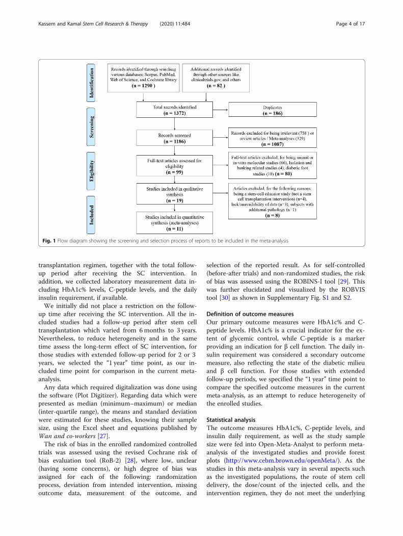
employed UCB-derived stem cells in a relatively recentintervention for treating DM, called “Stem Cell EducatorTherapy.” Briefly, in these studies, mononuclear cells(MNCs) are isolated from the patient’s whole blood andare co-cultured with adherent UCB-derived stem cells,and afterwards, those educated autologous MNCs arereturned back to the patient’s blood circulation [16–18,25]. For the current meta-analysis, we did not includethese “Stem Cell Educator” studies, because they did notactually make an UC-derived stem cell transplantationintervention, which is the focus of the current study.Additionally, we excluded studies in which the subjectshad any additional pathology besides DM. Finally, it isnoteworthy that we did not place language restrictionduring our initial screening/search, but for publishednon-English language studies, we limited inclusion forthose having at least a detailed abstract in English lan-guage. Thus, exclusion criteria can be summarized asanimal studies, in vitro molecular studies, studies with-out SC transplantation intervention, studies in which thesubjects were suffering an additional pathology to DM,and studies with incomplete/unavailable data. Figure 1shows the flow diagram illustrating our search strategyuntil reaching the selected included studies in our meta-analysis; this diagram was done according to the PRISMA statement guidelines [26]. In addition, the PRISMAchecklist for this meta-analysis study is presented in sup-plementary Table S1.
Data extraction and assessment of risk of biasData extraction was independently done by the two au-thors according to a standardized specified strategy. Anydisagreement was resolved through referring to the ori-ginal publication and discussion/consensus. For the se-lected studies, we reported clinical trial type (i.e., eitherrandomized, non-randomized, or self-controlled trial),the country of origin, the authors’ names, year of publi-cation, the number of enrolled subjects, and their meanage and duration of DM. We also collected data con-cerned with the type of DM, the type of UC-derivedstem cells employed (i.e., either WJ-MSCs or UCB), andthe count of injected cells, route of delivery, and
Kassem and Kamal Stem Cell Research & Therapy (2020) 11:484 Page 3 of 17
transplantation regimen, together with the total follow-up period after receiving the SC intervention. Inaddition, we collected laboratory measurement data in-cluding HbA1c% levels, C-peptide levels, and the dailyinsulin requirement, if available.We initially did not place a restriction on the follow-
up time after receiving the SC intervention. All the in-cluded studies had a follow-up period after stem celltransplantation which varied from 6months to 3 years.Nevertheless, to reduce heterogeneity and in the sametime assess the long-term effect of SC intervention, forthose studies with extended follow-up period for 2 or 3years, we selected the “1 year” time point, as our in-cluded time point for comparison in the current meta-analysis.Any data which required digitalization was done using
the software (Plot Digitizer). Regarding data which werepresented as median (minimum–maximum) or median(inter-quartile range), the means and standard deviationwere estimated for these studies, knowing their samplesize, using the Excel sheet and equations published byWan and co-workers [27].The risk of bias in the enrolled randomized controlled
trials was assessed using the revised Cochrane risk ofbias evaluation tool (RoB-2) [28], where low, unclear(having some concerns), or high degree of bias wasassigned for each of the following: randomizationprocess, deviation from intended intervention, missingoutcome data, measurement of the outcome, and
selection of the reported result. As for self-controlled(before-after trials) and non-randomized studies, the riskof bias was assessed using the ROBINS-I tool [29]. Thiswas further elucidated and visualized by the ROBVIStool [30] as shown in Supplementary Fig. S1 and S2.
Definition of outcome measuresOur primary outcome measures were HbA1c% and C-peptide levels. HbA1c% is a crucial indicator for the ex-tent of glycemic control, while C-peptide is a markerproviding an indication for β cell function. The daily in-sulin requirement was considered a secondary outcomemeasure, also reflecting the state of the diabetic milieuand β cell function. For those studies with extendedfollow-up periods, we specified the “1 year” time point tocompare the specified outcome measures in the currentmeta-analysis, as an attempt to reduce heterogeneity ofthe enrolled studies.
Statistical analysisThe outcome measures HbA1c%, C-peptide levels, andinsulin daily requirement, as well as the study samplesize were fed into Open-Meta-Analyst to perform meta-analysis of the investigated studies and provide forestplots (http://www.cebm.brown.edu/openMeta/). As thestudies in this meta-analysis vary in several aspects suchas the investigated populations, the route of stem celldelivery, the dose/count of the injected cells, and theintervention regimen, they do not meet the underlying
Fig. 1 Flow diagram showing the screening and selection process of reports to be included in the meta-analysis
Kassem and Kamal Stem Cell Research & Therapy (2020) 11:484 Page 4 of 17
assumption of a fixed-effects model in which only thesampling error is the source of variability; hence, theoverall effect size was estimated using the random-effects model, utilizing the Der Simonian−Laird method.The random-effects model takes into account the vari-ability between studies and was therefore adequate forthe purpose of this meta-analysis. Heterogeneity wasassessed using two parameters: the Cochran’s Q statisticand the I2 index. The Q statistic indicates the presenceor absence of heterogeneity among a set of studies re-lated to differences in the measurements, whereas the I2
index implies the degree of heterogeneity; observedvalues up to 30 imply mild heterogeneity, 31–50 implymoderate, while more than 50 imply markedheterogeneity.The standardized mean difference and a 95% confi-
dence interval (CI) were calculated and represented inthe forest plot. It is noteworthy that we preferred to usethe standardized mean difference rather than the meandifference in our meta-analysis to improve the homogen-eity, since some studies were using different measure-ments for the specified outcome. Significance wasemployed by the p value, where values < 0.05 were con-sidered statistically significant. In case of having a singlestudy which weighed too much, in such a way raisingconcerns regarding the results of the performed meta-analysis, a sensitivity of the study was evaluated usingthe leave-one-out meta-analysis, to further assess theoutcome of such meta-analysis.Finally, it is important to point here that the included
studies were initially classified into two major groupsbased on the type of UC-SC intervention; WJ-MSCs orUCB, and after wards when performing the meta-analysis, each of these groups was classified into 2 sub-groups according to the type of DM while performing asub-group meta-analysis. As for the randomized con-trolled trials, due to their limited number, they were ori-ginally pooled together (T1DM and T2DM studies), andthe sub-group meta-analysis was afterwards done ac-cording to the type of intervention; either WJ-MSCs orUCB.
ResultsSearch results and description of studiesAs shown in Fig. 1, the initial databases’ search and elec-tronic data mining revealed a total of 1372 records.Within these, 186 duplicates were identified and ex-cluded. Thus, a total of 1186 records were screened bytitle to identify relevant studies. During this screening,758 records were excluded for being irrelevant (such asthose concerned with gestational DM), and 329 citationswere excluded for being either review or meta-analysisarticles. Afterwards, the remaining 99 citations passedthrough a further thorough investigation for eligibility.
Of these, 66 citations were excluded for being either ani-mal or in vitro molecular studies, 4 citations were ex-cluded for being UC banking-related studies, and finally10 citations excluded for being diabetic foot-relatedstudies. Next, the full text was carefully assessed for theremaining 19 citations. In that phase, 8 citations werefurther excluded for the following reasons; 4 studies forbeing a stem-cell educator study in which stem celltransplantation was not applied as an intervention [16–18, 25], 1 study for having an additional pathology toDM [31], and finally 3 studies were excluded due to lackof data [32–34]. Thus, conclusively, 11 eligible clinicaltrials were identified and included in the current meta-analysis study.The characteristics of the 11 eligible included studies
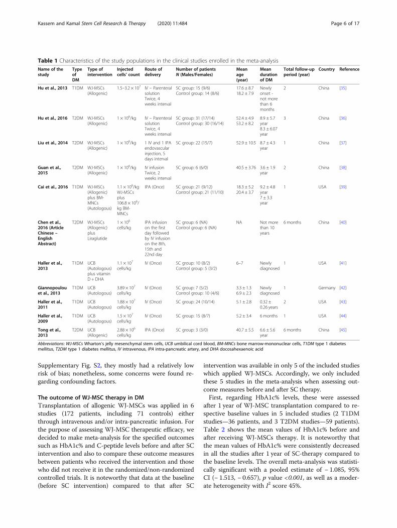
are presented in Table 1. When considering the countryof origin of these studies, 6 studies came from China, 4from the USA, and 1 from Germany. These 11 studiesincluded a total of 246 diabetic patients; 6 studies in-cluded patients with T1DM (142 patients, including 50controls), and 5 studies included patients with T2DM(104 patients, including 36 controls). When consideringthe design of these studies, we found 5 of them wererandomized controlled trials [35, 36, 39–41], 1 study wasa non-randomized controlled trial [42], and 5 studieswere self-controlled (before-after) clinical studies [37,38, 43–45].When considering the type of UC-derived stem cell
intervention, we found 6 studies (172 patients, including71 controls) which applied WJ-MSCs either throughintravenous and/or intra-pancreatic infusion. Of these, 3studies applied WJ-MSCs solely as their therapeuticintervention for T2DM, 1 study applied WJ-MSCs solelyfor T1DM, 1 study applied WJ-MSCs plus bonemarrow-derived mononuclear cells for T1DM, and 1study applied WJ-MSCs plus Liraglutide for T2DM. Onthe other hand, we found 5 studies (74 patients, includ-ing 15 controls) which applied UCB also via intravenousor intra-pancreatic infusion. Of these, 3 studies appliedUCB solely, and 1 study applied UCB followed by vita-min D and docosahexaenoic acid (DHA) for T1DM, anda single study applied UCB for T2DM. The total follow-up period after the SC transplantation ranged from 6months to 3 years.When assessing the risk of bias in these 11 included
studies, the 5 randomized controlled trials were assessedby Cochran’s RoB2 tool. As illustrated in SupplementaryFig. S1, they all showed a low to moderate risk of bias.The noticed concerns were mostly attributed to therandomization process, as well as lack of information re-garding the concealment methods, and avoiding devi-ation from intended intervention. The risk of bias forthe non-randomized as well as self-controlled studieswas assessed by the ROBINS-I tool. As shown in
Kassem and Kamal Stem Cell Research & Therapy (2020) 11:484 Page 5 of 17
Supplementary Fig. S2, they mostly had a relatively lowrisk of bias; nonetheless, some concerns were found re-garding confounding factors.
The outcome of WJ-MSC therapy in DMTransplantation of allogenic WJ-MSCs was applied in 6studies (172 patients, including 71 controls) eitherthrough intravenous and/or intra-pancreatic infusion. Forthe purpose of assessing WJ-MSC therapeutic efficacy, wedecided to make meta-analysis for the specified outcomessuch as HbA1c% and C-peptide levels before and after SCintervention and also to compare these outcome measuresbetween patients who received the intervention and thosewho did not receive it in the randomized/non-randomizedcontrolled trials. It is noteworthy that data at the baseline(before SC intervention) compared to that after SC
intervention was available in only 5 of the included studieswhich applied WJ-MSCs. Accordingly, we only includedthese 5 studies in the meta-analysis when assessing out-come measures before and after SC therapy.First, regarding HbA1c% levels, these were assessed
after 1 year of WJ-MSC transplantation compared to re-spective baseline values in 5 included studies (2 T1DMstudies—36 patients, and 3 T2DM studies—59 patients).Table 2 shows the mean values of HbA1c% before andafter receiving WJ-MSCs therapy. It is noteworthy thatthe mean values of HbA1c% were consistently decreasedin all the studies after 1 year of SC-therapy compared tothe baseline levels. The overall meta-analysis was statisti-cally significant with a pooled estimate of − 1.085, 95%CI (− 1.513, − 0.657), p value <0.001, as well as a moder-ate heterogeneity with I2 score 45%.
Table 1 Characteristics of the study populations in the clinical studies enrolled in the meta-analysisName of thestudy
TypeofDM
Type ofintervention
Injectedcells’ count
Route ofdelivery
Number of patientsN (Males/Females)
Meanage(year)
Meandurationof DM
Total follow-upperiod (year)
Country Reference
Hu et al., 2013 T1DM WJ-MSCs(Allogenic)
1.5–3.2 × 10⁷ IV – ParenteralsolutionTwice, 4weeks interval
SC group: 15 (9/6)Control group: 14 (8/6)
17.6 ± 8.718.2 ± 7.9
Newlyonset -not morethan 6months
2 China [35]
Hu et al., 2016 T2DM WJ-MSCs(Allogenic)
1 × 10⁶/kg IV – ParenteralsolutionTwice, 4weeks interval
SC group: 31 (17/14)Control group: 30 (16/14)
52.4 ± 4.953.2 ± 8.2
8.9 ± 5.7year8.3 ± 6.07year
3 China [36]
Liu et al., 2014 T2DM WJ-MSCs(Allogenic)
1 × 10⁶/kg 1 IV and 1 IPAendovascularinjection, 5days interval
SC group: 22 (15/7) 52.9 ± 10.5 8.7 ± 4.3year
1 China [37]
Guan et al.,2015
T2DM WJ-MSCs(Allogenic)
1 × 10⁶/kg IV infusionTwice, 2weeks interval
SC group: 6 (6/0) 40.5 ± 3.76 3.6 ± 1.9year
2 China [38]
Cai et al., 2016 T1DM WJ-MSCs(Allogenic)plus BM-MNCs(Autologous)
1.1 × 10⁶/kgWJ-MSCsplus106.8 × 106/kg BM-MNCs
IPA (Once) SC group: 21 (9/12)Control group: 21 (11/10)
18.3 ± 5.220.4 ± 3.7
9.2 ± 4.8year7 ± 3.3year
1 USA [39]
Chen et al.,2016 (ArticleChinese –EnglishAbstract)
T2DM WJ-MSCs(Allogenic)plusLiraglutide
1 × 10⁶
cells/kgIPA infusionon the firstday followedby IV infusionon the 8th,15th and22nd day
SC group: 6 (NA)Control group: 6 (NA)
NA Not morethan 10years
6 months China [40]
Haller et al.,2013
T1DM UCB(Autologous)plus vitaminD + DHA
1.1 × 10⁷
cells/kgIV (Once) SC group: 10 (8/2)
Control group: 5 (3/2)6–7 Newly
diagnosed1 USA [41]
Giannopoulouet al., 2013
T1DM UCB(Autologous)
3.89 × 10⁷
cells/kgIV (Once) SC group: 7 (5/2)
Control group: 10 (4/6)3.3 ± 1.36.9 ± 2.3
Newlydiagnosed
1 Germany [42]
Haller et al.,2011
T1DM UCB(Autologous)
1.88 × 10⁷
cells/kgIV (Once) SC group: 24 (10/14) 5.1 ± 2.8 0.32 ±
0.26 years2 USA [43]
Haller et al.,2009
T1DM UCB(Autologous)
1.5 × 10⁷
cells/kgIV (Once) SC group: 15 (8/7) 5.2 ± 3.4 6 months 1 USA [44]
Tong et al.,2013
T2DM UCB(Allogenic)
2.88 × 10⁶
cells/kgIPA (Once) SC group: 3 (3/0) 40.7 ± 5.5 6.6 ± 5.6
year6 months China [45]
Abbreviations: WJ-MSCs Wharton’s jelly mesenchymal stem cells, UCB umbilical cord blood, BM-MNCs bone marrow-mononuclear cells, T1DM type 1 diabetesmellitus, T2DM type 1 diabetes mellitus, IV intravenous, IPA intra-pancreatic artery, and DHA docosahexaenoic acid
Kassem and Kamal Stem Cell Research & Therapy (2020) 11:484 Page 6 of 17
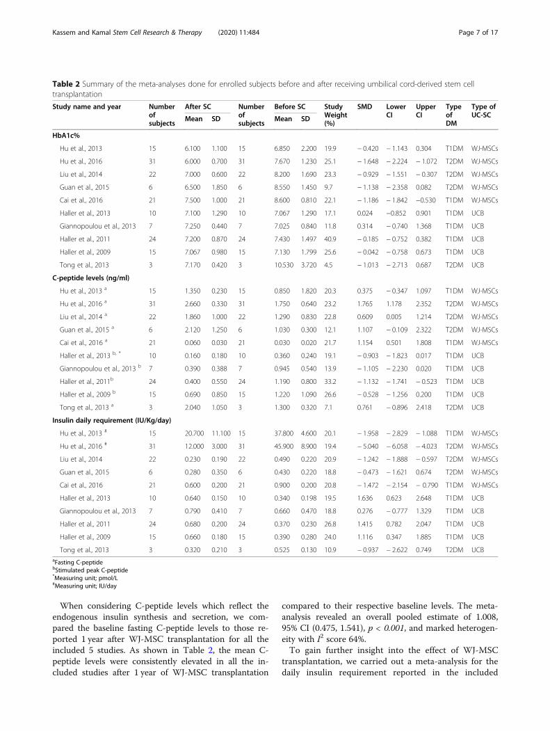
When considering C-peptide levels which reflect theendogenous insulin synthesis and secretion, we com-pared the baseline fasting C-peptide levels to those re-ported 1 year after WJ-MSC transplantation for all theincluded 5 studies. As shown in Table 2, the mean C-peptide levels were consistently elevated in all the in-cluded studies after 1 year of WJ-MSC transplantation
compared to their respective baseline levels. The meta-analysis revealed an overall pooled estimate of 1.008,95% CI (0.475, 1.541), p < 0.001, and marked heterogen-eity with I2 score 64%.To gain further insight into the effect of WJ-MSC
transplantation, we carried out a meta-analysis for thedaily insulin requirement reported in the included
Table 2 Summary of the meta-analyses done for enrolled subjects before and after receiving umbilical cord-derived stem celltransplantation
Study name and year Numberofsubjects
After SC Numberofsubjects
Before SC StudyWeight(%)
SMD LowerCI
UpperCI
TypeofDM
Type ofUC-SCMean SD Mean SD
HbA1c%
Hu et al., 2013 15 6.100 1.100 15 6.850 2.200 19.9 − 0.420 − 1.143 0.304 T1DM WJ-MSCs
Hu et al., 2016 31 6.000 0.700 31 7.670 1.230 25.1 − 1.648 − 2.224 − 1.072 T2DM WJ-MSCs
Liu et al., 2014 22 7.000 0.600 22 8.200 1.690 23.3 − 0.929 − 1.551 − 0.307 T2DM WJ-MSCs
Guan et al., 2015 6 6.500 1.850 6 8.550 1.450 9.7 − 1.138 − 2.358 0.082 T2DM WJ-MSCs
Cai et al., 2016 21 7.500 1.000 21 8.600 0.810 22.1 − 1.186 − 1.842 −0.530 T1DM WJ-MSCs
Haller et al., 2013 10 7.100 1.290 10 7.067 1.290 17.1 0.024 −0.852 0.901 T1DM UCB
Giannopoulou et al., 2013 7 7.250 0.440 7 7.025 0.840 11.8 0.314 − 0.740 1.368 T1DM UCB
Haller et al., 2011 24 7.200 0.870 24 7.430 1.497 40.9 − 0.185 − 0.752 0.382 T1DM UCB
Haller et al., 2009 15 7.067 0.980 15 7.130 1.799 25.6 − 0.042 − 0.758 0.673 T1DM UCB
Tong et al., 2013 3 7.170 0.420 3 10.530 3.720 4.5 − 1.013 − 2.713 0.687 T2DM UCB
C-peptide levels (ng/ml)
Hu et al., 2013 a 15 1.350 0.230 15 0.850 1.820 20.3 0.375 − 0.347 1.097 T1DM WJ-MSCs
Hu et al., 2016 a 31 2.660 0.330 31 1.750 0.640 23.2 1.765 1.178 2.352 T2DM WJ-MSCs
Liu et al., 2014 a 22 1.860 1.000 22 1.290 0.830 22.8 0.609 0.005 1.214 T2DM WJ-MSCs
Guan et al., 2015 a 6 2.120 1.250 6 1.030 0.300 12.1 1.107 − 0.109 2.322 T2DM WJ-MSCs
Cai et al., 2016 a 21 0.060 0.030 21 0.030 0.020 21.7 1.154 0.501 1.808 T1DM WJ-MSCs
Haller et al., 2013 b, * 10 0.160 0.180 10 0.360 0.240 19.1 − 0.903 − 1.823 0.017 T1DM UCB
Giannopoulou et al., 2013 b 7 0.390 0.388 7 0.945 0.540 13.9 − 1.105 − 2.230 0.020 T1DM UCB
Haller et al., 2011b 24 0.400 0.550 24 1.190 0.800 33.2 − 1.132 − 1.741 − 0.523 T1DM UCB
Haller et al., 2009 b 15 0.690 0.850 15 1.220 1.090 26.6 − 0.528 − 1.256 0.200 T1DM UCB
Tong et al., 2013 a 3 2.040 1.050 3 1.300 0.320 7.1 0.761 − 0.896 2.418 T2DM UCB
Insulin daily requirement (IU/Kg/day)
Hu et al., 2013 # 15 20.700 11.100 15 37.800 4.600 20.1 − 1.958 − 2.829 − 1.088 T1DM WJ-MSCs
Hu et al., 2016 # 31 12.000 3.000 31 45.900 8.900 19.4 − 5.040 − 6.058 − 4.023 T2DM WJ-MSCs
Liu et al., 2014 22 0.230 0.190 22 0.490 0.220 20.9 − 1.242 − 1.888 − 0.597 T2DM WJ-MSCs
Guan et al., 2015 6 0.280 0.350 6 0.430 0.220 18.8 − 0.473 − 1.621 0.674 T2DM WJ-MSCs
Cai et al., 2016 21 0.600 0.200 21 0.900 0.200 20.8 − 1.472 − 2.154 − 0.790 T1DM WJ-MSCs
Haller et al., 2013 10 0.640 0.150 10 0.340 0.198 19.5 1.636 0.623 2.648 T1DM UCB
Giannopoulou et al., 2013 7 0.790 0.410 7 0.660 0.470 18.8 0.276 − 0.777 1.329 T1DM UCB
Haller et al., 2011 24 0.680 0.200 24 0.370 0.230 26.8 1.415 0.782 2.047 T1DM UCB
Haller et al., 2009 15 0.660 0.180 15 0.390 0.280 24.0 1.116 0.347 1.885 T1DM UCB
Tong et al., 2013 3 0.320 0.210 3 0.525 0.130 10.9 − 0.937 − 2.622 0.749 T2DM UCBaFasting C-peptidebStimulated peak C-peptide*Measuring unit; pmol/L#Measuring unit; IU/day
Kassem and Kamal Stem Cell Research & Therapy (2020) 11:484 Page 7 of 17
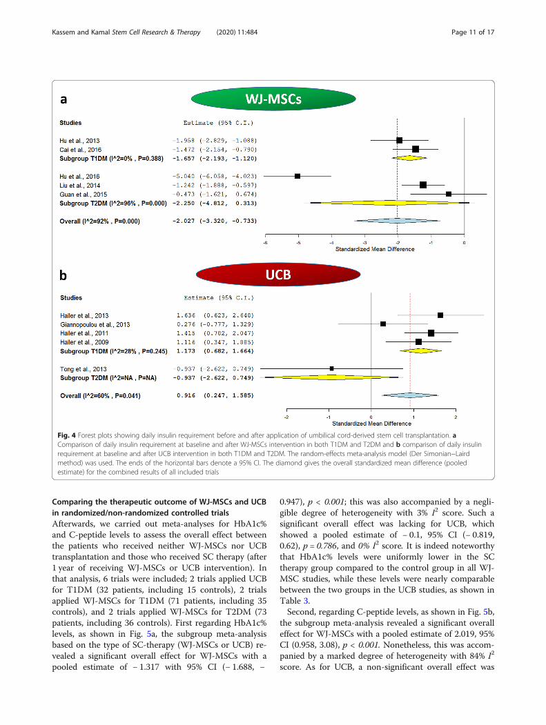
studies at the baseline and after 1 year of WJ-MSCtransplantation. This will indirectly reflect the statusof the diabetic milieu and endogenous insulin synthe-sis. Interestingly, the daily insulin requirement wasfound to be uniformly decreased in all the investi-gated studies after WJ-MSC therapy compared to re-spective baseline values as shown in Table 2.Additionally, in one of these studies, 3 out of 15T1DM patients became completely insulin independ-ent at the end of the 2-year follow-up period, and in8 of the remaining 12 patients, the insulin daily dos-age was reduced by more than 50% of the initial dailyrequirement at the baseline [35]. Likewise, Liu et al.reported that insulin suspension occurred for nearly41% of the T2DM patients who were receiving insulintherapy. This occurred within a time frame of 2 to 6months after WJ-MSC transplantation, and these pa-tients remained insulin-free for a mean time of 9months until the last follow-up of the study [37]. Thesame observation was also reported by Hu and co-workers; 32% of the T2DM patients who received WJ-MSC transplantation became insulin-free within aperiod ranging from 3 to 11 months after receivingWJ-MSC infusion and remained insulin-free for amean period of 12.5 ± 6.8 months [36]. The overallmeta-analysis for the daily insulin requirement in theincluded studies before and after 1 year of receivingSC therapy showed a pooled estimate of − 2.027, 95%CI (− 3.32, − 0.733), p = 0.002, with I2 score of 92%,implying a marked degree of heterogeneity.
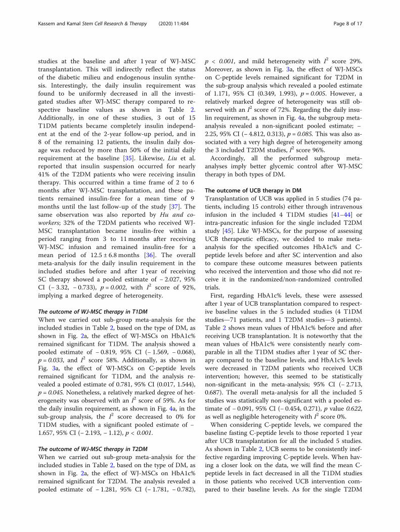
The outcome of WJ-MSC therapy in T1DMWhen we carried out sub-group meta-analysis for theincluded studies in Table 2, based on the type of DM, asshown in Fig. 2a, the effect of WJ-MSCs on HbA1c%remained significant for T1DM. The analysis showed apooled estimate of − 0.819, 95% CI (− 1.569, − 0.068),p = 0.033, and I2 score 58%. Additionally, as shown inFig. 3a, the effect of WJ-MSCs on C-peptide levelsremained significant for T1DM, and the analysis re-vealed a pooled estimate of 0.781, 95% CI (0.017, 1.544),p = 0.045. Nonetheless, a relatively marked degree of het-erogeneity was observed with an I2 score of 59%. As forthe daily insulin requirement, as shown in Fig. 4a, in thesub-group analysis, the I2 score decreased to 0% forT1DM studies, with a significant pooled estimate of −1.657, 95% CI (− 2.193, − 1.12), p < 0.001.
The outcome of WJ-MSC therapy in T2DMWhen we carried out sub-group meta-analysis for theincluded studies in Table 2, based on the type of DM, asshown in Fig. 2a, the effect of WJ-MSCs on HbA1c%remained significant for T2DM. The analysis revealed apooled estimate of − 1.281, 95% CI (− 1.781, − 0.782),
p < 0.001, and mild heterogeneity with I2 score 29%.Moreover, as shown in Fig. 3a, the effect of WJ-MSCson C-peptide levels remained significant for T2DM inthe sub-group analysis which revealed a pooled estimateof 1.171, 95% CI (0.349, 1.993), p = 0.005. However, arelatively marked degree of heterogeneity was still ob-served with an I2 score of 72%. Regarding the daily insu-lin requirement, as shown in Fig. 4a, the subgroup meta-analysis revealed a non-significant pooled estimate; −2.25, 95% CI (− 4.812, 0.313), p = 0.085. This was also as-sociated with a very high degree of heterogeneity amongthe 3 included T2DM studies, I2 score 96%.Accordingly, all the performed subgroup meta-
analyses imply better glycemic control after WJ-MSCtherapy in both types of DM.
The outcome of UCB therapy in DMTransplantation of UCB was applied in 5 studies (74 pa-tients, including 15 controls) either through intravenousinfusion in the included 4 T1DM studies [41–44] orintra-pancreatic infusion for the single included T2DMstudy [45]. Like WJ-MSCs, for the purpose of assessingUCB therapeutic efficacy, we decided to make meta-analysis for the specified outcomes HbA1c% and C-peptide levels before and after SC intervention and alsoto compare these outcome measures between patientswho received the intervention and those who did not re-ceive it in the randomized/non-randomized controlledtrials.First, regarding HbA1c% levels, these were assessed
after 1 year of UCB transplantation compared to respect-ive baseline values in the 5 included studies (4 T1DMstudies—71 patients, and 1 T2DM studies—3 patients).Table 2 shows mean values of HbA1c% before and afterreceiving UCB transplantation. It is noteworthy that themean values of HbA1c% were consistently nearly com-parable in all the T1DM studies after 1 year of SC ther-apy compared to the baseline levels, and HbA1c% levelswere decreased in T2DM patients who received UCBintervention; however, this seemed to be statisticallynon-significant in the meta-analysis; 95% CI (− 2.713,0.687). The overall meta-analysis for all the included 5studies was statistically non-significant with a pooled es-timate of − 0.091, 95% CI (− 0.454, 0.271), p value 0.622,as well as negligible heterogeneity with I2 score 0%.When considering C-peptide levels, we compared the
baseline fasting C-peptide levels to those reported 1 yearafter UCB transplantation for all the included 5 studies.As shown in Table 2, UCB seems to be consistently inef-fective regarding improving C-peptide levels. When hav-ing a closer look on the data, we will find the mean C-peptide levels in fact decreased in all the T1DM studiesin those patients who received UCB intervention com-pared to their baseline levels. As for the single T2DM
Kassem and Kamal Stem Cell Research & Therapy (2020) 11:484 Page 8 of 17
study, UCB seems to have resulted in a slight yet non-significant elevation of C-peptide levels. The meta-analysis revealed an overall pooled estimate of − 0.789,95% CI (− 1.252, − 0.325), p < 0.001, and mild hetero-geneity with I2 score 26%.As for the daily insulin requirement, we carried out a
meta-analysis for the included studies to assess the vari-ation of insulin daily dosage at the baseline and after 1year of UCB transplantation. In fact, in agreement withthe reported results concerned with HbA1c% and C-peptide levels, the daily insulin requirement was foundto be uniformly increased in all the included T1DMstudies and showed a slight decrease in the includedT2DM study as shown in Table 2. The overall meta-analysis for the daily insulin requirement in the includedstudies before and after 1 year of receiving UCB therapy
showed a pooled estimate of 0.916, 95% CI (0.247,1.585), p = 0.007, with I2 score of 60%, implying amarked degree of heterogeneity. These data imply thelack of improvement on insulin daily requirement andthe relatively poor therapeutic efficacy of UCB in thisregard.
The outcome of UCB therapy in T1DMWhen we carried out sub-group meta-analysis for theincluded studies in Table 2, based on the type of DM, asshown in Fig. 2b, the effect of UCB on HbA1c%remained non-significant for T1DM with a pooled esti-mate of − 0.047, 95% CI (− 0.418, 0.324), p = 0.803. Thisimplies that UCB does not have a significant beneficialeffect over glycemic control in T1DM. Afterwards, asshown in Fig. 3b, in the sub-group analysis to assess the
Fig. 2 Forest plots showing HbA1c% levels before and after application of umbilical cord-derived stem cell transplantation. a Comparison ofHbA1c% levels at baseline and after WJ-MSC intervention in both T1DM and T2DM and b comparison of HbA1c% levels at baseline and afterUCB intervention in both T1DM and T2DM. The random-effects meta-analysis model (Der Simonian−Laird method) was used. The ends of thehorizontal bars denote a 95% CI. The diamond gives the overall standardized mean difference (pooled estimate) for the combined results of allincluded trials
Kassem and Kamal Stem Cell Research & Therapy (2020) 11:484 Page 9 of 17
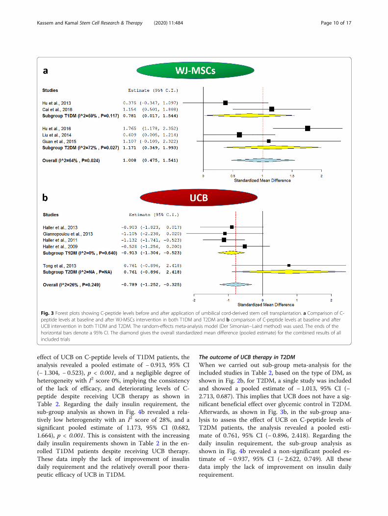
effect of UCB on C-peptide levels of T1DM patients, theanalysis revealed a pooled estimate of − 0.913, 95% CI(− 1.304, − 0.523), p < 0.001, and a negligible degree ofheterogeneity with I2 score 0%, implying the consistencyof the lack of efficacy, and deteriorating levels of C-peptide despite receiving UCB therapy as shown inTable 2. Regarding the daily insulin requirement, thesub-group analysis as shown in Fig. 4b revealed a rela-tively low heterogeneity with an I2 score of 28%, and asignificant pooled estimate of 1.173, 95% CI (0.682,1.664), p < 0.001. This is consistent with the increasingdaily insulin requirements shown in Table 2 in the en-rolled T1DM patients despite receiving UCB therapy.These data imply the lack of improvement of insulindaily requirement and the relatively overall poor thera-peutic efficacy of UCB in T1DM.
The outcome of UCB therapy in T2DMWhen we carried out sub-group meta-analysis for theincluded studies in Table 2, based on the type of DM, asshown in Fig. 2b, for T2DM, a single study was includedand showed a pooled estimate of − 1.013, 95% CI (−2.713, 0.687). This implies that UCB does not have a sig-nificant beneficial effect over glycemic control in T2DM.Afterwards, as shown in Fig. 3b, in the sub-group ana-lysis to assess the effect of UCB on C-peptide levels ofT2DM patients, the analysis revealed a pooled esti-mate of 0.761, 95% CI (− 0.896, 2.418). Regarding thedaily insulin requirement, the sub-group analysis asshown in Fig. 4b revealed a non-significant pooled es-timate of − 0.937, 95% CI (− 2.622, 0.749). All thesedata imply the lack of improvement on insulin dailyrequirement.
Fig. 3 Forest plots showing C-peptide levels before and after application of umbilical cord-derived stem cell transplantation. a Comparison of C-peptide levels at baseline and after WJ-MSCs intervention in both T1DM and T2DM and b comparison of C-peptide levels at baseline and afterUCB intervention in both T1DM and T2DM. The random-effects meta-analysis model (Der Simonian−Laird method) was used. The ends of thehorizontal bars denote a 95% CI. The diamond gives the overall standardized mean difference (pooled estimate) for the combined results of allincluded trials
Kassem and Kamal Stem Cell Research & Therapy (2020) 11:484 Page 10 of 17
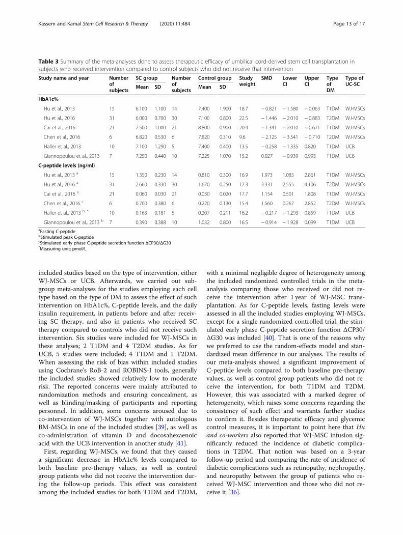
Comparing the therapeutic outcome of WJ-MSCs and UCBin randomized/non-randomized controlled trialsAfterwards, we carried out meta-analyses for HbA1c%and C-peptide levels to assess the overall effect betweenthe patients who received neither WJ-MSCs nor UCBtransplantation and those who received SC therapy (after1 year of receiving WJ-MSCs or UCB intervention). Inthat analysis, 6 trials were included; 2 trials applied UCBfor T1DM (32 patients, including 15 controls), 2 trialsapplied WJ-MSCs for T1DM (71 patients, including 35controls), and 2 trials applied WJ-MSCs for T2DM (73patients, including 36 controls). First regarding HbA1c%levels, as shown in Fig. 5a, the subgroup meta-analysisbased on the type of SC-therapy (WJ-MSCs or UCB) re-vealed a significant overall effect for WJ-MSCs with apooled estimate of − 1.317 with 95% CI (− 1.688, −
0.947), p < 0.001; this was also accompanied by a negli-gible degree of heterogeneity with 3% I2 score. Such asignificant overall effect was lacking for UCB, whichshowed a pooled estimate of − 0.1, 95% CI (− 0.819,0.62), p = 0.786, and 0% I2 score. It is indeed noteworthythat HbA1c% levels were uniformly lower in the SCtherapy group compared to the control group in all WJ-MSC studies, while these levels were nearly comparablebetween the two groups in the UCB studies, as shown inTable 3.Second, regarding C-peptide levels, as shown in Fig. 5b,
the subgroup meta-analysis revealed a significant overalleffect for WJ-MSCs with a pooled estimate of 2.019, 95%CI (0.958, 3.08), p < 0.001. Nonetheless, this was accom-panied by a marked degree of heterogeneity with 84% I2
score. As for UCB, a non-significant overall effect was
Fig. 4 Forest plots showing daily insulin requirement before and after application of umbilical cord-derived stem cell transplantation. aComparison of daily insulin requirement at baseline and after WJ-MSCs intervention in both T1DM and T2DM and b comparison of daily insulinrequirement at baseline and after UCB intervention in both T1DM and T2DM. The random-effects meta-analysis model (Der Simonian−Lairdmethod) was used. The ends of the horizontal bars denote a 95% CI. The diamond gives the overall standardized mean difference (pooledestimate) for the combined results of all included trials
Kassem and Kamal Stem Cell Research & Therapy (2020) 11:484 Page 11 of 17
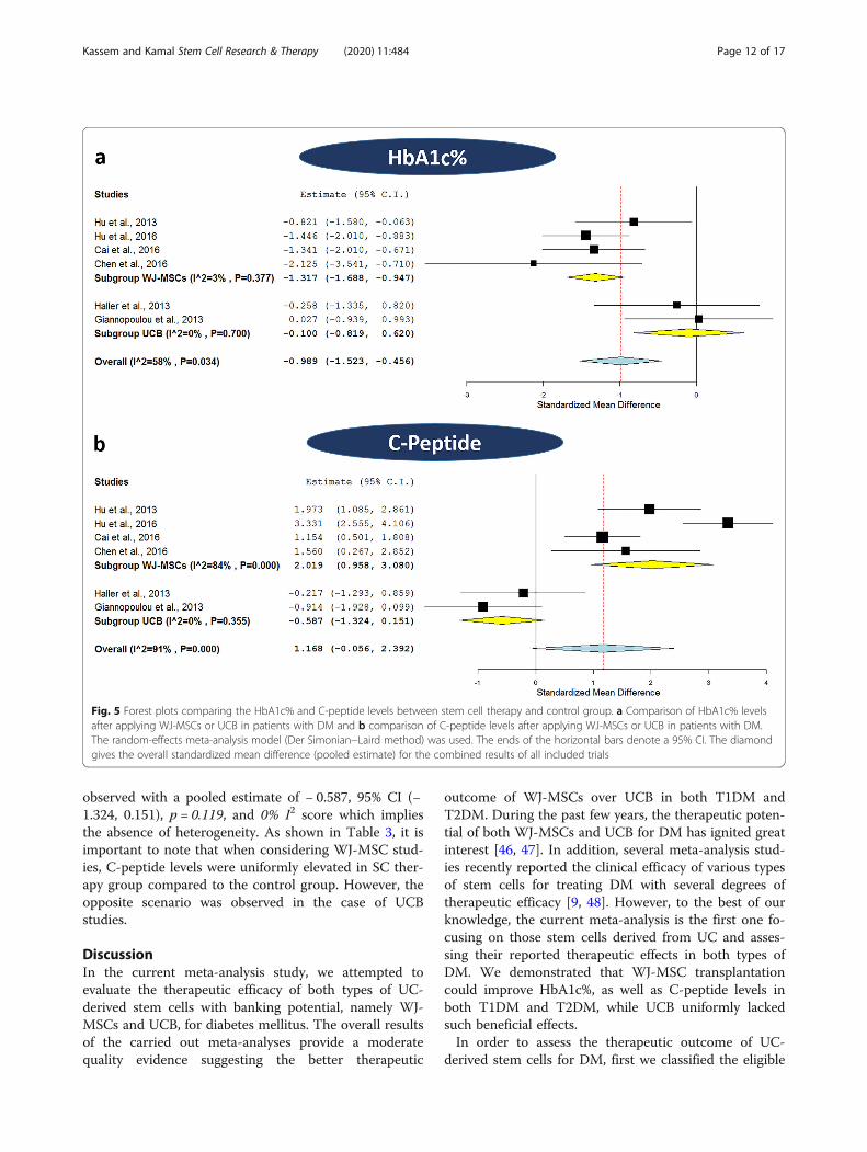
observed with a pooled estimate of − 0.587, 95% CI (−1.324, 0.151), p = 0.119, and 0% I2 score which impliesthe absence of heterogeneity. As shown in Table 3, it isimportant to note that when considering WJ-MSC stud-ies, C-peptide levels were uniformly elevated in SC ther-apy group compared to the control group. However, theopposite scenario was observed in the case of UCBstudies.
DiscussionIn the current meta-analysis study, we attempted toevaluate the therapeutic efficacy of both types of UC-derived stem cells with banking potential, namely WJ-MSCs and UCB, for diabetes mellitus. The overall resultsof the carried out meta-analyses provide a moderatequality evidence suggesting the better therapeutic
outcome of WJ-MSCs over UCB in both T1DM andT2DM. During the past few years, the therapeutic poten-tial of both WJ-MSCs and UCB for DM has ignited greatinterest [46, 47]. In addition, several meta-analysis stud-ies recently reported the clinical efficacy of various typesof stem cells for treating DM with several degrees oftherapeutic efficacy [9, 48]. However, to the best of ourknowledge, the current meta-analysis is the first one fo-cusing on those stem cells derived from UC and asses-sing their reported therapeutic effects in both types ofDM. We demonstrated that WJ-MSC transplantationcould improve HbA1c%, as well as C-peptide levels inboth T1DM and T2DM, while UCB uniformly lackedsuch beneficial effects.In order to assess the therapeutic outcome of UC-
derived stem cells for DM, first we classified the eligible
Fig. 5 Forest plots comparing the HbA1c% and C-peptide levels between stem cell therapy and control group. a Comparison of HbA1c% levelsafter applying WJ-MSCs or UCB in patients with DM and b comparison of C-peptide levels after applying WJ-MSCs or UCB in patients with DM.The random-effects meta-analysis model (Der Simonian−Laird method) was used. The ends of the horizontal bars denote a 95% CI. The diamondgives the overall standardized mean difference (pooled estimate) for the combined results of all included trials
Kassem and Kamal Stem Cell Research & Therapy (2020) 11:484 Page 12 of 17
included studies based on the type of intervention, eitherWJ-MSCs or UCB. Afterwards, we carried out sub-group meta-analyses for the studies employing each celltype based on the type of DM to assess the effect of suchintervention on HbA1c%, C-peptide levels, and the dailyinsulin requirement, in patients before and after receiv-ing SC therapy, and also in patients who received SCtherapy compared to controls who did not receive suchintervention. Six studies were included for WJ-MSCs inthese analyses; 2 T1DM and 4 T2DM studies. As forUCB, 5 studies were included; 4 T1DM and 1 T2DM.When assessing the risk of bias within included studiesusing Cochrane’s RoB-2 and ROBINS-I tools, generallythe included studies showed relatively low to moderaterisk. The reported concerns were mainly attributed torandomization methods and ensuring concealment, aswell as blinding/masking of participants and reportingpersonnel. In addition, some concerns aroused due toco-intervention of WJ-MSCs together with autologousBM-MSCs in one of the included studies [39], as well asco-administration of vitamin D and docosahexaenoicacid with the UCB intervention in another study [41].First, regarding WJ-MSCs, we found that they caused
a significant decrease in HbA1c% levels compared toboth baseline pre-therapy values, as well as controlgroup patients who did not receive the intervention dur-ing the follow-up periods. This effect was consistentamong the included studies for both T1DM and T2DM,
with a minimal negligible degree of heterogeneity amongthe included randomized controlled trials in the meta-analysis comparing those who received or did not re-ceive the intervention after 1 year of WJ-MSC trans-plantation. As for C-peptide levels, fasting levels wereassessed in all the included studies employing WJ-MSCs,except for a single randomized controlled trial, the stim-ulated early phase C-peptide secretion function ΔCP30/ΔG30 was included [40]. That is one of the reasons whywe preferred to use the random-effects model and stan-dardized mean difference in our analyses. The results ofour meta-analysis showed a significant improvement ofC-peptide levels compared to both baseline pre-therapyvalues, as well as control group patients who did not re-ceive the intervention, for both T1DM and T2DM.However, this was associated with a marked degree ofheterogeneity, which raises some concerns regarding theconsistency of such effect and warrants further studiesto confirm it. Besides therapeutic efficacy and glycemiccontrol measures, it is important to point here that Huand co-workers also reported that WJ-MSC infusion sig-nificantly reduced the incidence of diabetic complica-tions in T2DM. That notion was based on a 3-yearfollow-up period and comparing the rate of incidence ofdiabetic complications such as retinopathy, nephropathy,and neuropathy between the group of patients who re-ceived WJ-MSC intervention and those who did not re-ceive it [36].
Table 3 Summary of the meta-analyses done to assess therapeutic efficacy of umbilical cord-derived stem cell transplantation insubjects who received intervention compared to control subjects who did not receive that intervention
Study name and year Numberofsubjects
SC group Numberofsubjects
Control group Studyweight
SMD LowerCI
UpperCI
TypeofDM
Type ofUC-SCMean SD Mean SD
HbA1c%
Hu et al., 2013 15 6.100 1.100 14 7.400 1.900 18.7 − 0.821 − 1.580 − 0.063 T1DM WJ-MSCs
Hu et al., 2016 31 6.000 0.700 30 7.100 0.800 22.5 − 1.446 − 2.010 − 0.883 T2DM WJ-MSCs
Cai et al., 2016 21 7.500 1.000 21 8.800 0.900 20.4 − 1.341 − 2.010 − 0.671 T1DM WJ-MSCs
Chen et al., 2016 6 6.820 0.530 6 7.820 0.310 9.6 − 2.125 − 3.541 − 0.710 T2DM WJ-MSCs
Haller et al., 2013 10 7.100 1.290 5 7.400 0.400 13.5 − 0.258 − 1.335 0.820 T1DM UCB
Giannopoulou et al., 2013 7 7.250 0.440 10 7.225 1.070 15.2 0.027 − 0.939 0.993 T1DM UCB
C-peptide levels (ng/ml)
Hu et al., 2013 a 15 1.350 0.230 14 0.810 0.300 16.9 1.973 1.085 2.861 T1DM WJ-MSCs
Hu et al., 2016 a 31 2.660 0.330 30 1.670 0.250 17.3 3.331 2.555 4.106 T2DM WJ-MSCs
Cai et al., 2016 a 21 0.060 0.030 21 0.030 0.020 17.7 1.154 0.501 1.808 T1DM WJ-MSCs
Chen et al., 2016 c 6 0.700 0.380 6 0.220 0.130 15.4 1.560 0.267 2.852 T2DM WJ-MSCs
Haller et al., 2013 b, * 10 0.163 0.181 5 0.207 0.211 16.2 − 0.217 − 1.293 0.859 T1DM UCB
Giannopoulou et al., 2013 b 7 0.390 0.388 10 1.032 0.800 16.5 − 0.914 − 1.928 0.099 T1DM UCBaFasting C-peptidebStimulated peak C-peptidecStimulated early phase C-peptide secretion function ΔCP30/ΔG30*Measuring unit; pmol/L
Kassem and Kamal Stem Cell Research & Therapy (2020) 11:484 Page 13 of 17
In fact, the previous observations come in perfectagreement with our findings regarding the daily insulinrequirement assessment before and after receiving WJ-MSC transplantation in T1DM. The daily insulin re-quirement significantly decreased in patients after re-ceiving WJ-MSC therapy, not only that, but also 3 out of15 patients became insulin-free in one study [35]. As forT2DM, the results of meta-analysis for the daily insulinrequirement were quite in-consistent. On the one hand,non-significant pooled estimate was observed with amarked degree of heterogeneity with 96% I2 score. Onthe other hand, two of the included studies reportedinsulin-suspension in 30–40% of the patients who re-ceived WJ-MSC transplantation [36, 37]. Such discrep-ancy might be at least partially attributed to the differentpatients’ properties regarding the diabetes duration be-fore receiving WJ-MSC intervention, as well as the dif-ferent regimens used for WJ-MSC transplantation.Generally, our meta-analysis was limited by the relativelysmall sample size. Nevertheless, it is noteworthy that forT1DM specifically, the studies of WJ-MSCs were verylimited, we only found two studies, and one of these ap-plied WJ-MSCs together with bone marrow-derivedmono-nuclear cells (BM-MNCs) [39], so there are someconcerns regarding the bias of co-intervention. It isnoteworthy that we found an additional registered clin-ical trial currently recruiting patients in Vietnam to as-sess this same co-intervention of WJ-MSCs withautologous BM-MNCs in T1DM, NCT03484741 [49].Accordingly, given the reported safety of WJ-MSCs, fur-ther well-designed large scale studies are indeed war-ranted to confirm their therapeutic efficacy, especiallyfor T1DM. Luckily, we found two registered randomizedcontrolled trials in the USA (NCT04061746) andSweden (NCT03406585). These trials are currentlyrecruiting patients to assess the efficacy of allogenic WJ-MSCs in T1DM patients, with estimated completiondate by 2023 [49].It is important to point here that all the studies
which applied WJ-MSCs employed allogenic SC ther-apy (without the application of immuno-suppressivedrugs), while in the case of UCB, all the studiesapplied autologous SC therapy except Tong and co-workers who applied allogenic UCB in T2DM pa-tients [45]. Such observation shed lights on the factthat well-designed clinical studies to also assess theefficacy of autologous WJ-MSCs for DM in the fu-ture are indeed warranted. This might be compli-cated by the lack of standardized cryo-preservationprotocols for WJ-MSCs, unlike UCB whose bankingprocedures are well-established [50]. In fact, well-standardized cryo-preservation/banking protocols forGMP-compliant clinical grade WJ-MSCs are indeedwarranted and considered to be among the major
challenges for successful translation of WJ-MSCsfrom bench to bed-side [51].When considering the results of our meta-analysis for
UCB effect on HbA1c% levels, it was found to be uni-formly ineffective in both T1DM and T2DM. This ob-servation was consistent among all the included studieswith negligible or nearly absent heterogeneity, which re-flects the lack of efficacy on the glycemic control anddiabetic status in those patients who received the UCBintervention. It is noteworthy that when assessing the ef-fect of UCB transplantation on HbA1c%, the weight ofone of the studies was 40.9%. Accordingly, we carriedout a leave-one-out meta-analysis to exclude the studyof the highest weight [43]. However, this revealed thesame findings of the original meta-analysis as a furtherconfirmation for the lack of efficacy as shown in Supple-mentary Fig. S3. This comes in perfect agreement withthe consistent lack of improvement in C-peptide levels,either in comparison to the baseline values (before re-ceiving UCB infusion) or compared to those controlswho did not receive UCB transplantation. It is note-worthy here that for UCB studies, the stimulated peakC-peptide levels were reported in all the T1DM studiesas an indication of insulin synthesis and C-peptide secre-tion, rather than the fasting levels [41–44]. As for thesingle included T2DM, the fasting C-peptide levels werereported and included in our meta-analysis [45]. Again,that is why we preferred here to use the random-effectsmodel, as well as the standardized mean difference forour meta-analysis.Our analyses also revealed a consistent lack of im-
provement in the daily insulin requirement compared tothe baseline values before UCB therapy for both T1DMand T2DM. It is important to point here that unlike WJ-MSCs, the studies investigating UCB for T2DM were farmore limited than T1DM. In the current analysis, 4T1DM studies, while only a single T2DM study withonly 3 patients, were included [45]. This highlights thecrucial need for additional clinical studies to further elu-cidate the presence or absence of clinical efficacy ofUCB in T2DM. In fact, our findings regarding the lackof clinical efficacy of UCB in DM come in agreementwith the results of the previous meta-analysis by El-Badawy and El-Badri, who also reported the uniformlynegative effect of UCB in children with T1DM, and fail-ure to improve the glycemic status at 1 year post-transplantation [48]. Generally, autologous UCB infusionin the included studies was reported to be safe and didnot cause undesirable side effects. However, the lack oftherapeutic efficacy warrants further investigations tooptimize the transplantation regimen. The authors ex-plained the lack of efficacy of autologous UCB by theprobably insufficient number of cells with immuno-regulatory/regenerative potential [43]. In fact, it is
Kassem and Kamal Stem Cell Research & Therapy (2020) 11:484 Page 14 of 17
important to point here that all these studies employedautologous UCB nucleated cells, without any selectionor enrichment for any particular cell population. Inter-estingly, on the public clinical trial registry, there areregistered clinical trials currently recruiting patients toassess safety and efficacy of UCB-derived regulatory Tcells (T-Regs) for T1DM; NCT02932826 andNCT03011021 [49].It is indeed important to point here that no serious ad-
verse events were reported in any of the included stud-ies. Generally, the studies for both WJ-MSCs and UCBreported their safety, and the absence of any tumor for-mation throughout the whole follow-up period, based oncancer-screening tests like tumor marker assessmentand/or imaging examination [35, 36, 38]. Occasionally,only few transient adverse effects like mild fever, nausea,vomiting, or headache [37], as well as abdominal pain orpuncture-site bleeding [39] which recovered spontan-eously, were reported in nearly 5–20% of the patientswho received SC transplantation. As for the therapeuticefficacy, various mechanisms have been proposed bywhich WJ-MSCs could mediate their observed beneficialeffects in T1DM/T2DM [46]. Briefly, WJ-MSCs have theability for “homing” to sites of tissue injury and secretemultiple bioactive mediators which are capable of stimu-lating recovery of injured cells, as well as variousimmuno-modulatory functions [52, 53], and to a lesserextent, the observed improvement in C-peptide levelsmight also be attributed to their differentiation potentialinto insulin-producing β cells [54–56]. On the otherhand, the putative therapeutic potential of UCB for DMwas originally based on their proposed immuno-modulatory actions especially for T1DM being an auto-immune disease [57].It is noteworthy here that for the six enrolled stud-
ies which employed WJ-MSCs, three of those studiesapplied WJ-MSCs intravenously [35, 36, 38], a singlestudy transplanted the cells via the intra-pancreaticartery in combination with BM-MNCs [39], and twostudies applied the cells via both intravenous andintra-pancreatic routes [37, 40]. Generally, a dose of1 × 106/kg was commonly reported for WJ-MSCs asshown in Table 1. However, given the available data,it is indeed important to point here that the bestroute of delivery, number of cells, the frequency ofdoses, and the time intervals between multiple dosesare still controversial issues. Future large scale well-designed clinical studies will undoubtedly help to re-solve these controversies to reach the optimum trans-plantation/dosage regimen.It is indeed important to point here that both WJ-
MSCs and UCB are readily available, non-invasivesources of SC therapy, with banking potential. All ofthese traits boost the feasibility/applicability of their
therapeutic potential not only for DM, but for variousregenerative medicine applications. Nowadays, bankingof UCB is very well-established worldwide; however, forUC-tissue/WJ-MSCs, this is not the case. Unfortunately,neither the isolation/propagation, nor the cryopreserva-tion protocols of GMP-compliant clinical grade WJ-MSCs are standardized worldwide. That is not goingalong with the increasing number of reports highlightingthe therapeutic efficacy of WJ-MSCs in a wide-array ofdiseases including DM [58].Finally, although the current meta-analysis clearly
demonstrates the superior efficacy of WJ-MSCs overUCB transplantation for improving both glycemic con-trol and β cell function in patients with DM, several lim-itations must be kept in mind. First, the number ofincluded studies was quite limited and in most caseswith a relatively small number of patients. In addition,the efficacy of WJ-MSCs in T1DM was evaluated in twostudies—one of these applied a co-intervention of WJ-MSCs with autologous BM-MNCs. Most importantly,the effect of UCB in T2DM was evaluated in a singlestudy with a very limited number of enrolled patients,resulting in a low statistical power. However, these limi-tations reflect the scarcity of reliable published data,which employ these important readily available sourcesof cell therapy for DM. This also sheds lights on the cru-cial need for additional well-designed randomized con-trolled trials with larger cohorts, in order to fill theobvious gap between pre-clinical and clinical studies.Further large-scale clinical studies are indeed warrantedto address several un-answered questions and enlightenlots of dark spots, in order to maximize the therapeuticbenefit. These dark spots/un-answered questions includethe optimum transplantation regimen, route of adminis-tration, injected cell number, preference of autologousor allogenic UC-SC therapy, and putative synergistic co-interventions. Additionally, further clinical studies arerequired to investigate therapeutic efficacy of selected/enriched UCB-derived cell populations with immuno-modulatory/regenerative potential in DM.
ConclusionsTo the best of our knowledge, this is the first studywhich provides a focused consideration to evaluate theclinical efficacy of umbilical cord-derived stem cells,namely WJ-MSCs and UCB for DM. The results of ourstudy provide a clear evidence for the superior efficacyof WJ-MSCs over UCB in DM. Basically, WJ-MSCs ex-hibited safety, as well as significant improvement for gly-cemic control, as well as β cell function, in DM. WhileUCB, despite its demonstrated safety, it consistentlyshowed a lack of significant therapeutic effects. More-over, WJ-MSCs resulted in decreased incidence of dia-betic complications and ameliorated the need of
Kassem and Kamal Stem Cell Research & Therapy (2020) 11:484 Page 15 of 17
exogenous insulin injection in some of those patientswho received such intervention. Nevertheless, furtherlarge scale well-designed clinical trials are indeed war-ranted to confirm these encouraging observations, be-cause they were based on limited number of studies withrelatively small cohorts. The results of the current studyalso shed lights on the importance to consider cryo-preservation/banking of WJ-MSCs together with thewell-established routine banking of UCB, especially forthose with family history of DM. Additionally, thecurrent study highlights the crucial need for additionalwell-designed randomized controlled trials with largercohorts, in order to fill the obvious gap between pre-clinical and clinical studies. Undoubtedly, the future willunravel much more findings concerned with the thera-peutic mechanisms of action, as well as methods tomaximize the therapeutic benefits of WJ-MSCs.
Supplementary InformationThe online version contains supplementary material available at https://doi.org/10.1186/s13287-020-01996-x.
Additional file 1 : Supplementary Table S1. PRISMA checklist.
Additional file 2 : Supplementary Fig. S1. Risk of bias by revised RoB-2 tool.
Additional file 3 : Supplementary Fig. S2. Risk of bias by ROBINS-Itool.
Additional file 4 : Supplementary Fig. S3. Leave-One Out Meta-analysis.
AbbreviationsBM: Bone marrow; CI: Confidence interval; DM: Diabetes mellitus; GMP: Goodmanufacturing practice; HbA1c: Glycated hemoglobin; MNCs: Mononuclearcells; MSCs: Mesenchymal stem cells; PB-MNCs: Peripheral bloodmononuclear cells; T1DM: Type 1 diabetes mellitus; T2DM: Type 2 diabetesmellitus; UCB: Umbilical cord blood; UC-SC: Umbilical cord-stem cells; WJ-MSCs: Wharton’s jelly mesenchymal stem cells
AcknowledgementsNot applicable
Authors’ contributionsConceived and designed the experiments: DHK and MMK. Collected andextracted data: DHK and MMK. Analyzed the data: DHK and MMK. Wrote thepaper: DHK and MMK. The authors reviewed and approved the manuscript.
FundingThis research did not receive any specific grant from funding agencies in thepublic, commercial, or not-for-profit sectors.
Availability of data and materialsNot applicable
Ethics approval and consent to participateNot applicable
Consent for publicationNot applicable
Competing interestsThe authors declare that they have no conflict of interest.
Author details1Department of Biochemistry, Faculty of Pharmacy, Ain Shams University,Cairo 11566, Egypt. 2Pharmacology and Biochemistry Department, Faculty ofPharmacy, The British University in Egypt (BUE), Cairo, Egypt. 3The Center forDrug Research and Development (CDRD), Faculty of Pharmacy, The BritishUniversity in Egypt (BUE), Cairo 11837, Egypt.
Received: 29 July 2020 Accepted: 24 October 2020
References1. IDF: International Diabetes Federation. IDF Diabetes Atlas, 9th edn. http://
www.idf.org/diabetesatlas. 2019.2. Ilonen J, Lempainen J, Veijola R. The heterogeneous pathogenesis of type 1
diabetes mellitus. Nat Rev Endocrinol. 2019;15(11):635–50.3. Krentz NAJ, Gloyn AL. Insights into pancreatic islet cell dysfunction from
type 2 diabetes mellitus genetics. Nat Rev Endocrinol. 2020;16(4):202–12.4. Chen C, Cohrs CM, Stertmann J, Bozsak R, Speier S. Human beta cell mass
and function in diabetes: recent advances in knowledge and technologiesto understand disease pathogenesis. Mol Metab. 2017;6(9):943–57.
5. Rangel EB, Rodrigues CO, De Sá JR. Micro- and macrovascular complicationsin diabetes mellitus: preclinical and clinical studies. J Diab Res. 2019;2019:5.
6. Li S, Wang J, Zhang B, Li X, Liu Y. Diabetes mellitus and cause-specificmortality: a population-based study. Diabetes Metab J. 2019;43(3):319–41.
7. Senior PA, Pettus JH. Stem cell therapies for type 1 diabetes: current statusand proposed road map to guide successful clinical trials. Diabet Med. 2019;36(3):297–307.
8. The-Lancet. Stem-cell research: drawing the line. Lancet. 2001;358(9277):163.9. Zhang YZ, Chen WY, Feng B, Cao HC. The clinical efficacy and safety of
stem cell therapy for diabetes mellitus: a systematic review and meta-analysis. Aging Dis. 2020;11(1):141–53.
10. Liau LL, Ruszymah BHI, Ng MH, Law JX. Characteristics and clinicalapplications of Wharton's jelly-derived mesenchymal stromal cells. Curr ResTransl Med. 2020;68(1):5–16.
11. Mayani H, Wagner JE, Broxmeyer HE. Cord blood research, banking, andtransplantation: achievements, challenges, and perspectives. Bone MarrowTransplant. 2020;55(1):48–61.
12. Knudtzon S. In vitro growth of granulocytic colonies from circulating cells inhuman cord blood. Blood. 1974;43(3):357.
13. McElreavey K, Irvine A, Ennis K, McLean W. Isolation, culture andcharacterisation of fibroblast-like cells derived from the Wharton’s jellyportion of human umbilical cord. Biochem Soc Trans. 1991;19(1):29S.
14. Arutyunyan I, Elchaninov A, Makarov A, Fatkhudinov T. Umbilical cord asprospective source for mesenchymal stem cell-based therapy. Stem CellsInt. 2016;2016:6901286.
15. Han Y, Li X, Zhang Y, Han Y, Chang F, Ding J. Mesenchymal stem cells forregenerative medicine. Cells. 2019;8(8):886.
16. Zhao Y, Jiang Z, Zhao T, Ye M, Hu C, Zhou H, Yin Z, Chen Y, Zhang Y, WangS, et al. Targeting insulin resistance in type 2 diabetes via immunemodulation of cord blood-derived multipotent stem cells (CB-SCs) in stemcell educator therapy: phase I/II clinical trial. BMC Med. 2013;11(1):160.
17. Zhao Y, Jiang Z, Zhao T, Ye M, Hu C, Yin Z, Li H, Zhang Y, Diao Y, Li Y, et al.Reversal of type 1 diabetes via islet β cell regeneration following immunemodulation by cord blood-derived multipotent stem cells. BMC Med. 2012;10:3.
18. Delgado E, Perez-Basterrechea M, Suarez-Alvarez B, Zhou H, Revuelta EM,Garcia-Gala JM, Perez S, Alvarez-Viejo M, Menendez E, Lopez-Larrea C, et al.Modulation of autoimmune T-cell memory by stem cell educator therapy:phase 1/2 clinical trial. EBioMed. 2015;2(12):2024–36.
19. Cao JX, Zhao YQ, Ding GC, Li JL, Liu YS, Wang M, Xu BL, Liu JL, Wang ZX.Evaluation of the clinical efficacy of stem cell transplantation in patientswith type 1 diabetes mellitus. Int J Clin Exp Med. 2016;9(10):19034–51.
20. Hwang G, Jeong H, Yang HK, Kim H-S, Hong H, Kim NJ, Oh I-H, Yim HW.Efficacies of stem cell therapies for functional improvement of the β cell inpatients with diabetes: a systematic review of controlled clinical trials. Int JStem Cells. 2019;12(2):195–205.
21. Wang Z-X, Cao J-X, Li D, Zhang X-Y, Liu J-L, Li J-L, Wang M, Liu Y, Xu B-L,Wang H-B. Clinical efficacy of autologous stem cell transplantation for thetreatment of patients with type 2 diabetes mellitus: a meta-analysis.Cytotherapy. 2015;17(7):956–68.
Kassem and Kamal Stem Cell Research & Therapy (2020) 11:484 Page 16 of 17
22. Gan J, Wang Y, Zhou X. Stem cell transplantation for the treatment ofpatients with type 1 diabetes mellitus: a meta-analysis. Exp Ther Med. 2018;16(6):4479–92.
23. Rahim F, Arjmand B, Shirbandi K, Payab M, Larijani B. Stem cell therapy forpatients with diabetes: a systematic review and meta-analysis ofmetabolomics-based risks and benefits. Stem Cell Investig. 2018;5:40.
24. El-Demerdash RF, Hammad LN, Kamal MM, El Mesallamy HO. A comparisonof Wharton’s jelly and cord blood as a source of mesenchymal stem cellsfor diabetes cell therapy. Regen Med. 2015;10(7):841–55.
25. Zhao Y. Stem cell educator therapy and induction of immune balance. CurrDiab Rep. 2012;12(5):517–23.
26. Moher D, Liberati A, Tetzlaff J, Altman DG, The PG. Preferred reporting itemsfor systematic reviews and meta-analyses: the PRISMA statement. Plos Med.2009;6(7):e1000097.
27. Wan X, Wang W, Liu J, Tong T. Estimating the sample mean and standarddeviation from the sample size, median, range and/or interquartile range.BMC Med Res Methodol. 2014;14(1):135.
28. Sterne JAC, Savović J, Page MJ, Elbers RG, Blencowe NS, Boutron I, Cates CJ,Cheng H-Y, Corbett MS, Eldridge SM, et al. RoB 2: a revised tool forassessing risk of bias in randomised trials. BMJ. 2019;366:l4898.
29. Sterne JAC, Hernán MA, Reeves BC, Savović J, Berkman ND, Viswanathan M,Henry D, Altman DG, Ansari MT, Boutron I, et al. ROBINS-I: a tool forassessing risk of bias in non-randomised studies of interventions. BMJ. 2016;355:i4919.
30. McGuinness LA: Robvis: an R package and web application for visualisingrisk-of-bias assessments https://github.com/mcguinlu/robvis 2019.
31. Bhattacharya N. Placental umbilical cord blood transfusion: a new methodof treatment of patients with diabetes and microalbuminuria in thebackground of anemia. Clin Exp Obstet Gynecol. 2006;33(3):164–8.
32. Kong DX, Zhuang XH, Wang DQ, Qu HT, Jiang Y, Li XM, Wu WX, Xiao J, LiuXL, Liu JL, et al. Umbilical cord mesenchymal stem cell transfusionameliorated hyperglycemia in patients with type 2 diabetes mellitus. ClinLab. 2014;60(12):1969–76.
33. Al-Zoubi A, Haboob H, Al-Bakheet S, Tapponi M, Zalloum M, Abu Radi S,Khalifeh F, Sarayrah S, AlTwal F, Hermas J, et al. Utilization of purifiedautologous peripheral blood-derived stem cells, combined with immunemodulation by cord blood mesenchymal stem cells in treatment of type 1diabetes mellitus: a Jordanian pilot study. Transfusion. 2015;55(6):2A–3A.
34. Yu W, Gao H, Yu X, Wang L, Yan S, Wang Y. Umbilical cord mesenchymalstem cells transplantation for newly-onset type 1 diabetes. J Clin RehabilTissue Eng Res. 2011;15(23):4363–6.
35. Hu J, Yu X, Wang Z, Wang F, Wang L, Gao H, Chen Y, Zhao W, Jia Z, Yan S,et al. Long term effects of the implantation of Wharton's jelly-derivedmesenchymal stem cells from the umbilical cord for newly-onset type 1diabetes mellitus. Endocr J. 2013;60(3):347–57.
36. Hu J, Wang Y, Gong H, Yu C, Guo C, Wang F, Yan S, Xu H. Long term effectand safety of wharton's jelly-derived mesenchymal stem cells on type 2diabetes. Exp Ther Med. 2016;12(3):1857–66.
37. Liu X, Zheng P, Wang X, Dai G, Cheng H, Zhang Z, Hua R, Niu X, Shi J, An Y.A preliminary evaluation of efficacy and safety of Wharton’s jellymesenchymal stem cell transplantation in patients with type 2 diabetesmellitus. Stem Cell Res Ther. 2014;5(2):57.
38. Guan LX, Guan H, Li HB, Ren CA, Liu L, Chu JJ, Dai LJ. Therapeutic efficacy ofumbilical cord-derived mesenchymal stem cells in patients with type 2diabetes. Exp Ther Med. 2015;9(5):1623–30.
39. Cai J, Wu Z, Xu X, Liao L, Chen J, Huang L, Wu W, Luo F, Wu C, Pugliese A,et al. Umbilical cord mesenchymal stromal cell with autologous bonemarrow cell transplantation in established type 1 diabetes: a pilotrandomized controlled open-label clinical study to assess safety and impacton insulin secretion. Diabetes Care. 2016;39(1):149–57.
40. Chen P, Huang Q, Xu XJ, Shao ZL, Huang LH, Yang XZ, Guo W, Li CM, ChenC. The effect of liraglutide in combination with human umbilical cordmesenchymal stem cells treatment on glucose metabolism and β cellfunction in type 2 diabetes mellitus. Zhonghua nei ke za zhi. 2016;55(5):349–54.
41. Haller MJ, Wasserfall CH, Hulme MA, Cintron M, Brusko TM, McGrail KM,Wingard JR, Theriaque DW, Shuster JJ, Ferguson RJ, et al. Autologousumbilical cord blood infusion followed by oral docosahexaenoic acid andvitamin D supplementation for C-peptide preservation in children with type1 diabetes. Biol Blood Marrow Transpl. 2013;19(7):1126–9.
42. Giannopoulou EZ, Puff R, Beyerlein A, von Luettichau I, Boerschmann H,Schatz D, Atkinson M, Haller MJ, Egger D, Burdach S, et al. Effect of a singleautologous cord blood infusion on beta-cell and immune function inchildren with new onset type 1 diabetes: a non-randomized, controlled trial.Pediatr Diabetes. 2013;15(2):100–9.
43. Haller MJ, Wasserfall CH, Hulme MA, Cintron M, Brusko TM, McGrail KM,Sumrall TM, Wingard JR, Theriaque DW, Shuster JJ, et al. Autologousumbilical cord blood transfusion in young children with type 1 diabetesfails to preserve C-peptide. Diabetes Care. 2011;34(12):2567.
44. Haller MJ, Wasserfall CH, McGrail KM, Cintron M, Brusko TM, Wingard JR,Kelly SS, Shuster JJ, Atkinson MA, Schatz DA. Autologous umbilical cordblood transfusion in very young children with type 1 diabetes. DiabetesCare. 2009;32(11):2041–6.
45. Tong Q, Duan L, Xu Z, Wang H, Wang X, Li Z, Zhang W, Zheng H. Improvedinsulin secretion following intrapancreatic UCB transplantation in patientswith T2DM. J Clin Endocrinol Metab. 2013;98(9):E1501–4.
46. Kassem DH, Kamal MM. Wharton’s jelly MSCs: potential weapon to sharpenfor our battle against DM. Trends Endocrinol Metab. 2020;31(4):271–3.
47. Stiner R, Alexander M, Liu G, Liao W, Liu Y, Yu J, Pone EJ, Zhao W, Lakey JRT.Transplantation of stem cells from umbilical cord blood as therapy for typeI diabetes. Cell Tissue Res. 2019;378(2):155–62.
48. El-Badawy A, El-Badri N. Clinical efficacy of stem cell therapy for diabetesmellitus: a meta-analysis. Plos One. 2016;11(4):e0151938.
49. ClinicalTrials.gov: https://clinicaltrials.gov/. 2020. Accessed 26 Apr 2020.50. Shearer WT, Lubin BH, Cairo MS, Notarangelo LD, Section On HO, Section
On A. Immunology: cord blood banking for potential future transplantation.Pediatrics. 2017;140(5):e20172695.
51. Kamal MM, Kassem DH. Therapeutic potential of Wharton’s jellymesenchymal stem cells for diabetes: achievements and challenges. FrontCell Dev Biol. 2020;8:16.
52. Anzalone R, Opatrilova R, Kruzliak P, Gerbino A, La Rocca G. Chapter 20 -mesenchymal stromal cells from Wharton’s Jelly (WJ-MSCs): coupling theirhidden differentiative program to their frank immunomodulatoryphenotype. In: Atala A, Cetrulo KJ, Taghizadeh RR, Murphy SV, Cetrulo CL,editors. Perinatal Stem Cells. Boston: Academic Press; 2018. p. 271–9.
53. Yin Y, Hao H, Cheng Y, Gao J, Liu J, Xie Z, Zhang Q, Zang L, Han W, Mu Y.The homing of human umbilical cord-derived mesenchymal stem cells andthe subsequent modulation of macrophage polarization in type 2 diabeticmice. Int Immunopharmacol. 2018;60:235–45.
54. Kassem DH, Kamal MM, El-Kholy AELG, El-Mesallamy HO. Association ofexpression levels of pluripotency/stem cell markers with the differentiationoutcome of Wharton's jelly mesenchymal stem cells into insulin producingcells. Biochimie. 2016;127:187–95.
55. El-Asfar RK, Kamal MM, Abd El-Razek RS, El-Demerdash E, El-Mesallamy HO.Obestatin can potentially differentiate Wharton’s jelly mesenchymal stemcells into insulin-producing cells. Cell Tissue Res. 2018;372(1):91–8.
56. Kassem DH, Kamal MM, El-Kholy AELG, El-Mesallamy HO. Exendin-4enhances the differentiation of Wharton’s jelly mesenchymal stem cells intoinsulin-producing cells through activation of various β-cell markers. StemCell Res Ther. 2016;7(1):1–11.
57. He B, Li X, Yu H, Zhou Z. Therapeutic potential of umbilical cord blood cellsfor type 1 diabetes mellitus. J Diab. 2015;7(6):762–73.
58. Can A, Celikkan FT, Cinar O. Umbilical cord mesenchymal stromal celltransplantations: a systemic analysis of clinical trials. Cytotherapy. 2017;19(12):1351–82.
Publisher’s NoteSpringer Nature remains neutral with regard to jurisdictional claims inpublished maps and institutional affiliations.
Kassem and Kamal Stem Cell Research & Therapy (2020) 11:484 Page 17 of 17
Copyright © 2022 FDOKUMEN