Current View from Alzheimer Disease to Type 2 Diabetes Mellitus
10
Send Orders for Reprints to [email protected] CNS & Neurological Disorders - Drug Targets, 2014, 13, 000-000 1 1871-5273/14 $58.00+.00 © 2014 Bentham Science Publishers Current View from Alzheimer Disease to Type 2 Diabetes Mellitus Mahmood Rasool *,1 , Arif Malik 2 , Aamer Qazi 3 , Ishfaq Ahmed Sheikh 4 , Abdul Manan 2 , Sumaira Shaheen 5 , Mahmood Husain Qazi 5 , Adeel G. Chaudhary 1 , Adel M. Abuzenadah 1 , Muhammad Asif 6 , Mohammed Hussain Alqahtani 1 , Zafar Iqbal 7 , Munvar Miya Shaik 8 , Siew Hua Gan 8 and Mohammad Amjad Kamal 4 1 Center of Excellence in Genomic Medicine Research (CEGMR), King Abdulaziz University, Jeddah, Saudi Arabia 2 Institute of Molecular Biology and Biotechnology, (IMBB), the University of Lahore, Pakistan 3 Cancer Genomics, Ontario Institute for Cancer Research, MaRS Centre, Toronto, Canada 4 Fundamental and Applied Biology Group, King Fahd Medical Research Center, King Abdulaziz University, P. O. Box 80216, Jeddah 21589, Saudi Arabia 5 Centre for Research in Molecular Medicine (CriMM), The University of Lahore, Pakistan. 6 Department of Biotechnology and Informatics, (BUITEMS), Quetta. Pakistan 7 College of Applied Medical Sciences, King Saud Bin Abdulaziz University of Health Sciences, National Guards Health Affairs, Riyadh, Saudi Arabia 8 Human Genome Centre, School of Medical Sciences, Universiti Sains Malaysia, 16150 Kubang Kerian, Kelantan, Malaysia Abstract: Alzheimer’s disease (AD) is a neurodegenerative disorder that leads to memory problems. It has been associated with type 2 diabetes mellitus at both the molecular and biochemical level. Pancreatic cells have molecular similarities to the brain at the transcriptomic and proteomic levels. Several genes have been reported to be responsible for both AD and diabetes. Currently, no proper treatment is available but various therapeutic approaches are utilized worldwide for the management of these disorders and may be nanoparticles and herbal treatment of Bacopa monnieri will make promise for the treatment of AD in future. The formation of amyloids in neurons and the formation of amylin in pancreatic cells are potential links between these two disorders, which can be silent killers. Keywords: Neurodegenerative, type 2 diabetes mellitus, pancreatic cells, amyloid. 1. BACKGROUND AD is a neurodegenerative disease that is also the most common cause of dementia or mental deterioration among the population aged 65 years and older. It was discovered in 1906 by Alois Alzheimer, after whom the disease is named. It is believed that, currently, approximately 35 million people are suffering from AD worldwide [1]. The main features of AD are progressive and irreversible loss of memory and dementia, which occur due to the destructive and negative function of the parasympathetic neurons in the hippocampus and the cortex. The clinical attributes for the disorder are destruction of cognitive ability and impairment of short- and long-term memories [2]. In diabetes, the islets of Langerhans have a loss of beta cells and form islet amyloid, which is derived from islet amyloid polypeptide, a protein usually co-expressed and secreted with insulin (two peptidic chains joined by two *Address correspondence to this author at the Center of Excellence in Genomic Medicine Research, Post Box No. 80216, King Abdulaziz University, Jeddah 21589, Saudi Arabia; Tel: +966-582-254267; E-mail: [email protected] disulfide bonds) by pancreatic β-cells [3]. Brain dysfunction in the case of AD is characterized by amyloid deposits that are composed of locally expressed amyloid β protein, which occurs due to the neuronal pathology [2]. The pervasiveness and frequency of both type 2 diabetes (T2DM) and AD increases with age, and genetic variations play a role in the pathogenesis of both. The appearance of resistance to insulin in T2DM may be linked to the appearance of higher rates of AD. For example, impaired glucose tolerance is reported to be associated with vascular dementia, while insulin resistance and impaired insulin secretion are associated with a higher risk of dementia and cognitive impairment [4]. 2. PREVALENCE An estimated 35.6 million people had dementia (memory disorder) worldwide in 2010. It is estimated that this figure will double every 20 years, reaching an estimated 65.7 million by 2030 and 115.4 million by 2050. It has been predicted that 5.2 million Americans of all ages currently have AD, of which 5 million are 65 years of age and older [5]. Based on the World Alzheimer’s Report in 2010, the prevalence of AD in the Middle East (2010) was 3.7%.
-
Upload
independent -
Category
Documents
-
view
1 -
download
0
Transcript of Current View from Alzheimer Disease to Type 2 Diabetes Mellitus

Send Orders for Reprints to [email protected]
CNS & Neurological Disorders - Drug Targets, 2014, 13, 000-000 1
1871-5273/14 $58.00+.00 © 2014 Bentham Science Publishers
Current View from Alzheimer Disease to Type 2 Diabetes Mellitus
Mahmood Rasool*,1, Arif Malik2, Aamer Qazi3, Ishfaq Ahmed Sheikh4, Abdul Manan2, Sumaira Shaheen5, Mahmood Husain Qazi5, Adeel G. Chaudhary1, Adel M. Abuzenadah1, Muhammad Asif6, Mohammed Hussain Alqahtani1, Zafar Iqbal7, Munvar Miya Shaik8, Siew Hua Gan8 and Mohammad Amjad Kamal4
1Center of Excellence in Genomic Medicine Research (CEGMR), King Abdulaziz University, Jeddah, Saudi Arabia 2Institute of Molecular Biology and Biotechnology, (IMBB), the University of Lahore, Pakistan 3Cancer Genomics, Ontario Institute for Cancer Research, MaRS Centre, Toronto, Canada 4Fundamental and Applied Biology Group, King Fahd Medical Research Center, King Abdulaziz University, P. O. Box 80216, Jeddah 21589, Saudi Arabia 5Centre for Research in Molecular Medicine (CriMM), The University of Lahore, Pakistan. 6Department of Biotechnology and Informatics, (BUITEMS), Quetta. Pakistan 7College of Applied Medical Sciences, King Saud Bin Abdulaziz University of Health Sciences, National Guards Health Affairs, Riyadh, Saudi Arabia 8Human Genome Centre, School of Medical Sciences, Universiti Sains Malaysia, 16150 Kubang Kerian, Kelantan, Malaysia
Abstract: Alzheimer’s disease (AD) is a neurodegenerative disorder that leads to memory problems. It has been associated with type 2 diabetes mellitus at both the molecular and biochemical level. Pancreatic cells have molecular similarities to the brain at the transcriptomic and proteomic levels. Several genes have been reported to be responsible for both AD and diabetes. Currently, no proper treatment is available but various therapeutic approaches are utilized worldwide for the management of these disorders and may be nanoparticles and herbal treatment of Bacopa monnieri will make promise for the treatment of AD in future. The formation of amyloids in neurons and the formation of amylin in pancreatic cells are potential links between these two disorders, which can be silent killers.
Keywords: Neurodegenerative, type 2 diabetes mellitus, pancreatic cells, amyloid.
1. BACKGROUND
AD is a neurodegenerative disease that is also the most common cause of dementia or mental deterioration among the population aged 65 years and older. It was discovered in 1906 by Alois Alzheimer, after whom the disease is named. It is believed that, currently, approximately 35 million people are suffering from AD worldwide [1]. The main features of AD are progressive and irreversible loss of memory and dementia, which occur due to the destructive and negative function of the parasympathetic neurons in the hippocampus and the cortex. The clinical attributes for the disorder are destruction of cognitive ability and impairment of short- and long-term memories [2]. In diabetes, the islets of Langerhans have a loss of beta cells and form islet amyloid, which is derived from islet amyloid polypeptide, a protein usually co-expressed and secreted with insulin (two peptidic chains joined by two
*Address correspondence to this author at the Center of Excellence in Genomic Medicine Research, Post Box No. 80216, King Abdulaziz University, Jeddah 21589, Saudi Arabia; Tel: +966-582-254267; E-mail: [email protected]
disulfide bonds) by pancreatic β-cells [3]. Brain dysfunction in the case of AD is characterized by amyloid deposits that are composed of locally expressed amyloid β protein, which occurs due to the neuronal pathology [2]. The pervasiveness and frequency of both type 2 diabetes (T2DM) and AD increases with age, and genetic variations play a role in the pathogenesis of both. The appearance of resistance to insulin in T2DM may be linked to the appearance of higher rates of AD. For example, impaired glucose tolerance is reported to be associated with vascular dementia, while insulin resistance and impaired insulin secretion are associated with a higher risk of dementia and cognitive impairment [4].
2. PREVALENCE
An estimated 35.6 million people had dementia (memory disorder) worldwide in 2010. It is estimated that this figure will double every 20 years, reaching an estimated 65.7 million by 2030 and 115.4 million by 2050. It has been predicted that 5.2 million Americans of all ages currently have AD, of which 5 million are 65 years of age and older [5]. Based on the World Alzheimer’s Report in 2010, the prevalence of AD in the Middle East (2010) was 3.7%.

2 CNS & Neurological Disorders - Drug Targets, 2014, Vol. 13, No. 3 Rasool et al.
There will be a 125% increase from 2010 to 2030. Moreover, there is a projected increase by 2050 of 438% [6]. The large-scale scourge of people who have T2DM is mainly due to population growth, urbanization, aging, obesity and physical inactivity. Internationally, the total number of people who have T2DM is likely to increase from 171 million in 2000 to 366 million by 2030. The prevalence of T2DM has increased dramatically in the Arabic-speaking countries over the last three decades, an increase that corresponds to increased industrial development. As many as six Arabic-speaking countries have the world’s highest T2DM prevalence; this includes Kuwait (21.2%), Lebanon (20.1%), Qatar (20.1%), Saudi Arabia (20.0%), Bahrain (19.8%) and the United Arab Emirates (19.2%). Furthermore, an estimated 9.1% of the population from the Middle East or North Africa had T2DM (32.8 millions) in 2011, and this figure is projected to reach 60 million by 2030 [7]. Evidence advocates that patients with type 2 diabetes are at an increased threat of getting AD, moreover, hyperinsulinaemia and insulin resistance can lead to impairment of memory [8-10]. A study was conducted [11] on two new mouse models that give an idea of potential underlying mechanisms that link AD and T2D. Low levels of insulin and insulin sensitivity was observed to contribute in decreasing synthesis of acetylcholine that leads to AD [12]. Both AD and Diabetes showed co-localization of Islet Brain-1, c-Jun N-terminal Kinase and hyperphosphorylation of tau with amyloid deposits [13]. Miklossy et al., [14] found hyperphosphorylated tau and islet amyloid polypeptide in islet cells of the pancreas in diabetes type 2.
3. PATHOLOGY
AD is characterized by regular pathological findings that include signs of amyloid-beta deposits, oxidative stress (OS) and neurofibrillary tangles (NFTs). Weakened or impaired insulin signaling also considerably contribute to the pathogenesis of AD and lead to the notion that it is a neuroendocrine disease [15]. Amyloid beta-derived diffusible ligands (ADDLs), which are also neurotoxins, have been suspected to underlie impaired insulin signaling [16]. This study investigated the brains of patients who had AD at varying Braak stages of neurodegeneration. It reported that insulin expression was inversely correlated to the Braak stage, and there was an 80% decrease in the number of insulin receptors in AD patients when compared to normal individuals. Moreover, there were decreased levels of messenger ribonucleic acid (mRNA) for insulin, insulin-like growth factor-1 (IGF-1) and their receptors. Additionally, a reduction of the tau protein was observed, levels of which are regulated by insulin and IGF-1 [12, 17]. Insulin is secreted by the β-cells of the pancreas and is released into the circulatory system through the portal vein and catalyzed by insulin degrading enzyme (IDE) in the liver, kidneys and muscles [18]. Insulin located within the brain is of pancreatic origin, although there is some discussion about a quantity that could be produced de novo within the central nervous system (CNS) [19]. Insulin influences the function of the hypothalamus and possibly the function of other brain regions. Insulin also plays a vital role
in many neurological disorders (Fig. 1). The most important recognized biological function of insulin in the brain is the control of food intake by acting on insulin receptors located in various brain regions, which can affect cognitive function including memory [20]. Insulin receptors in areas of the brain are accountable for cognition because insulin activates signaling pathways related with long-term learning and memory [21]. The decreases in glucose utilization of neurons and impaired insulin function have been implicated in memory impairment. Normally, insulin, which plays a vital role in memory processing, can cross the blood-brain barrier and can even, be present in brain tissue itself [22]. However, insulin levels and insulin receptors were decreased in neurons from AD patients when compared to neurons from non-demented patients. Insulin receptors are spread over cognitive regions of the brain and play a vital role in binding to insulin in various areas of the brain (cerebral cortex, hypothalamus, cerebellum, hippocampus and olfactory bulb). Therefore, insulin plays a vital role, directly and indirectly, in cognition [22]. Moreover, AD is characterized by both low levels of insulin and insulin resistance within the CNS. In contrast, T2DM is characterized by high levels of insulin and insulin resistance involving the areas outside of the CNS. Both insulin resistance and hyperinsulinemia cause a reduction in brain insulin levels [23]. Insulin receptors found in the brain is reported to be different from insulin receptors found in the peripheral part of the body in size, insulin-binding specifics and during glycosylation process [24, 25]. Insulin receptors in brain are haphazardly and extensively distributed throughout the CNS in distinctive patterns, with higher concentrations found in the cerebral cortex, olfactory bulb, cerebellum, hypothalamus and choroid plexus [25]. Moreover, de la Monte et al., [17] examined postmortem brain tissue of AD patients and found that AD may be a neuroendocrine disease linked with insulin signaling. The term type 3 diabetes was coined for such state of disease because it showed both types 1 and 2 diabetes, in other words there was a decrease in the synthesis of insulin and resistance to insulin receptors [12, 15]. Energy metabolism of neurons, neuronal survival and neuronal plasticity play major roles in memory and learning [26], and such processes are facilitated by insulin. In addition, insulin also plays a functional role as a growth factor for all cells, including CNS neurons. Insulin resistance or a lack of insulin, in addition to unfavorably affecting blood sugar levels, contributes to progressive neurodegeneration. In the presence of high levels of insulin and β-amyloid peptide, which is built up in the brains of AD patients, are regulated [26, 27]. However, exaggerated increases of plasma insulin levels amplify amyloid peptide levels in the cerebrospinal fluid, which results in memory impairment [26-28]. The accurate pathological imperfections in AD are mysterious, but widespread studies correlate AD with build-up of soluble β-amyloid oligomers or NFTs or insoluble plaques [29, 30]. One of the greatest risk factors for AD is increase in age [25, 29, 30]. The β-Amyloid [(39-43 amino acid sequence formed by cleavage of amyloid precursor protein (APP)] is a normal and naturally occurring transmembrane glycoprotein, which does not have an

Current View from Alzheimer Disease to Type 2 Diabetes Mellitus CNS & Neurological Disorders - Drug Targets, 2014, Vol. 13, No. 3 3
identified function [25]. NFTs are hyperphosphorylated tau proteins linked with microtubules in the axons of neurons [31].
4. MITOCHONDRIA DYSFUNCTION AND OS
OS and mitochondrial dysfunction play key roles in the pathogenesis of both AD and T2DM, and therefore, these mechanisms offer a possible link between the two diseases [109]. In T2DM, there is increased OS and decreased antioxidant capacity [32] that particularly affects mitochondria, which can lead to neuronal damage [33]. Mitochondria provide approximately 90% of the adenosine triphosphate required for the normal functioning of neurons and mitochondrial dysfunction results in the loss of metabolic ability and neural degeneration. Because the CNS mainly relies upon adenosine triphosphate production for energy, it is more predisposed to this dysfunction compared to other systems in the body [34, 35].
5. ADDLs
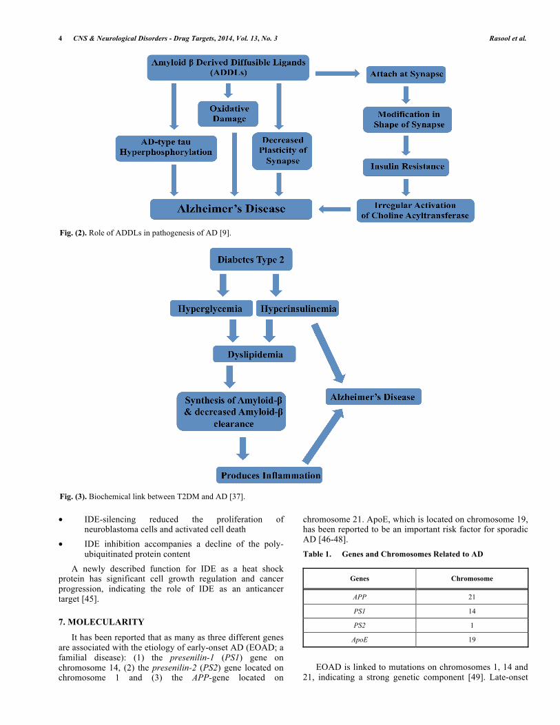
ADDLs are oligomers comparable in size and morphology to prion aggregates that are also associated with neurodegenerative disease. ADDLs are associated with reduced insulin levels and insulin resistance in the brains of AD patients. They are more diffusible and injurious than amyloid plaques [16]. A schematic representation explaining the role of ADDLs in AD is explained in Fig. (2). Under physiological conditions, insulin binds to receptors at neuronal synapses and facilitates memory development. ADDLs can interrupt synaptic communication by binding to the synapse and causing dysfunction. Furthermore, the structure and shape of the synapse is distorted preventing the normal binding of insulin. This process may interrupt signal transduction and results in insulin resistance. ADDLs have been observed to decrease the plasticity of the synapse, cause oxidative damage, potentiate synapse loss and result in AD type 2 hyperphosphorylation, which are all possible links to AD (Fig. 3) [36].
6. INSULIN-DEGRADING ENZYME (IDE)
Two substrates of IDE (previously known as insulinase), insulin and amyloid β-protein (Aβ), are significantly involved in the pathogenesis of T2DM and AD, respectively. IDE has been identified as a principal regulator of Aβ levels in microglial and neuronal cells [38]. IDE is an entity that potentially underlies an association between T2DM and AD. IDE consists of a 110 kDa zinc metalloendopeptidase present in liver extracts. IDE is greatly expressed in the brain, muscle, and testis and in the hepatocytes. IDE is mainly cytosolic, with smaller quantities detected in the endoplasmic reticulum, peroxisomes and plasma membranes [39]. Numerous peptides with molecular weights of 3-10 kDa including insulin, amylin, IGF-I, IGF-II and ß-amyloid have been revealed to be substrates of IDE. Theses peptide substrates have little to no homology between their primary amino acid sequences; however, they have a comparable secondary structure with amyloidogenic nature [40]. A small fraction (up to 10%) of the total IDE is usually transported to the extracellular space, most likely by an unconventional protein secretion passageway, despite the lack of a typical signal peptide [41]. The levels of IDE protein and transcripts are reduced in the hippocampus of AD patients who have the apolipoprotein E (apoE)-ε4 allele when compared to those who do not have this allele [42]. Recently, a report revealed that β amyloid degradation by IDE outside the cell is facilitated by apoE [43]. A similar region of chromosome 10 is genetically associated with T2DM [44]. Several experimental observations have recognized that IDE is also involved in a wide variety of pathological processes including ubiquitin clearance and infection of the varicella zoster virus. A study [45] was conducted to observe the behavior of IDE in different stressful conditions in both normal and malignant cells. Up-regulation of IDE in a heat shock protein (HSP)-like fashion was observed. Furthermore, it was revealed that • IDE was over-expressed in vivo in tumors of the CNS
Fig. (1). Insulin dysfunction and CNS problems.
4 CNS & Neurological Disorders - Drug Targets, 2014, Vol. 13, No. 3 Rasool et al.
• IDE-silencing reduced the proliferation of neuroblastoma cells and activated cell death
• IDE inhibition accompanies a decline of the poly-ubiquitinated protein content
A newly described function for IDE as a heat shock protein has significant cell growth regulation and cancer progression, indicating the role of IDE as an anticancer target [45].
7. MOLECULARITY
It has been reported that as many as three different genes are associated with the etiology of early-onset AD (EOAD; a familial disease): (1) the presenilin-1 (PS1) gene on chromosome 14, (2) the presenilin-2 (PS2) gene located on chromosome 1 and (3) the APP-gene located on
chromosome 21. ApoE, which is located on chromosome 19, has been reported to be an important risk factor for sporadic AD [46-48]. Table 1. Genes and Chromosomes Related to AD
Genes Chromosome
APP 21
PS1 14
PS2 1
ApoE 19
EOAD is linked to mutations on chromosomes 1, 14 and 21, indicating a strong genetic component [49]. Late-onset
Fig. (2). Role of ADDLs in pathogenesis of AD [9].
Fig. (3). Biochemical link between T2DM and AD [37].

Current View from Alzheimer Disease to Type 2 Diabetes Mellitus CNS & Neurological Disorders - Drug Targets, 2014, Vol. 13, No. 3 5
AD appears to be associated with a gene located on chromosome 19, which codes for the cholesterol transporter protein apoE. There are numerous alleles of the apoE gene of which apoE-ε2, apoE-ε3 and apoEε4 occur most frequently. AD is a complex disease and to date, a number of genes that may increase the risk of developing the disease have been revealed. The most recognizable link between AD and genetics is in familial EOAD for which three genes accounting for a major number of familial EOAD cases have been identified. The APP gene encodes the APP, which is cleaved to form Aβ. Mutations in the APP gene result in the erroneous cleavage of the protein, producing a version of Aβ that is more likely to form plaques [50]. Mutations in the APP gene are responsible for 10%-15% of familial EOAD cases [51]. Generally, the presenilin (PS) gene encode for proteins that function in the cleavage of APP. Mutations in both the PS1 and PS2 genes result in erroneous cleavage of APP and contribute to the development of familial EOAD [50]. Mutations in the PS1 gene account for 30%-70% of the familial EOAD cases, and mutations in PS2 are thought to be responsible for less than 5% of these cases [51]. Because familial EOAD is inherited in an autosomal dominant manner, the inheritance of a mutant allele of APP, PS1 or PS2 almost always results in the development of the disease [51]. Children of an affected parent have a 50% possibility of inheriting the mutation and developing the disease. However, it is imperative to note that mutations in APP, PS1 and PS2 genes do not account for all cases of familial EOAD, so there are most likely other genes that have not yet been elucidated that could play a significant role in familial EOAD. Furthermore, abnormalities in the allele of the apolipoprotein E (ApoE) gene located on chromosome 19 have been implicated in not only EOAD but have also been reported to augment the severity of hereditary and sporadic AD [52]. Table 2. The Role of ApoE in AD (Adapted from Liu et al.,
[53])
Loss of Neuroprotective Role Due to ApoE Toxic Role of ApoE
Decreased synaptic action Increased brain deterioration
Decreased glucose metabolism Increased neuronal toxicity
Decreased neurogenesis Increased amyloid β aggregation
Decreased amyloid β clearance Increased tangle synthesis
Decreased vascular function Increased anomalous brain action
Mitochondrial dysfunction
Decreased lipid and cholesterol metabolisms
As far as neurons are concerned, insulin binds to the insulin receptor’s β-subunit and activates tyrosine kinase phosphorylation of that β-subunit, consequently leading to the activation of several second-messenger transduction pathways. The neural Shc/MAP (Shc homology collagen mitogen-activated protein) kinase pathway stimulates the
gene expression necessary for neurons’ repair and maintenance processes and the growth of synapses. It also serves as a modulator of hippocampal synaptic plasticity that promotes learning and memory [54]. Another pathway involved is the binding of insulin receptor substrates (IRS-1 and IRS-2) to phosphatidylinositol 3-kinase, which is essential for synaptic plasticity, memory integration, extinction and recall of contextual memory and Aβ-induced memory loss [55-57]. This pathway also encourages the synthesis of nitric oxide, which plays a vital role in learning and memory processes. Insulin receptors also affect neurotransmission by phosphorylation of N-methyl-D-Aspartate glutamate receptors (increasing the opening of the associated calcium channels), by influencing the internalization of the α-amino-3-hydroxy-5-methyl-4-isoxazolepropionic acid receptor and by recruiting gamma-aminobutyric acid receptors to the postsynaptic sites [58]. The mechanisms that operate insulin resistance and AD could be explained by the transcriptional factors e.g. Forkhead/winged helix transcription factors (FOXO) which are crucial contributors in cell survival, cell proliferation and differentiation, arresting of cell cycle and apoptosis [59-61]. Hence, these transcription factors are major molecular entities in cell death and cell survival pathways. Transcriptional activity of FOXO is controlled by insulin with help of the phosphatidylinositol 13-kinase/Alternative kinase thymoma (Akt) signaling pathway. In presence of insulin, activated Akt is transported to the nucleus where directly phosphorylates FOXO [62]. Successive activation of c-Jun N-terminal kinases and inhibition of Wingless signaling possibly will effect in the formation of Amyloid β plaques and tau protein phosphorylation. Wingless signaling inhibition might also outcome in a continuous activation of FOXO proteins with stimulation of apoptosis and neuronal loss or neurodegeneration [63]. FOXO transcription factors are responsible for the insulin action and the cell responses towards OS, in that way providing a potential centralizing link between AD and insulin resistance [63].
8. AUTOPHAGY DYSFUNCTIONING
The autophagy pathway plays a vital role in the degradation of long-lasting proteins and organelles in a cell. Autophagy is an evolutionarily preserved process and is activated in response to nutrient deprivation as well as to endogenous and exogenous stresses. Various organisms, including mammals, carry a comparable set of autophagy related genes. Autophagy is a highly synchronized process recognized to contribute to cellular cleaning by eliminating intracellular components of lysosomes, thus further showing an important role in cellular quality control. Autophagy ensures that damaged or incorrectly synthesized proteins are removed from the cells by degradation while avoiding the destructive cellular consequences linked to accumulation of malfunctioning proteins inside the cells. Autophagy may also transform synaptic plasticity. This involves structural and functional remodeling of nerve terminals and the trafficking, degradation and recycling of receptors as well as other synaptic proteins [64]. AD is characterized by two aggregate forms: tau tangles and amyloid-β plaques in neurons. Autophagy has been

6 CNS & Neurological Disorders - Drug Targets, 2014, Vol. 13, No. 3 Rasool et al.
implicated in the pathogenesis of AD by its involvement with the endosomal-lysosomal system, which plays a key role in the arrangement of the amyloid-β plaques. Analyzing the role of autophagy in AD began because it was found there is an association of the endosomal-lysosomal system in AD pathogenesis and exclusively in Aβ-amyloidogenesis. Numerous studies have documented that the endosomal-lysosomal pathway is a central controller or regulator of APP processing [65, 66]. The endocytic pathway facilitates a cell’s interaction with its surrounding environment and plays a significant role in the uptake of extracellular nutrients and regulation of surface receptor expression. The arrangement of internalized molecules occurs in the early endosome, which directs the material back to the plasma membrane for recycling and towards the trans-Golgi apparatus for additional processing or to late endosomes and lysosomes for degradation [67]. The failure of autophagic pathways depends upon two main aggravating factors, OS and aging. Aging may lead to a decline in autophagosome development and the fusion of autophagosome and lysosomes [68]. Moreover, it may lead to lysosomal modifications such as enlarged lysosomal volume, decreased lysosomal stability, intra-lysosomal accretion of indigestible material such as lipofuscin and altered activity of hydrolases [69]. These modifications are dependable with a decrease in autophagy, more distinctively macroautophagy. It results in a decrease of turnover of intracellular components as well as a decrease in the ability of cells to adjust to changes in the extracellular environment [70]. Alternatively, OS burdens the macroautophagic system – oxidized proteins and smashed organelles are engulfed by autophagosomes that can become a cause of reactive oxygen species inside the autophagosomes or lysosomes. Following that, reactive oxygen species can react and damage lysosomal hydrolases and/or other mechanisms that are involved in the fusion of lysosomes and autophagosomes resulting in the accretion of non-degraded products around these cellular compartments. If autophagy can directly degrade some cellular lipid components, it is plausible that lipids can also directly excite this process, particularly during lipotoxicity (the cytotoxic effects of excessive fat accumulation in cells, but not in adipocytes), and autophagy has been implicated as one of the contributing factors to diseases such as non-alcoholic fatty liver disease, obesity and diabetes [71]. T2DM patients are characterized by an accumulation of autophagosomes, which further implies an obstruction of autophagy in the pancreatic β-cells [72]. In the presence of sufficient amount of nutrients, insulin can activate class I phosphatidylinositol 3-kinase proteins that can, with the help of the Akt pathway, in turn amplify the signal to stimulate the mammalian target of rapamycin, which restrains the activation of autophagy.
9. EPIGENETICS
The progressive damage or death of neurons can lead to AD and Parkinson’s disease. Environmental factors have the ability to damage the developing and mature nervous system, resulting in neurodegenerative disorders. Epigenetics refers to the regulation of deoxyribonucleic acid (DNA) sequences that do not involve alteration of their actual base
composition. These alterations include downstream modification of DNA and histones by methylation, acetylation and phosphorylation. Environmental factors, such as dietary folate intake and heavy metals, agitate neurodegenerative genes by epigenetic means, leading to distorted gene expression and late-onset neurodegenerative disorders. Metabolic control without help does not predict an individual’s danger for diabetic complications [73]. Stable gene expression plays a crucial role in long-lasting forms of memories [74], which are in part maintained by chromatin-templated epigenetic processes [75]. Among the epigenetic modifications identified in the nervous system, histone acetylation has been unequivocally associated with learning and memory [75]. Acetylation diminishes the electrostatic affinity between neighboring histones and the DNA within them and consequently can promote a more open chromatin structure that allows for memory-related gene transcription [76]. Understanding epigenetic mechanisms will likely permit the ability to recognize novel remedial targets. Small regulative RNAs or microRNAs are established regulators of mammalian cell phenotype. They are involved in biological functions related to the pathogenesis of diabetes such as insulin secretion, immune response and metabolism processes. Patterns of microRNA in a variety of cells and tissues may offer functional disease biomarkers, while in vivo management of precise subsets of small regulatory RNAs may be employed for novel remedial strategies. Studies have suggested that epigenetic mechanisms may play a decisive role in the development of AD. Age-related disorders demonstrate a range of modifications in the anatomy of the brain and its function. The irregular processing of APP and the hyperphosphorylation of tau protein that results in NFTs are pathological characteristics of AD. Chemical alterations of the DNA structure and related proteins as well as epigenetic mechanisms can affect gene expression/transcription and may play a role in the relation between genetic and environmental factors when defining an individual’s phenotype [77]. Vascular risk factors such as hyperinsulinaemia, hypertension, obesity and T2DM have been associated with a higher risk of dementia and cognitive impairment [78]. Social factors are also related to the risk of AD [79]. A reduced risk of AD has repeatedly been associated with dietary factors such as consumption of vegetables and fruit, omega-3 fatty acids caloric, dietary limit of calories as well as to physical activity [80]. Epigenetic variations can be propagated to new cells either by mitosis or by meiosis; propagation through meiosis will transfer these variations between generations. Alterations can take place at specific gene loci in specific cells to give distinct cellular phenotypes or can comprise many genes in several cells, a mechanism implicated in the synchronization of biological processes, such as development and aging. Epigenetic alterations have the ability to appear as genetic mutations. Epigenetic suppression of tumor suppressor genes can imitate the loss of function mutations of tumor suppressor genes and both are considerably linked to cancer development [81]. Epigenetic alterations can also exaggerate the effect of gene mutations [82].

Current View from Alzheimer Disease to Type 2 Diabetes Mellitus CNS & Neurological Disorders - Drug Targets, 2014, Vol. 13, No. 3 7
On the other hand, DNA methylation plays a crucial role in both AD and T2DM. As far as diabetes is concerned, several genetic factors including common variants as the Peroxisome Proliferator-activated receptor gamma Coactivator, calpain 10, Peroxisome Proliferator-Activated Receptor gamma and Transcription factor 7-like 2 genes have been implicated with an increased risk of diabetes. The interaction between environment and genes may be even more intricate and involve epigenetic factors such as histone acetylation and DNA methylation that can encourage the development of T2DM. For example, cytosine residues at CG dinucleotides are a possible site for DNA methylation, and gene expression is frequently decreased when DNA methylation takes place at a promoter region [83]. Hepatic insulin resistance is an additional significant feature of both aging and T2DM. Glucokinase is a key enzyme for glucose utilization in hepatocytes, and its activity is diminished in the hepatocytes of diabetic patients. Alterations in the glucokinase gene can enhance the risk of getting a monogenic form of T2DM [84]. The Peroxisome Proliferator-Activated Receptor Gamma Coactivator 1-Aplpha (PPARGC1A, transcriptional coactivator) synchronizes gene expression that activates the metabolism of mitochondrial oxidation in several different tissues [85]. On the other hand, DNA methylation of the PPARGC1A promoter is prominent in pancreatic islets from patients with T2DM in contrast to those of healthy individuals. PPARGC1A expression is decreased in diabetic islets, and the links are inversely proportional with the extent of DNA methylation [86]. Notably, PPARGC1A expression is positively associated with glucose-stimulated insulin secretion in human pancreatic islets, indicating that epigenetic mechanisms may control gene expression and, consequently, insulin secretion in human islets [86].
10. THE ROLE OF BUTYRYLCHOLINESTERASE (BCHE) AND ACETYLCHOLINESTERASE (ACHE)
The levels of BChE are augmented in the cerebral cortex of AD patients, especially in those having the ε4 allele of the apolipoprotein E gene (ApoE), and the presence of certain BChE variants can predict increased AD risk and poor response to anticholinesterase treatment. BChE and acetylcholinesterase activities are connected to tangles and plaques in AD. These diseased cholinesterases, with changed properties, are reported to be involved in the formation of plaques [87]. The activity of BChE in serum is correlated to serum fasting triacylglycerol levels and insulin sensitivity in patients who have type 1 diabetes mellitus or T2DM. Interestingly, non-diabetic individuals who have a BChE insufficiency have serum triacylglycerol levels in the normal range, indicating that BChE may contribute to the difference in lipoprotein metabolism in hyper-triglyceridemia associated with insulin resistance or insulin deficiency in diabetes [88]. The levels of BChE and AChE were found to be significantly higher in various parts of the brain in AD patients. The higher activities of BChE and AChE have been reported to contribute to a higher incidence of AD and to also increase the number of neocortical and cortical amyloid-rich NFTs and neuritic plaques among AD patients [89].
In diabetes, there is enhanced OS due to an increase in lipoxidation and glycoxidation products in both tissue and plasma proteins. Among uremic diabetes patients, increased carbonyl stress (carbonyl overload) will lead to increased chemical variation of proteins by lipids and carbohydrates. This may be caused by a widespread increase in the level of reactive carbonyl precursors of advanced glycation and lipoxidation end-products. Carbonyl stress may result from an increase in substrate stress as well as a reduction in the effectiveness of detoxification of reactive carbonyl compounds, i.e., an imbalance between the rates of synthesis and detoxification of reactive carbonyls.
11. NANOPARTICLES
In the management of neurological disorders, encapsulation of nanoparticles has been explored for transporting therapeutic molecules directly to the eye beyond the blood retina barrier as well as to the CNS beyond the blood-brain barrier. Applications for nanoparticles have been extended to neurodegenerative diseases such as Huntington’s, Parkinson’s and AD. Protein aggregation and amyloid deposits are related to more than 40 different diseases ranging from neurodegenerative diseases such as AD and Parkinson’s disease to systemic amyloidosis, such as T2DM [90-94]. Although amyloids occur naturally in the body, the mechanism of their generation remains unidentified; this poses a challenge for drug development despite decades of research. Recently, a new class of extremely small peptides developed by the Institute of Bioengineering and Nanotechnology, encouraged other researchers to consider this technology, which can provide the necessary approach required to devise more effective treatments for these diseases. Micro and nanosized sensors can take advantage of a wide range of technologies that most effectively sense a targeted chemical or physical property. The use of polyethylene glycol beads coated with fluorescent molecules can help monitor blood sugar levels among diabetics. The beads are introduced under the skin and reside in the interstitial fluid. When glucose levels in the interstitial fluid drop to dangerous levels the fluorescent molecules glow. AD, breast cancer, types 1 diabetes, T2DM and major depression are the four therapeutic clinical research objectives of the Nano Axis LLC Technology and the new Quantum Materials Corporation Alliance (New York, USA). Currently, Nano Axis is developing new remedies and diagnostics for the treatment, measurement and detection of medical conditions and disorders including depression, chronic pain, AD, diabetes, cancer and influenza. Amyloids are composed of general polypeptide chains in a stable structural state and as such could be employed as a template for the improvement and manufacturing of new self-organized nanomaterials such as nanowires [95-97]. Computational approaches based on nanoscience will help determine the role of amyloids in biomedical disorders and are a promising approach for understanding the functional material properties of amyloids. Potential biological nanotechnology applications come into sight at scales of hundreds of nanometers and micrometers. Recently, gold and silver nanoparticles have been developed as inhibitors of protein glycation [98-101]. Gold nanoparticles have been
8 CNS & Neurological Disorders - Drug Targets, 2014, Vol. 13, No. 3 Rasool et al.
found to successfully inhibit glycation of α-crystallins and collagen, whereas silver nanoparticles can inhibit AGE-induced retinal complications.
12. TREATMENT FOR AD AND T2DM
Currently, AD patients appear to benefit from pharmacotherapy targeted for treating T2DM, and clinical trials for the effectiveness of such treatments are still in progress. There is no existing treatment available to stop or decrease the decline of brain function. Currently available drugs are only able to slow deterioration of symptoms for up to 6-12 months, and even then have been effective in only 50% of the patients [102]. Tau phosphorylation in both AD and T2DM plays a role in the stimulation of glycogen synthase kinase-3 (GSK-3) which in turn phosphorylates glycogen synthase in the rate-limiting step of glycogen biosynthesis [103-105]. GSK-3 plays an important role in the development NFTs and therefore, GSK-3 suppression could be a mutual target for the treatment of both AD and T2DM [106, 107]. Neurotoxicity induced by amyloid β can be averted by pre-treatment with insulin-like growth factors (IGFs), and the neuroprotective nature of IGFs is regulated by activation of phosphatidylinositol 13-kinase/Akt pathway and inhibition of GSK-3 [26]. Many AD patients have distorted insulin signaling, which is also apparent in brain specific diabetes [15]. Therefore, a probable prospective approach to AD could be pharmacological management of the insulin-signaling pathway. Moreover, as far as treatment is concerned, Bacopa monnieri has been suggested a potential neuroprotectant as well as cognitive enhancer against AD model [108].
13. CONCLUSION
Recent evidence suggests that there is a vital role for insulin and insulin receptors in the pathogenesis of AD. Additionally, direct and indirect evidence indicates the involvement of both genetics and epigenetics in the pathogenesis of AD. Further research is required to investigate the relationship between T2DM and AD using both genome wide and gene-specific targets. The pathological, genetic and epigenetic links between AD and hyperglycemic states can provide insight for better diagnostic and treatment strategies for AD. Because ADDLs also contribute to lowered insulin levels and insulin resistance in the brains of AD patients, future diagnosis may also be dependent upon measurement of ADDLs. It is believed that measurement of acetylcholinesterase and butyrylcholinesterase can also represent the role of the hypothalamus and other brain centers in the pathogenesis of AD. Both enzymes can be reliable and robust markers of inflammation, and measuring their activity could be useful in the diagnosis of AD as well as in new drug development for AD. Lastly, nanoparticles can play a role in the diagnosis, prevention and treatment of AD. These processes can take form in subject to either control or exterminate the toxicological risk factor, which may be caused by nanoparticles.
ACKNOWLEDGEMENTS
We wish to thank all the students for their valuable questions, which led us to write this updated review.
LIST OF ABBREVIATIONS
AD = Alzheimer’s Disease T2DM = Type 2 Diabetes Mellitus ADDLs = Amyloid beta-derived diffusible ligands IDE = Insulin degrading enzyme IGF = Insulin-like growth factor CNS = Central nervous system apoE = Apolipoprotein E PS = Presenilin APP = amyloid precursor protein EOAD = early-onset AD FOXO = Forkhead/winged helix transcription factors BChE = Butyrylcholinesterase AChE = Acetylcholinesterase GSK = glycogen synthase kinase PPARGC1A = Peroxisome Proliferator-Activated Receptor Gamma Coactivator 1-Aplpha OS = Oxidative stress NFTs = Neurofibrillary tangles Aβ = amyloid β-protein Akt = Alternative kinase thymoma Ribonucleic acid RNA = Ribonucleic acid DNA = Deoxyribonucleic acid
REFERENCES [1] Shaik AS, Raja AE, Vijayalakshmi M, Devalarao G. Alzhemier’s
Disease-Pathophysiology and Treatment. Intl J Pharm Biosci 2010; 1(2): 1-11.
[2] Dong S, Duan Y, Hu Y, Zhao Z. Advances in the pathogenesis of Alzheimer’s disease, a re-evaluation of amyloid cascade hypothesis. Transl Neurodegener 2012; 1: 18.
[3] Hebda JA, Saraogi I, Magzoub M, Hamilton AD, Miranker AD. A Peptidomimetic Approach to Targeting Pre-amyloidogenic States in Type II Diabetes. Chem Biol 2009; 16: 943-50.
[4] Rönnemaa E, Zethelius B, Sundelöf J. Impaired insulin secretion increases the risk of Alzheimer disease. Neurology 2008; 71: 1065-71.
[5] Herbert LE, Weuve J, Scherr PA, Evans DA. AD in the United States (2010-2050) estimated using the 2010 Census. Neurology 2013; 80(19): 1778-83.
[6] World Alzheimer Report, 2010. The Global Economic Impact of Dementia. Alzheimer’s Disease International, 2010; p. 15.
[7] Badran M, Laher I. Type 2 Diabetes Mellitus in Arabic-Speaking Countries. Int J Endocrin 2012; 11: 902873.
[8] Ott A, Stolk RP, van Harskamp F, et al. Diabetes mellitus and the risk of dementia, the Rotterdam Study. Neurology 1999; 53: 1937-42.
[9] Kroner Z. The relationship between Alzheimer’s disease and diabetes, type 3 diabetes? Altern Med Rev 2009; 14: 373-9.
[10] Jiang Q, Heneka M, Landreth GE. The role of peroxisome proliferator-activated receptor-gamma (PPARgamma) in Alzheimer’s disease, therapeutic implications. CNS Drugs 2008; 22: 1-14.
[11] Takeda S, Sato N, Uchio-Yamada K, et al. Diabetes accelerated memory dysfunction via cerebrovascular inflammation and Aβ deposition in Alzheimer mouse model with diabetes. Proc Natl Acad Sci 2010; 107: 7036-41.
[12] Rivera EJ, Goldin A, Fulmer N, et al. Insulin and insulin-like growth factor expression and function deteriorate with progression of

Current View from Alzheimer Disease to Type 2 Diabetes Mellitus CNS & Neurological Disorders - Drug Targets, 2014, Vol. 13, No. 3 9
Alzheimer’s disease, link to brain reductions in acetylcholine. J Alzheimers Dis 2005; 8: 247-68.
[13] Beeler N, Riederer BM, Waeber G, Abderrahmani A. Role of the JNK-interacting protein 1/islet brain 1 in cell degeneration in Alzheimer disease and diabetes. Brain Res Bull 2009; 80: 274-81.
[14] Miklossy J, Qing H, Radenovic A, et al. Beta amyloid and hyperphosphorylated tau deposits in the pancreas in type 2 diabetes. Neurobiol Aging 2010; 31: 1503-15.
[15] Steen E, Terry BM, Rivera EJ, et al. Impaired insulin and insulin-like growth factor expres-sion and signaling mechanisms in AD-is this type 3 diabetes? J Alzheimers Dis 2005; 7(1): 63-80.
[16] Viola KL, Velasco PT, Klein WL. Why Alzheimer’s is a disease of memory, the attack on synapses by A beta oligomers (ADDLs). J Nutr Health Aging 2008; 12: 51-7.
[17] de la Monte SM, Tong M, Lester-Coll N. Therapeutic rescue of neurodegeneration in experimental type 3 diabetes, relevance to AD. J Alzheimers Dis 2006; 10: 89-109.
[18] Watson GS, Craft S. The role of insulin resistance in the pathogenesis of AD, implications for treatment. CNS Drugs 2003; 17: 27-45.
[19] Woods SC, Seeley RJ, Baskin DG, Schwartz MW. Insulin and the blood-brain barrier. Curr Pharm Des 2003; 9: 795-800.
[20] Freychet P. Insulin receptors and insulin action in the nervous system. Diabetes Metab Res Rev 2000; 16: 390-2.
[21] Bingham EM, Hopkins D, Smith D. The role of insulin in human brain glucose metabolism, an 18fluoro-deoxyglucose positron emission tomography study. Diabetes 2002; 51: 3384-90.
[22] Craft S, Watson GS. Insulin and neurodegenerative disease, shared and specific mechanisms. Lancet Neurol 2004; 3: 169-78.
[23] Zhao WQ, Alkon DL. Role of insulin and insulin receptor in learning and memory. Mol. Cell Endocrinol 2001; 177: 125-34.
[24] Werner H, Roberts, CTJ, Raizada MK, et al. Developmental regulation of the insulin and insulin-like growth factor receptors in the central nervous system. In: Receptors in the Developing Nervous System; Zagon IS, McLaughlin PJ, Eds.; London, Chapman and Hall 1993; pp. 109-27.
[25] Akter K, Lanza EA, Martin SA, et al. Diabetes mellitus and AD, shared pathology and treatment? Br J Clin Pharmacol 2011; 71(3): 365-76.
[26] de la Monte SM. Insulin resistance and Alzheimers’s disease. BMB Rep 2009; 42: 475-81.
[27] Westerman MA, Cooper-Blacketer D, Mariash A. The relationship between Abeta and memory in the Tg2576 mouse model of AD. J Neurosci 2002; 22: 1858-67.
[28] Li L, Holscher C. Common pathological processes in Alzheimer disease and type 2 diabetes, a review. Brain Res Rev 2007; 56: 384-402.
[29] Maiorini AF, Gaunt MJ, Jacobsen TM, et al. Potential novel targets for Alzheimer pharmacotherapy: I. Secretases J Clin Pharm Ther 2002; 27(3): 169-83.
[30] Walsh DM, Klyubin I, Fadeeva JV, et al. Naturally secreted oligomers of amyloid beta protein potently inhibit hippocampal long-term potentiation in vivo. Nature 2002; 416: 535-9.
[31] Kosik KS, Joachim CL, Selkoe DJ. Microtubule-associated protein tau (tau) is a major antigenic component of paired helical filaments in Alzheimer disease. Proc Natl Acad Sci USA 1986; 83: 4044-8.
[32] Evans JL, Goldfine ID, Maddux BA, Grodsky GM. Oxidative stress and stress-activated signaling pathways, a unifying hypothesis of type 2 diabetes. Endocr Rev 2002; 23: 599-622.
[33] Schmeichel AM, Schmelzer JD, Low PA. Oxidative injury and apoptosis of dorsal root ganglion neurons in chronic experimental diabetic neuropathy. Diabetes 2003; 52: 165-71.
[34] Calabrese V, Scapagnini G, Giuffrida SAM, Bates TE, Clark JB. Mitochondrial involvement in brain function and dysfunction, relevance to aging, neurodegenerative disorders and longevity. Neurochem Res 2001; 26: 739-64.
[35] Orth M, Schapira AH. Mitochondria and degenerative disorders. Am J Med Genet 2001; 106: 27-36.
[36] De Felice FG, Wu D, Lambert MP. AD-type neuronal tau hyperphosphorylation induced by A beta oligomers. Neurobiol Aging 2008; 29: 1334-47.
[37] Carlsson CM. Type 2 Diabetes Mellitus, Dyslipidemia and AD. J Alzheimer Dis 2010; 20: 711-22.
[38] Farris W, Mansourian S, Chang Y, et al. Insulin-degrading enzyme regulates the levels of insulin, amyloid β-protein, and the β-amyloid precursor protein intracellular domain in vivo. PNAS 2003; 100(7): 4162-7.
[39] Miners JS, Baig S, Palmer J, et al. A beta-degrading enzymes in AD. Brain Pathol 2008; 18: 240-52.
[40] Qiu WQ, Folstein MF. Insulin, insulin-degrading enzyme and amyloid-beta peptide in AD, review and hypothesis. Neurobiol Aging 2006; 27: 190-8.
[41] Zhao J, Li L, Leissring MA. Insulin-degrading enzyme is exported via an unconventional protein secretion pathway. Mol Neurodegener 2009; 4: 4.
[42] Cook DG, Leverenz JB, McMillan PJ, et al. Reduced hippocampal insulin-degrading enzyme in late-onset AD is associated with the apolipoprotein E-epsilon4 allele. Am J Pathol 2003; 162: 313-9.
[43] Jiang Q, Lee CY, Mandrekar S, et al. ApoE promotes the proteolytic degradation of Abeta. Neuron 2008; 58: 681-93.
[44] Zeggini E, Weedon MN, Lindgren CM, et al. Replication of genome-wide association signals in UK samples reveals risk loci for type 2 diabetes. Science 2007; 316: 1336-41.
[45] Tundo GR, Sbardella D, Ciaccio C, et al. Insulin-degrading enzyme (IDE): a novel heat shock-like protein. J Biol Chem 2013; 288(4): 2281-9.
[46] Nowotny P, Kwon JM, Goate AM. Alzheimer Disease. Encyclopedia of Life Sciences, Nature Publishing Group (NPG): 2001; pp. 1-6.
[47] Hatters DM, Zhong N, Rutenber E, Weisgraber KH. Amino-terminal domain stability mediates apolipoprotein E aggregation into neurotoxic fibrils. J Mol Biol 2006; 361: 932-44.
[48] Martins IJ, Berger T, Sharman MJ, et al. Cholesterol metabolism and transport in the pathogenesis of Alzheimer’s disease. J Neurochem 2009; 111: 1275-308.
[49] Wolfe MS. Secretase targets for AD, identification and therapeutic potential. J Med Chem 2001; 44: 2039-60.
[50] Goedert M, Spillantini G. A Century of AD. Science 2006; 314: 777-81.
[51] Bird TD. Early-Onset Familial Alzheimer Disease. Gene Reviews 2007.
[52] Rocchi A, Pellegrini S, Siciliano G, Murri L. Causative and susceptibility genes for AD, a review. Brain Res Bull 2003; 61(1): 1-24.
[53] Liu CC, Kanekiyo T, Xu H, Bu G. Apolipoprotein E and Alzheimer disease, risk, mechanisms and therapy. Nat Rev Neurol 2013; 1-13.
[54] Miyamoto Y, Chen L, Sato M, et al. Hippocampal synaptic modulation by the phosphotyrosine adapter protein AhcC/N-Shc via interaction with the NMDA receptor. J Neurosci 2005; 16: 1826-35.
[55] Horwood JM, Dufour F, Laroche S, Davis S. Signalling mechanisms mediated by the phosphoinositide 3-kinase/Akt cascade in synaptic plasticity and memory in the rat. Eur J Neurosci 2006; 23: 3375-84.
[56] Chen X, Garelick MG, Wang H, et al. PI3 kinase signaling is required for retrieval and extinction of contextual memory. Nat Neurosci 2005; 8: 925-31.
[57] Chiang HC, Wang L, Xie Z, Yau A, Zhong Y. PI3 kinase signaling is involved in Aβ-induced memory loss in Drosophila. Proc Natl Acad Sci USA 2010; 107: 7060-5.
[58] Wan Q, Xiong ZG, Man HY, et al. Recruitment of functional GABAA receptors to postsynaptic domains by insulin. Nature 1997; 388: 686-90.
[59] Accili D, Arden KC. FOXOs at the crossroads of cellular metabolism, differentiation, and transformation. Cell 2004; 117: 421-6.
[60] van der Horst A, Burgering BM. Stressing the role of FOXO proteins in lifespan and disease. Nat Rev Mol. Cell Biol 2007; 8: 440-50.
[61] Huang H, Tindall DJ. Dynamic FOXO transcription factors. J Cell Sci 2007; 120: 2479-87.
[62] Greer EL, Brunet A. FOXO transcription factors at the interface between longevity and tumor suppression. Oncogene 2005; 24: 7410-25.
[63] Manolopoulos KN, Klotz L-O, Korsten P, Bornstein SR, Barthel A. Linking Alzheimer's disease to insulin resistance, the FOXO response to oxidative stress. Mol Psychiatry 2010; 15: 1046-52.
[64] Boland B, Nixon RA. Neuronal macroautophagy, from development to degeneration. Mol Aspects Med 2006; 27(5-6): 503-19.
[65] Grbovic OM, Mathews PM, Jiang Y. Rab5-stimulated up-regulation of the endocytic pathway increases intracellular beta-cleaved amyloid precursor protein carboxyl-terminal fragment levels and Abeta production. J Biol Chem 2003; 278: 31261-8.
[66] Pasternak SH, Bagshaw RD, Guiral M. Presenilin-1: nicastrin, amyloid precursor protein and gamma-secretase activity are co-localized in the lysosomal membrane. J Biol Chem 2003; 278: 26687-94.
[67] Nixon RA, Cataldo AM, Mathews PM. The endosomal-lysosomal system of neurons in AD pathogenesis, a review. Neurochem Res 2000; 25: 1161-72.
10 CNS & Neurological Disorders - Drug Targets, 2014, Vol. 13, No. 3 Rasool et al.
[68] Terman A, Brunk UT. Lipofuscin. Int J Biochem Cell Biol 2004; 36(8): 1400-4.
[69] Terman A, Dalen H, Brunk UT. Ceroid/lipofuscin-loaded human fibroblasts show decreased survival time and diminished autophagocytosis during amino acid starvation. Exp Gerontol 1999; 34(8): 943-57.
[70] Ward WF. Protein degradation in the aging organism. Prog Mol Subcell Biol 2002; 29: 35-42.
[71] Tan SH, Shui G, Zhou J, et al. Induction of autophagy by palmitic acid via a PKC-mediated signalling pathway independent of mTOR. J Biol Chem 2012; 287(18): 14364-76.
[72] Masini M, Bugliani M, Lupi R, et al. Autophagy in human type 2 diabetes pancreatic beta cells. Diabetologia 2009; 52: 1083-6.
[73] Kwok JBJ. Role of epigenetics in Alzheimer’s and Parkinson’s disease. Epigenomics 2010; 2(5): 671-82.
[74] Kandel ER. The molecular biology of memory storage, a dialogue between genes and synapses. Science 2001; 294(5544): 1030-8.
[75] Graff J, Kim D, Dobbin MM, Tsai LH. Epigenetic regulation of gene expression in physiological and pathological brain processes. Physiol Rev 2011; 91(2): 603-49.
[76] Brownell JE, Zhou J, Ranalli T, et al. Tetrahymena histone acetyltransferase A, a homolog to yeast Gcn5p linking histone acetylation to gene activation. Cell 1996; 84(6): 843-51.
[77] Reichenberg A, Mill J, MacCabe JH. Epigenetics, genomic mutations and cognitive function. Cogn Neuropsychiatry 2009; 14: 377-90.
[78] Kalaria RN, Maestre GE, Arizaga R, Friedland RP, Antuono P. AD and vascular dementia in developing countries, prevalence, management and risk factors. Lancet Neurol 2008; 7: 812-26.
[79] Hakansson K, Rovio S, Helkala EL, Vilska AR, Kivipelto M. Association between mid-life marital status and cognitive function in later life, population based cohort study. Br Med J 2009; 339: 2462.
[80] Scarmeas N, Luchsinger JA, Schupf N, et al. Physical activity, diet and risk of Alzheimer disease. JAMA 2009; 302: 627-37.
[81] Mund C, Brueckner B, Lyko F. Reactivation of epigenetically silenced genes by DNA methyltransferase inhibitors, basic concepts and clinical applications. Epigenetics 2006; 1: 7-13.
[82] Colnot S, Niwa-Kawakita M, Hamard G, Godard C, Perret C. Colorectal cancers in a new mouse model of familial adenomatous polyposis, influence of genetic and environmental modifiers. Lab Invest 2004; 84: 1619-30.
[83] Bird AP. CpG-rich islands and the function of DNA methylation. Nature 1986; 321: 209-13.
[84] Vaxillaire M, Froguel P. Monogenic diabetes in the young, pharmacogenetics and relevance to multifactorial forms of type 2 diabetes. Endocr Rev 2008; 29: 254-64.
[85] Puigserver P, Spiegelman BM. Peroxisome proliferator-activated receptor-gamma coactivator 1 alpha (PGC-1 alpha): transcriptional coactivator and metabolic regulator. Endocr Rev 2003; 24: 78-90.
[86] Ling C, Del Guerra S, Lupi R, et al. Epigenetic regulation of PPARGC1A in human type 2 diabetic islets and effect on insulin secretion. Diabetologia 2008; 51: 615-22.
[87] Mariam F, Eskander NGN, Elaine YL, Bahiyyih K. Rivastigmine is a potent inhibitor of acetyl-and butyrylcholinesterase in Alzheimer’s plaques and tangles. Brain Res 2005; 1060: 144-52.
[88] Sanchez CG, Salceda R. Effect of streptozotocin induced diabetes on activities of cholinesterases in the rat retina. IUBMB Life 2000; 49: 283-7.
[89] Greig NH, Utsuki T, Ingram DK, Wang Y, Pepeu G. Selective butyrylcholinsterase inhibition elevates brain acetylcholine, augments
learning and lowers Alzheimer’s ß-amyloid peptide in rodent. Proc Natl Acad Sci USA 2005; 102: 17213-8.
[90] Chiti F, Dobson CM. Protein misfolding, functional amyloid and human disease. Annu Rev Biochem 2006; 75(1): 333-66.
[91] Caughey B, Lansbury PT. Proto fibrils, pores, fibrils and neurodegeneration, separating the responsible protein aggregates from the innocent bystanders. Annu Rev Neurosci 2003; 26(1): 267-98.
[92] Schnabel J. Protein folding, the dark side of proteins. Nature 2010; 464(7290): 828-9.
[93] Giehm L, Svergun DI, Otzen DE, Vestergaard B. Low-resolution structure of a vesicle disrupting & α,-synuclein oligomer that accumulates during fibrillation. Proc Natl Acad Sci USA 2011; 108(8): 3246-51.
[94] Cooper GJ, Willis AC, Clark A, et al. Purification and characterization of a peptide from amyloid-rich pancreases of type 2 diabetic patients. Proc Natl Acad Sci USA 1987; 84(23): 8628-32.
[95] Chiti F, Webster P, Taddei N, et al. Designing conditions for in vitro formation of amyloid proptofilaments and fibrils. Proc Natl Acad Sci USA 1999; 96: 3590-4.
[96] Fandrich M, Dobson CM. The behavior of polyamino acids reveals an inverse side chain effect in amyloid structure formation. EMBO J 2002; 21: 5682-90.
[97] Scheibel T, Parthasarathy R, Sawicki G, et al. Conducting nanowires built by controlled self-assembly of amyloid fibers and selective metal deposition. Proc Natl Acad Sci USA 2003; 100(8): 4527-32.
[98] Singh S, Bhattacharya J, Datta H, Dasgupta AK. Anti-glycation activity of gold nanoparticles. Nanomed Nanotech Biol Med 2009; 5: 21-9.
[99] Seneviratne C, Narayanan R, Liu W, Dain J. The in vitro inhibition effect of 2nm gold nanoparticles on non-enzymatic glycation of human serum albumin. Biochem Biophys Res Commun 2012; 422: 447-54.
[100] Kim J, Hong C, Koo Y, Choi H, Lee K. Anti-glycation effect of gold nanoparticles on collagen. Biol Pharm Bull 2012; 35: 260-4.
[101] Sheikpranbabu S, Kalishwaralal K, Lee K, et al. The inhibition of advanced glycation end-products-induced retinal vascular permeability by silver nanoparticles. Biomaterials 2010; 31: 2260-71.
[102] Lanctot DL, Rajaram RD, Herrmann N. Review, therapy for AD, how effective are current treatments? Ther Adv Neurol 2009; 2: 163-80.
[103] Doble BW, Woodgett JR. Role of glycogen synthase kinase-3 in cell fate and epithelial-mesenchymal transitions. Cells Tissues Organs 2007; 185: 73-84.
[104] Phiel CJ, Wilson CA, Lee VM, Klein PS. GSK-3alpha regulates production of AD amyloid-beta peptides. Nature 2003; 423: 435-9.
[105] Sivaprakasam P, Xie A, Doerksen RJ. Probing the physicochemical and structural requirements for glycogen synthase kinase-3alpha inhibition, 2D-QSAR for 3-anilino-4-phenylmaleimides. Bioorg Med Chem 2006; 14: 8210-8.
[106] Bhat RV, Budd Haeberlein SL, Avila J. Glycogen synthase kinase 3: a drug target for CNS therapies. J Neurochem 2004; 89: 1313-7.
[107] Cole AR, Astell A, Green C, Sutherland C. Molecular connexions between dementia and diabetes. Neurosci Biobehav Rev 2007; 31: 1046-63.
[108] Uabundit N, Wattanathorn J, Mucimapura S, Ingkaninan K. Cognitive enhancement and neuroprotective effects of Bacopa monnieri in Alzheimer's disease model. J Ethnopharmacol 2010; 127(1): 26-31.
[109] Moreira PI, Santos MS, Seica R, Oliveira CR. Brain mitochondrial dysfunction as a link between AD and diabetes. J Neurol Sci 2007; 257: 206-14.
Received: May 29, 2013 Revised: September 9, 2013 Accepted: September 10, 2013 DISCLAIMER: The above article has been published in Epub (ahead of print) on the basis of the materials provided by the author. The Editorial Department reserves the right to make minor modifications for further improvement of the manuscript.
PMID: 24059295