The Odyssey’s Northern Origins and a Different Author Than Homer
Upload
independentCategory
view
1download
0
Disassembly of Shank and Homer Synaptic Clusters IsDriven by Soluble b-Amyloid1-40 through DivergentNMDAR-Dependent Signalling PathwaysFrancesco Roselli1,2*, Peter Hutzler1,3, Yvonne Wegerich1, Paolo Livrea2, Osborne F. X. Almeida1*
1 Max-Planck Institute of Psychiatry, Munich, Germany, 2 Department of Neurological and Psychiatric Sciences, University of Bari, Bari, Italy, 3 Institute of Pathology,
Helmholtz Center Munich, Neuherberg, Germany
Abstract
Disruption of the postsynaptic density (PSD), a network of scaffold proteins located in dendritic spines, is thought to beresponsible for synaptic dysfunction and loss in early-stage Alzheimer’s disease (AD). Extending our previous demonstrationthat derangement of the PSD by soluble amyloid-b (Ab) involves proteasomal degradation of PSD-95, a protein importantfor ionotropic glutamate receptor trafficking, we now show that Ab also disrupts two other scaffold proteins, Homer1b andShank1, that couple PSD-95 with ionotropic and metabotropic glutamate receptors. Treatment of fronto-cortical neuronswith soluble Ab results in rapid (within 1 h) and significant thinning of the PSD, decreased synaptic levels of Homer1b andShank1, and reduced synaptic mGluR1 levels. We show that de novo protein synthesis is required for the declustering effectsof Ab on Homer1b (but not Shank1) and that, in contrast to PSD-95, Ab-induced Homer1b and Shank1 cluster disassemblydoes not depend on proteasome activity. The regulation of Homer1b and Shank1 by Ab diverges in two other respects: i)whereas the activity of both NMDAR and VDCC is required for Ab-induced declustering of Homer1b, Ab-induceddeclustering of Shank1 only requires NMDAR activity; and ii) whereas the effects of Ab on Homer1b involve engagement ofthe PI-3K pathway and calcineurin phosphatase (PP2B) activity, those on Shank1 involve activation of the ERK pathway. Insummary, soluble Ab recruits discrete signalling pathways to rapidly reduce the synaptic localization of major componentsof the PSD and to regulate the availability of mGluR1 in the synapse.
Citation: Roselli F, Hutzler P, Wegerich Y, Livrea P, Almeida OFX (2009) Disassembly of Shank and Homer Synaptic Clusters Is Driven by Soluble b-Amyloid1-40
through Divergent NMDAR-Dependent Signalling Pathways. PLoS ONE 4(6): e6011. doi:10.1371/journal.pone.0006011
Editor: Hitoshi Okazawa, Tokyo Medical and Dental University, Japan
Received December 15, 2008; Accepted May 31, 2009; Published June 23, 2009
Copyright: � 2009 Roselli et al. This is an open-access article distributed under the terms of the Creative Commons Attribution License, which permitsunrestricted use, distribution, and reproduction in any medium, provided the original author and source are credited.
Funding: F.R. was partly supported by a Vigoni grant (Conferenza dei Rettori delle Universita Italiane, CRUI, Roma, Italy and the German Academic ExchangeService), the University of Bari (Italy), and the Max Planck Institute of Psychiatry (Munich, Germany). The funders had no role in study design, data collection andanalysis, decision to publish, or preparation of the manuscript.
Competing Interests: The authors have declared that no competing interests exist.
* E-mail: [email protected] (FR); [email protected] (OFXA)
Introduction
Loss of synaptic structure and function is thought to mark early
stages of Alzheimer’s disease [1]. Soluble amyloid-b (Ab) peptides
dramatically derange synaptic plasticity, dendritic spine number
and motility, and pre- and postsynaptic composition without overt
neuronal loss [2–5]. Notably, cognitive disturbances are signifi-
cantly correlated with levels of soluble Ab oligomers in both AD
patients and transgenic mouse models [3,6–8], and Ab oligomers
produce transient cognitive deficits when injected into the brains
of healthy animals [2].
The morphological and functional integrity of synapses, and in
particular of post-synaptic sites, requires the proper organization
of the post-synaptic density (PSD), a network of scaffold proteins
and enzymes located in dendritic spines [9]. The PSD is composed
of a large number of interacting scaffold proteins, including a
PSD95/NMDAR module and a Homer1b/mGlurI complex, both
of which interact with Shank family members [10]; PSD-95,
Homer1b and Shank are all involved in the regulation of dendritic
spine formation [11–13]. Homer 1b is localized at the periphery of
the PSD [14,15]; its N-terminal EVH1 domain binds and
modulates metabotropic glutamate receptor (mGluR) trafficking
and signalling [16,17]. The EVH1 domain of Homer also interacts
with Shank family proteins [14,18]. Shank1 is located on the
cytoplasmic side of the PSD [15] and forms a core element of the
framework of the PSD [19]. Among others, Shank1 has a PDZ
domain that is essential for its interaction with the GKAP/PSD-
95/NMDAR complex, and a proline-rich domain which serves as
a docking site for Homer1b/mGluR I and for several actin
cytoskeleton-related proteins such as cortactin [10,20]; these
features allow Shank1 to physically bridge the actin cytoskeleton
with ionotropic and metabotropic glutamate receptors.
Evidence supporting the hypothesis that disassembly of the PSD
may be an early event in Ab-induced synaptic dysfunction includes
the observation that Ab oligomers induce the proteasomal
degradation of a major post-synaptic density protein (PSD-95),
followed by endocytosis of NMDA and AMPA receptors [21,22].
However, the full extent of the PSD remodelling by Ab oligomers
has not yet been explored. Given the relative abundance and
important functions of Homer1b and Shank [12,23], this study
analyzed the dynamics and mechanisms of regulation of these
post-synaptic proteins by Ab. The results demonstrate that, in
addition to inducing degradation of PSD-95 [21,22], soluble Abcauses rapid changes in PSD architecture by depleting the synaptic
pool of Homer1b and Shank1 clusters, culminating in reduced
synaptic levels of mGluR1 in cortical neurons. Further, it is shown
PLoS ONE | www.plosone.org 1 June 2009 | Volume 4 | Issue 6 | e6011

that distinct signalling pathways mediate the actions of soluble Abon Homer1b and Shank levels.
Materials and Methods
CompoundsAb1–40 (American Peptides, Sunnyvale, CA) was prepared as
previously described [21] to yield predominantly low N-oligomers
(mainly monomeric to tetrameric) [24–26]. While the preparation
may have contained a few protofibrils, it is pertinent to note that
longer incubation times and higher concentrations of the peptide
(.10 mM) are required for fibrillogenesis [27,28]. Nifedipine, (6)-
verapamil, roscovitine, NiCl2, cycloheximide, cyclosporin and
FK506 were purchased from Sigma Chemicals (Deisenhofen,
Germany); NMDA, Bay-K4688, SL0101-1, MK801, UO126,
PD98059, API-2, wortmannin, cantharidine were from Tocris
(Bristol, UK); and sodium orthovanadate, rapamycin, TDZT,
SU6656 and MG132 were from Calbiochem (La Jolla, CA).
Primary cell cultureTrypsin-dissociated primary cell cultures were prepared from
frontal cortical tissue from 4-day-old (P4) Wistar rats (Charles
River, Sulzfeld, Germany), using a previously described method
[22,29]. Cells were plated onto gelatin/PDL-coated glass cover-
slips (450,000 cells/mm2). Experiments were started after 10–13
days in vitro (DIV 10–13). When required, cells were starved by
culturing them in Neurobasal A medium/1 mM glutamine
(Invitrogen, Karlsruhe, Germany) for 2 h before treatments.
Electron microscopyNeurons were grown on Permanox slides (LabTek, Nalgene,
Naperville, IL) at 600 cells/mm2 and used after DIV 10–13. After
treatment cells were washed in PBS and fixed in 3% PFA/3%
glutaraldehyde/0.1 M cacodylate buffer (Electron Microscopy
Sciences, Hatfield, PA) for 20 min at 25uC, before washing and
post-fixation in 2% osmium tetroxide. After dehydration,
specimens were embedded in Epon resin (Electron Microscopy
Sciences), cut (70 nm) and collected on copper grids, and stained
with 0.5% uranyl acetate and 3% lead citrate. Grids were
examined on a EM10 Zeiss electron microscope (acceleration
voltage: 60 kV). Images were acquired at 50,0006 original
magnification and at resolution of 137661036 pixels. Only
artifact-free synapses, with clearly identifiable presynaptic termi-
nals, synaptic clefts, postsynaptic membranes and PSD were
selected [30]; our analysis focused on glutamatergic synapses,
identified by the presence of PSD located in dendritic spines.
Thickness and length of the PSD were measured using ImageJ
software [31]. Images were acquired from 6 independent
specimens (N = 6); 15–20 synapses were evaluated in each block
(total number of spines: control, n = 112; treated, n = 94).
Western blottingCells were lysed, electrophoresed (6% or 12% acrylamide gels)
and blotted on nitrocellulose membranes as previously described
[21]. Membranes were incubated with one of the following
primary antibodies: anti-Homer1b/c (1:2,000 Santa Cruz Bio-
technology, Heidelberg, Germany or 1:3000, Synaptic Systems,
Goettingen, Germany), anti-synapsin I (1:2,000; Chemicon,
Chandlers Ford, UK), and either anti-actin (1:10,000; Chemicon)
or anti-b-tubulin (1:2,000; Oncogene Science, Schwalbach,
Germany) before staining with appropriate horseradish peroxi-
dase-IgG conjugates (GE Health, Freiburg, Germany). Immuno-
reactivity was revealed by enhanced chemoluminescence (GE
Health) and optical densities were measured, after checking for
linearity of signal, using TINA 3.0 bioimaging software (Raytest,
Straubenhardt, Germany). All values were normalized and
expressed as percentages of control; in pharmacological experi-
ments, percentages of responses to Ab treatment vs. control
treatment. Each set of numerical data shown was obtained from 3
to 5 independent sets of experiments, with 3 replicates in each run.
Immunostaining and confocal imagingCells were fixed and blocked as previously described [21] before
overnight incubation (4uC) with anti-Homer1b/c (1:500; Santa
Cruz), anti-Shank1 (1:200) or anti pan-Shank (1:400) (both kind
gifts of M. Sheng) and subsequent incubation with Alexa-488
conjugated goat anti-rabbit IgG (1:500; Molecular Probes,
Eugene, OR). Double-staining with synaptophysin was performed
using rabbit anti-synaptophysin (1:400; Sigma) and Alexa-543
conjugated goat anti-mouse IgG (1:500; Molecular Probes). For
mGluR1, p-PKB, p-Erk and p-RSK immunostaining, cells were
fixed in 4% p-formaldehyde (pH 7.4; 5 min; RT), before washing,
overnight (4uC) incubation with primary antibody (1:250 anti-
mGluR1, Sigma; 1:200 anti-p-PKB (Thr308), 1:250 anti-p-
ERK1/2 (Thr202/Tyr204), and 1:100 anti p-RSK 1/2/3 (Ser-
380), all from Cell Signalling/NEB, Frankfurt am Main,
Germany) and Alexa-488 conjugated goat anti-rabbit IgG
(1:500). These cells were subsequently stained for synaptophysin
as described above. Surface mGluR1 was detected after fixing cells
in 4% PFA, as above but with omission of the permeabilization
step. Cells were incubated with anti-mGluR1 (1:100, Alomone
laboratories, Jerusalem, Israel) for 1 h at 25uC and with Alexa-488
conjugated goat anti-rabbit IgG (1:500). In all cases, coverslips
were mounted with Vectashield (Vector Labs, Burlinghame, CA).
Optical section images and stacks of images from fluorescence-
labelled cells were obtained using a confocal laser scanning
microscope (LSM 510; Carl Zeiss, Jena, Germany) equipped with
a plan-apochromat 636/1.2 water lens, and where 1 pixel
corresponded to 0.1 mm2. Homer1b, Shank1, synaptophysin and
mGluR1 immunostaining was monitored in 275–375 puncta
within 8–12 randomly chosen dendrites from 6–8 neurons
(triplicate specimens). Dendrites showing less than 25 synapses
were not considered in the analysis. Image analysis was carried out
using ImageJ 1.37c software (NIH, Bethesda, MD). To evaluate
cluster size, single channel images were extracted, collapsed along
the z-axis using the maximum intensity algorithm, and thresholded
at the arbitrary values of 80 (Homer1b, Shank1 and synaptophy-
sin), 230 (surface mGluR1), or 135 (total mGluR1). Homer1b,
Shank1 and mGluR1 clusters that were juxtaposed to synapto-
physin puncta in overlay images were manually selected by an
investigator who was blind to the treatment; surface areas (pixels)
were measured with ImageJ software and logged into an Excel file.
Clusters separated by 1 pixel were considered to represent
individual clusters. To reduce noise effects, only clusters $3 pixels
were included in the analysis. This criterion possibly introduced a
bias toward smaller differences in treated vs. untreated groups in
cases where treatment caused a shrinkage of a substantial
proportion of clusters to ,3 pixels; however, since this set-point
would rather lead to an overestimation of average cluster size in
the treated groups, the statistical significance of detected
differences would not be undermined. The above cluster size
analysis was complemented by independent evaluation of the
images by a second investigator (also blind to treatments) who
ranked images according to cluster size; in all cases, there was a
100% match between these latter qualitative evaluations and the
quantitative analysis. Colocalization was calculated as the number
of puncta displaying positive synaptophysin and Homer1b
immunoreactivity/total number of synaptophysin-positive puncta.
PSD Disruption by Soluble Ab
PLoS ONE | www.plosone.org 2 June 2009 | Volume 4 | Issue 6 | e6011

Only puncta §3 pixels were considered to be clusters and
included in the analysis. For each condition, 8–12 neurons from
3–4 experiments were considered (N = 8–12), and at least 50
synapses on each neuron were evaluated (total puncta n = 400–
600).
The fluorescence intensity of p-PKB, p-ERK and p-RSK was
calculated using single optical sections 1 mm thick (full-width, half-
maximum); regions of interest were traced in synaptophysin-
positive cell bodies. For each condition, 30 to 50 neurons from
three replicates were evaluated. Average fluorescence intensities
were logged into Excel files, after subtraction of the local
background value for each spot.
Statistical analysis: All data are depicted as means6S.D. from 3–5
independent experiments. Data shown represent the number of
neurons (N) analyzed. Data were tested for statistical significance
using ANOVA and appropriate post-hoc tests, with p,0.05 being
set as the minimum level of significance.
Results
Ab induces ultrastructural changes in the PSDThe PSD, a prominent feature of excitatory post-synaptic sites,
can be visualized by transmission electron microscopy as an
electron-dense thickening of the post-synaptic membrane. Under
control conditions, the PSD of asymmetric synapses of cultured
neurons was found to display morphological features, including
length (302692.1 nm) and thickness (30.2267.8 nm), that are
typical of such synapses in adult brain (Fig. 1A) [cf. 15]. Treatment
of cultures with 1 mM soluble Ab1–40 (hereinafter referred to as
Ab) led to a rapid and significant thinning of the PSD
(22.1264.2 nm, n = 94, p,0.001; Fig. 1A, B and C) and a
leftward shift in the cumulative frequency distribution of PSD
thickness (not shown); a shorter exposure to Ab (30 min) did not
significantly affect PSD thickness (29.867.14 nm).
Reduced Homer1b and Shank clustering in synapsesafter Ab treatment
Since Homer1b and Shank1 are major structural constituents of
the PSD, the influence of Ab on the synaptic pools of these
proteins was studied by monitoring the size and distribution of
Homer1b and Shank1 clusters at synaptic sites. In untreated
fronto-cortical neurons, punctate Homer1b staining was clearly
evident along the dendrites whereas only faint staining was
detectable in dendritic shafts; the majority of Homer1b clusters
(83.366.5%) were juxtaposed to synaptophysin-positive puncta,
indicating synaptic localization of Homer1b in mature neurons.
Punctate Shank1 staining was observed in close apposition to
84.463.9% of synaptophysin-immunoreactive puncta. Homer and
Shank cluster sizes were dose-dependently influenced by Ab at
doses ranging between 10 nM and 1 mM (Fig. S1A and B); more
detailed analyses were performed with Ab at a dose of 1 mM.
Exposure of neurons to Ab (1 mM, 1 h) resulted in a significant
decrease in Homer1b cluster size (64.965.9% vs. 10067% in
controls, p,0.05; Fig. 2A and 2B) and a marked leftward shift in
cluster size distribution (Fig. 2C). Longer exposures to Ab (6 and
24 h) did not produce further changes in Homer1b cluster size
(Fig. 2A and B). Nevertheless, the number of synapses showing
undetectable levels of Homer1b increased with time of exposure to
Ab; thus, the abundance of apposed Homer1b/synaptophysin-
immunoreactive puncta decreased to 85.666.8% (1 h),
82.367.8% (6 h) and 61.9610.4% (24 h) of baseline (100%)
value after exposure to Ab (Fig. 2D).
Exposure of cells to Ab for 1 h resulted in a significant reduction
of Shank1 cluster size (77.669.4% of controls, p,0.05; Fig. 3A
and B) and a leftward shift in the Shank1 cluster size distribution
curve (Fig. 3C). Extended exposure to Ab resulted in further
reductions in Shank1 cluster size (74.563.6% after 6 h;
64.465.8% after 24 h; Fig. 2E and F). Interestingly, the rate at
which the ratio of Shank1/synaptophysin apposition shifted after
Ab treatment was slower than that observed for Homer1b/
synaptophysin, with the first significant change occurring after 6 h
(94.264.5% of control, p,0.05) and a further reduction after 24 h
(84.963.2% of control, p,0.05) of Ab application (Fig. 3D).
Ab-induced decreases in synaptic Homer1b and Shank1levels occur in a proteasome-independent manner
Synaptic protein abundance and turnover are determined by de
novo protein synthesis and proteasome activity [32]; the latter was
Figure 1. Ultrastructural effects of Ab on the post-synaptic density (PSD). Rat fronto-cortical neurons in culture were treated with vehicle orAb (1 mM, 1 h) before being fixed, embedded and sectioned at 70 nm for transmission electron microscopy (TEM). (A, B) Ab decreases the thicknessof the post-synaptic density. Representative TEM images show a significant decrease in PSD thickness after Ab treatment. Scale bars represent500 nm. (C) After Ab treatment, PSD thickness was reduced to 22.1264.2 nm vs. 30.2267.8 nm in vehicle-treated neurons (p,0.001). Images wereacquired from 6 independent specimens (N = 6), and 15–20 synapses in each block were evaluated (total number of spines: control, n = 112; treated,n = 94). Data shown are mean6SD.doi:10.1371/journal.pone.0006011.g001
PSD Disruption by Soluble Ab
PLoS ONE | www.plosone.org 3 June 2009 | Volume 4 | Issue 6 | e6011

previously demonstrated to be essential for Ab-induced degrada-
tion of PSD-95 [21]. Here, we found that pre-treatment of neurons
with the proteasome inhibitor MG132 (0.1 mM) did not prevent
Ab-induced reductions in synaptic Homer1b (Fig. 4A) and Shank1
(Fig. 4C) cluster sizes. Furthermore, whole-cell lysate levels of both
proteins remained constant after application of 1 mM (Fig. 4B and
D) or 10 mM (not shown) of Ab. Confirming the findings for
Homer1b, antibodies directed against either the N- or C-terminal
epitopes of Homer1b failed to detect truncated forms of the
protein (data not shown).
In examining the importance of protein synthesis for the
manifestation of the Ab effects on synaptic levels of Homer1b and
Shank1, we observed differential dependence of the regulation of
these two proteins by Ab. Specifically, co-application of the
protein synthesis inhibitor cycloheximide (CHX, 15 mM) prevent-
ed Ab-induced shrinkage of Homer1b clusters at synaptic sites
(Fig. 4A), but did not interfere with the ability of Ab to reduce the
size of Shank1 clusters (Fig. 3C).
Discrete Ca2+ sources contribute to Ab actions onHomer1b and Shank1
Previous studies demonstrated the important role of NMDAR
activity in the regulation of post-synaptic density scaffold proteins,
including PSD95 [33] and Homer1b [34]. In the present study,
pre-treatment of neurons with the NMDAR antagonist MK801
(10 mM) abolished the Ab-induced decrease in synaptic levels
(cluster size) of both Homer1b (Fig. 5A) and Shank1 (Fig. 5B),
indicating the requirement of NMDAR for the effects of Ab on the
synaptic localization of these proteins. In contrast, the Ab-induced
changes in Homer1b and Shank1 clustering were not influenced
by antagonism of AMPAR and class I/II mGluRs with NBQX
and E4CPG, respectively (Supplementary Fig. S2A and S2B).
Earlier work reported the requirement of voltage-dependent
calcium channel (VDCC) activity for the modulation of Homer1b
clustering by glutamate [34]. Here we show that the effects of Abon synaptic Homer1b levels are abolished after pre-treatment with
the L-type VDCC blockers, verapamil (50 mM; Fig. 5A) and
nifedipine (40 mM, Fig. S3), but not with NiCl2, a T-type calcium
channel blocker (Supplementary Fig. S3). The actions of Ab on
Shank1 levels were found to be verapamil-insensitive (Fig. 5B).
Specific NMDAR and VDCC agonists were next employed to
investigate the individual and combined roles of these signalling
pathways in the mediation of the Ab effects. Treatment with
NMDA alone resulted in the disassembly of Homer and Shank
clusters (p,0.05; Fig. 4C and 4D), an effect that was accentuated
in the presence of Ab (p,0.05; Fig. 5A and B). VDCC activation
with Bay-K8644 (10 mM) did not affect Homer or Shank cluster
sizes under any condition (Fig. 5A and B).
Having established a crucial role for NMDAR in Ab-induced
remodeling of the PSD, as well as the additional requirement of
VDCC in the manifestation of Ab effects on synaptic Homer1b
levels, we next examined the involvement of NMDAR in greater
detail. Focusing on the NR2B subunit of this receptor, we
determined the importance of tyrosine phosphorylation of the
Figure 2. Ab induces the dispersal of Homer1b synaptic clusters. Fronto-cortical neurons in culture were treated with vehicle or Ab (1 mM) for1, 6 or 24 h before immunostaining. (A, B) Homer1b cluster size is significantly decreased after 1 h treatment with Ab (64.965.9% vs. 10067% incontrol; p,0.05); a similar decrease is observed after 6 and 24 h of exposure to Ab (59.766.7% and 5564.9% of control, respectively; p,0.05). (C) Abalters Homer1b cluster size distribution. Homer1b cluster size distribution was analyzed after 1 h of treatment with Ab or vehicle. The cumulativefrequency plot shows a homogeneous leftward shift in the frequency curve after Ab treatment. (D) Ab decreases the number of synaptophysin-labelled synapses that co-localize Homer1b. The ratio of Homer1b to synaptophysin immunopositive puncta was decreased after 1 h treatment withAb (85.666.8% vs. control; p,0.05), and further decreases were observed after treatment for 6 and 24 h (82.367.8% and 61.9610.4% of control,respectively; p,0.05 in both cases). For each condition 8–12 neurons from 3–4 replicate experiments were considered (N = 8–12), and at least 50synapses on each neuron were evaluated (on average, n = 500 puncta/condition). Scale bars represent 5 mm.doi:10.1371/journal.pone.0006011.g002
PSD Disruption by Soluble Ab
PLoS ONE | www.plosone.org 4 June 2009 | Volume 4 | Issue 6 | e6011
C-terminal tail of this subunit. The NR2B subunit is the major
tyrosine-phosphorylated protein in the PSD and its phosphoryla-
tion by src family kinases at multiple sites results in a docking site
for various signalling molecules and enzymes [35]. In this study,
pre-treatment of neurons with the src family tyrosine kinase
inhibitor SU6656 (10 mM) significantly blocked the ability of Ab to
induce declustering of Homer1b (p,0.05, Fig. 5A), but not of
Shank1 (Fig. 5B). Consistently, the tyrosine-phosphatase inhibitor,
sodium orthovanadate (Na3VO4, 100 mM) mimicked the effects of
Ab on Homer clusters (p,0.05, Fig. 5A), while not influencing
Shank clusters (Fig. 5B); pretreatment with Na3VO4 occluded the
effects of Ab.
Homer1b and Shank1 are regulated by Ab throughdivergent signalling pathways
Several signalling pathways downstream of NMDAR and
VDCC may be potentially involved in Ab-triggered remodelling
of the PSD. Given the observation that new protein synthesis is
required for Ab-induced Homer declustering, we evaluated the
importance of the PI-3K/mTOR pathway, a major regulator of
protein synthesis. Inhibition of PI-3K signalling with wortmannin
(2 mM) abolished the ability of Ab to reduce synaptic levels of
Homer1b (p,0.05; Fig. 6A), but not of Shank 1 (Fig. 6B);
however, wortmannin alone caused significant shrinkage of
Shank1 clusters (p,0.05; Fig. 6B) without affecting basal
Homer1b cluster size (Fig. 6A). Blockade of the PI-3K downstream
kinases, PKB and mTOR with API-2 (30 mM) and rapamycin
(5 mM), respectively, also prevented the Ab-induced reduction in
synaptic Homer1b levels (p.0.05; Fig. 6A). However blockade of
GSK-3b, another PI-3K target, failed to abrogate Ab-induced
shrinkage of Homer1b and Shank 1 clusters (data not shown).
To examine whether activation of the PI-3K/PKB pathway
resulted from activation of NMDAR [36], VDCC [37] or both, we
monitored the intensity of phospho-PKB immunostaining in
neurons pre-treated with MK801, (10 mM), verapamil, (50 mM),
or vehicle before treatment with soluble Ab. Ab triggered a
significant increase in the phospho-PKB signal (143.2617.1% vs.
100614.2 in control, p,0.05; Supplementary Fig. S4). Whereas
pre-treatment with MK801 significantly decreased phospho-PKB
staining after Ab application (77.1%610.8, p,0.05; Supplemen-
tary Fig. S4A and B), verapamil did not significantly attenuate the
Ab-induced increase in PKB phosphorylation (139.1611.1% of
baseline, p,0.05; Ab vs. Ab+ verapamil, p.0.05). Together, these
data indicate that PI-3K activation by Ab is NMDAR-mediated.
Since the ERK pathway is known to regulate the interaction of
Shank1 with the actin cytoskeleton [38] and ERK signalling ranks
prominently among the signalling mechanisms activated by
NMDAR, involvement of this pathway in the regulation of
Shank1 and Homer1 by Ab was examined. Two structurally
unrelated MEK-ERK1/2 inhibitors, U0126 (10 mM) and
PD98059 (25 mM), abrogated the effects of Ab on the synaptic
levels of Shank1 (p,0.05, Fig. 6B), but not of Homer1b (Fig. 6A)
(data obtained with PD98059 are not shown). Confirming
NMDAR as the origin of Ab-triggered ERK activation, the
Figure 3. Ab induces the dispersal of Shank1 synaptic clusters. Fronto-cortical neurons in culture were treated with vehicle or Ab (1 mM) for 1,6 or 24 h before immunostaining. (A, B) Quantification of Shank1 clusters revealed a significant decrease in average size within 1 h of treatment withAb (77.669.4% vs. control; p,0.05); a similar decrease was seen after 6 and 24 h (74.563.6% and 64.465.8% of control, respectively; p,0.05). (C)Analysis of Shank1 cluster size distribution after 1 h of treatment with Ab (plotted according to cumulative frequency) revealed that Ab leads to ahomogeneous leftward shift in the frequency curve. (D) Ab decreases the number of Shank1-immunopositive synapses; synaptic sites were markedwith anti-synaptophysin. The ratio of Shank1 to synaptophysin immunopositive puncta was not decreased (compared to control) after 1 h Abtreatment (103.767.4; p = 0.075), but was significantly reduced after exposure to Ab for 6 and 24 h (94.264.5% and 84.963.2%, respectively; p,0.05).For each condition 8–12 neurons from 3–4 replicate experiments were considered (N = 8–12), and at least 50 synapses on each neuron wereevaluated (on average, n = 500 puncta/condition). Scale bars represent 5 mm.doi:10.1371/journal.pone.0006011.g003
PSD Disruption by Soluble Ab
PLoS ONE | www.plosone.org 5 June 2009 | Volume 4 | Issue 6 | e6011

significant increase in p-ERK levels following treatment with Abalone (184.6647% of control, p,0.05) was abolished by MK801
pre-treatment (77614.4 of control, Supplementary Fig. S5).
Further, we found that RSK2/3, a kinase that acts downstream
of ERK and is known to interact and directly phosphorylate Shank
family proteins [38], was activated within 15 min of application of
Ab; the increase in phospho-RSK staining (180.2624.5% from
barely detectable levels under baseline conditions) was sensitive to
NMDAR blockade with MK801 (114.0611.8%) and UO126
(122.3614.3%) (Supplementary Fig. S6). Finally, confirming the
role of RSK in Ab-induced Shank cluster dispersal, pretreatment
of the cultures with the RSK inhibitor SL0101-1 (2 mM) prevented
the dispersal of Shank clusters by Ab (100.469.8%, p.0.05;
Fig. 6B).
Protein phosphatase 2B (PP2B, calcineurin) signalling was
recently identified as a mediator of Ab-induced AMPAR
endocytosis [39] and dendritic spine loss [4]. Consistent with a
role of PP2B in Ab-induced synaptic dysfunction, the PP2B
inhibitors FK506 (10 mM) and cyclosporin (100 mM) abolished the
Ab-induced decrease in synaptic Homer1b (p,0.05; Fig. 6C), but
not Shank (Fig. 6D), levels. The effects of Ab on Homer1b could
not be significantly abrogated by either okadaic acid (a general
PP1/2A inhibitor, used at 2 mM) or cantharidine (a specific PP2A
inhibitor, used at 0.2 mM) (not shown).
Lastly, the involvement of cdk5 (previously shown to mediate
Ab-induced degradation of PSD-95 [21,40]) and of CaMKII and
PKC a/c (two Ca2+-dependent kinases implicated in synaptic
physiology) in Ab-induced Homer1b and Shank1 declustering
were ruled out on the basis of results obtained with the cdk5
inhibitor roscovitine (10 mM) (Fig. 6A and 6B), KN-93 (5 mM) and
Go6893 (5 mM), respectively (Supplementary Fig. S7A and S7B).
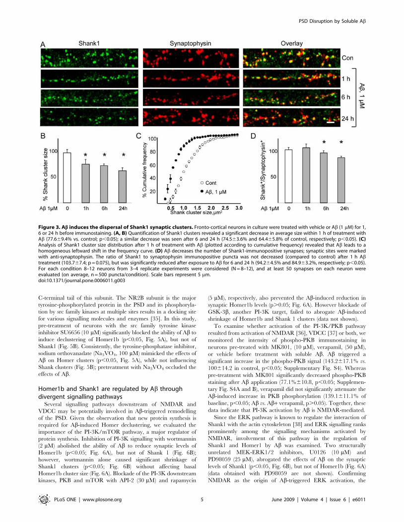
Ab treatment reduces synaptic mGluR1Since mGluR1 trafficking to the surface relies on interactions
with Homer1b [17,41], it was of interest to examine the
consequences of Ab-induced decreases in synaptic levels of
Homer1b on mGluR1 localization. Immunostaining revealed a
major decrease in surface mGluR1 cluster size after Ab treatment
(surface cluster size 66.062.8%, n = 10, p,0.05, Fig. 7A and 7B),
an effect that was prevented by treatment with either MK801
Figure 4. Ab-induced dispersal of Homer1b and Shank1 clusters does not depend upon protein degradation. Protein synthesis isrequired for Ab-induced Homer1b clusters dispersal. Neurons were pretreated with the proteasome inhibitor MG132 (0.1 mM) or with the proteinsynthesis inhibitor cycloheximide (CHX, 15 mM) before adding Ab (1 mM), before immunostaining for synaptophysin and either Homer1b or Shank1.(A) Proteasome inhibition did not block the Ab effects (69.164.2%, cf. Ab+MG132 and MG alone, p,0.05), while blockade of protein synthesiseffectively prevented the dispersal of Homer1b clusters (103.9610%, cf. CHX+Ab and CHX alone). For each condition, 8–12 neurons from 3–4replicate experiments were considered (N = 8–12), and at least 50 synapses on each neuron were evaluated (average, n = 500 puncta/condition). (B)Ab did not reduce whole cell levels of Homer1b levels after 30, 60, 120 or 180 min. (C) Neither proteasomal activity nor de novo protein synthesiswere found to be necessary for Ab-induced dispersal of Shank1 clusters. (D) Exposure to Ab for up to 120 min. did not lead to down-regulation ofShank1 levels in whole cell protein extracts; however, a significant decrease in Shank1 was observed after 180 min of exposure to Ab (80.769.7% ofbaseline; p,0.05). Since Shank1 displays multiple isoforms, the quantification shown refers to the most abundant (170 kDa) band.doi:10.1371/journal.pone.0006011.g004
PSD Disruption by Soluble Ab
PLoS ONE | www.plosone.org 6 June 2009 | Volume 4 | Issue 6 | e6011

(10 mM) or verapamil (50 mM). mGluR1 is regulated by
constitutive endocytosis, a process that is subject to retardation
upon binding of Homer proteins to mGluR1 [41–43]. Here, the
size of mGluR1 synaptic clusters was evaluated to examine
whether the Ab-induced loss of surface mGluR1 resulted from
endocytosis alone or whether it also involved decreases in overall
synaptic mGluR1 content. Under baseline conditions, mGluR1
immunoreactivity was predominantly found in the perikaryon and
dendrites of neurons [17] in approximately 60% of synaptophysin-
positive puncta (Fig. 7C and 7D). The number of mGluR1-
positive synapses was markedly decreased within 1 h of Abapplication (47.365.7% of control, n = 10; p,0.05; Fig. 7C and
7D). Likewise, the size of synaptic mGluR1 clusters was decreased
after exposure to Ab (6566.5% of control; p,0.05; Fig. 7C and
7E). The effects of Ab on synaptic mGluR1 localization (Fig. 7C
and 7D) and cluster size (Fig. 7E) proved to be sensitive to MK801
(10 mM) and verapamil (50 mM), indicating their mediation by
NMDAR and VDCC, respectively (Fig. 7C and 7D).
Discussion
Oligomeric Ab has been implicated in the pathogenesis of
Alzheimer’s disease [2,8] but a physiological role for Ab in the
regulation of synaptic homeostasis has also been hypothesized
[44]. Several lines of evidence suggest that soluble Ab may
contribute to the early stages of AD by impairing synaptic
Figure 5. Differential requirements of NMDAR and VDCC for Ab-induced declustering of Homer1b and Shank1. (A) Ab-induceddeclustering of Homer depends on NMDAR VDCC activity and on src activity. Cells were pre-treated with either the NMDAR antagonistMK801 (10 mM) or the VDCC blocker verapamil (50 mM) before exposure to Ab (1 mM, 1 h). Both MK801 and VDCC effectively prevented Ab-inducedHomer1b cluster dispersal (100.7615.5%, cf. MK+Ab and MK alone; p.0.05; 105.767%, cf. verapamil+Ab and verapamil alone; p.0.05). To evaluatethe sufficiency of each of these receptors for Ab-induced Homer cluster dispersal cells were pre-treated with either the NMDAR agonist NMDA(10 mM) or the VDCC activator Bay-K8644 (10 mM) before exposure to Ab (1 mM, 1 h). NMDA alone significantly reduced Homer (75.168.8%, p,0.05)and a further decrease was observed after NMDA+Ab cotreatment (50.265.2% of untreated control). To assess the role of NMDAR tyrosinephosphorylation in the signalling leading to Homer cluster dispersal, cells were pre-treated with the src-family inhibitor SU6656 (10 mM), whichblocked the ability of Ab to reduce Homer1b (100.8611.1%, cf. Ab+SU6656 and SU6656 alone; p.0.05), or with the tyrosine-phosphatase inhibitorsodium orthovanadate (Na3VO4, 100 mM) alone, which led to decreases in Homer1b (57.767.2% of control; p,0.05) cluster sizes and occluded Abeffect when Na3VO4 was applied before Ab: (9667.1%, cf. Na3VO4+Ab and Na3VO4 alone; p.0 05). (B) Ab-induced declustering of Shankdepends only on NMDAR activity. In contrast to Homer1b declustering, MK801, but not verapamil, blocked the effects of Ab on Shank1clustering (116.6610.4%, cf. MK801+Ab and MK801 alone; p.0.05; 63.365.7%, cf. verapamil+Ab and verapamil alone; p,0.05). Pretreatment with theNMDAR agonist NMDA (10 mM) or the VDCC activator Bay-K8644 (10 mM) before exposure to Ab (1 mM, 1 h) reduced Shank cluster size (53.964.1%and 77.769.9% respectively; p,0.05) and a further decrease was observed after NMDA+Ab and BayK8644+Ab cotreatment (43.466.7% and43.865.6% respectively, p,0.05). Pre-treatment with the src-family inhibitor SU6656 (10 mM) did not block the ability of Ab to reduce Shank1(90.5612.2%, cf. SU6656+Ab and SU6656 alone; p.0.05) clusters. Exposure to the tyrosine-phosphatase inhibitor sodium orthovanadate (Na3VO4,100 mM) alone led to decreases in Shank1 (77.8610.3% of control; cf. Na3VO4+Ab and Na3VO4 alone p,0.05) cluster sizes but further decreasewas observed after co-treatment with Ab (47. 69.8%, p,0.05). For each condition, 8–12 neurons from 3–4 replicate experiments were examined(N = 8–12); at least 50 synapses on each neuron were evaluated (on average, n = 500 puncta/condition).doi:10.1371/journal.pone.0006011.g005
PSD Disruption by Soluble Ab
PLoS ONE | www.plosone.org 7 June 2009 | Volume 4 | Issue 6 | e6011

physiology: (i) soluble Ab levels correlate strongly with loss of
synapses as well as with severity of cognitive impairment in AD
patients [6] and in transgenic mouse models [7,45], (ii) Aboligomers acutely impair synaptic plasticity and cognitive functions
[3,7,8], and (iii) Ab oligomers influence the overall levels,
localization and trafficking of several receptors (including
NMDAR and AMPAR) and signalling molecules [39,46,47].
In the present investigations, Ab was used at a dose (1 mM) and
over a period of time (1 h), conditions that do not favor its
aggregation into fibrils [24–26]. Our data identify the remodelling
of the PSD and the dispersal of scaffold proteins clustered in the
post-synaptic site as early events triggered by Ab oligomers. We
show that Ab rapidly disrupts the ultrastructural integrity and
thickness of the PSD, inducing morphological changes that are
reminiscent of the phenotype found in mice in which selected PSD
constituents, including Shank [30], are knocked out. Extending
previous findings that Ab treatment leads to a loss of PSD-95
[21,22], we here found this treatment to lead to a significant
decrease in the size of synaptic clusters (loss) of two other PSD
proteins, Homer1b and Shank1.
Interestingly, our investigations show that distinct mechanisms
underlie Ab-triggered loss of synaptic Homer1b and Shank1. In
contrast to previous demonstrations that the loss of synaptic PSD-
95 requires proteasomal activity [21,33], Shank1 loss was found to
occur independently of proteasomal activity over the timeframe
monitored in the present study, although the possibility of
degradation of the protein over longer periods cannot be ruled
out [cf.33]. Remarkably, a different mechanism, namely de novo
Figure 6. Divergent signalling pathways mediate the effects of Ab on Homer1b and Shank1 cluster dispersal. Cultured cortical neuronswere pre-treated for 45 min with the indicated pharmacological inhibitors before being treated for 1 h with Ab (1 mM). Inhibitors were present in themedium during Ab treatment. (A) PI-3K/mTOR pathway is required for Homer1b declustering. Pre-treatment with wortmannin (Wort, 2 mM)abolished the ability of Ab to disperse Homer1b clusters (101.1615.3%, cf. Ab+Wort and Wort alone). Likewise, inhibition of the PI-3K downstreamkinases PKB (with API-2, 30 mM) or mTor (with rapamycin, Rapa, 5 mM) prevented Ab-induced declustering of Homer1b (90.668.4%, cf. Ab+AP2 andAPI-2 alone; 9568.2%, cf. Ab+Rapa and rapamycin alone). Either the blockade of MEK/ERK pathway by pre-treatment with UO126 (10 mM; 70.964.8%UO126+Ab vs UO126 alone) or cdk-5 inhibition by roscovitine (10 mM;63.765.8%, cf. Ab+Rosco and Rosco alone) pre-treatment did affect Ab-inducedHomer declustering. (B) MEK/ERK pathway, but not PI-3K pathway, is required for Ab-induced dispersal of Shank1. Pre-treatment withthe PI-3K inhibitor wortmannin before Ab treatment did not affect Ab-induced Shank cluster dispersal (64.7610.3%, cf. Wort+Ab and Wort alone;p,0.05). Notably, wortmannin itself decreased Shank1 (but not Homer1b) cluster size (74.3610.3%, cf. Wort and vehicle; p,0.05). Two structurallyunrelated MEK inhibitors, UO126 (10 mM) and PD98059 (25 mM) prevented Ab-induced declustering of Shank1 (96.8612.1%, cf. PD98059+Ab andPD98059 alone; 92.2615.4%, cf. UO126+Ab and UO126 alone). Likewise, the blockade of the Erk targeted kinase RSK by pre-treatment with SL0101-1blocked Ab effects (100.469.8%, p.0.05). Pre-treatment of neurons with the specific cdk-5 inhibitor roscovitine (Rosco, 10 mM) did not interfere withthe ability of Ab to reduce the size of Shank1 (72.6610.8%, cf. Rosco+Ab and Ab alone) clusters. (C,D) PP2B is required for the dispersal of Homer1b,but not Shank1, clusters by Ab. Pre-treatment with structurally unrelated PP2B inhibitors FK506 (10 mM) or cyclosporin (Cyclo, 100 mM) significantlyblocked (p,0.05) Ab-induced Homer1b declustering (98.8610.4%, comparing FK506+Ab vs. Ab alone; 83.465.8%, comparing Ab+Cyclo and Cycloalone; and 6167.9% in vehicle+Ab-treated cells; see panel C); however, all PP2B inhibitors were ineffective in preventing Shank1 declustering after Abexposure (61.364.5%, cf. FK506+Ab and FK506 alone; 90.8612.4%, cf. Cyclo+Ab and Cyclo alone, see panel D). For each condition, 8–12 neurons from3–4 replicate experiments were considered (N = 8–12), and at least 50 synapses on each neuron were evaluated (on average, n = 500 puncta/condition).doi:10.1371/journal.pone.0006011.g006
PSD Disruption by Soluble Ab
PLoS ONE | www.plosone.org 8 June 2009 | Volume 4 | Issue 6 | e6011
protein synthesis, was found to underlie the loss of synaptic
Homer1b after application of Ab. Since the reduction in synaptic
Homer1b clusters was not accompanied by a change in whole-cell
levels of the protein (or its truncated forms), it would appear that
Ab treatment leads to cluster dissociation and relocation to
another cellular compartment [cf.34]. The presented data on
Shank1 and Homer1b, together with previous findings for PSD-95
[21], show that Ab can selectively employ either protein synthesis
or degradation to disrupt the PSD; our results complement other
work on a different model of synaptic plasticity [48].
Declustering of Homer and Shank, like that of PSD-95 [21],
was found to require NMDAR activity (and agonist-induced
Figure 7. Dispersal of synaptic mGluR1 clusters by Ab requires NMDAR and VDCC activity. Rat cortical neurons were treated with Ab(1 mM) for 1 h (with or without co-treatment with the NMDAR antagonist MK801, or the VDCC blocker verapamil) and then immunostained forsurface mGluR1 before fixation. (A, B) Ab downregulates surface mGluR1 (surface cluster size 66.062.8%, N = 10, n = 300, p,0.05); pre-treatmentwith either MK801 (10 mM) or verapamil (50 mM) blocked this effect (102.9610.5%, for MK801+Ab vs. MK alone; 109.864.1%, for verapamil+Ab vs.verapamil alone). (C, D) Ab treatment results in decreased synaptic localization of total mGluR1 (ratio of mGluR1/synaptophysin immunopositivepuncta, 47.365.7% of baseline, n = 600, p,0.05); pre-treatment with either MK801 or verapamil prevented this effect (102.067.2%, for MK801+Ab vs.MK alone; 106.7610.8% for verapamil+Ab vs. verapamil alone). (C, E) Ab treatment results in reduced size of total synaptic mGluR1 clusters(68.168.0%, N = 10, n = 600, p,0.05); pre-treatment with either MK801 or verapamil abolished Ab-induced cluster shrinkage (93611.1%, forMK801+Ab vs. MK alone; 112.4610.7%, for verapamil+Ab vs. verapamil alone). Scale bar represents 5 mm.doi:10.1371/journal.pone.0006011.g007
PSD Disruption by Soluble Ab
PLoS ONE | www.plosone.org 9 June 2009 | Volume 4 | Issue 6 | e6011

activation of NMDAR alone was sufficient to mimic the Ab effect).
Therefore, the NMDAR is a major hub for the regulation of PSD
structure and composition and its activation appears to be essential
for triggering disassembly of the PSD; however, our study
demonstrates that divergent downstream signalling pathways
regulate the demise of each PSD component. Specifically, we
found that Ab-induced Homer declustering requires activated
VDCC as an additional source of Ca++ and other signalling
molecules; importantly, VDCC activation alone however did not
mimic the actions of Ab. These observations are consistent with
those of Okabe et al. [34] who reported that NMDAR- and
VDCC-regulated Ca2+ spikes promote Homer1b clustering
whereas Ca2+ levels that rise and reach a plateau result in the
shrinkage of Homer1b clusters.
The PI-3K/mTor signalling pathway, acting downstream of
NMDAR, was identified as an important mediator of Ab-induced
Homer1b cluster dispersal (see Supplementary Fig. S8). Since PI-
3K/mTOR is a master regulator of protein synthesis [49] it is
plausible that this pathway links NMDAR activation to the
synthesis of protein(s) ultimately involved in Homer cluster
disassembly. Consistent with a previous demonstration that
NMDAR-dependent activation of PI-3K requires the binding to
a phospho-tyrosine residue on the cytoplasmic tail of the NR2
subunit of the NMDAR [50], inhibition of the src tyrosine kinase
in the present study interfered with Homer1b declustering. A role
for protein phosphatase 2B (PPB2), acting downstream of
NMDAR [51], in the control of Homer1b cluster disassembly
was also demonstrated in the present work. The recent implication
of PPB2 and its downstream target cofillin in Ab-induced loss of
dendritic spines [4] suggests that this molecule may serve as a
mechanistic link between Homer1b cluster dispersal and dendritic
spine loss [4,12].
Signalling pathways that are distinct from those involved in the
regulation of Homer1b declustering were found to mediate
NMDAR-triggered dispersal of Shank1 clusters. While roles for
the PI-3K and PP2B pathways were dismissed, we demonstrated
serial activation of ERK and RSK upon NMDAR activation.
Previous work demonstrated that ERK/RSK activation leads to
the phosphorylation of Shank protein and a reduction in its affinity
for the actin-binding protein, cortactin [38]. Since Shank
clustering depends upon actin dynamics [52], the probability of
Shank declustering after interference with the Shank-cortactin
interaction through the ERK-RSK pathway is high.
Homer1b is known to critically affect the clustering and
trafficking of mGluR1 as well as its delivery to the cell surface
from where mGluR1 is removed by constitutive and regulated
internalisation and degradation [17,41–43]. The present finding
that Ab causes a significant decrease in the surface expression of
mGluR1 implies endocytosis of the receptor. Further, our studies
show that the loss of surface mGluR1 can be prevented by
blockade of either NMDAR or VDCC, paralleling our observa-
tions with respect to Homer1b declustering; it may therefore be
inferred that loss of synaptic Homer1b is a key event in the
downregulation of surface levels of mGluR1. Interestingly, Abinduces a decrease in the total synaptic content of mGluR1; this
finding suggests receptor endocytosis and degradation rather than
receptor recycling. In brief, while the loss of Homer1b and
mGluR1 is likely to contribute to Ab-triggered demise of the PSD,
our results are compatible with the view that mGluR1 surface
clustering is tightly coupled to mGluR1 interactions with
Homer1b [41]. Thus, Homer1b declustering is directly involved
in the internalisation of mGluR1. Accordingly, we suggest that loss
of synaptic Homer1b and mGluR1 mediates a major part of the
impact of Ab on synaptic physiology [cf. 53–55].
In summary, our results suggest that soluble Ab affects the
composition and overall structure of the PSD by triggering its
disassembly and, ultimately, synaptic loss. The results presented
here and previously [21] highlight the remarkable diversity of
signaling mechanisms through which Ab regulates the synaptic
levels of three strongly interacting scaffold proteins in the mature
PSD. Despite its structural complexity, the PSD may be viewed as
a ‘‘flexible matrix’’ undergoing continuous turnover and structural
plasticity [52,56]. In this context, the relative abundance and
binding affinity of each component could be regulated indepen-
dently [cf. 57,58], ultimately leading to either changes in PSD
composition or its complete disassembly.
Supporting Information
Figure S1 Ab effect on Homer1b and Shank1 clusters are dose-
dependent. (A,B) Cultured rat fronto-cortical neurons were treated
with Ab at doses ranging from 100 pM to 1 mM for 1 h; thereafter
they were immunostained for Homer1b or Shank (significant
differences are indicated by asterisks, p,0.05).
Found at: doi:10.1371/journal.pone.0006011.s001 (4.38 MB TIF)
Figure S2 Homer1b and Shank1 cluster dispersal by Ab does
not depend upon AMPAR or mGluR activity. (A, B) Cultured
fronto-cortical neurons were pre-treated with the AMPAR blocker
NQBX (50 mM, 45 min) or the mGluR I/II blocker E4CPG
(10 mM) before exposure to Ab (1 mM, 1 h), after which they were
fixed and immunostained for synaptophysin and either Homer1b
(panel A) or Shank1 (panel B). Neither NBQX nor E4CPG
interfered with the ability of Ab to reduce Homer1b (6664.3%, cf.
NQBX+Ab and NQBX alone, p,0.05) or Shank1 (6264.9%,
cf. NQBX+Ab and NQBX alone, p,0.05; 69.364.4%, cf.
E4CPG+Ab and E4CPG alone, p,0.05; 59.262.7%, cf.
E4CPG+Ab vs. E4CPG alone, p,0.05) cluster sizes.
Found at: doi:10.1371/journal.pone.0006011.s002 (4.27 MB TIF)
Figure S3 L-type, but not T-type, calcium channels are required
for Ab-induced dispersal of Homer1b clusters. Neurons were pre-
treated with the L-type VDCC blocker nifedipine (40 mM, 45 min)
or with the T-type blocker NiCl2 (400 mM, 45 min) before being
treated with Ab (1 mM, 1 h). Consistent with the results obtained
with the structurally unrelated VDCC blocker verapamil (see
Fig. 4), nifedipine effectively prevented Homer1b cluster dispersal
by Ab (95.868.4%, cf. nifedipine+Ab and nifedipine alone,
p.0.05), whereas NiCl2 proved ineffective in blocking the actions
of Ab (64.765.2%, cf. Ab+NiCl2 and NiCl2 alone, p,0.05).
Found at: doi:10.1371/journal.pone.0006011.s003 (1.97 MB TIF)
Figure S4 Ab activates the PKB pathway through NMDAR.
Rat cortical neurons were starved (see Methods) for 2 h before
pre-treatment (45 min) with the NMDAR antagonist MK801
(10 mM), the VDCC blocker verapamil (50 mM) or vehicle before
exposure to Ab (1 mM, 10 min). Cells were fixed and immuno-
stained for p-PKB (Thr308) and synaptophysin. (A) Representative
images of p-PKB-immunostained cells (column 1: intensity-coded
image, column 2: actual palette), synaptophysin-stained cells
(column 3), and the resulting overlay (column 4) are shown. Cells
included in the evaluation expressed cytoplasmic synaptophysin as
well as punctate synaptophysin immunoreactivity along their
processes. (B) shows p-PKB immunofluorescence intensity (after
background subtraction). Ab treatment triggered an increase in p-
PKB fluorescence intensity (143.2617.1% of baseline, n = 50,
p,0.05). Pre-treatment with MK801 abrogated Ab-induced
increases in p-PKB immunoreactivity (77.1%610.8% of baseline,
n = 30), whereas pre-treatment with verapamil had only a minor
PSD Disruption by Soluble Ab
PLoS ONE | www.plosone.org 10 June 2009 | Volume 4 | Issue 6 | e6011

effect on p-PKB immunofluorescence (139.1611.1% of baseline,
p,0.05). Scale bar represents 10 mM.
Found at: doi:10.1371/journal.pone.0006011.s004 (8.06 MB TIF)
Figure S5 Ab activates ERK phosphorylation through
NMDAR. Fronto-cortical neurons were starved (see Methods)
for 2 h before pre-treatment (45 min) with the NMDAR
antagonist MK801 (10 mM) or vehicle before exposure to Ab(1 mM, 10 min). Cells were fixed and immunostained for p-ERK
(Thr202/Tyr204) and synaptophysin. (A) shows representative
images of p-ERK immunostaining (column 1: intensity-coded
image; column 2: actual palette), synaptophysin immunostaining
(column 3) and the resulting overlay (column 4). Cells included in
the evaluation expressed cytoplasmic synaptophysin as well as
punctate synaptophysin immunoreactivity along their processes.
(B) demonstrates p-ERK immuno-fluorescence intensity (after
background subtraction). Ab treatment led to an increase in p-
ERK fluorescence intensity (184.6647% of baseline, n = 50,
p,0.05). Pre-treatment with MK801 abrogated Ab-induced p-
ERK immunoreactivity (77614.4% of baseline, n = 30). Scale bar
represents 10 mM.
Found at: doi:10.1371/journal.pone.0006011.s005 (9.46 MB TIF)
Figure S6 Ab activates RSK phosphorylation through NMDAR
and Erk pathway. Fronto-cortical neurons were starved (see
Methods) for 2 h before pre-treatment (45 min) with the NMDAR
antagonist MK801 (10 mM), the MEK inhibitor UO126 (10 mM)
or vehicle before exposure to Ab (1 mM, 15 min). Cells were fixed
and immunostained for p-RSK and synaptophysin. (A) shows
representative images of p-RSK immunostaining (column 1:
intensity-coded image; column 2: actual palette), synaptophysin
immunostaining (column 3) and the resulting overlay (column 4).
Cells included in the evaluation expressed cytoplasmic synapto-
physin as well as punctate synaptophysin immunoreactivity along
their processes. (B) demonstrates p-ERK immuno-fluorescence
intensity (after background subtraction). Ab treatment led to an
increase in p-ERK fluorescence intensity (180.2624.5 of baseline,
n = 50, p,0.05). Pre-treatment with MK801 and UO126 largely
abrogated Ab-induced p-RSK immunoreactivity (114.3611.8%
and 122.3614.3% of baseline respectively, n = 30). Scale bar
represents 10 mM.
Found at: doi:10.1371/journal.pone.0006011.s006 (8.14 MB TIF)
Figure S7 Differential kinase requirements in the dispersal of
Homer1b and Shank1 clusters by Ab. Cortical neurons were pre-
treated with KN93 (5 mM) or Go6893 (5 mM) for 45 min before
addition of Ab (1 mM, 1 h). (A, B) demonstrate that CaMKII and
PKCa/c are not required for manifestation of the ability of Ab to
induce dispersal of Homer1b and Shank1 clusters. Neither KN93
nor Go6893 were effective at blocking the effects of Ab on
Homer1b (63.969.4% for KN93+Ab vs. KN93 alone, p,0.05;
and 68.464.4% for Go6893+Ab vs. Go6893 alone, p,0.05).
Similarly, neither inhibitor influenced the actions of Ab on Shank1
clusters (64.466.8% for KN93+Ab vs. KN93 alone, p,0.05; and
73.668% for Go6893+Ab vs. Go6893 alone, p,0.05). Notably,
Go6893 itself (but not KN93) led to a marked decrease in Shank1
cluster size (66.5610.9%, cf. Go6893 and control, p,0.05).
Found at: doi:10.1371/journal.pone.0006011.s007 (4.43 MB TIF)
Figure S8 Divergent signalling pathways mediate Ab effects on
Homer1b, Shank1 and PSD-95 and regulate Ab-induced PSD
remodelling. Ab-triggered signalling pathways leading to PSD
disruption are shown. Homer cluster disassembly relies on PI-3K/
mTot pathway and PP2B pathway activity, whereas Shank cluster
disassembly only requires ERK/RSK pathway activity (present
work). In contrast, PSD-95 loss involves the activity of cdk-5. For
each pathway, the inhibitors used in the paper are shown.
Found at: doi:10.1371/journal.pone.0006011.s008 (7.38 MB TIF)
Acknowledgments
The authors wish to thank Drs. Juergen Schlegel and Axel Walch, and
Luise Jennen (Institute of Pathology, Helmholtz Center Munich) for help
with electron and confocal microscopy, and Dr. Morgan Sheng (MIT,
Cambridge, MA) for kindly providing Homer1b and Shank1 antibodies.
Dieter Fischer and Rainer Stoffel gave dedicated technical assistance, and
Carola Hetzel provided administrative and editorial help. Members of the
Munich lab are thanked for helpful comments.
Author Contributions
Conceived and designed the experiments: FR OFXA. Performed the
experiments: FR. Analyzed the data: FR PH PL OFXA. Contributed
reagents/materials/analysis tools: PH YW. Wrote the paper: FR PL
OFXA.
References
1. Selkoe DJ (2002) Alzheimer’s disease is a synaptic failure. Science 298: 789–791.
2. Walsh DM, Klyubin I, Fadeeva JV, Cullen WK, Anwyl R, et al. (2002) Naturally
secreted oligomers of amyloid beta protein potently inhibit hippocampal long-term potentiation in vivo. Nature 416: 535–539.
3. Cleary JP, Walsh DM, Hofmeister JJ, Shankar GM, Kuskowski MA, et al. (2005)Natural oligomers of the amyloid-beta protein specifically disrupt cognitive
function. Nat Neurosci 8: 79–84.
4. Shankar GM, Bloodgood BL, Townsend M, Walsh DM, Selkoe DJ, et al. (2007)
Natural oligomers of the Alzheimer amyloid-beta protein induce reversiblesynapse loss by modulating an NMDA-type glutamate receptor-dependent
signalling pathway. J Neurosci 27: 2866–2875.
5. Calabrese B, Shaked GM, Tabarean IV, Braga J, Koo EH, et al. (2007) Rapid,
concurrent alterations in pre- and postsynaptic structure induced by naturally-secreted amyloid-beta protein. Mol Cell Neurosci 35: 183–193.
6. McLean CA, Cherny RA, Fraser FW, Fuller SJ, Smith MJ, et al. (1999) Solublepool of Abeta amyloid as a determinant of severity of neurodegeneration in
Alzheimer’s disease. Ann Neurol 46: 860–866.
7. Lesne S, Koh MT, Kotilinek L, Kayed R, Glabe CG, et al. (2006) A specific
amyloid-beta protein assembly in the brain impairs memory. Nature 440:352–357.
8. Walsh DM, Selkoe DJ (2007) A beta oligomers-a decade of discovery.J Neurochem 101: 1172–1184.
9. Sheng M, Hoogenraad CC (2007) The postsynaptic architecture of excitatory
synapses: a more quantitative view. Annu Rev Biochem 76: 823–47.
10. Tu JC, Xiao B, Naisbitt S, Yuan JP, Petralia RS, et al. (1999) Coupling of
mGluR/Homer1b and PSD-95 complexes by the Shank family of postsynaptic
density proteins. Neuron 23: 583–592.
11. El-Husseini AE, Schnell E, Chetkovich DM, Nicoll RA, Bredt DS (2001) PSD-
95 involvement in maturation of excitatory synapses. Science 290: 1364–1368.
12. Sala C, Piech V, Wilson NR, Passafaro M, Liu G, et al. (2001) Regulation ofdendritic spine morphology and synaptic function by Shank and Homer.
Neuron 31: 115–130.
13. Roussignol G, Ango F, Romorini S, Tu JC, Sala C, et al. (2005) Shankexpression is sufficient to induce functional dendritic spine synapses in aspiny
neurons. J Neurosci 25: 3560–3570.
14. Brakeman PR, Lanahan AA, O’Brien R, Roche K, Barnes CA, et al. (1997)
Homer: a protein that selectively binds metabotropic glutamate receptors.Nature 386: 284–288.
15. Valtschanoff JG, Weinberg RJ (2001) Laminar organization of the
NMDA receptor complex within the postsynaptic density. J Neurosci 21:1211–1217.
16. Mao L, Yang L, Tang Q, Samdani S, Zhang G, Wang JQ (2005) The
scaffold protein Homer1b/c links metabotropic glutamate receptor 5 toextracellular signal-regulated protein kinase cascades in neurons. J Neurosci
25: 2741–2752.
17. Das SS, Banker GA (2006) The role of protein interaction motifs in regulating
the polarity and clustering of the metabotropic glutamate receptor mGluR1a.J Neurosci 26: 8115–8125.
18. Tu JC, Xiao B, Yuan JP, Lanahan AA, Leoffert K, et al. (1998) Homer binds a
novel proline-rich motif and links group 1 metabotropic glutamate receptorswith IP3 receptors. Neuron 21: 717–726.
19. Baron MK, Boeckers TM, Vaida B, Faham S, Gingery M, et al. (2006) An
architectural framework that may lie at the core of the postsynaptic density.
Science 311: 531–535.
PSD Disruption by Soluble Ab
PLoS ONE | www.plosone.org 11 June 2009 | Volume 4 | Issue 6 | e6011
20. Naisbitt S, Kim E, Tu JC, Xiao B, Sala C, et al. (1999) Shank, a novel family of
postsynaptic density proteins that binds to the NMDA receptor/PSD-95/GKAPcomplex and cortactin. Neuron 23: 569–582.
21. Roselli F, Tirard M, Lu J, Hutzler P, Lamberti P, et al. (2005) Soluble beta-
amyloid1-40 induces NMDA-dependent degradation of postsynaptic density-95at glutamatergic synapses. J Neurosci 25: 11061–11070.
22. Almeida CG, Tampellini D, Takahashi RH, Greengard P, Lin MT, et al. (2005)Beta-amyloid accumulation in APP mutant neurons reduces PSD-95 and GluR1
in synapses. Neurobiol Dis 20: 187–198.
23. Peng J, Kim MJ, Cheng D, Duong DM, Gygi SP, et al. (2004) Semiquantitativeproteomic analysis of rat forebrain postsynaptic density fractions by mass
spectrometry. J Biol Chem 279: 21003–11.24. Walsh DM, Lomakin A, Benedek GB, Condron MM, Teplow DB (1997)
Amyloid beta-protein fibrillogenesis. Detection of a protofibrillar intermediate.J Biol Chem 272: 22364–22372.
25. Bitan G, Vollers SS, Teplow DB (2003) Elucidation of primary structure
elements controlling early amyloid beta-protein oligomerization. J Biol Chem278: 34882–34889.
26. Stine BW, Dahlgren KN, Krafft GA, LaDu MJ (2003) In vitro characterizationof conditions for amyloid-b peptide oligomerization and fibrillogenesis. J Biol
Chem 278: 11612–11622.
27. O’Nuallain B, Williams AD, Westermark P, Wetzel R (2004) Seeding specificityin amyloid growth induced by heterologous fibrils. J Biol Chem 279:
17490–17499.28. Wogulis M, Wright S, Cunningham D, Chilcote T, Powell K, et al. (2005)
Nucleation-dependent polymerization is an essential component of amyloid-mediated neuronal cell death. J Neurosci 25: 1071–1080.
29. Crochemore C, Lu J, Wu Y, Liposits Z, Sousa N, et al. (2005) Direct targeting of
hippocampal neurons for apoptosis by glucocorticoids is reversible bymineralocorticoid receptor activation. Mol Psychiatry 10: 790–798.
30. Hung AY, Futai K, Sala C, Valtschanoff JG, Ryu J, et al. (2008) Smallerdendritic spines, weaker synaptic transmission, but enhanced spatial learning in
mice lacking Shank1. J Neurosci 28: 1697–1708.
31. Rasband WS ImageJ, U. S. National Institutes of Health, Bethesda, Maryland,USA, http://rsb.info.nih.gov/ij/, 1997–2008.
32. Ehlers MD (2003) Activity level controls postsynaptic composition and signallingvia the ubiquitin-proteasome system. Nat Neurosci 6: 231–242.
33. Colledge M, Snyder EM, Crozier RA, Soderling JA, Jin Y, et al. (2003)Ubiquitination regulates PSD-95 degradation and AMPA receptor surface
expression. Neuron 40: 595–607.
34. Okabe S, Urushido T, Konno D, Okado H, Sobue K (2001) Rapidredistribution of the postsynaptic density protein PSD-Zip45 (Homer1c) and
its differential regulation by NMDA receptors and calcium channels. J Neurosci21: 9561–9571.
35. Salter MW, Kalia LV (2004) Src kinases: a hub for NMDA receptor regulation.
Nat Rev Neurosci 5: 317–328.36. Sutton G, Chandler LJ (2002) Activity-dependent NMDA receptor-mediated
activation of protein kinase B/Akt in cortical neuronal cultures. J Neurochem82: 1097–1105.
37. Baxter AW, Wyllie DJ (2006) Phosphatidylinositol 3 kinase activation andAMPA receptor subunit trafficking underlie the potentiation of miniature EPSC
amplitudes triggered by the activation of L-type calcium channels. J Neurosci 26:
5456–5469.38. Thomas GM, Rumbaugh GR, Harrar DB, Huganir RL (2005) Ribosomal S6
kinase 2 interacts with and phosphorylates PDZ domain-containing proteins andregulates AMPA receptor transmission. Proc Natl Acad Sci U S A 102:
15006–15011.
39. Hsieh H, Boehm J, Sato C, Iwatsubo T, Tomita T, et al. (2006) AMPARremoval underlies Abeta-induced synaptic depression and dendritic spine loss.
Neuron 52: 831–843.
40. Morabito MA, Sheng M, Tsai LH (2004) Cyclin-dependent kinase 5
phosphorylates the N-terminal domain of the postsynaptic density protein
PSD-95 in neurons. J Neurosci 24: 865–876.
41. Kammermeier PJ (2006) Surface clustering of metabotropic glutamate receptor
1 induced by long Homer proteins. BMC Neurosci 7: 1.
42. Dhami GK, Ferguson SS (2006) Regulation of metabotropic glutamate receptor
signaling, desensitization and endocytosis. Pharmacol Ther 111: 260–271.
43. Ciruela F, Soloviev MM, Chan WY, McIlhinney RA (2000) Homer-1c/Vesl-1L
modulates the cell surface targeting of metabotropic glutamate receptor type
1alpha: evidence for an anchoring function. Mol Cell Neurosci 15: 36–50.
44. Kamenetz F, Tomita T, Hsieh H, Seabrook G, Borchelt D, et al. (2003) APP
processing and synaptic function. Neuron 37: 925–937.
45. Westerman MA, Cooper-Blacketer D, Mariash A, Kotilinek L,
Kawarabayashi T, et al. (2002) The relationship between Abeta and memory
in the Tg2576 mouse model of Alzheimer’s disease. J Neurosci 22: 1858–1867.
46. Snyder EM, Nong Y, Almeida CG, Paul S, Moran T, et al. (2005) Regulation of
NMDA receptor trafficking by amyloid-beta. Nat Neurosci 8: 1051–1058.
47. Lacor PN, Buniel MC, Furlow PW, Clemente AS, Velasco PT, et al. (2007)
Abeta oligomer-induced aberrations in synapse composition, shape, and density
provide a molecular basis for loss of connectivity in Alzheimer’s disease.
J Neurosci 27: 796–807.
48. Fonseca R, Vabulas RM, Hartl FU, Bonhoeffer T, Nagerl UV (2006) A balance
of protein synthesis and proteasome-dependent degradation determines the
maintenance of LTP. Neuron 52: 239–245.
49. Schratt GM, Nigh EA, Chen WG, Hu L, Greenberg ME (2004) BDNF regulates
the translation of a select group of mRNAs by a mammalian target of
rapamycin-phosphatidylinositol 3-kinase-dependent pathway during neuronal
development. J Neurosci 24: 7366–7377.
50. Takagi N, Sasakawa K, Besshoh S, Miyake-Takagi K, Takeo S (2003) Transient
ischemia enhances tyrosine phosphorylation and binding of the NMDA receptor
to the Src homology 2 domain of phosphatidylinositol 3-kinase in the rat
hippocampus. J Neurochem 84: 67–76.
51. Waxman EA, Lynch DR (2005) N-methyl-D-aspartate receptor subtype
mediated bidirectional control of p38mitogen-activated protein kinase. J Biol
Chem 280: 29322–29333.
52. Kuriu T, Inoue A, Bito H, Sobue K, Okabe S (2006) Differential control of
postsynaptic density scaffolds via actin-dependent and -independent mecha-
nisms. J Neurosci 26: 7693–7706.
53. Van Keuren-Jensen K, Cline HT (2006) Visual experience regulates
metabotropic glutamate receptor-mediated plasticity of AMPA receptor synaptic
transmission by homer1a induction. J Neurosci 26: 7575–7580.
54. Wang QW, Walsh DM, Rowan MJ, Selkoe DJ, Anwyl R (2004) Block of long-
term potentiation by naturally occurring secreted and synthetic amyloid-peptide
in hippocampal slices is mediated by activation of the kinases c-jun terminal
kinases, cyclin-dependent kinase 5, and p38 mitogen-activated protein kinase as
well as metabotropic glutamate receptor type 5. J Neurosci 24: 3370–3378.
55. Tyszkiewicz JP, Yan Z (2005) beta-Amyloid peptides impair PKC-dependent
functions of metabotropic glutamate receptors in prefrontal cortical neurons.
J Neurophysiol 93: 3102–3111.
56. Blanpied TA, Kerr JM, Ehlers MD (2008) Structural plasticity with preserved
topology in the postsynaptic protein network. Proc Natl Acad Sci (USA) 105:
12587–12592.
57. Steiner P, Higley MJ, Xu W, Czervionke BL, Malenka RC, Sabatini BL (2008)
Destabilization of the postsynaptic density by PSD-95 serine 73 phosphorylation
inhibits spine growth and synaptic plasticity. Neuron 60: 788–802.
58. Hayashi MK, Tang C, Verpelli C, Narayanan R, Stearns MH, et al. (2009) The
postsynaptic density proteins Homer and Shank form a polymeric network
structure. Cell 137: 159–171.
PSD Disruption by Soluble Ab
PLoS ONE | www.plosone.org 12 June 2009 | Volume 4 | Issue 6 | e6011
Copyright © 2022 FDOKUMEN