Graft-versus-Host Disease: Role of Inflammation in the Development of Chromosomal Abnormalities of...
16
Graft-vs.-Host Disease: Role of Inflammation in the development of chromosomal abnormalities of keratinocytes Elaine Sloand, MD, Loretta Pfannes, Casey Ling, Monika Jasek, PhD, Rodrigo Calado, MD PhD, Zachary C. G. Tucker, Xingmin Feng, PhD, Peiman Hematti, MD, Jaroslaw Maciejewski, MD, Cynthia Dunbar, MD, John Barrett, MD, and Neal Young, MD National Heart, Lung, and Blood Institute, Building 10 CRC 4-5230, Bethesda, Md 20892 Abstract Graft-versus-host disease (GVHD) is a major risk factor for secondary malignancy after hematopoietic stem cell transplantation. Squamous cell carcinoma (SCC) of the skin and mucous membranes are especially frequent in this setting where aneuploidy and tetraploidy are associated with aggressive disease. The current study is directed to the mechanism of neoplasia in this setting. Un-manipulated keratinocytes from areas of oral GVHD in 9 patients showed tetraploidy in 10–46% of cells when examined by florescence in situ hybridization (FISH). Keratinocytes isolated from biopsy sites of GVHD but not from normal tissue showed even greater numbers of tetraploid cells (mean 78%, range 15–85%; N=9) after culture. To mimic the inflammatory process in GVHD, allogeneic HLA- mismatched lymphocytes were mixed with normal keratinocytes. After two weeks, substantial numbers of aneuploid and tetraploid cells were evident in cultures with lymphocytes and with purified CD8 but not CD4 cells. Telomere length was substantially decreased in the lymphocyte-treated sample. No mutations were present in p53 gene, although haploinsufficiency for p53 due to loss of chromosome 17 was common in cells exposed to lymphocytes. These findings suggest that in GVHD, inflammation and repeated cell division correlates with the development of karyotypic abnormalities. Introduction Recipients of solid organ and bone marrow transplants are at risk of developing solid tumors. Two to six percent of long-term survivors acquire some type of malignancy by 10 years of followup 1–4 . While radiation therapy increases the risk of non-squamous cell carcinomas of breast, brain, and bone and of melanoma 5, 6 , chronic graft versus host disease © 2010 Published by Elsevier Inc on behalf of The American Society for Blood and Marrow Transplantation. Corresponding author Elaine M Sloand, MD, 301-496-7309 (tel), 301-594-1290 (fax), [email protected]. Publisher's Disclaimer: This is a PDF file of an unedited manuscript that has been accepted for publication. As a service to our customers we are providing this early version of the manuscript. The manuscript will undergo copyediting, typesetting, and review of the resulting proof before it is published in its final citable form. Please note that during the production process errors may be discovered which could affect the content, and all legal disclaimers that apply to the journal pertain. Elaine Sloand, MD designed the study, performed the research, wrote the paper and analyzed the data Loretta Pfannes performed the research, wrote the paper Casey Ling performed the research Monika Jasek, PhD performed the research Rodrigo Calado, MD PhD performed the research Jaroslaw Maciejewski performed the research John Barrett, MD designed the study, analyzed the data, wrote the paper Neal Young, MD designed the study, analyzed the data No disclaimers No prior presentations NIH Public Access Author Manuscript Biol Blood Marrow Transplant. Author manuscript; available in PMC 2012 September 10. Published in final edited form as: Biol Blood Marrow Transplant. 2010 December ; 16(12): 1665–1673. doi:10.1016/j.bbmt.2010.07.014. NIH-PA Author Manuscript NIH-PA Author Manuscript NIH-PA Author Manuscript
Transcript of Graft-versus-Host Disease: Role of Inflammation in the Development of Chromosomal Abnormalities of...

Graft-vs.-Host Disease: Role of Inflammation in the developmentof chromosomal abnormalities of keratinocytes
Elaine Sloand, MD, Loretta Pfannes, Casey Ling, Monika Jasek, PhD, Rodrigo Calado, MDPhD, Zachary C. G. Tucker, Xingmin Feng, PhD, Peiman Hematti, MD, JaroslawMaciejewski, MD, Cynthia Dunbar, MD, John Barrett, MD, and Neal Young, MDNational Heart, Lung, and Blood Institute, Building 10 CRC 4-5230, Bethesda, Md 20892
AbstractGraft-versus-host disease (GVHD) is a major risk factor for secondary malignancy afterhematopoietic stem cell transplantation. Squamous cell carcinoma (SCC) of the skin and mucousmembranes are especially frequent in this setting where aneuploidy and tetraploidy are associatedwith aggressive disease. The current study is directed to the mechanism of neoplasia in thissetting. Un-manipulated keratinocytes from areas of oral GVHD in 9 patients showed tetraploidyin 10–46% of cells when examined by florescence in situ hybridization (FISH). Keratinocytesisolated from biopsy sites of GVHD but not from normal tissue showed even greater numbers oftetraploid cells (mean 78%, range 15–85%; N=9) after culture. To mimic the inflammatoryprocess in GVHD, allogeneic HLA- mismatched lymphocytes were mixed with normalkeratinocytes. After two weeks, substantial numbers of aneuploid and tetraploid cells were evidentin cultures with lymphocytes and with purified CD8 but not CD4 cells. Telomere length wassubstantially decreased in the lymphocyte-treated sample. No mutations were present in p53 gene,although haploinsufficiency for p53 due to loss of chromosome 17 was common in cells exposedto lymphocytes. These findings suggest that in GVHD, inflammation and repeated cell divisioncorrelates with the development of karyotypic abnormalities.
IntroductionRecipients of solid organ and bone marrow transplants are at risk of developing solidtumors. Two to six percent of long-term survivors acquire some type of malignancy by 10years of followup1–4. While radiation therapy increases the risk of non-squamous cellcarcinomas of breast, brain, and bone and of melanoma5, 6, chronic graft versus host disease
© 2010 Published by Elsevier Inc on behalf of The American Society for Blood and Marrow Transplantation.
Corresponding author Elaine M Sloand, MD, 301-496-7309 (tel), 301-594-1290 (fax), [email protected].
Publisher's Disclaimer: This is a PDF file of an unedited manuscript that has been accepted for publication. As a service to ourcustomers we are providing this early version of the manuscript. The manuscript will undergo copyediting, typesetting, and review ofthe resulting proof before it is published in its final citable form. Please note that during the production process errors may bediscovered which could affect the content, and all legal disclaimers that apply to the journal pertain.
Elaine Sloand, MD designed the study, performed the research, wrote the paper and analyzed the dataLoretta Pfannes performed the research, wrote the paperCasey Ling performed the researchMonika Jasek, PhD performed the researchRodrigo Calado, MD PhD performed the researchJaroslaw Maciejewski performed the researchJohn Barrett, MD designed the study, analyzed the data, wrote the paperNeal Young, MD designed the study, analyzed the dataNo disclaimersNo prior presentations
NIH Public AccessAuthor ManuscriptBiol Blood Marrow Transplant. Author manuscript; available in PMC 2012 September 10.
Published in final edited form as:Biol Blood Marrow Transplant. 2010 December ; 16(12): 1665–1673. doi:10.1016/j.bbmt.2010.07.014.
NIH
-PA Author Manuscript
NIH
-PA Author Manuscript
NIH
-PA Author Manuscript

(GVHD) and its therapy increase the probability of developing squamous cell carcinoma(SCC) of the skin and oral cavity.7 In one study, the risk for SCC among transplantrecipients with chronic GVHD was three-fold that of patients without GVHD. 7 Major riskfactors included long duration of chronic GVHD therapy, use of azathioprine, and severityof the chronic GVHD; the conditioning regimen or the use of high-dose radiation did notinfluence the incidence of SCC. In bone marrow transplant patients, the relativecontributions of immunosuppression and the inflammatory effects of chronic GVHD cannotbe ascertained. However, in patients receiving solid organ transplants, immunosuppressionalone has been associated with SCC and a randomized trial of immunosuppression reductionshowed significantly fewer cancers in those receiving the less intense regimen8, 9
Chronic inflammation alone is associated with increased risk of cancer in some conditions.Examples include bowel cancer in ulcerative colitis10 , esophageal cancer in Barrett’sesophagitis11,and hepatoma in hepatitis B12. DNA damage in areas of inflammation isbelieved to be related to reactive oxygen species,.13–15 and in vitro exposure to endogenousoxidants leads to degradation of deoxyribose residues13, 16 Polypoid and aneuploid cells areevident at sites of inflammation preceding the development of cancer in Barrett'sesophagus17, in hepatocytes after partial hepatectomy18, in ulcerative colitis10 andtransiently in skin wounds and burns.19 Mutation or loss of a pivotal oncogene, TP5320 alsooccurs in areas of inflammation21TP53 is a checkpoint gene which forces cells withsubstantial DNA damage into senescence or apoptosis22, 23. Approximately 85% of coloncancers resulting from inflammatory bowel disease have lost at least one TP53 allele21.Haplo-insufficiency, gene duplication, or mutation of TP53 allows cells with polyploidy tocontinue to live and divide24. It has been theorized that in inflammatory disorders, repeatedoxidative stress results in TP53 mutation or loss21. An alternative mechanism for thedevelopment of ploidy involves telomeric shortening resulting from repeated cell divisionaccompanying tissue repair25. In the telomerase-deficient mouse significant telomericshortening results in end-to-end fusion of chromatids with consequent non-disjunctionduring metaphase and aneuploidy26.
Tetraploidy and aneuploidy are present in most cases of SCC and complex chromosomalabnormalities correlate with poor prognosis27–29. Tetraploidy results from chromosome non-disjunction in late mitosis SCC28, 30–34 and precedes the development of aneuploidy in somemodels26. In this study, we examine sites of oral and skin GVHD in transplant patients anddevelop an in vitro model of GVHD using keratinocytes co-cultured with HLA mis-matchedallogeneic lymphocytes or inflammatory cytokines. We also assessed the effect ofinflammation on telomere length and TP53 in an effort to develop a mechanism for theoccurrence of squamous cell carcinoma in patients with GVHD.
Materials and MethodsPatients
Patients undergoing hematopoietic stem cell transplantation (HSCT ) were recruited forstudy using a protocol approved by the Institutional Review Board of the National Heart,Lung, and Blood Institute, after written informed consent. All patients were adults with ahematologic disorders who had received a granulocyte colony-stimulating factor (G-CSF) -mobilized peripheral blood allograft from a fully HLA-matched male sibling donor. Somepatients received full myeloablation with total body irradiation (1360 rad) andcyclophosphamide 60 mg/kg/ day for 2 days, followed by infusion of grafts that were T-celldepleted by positive selection for CD34 + cells via the Isolex immunoabsorption device.Other patients received non-manipulated G-CSF mobilized peripheral blood cells followinga non-myeloablative conditioning regimen of fludarabine and cyclophosphamide. Sampleswere obtained from patients who were transplanted a minimum of 2 months before the
Sloand et al. Page 2
Biol Blood Marrow Transplant. Author manuscript; available in PMC 2012 September 10.
NIH
-PA Author Manuscript
NIH
-PA Author Manuscript
NIH
-PA Author Manuscript

biopsy and were confirmed to have full donor engraftment by bone marrow cytogenetics orby PCR-based microsatellite chimerism analysis of the peripheral blood. Patients wererequired to have either active biopsy-proven cutaneous graft-vs.- host disease (GVHD) at thetime of skin biopsy or oral buccal mucosal smear. Patients with oral GVHD were diagnosedon clinical grounds.
Cell preparation and tissue cultureBuccal mucosal cells—Buccal mucosal cells were obtained by scraping the area of theoral mucosa that appeared to be most involved with GVHD. Cells were placed directly onthe slide after which they were fixed and prepared for FISH. Generally, several mucosalscrapings were obtained from the patient so that many cells could be examined. Buccalmucosal cells were also obtained from ten healthy volunteers.
Skin biopsies from GVHD patients—Two contiguous 4- to 6-mm punch biopsysamples from a site of ongoing or previous cutaneous GVHD were obtained from eachpatient with significant GVHD by standard techniques35. One specimen was fixed informalin and embedded in paraffin for routine histological evaluation. The second specimenwas treated at 4°C overnight with dispase (Becton-Dickinson Labware, Bedford, MA, USA),a type IV collagenase, to separate the epidermal layer from the basement membrane. Theepidermal sheet then was separated mechanically from the dermal layer and cells weredissociated by shaking and digesting with 0.5% trypsin-0.53 mM EDTA (Gibco BRL,Gaithersburg, MD, USA) at 37°C. The cells were plated on tissue culture dishes inkeratinocytes-serum-free media (Gibco BRL) supplemented with bovine pituitary extractand recombinant epidermal growth factor. Cells were expanded to larger flasks usingtrypsinization when the monolayers reached 60% to 70% confluence.
Culture of keratinocytes obtained from foreskins—In order to study normalkeratinocytes, we obtained keratinocyte isolated from foreskin fibroblasts from Carole Yeeet al (isolation detailed elsewhere)35 and cultured as previously described in keratinocytesgrowth media35. In order to assess the effect of inflammation, one flask of confluentkeratinocytes was co-cultured with normal peripheral blood mononuclear cells with orwithout IFN-γ (1000 units per ml), or in some cases with isolated CD4 or CD8 cells. Non-adherent lymphocytes were removed after 48 hours at 37°C, and fresh media was added tothe keratinocytes. Keratinocytes were allowed to become 70–80% confluent and passagedtwo times before preparation for FISH.
FISHKeratinocytes were exposed to trypsin, enumerated, and fixed to glass slides. Cells wereincubated in prewarmed hypotonic solution (0.3% KCl sol) at 37°C for 30 minutes, spun andresuspended in Carnoy's fixative (3 parts methanol and 1 part glacial acetic acid) 3 times.Fixed cells were dropped onto cleaned glass slides and dried overnight. Beforehybridization, slides were incubated in 2XSSC for 2 minutes at 73°C, then digested withpepsin for 5 min at 37°C and dehydrated in serial ethanol washes. CEP combined X/Y(spectrum orange and spectrum green, respectively) centromeric probe (Vysis, Inc, DownersGrove, IL 60515) was used for markers of tetraploidy since these probes were capable ofdistinguishing fusion cells from cells that are tetraploid secondary to failure of cytokinesis.Three hundred cells were scored by three “blinded” observers on all samples except for thebuccal smears were all available cells were counted. Cells were scored according to currentguidelines36; cells containing twice the normal complement of stained chromosomes weredefined as tetraploid, other abnormal gains or losses were defined as aneuploid.
Sloand et al. Page 3
Biol Blood Marrow Transplant. Author manuscript; available in PMC 2012 September 10.
NIH
-PA Author Manuscript
NIH
-PA Author Manuscript
NIH
-PA Author Manuscript

Centromeric probes for chromosomes 7, 8, and 9 (Abbott Laboratories, Abbott Park, Ill)were used for experiments with normal keratinocytes. Locus-specific p53 probe (Abbott)was used to assess loss of heterozygosity in keratinocytes. Slides were placed on the Hybritehybridization system (Abbott) and denaturated at 73°C for 2 minutes. After overnighthybridization at 42°C, the slides were washed in pre-warmed 0.4xSSC at 73°C for 2 minutesand 2xSSC/0.1percentage NP- 40 at room temperature for 1 minute, then allowed to dry inthe dark and counterstained with DAPI-I before examination using a fluorescencemicroscope. Overnight hybridization temperature for p53 locus specific probe was 37°C.
DNA cell cycle analysisTreated and untreated cells were prepared as directed by the NuCycle Dapi Kit (ExalphaBiologicals, Maynard, MA 01754, USA) and analyzed on a Beckman Coulter FC 500cytometer.
Cytokine AnalysisSupernatants from keratinocytes co-cultured in the presence of CD8+ T cells, CD4+ T cells,or in the absence of T cells (negative control) were analyzed for the following cytokines:chemokine ligand 5 (CXCL5), CXCL11, thrombopoietin (Tpo), tumor necrosis factor alpha(TNF-α), granulocyte colony-stimulating factor (G-CSF), epidermal growth factor (EGF),hepatocyte growth factor (HGF), interferon-inducible cytokine-10 (IP-10), interleukin 1receptor antagonist (IL-1RA), IL-2, IL-4, IL-6, IL-8, vascular endothelial growth factor(VEGF), eotaxin, CD40 ligand (CD40L), leptin, chemokine ligand 5 (RANTES),macrophage inflammatory protein 1 alpha (MIP-1α), and MIP-1β. Measurement of allcytokines was performed simultaneously by an immuno-bead-based multiplex assay(Luminex) according to the manufacturer’s instructions. Panels of capture antibody-coatedbeads and labeled detection antibodies were acquired from R&D Systems, Inc.(Minneapolis, MN). Control samples consisted of those keratinocytes cultured for an equaltime but without lymphocytes.
qPCR for measurement of Telomere LengthTelomere length of pre-treatment peripheral blood leukocytes was assessed by quantitativepolymerase chain reaction (qPCR) as previously described.12, 13 Total leukocytes wereseparated by ammonium-based lysis of red blood cells and DNA extracted using the DNeasyBlood kit (Qiagen, Maryland). PCRs were performed in a 7500 Real Time PCR System(Applied Biosystems, Foster City, CA). Each sample’s telomere length (x) was based on thetelomere to single copy gene ratio (T/S ratio) and based on the calculation of the ΔCt[Ct
(telomeres)/Ct(single gene)]. Telomere length was expressed as relative T/S ratio, which was
normalized to the average T/S ratio of reference sample [2−(ΔCtx − ΔCtr) = 2−(ΔΔCt].
P53 sequencing of keratinocytesScreening for TP53 mutations in patients was carried out using direct sequencing in bothdirections. PCR primers were designed to amplify exons 5, 6, 7, 8 and 9 of the TP53 gene(ENSG00000141510) from genomic DNA as three separate amplicons. The primersequences are available on request. The PCR products were analyzed using 3730xl DNAanalyzer (Applied Biosystems, Foster City, CA).
ResultsUn-manipulated, un-cultured buccal mucosal cells from sites of GVHD show significantnumbers of tetraploid cells
Sloand et al. Page 4
Biol Blood Marrow Transplant. Author manuscript; available in PMC 2012 September 10.
NIH
-PA Author Manuscript
NIH
-PA Author Manuscript
NIH
-PA Author Manuscript

In order to determine if chromosomal abnormalities could be detected in areas of the oralmucosa affected by GVHD, we obtained buccal mucosal cells from patients with andwithout GVHD and performed FISH using centromeric probes for X/Y. Cells were placeddirectly on a slide, fixed, and then stained with FISH X/Y probe. The X/Y probe was chosenfor our transplant patient experiments in order to distinguish tetraploid cells from fusioncells (in patients with sex-discordant transplants (XY/XY or XX/XX) from fusion cells (XY/XX)). When FISH was performed on multiple un-manipulated buccal smears obtained from9 patients at sites of oral GVHD, all demonstrated small numbers of tetraploid cells. None ofthe 6 bone marrow transplant patients without a recent history of GVHD or the 10 healthycontrols (ranging in age from 20 to 67) years showed tetraploidy (Table 1). None of the fourpatients from the GVHD cohort who had sex-discordant transplants demonstrated XX/XYfusion cells. When punch biopsies were obtained from individuals with biopsy-provenGVHD, fixed, embedded in paraffin, and prepared for FISH as previously described35.Tissue repair and active mitosis occur in areas recovering from GVHD. In order todetermine if repeated cell divisions predispose toward tetraploidy or aneuploidy, we studiedkeratinocytes isolated from biopsies of patients at sites of GVHD and from normalforeskins. Keratinocytes obtained from areas of GVHD in patients with sex-discordanttransplants were cultured in epithelial growth factor media for two weeks (the media doesnot support growth of hematopoietic cells or fibroblasts), passaged two times in 75cm2
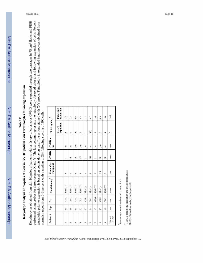
flasks and subjected to FISH. Tetraploidy was observed in many of the cultures (N=9;mean=45%) (Table 2). There was no evidence of fusion cells in the four patients whoreceived sex-discordant transplants stained with X/Y probe (Table 2; patients 6,7,8,9). Whenskin biopsies obtained from patients with GVHD and the healthy keratinocytes wereexpanded, tetraploidy was even more pronounced in patients samples (Table 2); simpleexpansion of keratinocytes derived from normal foreskins did not result in significanttetraploidy or aneuploidy.
Keratinocytes develop tetraploidy and aneuploidy after co-culture with PBMCsIn order to develop a model for GVHD in which individual, potentially inciting factors couldbe controlled; we examined the effect of HLA-mismatched allogeneic lymphocytes on thedevelopment of ploidy in keratinocytes in vitro. Keratinocytes for these experiments werederived from foreskins, which were cultured with allogeneic lymphocytes for two days, afterwhich the non-adherent lymphocytes were removed. We expanded the remainingkeratinocytes before performing FISH in order to select for the live replicating cells that hadsurvived the inflammatory insult; we chose to examine chromosomes 7, 8, 11, and 17because these are frequently abnormal in SCC33. However, when we examined healthykeratinocyte cultures with allogeneic PBMCs to mimic inflammation and cellular injury atsites of GVHD, we saw an increase in tetraploid cell number after two weeks of culture andtwo passages (example in Fig 1A; left panel). Tetraploidy could also be visualized whencells nuclei were stained with a nuclear stain and analyzed by flow cytometry (example seenin Fig 1A; right panel). Aneuploidy for either chromosome 7 or 8 developed after 10 days ofculture, while tetraploidy could be observed as early as three days (Fig 1B). Untreatedkeratinocytes isolated from normal human foreskins demonstrated only rare tetraploid cellsafter expansion in media (Table 1 N=20).
CD8+ cells produce more ploidy than CD4+ lymphocytesIn order to assess whether normal CD4 or CD8 cells were responsible for the changesobserved, we separated CD4 and CD8 cells by magnetic column and cultured eachpopulation with keratinocytes obtained from normal foreskins. Confluent keratinocytes werecultured with 50,000 CD4+ or CD8+ cells; as a control, one aliquot of 100,000 un-separatedPBLs were incubated with cyclosporine (400 ng/mL) before adding the lymphocytes to theflask in order to block lymphocyte proliferation and activation. Keratinocytes were passaged
Sloand et al. Page 5
Biol Blood Marrow Transplant. Author manuscript; available in PMC 2012 September 10.
NIH
-PA Author Manuscript
NIH
-PA Author Manuscript
NIH
-PA Author Manuscript

three times following addition of lymphocytes and harvested two weeks later. CD4 cellswere less effective in generation of aneuploid and tetraploid cells compared to CD8 cells.Addition of cyclosporine prevented these chromosomal changes (Fig 2A). Cultures withCD8 cells produced more inflammatory cytokines than those with CD4 cells (Fig 2B).
Inflammation produced in vitro by mis-matched allogeneic lymphocytes decreaseskeratinocytes telomere length
Inflammation and cell death accompanying GVHD results in significant cell mitosis andrepair37. In order to determine if an inflammatory insult was associated with decreases intelomere length we examined the effect of culture with un-separated PBLs, purified CD4+ Tcells, or CD8+ T cells on keratinocyte telomere length. Telomere length was shortened bythe addition of lymphocytes, most significantly after exposure to CD8+ cells (Fig 3).
Loss of P53 in keratinocytes results from deletion of chromosome 17In order to assess whether loss of TP53 could account for the frequency of tetraploidy andaneuploidy observed in keratinocyte culture, we stained with a locus-specific probe (LSP) top53 and a CEP probe to chromosome 11. When tetraploid and aneuploid cells werecompared with diploid cells, TP53 gain or loss was significantly greater in aneuploid cells(p<0.0001) (Fig 4). In order to determine if TP53 loss or gain resulted from loss of onlyTP53 or of the entire chromosome 17 (where TP53 resides), we stained cells with a LSP toTP53 and a CEP probe to 17. These experiments indicated loss of p53 was the resulted ofdeletion of chromosome 17 in the majority of cases (data not shown) (p<0.0001; Fisher’sexact test).
Keratinocytes exposed to lymphocytes demonstrate no p53 mutationIn order to determine if keratinocytes exposed to lymphocytes developed mutations in p53,we sequenced the p53 gene. We found no mutation in either the untreated or treatedkeratinocytes.
DiscussionIn this study, we observed tetraploid cells in un-manipulated buccal mucosal smearsobtained from sites of GVHD. Even greater numbers of polyploid cells were observedfollowing proliferation of these keratinocytes, suggesting that repeated cell division played arole in alteration of chromosome number. Keratinocytes exposed to allogeneic lymphocytesexhibited some of the chromosomal abnormalities frequently found in patients withsquamous cell carcinoma27: aneuploidy for chromosomes 8, 7, 17 and 11, and tetraploidy.We observed loss of p53 in many of the anueploid cells-a factor which may be responsiblefor their continued growth and survival. Telomere length decreased with keratinocyteexpansion but this decrease was more substantial in keratinocytes exposed to allogeneiclymphocytes, most notably CD8 cells.
In our experiments, loss of P53 in most cases appeared to be related to deletion ofchromosome 17, a frequent finding in squamous cell carcinoma27, rather than to a mutation.Others have observed this non-random deletion of chromosome 17 in cells aneuploid forother chromosomes38. TP53 triggers apoptosis in aneuploid and tetraploid cells, andhaploinsufficiency is associated with survival of aneuploid cells39. Tetraploidy has beenobserved in many pre-malignant states and may give rise to aneuploid cells38. In oneproposed model of tumorigenesis in ulcerative colitis (UC) 26, nitric oxide(NO) -inducedcell cycle arrest leads to transient tetraploidy and DNA repair. Reactive oxygen speciesrepeatedly exposing TP53 to oxidative stress may result in mutation or loss ofheterozygosity of this gene. These events may result in loss of this Tp53’s checkpoint
Sloand et al. Page 6
Biol Blood Marrow Transplant. Author manuscript; available in PMC 2012 September 10.
NIH
-PA Author Manuscript
NIH
-PA Author Manuscript
NIH
-PA Author Manuscript

function and a greater proclivity for malignant transformation. In the TP53 null mouse,tetraploid cells continue to divide but are genomically unstable, acquiring many newtranslocations or undergoing multi-polar mitosis because of duplication of the spindleapparatus, which results in aneuploidy.40
TP53 protein also plays an important role in eliminating cells with telomere dysfunction bytriggering senescence or apoptosis in cells with shortened telomeres41. Functional loss orgain of TP53 is associated with continued telomere erosion and subsequent formation ofdicentric chromosomes resulting from telomereic fusions42. Normal cells, adjacent to SCC,reportedly have shortened telomeres suggesting that reduction of telomere length could berelated to malignant transformation43. Inflammatory diseases with a proclivity for malignanttransformation such as Barrett’s esophagitis11, ulcerative colitis44, and hepatitis45, 46 allshow substantial decreases in telomere length. Telomere shortening is associated withaneuploidy both in human cancer as well as in the mouse model47, 48. End-to-end fusion oftelomeres with unequal segregation of chromosomes has been observed in the telomerase-deficient mouse and is presumed to be the mechanism for aneuploidy in this circumstance.Epithelial tissue has little telomerase activity, so that factors which increase cell turnoverwould be expected to give rise to more pronounced telomere shortening49.
These findings suggest that inflammation alone in the absence of immunosuppression maygive rise to tetraploid and aneuploid keratinocytes as well as haploinsufficincy of p53. Theseevents may lead to genomic instability resulting in malignant transformation50. This in vitrosystem may provide a model of GVHD with inflammation resulting in DNA damage, loss ofTP53, and shortened telomere length— all of which may be steps in the development ofSCC.
AcknowledgmentsResearch supported by the National Heart Lung and Blood Institute
Reference List1. Bhatia S, Louie AD, Bhatia R, et al. Solid cancers after bone marrow transplantation. J Clin Oncol.
2001; 19:464–471. [PubMed: 11208840]
2. Ramsay HM, Reece SM, Fryer AA, Smith AG, Harden PN. Seven-Year Prospective Study ofNonmelanoma Skin Cancer Incidence in U.K. Renal Transplant Recipients. Transplantation. 2007;84
3. Socie G, Curtis RE, Deeg HJ, et al. New Malignant Diseases After Allogeneic MarrowTransplantation for Childhood Acute Leukemia. J Clin Oncol. 2000; 18:348. [PubMed: 10637249]
4. Ulrich C, Kanitakis J, Stockfleth E, Euvrard S. Skin cancer in organ transplant recipients- -where dowe stand today? Am J Transplant. 2008; 8:2192–2198. [PubMed: 18782290]
5. Rizzo JD, Curtis RE, Socie G, et al. Solid cancers after allogeneic hematopoietic celltransplantation. Blood. 2009; 113:1175–1183. [PubMed: 18971419]
6. Travis LB, Hill DA, Dores GM, et al. Breast Cancer Following Radiotherapy and ChemotherapyAmong Young Women With Hodgkin Disease. JAMA. 2003; 290:465–475. [PubMed: 12876089]
7. Curtis RE, Metayer C, Rizzo JD, et al. Impact of chronic GVHD therapy on the development ofsquamous-cell cancers after hematopoietic stem-cell transplantation: an international case-controlstudy. Blood. 2005; 105:3802–3811. [PubMed: 15687239]
8. Dantal J, Hourmant M, Cantarovich D, et al. Effect of long-term immunosuppression in kidney-graftrecipients on cancer incidence: randomised comparison of two cyclosporin regimens. Lancet. 1998;351:623–628. [PubMed: 9500317]
9. Otley CC, Maragh SL. Reduction of immunosuppression for transplant-associated skin cancer:rationale and evidence of efficacy. Dermatol Surg. 2005; 31:163–168. [PubMed: 15762208]
Sloand et al. Page 7
Biol Blood Marrow Transplant. Author manuscript; available in PMC 2012 September 10.
NIH
-PA Author Manuscript
NIH
-PA Author Manuscript
NIH
-PA Author Manuscript
10. Svendsen LB, Larsen JK, Christensen IJ. Human skin fibroblast in vitro tetraploidy. Flowcytometric DNA assay used to confirm metaphase assay in patients with various colonic diseases.Cancer Genet Cytogenet. 1989; 39:245–251. [PubMed: 2752376]
11. Maley CC, Galipeau PC, Li X, et al. The combination of genetic instability and clonal expansionpredicts progression to esophageal adenocarcinoma. Cancer Res. 2004; 64:7629–7633. [PubMed:15492292]
12. Sherman M. Risk of hepatocellular carcinoma in hepatitis B and prevention through treatment.Cleve Clin J Med. 2009; 76(Suppl 3):S6–S9. [PubMed: 19465708]
13. Marnett LJ, Riggins JN, West JD. Endogenous generation of reactive oxidants and electrophilesand their reactions with DNA and protein. J Clin Invest. 2003; 111:583–593. [PubMed: 12618510]
14. West JD, Marnett LJ. Endogenous reactive intermediates as modulators of cell signaling and celldeath. Chem Res Toxicol. 2006; 19:173–194. [PubMed: 16485894]
15. West JD, Ji C, Marnett LJ. Modulation of DNA fragmentation factor 40 nuclease activity bypoly(ADP-ribose) polymerase-1. J Biol Chem. 2005; 280:15141–15147. [PubMed: 15703174]
16. Marnett LJ, Plastaras JP. Endogenous DNA damage and mutation. Trends in Genetics. 2001;17:214–221. [PubMed: 11275327]
17. Galipeau PC, Li X, Blount PL, et al. NSAIDs modulate CDKN2A, TP53, and DNA content riskfor progression to esophageal adenocarcinoma. PLoS Med. 2007; 4:e67. [PubMed: 17326708]
18. Lalwani ND, Dethloff LA, Haskins JR, Robertson DG, de l I. Increased nuclear ploidy, not cellproliferation, is sustained in the peroxisome proliferator-treated rat liver. Toxicol Pathol. 1997;25:165–176. [PubMed: 9125775]
19. Lothschutz D, Jennewein M, Pahl S, et al. Polyploidization and centrosome hyperamplification ininflammatory bronchi. Inflamm Res. 2002; 51:416–422. [PubMed: 12234059]
20. Rodin SN, Rodin AS. Origins and selection of p53 mutations in lung carcinogenesis. Seminars inCancer Biology. 2005; 15:103–112. [PubMed: 15652455]
21. Goodman JE, Hofseth LJ, Hussain SP, Harris CC. Nitric oxide and p53 in cancer-prone chronicinflammation and oxyradical overload disease. Environ Mol Mutagen. 2004; 44:3. [PubMed:15199542]
22. Ambs S, Hussain SP, Marrogi AJ, Harris CC. Cancer-prone oxyradical overload disease. IARC SciPubl. 1999:295–302. [PubMed: 10626229]
23. Hussain SP, Harris CC. p53 biological network: at the crossroads of the cellular-stress responsepathway and molecular carcinogenesis. J Nippon Med Sch. 2006; 73:54–64.
24. Ganem NJ, Storchova Z, Pellman D. Tetraploidy, aneuploidy and cancer. Curr Opin Genet Dev.2007; 17:157–162. [PubMed: 17324569]
25. Dabelsteen S, Hercule P, Barron P, Rice M, Dorsainville G, Rheinwald JG. Epithelial CellsDerived from Human Embryonic Stem Cells Display P16(INK4A) Senescence, Hypermotility,and Differentiation Properties Shared by Many P63(+) Somatic Cell Types. Stem Cells. 2009;27:1388–1399. [PubMed: 19489101]
26. Hofseth LJ, Saito S, Hussain SP, et al. Nitric oxide-induced cellular stress and p53 activation inchronic inflammation. PNAS. 2003; 100:143–148. [PubMed: 12518062]
27. Ozturk K, Acar H, Durmus E, Ozturk A, Mutlu N. Analysis of chromosomes 8 and 17 aneuploidiesin laryngeal squamous cell carcinoma by fluorescence in situ hybridization. Laryngoscope. 2004;114:1005–1010. [PubMed: 15179203]
28. Tralongo V, Rodolico V, Luciani A, Marra G, Daniele E. Prognostic factors in oral squamous cellcarcinoma. A review of the literature. Anticancer Res. 1999; 19:3503–3510. [PubMed: 10629643]
29. Akrish S, Buchner A, Dayan D. Oral cancer: diagnostic options as an aid to histology in order topredict patients at high risk for malignant transformation. Refuat Hapeh Vehashinayim. 2004;21:6–15. 93. [PubMed: 15672638]
30. Goldsmith MM, Cresson DH, Arnold LA, Postma DS, Askin FB, Pillsbury HC. DNA flowcytometry as a prognostic indicator in head and neck cancer. Otolaryngol Head Neck Surg. 1987;96:307–318. [PubMed: 3108817]
31. Ravindran A, Vijayakumar T, Sudha L, et al. Chromosome abnormalities in squamous cellcarcinoma of the human oral cavity. Neoplasma. 1990; 37:191–197. [PubMed: 2160621]
Sloand et al. Page 8
Biol Blood Marrow Transplant. Author manuscript; available in PMC 2012 September 10.
NIH
-PA Author Manuscript
NIH
-PA Author Manuscript
NIH
-PA Author Manuscript

32. Robinson JK, Rademaker AW, Goolsby C, Traczyk TN, Zoladz C. DNA ploidy in nonmelanomaskin cancer. Cancer. 1996; 77:284–291. [PubMed: 8625236]
33. Scully C, Field JK, Tanzawa H. Genetic aberrations in oral or head and neck squamous cellcarcinoma 2: chromosomal aberrations. Oral Oncology. 2000; 36:311–327. [PubMed: 10899669]
34. Struikmans H, Rutgers DH, Hordijk GJ, Slootweg PJ, van dT I, Battermann JJ. Prognosticsignificance of cell proliferation markers and DNA-ploidy in head and neck tumors. Int J RadiatOncol Biol Phys. 1998; 40:27–34. [PubMed: 9422554]
35. Hematti P, Sloand EM, Carvallo CA, et al. Absence of donor-derived keratinocyte stem cells inskin tissues cultured from patients after mobilized peripheral blood hematopoietic stem celltransplantation. Exp Hematol. 2002; 30:943–949. [PubMed: 12160846]
36. Zhao L, Khan Z, Hayes KJ, Glassman AB. Interphase fluorescence in situ hybridization analysis: astudy using centromeric probes 7, 8, and 12. Ann Clin Lab Sci. 1998; 28:51–56. [PubMed:9512785]
37. Klimczak A, Lange A. Apoptosis of keratinocytes is associated with infiltration of CD8+lymphocytes and accumulation of Ki67 antigen. Bone Marrow Transplant. 2000; 26:1077–1082.[PubMed: 11108306]
38. Olaharski AJ, Sotelo R, Solorza-Luna G, et al. Tetraploidy and chromosomal instability are earlyevents during cervical carcinogenesis. Carcinogenesis. 2006; 27:337–343. [PubMed: 16123119]
39. McNamee LM, Brodsky M. p53-independent Apoptosis Limits DNA Damage-inducedAneuploidy. Genetics. 2009
40. Borel F, Lohez OD, Lacroix FB, Margolis RL. Multiple centrosomes arise from tetraploidycheckpoint failure and mitotic centrosome clusters in p53 and RB pocket protein-compromisedcells. Proc Natl Acad Sci U S A. 2002; 99:9819–9824. [PubMed: 12119403]
41. Lechel A, Holstege H, Begus Y, et al. Telomerase deletion limits progression of p53-mutanthepatocellular carcinoma with short telomeres in chronic liver disease. Gastroenterology. 2007;132:1465–1475. [PubMed: 17433324]
42. Schwartz JL, Jordan R, Liber H, Murnane JP, Evans HH. TP53-dependent chromosome instabilityis associated with transient reductions in telomere length in immortal telomerase-positive celllines. Genes Chromosomes Cancer. 2001; 30:236–244. [PubMed: 11170280]
43. Kammori M, Poon SS, Nakamura K, et al. Squamous cell carcinomas of the esophagus arise froma telomere-shortened epithelial field. Int J Mol Med. 2007; 20:793–799. [PubMed: 17982685]
44. Kinouchi Y, Hiwatashi N, Chida M, et al. Telomere shortening in the colonic mucosa of patientswith ulcerative colitis. J Gastroenterol. 1998; 33:343–348. [PubMed: 9658312]
45. Lee YH, Oh BK, Yoo JE, et al. Chromosomal instability, telomere shortening, and inactivation ofp21WAF1/CIP1 in dysplastic nodules of hepatitis B virus-associated multistephepatocarcinogenesis. Mod Pathol. 2009
46. Kitay-Cohen Y, Goldberg-Bittman L, Hadary R, Fejgin MD, Amiel A. Telomere length inHepatitis C. Cancer Genet Cytogenet. 2008; 187:34–38. [PubMed: 18992639]
47. Griffith JK, Bryant JE, Fordyce CA, Gilliland FD, Joste NE, Moyzis RK. Reduced telomere DNAcontent is correlated with genomic instability and metastasis in invasive human breast carcinoma.Breast Cancer Res Treat. 1999; 54:59–64. [PubMed: 10369081]
48. Blasco MA, Lee HW, Hande MP, et al. Telomere shortening and tumor formation by mouse cellslacking telomerase RNA. Cell. 1997; 91:25–34. [PubMed: 9335332]
49. Krunic D, Moshir S, Greulich-Bode KM, et al. Tissue context-activated telomerase in humanepidermis correlates with little age-dependent telomere loss. Biochimica et Biophysica Acta(BBA) - Molecular Basis of Disease. 2009; 1792:297–308.
50. Storchova Z, Kuffer C. The consequences of tetraploidy and aneuploidy. J Cell Sci. 2008;121:3859–3866. [PubMed: 19020304]
Sloand et al. Page 9
Biol Blood Marrow Transplant. Author manuscript; available in PMC 2012 September 10.
NIH
-PA Author Manuscript
NIH
-PA Author Manuscript
NIH
-PA Author Manuscript

Sloand et al. Page 10
Biol Blood Marrow Transplant. Author manuscript; available in PMC 2012 September 10.
NIH
-PA Author Manuscript
NIH
-PA Author Manuscript
NIH
-PA Author Manuscript

Fig. 1. Coculture of keratinocytes from healthy foreskins with allogeneic mis-matchedlymphocytesA) When mismatched allogeneic lymphocytes were co-cultured with foreskin keratinocytesfor tetraploid cells could be observed in keratinocyte cultures co-cultured with allogeneiclymphocytes. An example of a FISH assay utilizing the X/Y probe is seen (left). Arrowspoint to tetraploid cells. Tetraploid cells are also distinguishable based on DNA content asmeasured by flow cytometric methods (right). B ) Keratinocytes cocultured with allogeneicwere stained with FISH probes for chromosomes 7 and 8. Samples cultured withlymphocytes had significantly more tetraploidy and aneuploidy than samples expanded inthe absence of lymphocytes. Tetraploidy appeared by day 3 of culture; aneuploidy was notseen until day 10.
Sloand et al. Page 11
Biol Blood Marrow Transplant. Author manuscript; available in PMC 2012 September 10.
NIH
-PA Author Manuscript
NIH
-PA Author Manuscript
NIH
-PA Author Manuscript

Fig. 2. Coculture of keratinocytes with CD8+ cells compared to CD4+ lymphocytesA) Samples of PBLs from healthy donors were separated into CD8+ cells and CD4+ cellsusing magnetic columns and these were cultured with keratinocytes. Keratinocytes treatedwith allogeneic CD8+ cells developed the most tetraploidy and aneuploidy; cyclosporineblocked the effect of CD4+ and CD8+ cells.B) Following culture of keratinocytes with purified CD4 and CD8 cells, media wascentrifuged to remove the cells and debris. Samples were then tested for cytokines using animmune-bead-based multiplex assay (Luminex). CD8 cells produced significantly moreinflammatory cytokines than CD4 cells following co-culture with keratinocytes. Controlsamples consisted of those keratinocytes cultured for an equal time but withoutlymphocytes. The fold change compared to controls is graphed for each cytokine.
Sloand et al. Page 12
Biol Blood Marrow Transplant. Author manuscript; available in PMC 2012 September 10.
NIH
-PA Author Manuscript
NIH
-PA Author Manuscript
NIH
-PA Author Manuscript

Fig. 3. Telomere lengths in keratinocytes cultured with lymphocytes
• Left panel: Keratinocytes were cultured with PBLs as described in Methods andMaterials and then passaged two times over the course of 17 days. Telomere lengthwas then measured. PBLs caused a decrease in keratinocyte telomere, which wasmost prominent immediately following co-cultivation with lymphocytes.
• Right panel: Keratinocytes were cultured with 30,000 purified CD4+, CD8+ and amixture of similar numbers of CD4 and CD8 cells (i.e. 60,000 total lymphocytes),which were pre-incubated with cyclosporine. CD8+ cells showed the most dramaticchange in telomere length.
Sloand et al. Page 13
Biol Blood Marrow Transplant. Author manuscript; available in PMC 2012 September 10.
NIH
-PA Author Manuscript
NIH
-PA Author Manuscript
NIH
-PA Author Manuscript

Fig. 4. Loss of TP53 gene following coculture with keratinocytesAn aliquot of lymphocyte-treated keratinocytes was probed with locus specific p53 signal.Cells were probed with CEP 11 and LSP p53. When this was analyzed statistically using theMann-Whitley test, an excess number of aneuploid and tetraploid cells showed either a gainor loss of p53 (p<0.001).
Sloand et al. Page 14
Biol Blood Marrow Transplant. Author manuscript; available in PMC 2012 September 10.
NIH
-PA Author Manuscript
NIH
-PA Author Manuscript
NIH
-PA Author Manuscript

NIH
-PA Author Manuscript
NIH
-PA Author Manuscript
NIH
-PA Author Manuscript
Sloand et al. Page 15
Tabl
e 1
Kar
yoty
pe A
naly
sis
of B
ucca
l Muc
osal
Cel
ls
Buc
cal m
ucos
al s
mea
rs w
ere
obta
ined
fro
m 8
pat
ient
s fr
om a
reas
of
GV
HD
and
6 p
atie
nts
from
uni
nvol
ved
area
s of
the
bucc
al m
ucos
a. K
erat
inoc
ytes
from
GV
HD
site
s fo
rced
into
rep
eate
d ce
ll di
visi
on b
y m
ultip
le in
vitr
o pa
ssag
ing
show
eve
n m
ore
pron
ounc
ed te
trap
loid
y th
an u
nman
ipul
ated
kera
tinoc
ytes
.
Pat
ient
GV
HD
Don
or s
exdi
scor
dant
Tim
e fr
omtr
ansp
lant
Tra
nspl
ant
regi
men
% t
etra
ploi
dar
ea o
f G
VH
D
11ch
roni
c sy
stem
icno
18 m
oFl
u/C
y23
/50
(46%
)
12ac
ute
skin
IV
yes
5 m
oFl
u/C
y5/
25 (
20%
)
13ch
roni
c lim
ited
no20
mo
Flu/
Cy
5/20
(25
%)
14ch
roni
c lim
ited
yes
38 m
oFl
u/C
y1/
5 (2
0%)
16ac
ute
skin
Iye
s2.
5 m
oFl
u/C
y2/
20 (
10%
)
17ac
ute
skin
1ye
s4m
oFl
u/C
y3/
7 (8
%)
18ch
roni
c lim
ited
no4
mo
Flu/
Cy
6/30
(20
%)
19ch
roni
c lim
ited
no1.
3 m
oFl
u/C
y3/
25 (
12%
)
20no
neno
6 m
oFl
u/C
y0/
72 (
0%)
21no
neno
2 m
oFl
u/C
y0/
21 (
0%)
22no
neno
22 m
oFl
u/C
y0/
3 (0
%)
23no
neno
2.6
mo
Flu/
Cy
0/37
(0%
)
24no
neno
36 m
oFl
u/C
y0/
11 (
0%)
25no
neno
12 m
oFl
u/C
y0/
12
(0%
)
Nor
mal
N=
10--
---
----
----
----
----
-0
Biol Blood Marrow Transplant. Author manuscript; available in PMC 2012 September 10.
NIH
-PA Author Manuscript
NIH
-PA Author Manuscript
NIH
-PA Author Manuscript
Sloand et al. Page 16
Tabl
e 2
Kar
yoty
pe a
naly
sis
of b
iops
ies
of s
kin
in G
VH
D p
atie
nt s
kin
kera
tino
cyte
s fo
llow
ing
expa
nsio
n
Ker
atin
ocyt
es o
btai
ned
from
ski
n bi
opsi
es o
f pa
tient
s w
ith a
his
tory
of
cuta
neou
s G
VH
D w
ere
expa
nded
thro
ugh
two
pass
ages
in 7
5 cm
2 fl
asks
and
FIS
Hpe
rfor
med
usi
ng p
robe
s fo
r ch
rom
osom
es X
and
Y. T
he la
st c
olum
n re
pres
ents
the
tetr
aplo
idy
obse
rved
pri
or to
and
fol
low
ing
expa
nsio
n of
cel
ls. P
erce
ntte
trap
loid
y pr
ior
to e
xpan
sion
is b
ased
on
coun
ts d
one
on p
araf
fin-
sect
ions
sta
ined
with
X/Y
pro
be. T
etra
ploi
dy in
exp
ande
d ke
ratin
ocyt
es o
btai
ned
from
norm
als
rang
ed f
rom
0–3
per
cent
with
a m
edia
n of
2%
fol
low
ing
scor
ing
of 3
00 c
ells
.
Pat
ient
#A
geD
xC
ondi
tion
ing*
Yea
rs a
fter
tran
spla
ntG
VH
DG
VH
D o
nbx
% t
etra
ploi
dy*
Bef
ore
expa
nsio
nF
ollo
win
gex
pans
ion
130
AM
LT
BI/
CY
3I
no1
15
236
CM
LT
BI/
CY
1II
no5
23
321
AM
LT
BI/
CY
2II
yes
1298
452
CL
LT
BI/
CY
3IV
yes
343
537
NH
LFl
u/C
y1
Ino
412
634
CM
LFl
u/C
y2
III
no13
67
742
MD
ST
BI/
CY
7I
no2
16
825
PNH
Flu/
Cy
0.6
IIye
s5
85
946
CM
LT
BI/
CY
6II
no1
10
Nor
mal
(N=
20)
----
----
----
----
----
---
----
01–
3
* Perc
enta
ges
wer
e ba
sed
on c
ell c
ount
s of
300
* TB
I/C
Y-t
otal
bod
y ir
radi
atio
n an
d cy
clop
hosp
ham
ide
Flu/
Cy-
Flud
arab
ine
and
cycl
opho
spha
mid
e
Biol Blood Marrow Transplant. Author manuscript; available in PMC 2012 September 10.