Impacts of climate change on the seasonal distribution of migratory caribou
Upload
independentCategory
view
3download
0
Urokinase Expression and Binding Activity AssociatedWith the Transforming Growth Factor b1-InducedMigratory and Invasive Phenotype of MouseEpidermal Keratinocytes
Juan F. Santibanez,1,2 Pilar Frontelo,1 Maite Iglesias,1 Jorge Martınez,2 and Miguel Quintanilla1*1Instituto de Investigaciones Biomedicas CSIC-UAM, 28029-Madrid, Spain2Unidad de Biologıa Celular, INTA, Universidad de Chile, Santiago, Chile
Abstract Transforming growth factor b1(TGF-b1) is a stimulator of malignant progression in mouse skin carcino-genesis. TGF-b1 exerts a differential effect on cultured nontumorigenic (MCA3D cell line) and transformed (PDV cellline) keratinocytes. Whereas MCA3D cells are growth arrested and committed to die in the presence of the factor, itinduces a reversible epithelial-fibroblastic conversion in PDV cells. This conversion is associated in vivo with asquamous-spindle cell carcinoma transition. Here we have investigated the role of urokinase (uPA) during malignantprogression of transformed epidermal keratinocytes. We show that the levels of uPA expression/secretion, and the uPAbinding activity to the cell surface, correlate with the invasive and malignant potentials of mouse epidermal cell lines.TGF-b1 enhanced uPA production, the number of uPA cell surface binding sites, and the expression of the plasminogenactivator inhibitor PAI-1, in transformed PDV cells, but had no major effect on nontumorigenic MCA3D keratinocytes.Increased uPA production depended on the presence of the factor in the culture medium and occurred concomitantly tothe stimulation of the migratory and invasive abilities of PDV cells. Synthetic peptides containing the amino terminalsequence of the mature mouse uPA inhibited the binding of uPA to the cell surface and decreased TGF-b1-induced cellmotility and invasiveness. These results demonstrate that the uPA system mediates at least part of the migratory andinvasive phenotype induced by TGF-b1 in transformed keratinocytes, and suggest a role for uPA on the changes that leadto the appearance of spindle carcinomas. J. Cell. Biochem. 74:61–73, 1999. r 1999 Wiley-Liss, Inc.
Key words: uPA; TGF-b1; migration; invasiveness; keratinocytes; carcinogenesis
During malignant progression the acquisi-tion of migratory and invasive properties arelinked to changes in cell-cell and cell-extracellu-lar matrix (ECM) adhesiveness, the reorganiza-tion of cytoskeletal components, and the expres-
sion and secretion of proteinases involved inECM degradation [Stetler-Stevenson et al.,1993]. Sometimes, the alterations in the differ-entiation program are profound and associatedto a drastic change of the cell phenotype. Thus,in natural and experimental carcinogenesissquamous cell carcinomas (SCCs) can progressto spindle carcinomas (SpCCs), a highly aggres-sive type of tumor formed by cells that have lostthe epithelial phenotype and acquired fibro-blast-like characteristics [Portella et al., 1994–95]. One of the best characterized animal mod-els for studying this transition is the mouseskin system, in which tumors are induced onthe dorsal skin of mice by a single topical appli-cation of a chemical carcinogen followed bysequential treatment with a tumor promoter. Aproportion of benign papillomas developed bychemical carcinogenesis progress spontane-ously to malignant SCCs. The later stages of
Abbreviations used: ECM, extracellular matrix; EGF, epi-dermal growth factor; FBS, fetal bovine serum; HGF/SF,hepatocyte growth factor/scatter factor; PA, plasminogenactivator; PAI, PA inhibitor; SCC, squamous cell carcinoma;SpCC, spindle cell carcinoma; TGF-b, transforming growthfactor b; uPA, urokinase-type PA; uPAR, uPA receptor.Grant sponsor: Comision Interministerial de Ciencia y Tec-nologıa; Grant number: SAF98–0085-C03–02; Grant spon-sor: Comunidad Autonoma de Madrid; Grant number: 8.1/22/97. Grant sponsor: Fondo Nacional de Ciencia y Tecno-logia de Chile; Grant number: 890028.*Correspondence to: Miguel Quintanilla, Instituto de Inves-tigaciones Biomedicas CSIC-UAM, Arturo Duperier 4,28029-Madrid, Spain. E-mail: [email protected] 20 May 1998; Accepted 19 January 1999
Journal of Cellular Biochemistry 74:61–73 (1999)
r 1999 Wiley-Liss, Inc.

tumor progression involve the loss of the epithe-lial phenotype and the development of SpCCs[Buchmann et al., 1991].
TGF-b growth factors affect proliferation anddifferentiation of many cell types [Massague,1990; Lahio and Keski-Oja, 1992]. TGF-b1 inhib-its the growth of cultured keratinocytes and isconsidered as a physiological negative regula-tor of basal cell proliferation in the epidermis[Glick et al., 1993]. However, skin carcinomacell lines are either less responsive or totallyescape to the growth inhibition exerted by thegrowth factor [Haddow et al., 1991]. We haveshown previously that TGF-b1 induces an epi-thelial-mesenchymal transdifferentiation intransformed epidermal keratinocytes [Caulınet al., 1995]. This change is associated with theloss of the differentiated phenotype of the tu-mors and the acquisition of metastatic abilities[Frontelo et al., 1998]. A role for TGF-b1 in thedevelopment of SpCCs has also been demon-strated in mice with TGF-b1 expression tar-geted to the epidermis [Cui et al., 1996]. TheTGF-b1-induced epithelial-fibroblastic conver-sion occurs in sequential steps with disruptionof intercellular contacts, cell dispersion andenhanced motility preceding the acquisition ofa spindle morphology [Caulın et al., 1995]. Incontrast, nontumorigenic keratinocytes that aregrowth arrested by the factor are commited tosquamous differentiation [Mansbridge andHanawalt, 1988] and cell death [Caulın et al.,1995] in the presence of TGF-b1.
It has been found that TGF-b1 is a potentmodulator of pericellular proteolysis by regulat-ing the expression and secretion of plasmino-gen activators (PAs) such as uPA and its inhibi-tor PAI-1 [Lahio and Keski-Oja, 1992]. uPA is aserin proteinase that, after secretion, is re-tained at the cell surface by a high affinityreceptor, and converts the inactive plasmino-gen into the broad-spectrum trypsin-like serinproteinase plasmin. Plasmin degrades severalcomponents of the ECM such as fibronectin,laminin, and proteoglycans, and activate othermatrix metalloproteinases. Several studies havedemonstrated a strong association between theexpression of catalytically active uPA and tu-moral invasiveness and metastasis [see An-dreasen et al., 1997, for a review]. Binding ofuPAto its receptor can also stimulate cell migra-tion, independently of its proteolytic activity, ina variety of cells including keratinocytes
[Odekon et al., 1992; Stahl and Mueller, 1994;Busso et al., 1994].
The present study was designed to investi-gate the role of uPA in TGF-b1-induced malig-nant progression of transformed epidermal ker-atinocytes.
MATERIALS AND METHODSCell Lines, Culture, and Treatment Conditions
The origin of the cell lines used in this studyhas been described elsewhere [Dıaz-Guerra etal., 1992; Caulın et al., 1995]. Cells were cul-tured in Ham’s F-12 medium supplementedwith aminoacids and vitamins (GIBCO Ltd.,Paisley, Scotland), 10% fetal bovine serum(FBS), and antibiotics (2.5 µg/ml amphotericinB, 100 µg/ml ampicillin, and 32 µg/ml gentami-cin; Sigma Chemical Co., St. Louis, MO). Cul-tures were maintained on plastic at 37°C in a5% CO2 humidified atmosphere.
For TGF-b1 treatments, human recombinantTGF-b1 (Calbiochem-Novabiochem Intl., LaJolla, CA) was used. The growth factor wasadded to the cell cultures in the presence ofserum at a final concentration of 10 ng/ml, asreported elsewhere [Caulın et al., 1995].
Synthesis of Murine uPA 41-54 and 44-63Peptides
Peptides comprising aminoacid residues 41-54(P41-54: VSYKYFSRIRRCSC) and 44-63 (P44-63: KYFSRIRRCSCPRKFQGEHC) of the aminoterminal sequence of uPA [Belin et al., 1985;Degen et al., 1987], corresponding to the epider-mal growth factor (EGF)-like domain, were syn-thesized in the Protein Chemistry Departmentof Centro de Biologıa Molecular (Molecular Bi-ology Center, CBM, Madrid, Spain). These se-quences were selected due to the lack of second-ary structure and high solvent accessibility, asdetermined by the PredictProtein computer pro-gram (EMBL, Heidelberg). As a control, weused a mixture of peptides derived from gelatinby trypsin degradation. A gelatin solution (10mg/ml) was incubated with trypsin (0.25 mg/ml) at 37°C for 2 h. Trypsin was inactivated byadding soybean trypsin inhibitor. By this proto-col a heterogeneous mixture of peptides of sizesbelow 10 kDa was obtained, as recorded bySDS-PAGE.
62 Santibanez et al.
Immunofluorescence Studies
Indirect immunofluorescence staining of uPAwas performed in nonpermeabilized cells fixedin paraformaldehyde (4% in PBS) using theanti-uPA mouse monoclonal antibody SAM-3,kindly provided by Dr. F. Castellino (Universityof Notre Dame, Indiana), and rabbit anti-mouse IgG coupled to fluorescein isothiocya-nate (FITC, Sigma Chemical Co.) as secondantibody.
Migration and Invasion Assays
The capacity of the cells to migrate throughMatrigel-coated filters was measured by usingTranswell chambers (Costar Corp., CambridgeMA) with 8-µm-pore polycarbonate filters coatedwith 30 µg of Matrigel (Collaborative Research,Bedford, MA). Cells at a density of 2 3 105 wereseeded in the upper compartment and incu-bated for 72 h in Ham’s F-12 medium plusserum. At the end of the incubation period, boththe viable nonmigrated cells, which remain onthe top of the filter, and the migrated cells, thatare attached to the underside of the filter and tothe bottom of the lower compartment, weredetermined by a colorimetric assay using thevital stain 3-(4,5-dimethylthiazol-2-yl)-2,5-di-phenyltetrazolium bromide after trypsiniza-tion. In experiments in which cells were treatedwith TGF-b1, the growth factor was added tothe medium at 10 ng/ml. To study the effect ofsynthetic peptides on invasiveness, amino ter-minal uPA peptides P41-54 and P44-63, or thecontrol peptide mixture, were added to the me-dia at 150 µg/ml together with TGF-b1.
The effect of TGF-b1 in cell motility was as-sayed by an in vitro wound model [Boyer et al.,1989]. Subconfluent cell monolayers were gen-tly scratched with a Gilson pipette yellow tip toproduce a ‘‘wound.’’ Then, the cultures wereallowed to growth for 24 h in the standardmedium in the presence or absence of TGF-b1
(10 ng/ml). Peptides P41-54, P44-63, or thecontrol peptide mixture, were added as before.
Caseinolysis Assays
Radial caseinolytic assays to study plasmino-gen activation were carried out in 1% agarosegels containing 0.5% casein and 2 µg/ml ofplasminogen as described previously [Santib-anez et al., 1995]. Cell cultures grown in T24plates in the presence or absence of TGF-b1
were incubated for the last 24 h in serum-freemedium. After this time, cells were trypsinizedand viable cells counted by Trypan blue dyeexclusion in a Neubauer chamber. Aliquots ofconditioned media normalized for the samenumber of viable cells (2 3 105) were applied toholes previously punched in the gels and incu-bated at 37°C for 16 h. Gels lacking plasmino-gen were used as controls. The diameters of theradial zones of caseinolysis were measured. Todetermine PA activity, different concentrationsof human uPA (Calbiochem-Navabiochem Intl.)were used as standards (0.01–20 U/ml). To iden-tify the molecular forms of PAs, a zymographicassay [Erickson et al., 1984] was used. For PAactivities secreted by the cells, aliquots of condi-tioned medium were first subjected to electro-phoresis in SDS-PAGE gels (10% acrylamide)under nonreducing conditions. SDS was re-moved by extensive washing in 2.5% TritonX-100, the gels placed on caseinolysis agarosegels and incubated at 37°C for 24 h. To analyzethe cell-associated activities, cultured cells werewashed extensively, scraped from the plates inPBS and sonicated. Total amount of protein inthe cell extracts was determined by Bradford(Bio-Rad, Madrid, Spain) and aliquots of thecell extracts containing the same amount ofprotein were diluted in Laemmli sample bufferand subjected to caseinolytic zymography. Themigrating position of the PA activities in thegels were determined respect to prestained mo-lecular weight protein markers (116, 84, 58, 45,36.5, and 26.6 kDa; Sigma Chemical Co.).
The PAI activity secreted by the cells wasanalyzed by reverse casein zymography [Keski-Oja et al., 1988]. Aliquots of conditioned mediaof cell cultures untreated or treated with TGF-b1
were subjected to SDS-PAGE in a 10% polyacril-amide gel containing plasminogen (2 mg/ml),casein (1 mg/ml), and uPA (0.2 U/ml). Afterwashing the gel to remove SDS, it was incu-bated at 37°C in buffer 50 mM Tris-HCl, pH 8.0,for 4 h, in which casein is cleaved by activatedplasminogen, and stained with Coomassie blue.Stained areas revealed the presence of activeinhibitor species.
Determination of uPA-Binding Sites
Human uPA was radioactively iodinated ac-cording to the method of Hunter and Green-wood [1962]. Cells were trypsinized and grown
uPA and TGF-b1-Induced Invasiveness of Keratinocytes 63

in suspension for at least 8 h in complete me-dium. Cells were then washed and treated withan acidic buffer (50 mM glycine pH 3.0, 100 mMNaCl) to remove cell surface-associated uPA,and resuspended in serum-free medium contain-ing 1% of bovine serum albumin (Sigma Chemi-cal Co.) at a density of 107 cells/ml. The cellsuspensions (100 µl) were incubated with 125I-uPA (105 acid-precipitable cpm; specific activity1.05 3 107 cpm/µg) for 45 min at 37°C. Specificbinding was determined by measuring the radio-activity bound in the presence of increasingconcentrations (up to a 400-fold excess) of non-radiolabeled uPA. The data were analyzed byScatchard transformation.
Northern Blot Analysis
Ten to 20 µg of poly A1 enriched RNA isolatedusing Mini RiboSepTM (Becton Dickinson Lab-ware, Bedford, MA) were fractioned on 1% aga-rose-formaldehyde gels and transferred to Ny-lon membranes (Zeta Probe; Bio-Rad, Madrid,Spain). For detection of uPA mRNA, the PstI-HindIII fragment of 600 bp from the pmu-PAplasmid containing the mouse uPA coding se-quence [Belin et al., 1985] was used; and forPAI-2, the PstI-PvuII fragment of 930 bp con-taining the full-length mouse cDNA [Belin etal., 1989].
Transient Transfection Assay
Cells in 20-cm2 culture dishes were trans-fected, using lipofectamine reagent (Life Tech-nologies Inc., Gaithersburg, MD), with 5 µg ofthe TGF-b responsive 3TPlux reporter con-struct, which contains a region of the PAI-1promoter in front of the luciferase gene [Wranaet al., 1992]. Transfection efficiency was moni-tored by cotransfection with 2 µg of the plasmid
TABLE I. uPA Production and Invasive/Tumorigenic Behaviour of Epidermal Cell Linesa
Cell lineTumori-genicity
Metastaticability
Migration throughMatrigel
(%)
uPA Production
Secretedactivity
(%)
Cell-associatedactivity
(%)
mRNAlevels
(%)
MCA3D 2 2 8 6 3 9 15 34PDV 1 2 57 6 4 40 42 55HaCa4 11 1 76 6 13 65 100 100Car C 11 1 100 6 11 100 74 85
aThe abilities of the cell lines to migrate through Matrigel were determined by seeding 2 3 105 cells in the upper compartmentof Transwells chambers and incubating for 72 h. The percentage of migrated cells was calculated from the total number ofviable cells at the end of the incubation period, as described in Materials and Methods. Values in the table are given aspercentages of the maximum migratory ability, corresponding to Car C. Each value represents the mean (6SD) of triplicates.Similar results were obtained in two independent experiments. To determine the uPA activity secreted by the cell lines, cellswere grown up to confluence in complete medium and changed to serum-free medium for 24 h. After this time, cells weretrypsinized and viable cells counted. Aliquots of conditioned medium normalized for the same number of viable cells weresubjected to casein zymography. For uPA cell-associated activity, cells grown up to confluence were washed, scraped from theplates, and sonicated. Aliquots of cell extracts containing the same amount of total protein were subjected to caseinzymography. The relative values of uPA activity were calculated by densitometric analysis of the clearing areas in the gel andare given as percentages of the maximum activity. The Table shows the results of a representative experiment. Similar resultswere obtained in four independent experiments. To determine the uPA mRNA levels of expression, 10 µg of a poly A1
RNA-enriched fraction isolated from the cell lines were loaded onto the gel and analyzed by Northern hybridization. Therelative values were calculated by densitometric analysis of the bands in the autorradiography normalized by the ethidiumbromide stain of the filter corresponding to the 18S RNA. In the table, values are given as percentages of the maximumintensity, corresponding to HaCa4. For additional details see the text.
TABLE II. Binding Activity of 125I-uPA to theCell Lines—Effect of Pretreatment
With TGF-b1a
Cell lineuPA bound
(fmol/106 cells) Kd (nM)
MCA3D 1.7 6 0.19 12.5 6 3.1MCA3D 1 TGF-b1 1.7 6 0.27 NDPDV 5.8 6 0.13 11.1 6 1.2PDV 1 TGF-b1 10.8 6 0.15 17.5 6 1.8HaCa4 9.7 6 0.68 14.3 6 4.1Car C 16.0 6 0.75 33.3 6 11.1
aCells cultured in the absence or presence of TGF-b1 (10ng/ml) for 3 days were acid treated and incubated with125I-uPA. Specific binding was determined by substractingthe activity bound in the presence of an excess of unlabeleduPA. Affinity values were calculated by Scatchard analysis.Each value represents the mean (6SD) of triplicates of arepresentative experiment. Similar results were obtainedin two independent experiments.
64 Santibanez et al.

pCH110 (Promega Corp., Madison, Wi) whichcontains the Escherichia coli lacZ gene underthe control of the simian virus 40 promoter.Cells were incubated in the presence or absenceof TGF-b1 (10 ng/ml) for 24 h, harvested andresuspended in 0.25 M Tris-HCl, pH 7.5, andcells extracts were obtained by three consecu-tive freeze and thaw cycles. b-galactosidase ac-tivity was first determined in cell extracts bymeasuring the conversion of o-nitro-phenyl b-D-galactopyranoside (Sigma Chemical Co.) at 420nm, and aliquots with equivalent b-galactosi-dase activities were then assayed for luciferaseactivity with a kit (Promega Corp.).
RESULTSIncreased Expression of uPA Correlates With
Malignant Behaviour of Skin CarcinomaCell Lines
We selected for this study four epidermal celllines (MCA3D, PDV, HaCa4, and Car C) pre-senting different phenotypes in culture and,when tumorigenic, producing histologically dis-
tinct types of tumors. A summary of the tumori-genic properties of the cell lines is presented inTable I. Both MCA3D and PDV cell lines havean epithelial phenotype. MCA3D is nontumori-genic but PDV produces well-differentiatedSCCs with relatively long latency periods afterinjection into athymic nude mice [Dıaz-Guerraet al., 1992; Caulın et al., 1995]. HaCa4 cells, onthe other hand, are epithelioid and induce mod-erately differentiated SCCs with short laten-cies [Caulın et al., 1996]. Car C cells exhibit aspindle phenotype in culture and give rise toSpCCs with short latency periods [Dıaz-Guerraet al., 1992]. Regarding to the metastatic abili-ties of the cell lines, PDV was found to benonmetastatic [Frontelo et al., 1998] whileHaCa4 and Car C were highly malignant pro-ducing a high number of lung metastatic foci inboth experimental and spontaneous metastaticassays [Buchmann et al., 1991; Caulın et al.,1996].
When the conditioned media of the cell lineswere subjected to casein zimography, cells re-
Fig. 1. Effect of TGF-b1 on the PA activity secreted by MCA3Dand PDV cells. A: Time-course of TGF-b1-induced uPA activitysecreted by MCA3D and PDV cell lines. Cells were cultured inthe presence of TGF-b1 (10 ng/ml) for 1, 2, 3, and 7 days and thePA activity of conditioned media determined by a radial caseino-lytic assay as described in Materials and Methods. Inset: Dose-dependent effect of TGF-b1 on the uPA activity secreted by PDVcells. Cells were cultured in the presence of different concentra-tions of TGF-b1 for 24 h and the PA activity of conditioned
media determined as before. B: Reversibility of TGF-b1-induceduPA production by PDV cells. The PA activity was determined inthe conditioned media of control cultures grown in the absenceof TGF-b1 (2), cells cultured in the presence of TGF-b1 (10ng/ml) for 3 and 7 days (1) and cultures treated with the growthfactor as above but 4 days after culturing into medium lackingTGF-b1 (1/2). Similar results were obtained in two independentexperiments.
uPA and TGF-b1-Induced Invasiveness of Keratinocytes 65
leased a PA activity of about 42 kDa correspond-ing to uPA (data not shown). No other band ofhigher molecular weight was observed, indicat-ing that none of the cell lines secreted thetissue-type plasminogen activator (tPA). uPAcaseinolytic activity was low in the conditionedmedium of MCA3D but increased in the extra-cellular media of carcinoma cell lines, Car Csecreting the greatest levels (Table I). Whenthe uPA activity was examined in the cell ly-sates, we also observed an increase in trans-formed cell lines respect to nontumorigenic ker-atinocytes. However, in this case, the greatestlevels of uPA activity corresponded to HaCa4.In fact, uPA mRNA expression was higher inHaCa4 than in Car C cells, as shown by North-ern blot hybridization analysis (Table I). uPAmRNA expression was low in MCA3D but in-creased in tumorigenic epidermal cell lines ac-cording to its malignant behavior. The fact thatHaCa4 secreted lower amounts of uPA than CarC, while uPA synthesis was higher in the formercell line, indicates that uPA production can beregulated at both the expression and secretionlevels. On the other hand, the profile of uPAactivity secreted by the cell lines correlatedwith their abilities to migrate through the recon-stituted basement membrane Matrigel (Table
I), an in vitro assay to test the invasive capacityof transformed cells.
We also measured the binding activity of thedifferent cell lines to radioactively iodinateduPA (Table II). The uPA binding activity washigher in transformed cell lines compared tonontumorigenic MCA3D keratinocytes, increas-ing three-, five-, and eight-fold in PDV, HaCa4,and Car C cells, respectively. These results arein good correlation with the secreted levels ofuPA and invasive abilities of the cell lines.Scatchard analysis of the 125I-uPA binding dataexhibited linearity, suggesting a single class ofbinding sites, and revealed that affinity, rang-ing from 12,5 nM (MCA3D) to 33,3 nM (Car C),did not vary significantly in the different celllines (Table II). These values are approximatelyone to two orders of magnitud higher thanthose reported for the binding of uPA to humanepidermal keratinocytes and other human celllines [Andreasen et al., 1997]. This difference islikely due to the fact that we used human uPAin our binding experiments and the mouse uPAreceptor binds poorly human uPA [Estreicher etal., 1989]. When binding to a human prostatecell line (PC-3) was carried out using the sameexperimental conditions, we obtained Kd val-
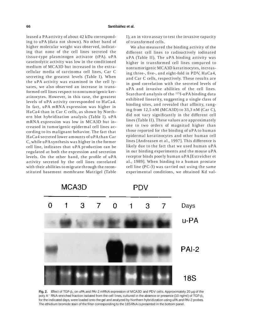
Fig. 2. Effect of TGF-b1 on uPA and PAI-2 mRNA expression of MCA3D and PDV cells. Approximately 20 µg of thepoly A1 RNA-enriched fraction isolated from the cell lines, cultured in the absence or presence (10 ng/ml) of TGF-b1
for the indicated days, were loaded onto the gel and analyzed by Northern hybridization using uPA and PAI-2 probes.The ethidium bromide stain of the filter corresponding to the 18S RNA is presented in the bottom panel.
66 Santibanez et al.

ues of about 0.6 nM similar to that reported inthe literature (not shown).
TGF-b1 Enhances uPA Expression, Secretion, andBinding Activity of Transformed PDV But Not of
Nontumorigenic MCA3D Keratinocytes
TGF-b1 affects differentially the morphologi-cal and growth properties of MCA3D and PDVcultures. MCA3D cells are growth arrested inthe presence of TGF-b1 while PDV continuesgrowing, although at a slower rate [Haddow etal., 1991; Caulın et al., 1995]. However, PDV-treated cells are dispersed and their membraneruffling activities increased. TGF-b1-induceddispersion of PDV cells is visible during thefirst week of treatment. After a longer exposure(2–3 weeks), PDV elicits an epithelial-fibroblas-tic conversion while MCA3D cells die [Caulın etal., 1995].
We, therefore, analyzed the effects of TGF-b1
on uPA expression and secretion in both celllines. Treatment of PDV cells with TGF-b1 for24 h enhanced PA secreted activity in a dose-dependent manner, as determined by a radialcaseinolytic assay (Fig. 1A, insert). This PAactivity corresponded to uPA as ascertained byzimography (data not shown). The greatest levelwas reached at 5 ng/ml, and higher concentra-tions (10 ng/ml) did not further increase uPAsecretion. Treatment of MCA3D cells with 10ng/ml of TGF-b1 for 24 h also increased (up tothree-fold) uPA secreted activity. However, af-ter longer treatments (2 to 7 days), uPA activitydropped to the basal level. In contrast, the uPAactivity secreted by PDV cells increased progres-sively along the time of treatment, reaching aplateau at about 3 days (Fig. 1A). At this time,uPA secreted activity was enhanced two-foldrespect to the basal level. Since the phenotypicalterations induced by TGF-b1 in PDV werereversible upon withdrawal of the factor fromthe cultures [Caulın et al., 1995], we studiedthe reversibility of uPA induction. As shown inFig. 1B, removal of the growth factor from PDVcultures after 3 and 7 days of treatment re-stored uPA activity to basal values.
Northern blot hybridization analysis demon-strated that these effects occurred at the mRNAlevel (Fig. 2). uPA mRNA expression was in-duced in MCA3D after 1 day of exposure toTGF-b1, but declined after longer treatment.Nevertheless, in PDV, uPA mRNA expressionenhanced at 1 and 3 days and still was highafter 7 days of treatment (Fig. 2). Rehybridiza-
tion of the filter with a probe for the uPA inhibi-tor PAI-2 showed that PDV cells expressed atremendous amount of PAI-2 message. PAI-2expression decreased after 1 and 3 days of treat-ment, although raised again at 7 days. Thesame effect was observed in MCA3D, althoughthe PAI-2 mRNA level was strikingly lower inthis cell line. However, TGF-b1 enhanced inPDV cells a PAI secreted activity, with a peak at3 days and a slight decline after 7 days oftreatment (Fig. 3A). This PAI activity corre-sponded to PAI-1, as shown by Northern experi-ments, and none effect was observed in MCA3Dcells (results not shown), suggesting that thiscell line does not respond to TGF-b1 for PAI-1induction. To study this further, MCA3D andPDV cells were transiently transfected with thep3TPlux construct, a TGF-b-responsive lucifer-ase reporter gene driven by the PAI-1 promoter[Wrana et al., 1992]. As shown in Figure 3B,TGF-b1 increased twice the luciferase basal ac-
Fig. 3. Effects of TGF-b1 on PAI-1 expression. A: Aliquots ofconditioned media of PDV cultures, untreated or treated withTGF-b1 (10 ng/ml) for the indicated days, were subjected toreverse casein zymography as described in Materials and Meth-ods. The conditioned media contained PAI activities migratingat about 45 kDa. B: Induction of PAI-1 promoter activity byTGF-b1 in MCA3D and PDV cells. Cells were transiently trans-fected with the p3TPlux reporter construct and assayed forluciferase activity after treatment with TGF-b1 for 24 h. Each barrepresents the mean of duplicate luciferase assays corrected fortransfection efficiency as indicated in Materials and Methods.The graphic shows a representative experiment of three.
uPA and TGF-b1-Induced Invasiveness of Keratinocytes 67

tivity of PDV but it was unable to activate thePAI-1 gene promoter in MCA3D cells.
On the other hand, binding activity analysisof 125I-uPA to untreated and TGF-b1-treatedMCA3D and PDV cell cultures showed that thegrowth factor enhanced the uPA binding sites ofPDV while did not alter the binding activity ofMCA3D (Table II).
Inhibition of uPA Binding to the Surface of PDVCells Reduces TGF-b1-Induced Motility and
Invasiveness
The effect of TGF-b1 on the invasive behav-iour of MCA3D and PDV cell lines was studiedby analyzing the ability of cells to migratethrough Matrigel. As expected, TGF-b1 had noeffect on the invasive capacity of MCA3D (datanot shown), but increased invasion of PDV cellsup to two-fold after 3 days of treatment (Fig. 4).To study whether TGF-b1-induced invasioncould be inhibited by interfering the binding ofuPA to the surface of PDV cells, we synthesizedtwo peptides comprising amino acid residues
41-54 and 44-63 of the EGF-like domain ofmouse uPA. This fragment of the uPA moleculehas been shown to be a potent inhibitor of thebinding of uPA to its high affinity receptor [Minet al., 1996]. Addition of the peptides (150 µg/ml) to the media during the Matrigel assaymarkedly inhibited (50%, P41-54 and 57%, P44-63) TGF-b1-induced invasiveness while a mix-ture of unrelated peptides obtained by trypsindigestion of gelatin, used at the same concentra-tion, had no significant effect (Fig. 4).
Previous experiments had demonstrated thatboth peptides inhibited the binding of uPA tothe surface of PDV keratinocytes, as ascer-tained by immunofluorescence experiments. In-direct immunofluorescence analysis, using ananti-uPA monoclonal antibody, on paraformal-dehyde-fixed nonpermeabilized PDV cultures,revealed an overall increase in the intensity offluorescence on cells treated with TGF-b1 for 3days (Fig. 5, compare a with b,c). uPA appearedlocated as clusters in certain regions of theplasma membrane, some of them resembling
Fig. 4. Stimulation by TGF-b1 of the invasive capacity of PDV cells through Matrigel and inhibition of PDV invasionby uPA synthetic peptides. Cells (2 3 105) seeded in the upper compartment of Transwell chambers were incubated inthe absence (C) and in the presence of TGF-b1 (10 ng/ml) for 3 days. P41-54, P44-63, and a mixture of trypsin-digestedgelatin peptides, used as a control, were added at 150 µg/ml together with TGF-b1. The percentage of migrated cellswas determined as indicated in Materials and Methods. Each bar represents the mean (6 SD) of triplicates. Similarresults were obtained in two independent experiments.
68 Santibanez et al.

membrane protrusions (Fig. 5b,c). The inten-sity of anti-uPA immunofluorescence stainingof TGF-b1-treated cells was highly decreasedwhen the peptide P44-63 was added to theculture for the last 16 or 24 h of treatment (Fig.5d,e). Similar results were obtained with P41-54(not shown) while control peptides had no ma-jor effect (Fig. 5c).
Moreover, TGF-b1 stimulated the motility ofPDV cells in an in vitro colonization assay (Fig.6). PDV cells cultured in standard mediumwere not able to cover the wound produced 24 hbefore, while PDV cells cultured in the presenceof TGF-b1 invaded the wound completely. Thiseffect was not due to enhanced proliferationsince TGF-b1 slows down the rate of growth ofPDV cells [Haddow et al., 1991; Caulın et al.,1995]. Addition of amino-terminal uPA peptidesstrongly inhibited (about 90% P44-63, 75% P41-54) TGF-b1-induced recolonization of the woundarea (Fig. 6). Peptides added in the absence ofTGF-b1 had no significant effect on basal cellinvasion and motility (not shown).
DISCUSSION
TGF-b1 modulates the epithelial phenotypeof transformed epidermal keratinocytes. Thisphenotypic modulation involves disruption of
cell-cell interactions and stimulation of migra-tion/scattering of the cells leading to the acqui-sition of fibroblast-like characteristics [Caulınet al., 1995]. The TGF-b1-induced epithelial-fibroblastic conversion is linked to progressionfrom a well differentiated SCC-type of tumor tohighly anaplastic SpCCs [Caulın et al., 1995;Cui et al., 1996]. A role for TGF-b1 as a modula-tor of the epithelial phenotype and/or invasive-ness of tumor cells has also been observed inother systems such as mammary [Oft et al.,1996] and colon [Huang et al., 1995] trans-formed cell lines. Cultured epidermal keratino-cytes that are growth arrested by TGF-b1 arenot responsive for this phenotypic conversionbut are induced to terminal differentiation andcell death [Mansbridge and Hanawalt, 1988;Caulın et al., 1995], a fact likely related to theobservation of TGF-b1 acting as a suppressor oftumor formation in early stages of carcinogen-esis [Glick et al., 1994; Cui et al., 1996]. One ofthe mechanisms by which TGF-b1 could modu-late cell migration and invasion is through re-modeling the ECM by regulating pericellularproteolysis [Lahio and Keski-Oja, 1992]. In thiswork, we show that i) increased expression,secretion and binding activity of uPA are associ-ated to a more malignant and undifferentiated
Fig. 5. Immunofluorescent detection of uPA in nonpermeabilized PDV cells untreated (a) or treated with TGF-b1 for3 days (b). P44-63 (150 µg/ml) added to TGF-b1-treated cultures for the last 16 h (d) or 24 h (e) reducedimmunofluorescence intensity, while the mixture of control peptides added for the last 24 h (c) had no significanteffect. Original magnifications: 3630.
uPA and TGF-b1-Induced Invasiveness of Keratinocytes 69

tumoral phenotype of skin carcinoma cell lines;ii) TGF-b1 stimulates uPA expression, secretionand binding activity in transformed keratino-cytes concomitantly to the induction of a migra-tory and invasive phenotype; and iii) syntheticpeptides antagonizing the binding of secreteduPA to the cell surface inhibit TGF-b1-inducedmigration and invasiveness.
Transformed PDV, but not nontumorigenicMCA3D keratinocytes, responded to TGF-b1 bya sustained increase in uPA expression/secre-tion and binding activity that correlated withenhanced invasion through Matrigel (Figs. 1, 2,and 4, Table II). The effect on uPA productionwas dose-dependent and required the continu-ous presence of the growth factor (Fig. 1).TGF-b1 also enhanced the expression of theinhibitor PAI-1 whereas that of PAI-2 was de-creased (Figs. 2 and 3). Interestingly, PAI-2 hasbeen found to inhibit invasion of tumor cells[Bruckner et al., 1992; Stahl and Mueller, 1994],while coexpression of uPA, its high affinity re-ceptor uPAR and PAI-1 appears to be necessary
for optimum invasiveness of cancer cells throughMatrigel [Liu et al., 1995]. On the other hand,uPA induction by TGF-b1 in PDV cultures coin-cided with cell dispersion [Caulın et al., 1995]and increased motility (Fig. 6), and uPA proteinappeared to be associated with defined regionsof plasma membrane protrusions on the surfaceof cells (Fig. 5). uPA and uPAR have been colo-calized with integrins in focal contacts at theleading edge of migrating cells [Pollanen et al.,1987; Wei et al., 1996], and a role for the uPAsystem (uPA, uPAR, PAI-1) in coordinating celladhesion and migration has gained consider-able support during the last years [Stefanssonand Lawrence, 1996; Kjoller et al., 1997; Planuset al., 1997].
Evidence for a functional role of uPA in TGF-b1-induced motility and invasiveness is ob-tained by the demonstration that synthetic pep-tides containing the EGF-like domain of theamino-terminal region of mouse uPA inhibitTGF-b1-induced migration through Matrigel(Fig. 4) and recolonization of an in vitro wound
Fig. 6. Effect of TGF-b1 on the motility of PDV cells andinhibition of TGF-b1-induced motility by uPA synthetic pep-tides. Areas free of cells (wounds) made in subconfluent PDVcultures were examined after 24 h of cultures in the absence orpresence of TGF-b1 (10 ng/ml). Where indicated, peptidesP44-63, P41-54, or the control mixture, were added at 150
µg/ml. Bottom right: Diagram showing quantification of the cellmigration assay. Cells invading the wound area were counted inthree different fields as those showed in the micrographs. Meanvalues (6 SD) are the percentages of the number of migratedcells in TGF-b1-treated cultures plus control peptides. Originalmagnifications: 3100.
70 Santibanez et al.

(Fig. 6). Those peptides also inhibited the bind-ing of uPA to the cell surface, as shown byreduced anti-uPA immunofluorescence inten-sity in nonpermeabilized cells, and it has beenreported by others that a purified EGF-likedomain of murine uPA antagonizes uPA bind-ing to its receptor [Min et al., 1996]. However,although we found an almost complete inhibi-tion (75–90%) of TGF-b1-induced motility in thewound assay, we were unable to block TGF-b1-induced invasion through Matrigel (reductionwas about 55%) by interfering uPA binding. Onthe other hand, peptides added in the absenceof TGF-b1 did not significantly affect the basalinvasive ability of transformed PDV cells. Theseresults suggest that other events are also in-volved in TGF-b1-stimulated invasiveness. Inthis respect, we have found that TGF-b1 en-hances MMP-9 (gelatinase B), but not MMP-2(gelatinase A), metalloproteinase production inPDV cells (unpublished results), and increasedexpression of MMP-9 in transformed keratino-cytes has been associated to the loss of cell-cellcontacts mediated by E-cadherin and the induc-tion of an invasive and metastatic phenotype[Llorens et al., 1998].
uPA mediates migration/invasion stimulatedalso by other growth factors such as fibroblastgrowth factor on endothelial cells [Odekon etal., 1992] and hepatocyte growth factor/scatterfactor (HGF/SF) on a sarcoma cell line [Jefferset al., 1996]. Therefore, induction of uPA couldbe a general mechanism for growth factors thatpromote cell motility. On the other hand, uPAconverts inactive forms of HGF/SF and TGF-binto their active proteins [Naldini et al., 1992;Odekon et al., 1994], and a feed back loop ofproHGF/SF activation by uPA which in turninduces more uPA has been proposed in sar-coma cells [Jeffers et al., 1996]. A similar mech-anism could be operating in epidermal carcino-genesis. Transformed keratinocytes expressincreased levels of TGF-b1 mRNA and protein[Glick et al., 1991; Frontelo et al., 1998]. LatentTGF-b1 secreted by these cells could be acti-vated by uPA and active TGF-b1 would gener-ate more uPA.
In conclusion, our findings demonstrate thatTGF-b1 stimulates cell migration and invasive-ness in transformed keratinocytes concomi-tantly to increased expression, secretion andbinding activity of uPA. This response precedesthe full conversion to a fibroblast-like pheno-type that takes place after long-term treatment
with the growth factor [Caulın et al., 1995].Since remodeling of the ECM has been shownto influence the cell phenotype and promoteepithelial-mesenchymal transitions [Hay, 1993;Roskelley et al., 1995], we also suggest thatincreased uPA secretion and activity, togetherwith other changes in ECM proteins and pro-teinases induced by TGF-b1, contribute to theconversion from SCCs to SpCCs.
ACKNOWLEDGMENTS
We thank Dr. F. Castellino for his useful giftof SAM-3 monoclonal antibody, and Dr. A. Canofor critical reading of the manuscript. J.F.S. wasthe recipient of a Mutis fellowship from the Agen-cia Espanola de Cooperacion Iberoamericana.
REFERENCES
Andreasen PA, Kjoller L, Christensen L, Duffy MJ. 1997.The urokinase-type plasminogen ativator system in can-cer metastasis: a review. Int J Cancer 72:1–22.
Belin D, Vasalli JD, Combepine C, Godeau F, Nagamine Y,Reich E, Kocher HP, Duvoisin RM. 1985. Cloning, nucleo-tide sequence and expression of cDNAs encoding mouseurokinase-type plasminogen activator. Eur J Biochem148:225–232.
Belin D, Wohlwend A, Schleuning W-D, Kruithof EKO,Vassalli J-D. 1989. Facultative polypeptide translocationallows a single mRNA to encode the secreted and cyto-solic forms of plasminogen activators inhibitor 2. EMBOJ 8:3287–3294.
Boyer B, Tucker GC, Valles AM, Franke WW, Thiery JP.1989. Rearrengements of desmosomal and cytoskeletalproteins during the transition from epithelial to fibroblas-toid organization in cultured rat bladder carcinoma cells.J Cell Biol 109:1495–1509.
Bruckner A, Filderman AE, Kirchheimer JC, Binder BR,Remold HG. 1992. Endogenous receptor-bound uroki-nase mediates tissue invasion of the human lung carci-noma cell line A549 and Calu-1. Cancer Res 52:3042–3047.
Buchmann A, Ruggeri B, Klein-Szanto AJP, Balmain A.1991. Progression of squamous carcinoma cells to spindlecarcinomas of mouse skin is associated with an imbal-ance of H-ras alleles on chromosome 7. Cancer Res 51:4097–4101.
Busso N, Masur SK, Lazega D, Waxman S, Ossowski L.1994. Induction of cell migration by pro-urokinase bind-ing to its receptor: possible mechanism for signal trans-duction in human epithelial cells. J Cell Biol 126:259–270.
Caulın C, Scholl FG, Frontelo P, Gamallo C, Quintanilla M.1995. Chronic exposure of cultured transformed mouseepidermal cells to transforming growth factor-b1 inducesan epithelial-mesenchymal transdifferentiation and aspindle tumoral phenotype. Cell Growth Differ 6:1027–1035.
Caulın C, Lopez-Barcons Ll, Gonzalez-Garrigues M, Na-varro P, Lozano E, Rodrigo I, Gamallo C, Cano A, FabraA, Quintanilla M. 1996. Suppression of the metastaticphenotype of a mouse skin carcinoma cell line indepen-dent of E-cadherin expression and correlated with re-duced levels of Ha-ras oncogene products. Mol Carc 15:104–114.
uPA and TGF-b1-Induced Invasiveness of Keratinocytes 71

Cui W, Fowlis DJ, Bryson S, Duffie E, Ireland H, BalmainA, Akhurst RJ. 1996. TGF-b1 inhibits the formation ofbenign skin tumors, but enhances progression to invasivespindle carcinomas in transgenic mice. Cell 86: 531–542.
Degen SJ, Heckel JL, Reich E, Degen JL. 1987. The murineurokinase-type plasminogen activator gene. Biochemis-try 26:8270–8279.
Dıaz-Guerra M, Haddow S, Bauluz C, Jorcano JL, Cano A,Balmain A, Quintanilla M. 1992. Expression of simpleepithelial cytokeratins in mouse epidermal keratinocytesharboring Harvey ras gene alterations. Cancer Res 52:680–687.
Erickson L, Lawrence D, Loskutoff J. 1984. Reverse fibrinautography: a method and partially characterized prote-ase inhibitors after dodecyl sulphate-polyacrilamide gelelectrophoresis. Anal Biochem 137:456–463.
Estreicher A., Wohlwend A, Belin D, Schlenning WD, Va-salli J-D. 1989. Characterization of the cellular bindingsite for the urokinase-type plasminogen activator. J BiolChem 264:1180–1189.
Frontelo P, Gonzalez-Garrigues M, Vilaro S, Gamallo C,Fabra A, Quintanilla M. 1998. Transforming growth fac-tor b1 induces squamous carcinoma cell variants withincreased metastatic abilities and a disorganized cytoskel-eton. Exp Cell Res 244:420–432.
Glick AB, Sporn MB, Yuspa SH. 1991. Altered regulation ofTGF-b1 and TGF-a in primary keratinocytes and papillo-mas expressing v-Ha-ras. Mol Carc 4:210–219.
Glick AB, Kulkarni AB, Tennenbaum T, Hennings H,Flanders KC, O’Reilly M, Sporn MB, Karlsson S, YuspaSH. 1993. Loss of expression of transforming growthfactor b in skin and skin tumors is associated withhyperproliferation and a high risk for malignant cover-sion. Proc Natl Acad Sci USA 90:6076–6080.
Glick AB, Lee MM, Darwiche N, Kulkarni AB, Karlsson S,Yuspa SH. 1994. Targeted deletion of the TGF-b1 genecauses rapid progression to squamous cell carcinoma.Genes Dev 8:2429–2440.
Haddow S, Fowlis DJ, Parkinson K, Akhurst RJ, BalmainA. 1991. Loss of growth control by TGF-b occurs at a latestage of mouse skin carcinogenesis and is independent ofras gene activation. Oncogene 6:1465–1570.
Hay ED. 1993. Extracellular matrix alters epithelial differ-entiation. Curr Opin Cell Biol 5:1029–1035.
Huang F, Newman E, Theodorescu D, Kerbel RS, FriedmanE. 1995. Transforming growth factor b1 (TGFb1) is anautocrine positive regulator of colon carcinoma U9 cellsin vivo as shown by transfection of a TGFb1 antisenseexpression plasmid. Cell Growth Differ 6:1635–1642.
Hunter W, Greenwood FC. 1962. Preparation of iodine-131labeled human growth hormone of high specific activity.Nature 194:495–496.
Jeffers M, Rong S, Vande Woude GF. 1996. Enhanced tu-morigenicity and invasion-metastasis by hepatocytegrowth factor/scatter factor-met signalling in human cellsconcomitant with induction of the urokinase proteolysisnetwork. Mol Cell Biol 16:1115–1125.
Keski-Oja J, Blasi F, Leof EB, Moses HL. 1988. Regulationof the synthesis and activity of urokinase plasminogenactivator in A549 human lung carcinoma cells by trans-forming growth factor-b. J Cell Biol 106:451–459.
Kjoller L, Kanse SM, Kirkegaard T, Rodenburg KW, RonneE, Goodman SL, Preisner KT, Ossovski L, Andreasen PA.1997. Plasminogen activator inhibitor-1 represses inte-grin- and vitronectin-mediated cell migration indepen-dently of its function as an inhibitor of plasminogenactivation. Exp Cell Res 232:420–429.
Lahio M, Keski-Oja J. 1992. Transforming growth factor-bas regulators of cellular growth and phenotype. Crit RevOncog 3:1–26.
Liu G, Shuman MA, Cohen RL. 1995. Co-expression ofurokinase, urokinase receptor and PAI-1 is necessary foroptimum invasiveness of cultured lung cancer cells. Int JCancer 60:501–506.
Llorens A, Rodrigo I, Lopez-Barcons L, Gonzalez-GarriguesM, Lozano E, Vinyals A, Quintanilla M, Cano A, Fabra A.1998. Down-regulation of E-cadherin in mouse skin carci-noma cells enhances a migratory and invasive phenotypelinked to matrix metalloproteinase-9 gelatinase expres-sion. Lab Invest 78:1131–1142.
Mansbridge JN, Hanawalt PC. 1988. Role of transforminggrowth factor beta in the maturation of human epidermalkeratinocytes. J Inv Dermatol 90:336–341.
Massague J. 1990. The transforming growth factor-b fam-ily. Annu Rev Cell Biol 6:597–641.
Min HY, Doyle LV, Vitt CR, Zandonella CL, Stratton-Thomas JR, Shuman MA, Rosenberg S. 1996. Urokinasereceptor antagonists inhibit angiogenesis and primarytumor growth in syngeneic mice. Cancer Res 56:2428–2433.
Naldini L, Tamagnone L, Vigna E, Sachs M, Hartmann G,Birchmeier V, Daikuhara Y, Tsubouchi H, Blasi F, Como-glio M. 1992. Extracellular proteolytic cleavage by uroki-nase is required for activation of hepatocyte growth factor/scatter factor. EMBO J 11:4825–4833.
Odekon LE, Sato Y, Rifkin DB. 1992. Urokinase-type plas-minogen activator mediates basic fibroblast growth factor-induced bovine endothelial cell migration independent ofits proteolytic activity. J Cell Physiol 150:258–263.
Odekon LE, Blasi F, Rifkin DB. 1994. Requirement forreceptor-bound urokinase in plasmin-dependent cellularconversion of latent TGF-b to TGF-b. J Cell Physiol158:398–407.
Oft, M, Peli J, Rudaz C, Schwartz H, Beug H, Reichmann E.1996. TGF-b1 and Ha-ras collaborate in modulating thephenotypic plasticity and invasiveness of epithelial tu-mor cells. Genes Dev 10:2462–2477.
Planus E, Barlovatz-Meimon G, Rogers RA, Bonavaud S,Ingber DE, Wang N. 1997. Binding of urokinase to plas-minogen activator inhibitor type-1 mediates cell adhe-sion and spreading. J Cell Sci 110:1091–1098.
Pollanen J, Saksela O, Salonen E-M, Andreasen P, NielsenL, Dano K, Vaheri A. 1987. Distinct localizations of uroki-nase-type plasminogen activator and its type 1 inhibitorunder cultured human fibroblasts and sarcoma cells. JCell Biol 104:1085–1096.
Portella G, Liddell J, Crombie R, Haddow S, Clarke M,Stoler AB, Balmain A. 1994–95. Molecular mechanismsof invasion and metastasis during mouse skin tumorprogression. Invas Metast 14:7–16.
Roskelley CD, Srebrow A, Bissell MJ. 1995. A hierarchy ofECM-mediated signalling regulates tissue-specific geneexpression. Curr Opin Cell Biol 7:736–747.
72 Santibanez et al.

Santibanez JF, Maccioni RB, Martinez J. 1995. The secre-tion of urokinase-like plasminogen activator is inhibitedby microtubule-interacting drugs. Cell Biochem Funct13:217–225.
Stahl, A, Mueller BM. 1994. Binding of urokinase toits receptor promotes migration and invasion of hu-man melanoma cells in vitro. Cancer Res 54:3066–3071.
Stefansson S, Lawrence DA. 1996. The serpin PAI-1 inhib-its cell migration by blocking integrin avb3 binding tovitronectin. Nature 383:441–443.
Stetler-Stevenson WG, Aznavoorian S, Liotta LA. 1993.Tumor cell interactions with the extracellular matrixduring invasion and metastasis. Annu Rev Cell Biol9:541–573.
Wei Y, Lukashev M, Simon DI, Bodary SC, Rosenberg S, DoyleMV, Chapman HA. 1996. Regulation of integrin function bythe urokinase receptor. Science 273:1551–1555.
Wrana JL, Attisano L, Carcamo J, Zentella A, Doody J,Lahio M, Wang X-F, Massague J. 1992. TGFb signalsthrough a heteromeric protein kinase receptor complex.Cell 71:1003–1014.
uPA and TGF-b1-Induced Invasiveness of Keratinocytes 73
Copyright © 2022 FDOKUMEN