6.-Human-Security-Bangladesh.pdf - Refugee and Migratory ...
Upload
khangminh22Category
view
9download
0
Migratory routes, domesticated birds and cercarial dermatitis:the distribution of Trichobilharzia franki in Northern Iran
Keyhan Ashrafi1, Meysam Sharifdini1, Abbas Darjani2, and Sara V. Brant3,*
1 Department of Medical Parasitology and Mycology, School of Medicine, Guilan University of Medical Sciences, Rasht 41996-13776, Iran2 Skin Research Center, Department of Dermatology, Razi Hospital, Guilan University of Medical Sciences, Rasht 41996-13776, Iran3 Museum of Southwestern Biology, Division of Parasites, Department of Biology, University of New Mexico, 1 University of NewMexico MSC03 2020, Albuquerque, New Mexico 87131, USA
Received 11 September 2020, Accepted 11 December 2020, Published online 12 January 2021
Abstract – Background: One of the major migration routes for birds going between Europe and Asia is the BlackSea-Mediterranean route that converges on the Volga Delta, continuing into the area of the Caspian Sea. Cercarialdermatitis is a disorder in humans caused by schistosome trematodes that use aquatic birds and snails as hosts andis prevalent in areas of aquaculture in Northern Iran. Before the disorder can be addressed, it is necessary to determinethe etiological agents and their host species. This study aimed to document whether domestic mallards are reservoirhosts and if so, to characterize the species of schistosomes. Previous work has shown that domestic mallards arereservoir hosts for a nasal schistosome. Results: In 32 of 45 domestic mallards (Anas platyrhynchos domesticus)(71.1%), the schistosome Trichobilharzia franki, previously reported only from Europe, was found in visceralveins. Morphological and molecular phylogenetic analysis confirmed the species designation. These findingsextend the range of T. franki from Europe to Eurasia. Conclusion: The occurrence of cercarial dermatitis in Iran ishigh in areas of aquaculture. Previous studies in the area have shown that domestic mallards are reservoir hosts ofT. regenti, a nasal schistosome and T. franki, as shown in this study. The genetic results support the conclusion thatpopulations of T. franki from Iran are not differentiated from populations in Europe. Therefore, the schistosomes aredistributed with their migratory duck hosts, maintaining the gene flow across populations with compatible snail hostsin Iran.
Key words: Trichobilharzia franki, Iran, Anas platyrhynchos domesticus, Cercarial dermatitis.
Resume – Routes migratoires, oiseaux domestiques et dermatite cercarienne : répartition de Trichobilharziafranki dans le nord de l’Iran. Contexte : L’une des principales voies de migration des oiseaux à destination et enprovenance de l’Europe et de l’Asie est la route mer Noire-Méditerranée qui converge vers le delta de la Volga etse poursuit dans la région de la mer Caspienne. La dermatite cercarienne est une affection causée chez l’hommepar des trématodes Schistosomatidae utilisant des oiseaux aquatiques et des mollusques comme hôtes, qui estrépandue dans les zones d’aquaculture du nord de l’Iran. Avant de pouvoir lutter contre cette parasitose, il estnécessaire de connaître les agents étiologiques et leurs espèces hôtes. Ce travail vise à documenter si les canardsdomestiques sont des hôtes réservoirs et si oui, à caractériser les espèces de schistosomes. Des travaux antérieursont montré que les canards domestiques sont des hôtes réservoirs pour un schistosome nasal. Résultats : Chez 32de 45 canards domestiques (Anas platyrhynchos domesticus) (71,1 %), le schistosome Trichobilharzia franki,précédemment signalé uniquement en Europe, a été trouvé dans les veines viscérales. L’identification de l’espèce aété vérifiée par une analyse morphologique et phylogénétique moléculaire. Cela étend l’aire de répartition del’Europe à l’Eurasie. Conclusion : La dermatite cercarienne est répandue dans les zones d’aquaculture en Iran. Destravaux antérieurs dans la région ont montré que les canards domestiques sont des hôtes réservoirs de T. regenti, unschistosome nasal et de T. franki, comme le montre cette étude. Les résultats génétiques soutiennent queles populations de T. franki d’Iran ne sont pas différenciées des populations d’Europe. Par conséquent, lesschistosomes se dispersent avec leur hôte canard lors de la migration, maintenant un flux génétique entre lespopulations avec des mollusques hôtes compatibles en Iran.
*Corresponding author: [email protected]
Parasite 28, 4 (2021)�K. Ashrafi et al., published by EDP Sciences, 2021https://doi.org/10.1051/parasite/2020073
Available online at:www.parasite-journal.org
This is an Open Access article distributed under the terms of the Creative Commons Attribution License (https://creativecommons.org/licenses/by/4.0),which permits unrestricted use, distribution, and reproduction in any medium, provided the original work is properly cited.
OPEN ACCESSRESEARCH ARTICLE
Introduction
One of the major migration routes for birds going betweenEurope and Asia is the Black Sea-Mediterranean route thatconverges on the Volga Delta, continuing into the area of theCaspian Sea. Birds along this route migrate twice a year, nest,or stay for the winter. Therefore, the surrounding areas arevisited year-round by migratory birds, particularly waterfowl.Aquaculture is a common occupation in many areas (coveringfive countries) around the Caspian Sea, but this type of farmingis often associated with parasitic diseases [18, 47]. The waterthat is used for plants, ducks, fish, or crustaceans and otherinvertebrates is often inhabited by aquatic gastropods that canhost a myriad of trematodes. Both domestic and wild mammalsand birds use the water, as do humans, creating many opportu-nities for life cycles of several species of parasites to establish.One of these life cycles can result in a disorder called cercarialdermatitis (CD) or swimmer’s itch [41], caused by digenetictrematodes in the family Schistosomatidae. These worms havea two-host life cycle where adult worms live in a mammalian oravian host, and the intermediate host is an aquatic gastropod.The emerging free-swimming larval stages (cercariae) fromthe gastropod penetrate the skin of humans causing an allergicreaction that can last up to a week [43]. In an aquacultureenvironment, this involves the gastropods that naturally estab-lish in water and domestic ducks, and sometimes migratorybirds. Discovering the species of schistosome and their hostdiversity along a migratory route is a foundational step to initi-ating targeted control programs for CD. It is more manageableto control one duck species in the life cycle than all duckspecies, so more specific knowledge facilitates control or miti-gation of the disease.
It is only within the last decade that there has been aconcerted effort to study the epidemiology of CD in regionsof the Middle East, as cases, particularly in rice fields, aregaining more attention [9, 26, 27, 30, 31, 35, 39, 51–53, 71].Much of the work on CD in this area has been conducted inIran, documenting the neglected status of the disease andnarrowing down the critical hosts and worm species for trans-mission. A summary of research in Iran thus far shows thatthere are at least three common species of TrichobilharziaSkrjabin and Zakharov, 1920 that have been found in ducksand snails in Northern Iran [6, 26, 31, 51, 53, 71]. Recently,at least one species has been found in the mesenteric veins(9, 30) and a second species in the nasal tissue of its duck hosts[6, 26], particularly Spatula clypeata (Linnaeus, 1758) andAnas platyrhynchos (Linnaeus, 1758). Avian schistosomes havebeen recovered from the snail hosts Radix gedrosiana(Annandale & Prashad, 1919) and Radix auricularia (Linnaeus,1758), and two species of Trichobilharzia from the mesentericveins of their duck hosts [71]. The molecular identity of theavian schistosome from R. gedrosiana has not yet beenconfirmed [9, 27, 71].
Recent studies have uncovered a more detailed patternof relationships among species of Trichobilharzia across abroader geographic range that encompasses the avian hostmigration routes [6, 25, 26, 37, 38, 61]. These patterns capture
genetic diversity in the worms that reflect the long distancestheir bird hosts move (e.g. [25]). Additionally, finding theseschistosomes in domestic or resident birds indicates that thesnail host species is available, or at least a susceptible snail host(often a congener). The work herein provides more data demon-strating the significant impact of host mobility and ecology onthe distribution and diversity of avian schistosomes [25]. Thiswork aims to continue the survey and documentation ofschistosomes in Iran, particularly in the rice-growing areas inthe north. Additionally, the role of domestic mallards Anasplatyrhynchos domesticus as reservoir hosts is furtherexamined.
Materials and methods
Study area
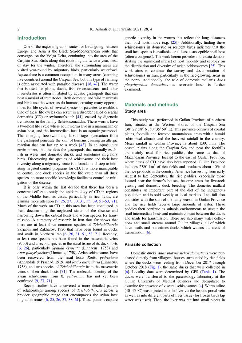
This study was performed in Guilan Province of northernIran, situated at the Western shores of the Caspian Sea(38� 280 5800 N, 50� 350 5900 E). This province consists of coastalplains, foothills and forested mountainous areas with a humidsubtropical climate and the heaviest rainfall in the country.Mean rainfall in Guilan Province is about 1500 mm. Thecoastal plains along the Caspian Sea and near the foothillsare mainly used for rice paddies, the same as that ofMazandaran Province, located to the east of Guilan Province,where cases of CD have also been reported. Guilan Provinceincludes 2380 km2 of rice fields and produces about 40% ofthe rice products in the country. After rice harvesting from earlyAugust to late September, the rice paddies, especially thoselocated near the farmer’s houses, become areas for livestockgrazing and domestic duck breeding. The domestic mallardconstitutes an important part of the diet of the indigenouspopulation and is sold weekly at local markets. Late fall alsocoincides with the start of the rainy season in Guilan Provinceand the rice fields receive large amounts of water. Thesepaddies then continue as suitable environments to sustain thesnail intermediate hosts and maintain contact between the ducksand snails for transmission. There are also many water collec-tions and small streams around Guilan villages, all of whichhave snails and sometimes ducks which widens the areas oftransmission [6].
Parasite collection
Domestic ducks Anas platyrhynchos domesticus were pur-chased directly from villagers’ houses surrounded by rice fieldswhere the ducks were feeding from December 2017 throughOctober 2018 (Fig. 1), the same ducks that were collected in[6]. Locality data were determined by GPS (Table 1). Theducks were transferred to the parasitology laboratory at theGuilan University of Medical Sciences and decapitated toexamine for presence of visceral schistosomes [4]. Warm saline(40–45 �C) was injected into the liver via the hepatic portal veinas well as into different parts of liver tissue (for frozen birds tapwater was used). Then, the liver was cut into small pieces in
2 K. Ashrafi et al.: Parasite 2021, 28, 4
saline and transferred to a series of different mesh size labora-tory sieves arranged from the largest to the smallest. The liverwas then slowly crushed by hand on the upper sieve, whilebeing washed using a trigger sprayer containing warm saline.This was done for each sieve size. The bottom sieve(106 lm) was slightly tilted and the remnants of the liver wash-ings were collected by a plastic pipette from the lower side ofthe sieve, and the same process was performed for 53 lmand 25 lm sieves. For microscopic examination, a smallpart of the collected materials was then transferred into a glassdish with clean saline solution to dilute the material andobtain a thin layer for examination under a dissection micro-scope for intact adults, fragments or eggs [4]. Some of the intactworms, fragments and eggs were transferred to the micro-tubes containing saline for rapid morphological studies (eggand adult measurements, their micrographs), and some trans-ferred to the microtubes containing 90% alcohol for molecularstudies. All procedures performed in studies involving animalswere in accordance with the ethical standards of the institutionor practice at which the studies were conducted. This study wasapproved by the Ethics Committee of the Guilan University ofMedical Sciences (IR.GUMS.REC.1398.109).
Morphological and genetic analyses
For morphological studies of adults and eggs, the intactworms, large fragments and eggs were transferred onto a glassslide, covered with a coverslip and measured (Tables 2, 3)under a microscope (Olympus BX50) equipped with a digitalcamera (TrueChrome Metrics, China) and Nomarski Piece(U-DICT, Olympus, Japan). The length and width of the eggswere measured, and the data analyzed in SPSS v. 22 (minimum,maximum, average, and SD). The remaining eggs, intact adultsand worm fragments, if any, were transferred to microtubescontaining 90% alcohol for molecular studies. Some of thecollected samples (full length worms, fragments and eggs) werealso kept in labeled microtubes in 90% alcohol in theDepartment of Parasitology and Mycology of the GuilanUniversity of Medical Sciences as a permanent museumvoucher. It is critical for the evolutionary characterization oforganisms to have a permanent museum voucher [33, 59, 68].
For the genetic studies, genomic DNA was extracted from90% ethanol-preserved worm fragments using a commercialkit (High Pure PCR Template Preparation Kit; Roche,Mannheim, Germany), according to the manufacturer’s
Figure 1. Map of Iran with Guilan province highlighted, showing collecting localities. Pink stars = provinces with positive ducks for both T.franki and T. regenti, and in some cases co-infections; Yellow stars = provinces with positive ducks for only T. franki; Blue stars = provinceswith positive ducks for only T. regenti; Circles without numbers = areas with negative ducks; and Circles with numbers = localities from whichworms were used for sequencing.
K. Ashrafi et al.: Parasite 2021, 28, 4 3
recommended protocol. Primers BD1 (50–GTCGTAACAAGG-TTTCCGTA–30) [12] and 4S (50–TCTAGATGCGTTCGAAR-TGTCGATG–30) [13] were used for amplification of a 1123 bpsequence of partial ITS1 nuclear rDNA. Also, Cox1_SchistoF(50–TCTTTRGATCATAAGCG–30) and Cox1_SchistoR (50–TAATGCATMGGAAAAAAACA–30) primers were employedto amplify a 1250 bp sequence of the partial mitochondrial cox1gene [48]. PCR reaction was performed in a 30 lL reactionmixture containing 15 lL of PCR mix including 1.25 U Taq
DNA polymerase, 200 lM of dNTPs and 1.5 mM MgCl2(2 � Master Mix RED Ampliqon, Denmark), 10 pmol of eachprimer, and 3 lL of DNA sample. The thermal PCR profiles forthe cox1 gene included an initial denaturation step at 94 �C for2 min followed by 35 cycles of denaturation at 94 �C for 30 s,annealing at 52 �C for 30 s and extension at 72 �C for 120 s,followed by a final extension step at 72 �C for 7 min. ThePCR conditions of ITS1 gene amplification consisted of initialdenaturation at 95 �C for 6 min, 30 cycles of 95 �C for 45 s,
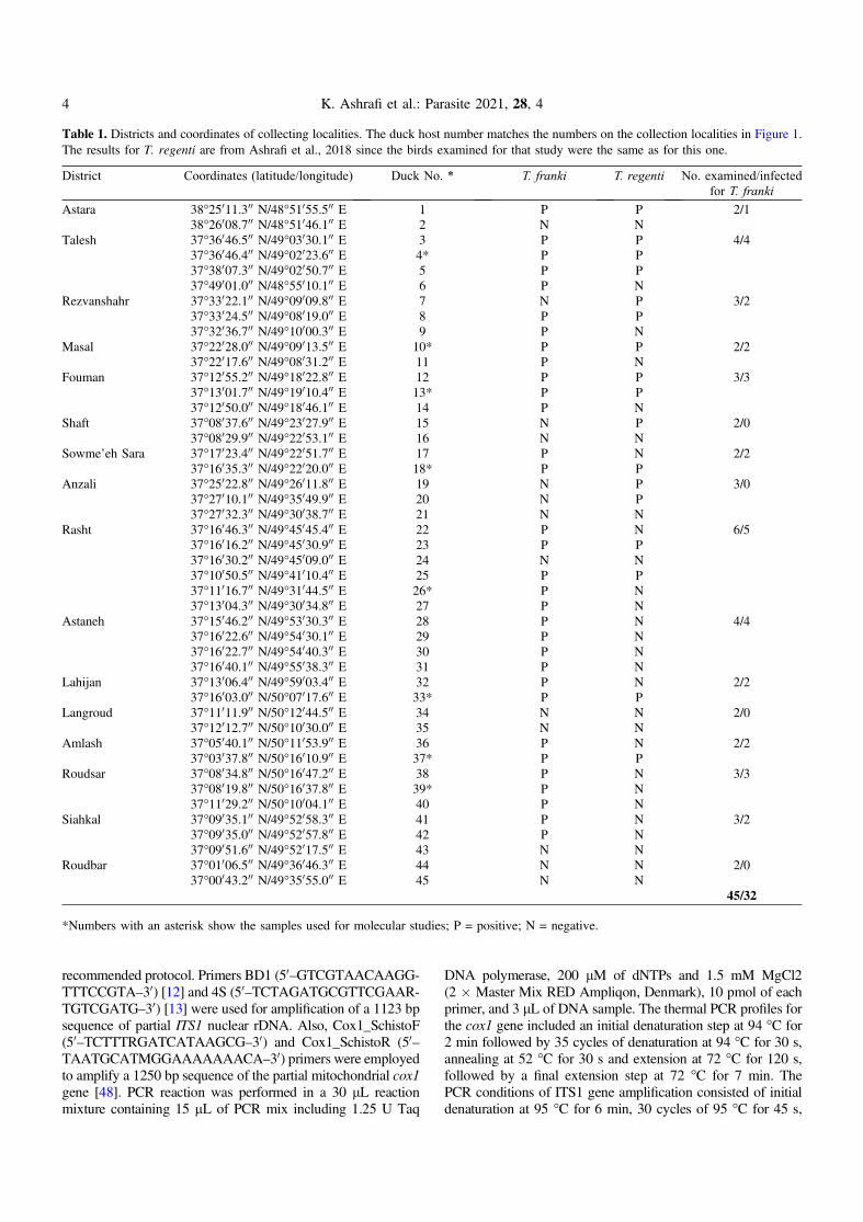
Table 1. Districts and coordinates of collecting localities. The duck host number matches the numbers on the collection localities in Figure 1.The results for T. regenti are from Ashrafi et al., 2018 since the birds examined for that study were the same as for this one.
District Coordinates (latitude/longitude) Duck No. * T. franki T. regenti No. examined/infectedfor T. franki
Astara 38�25011.300 N/48�51055.500 E 1 P P 2/138�26008.700 N/48�51046.100 E 2 N N
Talesh 37�36046.500 N/49�03030.100 E 3 P P 4/437�36046.400 N/49�02023.600 E 4* P P37�38007.300 N/49�02050.700 E 5 P P37�49001.000 N/48�55010.100 E 6 P N
Rezvanshahr 37�33022.100 N/49�09009.800 E 7 N P 3/237�33024.500 N/49�08019.000 E 8 P P37�32036.700 N/49�10000.300 E 9 P N
Masal 37�22028.000 N/49�09013.500 E 10* P P 2/237�22017.600 N/49�08031.200 E 11 P N
Fouman 37�12055.200 N/49�18022.800 E 12 P P 3/337�13001.700 N/49�19010.400 E 13* P P37�12050.000 N/49�18046.100 E 14 P N
Shaft 37�08037.600 N/49�23027.900 E 15 N P 2/037�08029.900 N/49�22053.100 E 16 N N
Sowme’eh Sara 37�17023.400 N/49�22051.700 E 17 P N 2/237�16035.300 N/49�22020.000 E 18* P P
Anzali 37�25022.800 N/49�26011.800 E 19 N P 3/037�27010.100 N/49�35049.900 E 20 N P37�27032.300 N/49�30038.700 E 21 N N
Rasht 37�16046.300 N/49�45045.400 E 22 P N 6/537�16016.200 N/49�45030.900 E 23 P P37�16030.200 N/49�45009.000 E 24 N N37�10050.500 N/49�41010.400 E 25 P P37�11016.700 N/49�31044.500 E 26* P N37�13004.300 N/49�30034.800 E 27 P N
Astaneh 37�15046.200 N/49�53030.300 E 28 P N 4/437�16022.600 N/49�54030.100 E 29 P N37�16022.700 N/49�54040.300 E 30 P N37�16040.100 N/49�55038.300 E 31 P N
Lahijan 37�13006.400 N/49�59003.400 E 32 P N 2/237�16003.000 N/50�07017.600 E 33* P P
Langroud 37�11011.900 N/50�12044.500 E 34 N N 2/037�12012.700 N/50�10030.000 E 35 N N
Amlash 37�05040.100 N/50�11053.900 E 36 P N 2/237�03037.800 N/50�16010.900 E 37* P P
Roudsar 37�08034.800 N/50�16047.200 E 38 P N 3/337�08019.800 N/50�16037.800 E 39* P N37�11029.200 N/50�10004.100 E 40 P N
Siahkal 37�09035.100 N/49�52058.300 E 41 P N 3/237�09035.000 N/49�52057.800 E 42 P N37�09051.600 N/49�52017.500 E 43 N N
Roudbar 37�01006.500 N/49�36046.300 E 44 N N 2/037�00043.200 N/49�35055.000 E 45 N N
45/32
*Numbers with an asterisk show the samples used for molecular studies; P = positive; N = negative.
4 K. Ashrafi et al.: Parasite 2021, 28, 4
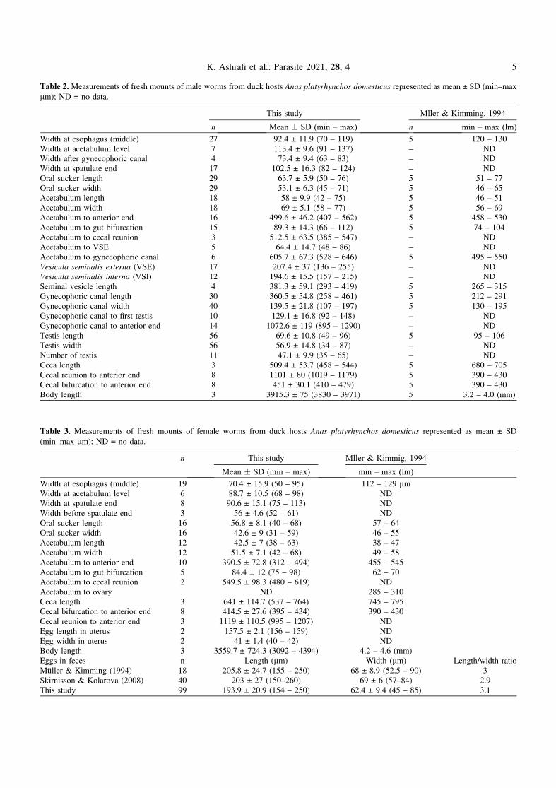
Table 2. Measurements of fresh mounts of male worms from duck hosts Anas platyrhynchos domesticus represented as mean ± SD (min–maxlm); ND = no data.
This study Mller & Kimming, 1994
n Mean � SD (min – max) n min – max (lm)
Width at esophagus (middle) 27 92.4 ± 11.9 (70 – 119) 5 120 – 130Width at acetabulum level 7 113.4 ± 9.6 (91 – 137) – NDWidth after gynecophoric canal 4 73.4 ± 9.4 (63 – 83) – NDWidth at spatulate end 17 102.5 ± 16.3 (82 – 124) – NDOral sucker length 29 63.7 ± 5.9 (50 – 76) 5 51 – 77Oral sucker width 29 53.1 ± 6.3 (45 – 71) 5 46 – 65Acetabulum length 18 58 ± 9.9 (42 – 75) 5 46 – 51Acetabulum width 18 69 ± 5.1 (58 – 77) 5 56 – 69Acetabulum to anterior end 16 499.6 ± 46.2 (407 – 562) 5 458 – 530Acetabulum to gut bifurcation 15 89.3 ± 14.3 (66 – 112) 5 74 – 104Acetabulum to cecal reunion 3 512.5 ± 63.5 (385 – 547) – NDAcetabulum to VSE 5 64.4 ± 14.7 (48 – 86) – NDAcetabulum to gynecophoric canal 6 605.7 ± 67.3 (528 – 646) 5 495 – 550Vesicula seminalis externa (VSE) 17 207.4 ± 37 (136 – 255) – NDVesicula seminalis interna (VSI) 12 194.6 ± 15.5 (157 – 215) – NDSeminal vesicle length 4 381.3 ± 59.1 (293 – 419) 5 265 – 315Gynecophoric canal length 30 360.5 ± 54.8 (258 – 461) 5 212 – 291Gynecophoric canal width 40 139.5 ± 21.8 (107 – 197) 5 130 – 195Gynecophoric canal to first testis 10 129.1 ± 16.8 (92 – 148) – NDGynecophoric canal to anterior end 14 1072.6 ± 119 (895 – 1290) – NDTestis length 56 69.6 ± 10.8 (49 – 96) 5 95 – 106Testis width 56 56.9 ± 14.8 (34 – 87) – NDNumber of testis 11 47.1 ± 9.9 (35 – 65) – NDCeca length 3 509.4 ± 53.7 (458 – 544) 5 680 – 705Cecal reunion to anterior end 8 1101 ± 80 (1019 – 1179) 5 390 – 430Cecal bifurcation to anterior end 8 451 ± 30.1 (410 – 479) 5 390 – 430Body length 3 3915.3 ± 75 (3830 – 3971) 5 3.2 – 4.0 (mm)
Table 3. Measurements of fresh mounts of female worms from duck hosts Anas platyrhynchos domesticus represented as mean ± SD(min–max lm); ND = no data.
n This study Mller & Kimmig, 1994
Mean � SD (min – max) min – max (lm)
Width at esophagus (middle) 19 70.4 ± 15.9 (50 – 95) 112 – 129 lmWidth at acetabulum level 6 88.7 ± 10.5 (68 – 98) NDWidth at spatulate end 8 90.6 ± 15.1 (75 – 113) NDWidth before spatulate end 3 56 ± 4.6 (52 – 61) NDOral sucker length 16 56.8 ± 8.1 (40 – 68) 57 – 64Oral sucker width 16 42.6 ± 9 (31 – 59) 46 – 55Acetabulum length 12 42.5 ± 7 (38 – 63) 38 – 47Acetabulum width 12 51.5 ± 7.1 (42 – 68) 49 – 58Acetabulum to anterior end 10 390.5 ± 72.8 (312 – 494) 455 – 545Acetabulum to gut bifurcation 5 84.4 ± 12 (75 – 98) 62 – 70Acetabulum to cecal reunion 2 549.5 ± 98.3 (480 – 619) NDAcetabulum to ovary ND 285 – 310Ceca length 3 641 ± 114.7 (537 – 764) 745 – 795Cecal bifurcation to anterior end 8 414.5 ± 27.6 (395 – 434) 390 – 430Cecal reunion to anterior end 3 1119 ± 110.5 (995 – 1207) NDEgg length in uterus 2 157.5 ± 2.1 (156 – 159) NDEgg width in uterus 2 41 ± 1.4 (40 – 42) NDBody length 3 3559.7 ± 724.3 (3092 – 4394) 4.2 – 4.6 (mm)Eggs in feces n Length (lm) Width (lm) Length/width ratioMüller & Kimming (1994) 18 205.8 ± 24.7 (155 – 250) 68 ± 8.9 (52.5 – 90) 3Skirnisson & Kolarova (2008) 40 203 ± 27 (150–260) 69 ± 6 (57–84) 2.9This study 99 193.9 ± 20.9 (154 – 250) 62.4 ± 9.4 (45 – 85) 3.1
K. Ashrafi et al.: Parasite 2021, 28, 4 5
55 �C for 60 s, and 70 �C for 1 min, followed by a final exten-sion at 72 �C for 6 min. These PCR products were submitted toBioneer Company (Korea) and sequenced in both directionsusing the same PCR primers.
Reconstruction of evolutionary relationships
The phylogenetic relationship of the schistosomes found inthis study were reconstructed using a mitochondrial gene regionof partial cytochrome oxidase 1 cox1 (695 bp) and a nucleargene region of the internal transcribed spacer regionsITS1-5.8S-ITS2 (945 bp). Sequences were aligned by eye inSe-Al v 2.0a11 (tree.bio.ed.ac.uk). Phylogenetic analyses ofthe cox1 and ITS datasets were performed using Bayesian Infer-ence in MrBayes [34] with default priors for the ITS genes(Nst = 6, rates = gamma, ngammacat = 4) and cox1 (parametersunlinked, each partition by codon had its own set of parameters;Nst = 6, rates = invgamma). The partitions by codon evolvedunder different rates (preset applyto = (all) ratepr = variable).Model selection was estimated using ModelTest [60]. Fourchains were run simultaneously for 4 � 105 generations, thefirst 4000 trees discarded as burn-in. The remaining trees wereused to calculate a 50% majority-rule consensus tree withposterior probabilities. Outgroups used were defined by rela-tionships from Brant and Loker [14] and Ebbs et al. [25].The new sequences generated by this study were deposited inGenBank (accession numbers: MF945587–MF953396;MH410291–MH410297). See Table 4 for the list of specimens,references and GenBank accession numbers.
Results
Morphological identification and molecularcharacterization
From our collection of 45 ducks from 45 localities in 16districts, Trichobilharzia franki Müller and Kimmig, 1994was found at 32 sites in 12 districts; worms were found inthe liver of 32/45 ducks, with 71.1% prevalence (Table 1).Because this study continues the efforts of Ashrafi et al. [6],some of these ducks were infected with both T. franki andthe neuropathic nasal species, T. regenti. There were 32/45infected with T. franki, 17/45 infected with T. regenti, and13/45 co-infected with both species (Table 1; [6]). These wormsaligned morphologically with those of the original descriptionby Müller and Kimmig (1994) of T. franki derived by infectionsof domestic dwarf mallards with cercariae from wild collectedRadix auricularia [54]. However, some of the measurementsherein were smaller. One explanation might be because Müllerand Kimmig [54] put the host tissue in a trichinelloscope, whichflattens the tissue to expose the live worms; their measurementsmight be larger with this type of preparation method. Theauthors even state that the measurements should not beregarded as absolute values [54]. In addition to the measure-ments (Tables 2 and 3) there were other features in commonwith the original description. Müller and Kimmig (1994) [54]found worms mostly in the veins of the liver, but in some cases,they found worms in the gut mucosa. If worms were found in
the mesenteric blood vessels, they were localized near the outerwall of the intestine and were irregular in density from the duo-denum to the cloaca. In the female and male worms, the oralsucker and acetabulum are spined (Figs. 2B, 2C and 3B) andin the males the gynecophoric canal is spined (Fig. 2D), butno body spines were observed. Other similarities: cecal reunionwas observed between the posterior seminal vesicle and anteriorgynecophoric canal and the tail is wide, spatulate and tri-lobed(Fig. 2E). Eggs in both studies are spindle-shaped with astraight longitudinal axis with one end rounded and the otherend slightly less rounded, but ending in a small spine(Fig. 3D). The uterus contained only one egg at a time(Fig. 3C), and the rounded end was pointed consistently ante-riad. Males and females were similarly sized in length, aswas also found in Müller and Kimmig [54]. We have morpho-logical adult comparisons only for the original description. Anyspecimens included in the gene trees were from fragments ofadults, eggs, or cercariae, and thus no morphological descrip-tions are available. Jouet et al. [37] included a description ofthe T. franki cercariae from R. auricularia and those fromAmpullaceana balthica (Linnaeus, 1758); the latter is largerthan T. franki.
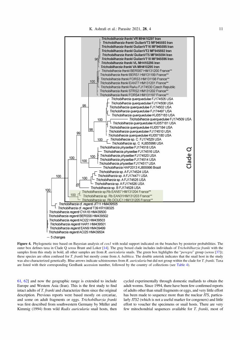
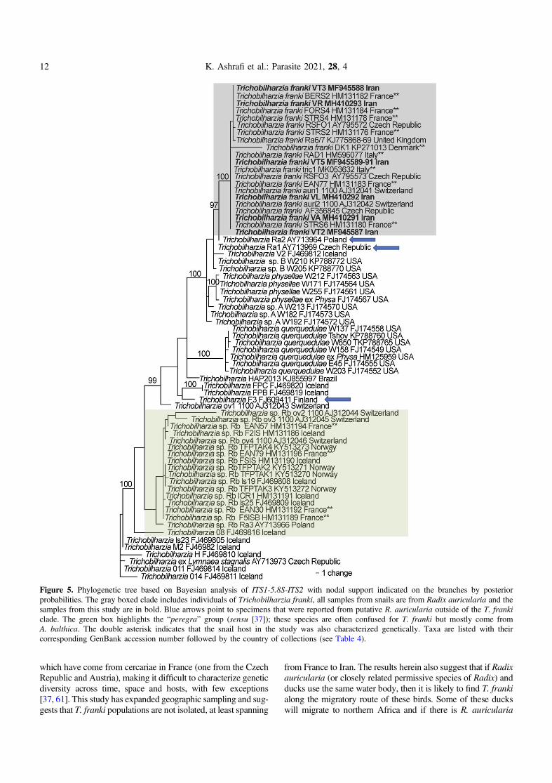
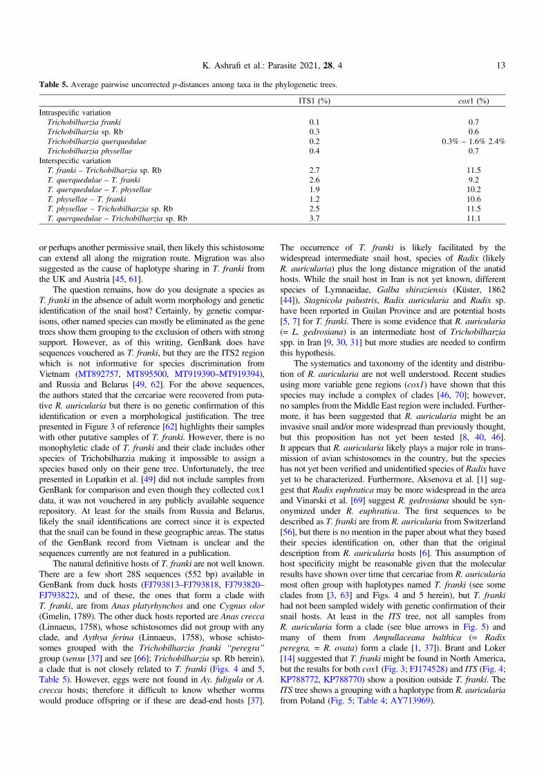
The phylogenetic analysis of both the cox1 (Fig. 4) andITS1-5.8S-ITS2 (Fig. 5) datasets placed the samples from thisstudy within specimens described as T. franki from Radixauricularia snail intermediate hosts. Some of the previous stud-ies that submitted sequences to GenBank labeled as T. frankiwere not monophyletic and most of those sequences belongto an undescribed species Trichobilharzia franki haplotype“peregra” (sensu [37] and were referred to as Trichobilharziasp. Rb from A. balthica snail intermediate hosts. Furthermore,there were many haplotypes labeled T. franki that did not groupwith any previously defined clade. The clades of T. franki andTrichobilharzia sp. Rb were also found by Soldanova et al.[67]. Using uncorrected p-distances as a measure of geneticdiversity and as a proxy for species differentiation, T. frankispecimens from Iran were not very divergent from the availablespecimens of T. franki from GenBank. The average withinspecies diversity was 0.1% for ITS and 0.7% for cox1, whichis consistent with other species of Trichobilharzia (Table 5).
Discussion
Our results show that T. franki, a species of avian schisto-some that occurs in the visceral veins of its anatid hosts, can befound in domestic ducks in Iran. Duck breeding is a routineactivity in almost all rural areas and towns in the flatlandsand foothills of Guilan Province, as well as other provincessuch as Mazandaran [6, 9, 26]. In these areas, Radix spp. andPhysa sp. snail are also found since they also do very well inthese modified habitats. It can be assumed that with 71% preva-lence in ducks found in this study, T. franki is a commonspecies maintained over time and space by the ubiquity ofthe intermediate host in the same modified aquaculture habitatsas well as the wide distribution and use of domestic mallards,an ideal reservoir host. In addition, the ducks examined herewere the same ducks examined in Ashrafi et al. [6] for theneuropathogenic species T. regenti. An interesting question to
6 K. Ashrafi et al.: Parasite 2021, 28, 4
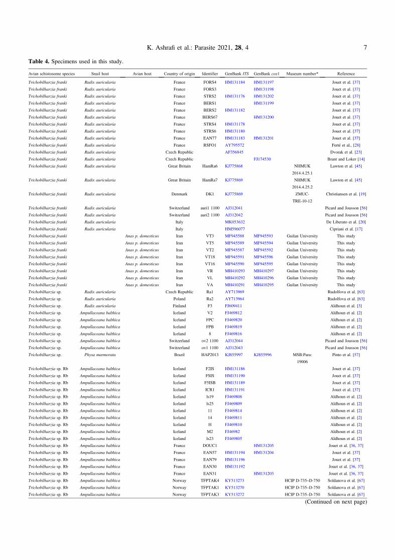
Table 4. Specimens used in this study.
Avian schistosome species Snail host Avian host Country of origin Identifier GenBank ITS GenBank cox1 Museum number* Reference
Trichobilharzia franki Radix auricularia France FORS4 HM131184 HM131197 Jouet et al. [37]
Trichobilharzia franki Radix auricularia France FORS3 HM131198 Jouet et al. [37]
Trichobilharzia franki Radix auricularia France STRS2 HM131176 HM131202 Jouet et al. [37]
Trichobilharzia franki Radix auricularia France BERS1 HM131199 Jouet et al. [37]
Trichobilharzia franki Radix auricularia France BERS2 HM131182 Jouet et al. [37]
Trichobilharzia franki Radix auricularia France BERS67 HM131200 Jouet et al. [37]
Trichobilharzia franki Radix auricularia France STRS4 HM131178 Jouet et al. [37]
Trichobilharzia franki Radix auricularia France STRS6 HM131180 Jouet et al. [37]
Trichobilharzia franki Radix auricularia France EAN77 HM131183 HM131201 Jouet et al. [37]
Trichobilharzia franki Radix auricularia France RSFO1 AY795572 Ferté et al., [28]
Trichobilharzia franki Radix auricularia Czech Republic AF356845 Dvorak et al. [23]
Trichobilharzia franki Radix auricularia Czech Republic FJ174530 Brant and Loker [14]
Trichobilharzia franki Radix auricularia Great Britain HamRa6 KJ775868 NHMUK
2014.4.25.1
Lawton et al. [45]
Trichobilharzia franki Radix auricularia Great Britain HamRa7 KJ775869 NHMUK
2014.4.25.2
Lawton et al. [45]
Trichobilharzia franki Radix auricularia Denmark DK1 KJ775869 ZMUC-
TRE-10-12
Christiansen et al. [19]
Trichobilharzia franki Radix auricularia Switzerland auri1 1100 AJ312041 Picard and Jousson [56]
Trichobilharzia franki Radix auricularia Switzerland auri2 1100 AJ312042 Picard and Jousson [56]
Trichobilharzia franki Radix auricularia Italy MK053632 De Liberato et al. [20]
Trichobilharzia franki Radix auricularia Italy HM596077 Cipriani et al. [17]
Trichobilharzia franki Anas p. domesticus Iran VT3 MF945588 MF945593 Guilan University This study
Trichobilharzia franki Anas p. domesticus Iran VT5 MF945589 MF945594 Guilan University This study
Trichobilharzia franki Anas p. domesticus Iran VT2 MF945587 MF945592 Guilan University This study
Trichobilharzia franki Anas p. domesticus Iran VT18 MF945591 MF945596 Guilan University This study
Trichobilharzia franki Anas p. domesticus Iran VT16 MF945590 MF945595 Guilan University This study
Trichobilharzia franki Anas p. domesticus Iran VR MH410293 MH410297 Guilan University This study
Trichobilharzia franki Anas p. domesticus Iran VL MH410292 MH410296 Guilan University This study
Trichobilharzia franki Anas p. domesticus Iran VA MH410291 MH410295 Guilan University This study
Trichobilharzia sp. Radix auricularia Czech Republic Ra1 AY713969 Rudolfova et al. [63]
Trichobilharzia sp. Radix auricularia Poland Ra2 AY713964 Rudolfova et al. [63]
Trichobilharzia sp. Radix auricularia Finland F3 FJ609411 Aldhoun et al. [3]
Trichobilharzia sp. Ampullaceana balthica Iceland V2 FJ469812 Aldhoun et al. [2]
Trichobilharzia sp. Ampullaceana balthica Iceland FPC FJ469820 Aldhoun et al. [2]
Trichobilharzia sp. Ampullaceana balthica Iceland FPB FJ469819 Aldhoun et al. [2]
Trichobilharzia sp. Ampullaceana balthica Iceland 8 FJ469816 Aldhoun et al. [2]
Trichobilharzia sp. Ampullaceana balthica Switzerland ov2 1100 AJ312044 Picard and Jousson [56]
Trichobilharzia sp. Ampullaceana balthica Switzerland ov1 1100 AJ312043 Picard and Jousson [56]
Trichobilharzia sp. Physa marmorata Brazil HAP2013 KJ855997 KJ855996 MSB:Para:
19006
Pinto et al. [57]
Trichobilharzia sp. Rb Ampullaceana balthica Iceland F2IS HM131186 Jouet et al. [37]
Trichobilharzia sp. Rb Ampullaceana balthica Iceland FSIS HM131190 Jouet et al. [37]
Trichobilharzia sp. Rb Ampullaceana balthica Iceland F5ISB HM131189 Jouet et al. [37]
Trichobilharzia sp. Rb Ampullaceana balthica Iceland ICR1 HM131191 Jouet et al. [37]
Trichobilharzia sp. Rb Ampullaceana balthica Iceland ls19 FJ469808 Aldhoun et al. [2]
Trichobilharzia sp. Rb Ampullaceana balthica Iceland ls25 FJ469809 Aldhoun et al. [2]
Trichobilharzia sp. Rb Ampullaceana balthica Iceland 11 FJ469814 Aldhoun et al. [2]
Trichobilharzia sp. Rb Ampullaceana balthica Iceland 14 FJ469811 Aldhoun et al. [2]
Trichobilharzia sp. Rb Ampullaceana balthica Iceland H FJ469810 Aldhoun et al. [2]
Trichobilharzia sp. Rb Ampullaceana balthica Iceland M2 FJ46982 Aldhoun et al. [2]
Trichobilharzia sp. Rb Ampullaceana balthica Iceland ls23 FJ469805 Aldhoun et al. [2]
Trichobilharzia sp. Rb Ampullaceana balthica France DOUC1 HM131205 Jouet et al. [36, 37]
Trichobilharzia sp. Rb Ampullaceana balthica France EAN57 HM131194 HM131204 Jouet et al. [37]
Trichobilharzia sp. Rb Ampullaceana balthica France EAN79 HM131196 Jouet et al. [37]
Trichobilharzia sp. Rb Ampullaceana balthica France EAN30 HM131192 Jouet et al. [36, 37]
Trichobilharzia sp. Rb Ampullaceana balthica France EAN31 HM131203 Jouet et al. [36, 37]
Trichobilharzia sp. Rb Ampullaceana balthica Norway TFPTAK4 KY513273 HCIP D-735–D-750 Soldanova et al. [67]
Trichobilharzia sp. Rb Ampullaceana balthica Norway TFPTAK1 KY513270 HCIP D-735–D-750 Soldanova et al. [67]
Trichobilharzia sp. Rb Ampullaceana balthica Norway TFPTAK3 KY513272 HCIP D-735–D-750 Soldanova et al. [67]
(Continued on next page)
K. Ashrafi et al.: Parasite 2021, 28, 4 7
consider is whether the diversity of Trichobilharzia in domesticmallards over time would reflect the diversity of schistosomespecies found in migratory birds that also cycle through thecommonly available snail intermediate hosts? Or given theprevalence of T. franki and T. regenti, are they well establishedenough in a domestic life cycle that finding the rarer speciesfrom migratory birds would be difficult, or competition in eitheror the snail or duck hosts? Mallards are listed to host at least10 named species (7 Trichobilharzia spp.) of schistosomes.Some adult schistosome species are only known from experi-mental infections of domestic mallards using cercariae from
captured wild snails, such as T. franki. Distinguishing betweenwild mallards and resident mallards is not often defined in mostpapers. It is known that mallards can host schistosomes, andcertainly they have made excellent experimental hosts, but itis difficult to ascertain the distribution and diversity of schisto-somes in wild mallards, or if the diversity reflects what can befound in the co-occurring anseriforms.
Until now, T. franki had been confirmed genetically only inEurope to include mostly northern countries (France, GreatBritain, Denmark, Switzerland, Czech Republic, Austria,western Russia) [3, 16, 17, 19, 20, 23, 28, 32, 36–38, 42, 45,
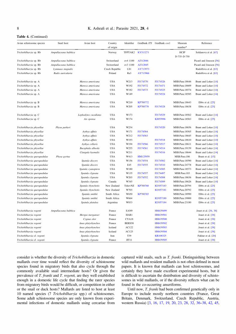
Table 4. (Continued)
Avian schistosome species Snail host Avian host Country
of origin
Identifier GenBank ITS GenBank cox1 Museum
number*
Reference
Trichobilharzia sp. Rb Ampullaceana balthica Norway TFPTAK2 KY513271 HCIP
D-735–D-750
Soldanova et al. [67]
Trichobilharzia sp. Rb Ampullaceana balthica Switzerland ov4 1100 AJ312046 Picard and Jousson [56]
Trichobilharzia sp. Rb Ampullaceana balthica Switzerland ov3 1100 AJ312045 Picard and Jousson [56]
Trichobilharzia sp. Rb Lymnaea stagnalis Czech Republic LS1 AY713973 Rudolfova et al. [63]
Trichobilharzia sp. Rb Radix auricularia Poland Ra3 AY713966 Rudolfova et al. [63]
Trichobilharzia sp. A Mareca americana USA W213 FJ174570 FJ174526 MSB:Para:18646 Brant and Loker [14]
Trichobilharzia sp. A Mareca americana USA W192 FJ174572 FJ174471 MSB:Para:18609 Brant and Loker [14]
Trichobilharzia sp. A Mareca americana USA W182 FJ174573 FJ174525 MSB:Para:18574 Brant and Loker [14]
Trichobilharzia sp. A Mareca americana USA W149 FJ174524 MSB:Para:18585 Brant and Loker [14]
Trichobilharzia sp. B Mareca americana USA W210 KP788772 MSB:Para:18643 Ebbs et al. [25]
Trichobilharzia sp. B Mareca americana USA W205 KP788770 FJ174528 MSB:Para:18638 Ebbs et al. [25]
Trichobilharzia sp. C Lophodytes cucullatus USA W173 FJ174529 MSB:Para:18562 Brant and Loker [14]
Trichobilharzia sp. C Aix sponsa USA W174 KJ855996 MSB:Para:18563 Ebbs et al. [25]
Trichobilharzia physellae Physa parkeri USA W234 FJ174520 MSB:Para:18656 Brant and Loker [14]
Trichobilharzia physellae Aythya affinis USA W171 FJ174564 MSB:Para:18565 Brant and Loker [14]
Trichobilharzia physellae Aythya affinis USA W212 FJ174563 MSB:Para:18645 Brant and Loker [14]
Trichobilharzia physellae Aythya affinis USA W193 FJ174518 MSB:Para:18610 Brant and Loker [14]
Trichobilharzia physellae Aythya collaris USA W194 FJ174566 FJ174517 MSB:Para:18611 Brant and Loker [14]
Trichobilharzia physellae Bucephala albeola USA W255 FJ174561 FJ174514 MSB:Para:19159 Brant and Loker [14]
Trichobilharzia physellae Clangula hyemalis USA W211 FJ174516 MSB:Para:18644 Brant and Loker [14]
Trichobilharzia querquedulae Physa gyrina USA W413 HM125959 MSB:Para:186 Brant et al. [15]
Trichobilharzia querquedulae Spatula discors USA W156 FJ174554 FJ174502 MSB:Para:18590 Brant and Loker [14]
Trichobilharzia querquedulae Spatula discors USA E45 FJ174555 FJ174510 MSB:Para:24778 Brant and Loker [14]
Trichobilharzia querquedulae Spatula cyanoptera USA W180 FJ174505 MSB:Para:18573 Brant and Loker [14]
Trichobilharzia querquedulae Spatula clypeata USA W135 FJ174557 FJ174497 MSB:Para:183 Brant and Loker [14]
Trichobilharzia querquedulae Spatula clypeata USA W203 FJ174552 FJ174508 MSB:Para:18636 Brant and Loker [14]
Trichobilharzia querquedulae Spatula clypeata Canada W345 FJ174509 MSB:Para:18626 Brant and Loker [14]
Trichobilharzia querquedulae Spatula rhynchotis New Zealand TshovNZ KP788760 KU057183 MSB:Para:20794 Ebbs et al. [25]
Trichobilharzia querquedulae Spatula rhynchotis New Zealand W703 KU057181 MSB:Para:20792 Ebbs et al. [25]
Trichobilharzia querquedulae Spatula smithii South Africa W650 KP788765 MSB:Para:18990 Ebbs et al. [25]
Trichobilharzia querquedulae Spatula smithii South Africa W664 KU057180 MSB:Para:19000 Ebbs et al. [25]
Trichobilharzia querquedulae Spatula platalea Argentina W833 KU057184 MSB:Para:23180 Ebbs et al. [25]
Trichobilharzia regenti Ampullaceana balthica France EAN9 HM439499 Jouet et al. [36, 38]
Trichobilharzia regenti Mergus merganser France HAR1 HM439501 Jouet et al. [38]
Trichobilharzia regenti Cygnus olor France CYA18 HM439500 Jouet et al. [38]
Trichobilharzia regenti Anas platyrhynchos France BERS58 HM439502 Jouet et al. [38]
Trichobilharzia regenti Anas platyrhynchos Iceland AC122 HM439503 Jouet et al. [38]
Trichobilharzia regenti Anas platyrhynchos Iceland AC125 HM439504 Jouet et al. [38]
Trichobilharzia cf. regenti Spatula clypeata Iran T39 KR108325 Fakhar et al. [26]
Trichobilharzia cf. regenti Spatula clypeata France JIT11 HM439505 Jouet et al. [38]
8 K. Ashrafi et al.: Parasite 2021, 28, 4
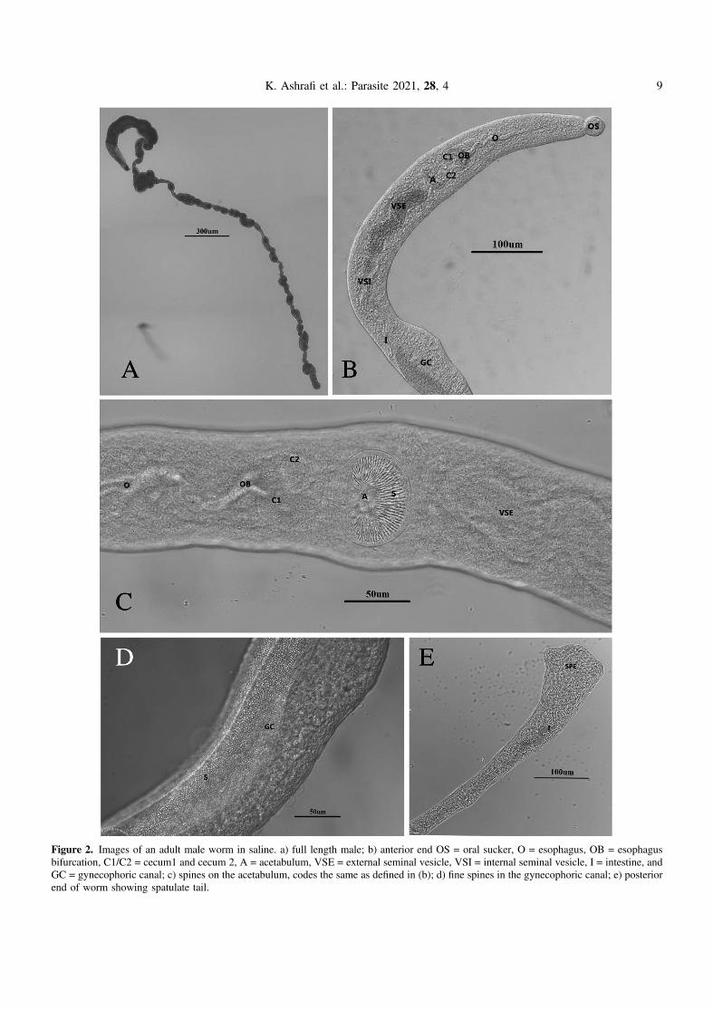
Figure 2. Images of an adult male worm in saline. a) full length male; b) anterior end OS = oral sucker, O = esophagus, OB = esophagusbifurcation, C1/C2 = cecum1 and cecum 2, A = acetabulum, VSE = external seminal vesicle, VSI = internal seminal vesicle, I = intestine, andGC = gynecophoric canal; c) spines on the acetabulum, codes the same as defined in (b); d) fine spines in the gynecophoric canal; e) posteriorend of worm showing spatulate tail.
K. Ashrafi et al.: Parasite 2021, 28, 4 9
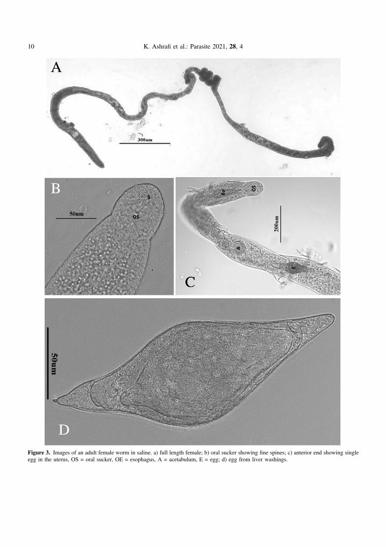
Figure 3. Images of an adult female worm in saline. a) full length female; b) oral sucker showing fine spines; c) anterior end showing singleegg in the uterus, OS = oral sucker, OE = esophagus, A = acetabulum, E = egg; d) egg from liver washings.
10 K. Ashrafi et al.: Parasite 2021, 28, 4
61, 62] and now the geographic range is extended to includeEurope and Western Asia (Iran). This is the first study to findintact adults of T. franki and characterize them since the originaldescription. Previous reports were based mostly on cercariae,and some on adult fragments or eggs. Trichobilharzia frankiwas first described from southwestern Germany by Müller andKimmig (1994) from wild Radix auricularia snail hosts, then
cycled experimentally through domestic mallards to obtain theadult worms. Since 1994, there have been few confirmed reportsof adults other than small fragments or eggs, and very little efforthas been made to sequence more than the nuclear ITS, particu-larly ITS2 (which is not a useful marker for congeners) and littleeffort to voucher the specimens or snail hosts. There are veryfew mitochondrial sequences available for T. franki, most of
Figure 4. Phylogenetic tree based on Bayesian analysis of cox1 with nodal support indicated on the branches by posterior probabilities. Theouter box defines taxa in Clade Q sensu Brant and Loker [14]. The gray boxed clade includes individuals of Trichobilharzia franki with thesamples from this study in bold, all other samples are from R. auricularia snails. The green box highlights the “peregra” group (sensu [37]);these species are often confused for T. franki but mostly come from A. balthica. The double asterisk indicates that the snail host in the studywas also characterized genetically. Blue arrows indicate schistosomes from R. auricularia but did not group within the clade for T. franki. Taxaare listed with their corresponding GenBank accession number, followed by the country of collections (see Table 4).
K. Ashrafi et al.: Parasite 2021, 28, 4 11
which have come from cercariae in France (one from the CzechRepublic and Austria), making it difficult to characterize geneticdiversity across time, space and hosts, with few exceptions[37, 61]. This study has expanded geographic sampling and sug-gests that T. franki populations are not isolated, at least spanning
from France to Iran. The results herein also suggest that if Radixauricularia (or closely related permissive species of Radix) andducks use the same water body, then it is likely to find T. frankialong the migratory route of these birds. Some of these duckswill migrate to northern Africa and if there is R. auricularia
Figure 5. Phylogenetic tree based on Bayesian analysis of ITS1-5.8S-ITS2 with nodal support indicated on the branches by posteriorprobabilities. The gray boxed clade includes individuals of Trichobilharzia franki, all samples from snails are from Radix auricularia and thesamples from this study are in bold. Blue arrows point to specimens that were reported from putative R. auricularia outside of the T. frankiclade. The green box highlights the “peregra” group (sensu [37]); these species are often confused for T. franki but mostly come fromA. balthica. The double asterisk indicates that the snail host in the study was also characterized genetically. Taxa are listed with theircorresponding GenBank accession number followed by the country of collections (see Table 4).
12 K. Ashrafi et al.: Parasite 2021, 28, 4
or perhaps another permissive snail, then likely this schistosomecan extend all along the migration route. Migration was alsosuggested as the cause of haplotype sharing in T. franki fromthe UK and Austria [45, 61].
The question remains, how do you designate a species asT. franki in the absence of adult worm morphology and geneticidentification of the snail host? Certainly, by genetic compar-isons, other named species can mostly be eliminated as the genetrees show them grouping to the exclusion of others with strongsupport. However, as of this writing, GenBank does havesequences vouchered as T. franki, but they are the ITS2 regionwhich is not informative for species discrimination fromVietnam (MT892757, MT895500, MT919390–MT919394),and Russia and Belarus [49, 62]. For the above sequences,the authors stated that the cercariae were recovered from puta-tive R. auricularia but there is no genetic confirmation of thisidentification or even a morphological justification. The treepresented in Figure 3 of reference [62] highlights their sampleswith other putative samples of T. franki. However, there is nomonophyletic clade of T. franki and their clade includes otherspecies of Trichobilharzia making it impossible to assign aspecies based only on their gene tree. Unfortunately, the treepresented in Lopatkin et al. [49] did not include samples fromGenBank for comparison and even though they collected cox1data, it was not vouchered in any publicly available sequencerepository. At least for the snails from Russia and Belarus,likely the snail identifications are correct since it is expectedthat the snail can be found in these geographic areas. The statusof the GenBank record from Vietnam is unclear and thesequences currently are not featured in a publication.
The natural definitive hosts of T. franki are not well known.There are a few short 28S sequences (552 bp) available inGenBank from duck hosts (FJ793813–FJ793818, FJ793820–FJ793822), and of these, the ones that form a clade withT. franki, are from Anas platyrhynchos and one Cygnus olor(Gmelin, 1789). The other duck hosts reported are Anas crecca(Linnaeus, 1758), whose schistosomes did not group with anyclade, and Aythya ferina (Linnaeus, 1758), whose schisto-somes grouped with the Trichobilharzia franki “peregra”group (sensu [37] and see [66]; Trichobilharzia sp. Rb herein),a clade that is not closely related to T. franki (Figs. 4 and 5,Table 5). However, eggs were not found in Ay. fuligula or A.crecca hosts; therefore it difficult to know whether wormswould produce offspring or if these are dead-end hosts [37].
The occurrence of T. franki is likely facilitated by thewidespread intermediate snail host, species of Radix (likelyR. auricularia) plus the long distance migration of the anatidhosts. While the snail host in Iran is not yet known, differentspecies of Lymnaeidae, Galba shiraziensis (Küster, 1862[44]), Stagnicola palustris, Radix auricularia and Radix sp.have been reported in Guilan Province and are potential hosts[5, 7] for T. franki. There is some evidence that R. auricularia(= L. gedrosiana) is an intermediate host of Trichobilharziaspp. in Iran [9, 30, 31] but more studies are needed to confirmthis hypothesis.
The systematics and taxonomy of the identity and distribu-tion of R. auricularia are not well understood. Recent studiesusing more variable gene regions (cox1) have shown that thisspecies may include a complex of clades [46, 70]; however,no samples from the Middle East region were included. Further-more, it has been suggested that R. auricularia might be aninvasive snail and/or more widespread than previously thought,but this proposition has not yet been tested [8, 40, 46].It appears that R. auricularia likely plays a major role in trans-mission of avian schistosomes in the country, but the specieshas not yet been verified and unidentified species of Radix haveyet to be characterized. Furthermore, Aksenova et al. [1] sug-gest that Radix euphratica may be more widespread in the areaand Vinarski et al. [69] suggest R. gedrosiana should be syn-onymized under R. euphratica. The first sequences to bedescribed as T. franki are from R. auricularia from Switzerland[56], but there is no mention in the paper about what they basedtheir species identification on, other than that the originaldescription from R. auricularia hosts [6]. This assumption ofhost specificity might be reasonable given that the molecularresults have shown over time that cercariae from R. auriculariamost often group with haplotypes named T. franki (see someclades from [3, 63] and Figs. 4 and 5 herein), but T. frankihad not been sampled widely with genetic confirmation of theirsnail hosts. At least in the ITS tree, not all samples fromR. auricularia form a clade (see blue arrows in Fig. 5) andmany of them from Ampullaceana balthica (= Radixperegra, = R. ovata) form a clade [1, 37]). Brant and Loker[14] suggested that T. franki might be found in North America,but the results for both cox1 (Fig. 3; FJ174528) and ITS (Fig. 4;KP788772, KP788770) show a position outside T. franki. TheITS tree shows a grouping with a haplotype from R. auriculariafrom Poland (Fig. 5; Table 4; AY713969).
Table 5. Average pairwise uncorrected p-distances among taxa in the phylogenetic trees.
ITS1 (%) cox1 (%)
Intraspecific variationTrichobilharzia franki 0.1 0.7Trichobilharzia sp. Rb 0.3 0.6Trichobilharzia querquedulae 0.2 0.3% – 1.6% 2.4%Trichobilharzia physellae 0.4 0.7
Interspecific variationT. franki – Trichobilharzia sp. Rb 2.7 11.5T. querquedulae – T. franki 2.6 9.2T. querquedulae – T. physellae 1.9 10.2T. physellae – T. franki 1.2 10.6T. physellae – Trichobilharzia sp. Rb 2.5 11.5T. querquedulae – Trichobilharzia sp. Rb 3.7 11.1
K. Ashrafi et al.: Parasite 2021, 28, 4 13
Much of the genetic diversity of Trichobilharzia lineages inClade Q (sensu [14]; Fig. 4) that includes T. franki fromGenBank sequences is represented by ITS sequences, and manyof these samples do not form clades (Fig. 5). This might suggestthat more diversity is yet to be discovered. Processes that maycontribute to our understanding of diversity in Trichobilharziathat emerges from sequencing surveys include the following:(A) Incomplete lineage sorting – the ITS tree is based onlyon a nuclear region, which may have a slower mutation raterelative to a faster evolving gene, thus ancestral polymorphismsare retained, or the ancestral population was large and thustakes more time. Also, it could be that speciation within atleast Clade Q has been relatively recent and nuclear copiesdo not match mitochondrial gene trees or species trees.However, in general, most sequences fall into taxa that groupaccording to species (or lineages if only cercariae) and thereis little or no evidence of widespread mito-nuclear discordance.(B) Hybridization – within avian schistosomes, hybridizationhas not been studied. However, mito-nuclear discordance isnot reliable evidence of hybridization (see [58]) but a morevariable gene certainly helps in diversity characterizations andspecies delineations. It is impossible to obtain mitochondrialor genomic data to explore hybridization or any other questionwith the individual worms available currently in GenBank,because there are no museum vouchers for re-evaluation. Thefew specimens that were vouchered are not available fordestructive sampling and thus it is strongly recommended thatvials of adult and cercariae (and hosts, particularly gastropods)are deposited so that we have a record of the past and materialavailable for new investigations. (C) Host-induced variation –
though poorly understood, host-induced variation can con-tribute to morphological variability, but it is not known howmuch it would affect genetic diversity in schistosomes, andmost studies have used morphology, not genetics in this con-text, with adult worms (see [10, 65]). (D) Ecological speciationis plausible given that the offspring of the worms in the migra-tory hosts are being distributed along the route, at each localitythe miracidia might be exposed to snails that are normallycompatible but might also be exposed to novel putativelysusceptible snails. For parasites, this is akin to host switchingevents. For a high-quality review see [64]. (E) Poor host taxon-omy. One of the consequences of genetic characterizations isthat it has provided a yardstick to define in more detail lineagediversity, which may not be reflected in morphological diver-sity. Invertebrates in particular have fallen in this category asthey can often have little variation to compare and some ofthe observed variation is subject to change based on a myriadof abiotic and biotic influences (such as parasitism, temperature,water chemistry, etc.) rather than phylogeny. Gastropods inparticular have been shown to be more species-rich than previ-ously considered based on morphology [1, 29]. If every geneticreport of Trichobilharzia spp. was accompanied by geneticassessment of the snail host, then we could understand moreabout host-parasite relationship specificity. It could be that thereare more species of Radix transmitting these species ofTrichobilharzia than is reported based solely on morphology(e.g. [21, 22, 46]).
Until there is more effort to include multiple and variablegene regions for schistosomes (or any organism) it is not
possible to understand the phylogeography or epidemiologyof disease-causing helminths. It has been shown repeatedly thatvariable mitochondrial genes are ideal for assessing crypticdiversity compared to nuclear genes. When only ITS2 is usedwithout ITS1, it is not possible to find enough variation withincongeners, particularly if they are closely related. However, astudy should not rely only on a single gene (and if it does so,it should be variable and useful for the future), as the diversityrevealed is gene diversity, not always directly reflecting species,which must be tested for congruency [50, 55]. The specimensavailable in GenBank that had variable cox1 sequences avail-able represent mostly Western Europe (Table 1). Yet, giventhe geographic distance between these specimens and NorthernIran, there was very little genetic differentiation in either cox1or ITS (Table 5). This result should not be surprising if thelong-distance migration of the wild hosts and the suitabilityof domestic mallards as reservoir hosts are considered intransmission dynamics (also see [4]). Furthermore, Ebbs et al.[25] showed that the intraspecific genetic diversity inTrichobilharzia querquedulae was within average range forspecies schistosomes, even though the comparison includedindividual worms from across 5 continents.
The occurrence of CD in Iran is high in areas of aquacul-ture. In addition to wild duck hosts, previous work in the areahas shown that domestic mallards are reservoir hosts ofT. regenti, a nasal schistosome [6], and as well for T. franki,shown in this study and thus maintain high prevalence ofCD. The genetic results support the finding that populationsof T. franki from Iran are not differentiated from populationsin Europe. Therefore, the schistosomes are dispersed with theirmigratory duck host, maintaining the gene flow across popula-tions with compatible snail hosts in Iran. It is not surprising thatspecies of Trichobilharzia are thought to be one of the commonetiological agents of CD; several of these species use snail hoststhat are widespread and/or invasive (e.g. [24]) and prefer or atleast are easily established in modified aquatic habitats used bydomestic animals and humans. Added to this, their definitivehosts travel long distances, further facilitating transmission fromone continent to another.
Conflict of interest
The authors declare that they have no conflict of interest.
Acknowledgements. This study was performed with the collabora-tion of the Guilan University of Medical Sciences, Iran and theUniversity of New Mexico, USA. The authors would like to thankMrs. Behnaz Rahmati and Mr. Alireza Noroosta for their kind helpin performing this study. The authors thank two anonymous review-ers for suggestions that improved the content of the manuscript.
References
1. Aksenova OV, Bolotov IN, Gofarov MY, Kondakov AV,Vinarski MV, Bespalaya YV, Kolosova YS, Palatov DM,Sokolova SE, Spitsyn VM, Tomilova AA, Travina OV, VikhrevIV. 2018. Species richness, molecular taxonomy and biogeog-raphy of the radicine pond snails (Gastropoda: Lymnaeidae) inthe Old World. Scientific Reports, 8, 11199.
14 K. Ashrafi et al.: Parasite 2021, 28, 4
2. Aldhoun J, Kolarova L, Skirnisson K, Horak P. 2009a. Birdschistosome diversity in Iceland: molecular evidence. Journal ofHelminthology, 83, 173–180.
3. Aldhoun J, Faltynkova A, Karvonen A, Horak P. 2009b.Schistosomes in the North: A unique finding from a proso-branch snail using molecular tools. Parasitology International,58, 314–317.
4. Ashrafi K, Brant SV. 2020. An efficient method for collectingthe full-length adults, fragments, and eggs of Trichobil-harzia spp. from the liver of definitive hosts. ParasitologyResearch, 119, 1167–1172.
5. Ashrafi K, Mas-Coma S. 2014. Fasciola gigantica transmissionin the zoonotic fascioliasis endemic lowlands of Guilan, Iran:experimental assessment. Veterinary Parasitology, 205, 96–106.
6. AshrafiK, Nouroosta A, Sharifdini M, Mahmoudi MR, Rahmati B,Brant SV. 2018. Genetic diversity of an avian nasal schistosomecausing cercarial dermatitis in the Black Sea-Mediterraneanmigratory route. Parasitology Research, 117, 3821–3833.
7. Ashrafi K, Valero MA, Peixoto RV, Artigas P, Panova M,Mas-Coma S. 2015. Distribution of Fasciola hepatica andF. gigantica in the endemic area of Guilan, Iran: Relationshipsbetween zonal overlap and phenotypic traits. Infections,Genetics and Evolution, 31, 95–109.
8. Aksenova O, Vinarski M, Bolotov I, Kondakov A, Besplalaya Y,Tomilova A, Palster I, Gofarov M. 2017. Two Radix spp.(Gastropoda: Lymnaeidae) endemic to thermal springs aroundLake Baikal represent ecotypes of the widespread Radix auric-ularia. Journal of Zoological Systematics and EvolutionaryResearch, 55, 298–309.
9. Athari A, Gohardehi S, Rostami-Jalilian M. 2006. Determina-tion of definitive and intermediate hosts of cercarial dermatitisproducing agents in northern Iran. Archives of IranianMedicine, 9, 11–15.
10. Bayssade-Dufour C, Jouet D, Rudolfova J, Horak P, Ferté H.2006. Seasonal morphological variations in bird schistosomes.Parasite, 13, 205–214.
11. Blair D, Islam KS. 1983. The life cycle and morphology ofTrichobilharzia australis n. sp. from the nasal blood vessels ofthe black duck in Australia with a review of the genusTrichobilharzia. Systematic Parasitology, 5, 89–117.
12. Bowles J, McManus DP. 1993. Rapid discriminationof Echinococcus species and strains using a PCR-based RFLPmethod. Molecular and Biochemical Parasitology, 57, 231–239.
13. Bowles J, Blair D, McManus DP. 1995. A molecular phylogenyof the human schistosomes. Molecular Phylogenetics andEvolution, 4, 103–109.
14. Brant SV, Loker ES. 2009. Molecular systematics of the avianschistosome genus Trichobilharzia (Trematoda: Schistosomati-dae) in North America. Journal of Parasitology, 95, 941–963.
15. Brant SV, Bochte CA, Loker ES. 2011. New intermediate hostrecords for the avian schistosomes Dendritobilharzia pulverulenta,Gigantobilharzia huonensis, and Trichobilharzia querquedulaefrom North America. Journal of Parasitology, 97, 946–949.
16. Caron Y, Cabaraux A, Marechal F, Losson B. 2017. Swimmer’sitch in Belgium: first recorded outbreaks, molecular identifica-tion of the parasite species and intermediate hosts. Vector-Borneand Zoonotic Diseases, 17, 3.
17. Cipriani P, Mattiucci S, Paoletti M, Scialanca F, Nascetti G.2011. Molecular evidence of Trichobilharzia franki Müller andKimmig, 1994 (Digenea: Schistosomatidae) in Radix auricu-laria from Central Italy. Parasitology Research, 109, 935–940.
18. Clausen JH, Madsen H, Van PT, Dalsgaard A, Murrell KD.2015. Integrated parasite management: path to sustainablecontrol of fishborne trematodes in aquaculture. Trends inParasitology, 31, 8–15.
19. Christiansen AO, Olsen A, Buchmann K, Kania PW, Nejsum P,Vennervald BJ. 2016. Molecular diversity of avian schisto-somes in Danish freshwater snails. Parasitology Research, 115,1027–1037.
20. De Liberato C, Berrilli F, Bossu T, Magliano A, Montalbano DiFilippo M, Di Cave D. 2019. Outbreak of swimmer’s itch inCentral Italy: description, causative agent and preventivemeasures. Zoonoses and Public Health, 66, 377–381.
21. Devkota R, Brant SV, Loker ES. 2015. The Schistosomaindicum species group in Nepal: presence of a new lineage ofschistosome and use of the Indoplanorbis exustus speciescomples of snail hosts. International Journal for Parasitology,45, 857–870.
22. Devkota R, Brant SV, Loker ES. 2016. A genetically distinctSchistosoma from Radix luteola from Nepal related to Schis-tosoma turkestanicum: a phylogenetic study of schistosome andsnail host. Acta Tropica, 164, 45–53.
23. Dvorák J, Vanácová S, Hampl V, Flegr J, Horák P. 2002.Comparison of European Trichobilharzia species based on ITS1and ITS2 sequences. Parasitology, 124, 307–313.
24. Ebbs ET, Loker ES, Brant SV. 2018. Phylogeography andgenetics of the globally invasive snail Physa acuta Draparnaud1805, and its potential to serve as an intermediate host to larvaldigenetic trematodes. BMC Evolutionary Biology, 18, 103.
25. Ebbs ET, Loker ES, Davis NE, Flores V, Veleizan A, Brant SV.2016. Schistosomes with wings: how host phylogeny andecology shape the global distribution of Trichobilharziaquerquedulae (Schistosomatidae). International Journal forParasitology, 46, 669–677.
26. Fakhar M, Ghobaditara M, Brant SV, Karamian M,Gohardehi S, Bastani R. 2016. Phylogenetic analysis of nasalavian schistosomes (Trichobilharzia) from aquatic birds inMazandaran Province, northern Iran. Parasitology International,65, 151–158.
27. Farahnak A, Essalat M. 2003. A study on cercarial dermatitis inKhuzestan province, southwestern Iran. BMC Public Health, 3, 35.
28. Ferté H, Depaquit J, Carre S, Villena I, Léger N. 2005. Thepresence of Trichobilharzia szidati in Lymnaea stagnalis andT. franki in Radix auricularia in northeastern France: molecularevidence. Parasitology Research, 95, 150–154.
29. Gauffre-Autelin P, von Rintelen T, Stelbrink B, Albrecht C.2017. Recent range expansion of an intermediate host foranimal schistosome parasites in the Indo-Australian Archipe-lago: phylogeography of the freshwater gastropod Indoplanorbisexustus in South and Southeast Asia. Parasites & Vectors,10, 126.
30. Ghobaditara M, Fakhar M, Sharif M. 2015. An overview on thepresent situation of cercarial dermatitis: a neglected zoonoticdisease in Iran and the World. Journal of Mazandaran Univer-sity of Medical Sciences, 24, 446–460.
31. Gohardehi S, Fakhar M, Madjidaei M. 2012. Avian schisto-somes and human cercarial dermatitis in a wildlife refuge inMazandaran Province, northern Iran. Zoonoses and PublicHealth, 60, 442–447.
32. Gulyas K, Soldanova M, Orosova M, Oros M. 2020.Confirmation of the presence of zoonotic Trichobilharziafranki following a human cercarial dermatitis outbreak inrecreational water in Slovakia. Parasitology Research, 119,2531–2537.
33. Hoberg EP, Pilitt PA, Galbreath KE. 2009. Why museumsmatter: a tale of pinworms (Oxyuroidea: Heteroxynematidae)among pikas (Ochotona princeps and O. collaris) in theAmerican west. Journal of Parasitology, 95, 490–501.
34. Huelsenbeck JP, Ronquist F. 2001. MRBAYES: Bayesianinference of phylogenetic trees. Bioinformatics ApplicationNotes, 17, 754–755.
K. Ashrafi et al.: Parasite 2021, 28, 4 15
35. Imani-Baran A, Yakhchali M, Malekzadeh-Viayeh R, Farahnak A.2013. Seasonal and geographical distribution of cercarial infectionin Lymnaea gedrosiana (Pulmunata: Lymnaeidae) in northwestIran. Iranian Journal of Parasitology, 8, 423–429.
36. Jouet D, Ferté H, Depaquit J, Rudolfova J, Latour P, Zanella D,Kaltenbach ML, Léger N. 2008. Trichobilharzia spp. in naturalconditions in Annecy Lake, France. Parasitology Research, 103,51–58.
37. Jouet D, Skirnisson K, Kolářová L, Ferté H. 2010a. Moleculardiversity of Trichobilharzia franki in two intermediate hosts(Radix auricularia and Radix peregra): a complex of species.Infection, Genetics and Evolution, 10, 1218–1227.
38. Jouet D, Skirnisson K, Kolářová L, Ferté H. 2010b. Final hostsand variability of Trichobilharzia regenti under natural condi-tions. Parasitology Research, 107, 923–930.
39. Karamian M, Aldhoun JA, Maraghi S, Hatam G, Farhangmehr B,Sadjjadi SM. 2011. Parasitological and molecular study of thefurcocercariae from Melanoides tuberculate as a probable agentof cercarial dermatitis. Parasitology Research, 108, 955–962.
40. Kipp RM, Benson AJ, Larson J, Fusaro A. 2017. Radixauricularia. USGS Nonindigenous Aquatic Species Database:Gainesville, FL. http://nas.er.usgs.gov/queries/factsheet.aspx?SpeciesID = 1012.
41. Kolářová L, Horák P, Skírnisson K, Marečková H, Doenhoff M.2013. Cercarial dermatitis, a neglected allergic disease. ClinicalReviews in Allergy and Immunology, 45, 63–74.
42. Korsunenka AV, Chrisanfova GG, Ryskov AP, MovseessianSO, Vasilyev VA, Semyenova SK. 2010. Detection of EuropeanTrichobilharzia schistosomes (T. franki, T. szidati, and T.regenti) based on novel genome sequences. Journal of Para-sitology, 96, 802–806.
43. Kourilová P, Hogg KG, Kolářová L, Mountford AP. 2004.Cercarial dermatitis caused by bird schistosomes comprises bothimmediate and late phase cutaneous hypersensitivity reaction.Journal of Immunology, 172, 3766–3774.
44. Küster HC. 1862. Die Gattungen Limnaeus, Amphipeplea, Chilina,Isidora und Physopsis, in Systematisches Conchylien-Cabinet,2nd edn. Martini C, Editor. Nürnberg: Bauer & Raspe, I.17 b:issues 180-182: 1-48, plates 1-11 (1862); issue 184: 49-77, plate 12(1863).
45. Lawton SP, Lim RM, Dukes JP, Cook RT, Walker AJ, Kirk RS.2014. Identification of a major causative agent of human cercarialdermatitis, Trichobilharzia franki (Müller and Kimmig, 1994), insouthern England and its evolutionary relationships with otherEuropean populations. Parasites & Vectors, 7, 277.
46. Lawton SP, Lim RM, Dukes JP, Kett SM, Cook RT, Walker AJ,Kirk RS. 2015. Unravelling the riddle of Radix: DNA barcodingfor species identification of freshwater snail intermediate host ofzoonotic digeneans and estimating their inter-population evolu-tionary relationships. Infection, Genetics and Evolution, 35, 63–74.
47. Lima dos Santos CAM, Howgate P. 2011. Fishborne zoonoticparasites and aquaculture: A review. Aquaculture, 318,253–261.
48. Lockyer AE, Olson PD, Ostergaard P, Rollinson D, JohnstonDA, Attwood SW, Southgate VR, Horak P, Snyder SD, Le TH,Agatsuma T, McManus DP, Carmichael AC, Naem S,Littlewood DT. 2003. The phylogeny of the Schistosomatidaebased on three genes with emphasis on the interrelationshipsof Schistosoma Weinland, 1858. Parasitology, 126, 203–224.
49. Lopatkin AA, Chrisanfova GG, Voronin MV, Zazornova OP,Beer SA, Semyenova SK. 2010. Polymorphism of the cox1 genein cercariae isolates of bird schistosomes (Trematoda: Schisto-somatidae) from ponds of Moscow and Moscow region.Russian Journal of Genetics, 46, 873–880.
50. Maddison WP. 1997. Gene trees in species trees. SystematicBiology, 46, 523–536.
51. Mahdavi SA, Farahnak A, Mobedi I, Rad MBM, Azadeh H.2013. Survey of migratory birds (Anatidae: Anas platyrhynchos)for schistosome parasites from Mazandaran Province, northernIran. Iranian Journal of Parasitology, 8, 333–336.
52. Mahdavi SA, Farahnak A, Mousavi SJ, Mobedi I, Rezaeian M,Golmohamadi T, Azadeh H, Gohardehi S. 2013. Prevalence ofschistosome induced cercarial dermatitis in north of Iran. AsianPacific Journal of Tropical Disease, 3, 37–40.
53. Maleki SH, Athari A, Haghighi A, Taghipour N, Gohardehi SH,Tabaei SS. 2012. Species identification of birds nasal Trichobil-harzia in Sari, north of Iran. Iranian Journal of Parasitology, 7,82–85.
54. Müller V, Kimmig P. 1994. Trichobilharzia franki n. sp.–thecause of swimmer’s dermatitis in southwest German dredgedlakes. Applied Parasitology, 35, 12–31.
55. Nakhleh L. 2013. Computational approaches to species phy-logeny inference and gene tree reconciliation. Trends inEcology and Evolution, 28, 719–728.
56. Picard D, Jousson O. 2001. Genetic variability among cercariaeof the Schistosomatidae (Trematoda: Digenea) causing swim-mer’s itch in Europe. Parasite, 8, 237–242.
57. Pinto HA, Brant SV, de Melo AL. 2014. Physa marmorata(Mollusca: Physidae) as a natural intermediate host ofTrichobilharzia (Trematoda: Schistosomatidae), a potentialcausative agent of avian mercurial dermatitis in Brazil. ActaTropica, 138, 38–43.
58. Platt RN III, McDew-White M, Le Clech W, Chevalier FD, AllanF, Emery AM, Garba A, Hamidou AA, Ame SM, Webster JP,Rollinson D, Webster BL, Anderson TJC. 2019. Ancient hybridiza-tion and adaptive introgression of an invadolysin gene in schisto-some parasites. Molecular Biology and Evolution, 36, 2127–2142.
59. Pleijel F, Jondelius U, Norlinder E, Nygren A, Oxelman B,Schander C, Sundberg P, Thollesson M. 2008. Phylogenies withoutroots? A plea for the use of vouchers in molecular phylogeneticstudies. Molecular Phylogenetics and Evolution, 48, 369–371.
60. Posada D, Crandall KA. 1998. Modeltest: testing the model ofDNA substitution. Bioinformatics, 14, 817–818.
61. Reier S, Haring E, Billinger F, Blatterer H, Duda M, Corofsky C,Grasser HP, Heinisch W, Horweg C, Druckenhauser L, SzucishNU, Wanka A, Sattmann H. 2020. First confirmed record ofTrichobilharzia franki Müller & Kimmig, 1994, from Radixauricularia (Linnaeus, 1758) for Austria. Parasitology Research,119, 4135–4141.
62. Rizevsky SV, Cherviakovsky EM, Kurchenko VP. 2010.Molecular taxonomic identification of Schistosomatidae fromNaroch Lake and Polonevichi Lake in Belarus. BiochemicalSystematics and Ecology, 39, 14–21.
63. Rudolfova J, Hampl V, Bayssade-Dufour C, Lockyer AE,Littlewood DT, Horák P. 2005. Validity reassessment ofTrichobilharzia species using Lymnaea stagnalis as the inter-mediate host. Parasitology Research, 95, 79–89.
64. Schluter D. 2001. The ecology and origin of species. Trends inEcology and Evolution, 16, 372–380.
65. Skírnisson K, Kolářová L, Horák P, Ferté H, Jouet D. 2012.Morphological features of the nasal blood fluke Trichobilharziaregenti (Schistosomatidae, Digenea) from naturally infectedhosts. Parasitology Research, 110, 1881–1892.
66. Skrjabin KI, Zakharov NP. 1920. Zweineue Trematodengat-tugen aus den Blutgefassen der Volgel. Izvestnik DonskovoVeterinarnovo Instituta, 2, 1–6.
67. Soldanova M, Georgieva S, Rohacova J, Knudsen R, Kuhn JA,Henriksen EH, Siwertsson A, Shaw JC, Kuris AM, Amundsen PA,Scholz T, Lafferty KD, Kostadinova A. 2017. Molecular analysesreveal high species diversity of trematodes in a sub-Arctic lake.International Journal for Parasitology, 47, 327–345.
16 K. Ashrafi et al.: Parasite 2021, 28, 4
68. Valkiunas G, Atkinson CT, Bensch S, Sehgal RNM, RicklefsRE. 2008. Parasite misidentifications in GenBank: how tominimize their number? Trends in Parasitology, 24, 247–248.
69. Vinarski MV, Aksenova OV, Bolotov IN. 2020. Taxonomicassessment of genetically-delineated species of radicine snails(Mollusca, Gastropoda, Lymnaeidae). Zoosystematics andEvolution, 96, 577–608.
70. Von Oheimb PV, Albrecht C, Riedel F, Du L, Yang J, AldridgeDC, Bößneck U, Zhang H, Wilke T. 2011. Freshwater
biogeography and limnological evolution of the TibetanPlateau – Insights from a plateau-wide distributed gastropodtaxon (Radix spp.). PLoS One, 6(10), e26307.
71. Yakhchali M, Hosseinpanahi A, Malekzadeh-Viayeh R. 2016.Molecular evidence of Trichobilharzia species (Digenea:Schistosomatidae) in the snails of Lymnaea auricularia fromUrmia suburb, northwest Iran. Iranian Journal of Parasitology,11, 296–302.
Cite this article as: Ashrafi K, Sharifdini M, Darjani A & Brant SV. 2021. Migratory routes, domesticated birds and cercarial dermatitis:the distribution of Trichobilharzia franki in Northern Iran. Parasite 28, 4.
An international open-access, peer-reviewed, online journal publishing high quality paperson all aspects of human and animal parasitology
Reviews, articles and short notes may be submitted. Fields include, but are not limited to: general, medical and veterinary parasitology;morphology, including ultrastructure; parasite systematics, including entomology, acarology, helminthology and protistology, andmolecularanalyses; molecular biology and biochemistry; immunology of parasitic diseases; host-parasite relationships; ecology and life history ofparasites; epidemiology; therapeutics; new diagnostic tools.All papers in Parasite are published in English. Manuscripts should have a broad interest and must not have been published or submittedelsewhere. No limit is imposed on the length of manuscripts.
Parasite (open-access) continues Parasite (print and online editions, 1994-2012) and Annales de Parasitologie Humaine et Comparée(1923-1993) and is the official journal of the Société Française de Parasitologie.
Editor-in-Chief: Submit your manuscript atJean-Lou Justine, Paris http://parasite.edmgr.com/
K. Ashrafi et al.: Parasite 2021, 28, 4 17
Copyright © 2022 FDOKUMEN