Redalyc.Atopic Dermatitis Guideline. Position Paper from the ...
35
Revista Alergia México ISSN: 0002-5151 [email protected] Colegio Mexicano de Inmunología Clínica y Alergia, A.C. México Sánchez, Jorge; Páez, Bruno; Macías, A; Olmos, C; de Falco, A Atopic Dermatitis Guideline. Position Paper from the Latin American Society of Allergy, Asthma and Immunology Revista Alergia México, vol. 61, núm. 3, julio-septiembre, 2014, pp. 178-211 Colegio Mexicano de Inmunología Clínica y Alergia, A.C. Ciudad de México, México Available in: http://www.redalyc.org/articulo.oa?id=486755157009 How to cite Complete issue More information about this article Journal's homepage in redalyc.org Scientific Information System Network of Scientific Journals from Latin America, the Caribbean, Spain and Portugal Non-profit academic project, developed under the open access initiative
-
Upload
khangminh22 -
Category
Documents
-
view
0 -
download
0
Transcript of Redalyc.Atopic Dermatitis Guideline. Position Paper from the ...

Revista Alergia México
ISSN: 0002-5151
Colegio Mexicano de Inmunología Clínica
y Alergia, A.C.
México
Sánchez, Jorge; Páez, Bruno; Macías, A; Olmos, C; de Falco, A
Atopic Dermatitis Guideline. Position Paper from the Latin American Society of Allergy,
Asthma and Immunology
Revista Alergia México, vol. 61, núm. 3, julio-septiembre, 2014, pp. 178-211
Colegio Mexicano de Inmunología Clínica y Alergia, A.C.
Ciudad de México, México
Available in: http://www.redalyc.org/articulo.oa?id=486755157009
How to cite
Complete issue
More information about this article
Journal's homepage in redalyc.org
Scientific Information System
Network of Scientific Journals from Latin America, the Caribbean, Spain and Portugal
Non-profit academic project, developed under the open access initiative

178 www.nietoeditores.com.mx
PoSition PaPEr
Atopic Dermatitis Guideline. Position Paper from the Latin American Society of Allergy, Asthma and Immunology
ABSTRACT
As in other regions, the incidence of atopic dermatitis in Latin America has been increasing in recent years. Although there are several clinical guidelines, many of their recommendations cannot be universal since they depend on the characteristics of each region. Thus, we decided to create a consensus guideline on atopic dermatitis applicable in Latin America and other tropical regions, taking into account socio-economic, geographical, cultural and health care system characteristics. The Latin American Society of Allergy Asthma and Immunology (SLAAI) conducted a systematic search for articles related to the pathophysiology, diagnosis and treatment of dermatitis using various electronic resources such as Google, Pubmed, EMBASE (Ovid) and Cochrane data base. We have also looked for all published articles in Latin America on the subject using LILACS (Latin American and Caribbean Literature on Health Sciences) database. Each section was reviewed by at least two members of the committee, and the final version was subsequently approved by all of them, using the Delphi methodology for consensus building. Afterward, the final document was shared for external evaluation with physicians, specialists (allergists, dermatologists and pediatricians), patients and academic institutions such as universities and scientific societies related to the topic. All recommendations made by these groups were taken into account for the final drafting of the document. There are few original studies conducted in Latin America about dermatitis; however, we were able to create a practical guideline for Latin America taking into account the particularities of the region. Moreover, the integral management was highlighted including many of the recommendations from different participants in the health care of this disease (patients, families, primary care physicians and specialists). This practical guide presents a concise approach to the diagnosis and management of atopic dermatitis that can be helpful for medical staff, patients and their families in Latin America.
Key words: allergy, allergen, atopy, dermatitis, eczema.
Jorge Sánchez1,2,3
Bruno Páez4
A Macías5
C Olmos6
A de Falco7
1 Institute for Immunological Research. University of Cartagena, Cartagena, Colombia.2 Foundation for the Development of Medical and Biological Sciences (FUNDEMEB), Cartagena, Colombia.3 Group of Clinical and Experimental Allergy (GACE) University of Antioquia, Medellín, Colombia. 4 Pediatric department, University of Para, Brazil.5 Regional Center of Allergy and Immunology (CRAIC), University Hospital Dr. José Eleuterio González, Mon-terrey, Nuevo León, México. 6 Unialergia Institution, Bogotá, Colombia. 7 National University of La Paz, Bolivia.
Corresponding author: Jorge Sánchez MD, MScCra 42 no. 7A Sur 92Medellín, [email protected]
This article must be quotedSánchez J, Páez B, Macías A, Olmos C, et al. Atopic dermatitis guideline. Position paper from the Latin American Society of Allergy, Asthma and Immunolo-gy. Revista Alergia México 2014;61:178-211.
Received: February 4, 2014 Accepted: May 29, 2014
Guía de dermatitis atópica. Consenso de la Sociedad Latinoamericana de Alergia, Asma e Inmunología
RESUMEN
La incidencia de dermatitis atópica en Latinoamérica muestra un in-cremento constante, si bien existen muchas guías clínicas de dermatitis
Revista Alergia México 2014;61:178-211.
RReevviissttaa
MMééxxiiccoo

179
Sánchez J et al. Atopic dermatitis guideline
RReevviissttaa
MMééxxiiccoo
BACKGROUND
Atopic dermatitis affects a large part of the po-pulation, particularly children under 5 years. It usually precedes the development of other allergic diseases such as food allergy, asthma, rhinitis and/or conjunctivitis, so it is conside-red an important risk factor for these diseases. Therefore, the evaluation and management of atopic dermatitis should be comprehensive and must include all participants in the process of health care: patients, families and health care system.
Although there are excellent guidelines that offer an appropriate approach for the management of this disease, the environmental characteristics of the tropics and subtropics make it necessary to create a guideline addressed to the particulari-ties of atopic dermatitis in Latin America. This guideline is not intended to restrict the treating physician about how to make their management approach. Since each patient must receive a personalized treatment, the recommendations presented here may not be appropriate for all patients but offer a starting point for management based on current scientific evidence.
atópica, muchas de las recomendaciones no pueden ser válidas de manera universal debido a las particularidades de cada región. Por ello, nos propusimos crear una guía de consenso de dermatitis atópica válida para Latinoamérica y otras regiones tropicales, que tome en cuenta las características socioeconómicas, geográficas, culturales y de los sistemas de salud. La Sociedad Latinoamericana de Alergia, Asma e Inmunología (SLAAI) realizó una búsqueda sistemática de artículos relacionados con la fisiopatología, el diagnóstico y el tratamiento de la dermatitis atópica usando diversas fuentes electrónicas, como Google, Pubmed, EMBASE (Ovid) y Cochrane. También realizamos una búsqueda extensa de las publicaciones realizadas en Latinoamérica utilizando el buscador LILACS (Literatura Latinoamericana y del Caribe en Ciencias de la Salud). Cada sección fue revisada por al menos dos miembros del comité y luego una versión final fue aprobada por todos los participan-tes, utilizando la metodología Delphi para la construcción de consensos. Finalmente, el documento final fue compartido para la evaluación externa por médicos, otros especialistas (alergólogos, dermatólogos, pediatras), pacientes e instituciones académicas, como universidades y sociedades científicas relacionadas con el tema. Todas las recomen-daciones dadas por estos grupos se tomaron en cuenta y se incluyeron en la versión final del documento. Existen pocos estudios realizados en Latinoamérica acerca de dermatitis; sin embargo, fue posible crear una guía que considera las particularidades de la región tropical. Ade-más, destacó el tratamiento integral porque se consideraron muchas de las recomendaciones ofrecidas por los diferentes participantes en el tratamiento de esta enfermedad (pacientes, familiares, médicos de atención primaria, especialistas).
Esta guía práctica expone una aproximación concisa del diagnóstico y tratamiento de la dermatitis atópica que puede ser útil para el personal médico de todos los niveles, el paciente y su familia en Latinoamérica.
Palabras clave: alergia, alergeno, atopia, dermatitis, eccema.

180
Revista Alergia México Volumen 61, Núm. 3, julio-septiembre 2014
METHODOLOGY
The committee of atopic dermatitis of the Latin American Society of Allergy Asthma and Immu-nology (SLAAI) developed this guideline. It was conceived because of the necessity to create a guide that takes into account the particular aspects of atopic dermatitis in Latin America and in tropical and subtropical regions. As a starting point, the committee organized a ta-ble of contents that was divided into sections, reviewed by at least two committee members and then discussed by all the staff. The points regarding the diagnosis and management were defined by vote using the Delphi method. Each management section concludes with a sum-mary of the topic, which includes the strength of the recommendation and a statement of the group based on current evidence in Latin America.
To facilitate understanding by health care staff and patients, recommendations on the diagno-sis and treatment were divided into “strong”, “moderate” or “weak” according to the GRA-DE system (Grading of Recommendations Assessment, Development and Evaluation). We classified as “strong recommendation” when the opinion of the working group was supported by scientific evidence of high quality; “moderate recommendation” when the opinion of the group was homogeneous (greater than 90%), but the scientific evidence was not of high quality; and “weak recommendation” when the opinion of the group was heterogeneous and/or the eviden-ce was of poor quality (Table 1).
This guideline had a process of external vali-dation to assess the clarity of the concepts and their applicability. The manuscript was presented to different allergists, dermatologists, general practitioners, allergy and dermatology residents, patients and family groups. External recom-mendations were then discussed again by the
members of the Committee and then included in the manuscript.
DEFINITIONS
For most of the terms used in this article, we use the nomenclature proposed by the World Allergy Organization (WAO) in 2004.1 Accor-ding to the recommendation of the WAO, the general term for a local inflammation of the skin should be “dermatitis”, while proposing the term “eczema” to replace the term previously used as “syndrome eczema/dermatitis”.1 They also recommend limiting the use of the term “atopic eczema” when a mediation IgE is demonstrated in the pathophysiology of the disease, and “non-atopic eczema” when it is discarded. While confirmatory immunological studies are done, they recommend only using the term eczema.
However, in many countries of Latin America the term “dermatitis” is used as equivalent to “eczema”, so in this guideline they are used a common term.2-4
EPIDEMIOLOGY
Atopic dermatitis is the most common skin aller-gic disease, affecting 1% to 20% of population.5 It has an onset in 80% of cases in children under 2 years of age; no significant differences between genders in the first years of life, but it is most frequent in women (60%) than in men (40%) after 6 years.6,7 Atopic dermatitis usually tends to remission symptoms before 5 years in 40% to 80% of patients8,9 and in 60% to 90% at 15 years of age. This disease has been recognized as an important risk factor for the development of other allergic diseases such as food allergy, rhinitis and asthma.10,11
Kemp et al.12 observed that stress and psychiatric problems in patients with moderate to severe dermatitis were higher than those in patients

181
Sánchez J et al. Atopic dermatitis guideline
RReevviissttaa
MMééxxiiccoo
with diabetes mellitus. In the 90s, Lapidus et al. estimated that the United States spent 365 mi-llion dollars annually to treat atopic dermatitis, including pharmacological management only.13
A British study that included payment for phy-sician visits, drug treatment (no skin hydration) and the loss of working days, estimated that the economic costs were 700 million per year.14
Differences in the prevalence and the incidence of atopic dermatitis may be due to many rea-sons, including the diagnostic criteria selected in each study.15 However, some international approaches using the same diagnostic tools have shown significant regional differences, perhaps due to genetic and environmental factors.16 The ISAAC study (International Study of Asthma and Allergies in Childhood)5,17 defined the presence of dermatitis using the Hanifin and Rajka diag-nostic criteria across surveys completed by the participants. In the phase three of that study, several centers from Latin American countries were included. It was observed that among children aged 6-7 years, the presence of “actual eczema” varied from 0.9% in Jodhpur (India) to
22.5% in Quito (Ecuador). Among children bet-ween 13-14 years, the prevalence ranged from 0.2% in Tibet (China) to 24.6% in Barranquilla (Colombia). In both age groups, the prevalence in Latin America was higher when compared with other countries, with values over 15% in several centers. This higher prevalence could have multiple causes including observational bias, but it may also reflects that may be some Latin American factors as high exposure to mites, the high genetic heterogeneity, have an important effect in the development of dermatitis.
PATHOPHYSIOLOGY
Atopic dermatitis is a complex and multifactorial disease. It is currently known that not only Th2 and IgE-mediated hypersensitivity are involved, but also the Th1 and even an autoimmune res-ponse.4,18 Multiple genes may be involved in its development, conferring risk or protection between populations.19 Several genes from the immune system has been involved (STAT-6, RANTES, TGF-beta);20-22 Filaggrin gene is located in the locus 1q21. This is a gene that encodes
Table 1. Strength of recommendation
Recommendation level assig-ned by the Working Group to interventions
Delphi method (recommen-ded or not recommended intervention)
GRADE clasification systemCategory of evidence accor-ding to GRADE system
Strength of recommendation
Strong > 90% of the voting agree-ment
Ia: evidence from meta-analysis of randomized controlled stu-dies. Ib: evidence from at least one controlled study. IIa: evidence from a non-rando-mized controlled study.
A: based on evidence from category I.B: based on evidence from category II or extrapolated recommendation from ca-tegory I
Moderate 70 to 89% of the voting agree-ment
IIb: evidence from at least one quasi-experimental study. III: evidence from nonexperi-mental descriptive study (exam-ple comparative studies)
C: based on category III evi-dence or recommendations from evidence class I or II
Weak 50 to 69% of the voting agree-ment
IV: evidence from opinions or clinical experience of experts in the field
D: based on category IV evidence or evidence from recommendations from cate-gory I, II, III

182
Revista Alergia México Volumen 61, Núm. 3, julio-septiembre 2014
a protein of the same name, whose metabolites are involved in the formation of the “natural moisturizing factor”.23 Several polymorphisms associated with non-expression of this gene have been strongly associated with the development of atopic dermatitis: 30% of patients with der-matitis have one of these polymorphisms, but 60% of all cases are concentrated in patients with severe presentations (SCORAD >40). However, as mentioned above, this disease is multifactorial and even though these mutations give a predisposition, there is not demonstrated a direct cause of the disease by the presence of these polymorphisms, and 15% of the popula-tion without dermatitis or other allergic diseases have it.24
The development of atopic and non-atopic dermatitis involves several mechanisms which can act together generating different pathways. However, two main points are present in all phenotypes: 1) an alteration of the integrity of the skin barrier and 2) an immune inflammatory process. In search of clarity, we comment those points separately.
Alteration of the skin barrier
The skin is a physical barrier that prevents the entry of multiple agents as organic and inorganic contaminants. Alterations in proteins or cells involved in the barrier function carry the entry of microorganisms, irritants and allergens, leading to a neuroimmune-inflammatory response with the consequent development of symptoms such as itching. It has been shown that patients with dermatitis have higher blood levels of substance P, nerve growth factor (NGF) and vasoactive intestinal polypeptide (VIP), and increased expo-sure and stimulation of Malpighian receptors.25 It has been observed that the skin damage persists caused by an inflammatory cycle difficult to break:26 skin disorders increase transepidermal water loss and inflammation, which in turn
stimulates scratching, increasing skin damage and inflammation which in turn causes more xerosis. There is an increased infiltration of T lymphocytes, eosinophils, macrophages and Langerhans cells in patients with dermatitis, even in apparently healthy skin.27
Keratinocytes play a major role in the innate immune response by producing antimicrobial peptides and preventing the invasion of micro-bes in the subcutaneous tissues.28 It has been observed that in a significant number of patients with atopic dermatitis, there is an accelerated apoptosis of keratinocytes, which favors the co-lonization of bacteria, including Staphylococcus aureus (S. aureus) that increases inflammation, either by the generation of an IgE response against the proteins or producing super antigens recognized by T cells.29 The overgrowth of S. au-reus or any other bacteria at the cutaneous level leads to the loss of balance of the microbiota, thereby disrupting natural barrier.
Immunological alterations
Several skin cells, including Langerhans cells, myeloid dendritic cells and inflammatory dendri-tic epidermal cells which, similar to innate cells, are in more quantity in patients with atopic der-matitis, especially during exacerbations.30 These antigen-presenting cells, especially Langerhans cells, favor an inflammatory response and present allergens to immature T lymphocytes (both CD4 + and CD8 +) which are activated and become mature T cells specific for the allergen that gene-rated activation. These lymphocytes may be Th1 or Th2;31,32 Th2 lymphocytes stimulate activation of B lymphocytes producing immunoglobulin E, which attaches to its high affinity receptors on the membrane of multiple cells located at skin level as basophils and mast cells.33 IgE may also be bonded to other effector cells at the level of the peripheral circulation as eosinophils.34 When a new allergen exposure occurs, this re-

183
Sánchez J et al. Atopic dermatitis guideline
RReevviissttaa
MMééxxiiccoo
sort Allergen/IgE/receptor can lead to a quickly degranulation of basophils and mast cells35 and to a production of chemokines, which promote inflammation and migration of new mature T lymphocytes, beginning the process again. This inflammatory process could be extended to other systems and this is why dermatitis is strongly associated with asthma, rhinitis and conjunctivitis.36 It has been demonstrated that a group of patients with dermatitis may have an au-toimmune response generated by cross-reactivity between allergens and endogenous proteins from the patient;37,38 this response appears to be associated with more severe symptoms.
RISK FACTORS
The increasing knowledge of the mechanisms of atopic dermatitis and the investigation over several birth cohorts, have allowed the identifi-cation of various factors that may be influencing directly or indirectly in its development. These factors and their clinical impact vary according to each region. Among the most strongly associated factors are family history of atopy, or personal development of asthma.39,40
The ISAAC study in Europe suggests that the urban environment,41 early sensitization to food and aeroallergens, high socioeconomic strata and few family members7,41 are factors that in-crease the risk of developing atopic dermatitis. These factors also appear to be important in Latin America, but cohort studies conducted in this area also indicate that additional factors may play a protective role or a risk.
The FRAAT (Risk Factors for Asthma and Atopy in the Tropics) birth cohort consists of 326 children from the lowest socioeconomic strata (lower income of $200 per month) of Cartagena (Colom-bia), and who have strong African ancestry.42 In this cohort, none of the children at age of three had developed atopic dermatitis, suggesting that
genetic inheritance and low sanitary conditions with greater exposure to endotoxin and other substances inherent to low economical income would be protective factors. These results are in stark contrast with data from the ISAAC study in Latin America, especially in the city of Barran-quilla, which is located very near to Cartagena. Both cities share similar geographical conditions, but the frequency of dermatitis in Barranquilla is one of the highest in Latin America. Given that the ISAAC study carried out the survey among families with children over 6 years, one possi-bility is that in some cities in Latin America, the onset of dermatitis is later (> 3 years) similar to that found in some European countries.6 The African heritage as a protective factor is supported when compared FRAAT cohort with a population of 600 children between 1 and 5 years in Buenos Aires (Argentina).43 Just as in the FRAAT cohort, the cohort in Buenos Aires was of low economical income population, but it was predominantly white and the prevalence of dermatitis was about 40% contrasting with 0% in the cohort of Cartagena.
The concept of “atopic march” and the “hygiene hypothesis” must also be interpreted in a particu-lar way in Latin America. The rapid urbanization in Latin American countries, economic develop-ment, the improvement of water quality, health coverage and the increasing adoption of Western lifestyle with consequent changes in diet, are important factors occurring in the region,44 rai-sing the possibility that these important changes can have unexpected consequences favoring the development of allergic diseases. The immune mechanism originally proposed to explain the high impact of allergies in developed countries was the decreasing number of infections by bacteria and virus, with the consequence of less Th1 stimulation, favoring the development of Th2 response. In Latin American populations, helminthes infection appears to have an impor-tant role in sensitization and some respiratory
184
Revista Alergia México Volumen 61, Núm. 3, julio-septiembre 2014
allergies. That has been demonstrated in some cohorts in Brazil, Colombia and Ecuador.45-47 Because helminthes are not currently a major problem in most European countries and the United States, the impact of helminthes infection in dermatitis should be studied as a particular factor in Latin America.
DIAGNOSIS
The diagnosis of atopic dermatitis is based on a set of clinical symptoms and signs, but to date, there is not a definitive diagnostic test. The presence of pruritus is an universal symptom in patients with dermatitis who also have eczematous lesions with periods of exacerbation and control. The distribution of eczema can change with time. In children under 2 years the involvement of the face and the extensor regions is usually more common that in the elderly, where the involvement of the
folds becomes more relevant; however, these distribution is not exclusive to each group. The major criteria of Hanifin and Rafka48 proposed over 30 years ago, adequately summarized the main criteria to be taken into account when eva-luating a patient with suspected atopic dermatitis. All proposals that emerged posteriorly as Williams criteria are based in original Hanifin and Rafka criteria:49 1) pruritus, 2) distribution and typical morphology (facial involvement and extension areas in children, and in the areas of flexion in adults), 3) chronic or recurrent symptoms and 4) personal or family history of asthma, rhinitis and/or dermatitis
For diagnosis, it is essential the presence of pruri-tus and at least two of the other criteria. Hanifin and Rafka proposed to support the diagnosis in the presence of at least three “minor criteria”. Minor criteria consist of some nonspecific signs
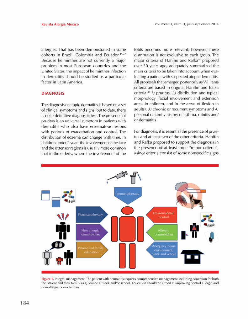
Figure 1. Integral management. The patient with dermatitis requires comprehensive management including education for both the patient and their family as guidance at work and/or school. Education should be aimed at improving control allergic and non-allergic comorbidities.

185
Sánchez J et al. Atopic dermatitis guideline
RReevviissttaa
MMééxxiiccoo
and symptoms which suggest allergy, like xerosis, pityriasis alba, cheilitis, follicular hyperkeratosis, white dermatographism, ichthyosis, high total IgE, conjunctivitis, tendency to skin infections, facial erythema, Dennie Morgan bifold, sensiti-zation to food, contact dermatitis and seborrheic dermatitis, among others.
Severity
Classifying the patients according to the severity and intensity of symptoms allows evaluating in an appropriate and effective manner the response to treatment. Several tests have been developed for this purpose and have been validated in di-fferent populations.50 Among the most frequently used are the SCORAD (Severity Scoring of Atopic Dermatitis), the objective SCORAD, EASI (Ec-zema Area and Severity Index), and the POEM (Patient-Oriented Eczema Measure).51,52 The full version of any of these scales and its usage can be obtained in the references cited, and there are several applications to computers, mobile phones and tablets that allow a quick and easy access. In these tests, the severity of dermatitis is basically defined according to three parameters; extension, severity and subjective perception.
Basically, the mentioned tests give a severity clas-sification according to the score obtained. It can be classified as mild, moderate or severe. Taking SCORAD as reference, the scale goes from 0 to 104 points, and ranks as “mild” when patient is below 15 points, 16 to 40 “moderate”, and over 40 “severe”.51 Nevertheless, the clinical history should be considered to assess the severity of symptoms, like the presence of comorbidities, the response to drug treatment and the duration of previous symptoms. All these parameters can help to predict the evolution and prognosis of the patient.50 Recently some European guideli-nes53 proposed to classify patients with transient symptoms as “mild”, recurring as “moderate” and persistent as “serious”. This is an interesting
proposal because new variables are included. However, it must be validated, and it carries the risk of many patients rated only by the persisten-ce of symptoms as serious, even if they are not (eg, patients with SCORAD <15).
Phenotypes
Phenotypes according to sensitization. Classi-cal dermatitis classification divides patients in intrinsic or extrinsic according to the presence or absence of sensitization to an allergen.54 Basically this classification divides patients in extrinsic dermatitis when they have high levels of total IgE (generally accepted > 200 kU/L), or a de-monstrated sensitization to aeroallergens or food allergens. The term intrinsic dermatitis is applied when patients do not meet any of these criteria. This division was made thinking that there were two separated immunological processes,55 but currently there is another hypothesis proposing that both immunological mechanisms are part of the same process in different periods of time, where the intrinsic dermatitis is the initial phase and extrinsic dermatitis the final phase, but this is under research.56 These hypotheses are not mutually exclusive and each one may represent a different group of patients.
The population characteristics in Latin America, especially in the tropical area, make it neces-sary to consider some issues when using this classification. We now know that up to 20% to 40% of the general population without allergic symptoms may have sensitization without cli-nical relevance.57 A big part of the non-allergic population in Latin American cities seem to have total IgE levels above 200 kU/L,58 so this cutoff would not serve as a criterion for classifying dermatitis as intrinsic or extrinsic. This higher concentration of total IgE in the tropical popu-lation seems to be due to the high frequency of helminthes infections. There is the additional complication that some of these parasites such as

186
Revista Alergia México Volumen 61, Núm. 3, julio-septiembre 2014
Ascaris lumbricoides, have cross-reactivity with some mite’s proteins,58,59 which makes it difficult to interpret the clinical relevance of sensitization.
Phenotypes according to immunological chan-ges. Parallel to the better understanding of the pathophysiology of AD, a more accurate classifi-cation has been developed to allow, through the use of multiple biomarkers, a greater certainty in the prediction of the evolution of dermatitis, and also to define a more effective treatment for each patient. Three processes that may occur in parallel or sequentially have been described in patients with dermatitis. In the first process, is observed a predominantly Th1 response charac-terized by the expression of cytokines such as IL-1, IL-6, TNF-beta, and dendritic cells with few exilon receptors in the membrane. This process predominates in those patients classified with intrinsic dermatitis and in patients with extrinsic dermatitis during inter-critical periods; in this process, defects in the epithelial barrier are gene-rally less severe, and in a significant percentage of patients, symptoms disappear with time. In the second process, there is a predominance of Th2 response characterized by both airbor-ne and food allergen sensitization and can be started in a spontaneous way or in patients who previously had a predominantly Th1 response.60 This process is often associated with asthma, has lower remission rate and greater severity. It is often associated with defects in filaggrin gene, which may be suspected from some clinical data such as palmar hiperlineality and eczema herpeticum.23 The third process is the presence of an autoimmune response mediated by IgE. It is suggested that this may be due to the homology between human proteins and allergens from other species, and represent the most serious phase in a patient with dermatitis as a result of the persistent exposure to intrinsic allergens.61-63
These three processes represent different “endo-phenotypes” of the dermatitis and their
identification would predict the likelihood of remission and the treatment required (whether or not avoidance of allergenic sources, treatment with topical or systemic immunomodulators, etc.).64 As mentioned in the previous section, although these processes may occur separately, can also be different stages of a single process where Th1 (process 1) is the first step response, the Th2 response (process 2) the second stage and sensitization to auto-allergens (process 3) the final stage.60 Although the identification of endo-phenotypes is promising in the diagnosis and treatment of atopic dermatitis, the procedu-res necessary to implement this classification, specially the final stage, are not widely available.
Classification according to age of presentation. 80% of the cases usually begin before age 2.65,66 Of the 192 children included in a multicenter birth cohort from Germany (Cohort MAS), 43.2% had a complete remission between 2 and 7 years of age, 18.7% persisted with symptoms and 38% had an intermittent pattern with occasional relapses. The persistence of symptoms seemed to be determined by the severity and the pre-sence of lower respiratory symptoms. 72.2% of children with persistent symptoms had an early onset (before the first year of life) and greater severity, while the majority of children with intermittent symptoms and minor scratching, had a later onset (Over first year) (OR = 5.86, 95% CI = 3.04 - 11.29).65 As mentioned in the “Risk Factors” section, in Latin American cities seems to predominate an even later onset (> 3 years)42 similar to that found in some European and Asiatic countries and it is not correlated with the severity of symptoms.6,36
The onset of disease in adulthood (> 14 years) may occur in up to 20% of patients and these cases have been little studied. A study conduc-ted in Germany by Garmhausen et al.67 found that in 725 adolescent and adult patients with dermatitis, 45% have had onset before 6 years,

187
Sánchez J et al. Atopic dermatitis guideline
RReevviissttaa
MMééxxiiccoo
10% between 6 and 14, 13% between 14 and 20 and 18% after 20 years. Sensitization and total IgE levels were higher in the groups with earlier onset, but the persistence of symptoms was hig-her in those who had onset after age 20. This is in contrast to the Cohort MAS, which found that over 80% of patients with dermatitis initiated symptoms before age 2. Since both studies were performed with German population, we can assume that environmental and anthropological changes rather than genetic inheritance could influence the different courses of dermatitis.65 One study evaluating the treatment of atopic dermatitis indicated that success in controlling eczema is directly related to early intervention and a multidisciplinary treatment. This is impor-tant considering as it may determine, at least in a subgroup of patients, the type of development that will have the disease in its future.68
Laboratory test
IgE total
Patients with dermatitis (and any other allergy) usually have high levels of total IgE. The clinical relevance of total IgE in the diagnosis and mo-nitoring of patients with atopic dermatitis has been studied broadly. A study in Japan found that between 16 biomarkers, only total IgE levels at 6 months of life in patients with dermatitis was an important predictor of persistent disease at 14 months of life.69,70 Similarly, in Spain, total IgE levels were higher among patients with dermatitis during an exacerbation.71,72 Other researchers found that 20 children with derma-titis and elevated IgE levels higher than 10,000 kU/L compared with 56 children with dermatitis and IgE levels between 400 to 1,000 kU/L had a higher rate of sensitization and greater severity.73 The clinical response of systemic treatments such as the use of azathioprine,74 gammaglobulins,75 immunotherapy76,77 and topical treatment with calcineurin inhibitors and steroids78 appears to
be associated with a reduction in total IgE, which would recommend the use of total IgE as a spe-cific marker of control. However, several factors preclude routinely recommendation of the use of Total IgE in patients with atopic dermatitis; not all studies show a clear correlation between total IgE levels and clinical improvement and in some patients high total IgE levels may persist elevated for a long time, even with a significant improvement in clinical symptoms.75,79,80 Another factor to consider is that parasites infection can elevate levels of total IgE, specially in Latin Ame-rica population, where parasites infections are an endemic problem, making it difficult to establish cutoff to predict the response to treatment.
Indication. Diagnostic extrinsic or intrinsic der-matitis. Evaluation and monitoring of patients with atopic dermatitis.
Committee recommendation. Weak. May be used in patients younger than 6 months with severe dermatitis and in patients over 5 years old with persistent severe symptoms.
Particular considerations in Latin America. It is necessary to know the “normal” values of total IgE in different regions of Latin America to re-commend performing this test routinely.
Allergen sensitization
Sensitization can be assessed by measurements of serum specific IgE or skin prick test. Sensitiza-tion to various sources of allergens in early life (especially food) may be transient, but atopic dermatitis patients are usually sensitized to a lar-ger number of sources than asthmatics or rhinitis patients.81 Some cohorts in Europe indicate that sensitization to food in children with dermatitis, occurs in the first years of life (<2 years) and is then replaced by sensitization to aeroallergens. This behavior does not seem to be shared by most of the tropical populations, where sensitization
188
Revista Alergia México Volumen 61, Núm. 3, julio-septiembre 2014
to mites usually starts even before the first year of life among patients with allergic symptoms.42,82
In Europe, levels of specific IgE (especially mites and cat dander) appear to be associated with the severity of symptoms.83 High serum concen-tration or skin prick test have been associated with an increased risk of reactions to foods, especially in patients with severe dermatitis.84-86 In the city of Medellin (Colombia), a correlation was observed between a pattern of sensitization to allergen sources (mites, dog dander, pigeon droppings, mold and cockroach) and the parallel development of atopic dermatitis and asthma, which indicates that the pattern of sensitization could predict the severity of disease and the development of the “atopic march”.87 However, care must be taken in the interpretation of the results because patients with dermatitis may have a high frequency of sensitizations without clinical relevance, and performing unnecessary avoidance measures can lead to poor patient adherence to therapy and important impairment to their quality of life. In a recent review of the epidemiology of food allergy in Latin America, it was observed that the behavior of food allergy is different to that reported in other countries; sensitization to milk and egg was important but less frequent than other sources as corn, tomato and pork.88
The sensitization to microbial proteins was ob-served in 50% to 80% of patients with dermatitis and has been correlated with AD severity.89-91 There has also been observed a greater sensi-tization to Malassezia furfur (previously called Pityrosporum ovale), although this has not been clearly correlated with the severity of symp-toms.92,93 The usefulness of measuring these extracts in patients with atopic dermatitis is still unclear.
The response against some auto-allergens (Hom s called because they are derived from the Homo
sapiens) appears to be specific for patients with severe atopic dermatitis,61-63,94 which would allow predicting patient prognosis. However, extracts required for these tests are not commer-cially available.
Indication. Diagnosis and monitoring of patients with atopic dermatitis. Identification of environ-mental sources exacerbating symptoms.
Committee recommendation. Aeroallergens: Strong. All patients with dermatitis. Food aller-gens: Strong. Recommended only in patients with clinical suspicion or serious and/or per-sistent presentations. The test battery should be consistent with the geographical area where the patient lives.
Particular considerations in Latin America. In Latin America there are many studies that provide insight into the most relevant aeroallergens, but there are few studies evaluating food allergens from the region.88,95
Patch test with food and/or aeroallergens
The underlying mechanism to be detected by the path test is the presence of T lymphocytes that mediate late reactions in patients after exposure to different sources.
The food patch test usually includes soy, wheat, egg and milk, but many other foods have been tested. Several articles support the usefulness of this test, especially in patients with atopic dermatitis.96,97 Due to the wide range in the predictive values (40% to 80%) and the lack of standardization of the technique, the test has been criticized and rejected by several groups.98 However, it is used in several centers because of its easy realization and potential utility detecting allergic processes mediated by T lymphocyte. In addition, some studies suggest that this test can reduce the requirement for provocation test and

189
Sánchez J et al. Atopic dermatitis guideline
RReevviissttaa
MMééxxiiccoo
avoid unnecessary restriction diets when is done with skin prick test.99,100 The patch with aeroaller-gens especially mites has also been studied;101 however, there are very few studies that validate its routinely use and also lacks a standardized universally accepted method.
Indication. Evaluation and monitoring of patients with atopic dermatitis and suspected delayed reactions with food allergens or aeroallergens.
Recommendation of the Committee. Recom-mendation for food test: Moderate. Patients with clinical suspicion of a particular food with negative IgE response or late-onset symptoms. Recommendation for aeroallergens path test: Weak. Few controlled studies. The battery must be consistent with the geographic area where the patient lives, yet few controlled studies are available and these usually include only soy, wheat, milk and egg.
Particular considerations in Latin America. In Latin America there are few studies evaluating the usefulness of the patch test, and results are in favor of its use;102 however, it is necessary to standardize the technique and taste local pro-ducts that could be cause sensitization.
Patch with standard battery and other types of patch
Contact dermatitis occurs frequently in patients with atopic dermatitis (15%-30%).103,104 The inflammatory process in the skin, which facili-tates the uptake of environmental antigens, can explain this. Patch test with standard battery is extremely useful in the identification of contact antigens.105 However, in patients with dermatitis there is a high risk of false positives, so its use in patients should be limited to cases with a strong suspicion of exacerbation for a contact, or in those patients with persistent refractory to treatment presentations.
The use of photo-patch for drugs and path for any other battery (cosmetics, shoes, etc.), should also be performed when there is a strong cli-nical suspicion, and it should be remembered that the appropriate concentration of many cosmetics and medicines to patch test are not standardized. When there is not available a reference concentration, it is recommended to perform the test in ten healthy subjects as a control group, that reduces the risk of false positives by irritation, but does not reduce the risk of false negatives.
Indication. Patients with strong suspicion of contact dermatitis. Patients with persistent and severe AD, refractory to medical therapy.
Committee recommendation. Standard battery: Strong. Other types of patch: Moderate. Routine use is not recommended in patients with atopic dermatitis.
Particular considerations in Latin America. Studies in Latin America show the availability and the high value of these tests as diagnostic support.106-108
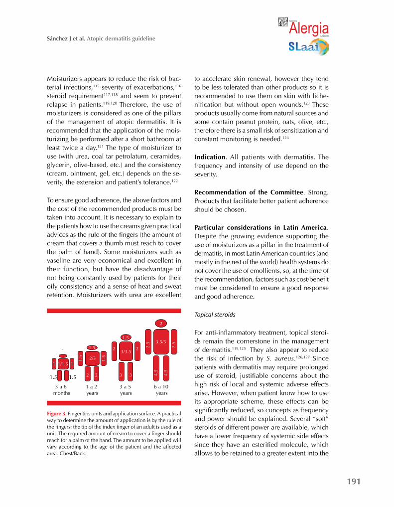
Very high power: hyperkeratotic region
(palms and plants) and severe injuries
Medium power: trunk, arms and legs
Low power: face, flexures,
perineum
High power: trunk, arms and legs. Thick skin
Palm
Front
Overleaf
Back
Figure 2. Topical steroids. Power of the steroid and applica-tion site.

190
Revista Alergia México Volumen 61, Núm. 3, julio-septiembre 2014
Provocation and food elimination diet
The food challenge is the gold standard for iden-tifying whether a suspected food is the cause of the patient’s symptoms, but due to the risks of ana-phylaxis and other severe symptoms, provocation should only be done when there are doubts in the diagnostic that cannot be clarified with skin tests and laboratory studies. Food symptoms can start immediately (pruritus, erythema) or later (worse-ning of eczema, new plates). In atopic dermatitis patients, is required long observation for days, even weeks, intercalated with food administra-tions to evaluate clinical changes.109,110 Because of these difficulties, food restriction for 4 to 6 weeks with the suspect food (and all products containing it), may be preferable in certain situations. If doubt persists then the provocation is necessary.
Indication. Patients with suspected food allergy that has not been cleared with skin or serum tests.
Committee recommendation. Strong. We recom-mend initially performing the diet restriction, and if the relationship with food is not clarified it should be performed a controlled provocation.
Particular considerations in Latin America. As in the rest of the world, there are few studies in Latin America using provocation tests in the evaluation of food allergy in patients with der-matitis.111 It is necessary to establish protocols with native foods.
Complementary studies
Laboratory tests as CBC, electrolytes, measure-ment of cortisol, liver function, kidney function, etc., are not indicated as routine exams. They could be indicated as part of the follow up when the patient requires the use of immunosup-pressants such as cyclosporine, prolonged oral steroids, etc. Skin biopsy could be indicated for differential diagnosis.
ACTIVE MANAGEMENT
First line management
Skin care and hydration
Dry skin (xerosis) is one of the main symptoms of dermatitis and a key point in its patho-physiology. Xerosis may occur as a result of defects in filaggrin or lack of lipids and other particles in the stratum corneum leading to a lack of continuity of the barrier.112 Due to the continuous skin peeling in these patients, the skin should be thoroughly cleaned during bathing, removing all debris that could sti-mulate bacterial growth. Drying seems to be even more effective than antiseptics to remove debris and prevent superinfection. Because long baths with very hot or very cold water may promote xerosis and make mechanical irritation, it is recommended short baths (<5 min) with slightly cold water. In patients with a history of skin infection, or in patients with risk of infection, is recommended to add one or two drops of hypochlorite per liter of water during bath to prevent bacterial growth.113 The use of oils or bath salts in the final two minutes of the bathroom also favors greater skin cleansing and improved skin hydration. However, using soaps must be avoided, or if necessary, neutral products can be used in areas that require it (armpits, pubic areas, etc.). Moisturizing lipstick is also recommended for patients with cheilitis. The nails of patients with dermatitis should be cut frequently to avoid scratching during sleep, and baggy clothing is recommended, preferably make of cotton to avoid heat and irritation.114 For what we know many of these products and measures appear to be useful, but there are few controlled stu-dies demonstrating their effectiveness. Since in most health systems these products are not covered and are funded directly by patients, the cost/benefit relation should be considered.
191
Sánchez J et al. Atopic dermatitis guideline
RReevviissttaa
MMééxxiiccoo
Moisturizers appears to reduce the risk of bac-terial infections,115 severity of exacerbations,116 steroid requirement117,118 and seem to prevent relapse in patients.119,120 Therefore, the use of moisturizers is considered as one of the pillars of the management of atopic dermatitis. It is recommended that the application of the mois-turizing be performed after a short bathroom at least twice a day.121 The type of moisturizer to use (with urea, coal tar petrolatum, ceramides, glycerin, olive-based, etc.) and the consistency (cream, ointment, gel, etc.) depends on the se-verity, the extension and patient’s tolerance.122
To ensure good adherence, the above factors and the cost of the recommended products must be taken into account. It is necessary to explain to the patients how to use the creams given practical advices as the rule of the fingers (the amount of cream that covers a thumb must reach to cover the palm of hand). Some moisturizers such as vaseline are very economical and excellent in their function, but have the disadvantage of not being constantly used by patients for their oily consistency and a sense of heat and sweat retention. Moisturizers with urea are excellent
to accelerate skin renewal, however they tend to be less tolerated than other products so it is recommended to use them on skin with liche-nification but without open wounds.123 These products usually come from natural sources and some contain peanut protein, oats, olive, etc., therefore there is a small risk of sensitization and constant monitoring is needed.124
Indication. All patients with dermatitis. The frequency and intensity of use depend on the severity.
Recommendation of the Committee. Strong. Products that facilitate better patient adherence should be chosen.
Particular considerations in Latin America. Despite the growing evidence supporting the use of moisturizers as a pillar in the treatment of dermatitis, in most Latin American countries (and mostly in the rest of the world) health systems do not cover the use of emollients, so, at the time of the recommendation, factors such as cost/benefit must be considered to ensure a good response and good adherence.
Topical steroids
For anti-inflammatory treatment, topical steroi-ds remain the cornerstone in the management of dermatitis.119,125 They also appear to reduce the risk of infection by S. aureus.126,127 Since patients with dermatitis may require prolonged use of steroid, justifiable concerns about the high risk of local and systemic adverse effects arise. However, when patient know how to use its appropriate scheme, these effects can be significantly reduced, so concepts as frequency and power should be explained. Several “soft” steroids of different power are available, which have a lower frequency of systemic side effects since they have an esterified molecule, which allows to be retained to a greater extent into the
Figure 3. Finger tips units and application surface. A practical way to determine the amount of application is by the rule of the fingers: the tip of the index finger of an adult is used as a unit. The required amount of cream to cover a finger should reach for a palm of the hand. The amount to be applied will vary according to the age of the patient and the affected area. Chest/Back.
3 a 6 months
1 a 2 years
3 a 5 years
6 a 10 years
1 1
1.51.5
1/1.5
1
22 3 3 4.5
4.5
2/33/3.5
3.5/52 2 2.
5
2.5
1.5
1.5
1.5
1.5
2

192
Revista Alergia México Volumen 61, Núm. 3, julio-septiembre 2014
skin, and are easily degraded when they enter to the circulation.128-130 Despite the widespread and undeniable usefulness of steroids in dermatitis, there are few controlled studies supporting their uses or how to use them. Different schemes have been proposed in the use of steroids and some common points are present:
Steroids with high potency should be used only in patients with moderate to severe atopic dermatitis, and should be avoided in the facial, folds and perennial regions, and they must be used with caution in children under two years. They could be considered in those three areas previously described in exceptional cases and for periods not exceeding 7 days. In all patients they should be used for the minimum possible time and switching to medium or low power ste-roids according to the control of the patient. The
continuous use of steroids for prolonged periods in wide body extensions (even mild steroids) can have similar risk of adverse effects than oral or intravenous steroids. The use of intermittent treatment appears to reduce this risk even with high potency steroids.131
Steroid use with moisturizer seems to impro-ve the power of the steroid and increase the time of its effect on the skin, so it is recom-mended their joint application in mixtures or separately, according to the severity of the symptoms. Most oily moisturizers may promote absorption of steroids for its occlu-sive effect. Proactive management consisting of intermittent application of a low power steroid or calcineurin inhibitors, appears to significantly reduce the risk of relapse in patients under control.119
Patient with suspected dermatitis
Yes Clinical history supports the diagnosis?
No Consider other diagnoses
Allergic sensitization?
Skin care, avoiding irritants.
Topical treatment
Avoiding triggers and/or causal agents, exclusion diet (if necessary) and
evaluate potencial benefit of immunotherapy
Successful management?
Satisfactory remission
Continuous hydration and consider proactive
management
Refractory or severe dermatitis
Consider other diagnoses. Consider systemic immunomodulators
Yes
No
No
Yes
Figure 4. Management algorithm.

193
Sánchez J et al. Atopic dermatitis guideline
RReevviissttaa
MMééxxiiccoo
The application of steroids more than once a day seems to report no advantage but increases the risk of adverse effects, especially in the sensitive areas of the face or skin folds.
In acute injuries, it is advisable to mix the steroid with an emollient to prevent irritation in the area ant to increase absorption.
The application of occlusive steroid systems should be performed only by the indication of specialists (allergists or dermatologists).130
Indication. All patients with atopic dermatitis. The power of steroid and frequency of use will depend on the course and severity of patients.
Recommendation of the Committee. Strong. However, more controlled studies do not select the best scheme for each patient.
Particular considerations in Latin America. Latin America has a wide variety of steroids, allowing to calibrate the potency according to the needs of the patient. It must be taken into account the characteristics of the tropics and subtropics regions when choosing the consistency (cream, ointment, etc.) to improve patient adherence.
Calcineurin inhibitors
There are two topical calcineurin inhibitors: tacrolimus and pimecrolimus. Both have proven efficacy in dermatitis132-135 in active and proac-tive treatment.136 In practice, they can be used for the same indications as a steroid of medium (tacrolimus 1%) or low power (tacrolimus 0.03%, pimecrolimus 1%),137,138 with the advantage that if continuous treatment is required, it will have a lower risk of adverse effects and it will not cause skin atrophy. However, it is necessary to avoid open injuries because they often produce bur-ning feeling.139 Other less common side effects include eczema herpeticum or molluscum.140-142
Although there is no evidence to show a causal relationship between cancer and the use of to-pical calcineurin inhibitors, it is recommended to be aware of the possible association during follow-up of patients.138
Indication. All patients with atopic dermatitis for active and proactive management.
Recommendation of the Committee. Strong in the above indications.
Particular considerations in Latin America. Cu-rrently in most Latin American countries both tacrolimus and pimecrolimus are available.
Allergen-specific immunotherapy
In the last two decades several controlled studies have been conducted showing that a significant percentage of patients with atopic (or extrinsic) dermatitis can benefit from this therapy, although impact varies according to the severity of patients.143-147 A study conducted in the city of Medellin (Colombia) showed that patients with moderate dermatitis according to the SCORAD, had a greater and more significant reduction in symptoms compared to placebo, as well as a significant increase in IgG4.80 These results are similar to those observed in other studies (147), but there is a need of additional studies to characterize better the patients who can benefit from this therapy. Several reports have shown that some patients may experien-ce exacerbation of cutaneous symptoms and even systemic symptoms with immunotherapy, however, when the administration is controlled especially with modified extracts, the risk of systemic reactions is greatly reduced as obser-ved in a retrospective study, where 114 patients with dermatitis which were applied over 1000 injections, and none had a systemic reaction nor abandoned therapy for the exacerbation of symptoms during treatment.
194
Revista Alergia México Volumen 61, Núm. 3, julio-septiembre 2014
Indication. Patients with persistent moderate or severe atopic dermatitis who have a clear relationship of exacerbation with aeroallergens.
Recommendation of the Committee. Moderate. There are needed further studies to characterize which patients benefit most from this therapy.
Particular considerations in Latin America. There are some studies in Latin America that support the efficacy and safety of using the specific aller-gen immunotherapy with Dermatophagoides farinae and Dermatophagoides pteronyssinus in patients with dermatitis, but studies using other common allergen sources in the region as Blomia tropicalis, Dermatophagoides siboney and some pollen grains are needed.
Environmental and dietary control
Since the skin of patients with dermatitis is very sensitive, many agents can act as irritants increasing the inflammatory process and the-refore should be avoided. Patients must learn to recognize irritant substances such as soap, detergents, some creams, polluted air and other specific factors present in their environment.148,149 How strict must be patient with these measures will depend on its severity. If possible, patients should also perform a control of temperature and humidity of the room where they live.
Allergenic sources which patient is sensitized should be avoided. “Prophylactic” restrictions (removal of pets, restricted diets, etc.) when there is not a clinical relevance are not recommen-ded. Recommendations should be very careful, particularly with diet, because in patients with dermatitis the number of irrelevant sensitizations can be high, so it is necessary to test only those foods with clinical suspicion to avoid confusion and unnecessary restrictions that can lead to nutritional problems in the patient.150 Some steps to reduce the amount of allergens in the home
as mop, cleaning with damp cloth, or removing pets (only when patient is sensitized) have been proposed, but few studies support that these restrictions lead to a significant improvement in the patient due to indirect exposure.151-153 Unless there is a clear clinical relationship and sensitization is demonstrated, other factors such as emotional attachment should be taken into ac-count before recommending the removal of pets, and it is necessary to consider that the amount of allergens from pets only starts to decrease significantly 3-4 months after removal.
Indication. All patients with dermatitis need to identify and avoid possible triggers of their ill-ness. Allergy food studies should be performed in patients with clinical suspicion or persistent presentations.
Recommendation of the committee. Strong to the above recommendations.
Particular considerations in Latin America. It should be evaluated the environmental condi-tions of each patient and dietary customs, which are different in Latin America countries.95,154
Second line management
Antihistamines
Antihistamines have been used for many years in patients with dermatitis to reduce itching; howe-ver, the majority of controlled studies evaluating their effect show little or no effect in reducing pruritus,155-157 perhaps because the itching in dermatitis have several pathways including the increased production of IL-33.33 The preference of many physicians to use first-generation an-tihistamines for their sedative effect, must be balanced by the risk of side effects that chronic use of these drugs may have (low concentra-tion, drowsiness, etc.).158 Among the second generation, controlled studies with loratadine,

195
Sánchez J et al. Atopic dermatitis guideline
RReevviissttaa
MMééxxiiccoo
fexofenadine and cetirizine show that these drugs have small effect in the control of pruritus.159-161 A recent study shows that antihistamines might promote a faster skin repair;162 however, more studies are needed to assess the true impact of these medications as repairers of the skin barrier. Since patients with dermatitis often have other comorbidities, such as rhinitis, it is frequent the use of antihistamines.
Indication. According to the comorbidities of each patient.
Recommendation of the Committee. Weak. There is needed more studies evaluating the ad-vantages and disadvantages of potential sedative and restorative effect in skin.
Particular considerations in Latin America. Due to the high frequency of comorbidities in patients with dermatitis in Latin America, the use of antihistamines is common, however, it should not be expected to control the itchy with this treatment alone.
Systemic steroids
It is clear that systemic steroids are useful in patients with severe disease, especially during exacerbations.163,164 However, due to the high risk of adverse effects (cataracts, osteoporosis, height, etc.) they are not recommended for prolonged use. Oral stPPeroids have been associated with higher relapse rate after suspension compared with other immunosuppressants such as cyclos-porine.164 To avoid these adverse effects, it is recommended to adjust the dose according to patient weight and to reduce the dose. To achieve complete suspension reduce the doses until fully suspension once the patient gets control.165 A used scheme is the administration of the full dose for 5-7 days, then half dose for another 5-7 days and last for three days and suspend interspersed. However there is no standard way to do this.
Indication. Patient with severe acute cases that do not respond to first-line management. It is not recommended chronically, even at low doses.
Recommendation of the committee. Strong for acute exacerbations.
Particular considerations in Latin America. The use of systemic steroids is quite popular in Latin America, unfortunately in many cases as chronic treatment. It is necessary to educate patient and physician to avoid overuse.
Sun exposure and phototherapy
An European study found that 74% of patients with mild to moderate dermatitis had a signifi-cant improvement over the summer with relapse in the other seasons.166 Additionally, those who spent their summer days near the sea had grea-ter improvement than those who passed it near the mountains. These results suggest that sun exposure (15 to 20 minutes a day from 7:00 to 8:00 am or 3:00 to 4:00 pm) has a beneficial effect. Since in the tropics high temperatures and humidity often accompany sun exposure, care should be taken when recommending controlled exposures because these conditions can exacerbate patient’s pruritus. Phototherapy has the advantage that it is done in controlled environments and gives substantial improvement in 40 to 50% of patients with moderate or mild dermatitis.167,168 Mechanisms leading to this effect are not clear yet, but it seems to be influenced by various pathways that produce an antimicrobial effect, inhibiting the activity of Langerhans cells and favoring the production of vitamin D.169,170 Phototherapy can be performed with various wavelengths (UVB, broadband UVB, UVA1) being preferred short waves. Although its use has been studied primarily in adults, some data suggest that narrow band UVB can be used safely in children. Its indication is mainly in patients with refractory signs of lichenification, however,

196
Revista Alergia México Volumen 61, Núm. 3, julio-septiembre 2014
some studies also suggest its use in acute exacer-bations.171,172 Exacerbations during phototherapy can be frequent (3%-20%) so the tolerance of each person must be carefully evaluated. Other side effects such as burns, hyperpigmentation fatigue, nausea and headaches can also occur with little frequency, while the more serious side effects, such as skin cancer, are less com-mon, but patients should be warned.173 There are few studies comparing the different types of phototherapy in dermatitis, so the advantages or disadvantages of one form over the other are not demonstrated for this disease.174
Indication. Sun exposure: All patients with annotated considerations to avoid itching. Phototherapy: Adult patient with recalcitrant symptoms that do not respond to first-line ma-nagement.
Recommendation of the Committee. Sun expo-sure: Weak. There are no studies in the tropical and subtropical region. Phototherapy: Strong for chronic conditions in adults.
Particular considerations in Latin America. Although currently several centers in different countries of Latin America have phototherapy units, its use for dermatitis is rare.175 This may be due to the difficulty in the mobilization and poor dissemination of this approach for dermatitis. Studies are needed in tropical and subtropical regions.
Cyclosporine A
Cyclosporine is a potent inhibitor of T lym-phocytes immune response through binding to cyclophilin. Cyclosporine have a lot of studies evaluating its efficacy and safety.176,177 A syste-matic review of the literature that included more than 10 trials in children and adults, concluded that this therapy is clinically effective but with a high relapse rate when suspended.178 The clinical
response is observed after two weeks reaching its greatest effect at 2 or 3 months. Despite its high efficacy, there is a significant risk of nephro-toxicity and hypertension, so the dose should be reduced to the minimum necessary and regular monitoring of blood test, blood pressure and renal function is required. Other common effects are nausea, abdominal pain and paresthesias.
Indication. Recalcitrant patient with severe symptoms that do not respond to first-line ma-nagement.
Recommendation of the Committee. Strong to severe chronic conditions.
Particular considerations in Latin America. Currently there are no studies with cyclosporine and dermatitis in Latin America. However, it is available in most countries.
Third line management
Mycophenolate mofetil
Mycophenolate is an inhibitor of purine synthe-sis and it stops the division of several cell lines, including lymphocytes. Although there are numerous reports showing its positive effect in patients with dermatitis,179 there are few con-trolled studies. Most reports show that in adults mycophenolate is generally well tolerated. Between its side effects are nausea, vomiting, retinitis and herpes. In an uncontrolled study with 14 children under 15 years of age, it showed a beneficial effect and a low rate of adverse effects, being mostly mild.180 In a controlled study comparing the effect of mycophenolate and cyclosporine, it was observed that the rate of adverse reactions was lower for mycophenolate. However, at 6 weeks, patients with cyclosporine had fewer exacerbations and clinical impro-vement was superior than in the group with mycophenolate.181

197
Sánchez J et al. Atopic dermatitis guideline
RReevviissttaa
MMééxxiiccoo
Indication. Recalcitrant patients with severe symptoms that do not respond to the manage-ment of first and second line.
Recommendation of the Committee. Weak. Further studies are required.
Particular considerations in Latin America. Whi-le this drug is widely available in Latin America, there are currently no studies with mycophe-nolate mofetil and dermatitis in Latin America.
Azathioprine
Although the precise mechanism of action of azathioprine is not known, it has been used for many years in the management of dermatitis. Se-veral controlled studies support its use, especially in severe cases with population over 6 years of age.182-184 However, in these studies the rate of withdrawal is high due to the frequent incidence of adverse effects (nausea, vomiting, abdominal pain). It is necessary to monitorize patients with laboratory test. Four to eight weeks are usually enough time to evaluate the clinical response.
Indication.Recalcitrant patient with severe symptoms unresponsive to handling first and second line.
Recommendation of the Committee. Moderate. Although more studies are needed, it can be an alternative when cyclosporine is contraindicated.
Particular considerations in Latin America. Currently there are no studies with azathioprine and dermatitis in Latin America. However, it is available in most countries.
Methotrexate
Methotrexate has been widely used in various skin problems, especially in psoriasis. In the case of dermatitis, there are few controlled studies and
therefore the appropriate dose and frequency of adverse effects is limited. In a comparative study, a similar effect was observed between metho-trexate and azathioprine185 and in two other studies doses of 10 to 25 mg per week showed a reduction in the severity of eczema.186,187
Indication. Adult patient with severe recalcitrant symptoms that do not respond to the manage-ment of first and second line.
Recommendation of the Committee. Weak. Further studies are required.
Particular considerations in Latin America. Cu-rrently there are no studies with Methotrexate and dermatitis in Latin America. However, it is available in most countries.
Fourth line management
Probiotics and prebiotics
Probiotics and prebiotics have been used on the prophylactic and active management of dermati-tis. In a case-control study, Kalliomäki et al. found that early administration of Lactobacillus rham-nosus prevents the development of eczema in children under 4 years.188,189 A review of Cochrane in 2007 based on 6 controlled studies published to that date, observed a reduction in eczema of children receiving probiotics prophylactically. But due to methodological biases, the authors concluded that there is not yet enough evidence to recommend adding probiotics in children at risk of dermatitis.190 Another meta-analysis pu-blished in 2010 shows that the administration of Lactobacillus spp during pregnancy prevents development of eczema in children with 2 to 7 years.191 These studies highlight that probiotics may have a positive effect in the prophylactic treatment, however there are still questions about the dosage and time of administration. Other important questions are what kind of strain is

198
Revista Alergia México Volumen 61, Núm. 3, julio-septiembre 2014
the most appropriate for each population, since the effect of probiotics in part depends on the characteristics of the intestinal microflora of the population. Currently in Latin America, a study evaluating the intestinal flora observed that the presence of Lactobacillus spp, reduces the risk of wheeze in children under two years, and in this cohort the frequency of dermatitis before three years was zero. It is necessary to evaluate in the tropics if supplementation of probiotics or prebiotics produces the same effect.192 Unlike prophylactic management, where the results are positive, most studies have shown little or no effect in the control of symptoms.193,194 A meta-analysis published in 2013 that included 13 studies, concluded that more studies are needed before recommending the routine use of prebio-tics in the prevention of allergies in children.195
Omalizumab
Although the current understanding of the patho-physiology of atopic dermatitis is incomplete, it is known that IgE play an essential role. Few studies have evaluated the effect of monoclonal anti-IgE in dermatitis but the results are promising.196,197 Patients with atopic dermatitis have much higher levels of IgE than patients with asthma, so doubts arise about the required dose needed to achieve control of patients with dermatitis. The maximum recommended dose (450 mg) in patients with asthma by the laboratory is calculated for total IgE levels of 750 kU/L, but several reports suggest that even with this dosage, patients with severe dermatitis can achieve positive results even with total IgE levels greater than 1,000 kU/L.198-200 Other studies have shown changes in immu-nological response but not clinical effect.201,202
IFN-γ
The gamma interferon (IFN-γ) is a cytokine that exerts its anti-inflammatory effect in dermatitis, inhibiting IgE synthesis and proliferation of
T lymphocytes. IFN-γ has been shown to be effective in reducing eosinophil count of patients with dermatitis and to further improve control of symptoms in patients with severe disease.203 A controlled study including 51 patients with severe recalcitrant dermatitis compared the clinical effect of low doses of IFN-γ (0.5 × 10⁶ IU/m2), high dose (1.5 × 10⁶ IU/m2), and placebo for 3 months of follow-up. Both groups treated with IFN-γ showed a significant reduction in the severity of symptoms compared to the placebo group, being faster in the higher-dose group but stable in both groups after two months. Potential adverse effects associated with IFN-γ are transient fever, myalgia, respiratory distress and elevation of transaminases and lipid profile.203
Other therapies
Some case reports have been published with several monoclonal like rituximab,204-206 efali-zumab,207 aterizumab, alafacept, mepolizumab, and eternacept, but so far the results have been contradictory and in few patients, so they cannot be recommended in routinely use.208 Intravenous immunoglobulin,209 therapy with autologous serum210 and some herbal products211,212 are often used in some countries with satisfactory results, but the dosage, availability of extracts and frequency of use is not standardized and therefore it is difficult to recommend their use in Latin America.
Hospital management
The hospital management should be avoided because of the high risk of complications. Howe-ver, when a patient with atopic dermatitis has a severe exacerbation with high risk of complica-tion, hospitalization should be consider.213,214 Some warning signs that suggest an imminent complication are:
Involvement of more than 50% of the skin surface with moist lesions or erythrodermia.

199
Sánchez J et al. Atopic dermatitis guideline
RReevviissttaa
MMééxxiiccoo
Sepsis or severe cutaneous infection, extensive or disseminated.
Involvement of other systems (respiratory, renal, etc.).
Limitation to perform their routine activities.
Failure to follow the established treatment.
The treating physician determines rapid dete-rioration.
Hospital management can ensure a continuous and adequate treatment for the patient and prevent further complications. However, due to the high risk of nosocomial infections and com-plications that it represent for the patient with dermatitis, other measures such as “hospital at home” may be more appropriate.
PREVENTION
Currently, there is no an intervention that has proven to be 100% effective in preventing the development of dermatitis or reduce its severity. However, the identification of modifiable risk factors permits to introduce indications that at least help to some of the population.
Primary prevention
Because dermatitis usually occurs in early child-hood, primary prevention is intended, principally for newborn. Although some genetic factors appear to be protective as black heritage, they can hardly be used to create preventive policies. Most studies looking protective factors have been directed to the type of diet and avoidance of potential triggers as we discussed earlier. The preventive effect of vitamin D is extensively studied. Although several studies show a clear association between low levels of vitamin D and the development of atopy, asthma and der-matitis,215 contrary to what would be expected,
vitamin D supplement in diet as primary preven-tion in children younger than 3 years seems to be a risk factor for dermatitis.216,217 The supply of vitamin D during pregnancy has been little stu-died and the results are also contradictory.215,218 In a systematic review of the literature, it was observed that some foods could have a pre-ventive effect on the development of dermatitis such as fruits, vegetables, unsaturated fatty acids, etc.219 Supplementation with polyunsaturated fatty acids n-3 during pregnancy appears to reduce the risk of dermatitis in the newborn;220 however, more controlled studies are required. Regarding pets, in a meta-analysis conducted in 2013, from 21 studies from birth cohorts, it was observed that the presence of dogs in the houses had a protective factor that reduced the risk of dermatitis by 25%. In the case of cats, it was not observed any risk or protective role.221
Secondary prevention
The goal of secondary prevention is to avoid common complications like exacerbations, bacterial superinfection with a worsening of severity. “Proactive management” has shown to be effective in preventing these complications as previously discussed (see “first line of mana-gement”). Probiotics have shown encouraging results in this regard and were treated in detail in the “fourth line management” section.
Several studies have shown that the use of topi-cal antibiotics for a week every month seems to prevent new superinfections and it also decreases the severity of symptoms.222 However, a meta-analysis conducted in 2010 did not observe significant statistically advantage with this thera-py, and there is also a warning about the risk of microbial resistance to antibiotics.194,223 Likewise, the use of oral antibiotics is not recommended unless the patient has an active infection. Des-pite disappointing results in primary prevention, vitamin D supplementation could be useful in a group of patients in secondary prevention. A

200
Revista Alergia México Volumen 61, Núm. 3, julio-septiembre 2014
controlled study showed that supplementation of vitamin D during the winter appears to redu-ce the severity of injuries,224 perhaps due to a possible antimicrobial effect on the skin.225 The supply of other vitamins (E and K) and minerals have also been proposed as adjuvant therapy, but there is not enough information to recommend these alternatives.211
SPECIAL SITUATIONS
Pregnancy
Dermatitis is the most common skin disease during pregnancy (36-49%).226 Histopathologi-cally there is no difference between pregnant and non-pregnant patients.227 Usually during the second half of gestation, 66% of patients present exacerbation of symptoms. Although the cause is unknown, this is attributed to the increase in Th2 polarization that normally occurs during pregnancy. However, among patients with non-atopic dermatitis there is also a worsening during pregnancy, therefore hormonal changes have been proposed as a possible cause.227,228 Although dermatitis seems not to cause direct problems in pregnancy, bacterial infections could potentially promote premature births, abortions or fetal growth restriction.229
Treatment in pregnant patients is essentially the same as in the rest of patients. It is essential to try to get control using the least quantity of topical steroids to decrease the risk of syste-mic reactions.226 It is also necessary to inform the patient the possibility of worsening during gestation. Although topical steroids are consi-dered category C during pregnancy, the first-line treatment (hydration, steroids, etc.) is the same. Calcinuerin inhibitors, oral steroids, cyclospo-rine and azathioprine can be used only in case of extreme necessity, while the methotrexate, mycophenolate mofetil, psoralens and PUVA therapy should be completely avoided.226 In the
case of antihistamines, several first generation are considered category B (chlorpheniramine, cy-proheptadine, diphenhydramine), this is because there are few studies with second-generation antihistamines, although loratadine appears to be a safe option.230
Breastfeeding
During lactation, it is necessary to note that the mother should restrict from their diet those foods to which the child is allergic, because some proteins can pass into breast milk and perpetuate symptoms in children.231,232 This should be done only in cases of severe disease that fail to control with first-line therapy. Breastfeeding appears to have a beneficial effect inducing a tolerogenic response to different allergens from the diet, so it should not be suspended.233 If the mother is recei-ving immunosuppressive drugs for dermatitis, she should take into account some considerations: Steroids can pass into breast milk but it seems that in small quantities. Cyclosporine should ideally be suspended during lactation, however it is not an absolute contraindication. No other immunosuppressive drugs are advised. Currently several second-generation antihistamines are ap-proved for use after 6 months of age (loratadine, fexofenadine, cetirizine, ebastine, bilastine).
Adult dermatitis
Although a group of patients with dermatitis of childhood onset may reach adulthood without being able to con trol the disease, in 5 to 15% of patients the disease onset is after the age of 14.67 The clinical action in these patients is essen-tially the same, however, the severity is usually higher and have a tendency to a greater number of non-allergic comorbidities.234 In addition, the proportion of patients with allergic dermatitis is usually higher. In these patients, it may be necessary a first line biopsy and a patch test to rule out other processes.

201
Sánchez J et al. Atopic dermatitis guideline
RReevviissttaa
MMééxxiiccoo
Table 2. Immunosuppressive drugs
Medicament Mechanism of action
Contraindications Laboratory test Efficacy Recommendation of the committee
Cyclosporine: 2.5-4 mg/kg/day
Inhibits the prolifera-tion of T lymphocytes
Relative: renal failu-re, hepatic disorders, pregnancy. Absolute: breastfeeding
Basal: BP, RF, HF, CBC Track: PA (fortnightly), RF, HF, CBC ideally graduate dose accor-ding to blood levels
50-70% Strong
Phototherapy:Number of sessions d e p e n d s o n t h e patient's age and se-verity
No clearly defined Relative: Pregnancy, children under 6 years
Clinical follow. CBC quarterly
40-70% Strong
Azathioprine:1 mg/kg/day, after 4 weeks two raised to 2 to 2.5mg/kg. Adminis-ter with meals
Inhibits purine synthe-sis and incorporation into DNA thioguanine
Relative: interact with allopurinol and war-farin. Absolute: pregnancy
Basal: BP, RF, HF, CBC pregnancy test, gra-duating Ideally dose according to levels of TPMT, evaluate lymphadenopathy. Track: (1, 2, 3 month then bimonthly): Sam-pling every 5-6 days after dose changes
30-80% Moderate
Mycophenolate mo-fetil: 1 to 2g daily (max. 3g)
Inhibits the synthesis of guanosine nucleo-tides
Relative: infections, kidney failure, liver disease, pregnancy. Absolute: breastfee-ding
Basal: BP, RF, HF, CBC pregnancy test. Track (quarterly): trimestral paraclinical
60-80% Weak
Methotrexate 5 to 25mg once a week
Folic acid analogue Relative: deficit fo-lic acid. Absolute: breastfeeding. Renal dysfunction, liver, DM, recurrent infec-tions
Basal: HF, CBC, he-patitis A/B/C, FR, HIV (optional). Track: (2-4sem and then quarterly): CBC, platelets, RF
50-70% Weak
Omalizumab Blocks free IgE Relative: parasites in-fection, dyslipidemia, abnormal ECG
Basal: CBC, lipid pro-file, ECG
30-50% Weak
INF-gamma Inhibits IgE produc-tion and T cell proli-feration
Relative: recurrent infections
Basal: RF, HF, lipid profile, CBC, ECG. Track (quarterly) : CBC, platelets
40-62% Weak
BP: blood pressure; RF: renal function; LF: liver function; CBC: blood count; ECG: electrocardiogram; G6PD: glucose-6-phosphate dehydrogenase; TPMT: thiopurine methyltransferase.
Interdisciplinary management
Patients with atopic dermatitis, especially those with severe presentations, require a multidisci-plinary approach. Along with a disease specialist (allergist and/or dermatologist), there must be a close accompaniment with the pediatrician if the
patient is a child.235 Usually allergic comorbidi-ties can be managed by the allergist, however, in places where there is no availability for this specialty or when a differential diagnosis is needed, the cooperation of the pulmonologist, the ophthalmologist or otolaryngologist may be required. All patients with dermatitis, must

202
Revista Alergia México Volumen 61, Núm. 3, julio-septiembre 2014
have at least an annual assessment for ophthal-mology and dentistry due to the high frequency of non-allergic comorbidities in the oral cavity and eyes.235
Patients with severe dermatitis, especially during adolescence, often have a higher frequency of psychological and psychiatric disorders like depression, anxiety, conduct disorders, autism, adaptive syndromes, or even suicide.236 There seems to be a clear relationship between the severity of dermatitis and the severity of psy-chiatric disorders. Kemp et al.12 observed that stress and psychiatric problems were presented with greater frequency and severity in patients with dermatitis than among patients with type 1 diabetes mellitus. Therefore, we recommend at least an annual assessment for psychology in all patients with severe symptoms.
COMPARISON WITH OTHER DERMATITIS GUIDELINES
Currently, there are available multiple guidelines and consensus for the management of dermatitis (Table 3).165,237-241 All guides mentioned are fo-cused on the management and were conducted by universities or scientific associations. Only
ETFAD guideline and the guideline from the American Academy of Allergy Asthma and Im-munology (AAAAI), use the GRADE system to define the level of evidence and to weigh the strength of the recommendation. The other gui-delines were based on a review of the literature and on consensus opinion. These guidelines share several similarities in management of the disease, but since many of the treatments have no well-designed studies, guidelines also show some differences in the recommendations and in the strength of the recommendation. AAAAI guideline for example, recommends that the maximum duration of the shower should be ten minutes, while ETFAD guideline recommends five minutes. ETFAD guideline supports the use of phototherapy in a stronger way than in the AAAAI guideline, but the AAAAI guideline recommends its use in acute exacerbation, where ETFAD guideline does not recommend it. Regarding immunotherapy, AAAAI and ETFAD guidelines recommend it, while the Asian guidelines do not recommend it.
In the SLAAI guideline, we use the Delphi method for the development of the guidelines, and we used consensus and the GRADE system to assess the quality of evidence. The recom-
Table 3. Dermatitis guideline comparison
Feature American Academy Allergy Asthma Immunology
European Task Force on Atopic Dermatitis
Asia and Japanese Latin American Society of Allergy Asthma and
Immunology
Year 2013 2012 2013/2009 2014 Methodology GRADE GRADE and
DELPHI Literature review and recommendation by
consensus
GRADE andDELPHI
Institutions involved AAAAI, ACAAI EADV, ETFAD, EFA, ESPD, GA2LEN
Different Universities from Asia
SLAAI
Objective Pathophysiology, diagnosis and treatment
Treatment Treatment Pathophysiology, diag-nosis and treatment
AAAAI: American Academy Allergy Asthma Immunology; ACAAI: American College of Allergy Asthma and Immunology; EADV: European Academy of Dermatology and Venereology; ETFAD: European Task Force on Atopic Dermatitis; EFA: Euro-pean Federation of Allergy; ESPD: European Society of Pediatric Dermatology; GA2LEN: Global Allergy and Asthma European Network; SLAAI: Latin American Society of Allergy Asthma and Immunology.

203
Sánchez J et al. Atopic dermatitis guideline
RReevviissttaa
MMééxxiiccoo
mendations are focused on the Latin American population considering sociodemographic characteristics, making this guide an important reference text when making decisions throughout Latin America. As mentioned in the introduction, it is necessary that all locations have their own management guidelines taking into account different geographical and cultural factors. However, as many other countries especially in Africa and Asia share the characteristics of the tropics and subtropics, this guideline can serve as a basis for the future development of similar documents considering particular factors present in those continents.
Authors’ contributions
All authors contributed to literature review, writing the manuscript and editing the figures. All authors have read and approved the final manuscript.
Acknowledgements
We acknowledge the support of the SLAAI (Chair, Dr. Alfonso Cepeda) and the University of An-tioquia. We thank Isabel Lopez from (University of Antioquia), Iris Casteblanco (University of An-tioquia), Jorge Caraballo (Editorial Tragaluz) for their assistance in the revision of the manuscript.
Abbreviations
Risk factors for the development of asthma and atopy in a tropical region (FRAAT), Ame-rican Academy Allergy Asthma Immunology (AAAAI), American College of Allergy Asthma and Immunology (ACAAI), European Academy of Dermatology and Venereology (EADV), the Eu-ropean Task Force on Atopic Dermatitis (ETFAD), European Federation of Allergy (EFA), the Euro-pean Society of Pediatric Dermatology (ESPD), and the Global Allergy and Asthma European Network (GA2LEN). Latin American Society of Allergy Asthma and Immunology (SLAAI).
Competing interests
The authors declare that they have no competing interests.
REFERENCES
1. Johansson SG, Bieber T, Dahl R, Friedmann PS, et al. Revised nomenclature for allergy for global use: Report of the Nomenclature Review Committee of the World Allergy Organization, October 2003. J Allergy Clin Immunol 2004;113:832-836.
2. Schäfer T, Krämer U, Vieluf D, Abeck D, et al. The excess of atopic eczema in East Germany is related to the intrinsic type. Br J Dermatol 2000;143:992-998.
3. Böhme M, Wickman M, Lennart Nordvall S, Svartengren M, Wahlgren CF. Family history and risk of atopic dermatitis in children up to 4 years. Clin Exp Allergy 2003;33:1226-1231.
4. Schmid-Grendelmeier P, Flückiger S, Disch R, Trautmann A, et al. IgE-mediated and T cell-mediated autoimmunity against manganese superoxide dismutase in atopic derma-titis. J Allergy Clin Immunol 2005;115:1068-1075.
5. Odhiambo JA, Williams HC, Clayton TO, Robertson CF, Asher MI, Group IPTS. Global variations in prevalence of eczema symptoms in children from ISAAC Phase Three. J Allergy Clin Immunol 2009;124(6):1251-1258.
6. Grize L, Gassner M, Wüthrich B, Bringolf-Isler B, et al. Trends in prevalence of asthma, allergic rhinitis and atopic dermatitis in 5-7-year old Swiss children from 1992 to 2001. Allergy 2006;61:556-562.
7. Weber AS, Haidinger G. The prevalence of atopic dermatitis in children is influenced by their parents’ education: results of two cross-sectional studies conducted in Upper Austria. Pediatr Allergy Immunol 2010;21:1028-1035.
8. Spergel JM. From atopic dermatitis to asthma: the atopic march. Ann Allergy Asthma Immunol 2010;105:99-106.
9. Williams HC. Is the prevalence of atopic dermatitis increa-sing? Clin Exp Dermatol 1992;17:385-391.
10. Barnetson RS, Rogers M. Childhood atopic eczema. BMJ 2002;324:1376-1379.
11. Gustafsson D, Sjöberg O, Foucard T. Development of allergies and asthma in infants and young children with atopic dermatitis--a prospective follow-up to 7 years of age. Allergy 2000;55:240-245.
12. Kemp AS. Cost of illness of atopic dermatitis in children: a societal perspective. Pharmacoeconomics 2003;21:105-113.
13. Lapidus CS, Schwarz DF, Honig PJ. Atopic dermatitis in children: who cares? Who pays? J Am Acad Dermatol 1993;28:699-703.
14. Lawson V, Lewis-Jones MS, Finlay AY, Reid P, Owens RG. The family impact of childhood atopic dermatitis: the

204
Revista Alergia México Volumen 61, Núm. 3, julio-septiembre 2014
Dermatitis Family Impact Questionnaire. Br J Dermatol 1998;138:107-113.
15. DaVeiga SP. Epidemiology of atopic dermatitis: a review. Allergy Asthma Proc 2012;33:227-234.
16. Williams HC. Diagnostic criteria for atopic dermatitis: where do we go from here? Arch Dermatol 1999;135:583-586.
17. Solé D, Mallol J, Wandalsen GF, Aguirre V, Group LAIPS. Prevalence of symptoms of eczema in Latin America: re-sults of the International Study of Asthma and Allergies in Childhood (ISAAC) Phase 3. J Investig Allergol Clin Immunol 2010;20:311-323.
18. Lugović L, Lipozencić J, Jakić-Razumović J. Prominent in-volvement of activated Th1-subset of T-cells and increased expression of receptor for IFN-gamma on keratinocytes in atopic dermatitis acute skin lesions. Int Arch Allergy Immunol 2005;137:125-133.
19. Segat L, Guimarães RL, Brandão LA, Rocha CR, et al. Beta defensin-1 gene (DEFB1) polymorphisms are not associated with atopic dermatitis in children and adolescents from northeast Brazil (Recife, Pernambuco). Int J Dermatol 2010;49:653-657.
20. Tamura K, Suzuki M, Arakawa H, Tokuyama K, Morikawa A. Linkage and association studies of STAT6 gene polymor-phisms and allergic diseases. Int Arch Allergy Immunol 2003;131:33-38.
21. Novak N, Kruse S, Kraft S, Geiger E, et al. Dichotomic na-ture of atopic dermatitis reflected by combined analysis of monocyte immunophenotyping and single nucleotide polymorphisms of the interleukin-4/interleukin-13 recep-tor gene: the dichotomy of extrinsic and intrinsic atopic dermatitis. J Invest Dermatol 2002;119:870-875.
22. Weidinger S, Klopp N, Wagenpfeil S, Rümmler L, et al. Association of a STAT 6 haplotype with elevated serum IgE levels in a population based cohort of white adults. J Med Genet 2004;41:658-663.
23. McAleer MA, Irvine AD. The multifunctional role of filaggrin in allergic skin disease. J Allergy Clin Immunol 2013;131:280-291.
24. Van den Oord RA, Sheikh A. Filaggrin gene defects and risk of developing allergic sensitisation and allergic disorders: systematic review and meta-analysis. BMJ 2009;339:2433.
25. Madva EN, Granstein RD. Nerve-derived transmitters in-cluding peptides influence cutaneous immunology. Brain Behav Immun 2013.
26. Sator PG, Schmidt JB, Hönigsmann H. Comparison of epidermal hydration and skin surface lipids in healthy individuals and in patients with atopic dermatitis. J Am Acad Dermatol 2003;48:352-358.
27. Wong CK, Leung KM, Qiu HN, Chow JY, et al. Activation of eosinophils interacting with dermal fibroblasts by pruri-togenic cytokine IL-31 and alarmin IL-33: implications in atopic dermatitis. PLoS One 2012;7:29815.
28. Sayama K, Komatsuzawa H, Yamasaki K, Shirakata Y, et al. New mechanisms of skin innate immunity: ASK1-mediated
keratinocyte differentiation regulates the expression of be-ta-defensins, LL37, and TLR2. Eur J Immunol 2005;35:1886-1895.
29. Lee HW, Kim SM, Kim JM, Oh BM, et al. Potential immu-noinflammatory role of staphylococcal enterotoxin a in atopic dermatitis: Immunohistopathological analysis and in vitro assay. Ann Dermatol 2013;25:173-180.
30. Gros E, Novak N. Cutaneous dendritic cells in allergic inflammation. Clin Exp Allergy 2012;42:1161-1175.
31. Nakajima S, Igyártó BZ, Honda T, Egawa G, et al. Langerhans cells are critical in epicutaneous sensitization with protein antigen via thymic stromal lymphopoietin receptor signa-ling. J Allergy Clin Immunol 2012;129:1048-1055.
32. Hijnen D, Knol EF, Gent YY, Giovannone B, et al. CD8(+) T cells in the lesional skin of atopic dermatitis and psoriasis patients are an important source of IFN-γ, IL-13, IL-17, and IL-22. J Invest Dermatol 2013;133:973-979.
33. Stott B, Lavender P, Lehmann S, Pennino D, et al. Human IL-31 is induced by IL-4 and promotes TH2-driven inflam-mation. J Allergy Clin Immunol 2013;132:446-454.
34. Matsuoka K, Shitara H, Taya C, Kohno K, et al. Novel baso-phil- or eosinophil-depleted mouse models for functional analyses of allergic inflammation. PLoS One 2013;8:60958.
35. Szalai K, Kopp T, Lukschal A, Stremnitzer C, et al. Establishing an allergic eczema model employing recombinant house dust mite allergens Der p 1 and Der p 2 in BALB/c mice. Exp Dermatol 2012;21:842-846.
36. Shen CY, Lin MC, Lin HK, Lin CH, et al. The natural course of eczema from birth to age 7 years and the association with asthma and allergic rhinitis: a population-based birth cohort study. Allergy Asthma Proc 2013;34:78-83.
37. Valenta R, Duchêne M, Pettenburger K, Sillaber C, et al. Iden-tification of profilin as a novel pollen allergen; IgE autoreac-tivity in sensitized individuals. Science 1991;253:557-560.
38. Mothes N, Niggemann B, Jenneck C, Hagemann T, et al. The cradle of IgE autoreactivity in atopic eczema lies in early infancy. J Allergy Clin Immunol 2005;116:706-709.
39. Bleiker TO, Shahidullah H, Dutton E, Graham-Brown RA. The prevalence and incidence of atopic dermatitis in a birth cohort: the importance of a family history of atopy. Arch Dermatol 2000;136:274.
40. Matsuoka S, Nakagawa R, Nakayama H, Yamashita K, Kuroda Y. Prevalence of specific allergic diseases in school children as related to parental atopy. Pediatr Int 1999;41:46-51.
41. Von Mutius E. The environmental predictors of allergic disease. J Allergy Clin Immunol 2000;105:9-19.
42. Acevedo N, Sánchez J, Zakzuk J, Bornacelly A, et al. Par-ticular characteristics of allergic symptoms in tropical environments: follow up to 24 months in the FRAAT birth cohort study. BMC Pulm Med 2012;12:13.
43. Dei-Cas I, Dei-Cas P, Acuña K. Atopic dermatitis and risk factors in poor children from Great Buenos Aires, Argenti-na. Clin Exp Dermatol 2009;34:299-303.

205
Sánchez J et al. Atopic dermatitis guideline
RReevviissttaa
MMééxxiiccoo
44. Barreto ML, Teixeira MG, Bastos FI, Ximenes RA. Successes and failures in the control of infectious diseases in Brazil: social and environmental context, policies, interventions, and research needs. Lancet 2011;377:1877-1889.
45. Figueiredo CA, Amorim LD, Alcantara-Neves NM, Matos SM, et al. Environmental conditions, immunologic phe-notypes, atopy, and asthma: new evidence of how the hygiene hypothesis operates in Latin America. J Allergy Clin Immunol 2013;131:1064-1068.
46. Alcantara-Neves NM, Veiga RV, Dattoli VC, Fiaccone RL, et al. The effect of single and multiple infections on atopy and wheezing in children. J Allergy Clin Immunol 2012;129:359-567.
47. Figueiredo CA, Alcantara-Neves NM, Amorim LD, Silva NB, et al. Evidence for a modulatory effect of IL-10 on both Th1 and Th2 cytokine production: the role of the environment. Clin Immunol 2011;139:57-64.
48. Hanifin JM. Diagnostic criteria for atopic dermatitis: con-sider the context. Arch Dermatol 1999;135:1551.
49. Williams HC. Clinical practice. Atopic dermatitis. N Engl J Med 2005;352:2314-2324.
50. Schram ME, Spuls PI, Leeflang MM, Lindeboom R. EASI (objective) SCORAD and POEM for atopic eczema: res-ponsiveness and minimal clinically important difference. Allergy 2012;67:99-106.
51. Severity scoring of atopic dermatitis: the SCORAD index. Consensus Report of the European Task Force on Atopic Dermatitis. Dermatology 1993;186:23-31.
52. Charman CR, Venn AJ, Williams HC. The patient-oriented eczema measure: development and initial validation of a new tool for measuring atopic eczema severity from the patients’ perspective. Arch Dermatol 2004;140:1513-1519.
53. Darsow U, Wollenberg A, Simon D, Taïeb A, et al. ETFAD/EADV eczema task force 2009 position paper on diagnosis and treatment of atopic dermatitis. J Eur Acad Dermatol Venereol 2010;24:317-328.
54. Wüthrich B. Atopic dermatitis flare provoked by inhalant allergens. Dermatologica 1989;178:51-53.
55. Oppel T, Schuller E, Günther S, Moderer M, et al. Phenoty-ping of epidermal dendritic cells allows the differentiation between extrinsic and intrinsic forms of atopic dermatitis. Br J Dermatol 2000;143:1193-1198.
56. Bieber T. Atopic dermatitis. N Engl J Med 2008;358:1483-1494.
57. Heinzerling LM, Burbach GJ, Edenharter G, Bachert C, et al. GA(2)LEN skin test study I: GA(2)LEN harmonization of skin prick testing: novel sensitization patterns for inhalant allergens in Europe. Allergy 2009;64:1498-1506.
58. Caraballo L, Acevedo N. Allergy in the tropics: the impact of cross-reactivity between mites and ascaris. Front Biosci (Elite Ed) 2011;3:51-64.
59. Acevedo N, Erler A, Briza P, Puccio F, et al. Allergenicity of Ascaris lumbricoides tropomyosin and IgE sensitization among asthmatic patients in a tropical environment. Int Arch Allergy Immunol 2011;154:195-206.
60. Bieber T, Cork M, Reitamo S. Atopic dermatitis: a candidate for disease-modifying strategy. Allergy 2012;67:969-975.
61. Valenta R, Natter S, Seiberler S, Wichlas S, et al. Molecular characterization of an autoallergen, Hom s 1, identified by serum IgE from atopic dermatitis patients. J Invest Derma-tol 1998;111:1178-1183.
62. Valenta R, Natter S, Seiberler S, Grote M. Isolation of cDNAs coding for IgE autoantigens: a link between atopy and au-toimmunity. Int Arch Allergy Immunol 1997;113:209-212.
63. Valenta R, Maurer D, Steiner R, Seiberler S, et al. Immuno-globulin E response to human proteins in atopic patients. J Invest Dermatol 1996;107:203-208.
64. Bieber T. Atopic dermatitis 2.0: from the clinical phenotype to the molecular taxonomy and stratified medicine. Allergy 2012;67:1475-1482.
65. Illi S, von Mutius E, Lau S, Nickel R, et al. The natural course of atopic dermatitis from birth to age 7 years and the asso-ciation with asthma. J Allergy Clin Immunol 2004;113:925-931.
66. Perkin MR, Strachan DP, Williams HC, Kennedy CT, et al. Natural history of atopic dermatitis and its relationship to serum total immunoglobulin E in a population-based birth cohort study. Pediatr Allergy Immunol 2004;15:221-229.
67. Garmhausen D, Hagemann T, Bieber T, Dimitriou I, et al. Characterization of different courses of atopic dermatitis in adolescent and adult patients. Allergy 2013;68:498-506.
68. Chou JS, LeBovidge J, Timmons K, Elverson W, et al. Predic-tors of clinical success in a multidisciplinary model of atopic dermatitis treatment. Allergy Asthma Proc 2011;32:377-383.
69. Kawamoto N, Fukao T, Kaneko H, Hirayama K, et al. Total IgE at 6 months predicts remittance or persistence of atopic dermatitis at 14 months. Allergy Asthma Proc 2013;34:362-369.
70. Liu FT, Goodarzi H, Chen HY. IgE, mast cells, and eosinophils in atopic dermatitis. Clin Rev Allergy Immunol 2011;41:298-310.
71. Antúnez C, Torres MJ, Mayorga C, Cornejo-García JA, et al. Different cytokine production and activation marker profiles in circulating cutaneous-lymphocyte-associated antigen T cells from patients with acute or chronic atopic dermatitis. Clin Exp Allergy 2004;34:559-566.
72. Antúnez C, Torres MJ, Corzo JL, Pena RR, et al. Different lymphocyte markers and cytokine expression in periphe-ral blood mononuclear cells in children with acute atopic dermatitis. Allergol Immunopathol 2004;32:252-258.
73. Laske N, Bunikowski R, Niggemann B. Extraordinarily high serum IgE levels and consequences for atopic phenotypes. Ann Allergy Asthma Immunol 2003;91:202-204.
74. Kuanprasert N, Herbert O, Barnetson RS. Clinical improve-ment and significant reduction of total serum IgE in patients suffering from severe atopic dermatitis treated with oral azathioprine. Australas J Dermatol 2002;43:125-127.

206
Revista Alergia México Volumen 61, Núm. 3, julio-septiembre 2014
75. Jolles S, Hughes J, Rustin M. The treatment of atopic dermatitis with adjunctive high-dose intravenous immu-noglobulin: a report of three patients and review of the literature. Br J Dermatol 2000;142:551-554.
76. Pajno GB, Caminiti L, Vita D, Barberio G, et al. Sublingual immunotherapy in mite-sensitized children with atopic dermatitis: a randomized, double-blind, placebo-controlled study. J Allergy Clin Immunol 2007;120:164-170.
77. Pajno GB, Barberio G, De Luca F, Morabito L, Parmiani S. Prevention of new sensitizations in asthmatic children monosensitized to house dust mite by specific immu-notherapy. A six-year follow-up study. Clin Exp Allergy 2001;31:1392-1397.
78. Beriat GK, Akmansu SH, Doğan C, Taştan E, et al. Is pime-crolimus cream (1%) an appropriate therapeutic agent for the treatment of external ear atopic dermatitis? Med Sci Monit 2012;18:135-143.
79. Cordero Miranda MA, Flores Sandoval G, Orea Solano M, Estrada Parra S, Serrano Miranda E. [Safety and efficacy of treatment for severe atopic dermatitis with cyclosporin A and transfer factor]. Rev Alerg Mex 1999;46:49-57.
80. Sánchez Caraballo JM, Cardona Villa R. Clinical and im-munological changes of immunotherapy in patients with atopic dermatitis: randomized controlled trial. ISRN Allergy 2012;2012:183983.
81. Jøhnke H, Norberg LA, Vach W, Høst A, Andersen KE. Pat-terns of sensitization in infants and its relation to atopic dermatitis. Pediatr Allergy Immunol 2006;17:591-600.
82. Lopez N, de Barros-Mazon S, Vilela MM, Condino Neto A, Ribeiro JD. Are immunoglobulin E levels associated with early wheezing? A prospective study in Brazilian infants. Eur Respir J 2002;20:640-645.
83. Schäfer T, Heinrich J, Wjst M, Adam H, et al. Association between severity of atopic eczema and degree of sensi-tization to aeroallergens in schoolchildren. J Allergy Clin Immunol 1999;104:1280-1284.
84. Hill DJ, Hosking CS. Food allergy and atopic dermatitis in infancy: an epidemiologic study. Pediatr Allergy Immunol 2004;15:421-427.
85. Somani VK. A study of allergen-specific IgE antibodies in Indian patients of atopic dermatitis. Indian J Dermatol Venereol Leprol 2008;74:100-104.
86. Wahn U, Warner J, Simons FE, de Benedictis FM, et al. IgE antibody responses in young children with atopic derma-titis. Pediatr Allergy Immunol 2008;19:332-336.
87. Sánchez J, Diez S, Cardona R. Sensibilización a aeroalerge-nos en pacientes alérgicos de Medellín, Colombia. Revista Alergia México 2012;59:139-147.
88. Sánchez J, Sánchez A. Epidemiology of food allergy in Latin America. Allergol Immunopathol 2013.
89. Leung DY, Harbeck R, Bina P, Reiser RF, et al. Presence of IgE antibodies to staphylococcal exotoxins on the skin of patients with atopic dermatitis. Evidence for a new group of allergens. J Clin Invest 1993;92:1374-1380.
90. Langer K, Breuer K, Kapp A, Werfel T. Staphylococcus aureus-derived enterotoxins enhance house dust mite-induced patch test reactions in atopic dermatitis. Exp Dermatol 2007;16:124-129.
91. Ide F, Matsubara T, Kaneko M, Ichiyama T, et al. Staphylo-coccal enterotoxin-specific IgE antibodies in atopic derma-titis. Pediatr Int 2004;46:337-341.
92. Wessels MW, Doekes G, Van Ieperen-Van Kijk AG, Koers WJ, Young E. IgE antibodies to Pityrosporum ovale in atopic dermatitis. Br J Dermatol 1991;125:227-232.
93. Jensen-Jarolim E, Poulsen LK, With H, Kieffer M, et al. Ato-pic dermatitis of the face, scalp, and neck: type I reaction to the yeast Pityrosporum ovale? J Allergy Clin Immunol 1992;89:44-51.
94. Altrichter S, Kriehuber E, Moser J, Valenta R, et al. Serum IgE autoantibodies target keratinocytes in patients with atopic dermatitis. J Invest Dermatol 2008;128:2232-2239.
95. Boye JI. Food allergies in developing and emerging econo-mies: need for comprehensive data on prevalence rates. Clin Transl Allergy 2012;2:25.
96. Isolauri E, Turjanmaa K. Combined skin prick and patch testing enhances identification of food allergy in infants with atopic dermatitis. J Allergy Clin Immunol 1996;97:9-15.
97. Niggemann B, Reibel S, Wahn U. The atopy patch test (APT)– a useful tool for the diagnosis of food allergy in children with atopic dermatitis. Allergy 2000;55:281-285.
98. Vanto T, Juntunen-Backman K, Kalimo K, Klemola T, et al. The patch test, skin prick test, and serum milk-specific IgE as diagnostic tools in cow’s milk allergy in infants. Allergy 1999;54:837-842.
99. Niggemann B, Reibel S, Roehr CC, Felger D, et al. Predictors of positive food challenge outcome in non-IgE-mediated reactions to food in children with atopic dermatitis. J Allergy Clin Immunol 2001;108:1053-1058.
100. Majamaa H, Moisio P, Holm K, Turjanmaa K. Wheat aller-gy: diagnostic accuracy of skin prick and patch tests and specific IgE. Allergy 1999;54:851-856.
101. Darsow U, Laifaoui J, Kerschenlohr K, Wollenberg A, et al. The prevalence of positive reactions in the atopy patch test with aeroallergens and food allergens in subjects with atopic eczema: a European multicenter study. Allergy 2004;59:1318-1325.
102. Levy SA, Dortas Junior SD, Pires AH, Abe AT, et al. Atopy patch test (APT) in the diagnosis of food allergy in children with atopic dermatitis. An Bras Dermatol 2012;87:724-728.
103. White JM. Patch testing: what allergists should know. Clin Exp Allergy 2012;42:180-185.
104. Spiewak R. Contact dermatitis in atopic individuals. Curr Opin Allergy Clin Immunol 2012;12:491-497.
105. Schnuch A, Geier J, Lessmann H, Arnold R, Uter W. Sur-veillance of contact allergies: methods and results of the Information Network of Departments of Dermatology (IVDK). Allergy 2012;67:847-857.

207
Sánchez J et al. Atopic dermatitis guideline
RReevviissttaa
MMééxxiiccoo
106. Rodrigues DF, Neves DR, Pinto JM, Alves MF, Fulgêncio AC. Results of patch-tests from Santa Casa de Belo Horizonte Dermatology Clinic, Belo Horizonte, Brazil, from 2003 to 2010. An Bras Dermatol 2012;87:800-803.
107. Blancas-Espinosa R, Conde-Salazar L, Pérez-Hortet C. Oc-cupational airborne contact dermatitis from pristinamycin. Contact Dermatitis 2006;54:63-65.
108. Rivas A, Kepa J, Gaviria M, Rodrigo N. Estudio descriptivo de dermatitis de contacto por cosméticos en Medellín, Colombia. Revista Asociación Colombiana Dermatología 2011;19:262.
109. Sampson HA, Gerth van Wijk R, Bindslev-Jensen C, Sicherer S, et al. Standardizing double-blind, placebo-controlled oral food challenges: American Academy of Allergy, Asthma & Immunology-European Academy of Allergy and Clinical Immunology PRACTALL consensus report. J Allergy Clin Immunol 2012;130:1260-1274.
110. Niggemann B, Lange L, Finger A, Ziegert M, et al. Accurate oral food challenge requires a cumulative dose on a sub-sequent day. J Allergy Clin Immunol 2012;130:261-263.
111. Madrigal BI, Alfaro AN, Jiménez CC, González GJ. [Adverse reactions to food in daycare children]. Rev Alerg Mex 1996;43:41-44.
112. Briot A, Deraison C, Lacroix M, Bonnart C, et al. Kallikrein 5 induces atopic dermatitis-like lesions through PAR2-mediated thymic stromal lymphopoietin expression in Netherton syndrome. J Exp Med 2009;206:1135-1147.
113. Huang JT, Abrams M, Tlougan B, Rademaker A, Paller AS. Treatment of Staphylococcus aureus colonization in atopic dermatitis decreases disease severity. Pediatrics 2009;123:808-814.
114. Méndez J. Manejo de la dermatitis atópica en atención primaria. MEDIFAM 2003;13:75-84.
115. Verallo-Rowell VM, Dillague KM, Syah-Tjundawan BS. Novel antibacterial and emollient effects of coconut and virgin olive oils in adult atopic dermatitis. Dermatitis 2008;19:308-315.
116. Breternitz M, Kowatzki D, Langenauer M, Elsner P, Fluhr JW. Placebo-controlled, double-blind, randomized, prospective study of a glycerol-based emollient on eczematous skin in atopic dermatitis: biophysical and clinical evaluation. Skin Pharmacol Physiol 2008;21:39-45.
117. Grimalt R, Mengeaud V, Cambazard F, Group SI. The steroid-sparing effect of an emollient therapy in infants with atopic dermatitis: a randomized controlled study. Dermatology 2007;214:61-67.
118. Szczepanowska J, Reich A, Szepietowski JC. Emollients improve treatment results with topical corticosteroids in childhood atopic dermatitis: a randomized comparative study. Pediatr Allergy Immunol 2008;19:614-618.
119. Berth-Jones J, Damstra RJ, Golsch S, Livden JK, et al. Twice weekly fluticasone propionate added to emollient maintenance treatment to reduce risk of relapse in atopic dermatitis: randomised, double blind, parallel group study. BMJ 2003;326:1367.
120. Glazenburg EJ, Wolkerstorfer A, Gerretsen AL, Mulder PG, Oranje AP. Efficacy and safety of fluticasone propionate 0.005% ointment in the long-term maintenance treatment of children with atopic dermatitis: differences between boys and girls? Pediatr Allergy Immunol 2009;20:59-66.
121. Chiang C, Eichenfield LF. Quantitative assessment of combi-nation bathing and moisturizing regimens on skin hydration in atopic dermatitis. Pediatr Dermatol 2009;26:273-278.
122. Varothai S, Nitayavardhana S, Kulthanan K. Moisturizers for patients with atopic dermatitis. Asian Pac J Allergy Immunol 2013;31:91-98.
123. Lodén M, Andersson AC, Anderson C, Bergbrant IM, et al. A double-blind study comparing the effect of glycerin and urea on dry, eczematous skin in atopic patients. Acta Derm Venereol 2002;82:45-47.
124. Lack G, Fox D, Northstone K, Golding J, Team ALSoPaCS. Factors associated with the development of peanut allergy in childhood. N Engl J Med 2003;348:977-985.
125. Hoare C, Li Wan Po A, Williams H. Systematic review of treatments for atopic eczema. Health Technol Assess 2000;4:1-191.
126. Stalder JF, Fleury M, Sourisse M, Rostin M, et al. Local ste-roid therapy and bacterial skin flora in atopic dermatitis. Br J Dermatol 1994;131:536-540.
127. Nilsson EJ, Henning CG, Magnusson J. Topical corticoste-roids and Staphylococcus aureus in atopic dermatitis. J Am Acad Dermatol 1992;27:29-34.
128. Korting HC, Kerscher MJ, Schäfer-Korting M. Topical gluco-corticoids with improved benefit/risk ratio: do they exist? J Am Acad Dermatol 1992;27:87-92.
129. Kerscher MJ, Hart H, Korting HC, Stalleicken D. In vivo assessment of the atrophogenic potency of mometasone furoate, a newly developed chlorinated potent topical glucocorticoid as compared to other topical glucocorticoids old and new. Int J Clin Pharmacol Ther 1995;33:187-189.
130. Chambergo R, Ballona C. Corticoides tópicos en pediatría: una puesta al día. Dermatología peruana 2003;13:163-170.
131. Thomas KS, Armstrong S, Avery A, Po AL, et al. Rando-mised controlled trial of short bursts of a potent topical corticosteroid versus prolonged use of a mild preparation for children with mild or moderate atopic eczema. BMJ 2002;324:768.
132. Ruzicka T, Bieber T, Schöpf E, Rubins A, Dobozy A, Bos JD, et al. A short-term trial of tacrolimus ointment for atopic dermatitis. European Tacrolimus Multicenter Atopic Der-matitis Study Group. N Engl J Med 1997;337:816-821.
133. Van Leent EJ, Gräber M, Thurston M, Wagenaar A, et al. Effectiveness of the ascomycin macrolactam SDZ ASM 981 in the topical treatment of atopic dermatitis. Arch Dermatol 1998;134:805-809.
134. Reitamo S, Wollenberg A, Schöpf E, Perrot JL, et al. Safety and efficacy of 1 year of tacrolimus ointment monotherapy in adults with atopic dermatitis. The European Tacrolimus Ointment Study Group. Arch Dermatol 2000;136:999-1006.

208
Revista Alergia México Volumen 61, Núm. 3, julio-septiembre 2014
135. Meurer M, Fölster-Holst R, Wozel G, Weidinger G, et al. Pimecrolimus cream in the long-term management of atopic dermatitis in adults: a six-month study. Dermatology 2002;205:271-277.
136. Wollenberg A, Reitamo S, Atzori F, Lahfa M, et al. Proactive treatment of atopic dermatitis in adults with 0.1% tacroli-mus ointment. Allergy 2008;63:742-750.
137. Luger T, Van Leent EJ, Graeber M, Hedgecock S, et al. SDZ ASM 981: an emerging safe and effective treatment for atopic dermatitis. Br J Dermatol 2001;144:788-794.
138. Reitamo S, Rustin M, Ruzicka T, Cambazard F, et al. Efficacy and safety of tacrolimus ointment compared with that of hydrocortisone butyrate ointment in adult patients with atopic dermatitis. J Allergy Clin Immunol 2002;109:547-555.
139. Bornhövd EC, Burgdorf WH, Wollenberg A. Immunomodu-latory macrolactams for topical treatment of inflammatory skin diseases. Curr Opin Investig Drugs 2002;3:708-712.
140. Lübbe J, Pournaras CC, Saurat JH. Eczema herpeticum du-ring treatment of atopic dermatitis with 0.1% tacrolimus ointment. Dermatology 2000;201:249-251.
141. Ashcroft DM, Dimmock P, Garside R, Stein K, Williams HC. Efficacy and tolerability of topical pimecrolimus and tacro-limus in the treatment of atopic dermatitis: meta-analysis of randomised controlled trials. BMJ 2005;330:516.
142. Kleinböhl D, Trojan J, Konrad C, Hölzl R. Sensitization and habituation of AMH and C-fiber related percepts of repetitive radiant heat stimulation. Clin Neurophysiol 2006;117:118-130.
143. Bussmann C, Maintz L, Hart J, Allam JP, et al. Clinical impro-vement and immunological changes in atopic dermatitis patients undergoing subcutaneous immunotherapy with a house dust mite allergoid: a pilot study. Clin Exp Allergy 2007;37:1277-1285.
144. Einarsson R, Dreborg S, Hammarström L, Löfkvist T, et al. Monitoring of mite dermatophagoides farinae allergen-specific IgG and IgG subclass distribution in patients on immunotherapy. Allergy 1992;47:76-82.
145. Werfel T, Breuer K, Ruéff F, Przybilla B, et al. Usefulness of specific immunotherapy in patients with atopic der-matitis and allergic sensitization to house dust mites: a multi-centre, randomized, dose-response study. Allergy 2006;61:202-205.
146. Glover MT, Atherton DJ. A double-blind controlled trial of hyposensitization to dermatophagoides pteronyssinus in children with atopic eczema. Clin Exp Allergy 1992;22:440-446.
147. Novak N, Bieber T, Hoffmann M, Fölster-Holst R, et al. Efficacy and safety of subcutaneous allergen-specific im-munotherapy with depigmented polymerized mite extract in atopic dermatitis. J Allergy Clin Immunol 2012;130:925-931.
148. Huss-Marp J, Eberlein-König B, Breuer K, Mair S, et al. Influence of short-term exposure to airborne Der p 1 and
volatile organic compounds on skin barrier function and dermal blood flow in patients with atopic eczema and healthy individuals. Clin Exp Allergy 2006;36:338-345.
149. Acevedo N, Mercado D, Vergara C, Sánchez J, et al. Associa-tion between total immunoglobulin E and antibody respon-ses to naturally acquired ascaris lumbricoides infection and polymorphisms of immune system-related LIG4, TNFSF13B and IRS2 genes. Clin Exp Immunol 2009;157:282-290.
150. Boyce JA, Assa’ad A, Burks AW, Jones SM, et al. Guidelines for the diagnosis and management of food allergy in the United States: report of the NIAID-sponsored expert panel. J Allergy Clin Immunol 2010;126:1-58.
151. Montealegre F, Meyer B, Chardon D, Vargas W, et al. Com-parative prevalence of sensitization to common animal, plant and mould allergens in subjects with asthma, or atopic dermatitis and/or allergic rhinitis living in a tropical environment. Clin Exp Allergy 2004;34:51-58.
152. Mandhane PJ, Sears MR, Poulton R, Greene JM, et al. Cats and dogs and the risk of atopy in childhood and adulthood. J Allergy Clin Immunol 2009;124:745-750.
153. Simpson A. Effect of household pet ownership on infant immune response and subsequent sensitization. J Asthma Allergy 2010;3:131-137.
154. Sanchez J. Physicochemical characteristics of gaseous and particulate air pollutants. Their impact on asthma Iatreia 2012;25:369-379.
155. Henz BM, Metzenauer P, O’Keefe E, Zuberbier T. Differen-tial effects of new-generation H1-receptor antagonists in pruritic dermatoses. Allergy 1998;53:180-183.
156. La Rosa M, Ranno C, Musarra I, Guglielmo F, et al. Double-blind study of cetirizine in atopic eczema in children. Ann Allergy 1994;73:117-122.
157. Wahlgren CF, Hägermark O, Bergström R. The antipruritic effect of a sedative and a non-sedative antihistamine in atopic dermatitis. Br J Dermatol 1990;122:545-551.
158. Munday J, Bloomfield R, Goldman M, Robey H, et al. Chlorpheniramine is no more effective than placebo in re-lieving the symptoms of childhood atopic dermatitis with a nocturnal itching and scratching component. Dermatology 2002;205:40-45.
159. Kawashima M, Tango T, Noguchi T, Inagi M, et al. Addition of fexofenadine to a topical corticosteroid reduces the pruritus associated with atopic dermatitis in a 1-week randomized, multicentre, double-blind, placebo-controlled, parallel-group study. Br J Dermatol 2003;148:1212-1221.
160. Hannuksela M, Kalimo K, Lammintausta K, Mattila T, et al. Dose ranging study: cetirizine in the treatment of atopic dermatitis in adults. Ann Allergy 1993;70:127-133.
161. Diepgen TL, Group ETotACS. Long-term treatment with cetirizine of infants with atopic dermatitis: a multi-country, double-blind, randomized, placebo-controlled trial (the ETAC trial) over 18 months. Pediatr Allergy Immunol 2002;13:278-286.
209
Sánchez J et al. Atopic dermatitis guideline
RReevviissttaa
MMééxxiiccoo
162. Gschwandtner M, Mildner M, Mlitz V, Gruber F, et al. His-tamine suppresses epidermal keratinocyte differentiation and impairs skin barrier function in a human skin model. Allergy 2013;68:37-47.
163. Heddle RJ, Soothill JF, Bulpitt CJ, Atherton DJ. Combined oral and nasal beclomethasone diproprionate in children with atopic eczema: a randomised controlled trial. Br Med J (Clin Res Ed) 1984;289:651-654.
164. Schmitt J, Schäkel K, Fölster-Holst R, Bauer A, et al. Prednisolone vs ciclosporin for severe adult eczema. An investigator-initiated double-blind placebo-controlled multicentre trial. Br J Dermatol 2010;162:661-668.
165. Akdis CA, Akdis M, Bieber T, Bindslev-Jensen C, et al. Diagnosis and treatment of atopic dermatitis in children and adults: European Academy of Allergology and Clinical Immunology/American Academy of Allergy, Asthma and Immunology/PRACTALL Consensus Report. J Allergy Clin Immunol 2006;118:152-169.
166. Patrizi A, Savoia F, Giacomini F, Tabanelli M, Gurioli C. The effect of summer holidays and sun exposure on atopic dermatitis. G Ital Dermatol Venereol 2009;144:463-466.
167. Tintle S, Shemer A, Suárez-Fariñas M, Fujita H, et al. Reversal of atopic dermatitis with narrow-band UVB phototherapy and biomarkers for therapeutic response. J Allergy Clin Immunol 2011;128:583-593.
168. Clayton TH, Clark SM, Turner D, Goulden V. The treatment of severe atopic dermatitis in childhood with narrowband ultraviolet B phototherapy. Clin Exp Dermatol 2007;32:28-33.
169. Dotterud LK, Wilsgaard T, Vorland LH, Falk ES. The effect of UVB radiation on skin microbiota in patients with atopic dermatitis and healthy controls. Int J Circumpolar Health 2008;67:254-260.
170. Hong SP, Kim MJ, Jung MY, Jeon H, et al. Biopositive effects of low-dose UVB on epidermis: coordinate upregulation of antimicrobial peptides and permeability barrier reinforce-ment. J Invest Dermatol 2008;128:2880-2887.
171. Suh KS, Kang JS, Baek JW, Kim TK, et al. Efficacy of ultra-violet A1 phototherapy in recalcitrant skin diseases. Ann Dermatol 2010;22:1-8.
172. Meduri NB, Vandergriff T, Rasmussen H, Jacobe H. Pho-totherapy in the management of atopic dermatitis: a sys-tematic review. Photodermatol Photoimmunol Photomed 2007;23:106-112.
173. Chuang TY, Heinrich LA, Schultz MD, Reizner GT, et al. PUVA and skin cancer. A historical cohort study on 492 patients. J Am Acad Dermatol 1992;26:173-177.
174. Tzaneva S, Kittler H, Holzer G, Reljic D, et al. 5-Methoxyp-soralen plus ultraviolet (UV) A is superior to medium-dose UVA1 in the treatment of severe atopic dermatitis: a ran-domized crossover trial. Br J Dermatol 2010;162:655-660.
175. Casara C, Eidt L, Cunha V. Prevalence study of dermatoses referred to the phototherapy unit at the Dermatology Service of the Clinics Hospital of Porto Alegre, RS, Brazil. An Bras Dermatol 2013;88:211-215.
176. Salek MS, Finlay AY, Luscombe DK, Allen BR, et al. Cyclos-porin greatly improves the quality of life of adults with severe atopic dermatitis. A randomized, double-blind, placebo-controlled trial. Br J Dermatol 1993;129:422-430.
177. Van Joost T, Heule F, Korstanje M, van den Broek MJ, et al. Cyclosporin in atopic dermatitis: a multicentre placebo-controlled study. Br J Dermatol 1994;130:634-640.
178. Schmitt J, Schmitt N, Meurer M. Cyclosporin in the treatment of patients with atopic eczema-a systematic review and meta-analysis. J Eur Acad Dermatol Venereol 2007;21:606-619.
179. Ballester I, Silvestre JF, Pérez-Crespo M, Lucas A. [Severe adult atopic dermatitis: treatment with mycophenolate mofetil in 8 patients]. Actas Dermosifiliogr 2009;100:883-887.
180. Heller M, Shin HT, Orlow SJ, Schaffer JV. Mycophenolate mofetil for severe childhood atopic dermatitis: experience in 14 patients. Br J Dermatol 2007;157:127-132.
181. Haeck IM, Knol MJ, Ten Berge O, van Velsen SG, et al. Enteric-coated mycophenolate sodium versus cyclosporin a as long-term treatment in adult patients with severe atopic dermatitis: a randomized controlled trial. J Am Acad Dermatol 2011;64:1074-1084.
182. Berth-Jones J, Takwale A, Tan E, Barclay G, Agarwal S, et al. Azathioprine in severe adult atopic dermatitis: a double-blind, placebo-controlled, crossover trial. Br J Dermatol 2002;147:324-330.
183. Meggitt SJ, Gray JC, Reynolds NJ. Azathioprine dosed by thiopurine methyltransferase activity for moderate-to-severe atopic eczema: a double-blind, randomised con-trolled trial. Lancet 2006;367:839-846.
184. Murphy LA, Atherton D. A retrospective evaluation of azathioprine in severe childhood atopic eczema, using thiopurine methyltransferase levels to exclude patients at high risk of myelosuppression. Br J Dermatol 2002;147:308-315.
185. Schram ME, Roekevisch E, Leeflang MM, Bos JD, et al. A randomized trial of methotrexate versus azathiopri-ne for severe atopic eczema. J Allergy Clin Immunol 2011;128:353-359.
186. Weatherhead SC, Wahie S, Reynolds NJ, Meggitt SJ. An open-label, dose-ranging study of methotrexate for moderate-to-severe adult atopic eczema. Br J Dermatol 2007;156:346-351.
187. Lyakhovitsky A, Barzilai A, Heyman R, Baum S, et al. Low-dose methotrexate treatment for moderate-to-severe atopic dermatitis in adults. J Eur Acad Dermatol Venereol 2010;24:43-49.
188. Kalliomäki M, Salminen S, Arvilommi H, Kero P, et al. Probio-tics in primary prevention of atopic disease: a randomised placebo-controlled trial. Lancet 2001;357:1076-1079.
189. Kalliomäki M, Salminen S, Poussa T, Arvilommi H, Isolauri E. Probiotics and prevention of atopic disease: 4-year follow-up of a randomised placebo-controlled trial. Lancet 2003;361:1869-1871.

210
Revista Alergia México Volumen 61, Núm. 3, julio-septiembre 2014
190. Osborn DA, Sinn JK. Probiotics in infants for prevention of allergic disease and food hypersensitivity. Cochrane Database Syst Rev. 2007;4:6475.
191. Dotterud CK, Storrø O, Johnsen R, Oien T. Probiotics in pregnant women to prevent allergic disease: a randomized, double-blind trial. Br J Dermatol. 2010;163:616-623.
192. Zakzuk J, Solano A, Sanchez J, Acevedo N, et al. Altered composition of gut microflora in wheezing infants from cartagena, a tropical city of Colombia. J Allergy Clin Im-munol 2011;127:224.
193. Williams HC, Grindlay DJ. What’s new in atopic eczema? An analysis of systematic reviews published in 2007 and 2008. Part 2. Disease prevention and treatment. Clin Exp Dermatol 2010;35:223-227.
194. Bath-Hextall FJ, Jenkinson C, Humphreys R, Williams HC. Dietary supplements for established atopic eczema. Co-chrane Database Syst Rev 2012;2:5205.
195. Osborn DA, Sinn JK. Prebiotics in infants for prevention of allergy. Cochrane Database Syst Rev 2013;3:6474.
196. Caruso C, Gaeta F, Valluzzi RL, Romano A. Omalizumab efficacy in a girl with atopic eczema. Allergy 2010;65:278-279.
197. Park SY, Choi MR, Na JI, Youn SW, et al. Recalcitrant ato-pic dermatitis treated with omalizumab. Ann Dermatol 2010;22:349-352.
198. Lane JE, Cheyney JM, Lane TN, Kent DE, Cohen DJ. Treatment of recalcitrant atopic dermatitis with omalizu-mab. J Am Acad Dermatol 2006;54:68-72.
199. Belloni B, Ziai M, Lim A, Lemercier B, et al. Low-dose anti-IgE therapy in patients with atopic eczema with high serum IgE levels. J Allergy Clin Immunol 2007;120:1223-1225.
200. Sheinkopf LE, Rafi AW, Do LT, Katz RM, Klausterme-yer WB. Efficacy of omalizumab in the treatment of atopic dermatitis: a pilot study. Allergy Asthma Proc 2008;29:530-537.
201. Heil PM, Maurer D, Klein B, Hultsch T, Stingl G. Omalizu-mab therapy in atopic dermatitis: depletion of IgE does not improve the clinical course-a randomized, placebo-controlled and double blind pilot study. J Dtsch Dermatol Ges 2010;8:990-998.
202. Iyengar SR, Hoyte EG, Loza A, Bonaccorso S, et al. Immuno-logic effects of omalizumab in children with severe refrac-tory atopic dermatitis: a randomized, placebo-controlled clinical trial. Int Arch Allergy Immunol 2013;162:89-93.
203. Jang IG, Yang JK, Lee HJ, Yi JY, et al. Clinical improvement and immunohistochemical findings in severe atopic derma-titis treated with interferon gamma. J Am Acad Dermatol 2000;42:1033-1040.
204. Simon D, Hösli S, Kostylina G, Yawalkar N, Simon HU. Anti-CD20 (rituximab) treatment improves atopic eczema. J Allergy Clin Immunol 2008;121:122-128.
205. Sedivá A, Kayserová J, Vernerová E, Poloucková A, et al. Anti-CD20 (rituximab) treatment for atopic eczema. J Allergy Clin Immunol 2008;121:1515-1516.
206. Ponte P, Lopes MJ. Apparent safe use of single dose rituxi-mab for recalcitrant atopic dermatitis in the first trimester of a twin pregnancy. J Am Acad Dermatol 2010;63:355-356.
207. Ibler K, Dam TN, Gniadecki R, Kragballe K, et al. Efalizumab for severe refractory atopic eczema: retrospective study on 11 cases. J Eur Acad Dermatol Venereol 2010;24:837-839.
208. Bremmer MS, Bremmer SF, Baig-Lewis S, Simpson EL. Are biologics safe in the treatment of atopic dermatitis? A re-view with a focus on immediate hypersensitivity reactions. J Am Acad Dermatol 2009;61:666-676.
209. Jee SJ, Kim JH, Baek HS, Lee HB, Oh JW. Long-term Efficacy of Intravenous Immunoglobulin Therapy for Moderate to Severe Childhood Atopic Dermatitis. Allergy Asthma Immunol Res 2011;3:89-95.
210. Pittler MH, Armstrong NC, Cox A, Collier PM, et al. Ran-domized, double-blind, placebo-controlled trial of auto-logous blood therapy for atopic dermatitis. Br J Dermatol. 2003;148:307-313.
211. DiNicola C, Kekevian A, Chang C. Integrative medicine as ad-junct therapy in the treatment of atopic dermatitis–the role of traditional Chinese medicine, dietary supplements, and other modalities. Clin Rev Allergy Immunol 2013;44:242-253.
212. Zhang W, Leonard T, Bath-Hextall F, Chambers CA, et al. Chinese herbal medicine for atopic eczema. Cochrane Database Syst Rev 2005:2291.
213. Buhles N, Wehrmann J, Hinsch KD, Nürnberg W, et al. S1 Guideline: Dermatological inpatient rehabilitation in adult atopic dermatitis. J Dtsch Dermatol Ges 2011;9:558-561.
214. Holling H, Depner C, Musekamp G, Stachow R, Janssen H. Effects of inpatient rehabilitation for children with atopic dermatitis: a prospective controlled evaluation study. J Eval Clin Pract 2010;16:1364-1367.
215. Reinholz M, Ruzicka T, Schauber J. Vitamin D and its role in allergic disease. Clin Exp Allergy 2012;42:817-826.
216. Bäck O, Blomquist HK, Hernell O, Stenberg B. Does vitamin D intake during infancy promote the development of atopic allergy? Acta Derm Venereol 2009;89:28-32.
217. Hyppönen E, Sovio U, Wjst M, Patel S, et al. Infant vitamin d supplementation and allergic conditions in adulthood: northern Finland birth cohort 1966. Ann N Y Acad Sci 2004;1037:84-95.
218. Nwaru BI, Ahonen S, Kaila M, Erkkola M, et al. Maternal diet during pregnancy and allergic sensitization in the offspring by 5 yrs of age: a prospective cohort study. Pediatr Allergy Immunol 2010;21:29-37.
219. Foolad N, Brezinski EA, Chase EP, Armstrong AW. Effect of nutrient supplementation on atopic dermatitis in children: a systematic review of probiotics, prebiotics, formula, and fatty acids. JAMA Dermatol 2013;149:350-355.
220. Palmer DJ, Sullivan T, Gold MS, Prescott SL, et al. Effect of n-3 long chain polyunsaturated fatty acid supplementation in pregnancy on infants’ allergies in first year of life: ran-domised controlled trial. BMJ 2012;344:184.
211
Sánchez J et al. Atopic dermatitis guideline
RReevviissttaa
MMééxxiiccoo
221. Pelucchi C, Galeone C, Bach JF, La Vecchia C, Chatenoud L. Pet exposure and risk of atopic dermatitis at the pediatric age: A meta-analysis of birth cohort studies. J Allergy Clin Immunol 2013;132:616-622.
222. Boguniewicz M, Leung DY. Recent insights into atopic dermatitis and implications for management of infectious complications. J Allergy Clin Immunol 2010;125:4-13.
223. Bath-Hextall FJ, Birnie AJ, Ravenscroft JC, Williams HC. Interventions to reduce Staphylococcus aureus in the ma-nagement of atopic eczema: an updated Cochrane review. Br J Dermatol 2010;163:12-26.
224. Sidbury R, Sullivan AF, Thadhani RI, Camargo CA. Rando-mized controlled trial of vitamin D supplementation for winter-related atopic dermatitis in Boston: a pilot study. Br J Dermatol 2008;159:245-247.
225. Hata TR, Kotol P, Jackson M, Nguyen M, et al. Administration of oral vitamin D induces cathelicidin production in atopic individuals. J Allergy Clin Immunol 2008;122:829-831.
226. Babalola O, Strober BE. Treatment of atopic dermatitis in pregnancy. Dermatol Ther 2013;26:293-301.
227. Cho S, Kim HJ, Oh SH, Park CO, et al. The influence of pregnancy and menstruation on the deterioration of atopic dermatitis symptoms. Ann Dermatol 2010;22:180-185.
228. Koutroulis I, Papoutsis J, Kroumpouzos G. Atopic dermatitis in pregnancy: current status and challenges. Obstet Gyne-col Surv 2011;66:654-663.
229. Weatherhead S, Robson SC, Reynolds NJ. Eczema in preg-nancy. BMJ 2007;335:152-154.
230. Kar S, Krishnan A, Preetha K, Mohankar A. A review of antihistamines used during pregnancy. J Pharmacol Phar-macother 2012;3:105-108.
231. Orru S, Di Nicola P, Giuliani F, Fabris C, et al. Detection of bovine alpha-S1-casein in term and preterm human
colostrum with proteomic techniques. Int J Immunopathol Pharmacol 2013;26:435-444.
232. Paveglio S, Puddington L, Rafti E, Matson AP. FcRn-mediated intestinal absorption of IgG anti-IgE/IgE immune complexes in mice. Clin Exp Allergy 2012;42:1791-1800.
233. Verhasselt V, Milcent V, Cazareth J, Kanda A, et al. Breast milk-mediated transfer of an antigen induces tolerance and protection from allergic asthma. Nat Med 2008;14:170-175.
234. De Bruin Weller MS, Rockmann H, Knulst AC, Bruijnzeel-Koomen CA. Evaluation of the adult patient with atopic dermatitis. Clin Exp Allergy 2013;43:279-291.
235. Silverberg JI, Simpson EL. Association between severe eczema in children and multiple comorbid conditions and increased healthcare utilization. Pediatr Allergy Immunol 2013;24:476-486.
236. Yaghmaie P, Koudelka CW, Simpson EL. Mental health comorbidity in patients with atopic dermatitis. J Allergy Clin Immunol 2013;131:428-433.
237. Ring J, Alomar A, Bieber T, Deleuran M, et al. Guidelines for treatment of atopic eczema (atopic dermatitis) part I. J Eur Acad Dermatol Venereol 2012;26:1045-1060.
238. Ring J, Alomar A, Bieber T, Deleuran M, et al. Guidelines for treatment of atopic eczema (atopic dermatitis) Part II. J Eur Acad Dermatol Venereol 2012;26:1176-1193.
239. Saeki H, Furue M, Furukawa F, Hide M, et al. Guidelines for management of atopic dermatitis. J Dermatol 2009;36:563-577.
240. Rubel D, Thirumoorthy T, Soebaryo RW, Weng SC, et al. Con-sensus guidelines for the management of atopic dermatitis: an Asia-Pacific perspective. J Dermatol 2013;40:160-171.
241. Schneider L, Tilles S, Lio P, Boguniewicz M, et al. Atopic dermatitis: a practice parameter update 2012. J Allergy Clin Immunol 2013;131:295-299.