Temporal Regulation of RNA Polymerase II by Srb10 and Kin28 Cyclin-Dependent Kinases
11
Molecular Cell, Vol. 2, 43–53, July, 1998, Copyright 1998 by Cell Press Temporal Regulation of RNA Polymerase II by Srb10 and Kin28 Cyclin-Dependent Kinases II molecules in the cell (Cadena and Dahmus, 1987; Ko- lodziej et al., 1990; reviewed in Dahmus, 1996). Several lines of evidence indicate that PIC formation involves Christoph J. Hengartner,* ²§ Vic E. Myer,* ²§ Sha-Mei Liao, k * ² Christopher J. Wilson,* ² Sang Seok Koh,* ² and Richard A. Young ‡ * ² RNA polymerase II molecules with unphosphorylated * Whitehead Institute for Biomedical Research CTDs and that these molecules become phosphorylated Cambridge, Massachusetts 02142 during or after the transition to active elongation. The ² Department of Biology form of pol II found in holoenzymes lacks phosphate on Massachusetts Institute of Technology its CTD (Kim et al., 1994; Koleske and Young, 1994). The Cambridge, Massachusetts 02139 unphosphorylated form of pol II preferentially assembles into a PIC reconstituted with purified transcription fac- tors (Bartholomew et al., 1986; Laybourn and Dahmus, Summary 1990; Lu et al., 1991; Chesnut et al., 1992; Usheva et al., 1992; Kang et al., 1993; Maxon et al., 1994). Since the Two cyclin-dependent kinases have been identified in phosphorylated CTD has a role in recruiting the mRNA yeast and mammalian RNA polymerase II transcription capping enzyme to the nascent transcript, and mRNA initiation complexes. We find that the two yeast ki- capping occurs soon after promoter clearance (Coppola nases are indistinguishable in their ability to phosphor- et al., 1983; Cho et al., 1997; McCracken et al., 1997a, ylate the RNA polymerase II CTD, and yet in living 1997b; Yue et al., 1997), CTD phosphorylation most likely cells one kinase is a positive regulator and the other occurs during the transition from transcription initiation a negative regulator. This paradox is resolved by the to elongation. Pol II molecules in the midst of elongation observation that the negative regulator, Srb10, is contain CTDs that are highly phosphorylated (Bartholo- uniquely capable of phosphorylating the CTD prior to mew et al., 1986; Cadena and Dahmus, 1987; Weeks et formation of the initiation complex on promoter DNA, al., 1993; O’Brien et al., 1994). with consequent inhibition of transcription. In con- The TFIIH kinase is apparently responsible for CTD trast, the TFIIH kinase phosphorylates the CTD only phosphorylation subsequent to PIC formation (Laybourn after the transcription apparatus is associated with and Dahmus, 1990; Ohkuma and Roeder, 1994; Akoulit- promoter DNA. These results reveal that the timing of chev et al., 1995; reviewed in Dahmus, 1996). This view CTD phosphorylation can account for the positive and is reinforced by evidence that HIV-1 Tat stimulates tran- negative functions of the two kinases and provide a scription elongation by interacting with TFIIH and stimu- model for Srb10-dependent repression of genes in- lating CTD phosphorylation (Parada and Roeder, 1996; volved in cell type specificity, meiosis, and sugar utili- Garcia-Martinez et al., 1997). Loss-of-function muta- zation. tions in the yeast TFIIH kinase subunit cause a general defect on class II transcription in vivo (Cismowski et al., Introduction 1995; Valay et al., 1995), confirming the positive role in transcription inferred from in vitro studies. Cyclin-dependent kinases (CDKs), originally described RNA polymerase II holoenzymes have been purified as cell cycle regulators, also have roles in transcription from yeast and mammalian cells that contain a second (reviewed in Dynlacht, 1997). Two distinct cyclin-depen- CDK implicated in CTD phosphorylation. Genes encod- dent kinases are associated with eukaryotic RNA poly- ing the Srb10 kinase and its cyclin partner Srb11 were merase II transcription initiation complexes (Liao et al., initially identified in a yeast genetic screen designed 1995; Maldonado et al., 1996; Pan et al., 1997; Scully et to reveal factors involved in CTD function; subsequent al., 1997). The yeast kinase Kin28 and its mammalian analysis revealed that their protein products copurify homolog Cdk7 are subunits of the general transcription with the other SRB proteins in the RNA polymerase II factor TFIIH, which phosphorylates RNA polymerase II holoenzyme (Nonet and Young, 1989; Liao et al., 1995). subsequent to formation of the preinitiation complex Holoenzymes with a catalytically inactive Srb10 subunit (PIC) on promoter DNA (Feaver et al., 1994; Roy et al., have substantially reduced CTD kinase activity, sug- 1994; Serizawa et al., 1995; Shiekhattar et al., 1995). gesting that Srb10 is a CTD kinase (Liao et al., 1995), Yeast Srb10 and its mammalian homolog Cdk8 are sub- but this has yet to be directly demonstrated. Mammalian units of the RNA polymerase II holoenzyme, but their Cdk8 and cyclin C are apparently homologs of Srb10 functions are not yet understood (Liao et al., 1995; Mal- and Srb11, as they share strong sequence similarity and donado et al., 1996; Pan et al., 1997; Scully et al., 1997). are both found in mammalian holoenzymes (Maldonado The C-terminal domain (CTD) of the large subunit of et al., 1996; Pan et al., 1997; Scully et al., 1997; D. Chao eukaryotic RNA polymerase II (pol II) contains a repeated and R. Y., unpublished data). heptapeptide that is phosphorylated in a portion of pol While the function of the TFIIH kinase has been thor- oughly studied, the function of Srb10 is poorly under- stood. It is clear, however, that yeast Kin28 and Srb10 ‡ To whom correspondence should be addressed (e-mail: young@ CDKs are not functionally redundant. Substantial ge- wi.mit.edu). netic and biochemical evidence indicates that Kin28 § These authors contributed equally to this work. plays an essential, general, and positive role in transcrip- k Present address: Proscript Inc., 38 Sidney Street, Cambridge, MA 02139. tion. In contrast, the evidence suggests that Srb10 is
-
Upload
independent -
Category
Documents
-
view
0 -
download
0
Transcript of Temporal Regulation of RNA Polymerase II by Srb10 and Kin28 Cyclin-Dependent Kinases

Molecular Cell, Vol. 2, 43–53, July, 1998, Copyright 1998 by Cell Press
Temporal Regulation of RNA Polymerase IIby Srb10 and Kin28 Cyclin-Dependent Kinases
II molecules in the cell (Cadena and Dahmus, 1987; Ko-lodziej et al., 1990; reviewed in Dahmus, 1996). Severallines of evidence indicate that PIC formation involves
Christoph J. Hengartner,*†§ Vic E. Myer,*†§
Sha-Mei Liao,‖*† Christopher J. Wilson,*†
Sang Seok Koh,*† and Richard A. Young‡*†
RNA polymerase II molecules with unphosphorylated*Whitehead Institute for Biomedical ResearchCTDs and that these molecules become phosphorylatedCambridge, Massachusetts 02142during or after the transition to active elongation. The†Department of Biologyform of pol II found in holoenzymes lacks phosphate onMassachusetts Institute of Technologyits CTD (Kim et al., 1994; Koleske and Young, 1994). TheCambridge, Massachusetts 02139unphosphorylated form of pol II preferentially assemblesinto a PIC reconstituted with purified transcription fac-tors (Bartholomew et al., 1986; Laybourn and Dahmus,Summary1990; Lu et al., 1991; Chesnut et al., 1992; Usheva etal., 1992; Kang et al., 1993; Maxon et al., 1994). Since theTwo cyclin-dependent kinases have been identified inphosphorylated CTD has a role in recruiting the mRNAyeast and mammalian RNA polymerase II transcriptioncapping enzyme to the nascent transcript, and mRNAinitiation complexes. We find that the two yeast ki-capping occurs soon after promoter clearance (Coppolanases are indistinguishable in their ability to phosphor-et al., 1983; Cho et al., 1997; McCracken et al., 1997a,ylate the RNA polymerase II CTD, and yet in living1997b; Yue et al.,1997), CTD phosphorylation most likelycells one kinase is a positive regulator and the otheroccurs during the transition from transcription initiationa negative regulator. This paradox is resolved by theto elongation. Pol II molecules in the midst of elongationobservation that the negative regulator, Srb10, iscontain CTDs that are highly phosphorylated (Bartholo-uniquely capable of phosphorylating the CTD prior tomew et al., 1986; Cadena and Dahmus, 1987; Weeks etformation of the initiation complex on promoter DNA,al., 1993; O’Brien et al., 1994).
with consequent inhibition of transcription. In con-The TFIIH kinase is apparently responsible for CTD
trast, the TFIIH kinase phosphorylates the CTD onlyphosphorylation subsequent toPIC formation (Laybourn
after the transcription apparatus is associated withand Dahmus, 1990; Ohkuma and Roeder, 1994; Akoulit-
promoter DNA. These results reveal that the timing of chev et al., 1995; reviewed in Dahmus, 1996). This viewCTD phosphorylation can account for the positive and is reinforced by evidence that HIV-1 Tat stimulates tran-negative functions of the two kinases and provide a scription elongation by interacting with TFIIH and stimu-model for Srb10-dependent repression of genes in- lating CTD phosphorylation (Parada and Roeder, 1996;volved in cell type specificity, meiosis, and sugar utili- Garcia-Martinez et al., 1997). Loss-of-function muta-zation. tions in the yeast TFIIH kinase subunit cause a general
defect on class II transcription in vivo (Cismowski et al.,Introduction 1995; Valay et al., 1995), confirming the positive role in
transcription inferred from in vitro studies.Cyclin-dependent kinases (CDKs), originally described RNA polymerase II holoenzymes have been purifiedas cell cycle regulators, also have roles in transcription from yeast and mammalian cells that contain a second(reviewed in Dynlacht, 1997). Two distinct cyclin-depen- CDK implicated in CTD phosphorylation. Genes encod-dent kinases are associated with eukaryotic RNA poly- ing the Srb10 kinase and its cyclin partner Srb11 weremerase II transcription initiation complexes (Liao et al., initially identified in a yeast genetic screen designed1995; Maldonado et al., 1996; Pan et al., 1997; Scully et to reveal factors involved in CTD function; subsequental., 1997). The yeast kinase Kin28 and its mammalian analysis revealed that their protein products copurifyhomolog Cdk7 are subunits of the general transcription with the other SRB proteins in the RNA polymerase IIfactor TFIIH, which phosphorylates RNA polymerase II holoenzyme (Nonet and Young, 1989; Liao et al., 1995).subsequent to formation of the preinitiation complex Holoenzymes with a catalytically inactive Srb10 subunit(PIC) on promoter DNA (Feaver et al., 1994; Roy et al., have substantially reduced CTD kinase activity, sug-1994; Serizawa et al., 1995; Shiekhattar et al., 1995). gesting that Srb10 is a CTD kinase (Liao et al., 1995),Yeast Srb10 and its mammalian homolog Cdk8 are sub- but this has yet to be directly demonstrated. Mammalianunits of the RNA polymerase II holoenzyme, but their Cdk8 and cyclin C are apparently homologs of Srb10functions are not yet understood (Liao et al., 1995; Mal- and Srb11, as they share strong sequence similarity anddonado et al., 1996; Pan et al., 1997; Scully et al., 1997). are both found in mammalian holoenzymes (Maldonado
The C-terminal domain (CTD) of the large subunit of et al., 1996; Pan et al., 1997; Scully et al., 1997; D. Chaoeukaryotic RNA polymerase II (pol II) containsa repeated and R. Y., unpublished data).heptapeptide that is phosphorylated in a portion of pol While the function of the TFIIH kinase has been thor-
oughly studied, the function of Srb10 is poorly under-stood. It is clear, however, that yeast Kin28 and Srb10‡To whom correspondence should be addressed (e-mail: young@CDKs are not functionally redundant. Substantial ge-wi.mit.edu).netic and biochemical evidence indicates that Kin28§These authors contributed equally to this work.plays an essential, general, and positive role in transcrip-‖ Present address: Proscript Inc., 38 Sidney Street, Cambridge, MA
02139. tion. In contrast, the evidence suggests that Srb10 is

Molecular Cell44
Figure 1. Srb10 Is a Negative Regulator In Vivo
(A) Loss-of-function mutations in SRB10 rescue the conditional lethality of a CTD truncation mutant. Strains with a truncated CTD (11heptapeptide repeats) are inviable when grown at 128C. Three different loss-of-function mutations in the SRB10 gene restore viability to theCTD truncation strain. srb10-1, the original SRB suppressor, is a C-terminal truncation of the kinase, srb10D1 is a deletion of the entire Srb10coding sequence, and srb10-3 is an engineered point mutation (D290A) that renders the kinase catalytically inactive. CTD repeat length isindicated on the left, growth temperature on the right, and SRB10 genotypes across the top.(B) SRB10 loss-of-function alleles suppress growth defects across a spectrum of CTD truncation mutations. The effect of loss-of-functionmutations in SRB2, SRB4, and SRB10 was investigated in strains containing progressively truncated CTDs. The number of CTD repeats isshown on the horizontal axis, and the plasmid carrying each CTD truncation allele is indicated (i.e., pN51). The growth phenotypes exhibitedby each CTD truncation mutant in the presence of wild-type SRB genes or with mutations in SRB2, SRB4, or SRB10 are shown. Nonviable(N) cells are indicated by a dashed line, conditional (C) cells that are inviable at 128C but can grow at 248C are indicated with a thin line, andviable (V) cells that exhibit wild-type growth characteristics under all conditions tested are indicated by a heavy line. The loss of Srb10increases the viability of CTD truncation mutants, whereas the loss of Srb2 or Srb4 decreases the viability of the CTD mutants.
essential for regulation of a subset of genes that is in- temperature (reviewed in Koleske and Young, 1995). Wehave found that the suppressing phenotype of thevolved in cell type specificity (Wahi and Johnson, 1995),
meiosis (Surosky et al., 1994), and sugar utilization srb10-1 allele, the original recessive suppressor ob-tained in the selection, can be duplicated by altering a(Kuchin et al., 1995). How Srb10 contributes to the regu-
lation of these important genes is not yet clear. single amino acid residue in Srb10 that is critical for itskinase function (srb10-3; Srb10(D290A)) or by deletingHere, we describe evidence that the two holoenzyme
CDKs are indistinguishable in their ability to phosphory- the entire SRB10 gene (srb10D1) (Figure 1A). The obser-vation that loss-of-function mutations in SRB10 can re-late the CTD; yet, in living cells Kin28 functions as a
positive regulator and Srb10 as a negative regulator. store viability to yeast cells with a CTD truncation indi-cates that the Srb10 kinase normally has a negative roleThe different regulatory consequences appear to be due
to the fact that the Srb10 kinase is able to phosphorylate in transcription in vivo.The effect of SRB10 mutations on yeast cells with athe CTD prior to holoenzyme binding to promoter DNA,
with consequent inhibition of transcription. In contrast, spectrum of CTD truncation mutations (Figure 1B) sup-ports a negative role for Srb10 in transcription. We pre-Kin28 is active only after PIC formation and plays a
positive role through CTD phosphorylation. These re- viously demonstrated that progressive truncation of theRNA polymerase II CTD produced cellswith increasinglysults support a novel model for transcriptional regulation
in which the negative and positive roles of the two ki- severe growth phenotypes, and that these phenotypeswere due to functional defects rather than reducednases, which act on the same substrate, are a conse-
quence of the time at which they are activated. This stability of the pol II molecules (Nonet et al., 1987; Scafeet al., 1990). The phenotypes exhibited by each of eigh-model describes how Srb10 contributes to repression
of yeast genes involved in cell type specificity, meiosis, teen different strains, which differ only in pol II CTDlength, were classified into three categories: nonviableand sugar utilization.(N) strains that failed to grow under any condition, condi-tional lethal (C) strains that were cold sensitive, andResultsviable (V) strains that exhibited essentially wild-typegrowth phenotypes. The wild-type SRB10 gene was re-Srb10 Is a Negative Regulator of Transcription
In Vivo placed with the srb10-1 or the srb10D1 allele, and thegrowth phenotypes of these cells were examined. TheThe SRB10 gene was identified in a genetic screen de-
signed to reveal genes whose products interact func- results, summarized in Figure 1B, demonstrate that theloss of Srb10 function restores full viability to CTD-tionally with the RNA polymerase II carboxy-terminal
domain (CTD) (Liao et al., 1995). Cells containing a CTD mutants that exhibited conditional lethal phenotypes inthe presence of wild-type Srb10 kinase activity. In addi-truncation mutation exhibit conditional lethality, and ex-
tragenic suppressors (SRBs) were identified that restore tion, the loss of Srb10 function rescues N15 cells, whichwere inviable with wild-type Srb10. Thus, the loss ofthe ability of these cells to grow at the nonpermissive
Transcriptional Regulation by CDKs45
this putative CTD kinase increases the viability of cellswhose pol II molecules have shortened CTDs. In con-trast, the loss of Srb2 or Srb4, both positive acting tran-scription factors, decreases the viability of these cells(Figure 1B). These results provide strong evidence thatSrb10 is a negative regulator of transcription in vivo.
An artificial holoenzyme recruitment assay (Barberiset al., 1995; Farrell et al., 1996; reviewed in Ptashneand Gann, 1997) provides another in vivo test of thehypothesis that Srb10 is a negative regulator. Tetheringof a holoenzyme component (such as Gal11, Srb2, etc.)to a sequence-specific DNA-binding domain (LexA) issufficient to activate transcription from a promoter con-taining the appropriate upstream element, as the teth-ered holoenzyme component apparently recruits the re-maining transcription apparatus to the promoter. If thekinase activity of Srb10 has a negative function in vivo,a mutation that eliminates kinase activity but does notalter the ability of the protein to interact with the holoen-zyme should produce a better artificial activator than itswild-type counterpart because it would recruit holo-enzyme as efficiently as its wild-type counterpart butwould have no inhibitory effect on transcription (Figure2A). Strains were constructed that contain the LexADNA-binding domain fused with either the wild-typeSrb10 sequence or the Srb10(D290A) sequence. TheD290A mutationrenders Srb10 protein catalytically inac-tive but fully capable of being incorporated into theholoenzyme (Liao et al., 1995). The results of the experi-ment (Figure 2B) demonstrate that LexA-Srb10 has sub-stantially less activity in artificial recruitment than LexA-Srb10(D290A). This result supports the model that theSrb10 kinase is a negative regulator of transcriptionin vivo.
Figure 2. Artificial Recruitment of Holoenzyme with LexA-Srb Fu-sion Proteins
(A) Diagram of the experimental concept. If Srb10 is a negativeCTD Phosphorylation by Srb10 and Kin28 CDKsregulator of the transcription initiation complex, then a LexA-Srb10Although two CDKs have been identified in yeast andfusion protein should recruit the transcription apparatus yet repress
mammalian holoenzyme preparations (Liao et al., 1995; transcription. In contrast, a LexA-Srb10(D290A) fusion protein, inMaldonado et al., 1996; Pan et al., 1997; Scully et al., which the kinase is catalyticaly inactive, should recruit the apparatus1997), only the TFIIH kinases have been demonstrated and produce levels of transcription similar to those observed with
Srb proteins that have positive roles in the holoenzyme.to phosphorylate the CTD directly (Feaver et al., 1991;(B) A wild-type strain containing the LexA-lacZ reporter plasmidLu et al., 1992; Serizawa et al., 1992). The Srb10 kinasepSH18-34 was transformed with plasmids expressing LexA alonehas been proposed to be a CTD kinase based uponor LexA fused to Srb6, Srb10, or Srb10(D290A) as indicated. The
genetic evidence for an interaction with the CTD and specific activity of b-galactosidase is expressed in nmoles ofevidence that holoenzyme preparations lacking Srb10 o-nitrophenol produced per min/mg of total protein assayed. Asactivity have substantially reduced CTD kinase activity predicted by the model in (A), the LexA-Srb10 protein is a very poor
artificial activator, whereas the LexA-Srb10(D290A) fusion protein(Liao et al., 1995). We tested whether purified Srb10 hasis a good activator. The LexA-Srb10(D290A) fusion activates as wellCTD kinase activity and, if so, how it compares to Kin28as LexA-fusions with Srb proteins that have positive functions;CTD kinase activity. Epitope-tagged recombinant Srb10/LexA-Srb6 is shown as an example. The Srb10 protein is active
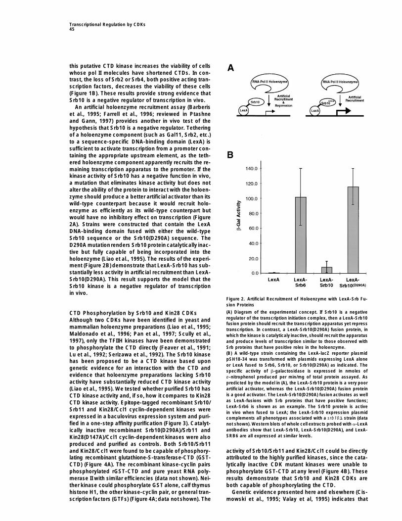
Srb11 and Kin28/Ccl1 cyclin-dependent kinases were in vivo when fused to LexA; the LexA-Srb10 expression plasmidexpressed in a baculovirus expression system and puri- complements all phenotypes associated with a srb10D strain (datafied in a one-step affinity purification (Figure 3). Catalyt- not shown). Western blots of whole cell extracts probed with a-LexA
antibodies show that LexA-Srb10, LexA-Srb10(D290A), and LexA-ically inactive recombinant Srb10(D290A)/Srb11 andSRB6 are all expressed at similar levels.Kin28(D147A)/Ccl1 cyclin-dependent kinases were also
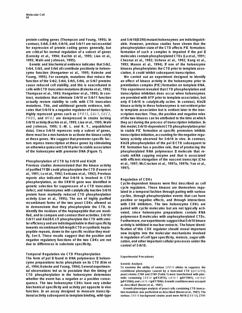
produced and purified as controls. Both Srb10/Srb11and Kin28/Ccl1 were found to be capable of phosphory- activity of Srb10/Srb11 and Kin28/Ccl1 could be directly
attributed to the highly purified kinases, since the cata-lating recombinant glutathione-S-transferase-CTD (GST-CTD) (Figure 4A). The recombinant kinase-cyclin pairs lytically inactive CDK mutant kinases were unable to
phosphorylate GST-CTD at any level (Figure 4B). Thesephosphorylated rGST-CTD and pure yeast RNA poly-merase II with similar efficiencies (data not shown). Nei- results demonstrate that Srb10 and Kin28 CDKs are
both capable of phosphorylating the CTD.ther kinase could phosphorylate GST alone, calf thymushistone H1, the other kinase-cyclin pair, or general tran- Genetic evidence presented here and elsewhere (Cis-
mowski et al., 1995; Valay et al, 1995) indicates thatscription factors (GTFs) (Figure 4A; data not shown). The

Molecular Cell46
kinase. In contrast, substitution of Ser-5 with alanineled to a dramatic loss in peptide phosphorylation, sug-gesting that Ser-5 is the principal phosphoacceptor inthe heptapeptide repeat. Substitutions of Tyr-1, Pro-3,or Pro-6 reduced phosphorylation of the synthetic pep-tides, probably due to the effects such alterations haveon their structure. These results indicate that Srb10 andKin28 CDKs are indistinguishable in substrate specificityand activity in these phosphorylation assays.
Srb10-Dependent Inhibition of Transcription In VitroThe existence of two CDKs in the holoenzyme with simi-lar biochemical specificity and activity, yet opposite invivo function, led us to entertain the possibility that thetiming of CTD phosphorylation in the holoenzyme coulddetermine whether the event had a negative or a positiveconsequence. Although both kinases are capable ofCTD phosphorylation as purified, recombinant kinase-cyclin pairs, it is possible that they can function only atcertain times when assembled in the holoenzyme. Wetherefore considered a temporal model for the action ofthese kinases, in which Srb10 is uniquely capable of CTDphosphorylation prior to initiation complex formation bythe holoenzyme, thereby repressing transcription. Incontrast, Kin28, when assembled into the holoenzyme,is capable of CTD phosphorylation only after preinitia-tion complex formation, when such activity would notinterfere with transcription.
Figure 3. Purification of Recombinant Srb10/Srb11 and Kin28/Ccl1 This temporal model predicts that holoenzymes withCyclin-Dependent Kinases catalytically active Srb10 should be transcriptionally in-(A) Scheme for production and purification of recombinant holoen- hibited when the kinase functions prior to associationzyme CDKs from Sf21 cells coinfected with baculovirus encoding
with template DNA. RNA polymerase II holoenzymesa kinase (Srb10 or Kin28) and the corresponding cyclin partnercontaining Kin28 and either wild-type Srb10 or catalyti-(Srb11 or Ccl1, respectively). FLAG-epitope-tagged recombinantcally inactive Srb10(D290A) were purified in parallel andSrb10/Srb11 and Kin28/Ccl1 and their inactive mutant derivatives,
Srb10D290A/Srb11 and Kin28D147A/Ccl1, were purified in a single step assayed for kinase and transcriptional activities (Figurefrom whole cell extracts of baculovirus-infected insect cells using 5). The two purified holoenzymes contained comparablean anti-FLAG affinity column. amounts of Rpb1, Srb2, Srb4, Srb5, Srb10, and Kin28(B) Purity of recombinant kinase-cyclin pairs. Onput (OP), flow-
(Figure 5A). To determine whether Srb10 kinase activitythrough (FT), wash (W), and eluate (E) fractions of the anti-FLAGcan inhibit transcription by acting prior to PIC formation,affinity column were subjected to SDS-PAGE electrophoresis fol-we performed an in vitro transcription experiment inlowed by Coomassie (upper) or silver staining (lower). The identities
of the kinase and cyclin subunits and position of the molecular which both wild-type and mutant Srb10 containing holo-weight markers (MW) in kilodaltons is shown. enzymes were preincubated with ATP prior to addition
of template DNA, additional GTFs, and nucleoside tri-phosphates (NTPs) (Figure 5B). Preincubation with ATPproduced a significant inhibition of transcription withKin28 and Srb10 contribute positive and negative func-
tions, respectively, to transcription in vivo. We investi- the wild-type holoenzyme but not with the holoenzymelacking Srb10 catalytic activity (Figure 5B, comparegated the possibility that differential phosphorylation of
the CTD by the two CDKs might account for their differ- lanes 2 and 4). These data show that transcription byRNA polymerase II holoenzyme is inhibited when theent functions. The amino acid residues of the CTD phos-
phorylated in vitro by the two CDKs were identified by Srb10 kinase is allowed to function prior to PIC for-mation.two-dimensional thin layer chromatography. The results
demonstrate that both Srb10 and Kin28 phosphorylate Pol II CTD phosphorylation was monitored in the holo-enzymes that were subjected to preincubation with andserine residues (Figure 4C).
To investigate further the substrate specificity of the without ATP (Figure 5B). CTD phosphorylation occurredonly in holoenzymes containing catalytically active Srb10two CTD kinases, a battery of synthetic peptides were
used as substrates to determine which amino acid resi- (Figure 5B, compare lanes 2 and 4). Kin28 is apparentlynot active in the holoenzyme prior to PIC formation,dues in the heptapeptide consensus repeat are critical
for CTD phosphorylation (Figure 4D). The results show because the Srb10 mutant holoenzyme exhibits essen-tially no CTD phosphorylation activity during the prein-that the activities of Srb10 and Kin28 on these peptide
substrates are indistinguishable. Substitution of Ser-2, cubation. The Srb10-dependent phosphorylation of theCTD was highly efficient; most of the pol II moleculesThr-4, or Ser-7 with alanine did not significantly affect
the ability of the peptide to act as a substrate for either in the wild-type holoenzyme became phosphorylated in

Transcriptional Regulation by CDKs47
Figure 4. Recombinant CDKs Are Indistin-guishable in CTD Phosphorylation Activity
(A) Holoenzyme-associated CDKs phosphor-ylate the CTD but not histone H1 in vitro. Re-combinant GST, GST-CTD, or calf thymus H1substrates were incubated with pure recom-binant CDK/cyclin pairs in the presence ofg-32P-ATP, separated by SDS-PAGE, and vi-sualized by autoradiography. Label is trans-ferred only to the GST-CTD fusion and notto GST or histone H1, a well studied kinasesubstrate.(B) Purified mutant recombinant CDKs exhibitno kinase activity. Wild-type and mutantCDKs were incubated with GST-CTD in thepresence of g-32P-ATP as in panel A. The inac-tive CDK/cyclin pairs contained a point muta-tion at a highly conserved aspartate residuecritical for catalytic activity. The absence ofa labeled product in the mutant CDK/cyclinpreparations suggests the observed activityis not due to a contaminating kinase.(C) Phosphoamino acid analysis of in vitrophosphorylated CTD.Recombinant GST-CTD was incubated withrecombinant CDK/cyclin pairs in the pres-ence of g-32P-ATP. After SDS-PAGE andtransfer to a PVDF membrane, the labeledCTD band was cut out and subjected to acidhydrolysis. The phosphoamino acids wereseparated by two-dimensional thin layer elec-trophoresis. Amino acid standards were visu-alized by ninhydrin and their mobilities shownon the left, while the labeled phosphoaminoacids were visualized by autoradiography asshown in the middle and right panel. Serineis the primary phosphoacceptor on the GST-CTD substrate for both Srb10/Srb11 andKin28/Ccl1 kinases.(D) Holoenzyme CDKs show identical speci-ficity for synthetic CTD peptide substrates.Synthetic peptides consisting of three hepta-peptide consensus repeats, or mutant varia-tions thereof, were used as substrates for re-combinant holoenzyme CDKs. The wild-type(WT) heptapeptide consensus sequence aswell as the amino acid numbering used indescribing various mutants is shown at the
bottom of the figure. After SDS-PAGE, the phosphorylated peptides were visualized by autoradiography. The Ser-5 to Ala mutant peptidewas unable to serve as a substrate for either CDK, strongly suggesting that Ser-5 is the primary substrate labeled by the CDKs.
this reaction (Figure 5B, compare lanes 1 and 2). A con- assayed after the transcription reaction, revealing thatCTD phosphorylation occurs in RNA polymerase II mole-trol experiment confirmed the specificity of the antibod-
ies used to detect unphosphorylated and phosphory- cules from holoenzymes with or without functionalSrb10, albeit the levels are 3-fold less in holoenzymeslated CTDs (Figure 5C). Thus, Srb10-dependent CTD
phosphorylation is coincident with repression of tran- lacking catalytically active Srb10 kinase. These resultsindicate that Srb10-independent CTD phosphorylationscription, a result consistent with previous evidence that
formation of a functional preinitiation complex is im- occurs during the in vitro transcription reaction, aswould be expected from the action of Kin28.paired if the pol II molecules contain phosphorylated
CTDs (Lu et al., 1991; Chesnut et al., 1992; Usheva et We and others have found that Srb10 is critical forregulation of a subset of genes in yeast cells, includingal., 1992; Kang et al., 1993; Maxon et al., 1994).
The temporal model also predicts that RNA polymer- those involved in cell type specificity (Wahi and John-son, 1995), meiosis (Surosky et al., 1994), and sugarase II holoenzymes that are allowed to bind template
DNA prior to addition of nucleoside triphosphates should utilization (Kuchin et al., 1995; Liao et al., 1995). Srb10is not a general repressor of transcription, as a varietynot be transcriptionally inhibited by Srb10 activity. The
experiment shown in Figure 5D shows that the wild-type of genes are expressed normally in Srb10 mutant cells(Surosky et al., 1994; Liao et al., 1995), and the levelsand Srb10 mutant holoenzymes are in fact equally active
in transcription under these conditions, confirming the of active holoenzyme are similar in wild-type and Srb10mutant cells (S. S. K. and R.Y., unpublished data). Theprediction. The state of CTD phosphorylation was also

Molecular Cell48
observation that Srb10 is not a general repressor ofprotein-coding genes suggests that in living cells, wherethere is abundant ATP, Srb10 activity in holoenzymesmust be inhibited to prevent constitutive inactivation ofthe general transcription initiation apparatus. To testthis idea, we produced nuclear extracts from wild-type and Srb10(D290A) mutant strains and investigatedwhether the wild-type extract showed an ATP-depen-dent inhibition of transcription prior to PIC formation.We previously showed that the transcriptional activityin these extracts is entirely dependent on componentsof the Srb/mediator complex (Koleske et al., 1992;Thompson et al., 1993), which are tightly associatedwith pol II holoenzymes (Koleske and Young, 1994). Theresults, shown in Figure 6, demonstrate that transcrip-tion in nuclear extracts is not inhibited by preincubationwith ATP, suggesting that these extracts contain anSrb10 inhibitory activity that is lost during holoenzymepurification.
Discussion
Yeast and mammalian RNA polymerase II holoenzymeshave been described that contain two cyclin-dependentkinases. Previous studies established that Kin28 is aCTD kinase with a positive role in transcription, that ofproducing a phosphorylated form of the enzyme that isassociated with active elongation. Genetic and bio-chemical evidence described here reveals that the Srb10kinase is a CTD kinase with a negative role in transcrip-tion. Srb10 is uniquely capable of phosphorylating theCTD in purified holoenzymes prior to template binding,and this phosphorylation inhibits subsequent transcrip-tion by the holoenzyme. Srb10 does not appear to inhibittranscription after formation of a stable preinitiationcomplex. Thus, the transcription initiation apparatus canbe regulated positively or negatively via modification of
phosphorylation occurs during preincubation only in holoenzymescontaining functional Srb10 kinase (compare lanes 2 and 4). Controlexperiments indicate that the Srb10 CTD kinase activity is largelyrestricted to the holoenzyme in which it resides (data not shown).Srb4 is probed as a loading control.(C) Changes in MAb reactivity to RPB1 is due to phosphorylation.
Figure 5. Catalytically Active Srb10 Can Inhibit Transcription by Holoenzyme containing functional Srb10 was incubated with ATPRNA Polymerase II Holoenzyme In Vitro (lanes 1 and 2). The signal obtained when the phosphorylated prepa-(A) Purified holoenzymes contain similar amounts of Rpb1, Srb2, ration is probed with MAb 8WG16 (a-P2 CTD) is reduced, and theSrb4, Srb5, and the kinases Srb10 and Kin28. Wild-type and Srb10 mobility of RPB1 is retarded (lane 3). The same preparation then(D290A) mutant holoenzymes were purified in parallel and analyzed reacts with the phospho-serine CTD-specific H5 MAb (a-P1 CTD).by Western blot. Monoclonal antibodies specific to unphosphory- Subsequent treatmentof the sample with protein phosphatase elimi-lated CTD (a-P- CTD; 8WG16) were used to detect Rpb1. nates the H5 reactiveband andrestores 8WG16 reactivity andmobil-(B) Holoenzymes containing either wild-type Srb10 kinase (lanes 1 ity to that seen prior to ATP incubation (compare lanes 1 and 4).and 2) or catalytically inactive Srb10(D290A) kinase (lanes 3 and 4) This indicates the MAbs are accurately probing the phosphorylationwere preincubated with or without ATP prior to PIC formation and state of the CTD.analyzed as diagrammed. Only holoenzymes containing functional (D) Srb10 kinase does not affect transcription post PIC formation.Srb10 are inhibited for transcription when kinases are allowed to Holoenzymes containing either wild-type Srb10 kinase (WT) or cata-function before PIC formation (compare lanes 2 and 4). In vitro lytically inactive Srb10(D290A) kinase, preincubated with templatetranscription is performed in the presence of a-32P-UTP resulting in DNA and GTFs prior to addition of NTPs, exhibit identical transcrip-internal labeling of a 400 nucleotide transcript derived from a G-less tional activity (top). The state of CTD phosphorylationafter transcrip-cassette driven by the CYC1 promoter. The state of CTD phosphory- tion was monitored with the phospho-serine specific H5 MAb (a-P1
lation after ATP preincubation was monitored by Western analysis CTD). Both wild-type and srb10(D290A) containing holoenzymes areusing monoclonal antibodies specific to unphosphorylated (a-P2 able to phosphorylate the CTD. Srb4 is probed as a loading controlCTD; 8WG16) or Ser-phosphorylated CTD (a-P1 CTD; H5). CTD (bottom).

Transcriptional Regulation by CDKs49
that the Srb10 and Kin28 kinases could act on othersubstrates in the transcription initiation apparatus, butwe did not detect phosphorylation of general transcrip-tion factors or histones, nor didwe find that either kinasecould phosphorylate the other. It was also possible thatthe two kinases phosphorylated different residues onthe CTD, but our experiments indicate that they exhibitvery similar substrate recognition and modification be-haviors. The one clear difference in behavior was theunique ability of Srb10 to phosphorylate the pol II CTDprior to initiation complex formation when it is a compo-
Figure 6. Nuclear Extracts Show No Srb10-Dependent Inhibition of nent of the holoenzyme. We conclude that the temporalTranscription by RNA Polymerase II Holoenzyme regulation of transcription by CDKs is an instance whereNuclear extracts from cells containing either wild-type Srb10 kinase a specific phosphorylation event, carried out at differentor catalytically inactive Srb10(D290A) kinase were preincubated with times, can produce opposite regulatory effects in theor without ATP prior to PIC formation, as diagrammed (the experi- cell.mental design is identical to that in Figure 5B). In vitro transcriptionis performed in the presence of a-32P-UTP resulting in internal label-
Negative Regulation by Srb10 In Vivoing of a 400 nucleotide transcript derived from a G-less cassetteProgressive truncation of the RNA polymerase II CTDdriven by the CYC1 promoter. No inhibition of transcription was
observed after ATP preincubation with either extract. produces cells with increasingly severe growth pheno-types (Nonet et al., 1987). The greater the truncationof the CTD, the larger the number of genes affected,
the CTD, depending on the timing of the phosphorylation accounting for the increasingly severe growth pheno-event (Figure 7). types (Scafe et al., 1990). The SRB genes were originally
In arriving at this temporal model, we first examined identified as suppressors of defects due to CTD trunca-the two most obvious models that could account for tion. A subset of these genes, for example those encod-
ing Srb4 and Srb6, is essential for expression of mostdifferential regulation by the two CDKs. It was possible
Figure 7. Model for Temporal Function of Holoenzyme CDKs in Transcription Initiation
The two holoenzyme cyclin-dependent kinases are CTD kinases that function at different times. Srb10-dependent CTD phosphorylation canoccur prior to stable preinitiation complex (PIC) formation at a subset of promoters, presumably activated by factors associated with thesepromoters, with consequent inhibition of transcription. The Kin28 kinase functions after stable PIC formation at promoters generally, producingthe hyperphosphorylated form of pol II associated with productive elongation.
Molecular Cell50
protein-coding genes (Thompson and Young, 1995). In and Srb10(D290) mutant holoenzymes are indistinguish-able. However, previous studies have shown that thecontrast, Srb8, Srb9, Srb10, and Srb11 are not essential
for expression of protein coding genes generally, but phosphorylation state of the CTD affects PIC formation;formation of such a complex is impaired if the pol IIare critical for normal regulation of a subset of genes
(Surosky et al., 1994; Kuchin et al., 1995; Liao et al., molecules contain phosphorylated CTDs (Lu et al., 1991;Chesnut et al., 1992; Usheva et al., 1992; Kang et al.,1995; Wahi and Johnson, 1995).
Genetic and biochemical evidence indicates that Srb2, 1993; Maxon et al., 1994). If one of the holoenzymekinases phosphorylates the CTD prior to template asso-Srb4, Srb5, and Srb6 all contribute positively to holoen-
zyme function (Hengartner et al., 1995; Koleske and ciation, it could inhibit subsequent transcription.We carried out an experiment designed to identifyYoung, 1995). For example, mutations that reduce the
function of the Srb2, Srb4, Srb5, Srb6, or Srb7 proteins an effect of kinase activity in the holoenzyme prior topreinitiation complex (PIC) formation on template DNA.cause reduced cell viability, and this is exacerbated in
cells with CTD truncation mutations (Koleske et al., 1992; This experiment revealed that CTD phosphorylation andtranscription inhibition does occur when holoenzymesThompson et al., 1993; Hengartner et al., 1995). In con-
trast, mutations that eliminate Srb10 or Srb11 function are provided with ATP prior to template association, butonly if Srb10 is catalytically active. In contrast, Kin28actually restore viability to cells with CTD truncation
mutations. This, and additional genetic evidence, indi- kinase activity in these holoenzymes is not evident priorto template association but is evident later in the tran-cates that Srb10 is a negative regulator of transcription.
Highly repressed genes such as SPO13, GAL1, SUC2, scription reaction. Thus, the positive and negative rolesof the two kinases can be attributed to the time at whichPHO5, and MFA2 are derepressed in strains lacking
Srb10 activity (Kuchin et al., 1995; Liao et al., 1995; Wahi they act during the process of transcription initiation. Inthis model, Srb10-dependent CTD phosphorylation priorand Johnson, 1995; S.-M. L. and R. Y., unpublished
data). Since Srb10 represses only a subset of genes, to stable PIC formation at specific promoters inhibitstranscription initiation, accounting for thenegative regu-there must be a mechanism to activate the kinase solely
at these genes. We suggest that promoter-specific fac- latory activity observed for Srb10 in vivo. In contrast,Kin28 phosphorylation of the pol II CTD subsequent totors repress transcription at these genes by stimulating
an otherwise quiescent Srb10 prior to stable association PIC formation has a positive role, that of producing thephosphorylated RNA polymerase II molecule that re-of the holoenzyme with promoter DNA (Figure 7).cruits mRNA capping enzyme and that is associatedwith efficient elongation of the nascent transcript (ChoPhosphorylation of CTD by Srb10 and Kin28et al., 1997; McCracken et al., 1997a, 1997b; Yue et al.,Previous studies demonstrated that the kinase activity1997).of purified TFIIH could phosphorylate the CTD (Feaver et
al., 1991; Lu et al., 1992; Serizawa et al., 1992). Previousreports also indicated that Srb10 is involved in CTD Regulation of CDKsphosphorylation, as the SRB10 gene was identified in Cyclin-dependent kinases were first described as cellgenetic selection for suppressors of a CTD truncation cycle regulators. These kinases are themselves regu-defect, and holoenzymes with catalytically inactive Srb10 lated in a temporal fashion through pairing with variousprotein have markedly reduced CTD phosphorylation cyclins, through phosphorylation events that can haveactivity (Liao et al., 1995). The use of highly purified positive or negative effects, and through interactionsrecombinant forms of the two yeast CDKs allowed us with CDK inhibitors. The two holoenzyme CDKs areto demonstrate that they phosphorylate the CTD, to paired with cyclin molecules but are not typically acti-identify the residues of the heptapeptide that are modi- vated, since holoenzyme preparations contain RNAfied, and to compare and contrast their activities. Srb10/ polymerase II molecules with unphosphorylated CTDs.Srb11 and Kin28/Ccl1 phosphorylate the CTD with simi- Furthermore, ourexperiments suggest that Srb10 kinaselar efficiency and are indistinguishable in their specificity activity is inhibited in nuclear extracts. The future identi-towards recombinant full-length CTD or synthetic hepta- fication of this CDK regulator should reveal importantpeptide repeats, down to the specific residue they mod- new insights into the molecular mechanisms involvedify, Ser-5. These results suggest that the positive and in regulation of cell type specificity, meiosis, sugar utili-negative regulatory functions of the two CDKs are not zation, and other important cellular processes under thedue to differences in substrate specificity. control of Srb10.
Temporal Regulation via CTD PhosphorylationExperimental ProceduresThe form of pol II found in RNA polymerase II holoen-
zyme preparations lacks phosphate on its CTD (Kim et Genetic Analysisal., 1994; Koleske and Young, 1994). Several experimen- To examine the ability of various SRB10 alleles to suppress the
conditional phenotypes caused by a truncated CTD (rpb1D104),tal observations led us to postulate that the timing ofyeast strains Z768 and Z769 (Table 1) were transformed with plas-CTD phosphorylation in the holoenzyme determinesmids containing SRB10 (pRY2973), srb10-1 (pRY7091), srb10D1whether the event has a negative or a positive conse-(pRY2966), and srb10-3 (pRY7096). Growth conditionswere assayedquence. The two holoenzyme CDKs have very similaras described (Nonet et al., 1987).
biochemical specificity and activity yet opposite in vivo Growth phenotype analysis of yeast cells containing CTD trunca-function. In an assay designed to measure transcrip- tion mutations was performed as described (Nonet et al., 1987). The
various SRB10 background strains used were N418 (SRB10), Z741tional activity subsequent to template binding, wild-type

Transcriptional Regulation by CDKs51
Table 1. Yeast Strains
Strain Alias Genotype Reference
Z768 SLY67 Mata ura3-52 his3D200 leu2-3, -112 rpb1D187::HIS3 srb10D1::hisG [L14 (LEU2 CEN RPB1)] This studyZ769 SLY69 Mata ura3-52 his3D200 leu2-3, -112 rpb1D187::HIS3 srb10D1::hisG [C6 (LEU2 CEN rpb1D104)] This studyN418 Mata ura3-52 his3D200 leu2-3, -112 rpb1D187::HIS3 SRB10 [pRP112 (URA3 CEN RPB1)] Nonet et al., 1989Z741 SLY37 Mata ura3-52 his3D200 leu2-3, -112 rpb1D187::HIS3 srb10D1::hisG [pRP112 (URA3 CEN RPB1)] This studyZ735 SLY26 Mata ura3-52 his3D200 leu2-3, -112 rpb1D187::HIS3 srb10-1::hisG [pRP112 (URA3 CEN RPB1)] This studyZ687 SLY7 Mata ura3-52 his3D200 leu2-3, -112 RPB1 srb10D1::hisG Liao et al., 1995Z690 SLY96 Mata ura3-52 his3D200 leu2-3, -112 RPB1 srb10-3::hisG Liao et al., 1995Z719 SLY3 Mata ura3-52 his3D200 leu2-3, -112 RPB1 SRB10 Liao et al., 1995
(srb10D1), and Z735 (srb10-1) (Table 1). The viability of cells con- and 40 mM EDTA) and then resolved by 4%–20% acrylamide gradi-ent SDS-PAGE. The dried gels were exposed directly to autoradio-taining CTD truncations in those backgrounds was assayed by plas-
mid shuffle, and surviving strains were tested for cold sensitivity. graphic films.Kinase substrates GST and GST-CTD were purified from bacteria
as described (Thompson et al., 1993) and purified calf thymus his-In Vivo Recruitment Assays
tone H1 (Boehringer Mannheim). Triple CTD heptapeptide consen-b-galactosidase assays were performed as described (Rose and
sus repeats were synthesized (Research Genetics) and were pro-Botstein, 1983). The strains are derivatives of Z719 transformed with
vided in the form H-YSPTSPSYSPTSPSYSPTSPS-Amide. CTDthe reporter pSH18–34 and the appropriate LexA fusion. To make
peptide variants where one or more amino acids of the consensusthe LexA fusions, SRB10, srb10-3, and SRB6 open reading frames
sequence have been systematically replaced by alanine were alsowere subcloned into the LexA fusion plasmid pEG202 (Ausubel et
synthesized by Research Genetics.al., 1997).
Phosphoamino Acid AnalysisRecombinant CDK/Cyclin Production and PurificationUsing GST-CTD as a substrate, a kinase reaction was performed.
from Insect CellsAfter SDS-PAGE, the samples weretransferred to a PVDFmembrane
Recombinant CDK/cyclin pairs were produced using a baculovirusand the labeled phosphorylated GST-CTD band, localized, and cut
expression system. For expression of CDKs, genes for Srb10 andout after a short film exposure. Two-dimensional electrophoretic
Kin28 were amplified by polymerase chain reactions (PCR) andanalysis of phosphoamino acid content was performed subsequent
cloned into baculoviral transfer vectors pSK277 or pSK278 (Koh etto acid hydrolysis as described in Coligan et al. (1997). The phospho-
al., 1997) to produce recombinants with FLAG epitope-tag at theirlabeled phosphoamino acids were visualized by autoradiography.
N termini. For expression of cyclins, genes for Srb11 and Ccl1 wereamplified by PCR and cloned into baculoviral transfer vectors pBac-PAK8 or pBacPAK9 (Clontech). PCRs were performed with Vent Nuclear Extract Transcription
Nuclear extracts from Z719 and Z690 were prepared according toDNA polymerase (New England Biolabs). All the PCR clones wereverified by DNA sequencing. Mutant CDK clones, Srb10(D290A) and Lue et al. (1991) with the modifications described by Liao et al.
(1991), yielding a final protein concentration of 85 and 75 mg/ml,Kin28(D147A), were produced by site-directed in vitro mutagenesis(Kunkel et al., 1987) using oligonucleotides CAAAACCTAAAGCACC respectively. In vitro transcription was carried out essentially as
described (Liao et al., 1991). Each reaction contained 90 mg of Z719AATTTT and CCTTGCTAGACCGAAAGCTGCTACTTTTATCTG, re-spectively. All mutations were verified by DNA sequencing. protein or 120 mg of Z690 protein with 250 ng of template.
Recombinant baculoviruses were generated from the recombi-nant transfer plasmids containing CDKs or cyclins by cotransfection
Transcription and Western Blot Analysisof the plasmids with wild-type viral DNA as recommended by the
Holoenzyme was purified according to Liao et al. (1995). In vitromanufacturer (Clontech). Spodoptera frugiperda (Sf21) cells were
transcription reactions were performed essentially as describedcoinfected with recombinant baculoviruses expressing CDKs and
(Gadbois et al., 1997), with the following modifications. Preincuba-their cyclin partners at a multiplicity of infection (m.o.i.) of 5–10. The
tions (19 ml total) contained all reaction components except TBP,cells from 200 mL of culture (approximately 3 3 108) were collected TFIIE, TFIIB, nucleotides, and DNA template; ATP containing reac-60–72 hr postinfection and lysed by sonication. The lysates were tions were brought to a final ATP concentration of 1 mM with 100then clarified by centrifuging for 3 hr at 100,000 3 g yielding 20–40 mM stock (Pharmacia). After a 15 min preincubation at room temper-mg of total protein in 10 mL. CDK/cyclin pairs were purified from ature, GTFs (3 ml) and NTP mix (4 ml; 5 mM ATP, CTP, 0.156 mMthe lysates as described (Koh et al., 1997) using 1 mL of anti-FLAG
UTP, 0.25 mM 39-O-Me GTP and 10 mCi [a-32P] UTP 3000 Ci/mmole)M2 affinity gel and 73 mg/mL of FLAG peptide in the elution buffer.
containing 100 ng DNA template (pGALD) were added for a finalTypical yields were 0.2%–0.4% of total protein from cell lysate.
reaction volume of 26 ml. After allowing transcription to proceed for30 min at room temperature, reactions were stopped by addition of125 ml stop buffer (10 mM Tris-HCl [pH 7.5], 20 mM EDTA, 2 MKinase Assay
Kinase assays were performed using 1 mg of protein substrate (GST, ammonium acetate, and 10 mg/ml glycogen) and 150 ml isopropanol.Samples were placed on dry ice for 10 min, microcentrifuged at 14Krecombinant GST-CTD, or calf thymus histone H1) or 15 mg of syn-
thetic CTD peptide substrate with 100 ng of pure recombinant CDK/ RPM for 10 min, and pellets resuspended in 6 ml formamide loadingdyes and electrophoresed on a 4% Urea-containing denaturingcyclin pairs in 15 ml reaction containing 20 mM Hepes-KOH (pH 7.3),
10% glycerol, 2.5 mM EGTA, 15 mM magnesium acetate, 1 mM polyacrylamide gel. Gels were dried and exposed to Kodak X-ARfilm at 2808C with an intensifying screen.DTT, 100 mM potassium acetate, 200 mM ATP, 10 mCi [g-32P] ATP
(NEN, 6000 Ci/mmol, 10 mCi/ml), a mixture of phosphatase inhibitors Samples for Western blot analysis were fractionated on 5% SDS-PAGE gels and transferred according to standard procedures.(1 mM NaN3, 1 mM NaF, 0.4 mM NaVO3, and 0.4 mM Na3VO4), a
cocktail of protease inhibitors (0.5 mM PMSF and 1 mM benzami- 8WG16 monoclonal antibody (Babco) and Srb4 rabbit anti-serumwere used at 1:1000, and H5 monoclonal antibody (Babco) wasdine, 1 mM pepstatin, 0.3 mM leupeptin, and 1 mg/ml chymostatin)
and 0.5 mg/ml of acetylated BSA. Reactions were assembled on used at 1:250. HRP-conjugated anti-mouse (Pierce) and anti-rabbit(Amersham) secondary antibodies were used at 1:2000. Detectionice and initiated with the addition of ATP. After 60 min at 258C , the
reactions were terminated by adding 15 ml of stop buffer (23 SDS- was performed by ECL according to the manufacturer’s directions(Amersham).PAGE loading buffer supplemented with 100 mM Tris-HCl, [pH 6.8],

Molecular Cell52
Acknowledgments S.-M., Koleske, A.J., Okamura, S., and Young, R.A. (1995). Associa-tion of an activator with an RNA polymerase II holoenzyme. GenesDev. 9, 897–910.We thank Frank Holstege, David Chao, and Tony Lee for reagents
and discussion. This work was supported by National Institutes of Kang, M.E., and Dahmus, M.E. (1993). RNA polymerases IIA and IIOHealth grant GM34365. have distinct roles during transcription from the TATA-less murine
dihydrofolate reductase promoter. J. Biol. Chem. 268, 25033–25040.
Received March 3, 1998; revised April 13, 1998. Kim, Y.-J., Bjorkland, S., Li, Y., Sayre, M.H., and Kornberg, R.D.(1994). A multiprotein mediator of transcriptional activation and itsinteraction with the C-terminal repeat domain of RNA polymeraseReferencesII. Cell 77, 599–608.
Koh, S.S., Hengartner, C.J., and Young, R.A. (1997). BaculoviralAkoulitchev, S., Makela, T.P., Weinberg, R.A., and Reinberg, D.transfer vectors for expression of FLAG fusion proteins in insect(1995). Requirement for TFIIH kinase activity in transcription by RNAcells. Biotechniques 23, 622–627.polymerase II. Nature 377, 557–560.Koleske, A.J., Buratowski, S., Nonet, M., and Young, R.A. (1992). AAusubel, F.M., Brent, R., Kingston, R.E., Moore, D.D., Seidman, J.G.,novel transcription factor reveals a functional link between the RNASmith, J.A., and Struhl, K. (1997). Interaction trap/two-hybrid systempolymerase II CTD and TFIID. Cell 69, 883–894.to identify interacting proteins. In Current Protocols, V.B. Chanda,Koleske, A.J., and Young, R.A. (1994). An RNA polymerase II holoen-ed. (New York: John Wiley & Sons), pp. 20.1.1–20.1.28.zyme responsive to activators. Nature 368, 466–469.Barberis, A., Pearlberg, J., Simkovich, N., Farrell, S., Reinagel, P.,Koleske, A.J., and Young, R.A. (1995). The RNA polymerase II holo-Bamdad, C., Sigal, G., and Ptashne, M. (1995). Contact with a com-enzyme and its implications for gene regulation. Trends Biochem.ponent of the polymerase II holoenzyme suffices for gene activation.Sci. 20, 113–116.Cell 81, 359–368.Kolodziej, P.A., Woychik, N., Liao, S.M., and Young, R.A. (1990).Bartholomew, B., Dahmus, M.E., and Meares, C.F. (1986). RNA con-RNA polymerase II subunit composition, stoichiometry, and phos-tacts subunits IIo and IIc in HeLa RNA polymerase II transcriptionphorylation. Mol. Cell. Biol. 10, 1915–1920.complexes. J. Biol. Chem. 261, 14226–14231.Kuchin, S., Yeghiayan, P., and Carlson, M. (1995). Cyclin-dependantCadena, D.L., and Dahmus, M.E. (1987). Messenger RNA synthesisprotein kinase and cyclin homologs SSN3 and SSN8 contribute toin mammalian cells is catalyzed by the phosphorylated form of RNAtranscriptional control in yeast. Proc. Natl. Acad. Sci. USA 92, 4006–polymerase II. J. Biol. Chem. 262, 12468–12474.4010.
Chesnut, J.D., Stephens, J.H., and Dahmus, M.E. (1992). The interac-Kunkel, T.A., Roberts, J.D., and Zakour, R.A. (1987). Rapid and effi-tion of RNA polymerase II with the adenovirus-2 major late promotercient site-specific mutagenesis without phenotypic selection. Meth-is precluded by phosphorylation of the C-terminal domain of subunitods Enzymol. 154, 367–382.IIa. J. Biol. Chem. 267, 10500–10506.Laybourn, P.J., and Dahmus, M.E. (1990). Phosphorylation of RNA
Cho, E.J., Takagi, T., Moore, C.R., and Buratowski, S. (1997). mRNA polymerase IIA occurs subsequent to interaction with the promotercapping enzyme is recruited to the transcription complex by phos- and before the initiation of transcription. J. Biol. Chem. 265, 13165–phorylation of the RNA polymerase II carboxy-terminal domain. 13173.Genes Dev. 11, 3319–3326.
Liao, S.M., Taylor, I.C., Kingston, R.E., and Young, R.A. (1991). RNACismowski, M.J., Laff, G.M., Solomon, M.J., and Reed, S.I. (1995). polymerase II carboxy-terminal domain contributes to the responseKIN28 encodes a C-terminal domain kinase that controls mRNA to multiple acidic activators in vitro. Genes Dev. 5, 2431–2440.transcription in Saccharomyces cerevisiae but lacks cyclin-depen-
Liao, S.-M., Zhang, J., Jeffery, D.A., Koleske, A.J., Thompson, C.M.,dent kinase-activating kinase (CAK) activity. Mol. Cell. Biol. 15,
Chao, D.M., Viljoen, M., van Vuuren, H.J.J., and Young, R.A. (1995).2983–2992. A kinase-cyclin pair in the RNA polymerase II holoenzyme. NatureColigan, J.E., Dunn, B.M., Ploegh, H.L., Speicher, D.W., and Wing- 374, 193–196.field, P.T. (1997). Phosphoamino Acid Analysis. In Current Protocols, Lu, H., Flores, O., Weinmann, R., and Reinberg, D. (1991). The non-V.B. Chanda, ed. (New York: John Wiley & Sons), pp. 13.3.1–13.3.7. phosphorylated form of RNA polymerase II preferentially associatesCoppola, J.A., Field, A.S., and Luse, D.S. (1983). Promoter-proximal with the preinitiation complex. Proc. Natl. Acad. Sci. USA 88, 10004–pausing by RNA polymerase II in vitro: transcripts shorter than 20 10008.nucleotides are not capped. Proc. Natl. Acad. Sci. USA 80, 1251– Lu, H., Zawel, L., Fisher, L., Egly, J.M., and Reinberg, D. (1992).1255. Human general transcription factor IIH phosphorylates the C-termi-
nal domain of RNA polymerase II. Nature 358, 641–645.Dahmus, M.E. (1996). Reversible phosphorylation of the C-terminaldomain of RNA polymerase II. J. Biol. Chem. 271, 19009–19012. Lue, N.F., Flanagan, P.M., Kelleher, R., III, Edwards, A.M., and Korn-
berg, R.D. (1991). RNA polymerase II transcription in vitro. MethodsDynlacht, B.D. (1997). Regulation of transcription by proteins thatEnzymol. 194, 545–550.control the cell cycle. Nature 389, 149–152.Maldonado, E., Shiekhattar, R., Sheldon, M., Cho, H., Drapkin, R.,Farrell, S., Simkovich, N., Wu, Y., Barberis, A., and Ptashne, M.Rickert, P., Lees, E., Anderson, C.W., Linn, S., and Reinberg, D.(1996). Gene activation by recruitment of the RNA polymerase II(1996). A human RNA polymerase II complex associated with SRBholoenzyme. Genes Dev. 10, 2359–2367.and DNA-repair proteins. Nature 381, 86–89.
Feaver, W.J., Gileadi, O., Li, Y., and Kornberg, R.D. (1991). CTDMaxon, M.E., Goodrich, J.A., and Tjian, R. (1994). Transcription fac-kinase associated with yeast RNA polymerase II initiation factor b.tor IIE binds preferentially to RNA polymerase IIa and recruits TFIIH:Cell 67, 1223–1230.a model for promoter clearance. Genes Dev. 8, 515–524.
Feaver, W.J., Svejstrup, J.Q., Henry, N.L., and Kornberg, R.D. (1994).McCracken, S., Fong, N., Rosonina, E., Yankulov, K., Brothers, G.,Relationship of CDK-activating kinase and RNA polymerase II CTDSiderovski, D., Hessel, A., Foster, S., Shuman, S., and Bentley, D.L.kinase TFIIH/TFIIK. Cell 79, 1109–1109.(1997a). 59-capping enzymes are targeted to pre-mRNA by binding
Gadbois, E.L., Chao, D.M., Reese, J.C., Green, M.R., and Young, to the phosphorylated carboxy-terminal domain of RNA polymeraseR.A. (1997). Functional antagonism between RNA polymerase II ho- II. Genes Dev. 11, 3306–3318.loenzyme and global negative regulator NC2 in vivo. Proc. Natl. McCracken, S., Fong, N., Yankulov, K., Ballantyne, S., Pan, G.,Acad. Sci. USA 94, 3145–3150. Greenblatt, J., Patterson, S.D., Wickens, M., and Bentley, D.L.Garcia-Martinez, L.F., Mavankal, G., Neveu, J.M., Lane, W.S., Ivanov, (1997b). The C-terminal domain of RNApolymerase II couples mRNAD., and Gaynor, R.B. (1997). Purification of a Tat-associated kinase processing to transcription. Nature 385, 357–361.reveals a TFIIH complex that modulates HIV-1 transcription. EMBO Nonet, M.L., and Young, R.A. (1989). Intragenic and extragenic sup-J. 16, 2836–2850. pressors of mutations in the heptapeptide repeat domain if Sacchar-
omyces cerevisiae RNA polymerase II. Genetics 123, 715–724.Hengartner, C.J., Thompson, C.M., Zhang, J., Chao, D.M., Liao,

Transcriptional Regulation by CDKs53
Nonet, M., Sweetser, D., and Young, R.A. (1987). Functional redun- Saccharomyces cerevisiae lacking mRNA guanylyltransferase andselectively binds the elongating form of RNA polymerase II. Proc.dancy and structural polymorphism in the large subunit of RNA
polymerase II. Cell 50, 909–915. Natl. Acad. Sci. USA 94, 12898–12903.
O’Brien, T., Hardin, S., Greenleaf, A., and Lis, J.T. (1994). Phosphory-lation of RNA polymerase II C-terminal domain and transcriptionalelongation. Nature 370, 75–77.
Ohkuma, Y., and Roeder, R.G. (1994). Regulation of TFIIH ATPaseand kinase activities by TFIIE during active initiation complex forma-tion. Nature 368, 160–163.
Pan, G., Aso, T., and Greenblatt, J. (1997). Interaction of elongationfactors TFIIS and elongin A with a human RNA polymerase II holoen-zyme capable of promoter-specific initiation and responsive to tran-scriptional activators. J. Biol. Chem. 272, 24563–24571.
Parada, C.A., and Roeder, R.G. (1996). Enhanced processivity ofRNA polymerase II triggered by Tat-induced phosphorylation of itscarboxy-terminal domain. Nature 384, 375–378.
Ptashne, M., and Gann, A. (1997). Transcriptional activation by re-cruitment. Nature 386, 569–577.
Rose, M., and Botstein, D. (1983). Construction and use of genefusions to lacZ (beta-galactosidase) that are expressed in yeast.Methods Enzymol. 101, 167–180.
Roy, R., Adamczewski, J.P., Seroz, T., Vermulen, W., Tassan, J.P.,Schaeffer, L., Nigg, E.A., Hoeijmakers, J.H., and Egly, J.M. (1994).The MO15 cell cycle kinase is associated with the TFIIH transcrip-tion-DNA repair factor. Cell 79, 1093–1101.
Scafe, C., Chao, D., Lopes, J., Hirsch, J.P., Henry, S., and Young,R.A. (1990). RNA polymerase II C-terminal repeat influences re-sponse to transcriptional enhancer signals. Nature 347, 491–494.
Scully, R., Anderson, S.F., Chao, D.M., Wei, W., Ye, L., Young, R.A.,Livingston, D.M., and Parvin, J.D. (1997). BRCA1 is a component ofthe RNA polymerase II holoenzyme. Proc. Natl. Acad. Sci. USA 94,5605–5610.
Serizawa, H., Conaway, R.C., and Conaway, J.W. (1992). A carboxyl-terminal-domain kinase associated with RNA polymerase II tran-scription factor delta from rat liver. Proc. Natl. Acad. Sci. USA 89,7476–7480.
Serizawa, H., Makela, T.P., Conaway, J.W., Conaway, R.C., Wein-berg, R.A., and Young, R.A. (1995). Association of Cdk-activatingkinase subunits with transcription factor TFIIH. Nature 374, 280–282.
Shiekhattar, R., Mermelstein, F., Fisher, R.P., Drapkin, R., Dynlacht,B., Wessling, H.C., Morgan, D.O., and Reinberg, D. (1995). Cdk-activating kinase complex is a component of human transcriptionfactor TFIIH. Nature 374, 283–287.
Surosky, R.T., Strich, R., and Esposito, R.E. (1994). The yeast UME5gene regulates the stability of meiotic mRNAs in response to glu-cose. Mol. Cell. Biol. 14, 3446–3458.
Thompson, C.M., and Young, R.A. (1995). General requirement forRNA polymerase II holoenzymes in vivo. Proc. Natl. Acad. Sci. USA92, 4587–4590.
Thompson, C.M., Koleske, A.J., Chao, D.M., and Young, R.A. (1993).A multisubunit complex associated with the RNA polymerase II CTDand TATA-binding protein in yeast. Cell 73, 1361–1375.
Usheva, A., Maldonado, E., Goldring, A., Lu, H., Houbavi, C., Rein-berg, D., and Aloni, Y. (1992). Specific interaction between the non-phosphorylated form of RNA polymerase II and the TATA-bindingprotein. Cell 69, 871–881.
Valay, J.G., Simon, M., Dubois, M.F., Bensaude, O., Facca, C., andFaye, G. (1995). The KIN28 gene is required both for RNA polymeraseII mediated transcription and phosphorylation of the Rpb1p CTD.J. Mol. Biol. 249, 535–544.
Wahi, M., and Johnson, A.D. (1995). Identification of genes requiredfor alpha 2 repression in Saccharomyces cerevisiae. Genetics 140,79–90.
Weeks, J.R., Hardin, S.E., Shen, J., Lee, J.M., and Greenleaf, A.L.(1993). Locus-specific variation in phosphorylation state of RNApolymerase II in vivo: correlations with gene activity and transcriptprocessing. Genes Dev. 7, 2329–2344.
Yue, Z., Maldonado, E., Pillutla, R., Cho, H., Reinberg, D., and Shat-kin, A.J. (1997). Mammalian capping enzyme complements mutant