The RNA Polymerase “Switch Region” Is a Target for Inhibitors
13
The RNA Polymerase ‘‘Switch Region’’ Is a Target for Inhibitors Jayanta Mukhopadhyay, 1,2,4,6 Kalyan Das, 3,4,6 Sajida Ismail, 1,2,4 David Koppstein, 1,2,4 Minyoung Jang, 1,2,4 Brian Hudson, 3,4 Stefan Sarafianos, 3,4 Steven Tuske, 3,4 Jay Patel, 3,4 Rolf Jansen, 5 Herbert Irschik, 5 Eddy Arnold, 3,4, * and Richard H. Ebright 1,2,4, * 1 Howard Hughes Medical Institute 2 Waksman Institute 3 Center for Advanced Biotechnology and Medicine 4 Department of Chemistry and Chemical Biology Rutgers University, Piscataway, NJ 08854, USA 5 Helmholtz Centre for Infection Research, Inhoffenstraße 7, D-38124 Braunschweig, Germany 6 These authors contributed equally to this work *Correspondence: [email protected] (E.A.), [email protected] (R.H.E.) DOI 10.1016/j.cell.2008.09.033 SUMMARY The a-pyrone antibiotic myxopyronin (Myx) inhibits bacterial RNA polymerase (RNAP). Here, through a combination of genetic, biochemical, and structural approaches, we show that Myx interacts with the RNAP ‘‘switch region’’—the hinge that mediates opening and closing of the RNAP active center cleft—to prevent interaction of RNAP with promoter DNA. We define the contacts between Myx and RNAP and the effects of Myx on RNAP conformation and propose that Myx functions by interfering with opening of the RNAP active-center cleft during tran- scription initiation. We further show that the structur- ally related a-pyrone antibiotic corallopyronin (Cor) and the structurally unrelated macrocyclic-lactone antibiotic ripostatin (Rip) function analogously to Myx. The RNAP switch region is distant from targets of previously characterized RNAP inhibitors, and, correspondingly, Myx, Cor, and Rip do not exhibit crossresistance with previously characterized RNAP inhibitors. The RNAP switch region is an attractive target for identification of new broad-spec- trum antibacterial therapeutic agents. INTRODUCTION Bacterial RNA polymerase (RNAP) is a proven target for broad- spectrum antibacterial therapy (Darst, 2004; Chopra, 2007). The suitability of bacterial RNAP as a target for broad-spectrum antibacterial therapy follows from the fact that bacterial RNAP is an essential enzyme (permitting efficacy), the fact that bacterial RNAP subunit sequences are highly conserved (permitting for broad-spectrum activity), and the fact that bacterial RNAP-sub- unit sequences and eukaryotic RNAP-subunit sequences are not highly conserved (permitting therapeutic selectivity). The rifamycin antibacterial agents—notably rifampicin, rifa- pentine, and rifabutin—function by binding to and inhibiting bac- terial RNAP (Campbell et al., 2001; Darst, 2004; Chopra, 2007). The rifamycins bind to a site on bacterial RNAP adjacent to the RNAP active center and prevent extension of RNA beyond a length of 2–3 nt. The rifamycins are of clinical importance in treatment of Gram-positive and Gram-negative bacterial infec- tions, are first-line antituberculosis agents, and are the only antituberculosis agents able to rapidly clear infection and prevent relapse. However, the clinical utility of the rifamycin anti- bacterial agents is threatened by the existence of bacterial strains resistant to rifamycins. Resistance to rifamycins typically involves substitution of residues in or adjacent to the rifamycin- binding site on bacterial RNAP, i.e., substitutions that directly decrease binding of rifamycins. In view of the public-health threat posed by rifamycin-resistant and multidrug-resistant bacterial infections, there is an urgent need for new classes of antibacterial agents that (1) target bacterial RNAP (and thus have the same biochemical effects as rifamycins) but that (2) target sites within bacterial RNAP dis- tinct from the rifamycin-binding site (and thus do not show cross- resistance with rifamycins) (Darst, 2004; Chopra, 2007). Structures have been determined for bacterial RNAP and eu- karyotic RNAP II (Zhang et al., 1999; Cramer et al., 2000, 2001; Ebright, 2000; Darst, 2001; Cramer, 2002; Young et al., 2002; Murakami and Darst, 2003). The structures reveal that RNAP, bacterial or eukaryotic, has dimensions of 150 A ˚ 3 100 A ˚ 3 100 A ˚ and has a shape reminiscent of a crab claw (Figure 1A). The two ‘‘pincers’’ of the ‘‘claw’’ define the active-center cleft, which has a diameter of 20 A ˚ —a diameter that can accommo- date a double-stranded nucleic acid—and which has the active- center Mg 2+ at its base. The largest subunit (b 0 in bacterial RNAP) makes up one pincer, termed the ‘‘clamp,’’ and part of the base of the active-center cleft. The second-largest subunit (b in bacte- rial RNAP) makes up the other pincer and part of the base of the active-center cleft. The structures further reveal that the RNAP clamp can exist in a range of distinct conformational states, from a fully open clamp conformation that permits unimpeded entry and exit of DNA Cell 135, 295–307, October 17, 2008 ª2008 Elsevier Inc. 295
-
Upload
independent -
Category
Documents
-
view
0 -
download
0
Transcript of The RNA Polymerase “Switch Region” Is a Target for Inhibitors

The RNA Polymerase ‘‘Switch Region’’Is a Target for InhibitorsJayanta Mukhopadhyay,1,2,4,6 Kalyan Das,3,4,6 Sajida Ismail,1,2,4 David Koppstein,1,2,4 Minyoung Jang,1,2,4
Brian Hudson,3,4 Stefan Sarafianos,3,4 Steven Tuske,3,4 Jay Patel,3,4 Rolf Jansen,5 Herbert Irschik,5 Eddy Arnold,3,4,*and Richard H. Ebright1,2,4,*1Howard Hughes Medical Institute2Waksman Institute3Center for Advanced Biotechnology and Medicine4Department of Chemistry and Chemical Biology
Rutgers University, Piscataway, NJ 08854, USA5Helmholtz Centre for Infection Research, Inhoffenstraße 7, D-38124 Braunschweig, Germany6These authors contributed equally to this work
*Correspondence: [email protected] (E.A.), [email protected] (R.H.E.)
DOI 10.1016/j.cell.2008.09.033
SUMMARY
The a-pyrone antibiotic myxopyronin (Myx) inhibitsbacterial RNA polymerase (RNAP). Here, througha combination of genetic, biochemical, and structuralapproaches, we show that Myx interacts with theRNAP ‘‘switch region’’—the hinge that mediatesopening and closing of the RNAP active centercleft—to prevent interaction of RNAP with promoterDNA. We define the contacts between Myx andRNAP and the effects of Myx on RNAP conformationand propose that Myx functions by interfering withopening of the RNAP active-center cleft during tran-scription initiation. We further show that the structur-ally related a-pyrone antibiotic corallopyronin (Cor)and the structurally unrelated macrocyclic-lactoneantibiotic ripostatin (Rip) function analogously toMyx. The RNAP switch region is distant from targetsof previously characterized RNAP inhibitors, and,correspondingly, Myx, Cor, and Rip do not exhibitcrossresistance with previously characterizedRNAP inhibitors. The RNAP switch region is anattractive target for identification of new broad-spec-trum antibacterial therapeutic agents.
INTRODUCTION
Bacterial RNA polymerase (RNAP) is a proven target for broad-
spectrum antibacterial therapy (Darst, 2004; Chopra, 2007).
The suitability of bacterial RNAP as a target for broad-spectrum
antibacterial therapy follows from the fact that bacterial RNAP is
an essential enzyme (permitting efficacy), the fact that bacterial
RNAP subunit sequences are highly conserved (permitting for
broad-spectrum activity), and the fact that bacterial RNAP-sub-
unit sequences and eukaryotic RNAP-subunit sequences are not
highly conserved (permitting therapeutic selectivity).
The rifamycin antibacterial agents—notably rifampicin, rifa-
pentine, and rifabutin—function by binding to and inhibiting bac-
terial RNAP (Campbell et al., 2001; Darst, 2004; Chopra, 2007).
The rifamycins bind to a site on bacterial RNAP adjacent to the
RNAP active center and prevent extension of RNA beyond
a length of 2–3 nt. The rifamycins are of clinical importance in
treatment of Gram-positive and Gram-negative bacterial infec-
tions, are first-line antituberculosis agents, and are the only
antituberculosis agents able to rapidly clear infection and
prevent relapse. However, the clinical utility of the rifamycin anti-
bacterial agents is threatened by the existence of bacterial
strains resistant to rifamycins. Resistance to rifamycins typically
involves substitution of residues in or adjacent to the rifamycin-
binding site on bacterial RNAP, i.e., substitutions that directly
decrease binding of rifamycins.
In view of the public-health threat posed by rifamycin-resistant
and multidrug-resistant bacterial infections, there is an urgent
need for new classes of antibacterial agents that (1) target
bacterial RNAP (and thus have the same biochemical effects
as rifamycins) but that (2) target sites within bacterial RNAP dis-
tinct from the rifamycin-binding site (and thus do not show cross-
resistance with rifamycins) (Darst, 2004; Chopra, 2007).
Structures have been determined for bacterial RNAP and eu-
karyotic RNAP II (Zhang et al., 1999; Cramer et al., 2000, 2001;
Ebright, 2000; Darst, 2001; Cramer, 2002; Young et al., 2002;
Murakami and Darst, 2003). The structures reveal that RNAP,
bacterial or eukaryotic, has dimensions of �150 A 3 �100 A 3
�100 A and has a shape reminiscent of a crab claw (Figure 1A).
The two ‘‘pincers’’ of the ‘‘claw’’ define the active-center cleft,
which has a diameter of �20 A—a diameter that can accommo-
date a double-stranded nucleic acid—and which has the active-
center Mg2+ at its base. The largest subunit (b0 in bacterial RNAP)
makes up one pincer, termed the ‘‘clamp,’’ and part of the base
of the active-center cleft. The second-largest subunit (b in bacte-
rial RNAP) makes up the other pincer and part of the base of the
active-center cleft.
The structures further reveal that the RNAP clamp can exist in
a range of distinct conformational states, from a fully open clamp
conformation that permits unimpeded entry and exit of DNA
Cell 135, 295–307, October 17, 2008 ª2008 Elsevier Inc. 295

(clamp perpendicular to floor of active-center cleft) to a fully
closed clamp conformation that prevents entry and exit of DNA
(clamp rotated into active-center cleft) (Figure 1A; Zhang et al.,
1999; Cramer et al., 2000, 2001; Ebright, 2000; Darst, 2001;
Cramer, 2002; Young et al., 2002; Murakami and Darst, 2003).
The transition between the fully open and fully closed clamp con-
formations involves a 30� swinging motion of the clamp, with
a 30 A displacement of residues at the distal tip of the clamp
(Figure 1A). It has been proposed that the clamp must open to
permit DNA to enter the active-center cleft during early stages
of transcription initiation, and that the clamp must close to retain
DNA in the active-center cleft during later stages of transcription
initiation and during transcription elongation.
The ‘‘switch region’’ is located at the base of the clamp and
serves as the hinge on which the clamp swings in clamp opening
and clamp closure (Figure 1B; Cramer et al., 2001; Gnatt et al.,
2001; Cramer, 2002). The switch region adopts different confor-
mations in open and closed clamp conformational states
(Figure 1B). Several residues of the switch region make direct
contacts with DNA phosphates in the transcription elongation
complex (Gnatt et al., 2001; Vassylyev et al., 2007). It has been
proposed that direct contacts between the switch region and
DNA phosphates might coordinate, and even might mechani-
cally couple, clamp closure and DNA binding (Cramer et al.,
2001; Gnatt et al., 2001; Cramer, 2002).
Figure 1. RNAP Clamp, RNAP Switch
Region, and Antibiotics Studied
(A) Conformational states of the RNAP clamp (two
orthogonal views). Structure of RNAP showing
open (red), partly closed (yellow), and fully closed
(green) clamp conformations, as observed in crys-
tal structures (PDB 1I3Q, PDB 1HQM, PDB 1I6H).
Circle, switch region; dashed circle, binding site
for rifamycins; violet sphere, active-center Mg2+.
(B) Conformational states of the RNAP switch re-
gion (stereoview). Structure of RNAP switch 1
and RNAP switch 2 (b0 residues 1304–1329 and
b0 residues 330–349; residues numbered as in
E. coli RNAP) showing conformational states
associated with open (red), partly closed (yellow),
and fully closed (green) clamp conformations, as
observed in crystal structures (PDB 1I3Q, PDB
1HQM, PDB 1I6H). Gray squares, points of con-
nection of switch 1 and switch 2 to the RNAP
main mass. Colored circles, points of connection
of switch 1 and switch 2 to the RNAP clamp.
(C) Structures of myxopyronin A (Myx), corallopy-
ronin A (Cor), and ripostatin A (Rip).
In this work, we show that three
antibiotics—the a-pyrone antibiotic
myxopyronin (Myx), the a-pyrone antibi-
otic corallopyronin (Cor), and the macro-
cyclic-lactone antibiotic ripostatin
(Rip)—function by binding to the RNAP
switch region and preventing interaction
of RNAP with promoter DNA, apparently
by preventing opening of the clamp to
permit entry of promoter DNA during tran-
scription initiation. The three compounds interact with residues
that are conserved in Gram-positive and Gram-negative bacte-
rial RNAP and, accordingly, exhibit broad-spectrum antibacterial
activity. The three compounds interact, in part, with residues that
are not conserved in eukaryotic RNAP I, RNAP II, and RNAP III
and, accordingly, do not exhibit crossinhibition of eukaryotic
RNAP. The three compounds interact with residues that are re-
mote from the binding site for rifamycins and from the binding
sites for other characterized RNAP inhibitors (Figure 1A) and, ac-
cordingly, do not exhibit crossresistance with rifamycins or other
characterized RNAP inhibitors (Table 1). Taken together, these
properties make the three compounds attractive candidates
for development as broad-spectrum antibacterial therapeutic
agents and make the switch region an attractive target for iden-
tification of new broad-spectrum antibacterial therapeutic
agents.
RESULTS
Target of Transcription Inhibition by MyxMyx
Myx is polyketide-derived a-pyrone antibiotic produced by the
myxobacterium Myxococcus fulvus Mxf50 (Figure 1C; Irschik
et al., 1983; Kohl et al., 1983). The compound inhibits growth
of a broad spectrum of Gram-positive and Gram-negative
296 Cell 135, 295–307, October 17, 2008 ª2008 Elsevier Inc.

bacterial species, including Mycobacterium tuberculosis, Staph-
ylococcus aureus, Bacillus anthracis, Enterococcus faecium, En-
terobacter cloacae, Acinetobacter calcoaceticus, Pseudomonas
aeruginosa, and Escherichia coli DH21tolC (minimum inhibitory
concentrations [MICs] % 12.5 mg/ml for all; MICs % 1 mg/ml for
S. aureus, A. calcoaceticus, and E. coli DH21tolC; Irschik et al.,
1983; Kohl et al., 1983; Hu et al., 1998; M. Talaue, N. Connell,
J.M., and R.H.E., unpublished data). The compound is bacterio-
cidal, as assessed with E. coli DH21f2tolC (J.M. and R.H.E.,
unpublished data). The compound inhibits bacterial RNAP
(IC50 �1 mM) but does not inhibit eukaryotic RNAP II (Irschik
et al., 1983). The compound exhibits no acute toxicity in mice
at concentrations up to 100 mg/kg (Irschik et al., 1983). Total
syntheses of Myx have been reported, and analogs of Myx
have been prepared (Hu et al., 1998; Doundoulakis et al., 2004;
Lira et al., 2007). However, in the absence of information regard-
Table 1. Myx/Cor/Rip Crossresistance Patterns
Amino Acid
Substitution
Selected
Resistance(s)
MIC Ratio (MIC/MICwild-type)a
Myx Cor Rip Rif
RNAP b0 Subunit
345 Lys/Arg Cor, Rip >32 >8 >16 1
345 Lys/Asn Myx, Cor 32 8 8 1
345 Lys/Thr Cor 32 8 >16 1
1346 Gly/Asp Cor 1 4 4 1
1351 Val/Phe Myx, Cor, Rip >32 >8 >16 1
1352 Ile/Asn Rip 2 4 >16 1
1352 Ile/Ser Rip 2 4 >16 1
1354 Gly/Cys Cor 2 2 4 1
RNAP b Subunit
1232 Met/Ile Myx 2 4 4 1
1255 Thr/Ile Myx 2 4 4 1
1275 Val/Met Myx, Cor >32 >8 8 1
1275 Val/Phe Myx >32 >8 >16 1
1278 Leu/Val Myx 2 4 >16 1
1279 Glu/Gly Rip 32 4 >16 1
1279 Glu/Lys Myx, Cor >32 8 4 1
1283 Ala/Val Rip 2 4 4 1
1285 Tyr/Asp Myx 2 1 4 1
1291 Leu/Phe Myx,Rip 2 1 4 1
1298 Val/Leu Myx 2 2 4 1
1315 Met/Leu Myx 2 2 4 1
1317 Pro/Leu Myx 2 2 2 1
1320 Pro/Ala Myx 2 4 4 1
1322 Ser/Pro Rip 32 4 >16 1
1322 Ser/Thr Myx 2 4 4 1
1322 Ser/Tyr Myx 2 4 2 1
1322 Ser/Val Myx 16 8 >16 1
1325 Val/Leu Myx 2 4 4 1
1326 Leu/Trp Rip 1 >8 >16 1a MIC values with wild-type RNAP b0 and wild-type RNAP b for Myx, Cor,
Rip, and Rif are 0.1, 1.6, 0.1, and 0.1 mg/ml, respectively.
ing the target, biochemical basis, and structural basis of
transcription inhibition by Myx, efforts to prepare more potent
analogs have been unsuccessful.
The RNAP Switch Region Contains a Determinant
for Function of Myx
As a first step to identify the target within RNAP for Myx,
we performed random mutagenesis of the genes encoding
E. coli RNAP b0 and b subunits and isolated and characterized
mutants conferring resistance to Myx (Myxr). We identified
four different single-substitution Myxr mutants: two involving
substitution of residue 345 within conserved region C of
b0 subunit, and one each involving substitution of residues
1275 and 1291 within conserved region I of b subunit (Table
S1; Figure S1A available online). MIC assays indicate that all
four Myxr mutants exhibit R2-fold increases in MIC, and that
three of four Myxr mutants exhibit R32-fold increases in MIC
(Table S1). Complementation assays indicate that each Myxr
mutant is able to complement a corresponding temperature-
sensitive mutant for growth at the nonpermissive temperature,
indicating that each Myxr RNAP derivative is functional in tran-
scription, sufficiently functional to support viability (Table S1).
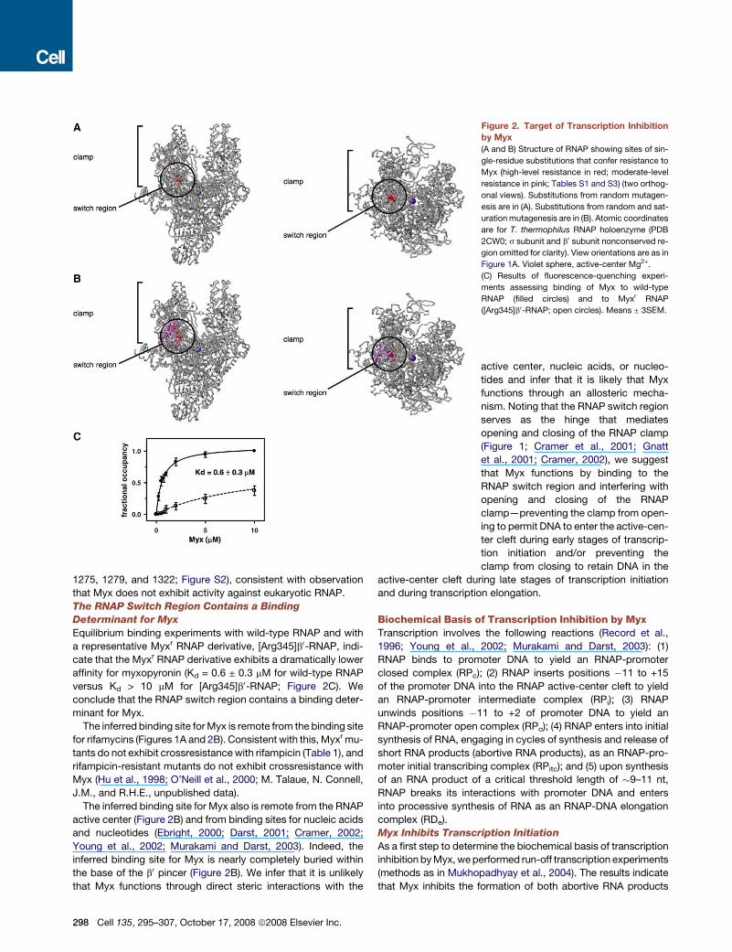
In the three-dimensional structure of RNAP, the sites of the
Myxr substitutions cluster tightly and are located in the RNAP
switch region (Figure 2A). We conclude that the RNAP switch
region contains a determinant required for transcription inhibi-
tion by Myx.
The RNAP Switch Region Contains an Extensive
Determinant for Function of Myx
To define systematically the determinant for Myx within the
RNAP switch region, we performed saturation mutagenesis of
the genes encoding the RNAP b0 and b subunits and isolated
and characterized additional Myxr mutants. We performed satu-
ration mutagenesis using a set of seventeen ‘‘doped’’ oligonu-
cleotide primers designed to introduce all possible nucleotide
substitutions at all codons for all residues located within 30 A
of the Myxr substitutions of the preceding paragraph (sequences
in Table S2). We isolated 125 independent Myxr mutants (Table
S3). Sequencing indicates that 106 of the 125 independent
Myxr mutants are single-substitution mutants. The single-substi-
tution mutants comprise 18 distinct substitutions, involving 2
sites within b0 (residue 345 in b0 conserved region C and residue
1351 in b0 conserved region H) and 12 sites within b (residues
1232, 1255, 1275, 1278, 1279, 1285, 1298, 1315, 1317, 1320,
1322, and 1325 in and near b conserved region I; Table S3; Fig-
ure S1B). In the three-dimensional structure of RNAP, the sites of
the Myxr substitutions define a single determinant with dimen-
sions of �20 A 3 �20 A 3 �10 A (Figure 2B). The determinant
is located in the RNAP switch region and encompasses a signif-
icant fraction of the RNAP switch region, including switch 2 and
segments of b0 and b adjacent to switch 2 (Figure 2B). We con-
clude that the RNAP switch region contains an extensive deter-
minant for function of Myx.
All identified Myxr substitutions involve residues that are
conserved in bacterial RNAP (Figure S2), consistent with the
observation that Myx exhibits broad-spectrum activity against
bacterial RNAP. Three identified Myxr substitutions involve resi-
dues that are not conserved—and indeed are radically differ-
ent—in eukaryotic RNAP I, RNAP II, and RNAP III (b residues
Cell 135, 295–307, October 17, 2008 ª2008 Elsevier Inc. 297
1275, 1279, and 1322; Figure S2), consistent with observation
that Myx does not exhibit activity against eukaryotic RNAP.
The RNAP Switch Region Contains a Binding
Determinant for Myx
Equilibrium binding experiments with wild-type RNAP and with
a representative Myxr RNAP derivative, [Arg345]b0-RNAP, indi-
cate that the Myxr RNAP derivative exhibits a dramatically lower
affinity for myxopyronin (Kd = 0.6 ± 0.3 mM for wild-type RNAP
versus Kd > 10 mM for [Arg345]b0-RNAP; Figure 2C). We
conclude that the RNAP switch region contains a binding deter-
minant for Myx.
The inferred binding site for Myx is remote from the binding site
for rifamycins (Figures 1A and 2B). Consistent with this, Myxr mu-
tants do not exhibit crossresistance with rifampicin (Table 1), and
rifampicin-resistant mutants do not exhibit crossresistance with
Myx (Hu et al., 1998; O’Neill et al., 2000; M. Talaue, N. Connell,
J.M., and R.H.E., unpublished data).
The inferred binding site for Myx also is remote from the RNAP
active center (Figure 2B) and from binding sites for nucleic acids
and nucleotides (Ebright, 2000; Darst, 2001; Cramer, 2002;
Young et al., 2002; Murakami and Darst, 2003). Indeed, the
inferred binding site for Myx is nearly completely buried within
the base of the b0 pincer (Figure 2B). We infer that it is unlikely
that Myx functions through direct steric interactions with the
Figure 2. Target of Transcription Inhibition
by Myx
(A and B) Structure of RNAP showing sites of sin-
gle-residue substitutions that confer resistance to
Myx (high-level resistance in red; moderate-level
resistance in pink; Tables S1 and S3) (two orthog-
onal views). Substitutions from random mutagen-
esis are in (A). Substitutions from random and sat-
uration mutagenesis are in (B). Atomic coordinates
are for T. thermophilus RNAP holoenzyme (PDB
2CW0; s subunit and b0 subunit nonconserved re-
gion omitted for clarity). View orientations are as in
Figure 1A. Violet sphere, active-center Mg2+.
(C) Results of fluorescence-quenching experi-
ments assessing binding of Myx to wild-type
RNAP (filled circles) and to Myxr RNAP
([Arg345]b0-RNAP; open circles). Means ± 3SEM.
active center, nucleic acids, or nucleo-
tides and infer that it is likely that Myx
functions through an allosteric mecha-
nism. Noting that the RNAP switch region
serves as the hinge that mediates
opening and closing of the RNAP clamp
(Figure 1; Cramer et al., 2001; Gnatt
et al., 2001; Cramer, 2002), we suggest
that Myx functions by binding to the
RNAP switch region and interfering with
opening and closing of the RNAP
clamp—preventing the clamp from open-
ing to permit DNA to enter the active-cen-
ter cleft during early stages of transcrip-
tion initiation and/or preventing the
clamp from closing to retain DNA in the
active-center cleft during late stages of transcription initiation
and during transcription elongation.
Biochemical Basis of Transcription Inhibition by MyxTranscription involves the following reactions (Record et al.,
1996; Young et al., 2002; Murakami and Darst, 2003): (1)
RNAP binds to promoter DNA to yield an RNAP-promoter
closed complex (RPc); (2) RNAP inserts positions �11 to +15
of the promoter DNA into the RNAP active-center cleft to yield
an RNAP-promoter intermediate complex (RPi); (3) RNAP
unwinds positions �11 to +2 of promoter DNA to yield an
RNAP-promoter open complex (RPo); (4) RNAP enters into initial
synthesis of RNA, engaging in cycles of synthesis and release of
short RNA products (abortive RNA products), as an RNAP-pro-
moter initial transcribing complex (RPitc); and (5) upon synthesis
of an RNA product of a critical threshold length of �9–11 nt,
RNAP breaks its interactions with promoter DNA and enters
into processive synthesis of RNA as an RNAP-DNA elongation
complex (RDe).
Myx Inhibits Transcription Initiation
As a first step to determine the biochemical basis of transcription
inhibition by Myx, we performed run-off transcription experiments
(methods as in Mukhopadhyay et al., 2004). The results indicate
that Myx inhibits the formation of both abortive RNA products
298 Cell 135, 295–307, October 17, 2008 ª2008 Elsevier Inc.

(products of transcription initiation; 3 nt and 4 nt species produced
in large stoichiometric excess over the DNA template) and full-
length RNA products (products of transcription initiation followed
by transcription elongation; species produced in stoichiometric
Figure 3. Biochemical Basis of Transcrip-
tion Inhibition by Myx
(A) Results of run-off transcription experiments as-
sessing effects of Myx on formation of abortive
products (top right) and full-length products
(bottom right). Left panel, promoter DNA fragment.
Right panels, data from experiments with wild-
type RNAP (filled circles), Myxr RNAP
([Arg345]b0-RNAP; open circles), and wild-type
RNAP with Myx added after addition of DNA
(open squares). Means ± 3SEM.
(B) Results of fluorescence-detected electropho-
retic mobility-shift experiments assessing effects
of Myx on interaction of RNAP with promoter
DNA. Left panel, promoter DNA fragment. Right
panels, data for experiments with wild-type
RNAP (filled circles), Myxr RNAP ([Arg345]b0-
RNAP; open circles), and wild-type RNAP with
Myx added after addition of DNA (open squares).
Means ± 3SEM.
(C–F) Results of fluorescence-detected electro-
phoretic mobility-shift experiments assessing
effects of Myx on interaction of RNAP with fork-
junction (C), artificial-bubble (D), gapped-tem-
plate-strand (E), and gapped-nontemplate-strand
(F) DNA fragments. Left panels, DNA fragments.
Right panels, data.
equivalence with the DNA template) (Fig-
ures 3A and S3A). Within experimental er-
ror, the median effective concentrations,
IC50s, for inhibition of formation of abor-
tive products and full-length products
are identical to each other (1.7 ± 0.6 mM
versus 1.3 ± 0.3 mM; Figure 3A) and are
identical to the equilibrium dissociation
constant, Kd, for RNAP-Myx interaction
(0.6 ± 0.3 mM; Figure 2C). The inhibition
is specific and requires the RNAP switch
region; a Myxr substitution in the RNAP
switch region abrogates inhibition (Fig-
ures 3A and S3B). The inhibition exhibits
a profound order-of-addition depen-
dence; inhibition is observed only when
interaction of RNAP with Myx is allowed
to precede interaction of RNAP with pro-
moter DNA (Figures 3A and S3C). Fluo-
rescence-detected abortive initiation as-
says confirm that Myx inhibits formation
of abortive products, confirm that inhibi-
tion is switch-region dependent, and con-
firm that inhibition is order-of-addition
dependent (Figure S4). The observation
that Myx inhibits formation of abortive
products indicates that Myx inhibits transcription initiation or in-
hibits both transcription initiation and transcription elongation.
The observation that inhibition requires interaction of RNAP
with Myx prior to interaction of RNAP with promoter DNA
Cell 135, 295–307, October 17, 2008 ª2008 Elsevier Inc. 299

suggests that Myx inhibits transcription initiation and does not (or
at least does not effectively) inhibit transcription elongation.
Myx Inhibits Interaction of RNAP with Promoter DNA
To determine whether Myx inhibits steps in transcription initiation
up to and including formation of a stable, heparin-resistant RNAP-
promoter open complex, we performed fluorescence-detected
electrophoretic mobility-shift experiments (methods as in Mukho-
padhyay et al., 2004). The results indicate that Myx inhibits forma-
tion of a stable, heparin-resistant RNAP-promoter open complex
(Figures 3B and S5). Within experimental error, the IC50 for the in-
hibition (0.8 ± 0.3 mM; Figures 3B and S5) is identical to the IC50 for
inhibition of transcription and the Kd for RNAP-Myx interaction
(1.3 ± 0.3 mM and 0.6 ± 0.3 mM; Figures 2C and 3A). The inhibition
is specific and requires the RNAP switch region; a Myxr substitu-
tion in the RNAP switch region abrogates inhibition (Figures 3B
and S5). The inhibition exhibits the same profound order-of-addi-
tion dependence as observed for inhibition of transcription; the
inhibition is observed only when interaction of RNAP with Myx is
allowed to precede interaction of RNAP with promoter DNA (Fig-
ures 3B and S5). Equivalent results are obtained with all
promoters tested, including consensus �35/�10 promoters,
nonconsensus �35/�10 promoters, and extended �10 pro-
moters (Figures 3B and S5). We conclude that Myx functions by
preventing interaction of RNAP with promoter DNA to form a sta-
ble, heparin-resistant RNAP-promoter open complex, preventing
DNA binding, DNA retention, or both. We note that this conclusion
is consistent with the proposal above that Myx functions by inter-
fering with opening and/or closing of the RNAP clamp—prevent-
ing clamp opening required for DNA binding and/or preventing
clamp closing required for DNA retention.
Myx Inhibits Interaction of RNAP with Promoter
Positions �11 to +15
To map the RNAP-promoter interaction inhibited by Myx, we
performed fluorescence-detected electrophoretic mobility-shift
experiments with promoter subfragments. The results indicate
that inhibition does not occur with promoter subfragments
that lack promoter positions �11 to +15 (Figures 3C and S6).
Thus Myx does not inhibit formation of a stable, heparin-resis-
tant complex of RNAP with a DNA fragment that lacks positions
�11 to +15 of the template strand and positions �6 to +15 of
the nontemplate strand (‘‘fork-junction’’ DNA fragment; Guo
and Gralla, 1998; Murakami et al., 2002b) (Figures 3C and
S6). Equivalent results are obtained with fork-junction DNA
fragments having sequences of other promoters and with
fork-junction DNA fragments having different-length nontem-
plate-strand overhangs (Figures 3C and S6). We infer that
Myx interferes with interactions of RNAP with the promoter
DNA segment comprising positions �11 to +15. We note that
positions �11 to +15 correspond, precisely, to the positions
that are proposed to bind within the RNAP active-center cleft,
and to be affected by opening and closing of the RNAP clamp,
in structural models of transcription-initiation complexes (Nar-
yshkin et al., 2000; Ebright, 2000; Young et al., 2002; Murakami
and Darst, 2003). We suggest that Myx functions by interfering
with opening and/or closing of the RNAP clamp, preventing
clamp opening required for entry of promoter positions �11
to +15 and/or preventing clamp closing required for retention
of promoter positions �11 to +15.
300 Cell 135, 295–307, October 17, 2008 ª2008 Elsevier Inc.
Myx Inhibits Interaction of RNAP with Promoter
Positions �11 to +15 in Double-Stranded Form
The promoter DNA segment comprising positions �11 to +15
contains the region that is unwound during transcription initiation
to form the ‘‘transcription bubble’’ (positions �11 to +2; Record
et al., 1996; Young et al., 2002; Murakami and Darst, 2003). To
determine whether the RNAP-promoter interaction inhibited by
Myx involves the transcription-bubble region in double-stranded
form, in single-stranded DNA form, or both, we performed fluo-
rescence-detected electrophoretic mobility-shift experiments
with promoter derivatives in which the transcription-bubble re-
gion was constrained to be in single-stranded form. The results
indicate that inhibition does not occur if the transcription-bubble
region is constrained to be in single-stranded form (Figures 3D–
3F and S7). Thus Myx does not inhibit formation of stable, hepa-
rin-resistant complexes of RNAP with DNA fragments in which
positions �11 to +2 are maintained in single-stranded form by
the presence of noncomplementary template- and nontem-
plate-strand sequences (‘‘artificial-bubble’’ DNA fragment; Tri-
patara and deHaseth, 1993; Helmann and deHaseth, 1999), by
the presence of a template-strand gap (‘‘template-strand-gap’’
DNA fragment), or by the presence of a nontemplate-strand
gap (‘‘nontemplate-strand-gap’’ DNA fragment) (Figures 3D–
3F). Myx also does not inhibit formation of stable, heparin-resis-
tant complexes of RNAP with an oligonucleotide comprising
positions �21 to +15 of the nontemplate strand (‘‘nontemplate-
strand oligonucleotide’’; Marr and Roberts, 1997) (Figure S7).
Equivalent results are obtained in experiments using RNAP and
DNA concentrations that are one-tenth those in the experiments
in Figure 3 and that are unequivocally subsaturating, ruling out the
possibility that inhibition occurs but is masked by a high affinity of
RNAP for DNA fragments in which positions�11 to +2 are in sin-
gle-stranded form (Figure S8). Further results indicate that Myx
has no effect on equilibrium dissociation constants for RNAP-
DNA interaction, Kd,RNAP-DNA, for DNA fragments in which posi-
tions �11 to +2 are in single-stranded form (Figure S9). We infer
that Myx interferes with interactions of RNAP with the promoter
DNA when, and only when, the transcription-bubble region is in
double-stranded form. We note that the transcription-bubble re-
gion interacts with RNAP in double-stranded form in early stages
of transcription initiation, before and during entry of DNA into the
RNAP active-center cleft (in RPc and RPi), but interacts with
RNAP in single-stranded form in late stages of transcription initi-
ation, after entry of DNA into the RNAP active-center cleft (in RPo
and RPitc; Record et al., 1996; Young et al., 2002; Murakami and
Darst, 2003). We propose that Myx functions in early stages of
transcription initiation, before or during entry of DNA into the
RNAP active-center cleft (in RPc or RPi). We note further that
structures of RNAP suggest that opening of the RNAP clamp is
required for entry of double-stranded DNA (diameter �20 A)
into the RNAP active-center cleft but not for entry of single-
stranded DNA (diameter �7 A) into the RNAP active-center cleft
(see Murakami et al., 2002a; Vassylyev et al., 2002). We propose
that Myx functions by interfering with opening of the RNAP clamp
in early stages of transcription initiation—preventing clamp open-
ing required for entry of promoter positions�11 to +15, in double-
stranded form, into the RNAP active-center cleft in early stages of
transcription initiation.

According to this proposal, Myx inhibits formation of stable,
heparin-resistant complexes of RNAP with DNA fragments that
contain promoter positions �11 to +15 in double-stranded
form by preventing clamp opening and blocking entry of dou-
ble-stranded DNA into the RNAP active-center cleft, thereby
blocking establishment of RNAP-DNA interactions essential for
heparin resistance (interactions that minimally involve positions
�11 to �7 of one DNA strand; Guo and Gralla, 1998; Helmann
and deHaseth, 1999). According to this proposal, Myx does
not inhibit formation of stable, heparin-resistant complexes of
RNAP with DNA fragments that contain the corresponding posi-
tions in single-stranded form since prevention of clamp opening
does not block entry of single-stranded DNA into the RNAP
active-center cleft.
The artificial-bubble DNA fragment of Figure 3D is able to
serve as a template for transcription (Tripatara and deHaseth,
1993; Helmann and deHaseth, 1999). Myx does not efficiently
inhibit transcription from the artificial-bubble DNA fragment at
concentrations at which it efficiently inhibits transcription from
promoter DNA fragments (Figure S10). A DNA fragment having
a 30-terminal single-stranded overhang also is able to serve as
a template for transcription (‘‘tailed template’’; Kadesch and
Chamberlin, 1982). Myx does not efficiently inhibit transcription
from a tailed template at concentrations at which it efficiently in-
hibits transcription from promoter DNA fragments (Figure S11). A
single-stranded closed-circular DNA construct also is able to
serve as a template for transcription (‘‘rolling-circle-transcription
template’’; Daubendiek and Kool, 1997). Myx does not efficiently
inhibit transcription from the rolling-circle-transcription template
at concentrations at which it efficiently inhibits transcription from
promoter DNA fragments (Figure S12). We infer that Myx, in the
relevant concentration range, does not efficiently inhibit steps in
transcription subsequent to interaction of RNAP with promoter
positions �11 to +15 in double-stranded form (the step by-
passed by use of artificial-bubble, tailed, and rolling-circle-tran-
scription templates).
Structural Basis of Transcription Inhibition by MyxWe have determined a crystal structure of Thermus thermophi-
lus RNAP holoenzyme in complex with Myx. (Myx inhibits
T. thermophilus RNAP holoenzyme with IC50 = 20 mM [J.M.
and R.H.E., unpublished data].) Crystals of RNAP-Myx were ob-
tained by soaking pre-existing crystals of RNAP in solutions
containing Myx, X-ray diffraction data were collected at the
Brookhaven National Light Source beamline X-25, and the
structure was solved by molecular replacement and refined to
a resolution of 3.0 A (96% complete), an Rwork of = 0.235, and
an Rfree of 0.289 (Table 2; Figures S13 and S14). The structure
defines the location of the binding site for Myx, defines contacts
between Myx and RNAP, and defines effects of Myx on RNAP
conformation.
Myx Interacts with the RNAP Switch Region
Myx binds within the RNAP switch region, consistent with the ge-
netic data above (Figure 4A). Myx makes direct interactions with
switch 1 and, especially, switch 2 (b0 residues 1319–1328 and
330–347) and also makes direct interactions with adjacent
segments of the b0 and b subunits (b0 residues 1346–1357 and
b residues 1270–1292 and 1318-1328) (Figure 4B). (Here and
elsewhere in the text, to facilitate comparison of structural data
to genetic and biochemical data obtained with E. coli RNAP, res-
idues are numbered as in E. coli RNAP. In the figures, residues
are numbered, in parallel, as in T. thermophilus RNAP and as
in E. coli RNAP.) The interactions with switch 1 and switch 2 in-
volve residues conserved both in bacterial RNAP and in eukary-
otic RNAP I, RNAP II, and RNAP III; the interactions with adjacent
segments of b0 and b involve residues conserved in bacterial
RNAP but not conserved in eukaryotic RNAP I, RNAP II, or
RNAP III, consistent with the selectivity of Myx. Myx contacts,
or is within 5 A of, all residues at which substitutions conferring
high-level (R16-fold) Myx resistance are obtained (Figure 4B).
Myx does not overlap the RNAP active-center cleft or the
predicted positions of nucleic acids in transcription initiation and
elongation complexes (Figure 4A). Indeed, Myx is nearly com-
pletely buried, with little surface accessibility on the interior of the
RNAP active-center cleft and with no surface accessibility on the
exterior of RNAP. These observations support the inference from
genetic data above that that Myx must inhibit transcription through
allosteric interactions, not through direct, steric interactions.
Myx Interacts with a Nearly Completely Enclosed,
Primarily Hydrophobic, Binding Pocket in the RNAP
Switch Region
Myx interacts with a nearly completely enclosed, primarily hydro-
phobic, binding pocket (Figure 5). The binding pocket is crescent
Table 2. Crystallographic Data and Refinement Statistics
Crystallographic Data
Beamline BNL-NSLS X25
Space group P65
Temperature (�C) �165
Wavelength (A) 1.1
Resolution range (A) 50.0�3.0
Cell parameters: a, b, c (A) 235.09, 235.09, 250.88
Completeness (%) (highest shell,
3.11�3.00 A)
96.0 (77.4)
Reflections (total/unique) 556,296/150,817
Rmergea 0.11 (0.71)
Refinement Statistics
Space group P32
Resolution range (A) 50.0�3.0
Number of reflections (Rfree set) 298,910 (2,351)
Cutoff criteria jFj% 0
Rworkb 0.235
Rfree 0.289
Number of refined atoms 56,149
Bond-length rmsd (A) 0.008
Bond-angle rmsd (�) 1.57a Rmerge = ShklSi jI(hkl)i � < I(hkl) > j / ShklSi < I(hkl)i >.b Rwork = Shkl jFo(hkl) � Fc(hkl)j / Shkl jFo(hkl)j, where Fo and Fc are ob-
served and calculated structure factors, respectively. The crystals have
the symmetry of space group P32 with perfect (50%) hemihedral twinning
and twinning operator (-h,-k,l), leading to apparent hexagonal (P6/m)
intensity symmetry. Processing and scaling were carried out in P65 fol-
lowed by expansion to P32 for structure solution and refinement.
Cell 135, 295–307, October 17, 2008 ª2008 Elsevier Inc. 301

Figure 4. Structural Basis of Transcription Inhibition by Myx: Structure of the RNAP-Myx Complex
(A) Overall structure (two orthogonal views; b0 nonconserved region and s omitted for clarity). View orientations are as in Figure 1A. Green, Myx; violet sphere,
active-center Mg2+.
(B) Myx binding region (stereoview). Residues are numbered both as in T. thermophilus RNAP and as in E. coli RNAP (in parentheses). Green, Myx; red, sites of
single-residue substitutions that confer high-level resistance to Myx.
shaped, has dimensions of �25 A (measured along the curve of
the crescent) 3�5 A 3�4 A, and has a volume of�500 A3 (Fig-
ures 5A and 5C). The binding pocket connects to an adjacent hy-
drophobic pocket having a volume of �120 A3 (Figures 5A and
5C). The adjacent hydrophobic pocket is located close to the ter-
minus of the Myx dienone side chain and, as such, potentially is
able to accommodate the one-carbon side-chain extension pres-
ent in myxopyronin B (see Irschik et al., 1983; Kohl et al., 1983)
and the seven-carbon side-chain extension present in Cor (see
Irschik et al., 1985; Jansen et al., 1985; see section on Cor, be-
low). The divider between the binding pocket and the adjacent
hydrophobic pocket is formed by the side chain of bLeu1326
(see section on Cor, below). The binding pocket connects to
the RNAP active-center cleft through an opening with dimensions
of�5 A 3�4 A (Figures 5A and 5C). The opening is located close
to the C16-C17 methyl and the C15 carbonyl of the Myx dienone
side chain and to the C4 hydroxyl of the Myx a-pyrone ring. We
propose that Myx accesses the binding pocket by entering the
RNAP active-center cleft and threading through this opening.
Contacts between residues of RNAP and Myx are shown in
Figure 5B and are summarized in Figure 5C. A network of
H-bonds centered on an ordered bound water molecule
engages the nitrogen atom and both oxygen atoms of the ene-
carbamate moiety of Myx. The network involves the ordered
bound water molecule (which yields unequivocal electron
density; Figure S14), the side-chain ammonium of b0Lys1348,
the side-chain carboxyl of b0Asp802, and the side-chain indole
NH of bTrp1276. The side-chain carboxyl of bGlu1279 forms
an H-bond with the carbonyl oxygen of the enecarbamate moiety
302 Cell 135, 295–307, October 17, 2008 ª2008 Elsevier Inc.
of Myx; formation of this H-bond is expected to require either
protonation of the carboxyl of bGlu1279 or tautomerization of
the enecarbamate of Myx. The side-chain hydroxyl of
bSer1322 potentially forms an H-bond with the C2 carbonyl ox-
ygen of the a-pyrone ring of Myx. The backbone NH of b0Gly620
potentially forms an H-bond with the C4 hydroxyl of the a-pyrone
ring of Myx. Residues 801, 805, and 1348 of b0 and residues
1034, 1271, 1275, 1276, 1279, and 1291 of b make van der Waals
interactions with the enecarbamate side chain of Myx. Residues
343, 344, 345, and 1352 of b0 and residue 1322 of b make van der
Waals interactions with the a-pyrone ring of Myx; side-chain
methylene groups of b0Lys345 underlie, and essentially form
a platform for, the a-pyrone ring of Myx. Residues 338, 1323,
1324, 1328, and 1352 of b0 and residue 1326 of b make extensive
van der Waals interactions with the dienone side chain of Myx.
Four substitutions conferring high-level (R16-fold) resistance
to Myx are predicted to disrupt RNAP-Myx H-bonds
(b1279Glu/Gly, b1279Glu/Lys, bSer1322/Pro, and
bSer1322/Val), and at least one of these is predicted also to
introduce steric conflict with Myx (b1279Glu/Lys). Five substi-
tutions conferring high-level resistance to Myx are predicted
to disrupt favorable RNAP-Myx van der Waals interactions
and to introduce steric clash with Myx (b0345Lys/Arg,
b0345Lys/Asn, b0345Lys/Thr, b1275 Val/Met, and
b1275Val/Phe). The remaining substitution conferring high-
level resistance to Myx is predicted to introduce steric conflict
with Myx (b01351Val/Phe).
With one exception, the structure is consistent with, and can
account for, published structure-activity relationships for
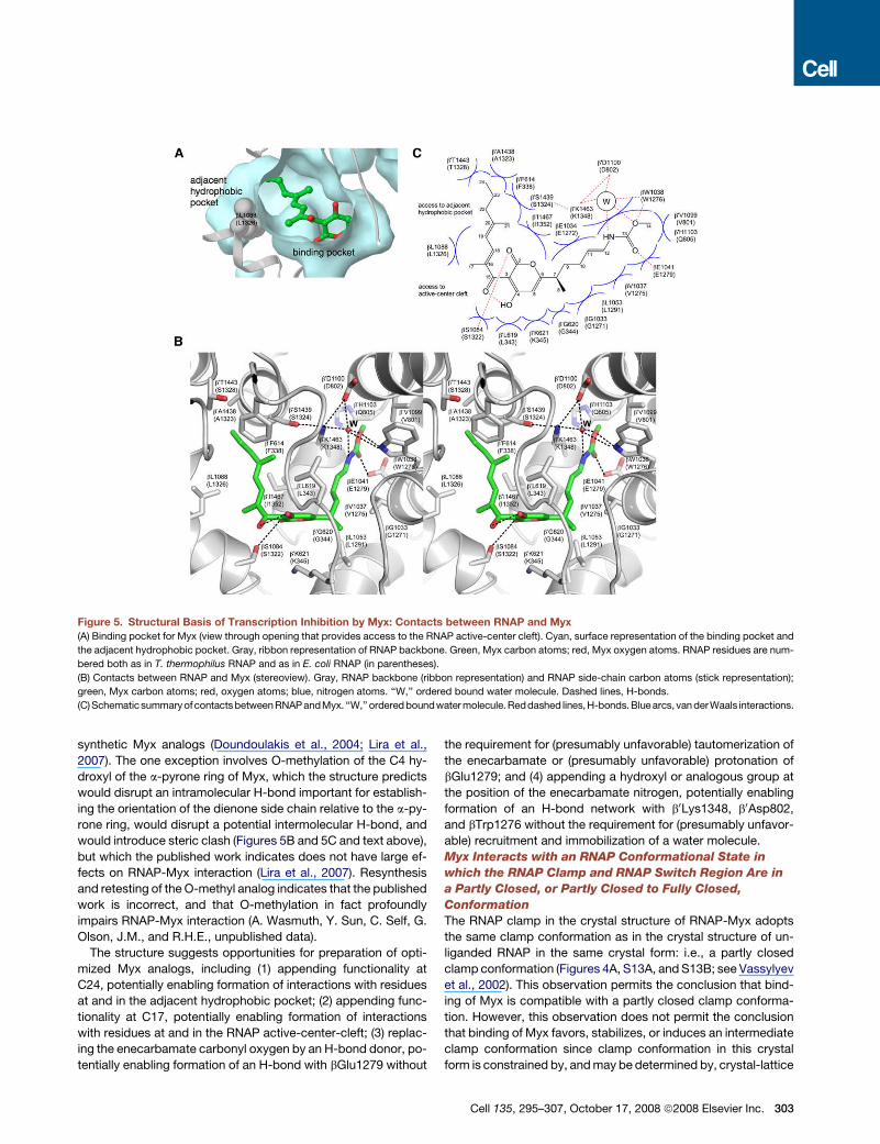
Figure 5. Structural Basis of Transcription Inhibition by Myx: Contacts between RNAP and Myx
(A) Binding pocket for Myx (view through opening that provides access to the RNAP active-center cleft). Cyan, surface representation of the binding pocket and
the adjacent hydrophobic pocket. Gray, ribbon representation of RNAP backbone. Green, Myx carbon atoms; red, Myx oxygen atoms. RNAP residues are num-
bered both as in T. thermophilus RNAP and as in E. coli RNAP (in parentheses).
(B) Contacts between RNAP and Myx (stereoview). Gray, RNAP backbone (ribbon representation) and RNAP side-chain carbon atoms (stick representation);
green, Myx carbon atoms; red, oxygen atoms; blue, nitrogen atoms. ‘‘W,’’ ordered bound water molecule. Dashed lines, H-bonds.
(C) Schematicsummary ofcontacts between RNAP andMyx. ‘‘W,’’ ordered boundwater molecule. Reddashed lines, H-bonds. Blue arcs, van derWaals interactions.
synthetic Myx analogs (Doundoulakis et al., 2004; Lira et al.,
2007). The one exception involves O-methylation of the C4 hy-
droxyl of the a-pyrone ring of Myx, which the structure predicts
would disrupt an intramolecular H-bond important for establish-
ing the orientation of the dienone side chain relative to the a-py-
rone ring, would disrupt a potential intermolecular H-bond, and
would introduce steric clash (Figures 5B and 5C and text above),
but which the published work indicates does not have large ef-
fects on RNAP-Myx interaction (Lira et al., 2007). Resynthesis
and retesting of the O-methyl analog indicates that the published
work is incorrect, and that O-methylation in fact profoundly
impairs RNAP-Myx interaction (A. Wasmuth, Y. Sun, C. Self, G.
Olson, J.M., and R.H.E., unpublished data).
The structure suggests opportunities for preparation of opti-
mized Myx analogs, including (1) appending functionality at
C24, potentially enabling formation of interactions with residues
at and in the adjacent hydrophobic pocket; (2) appending func-
tionality at C17, potentially enabling formation of interactions
with residues at and in the RNAP active-center-cleft; (3) replac-
ing the enecarbamate carbonyl oxygen by an H-bond donor, po-
tentially enabling formation of an H-bond with bGlu1279 without
the requirement for (presumably unfavorable) tautomerization of
the enecarbamate or (presumably unfavorable) protonation of
bGlu1279; and (4) appending a hydroxyl or analogous group at
the position of the enecarbamate nitrogen, potentially enabling
formation of an H-bond network with b0Lys1348, b0Asp802,
and bTrp1276 without the requirement for (presumably unfavor-
able) recruitment and immobilization of a water molecule.
Myx Interacts with an RNAP Conformational State in
which the RNAP Clamp and RNAP Switch Region Are in
a Partly Closed, or Partly Closed to Fully Closed,
Conformation
The RNAP clamp in the crystal structure of RNAP-Myx adopts
the same clamp conformation as in the crystal structure of un-
liganded RNAP in the same crystal form: i.e., a partly closed
clamp conformation (Figures 4A, S13A, and S13B; see Vassylyev
et al., 2002). This observation permits the conclusion that bind-
ing of Myx is compatible with a partly closed clamp conforma-
tion. However, this observation does not permit the conclusion
that binding of Myx favors, stabilizes, or induces an intermediate
clamp conformation since clamp conformation in this crystal
form is constrained by, and may be determined by, crystal-lattice
Cell 135, 295–307, October 17, 2008 ª2008 Elsevier Inc. 303
interactions (crystal-lattice interactions with the clamp, with
a b0 nonconserved domain appended to the clamp, and with
a s domain associated with the clamp).
The RNAP switch region in the crystal structure of RNAP-Myx
adopts a different conformation from that in the crystal structure
of unliganded RNAP in the same crystal form (Figure S15). The
difference in conformation involves a nine-residue segment of
switch 2—a nine-residue segment that differs in conformation
in open, partly closed, and fully closed clamp conformational
states (b0 residues 336–344; Figure 1B). The difference in confor-
mation involves 1–4 A displacements of Ca atoms of the nine-
residue segment toward positions intermediate between those
in partly closed and fully closed clamp conformational states
(Figures S15 and 1B). The nine-residue segment contains a resi-
due that contacts template-strand DNA in transcription com-
plexes (b0 residue 339; see Gnatt et al., 2001; Vassylyev et al.,
2007); however, since Myx does not efficiently inhibit interaction
between RNAP and template-strand DNA in relevant concentra-
tion ranges (Figures 3D, 3F and S10–S12), possible differences in
interaction between the nine-residue segment and template-
strand DNA cannot, in of themselves, account for transcription
inhibition by Myx.
Target and Mechanism of Transcription Inhibition by CorCor is a polyketide-derived a-pyrone antibiotic structurally re-
lated to Myx (Figure 1C; Irschik et al., 1985; Jansen et al.,
1985). Cor is produced by the myxobacterium Corallococcus
coralloides Cc c127 (Irschik et al., 1985; Jansen et al., 1985).
The compound inhibits growth of a broad spectrum of Gram-
positive and Gram-negative bacterial species, including M. tu-
berculosis, S. aureus, S. pneumoniae, B. anthracis, and E. coli
DH21tolC (MICs % 12.5 mg/ml for all; MIC % 0.1 mg/ml for S. au-
reus; Irschik et al., 1985; M. Talaue, N. Connell, J.M., and R.H.E.,
unpublished data). The compound is bacteriocidal, as assessed
in experiments with E. coli DH21f2tolC (J.M. and R.H.E., unpub-
lished data). The compound inhibits bacterial RNAP (IC50�4 mM)
but does not inhibit eukaryotic RNAP II (Irschik et al., 1985).
Cor differs from Myx only by possession of a seven-carbon ex-
tension of the dienone side chain (Figure 1C). Perhaps unsurpris-
ingly, in view of the structural similarity between Myx and Cor,
analysis of crossresistance patterns indicates that all mutants
that exhibit high-level (R16-fold) resistance to Myx also exhibit
resistance to Cor (Table 1). We infer that Cor interacts with a tar-
get that overlaps the target for Myx. To define further the target
for Cor, we performed saturation mutagenesis—targeting co-
dons for RNAP residues located within 30 A of sites of Myx-resis-
tant, Cor-crossresistant mutants—and we isolated and charac-
terized more than 75 additional Cor-resistant mutants (Table
S4; Figure S16). With one exception, all identified substitutions
that confer high-level (R8-fold) resistance to Cor also confer re-
sistance to Myx (Table 1). We conclude that Cor interacts with
the same target as Myx.
The one exception, a substitution that confers high-level resis-
tance to Cor but that does not confer resistance to Myx, involves
b residue 1326 (b1326Leu/Trp; Table 1). We infer that b residue
1326 interacts with the seven-carbon side-chain extension pres-
ent in Cor but not in Myx. Consistent with this inference, in the
three-dimensional structure of the RNAP-Myx complex, b1326
304 Cell 135, 295–307, October 17, 2008 ª2008 Elsevier Inc.
is located close to the ligand dienone side-chain terminus, the
point of attachment of the seven-carbon side-chain extension
present in Cor (Figures 5A–5C). Further consistent with this
inference, in the three-dimensional structure of the RNAP-Myx
complex, b1326 forms one wall of an adjacent hydrophobic
pocket that would have sufficient volume to accommodate the
seven-carbon side-chain extension present in Cor if b1326 is
Leu (as in the wild-type enzyme) but not if b1326 is Trp (as in
the mutant enzyme) (Figures 5A and 5C).
Experiments addressing the biochemical basis of transcription
inhibition by Cor indicate that Cor inhibits transcription initiation,
prevents interaction of RNAP with promoter DNA, prevents inter-
action with promoter positions�11 to +15, and prevents interac-
tion with promoter positions �11 to +15 when, and only when,
these positions are in double-stranded form (Figures S17 and
S18). We conclude that Cor shares the same target and same
mechanism as Myx.
Target and Mechanism of Transcription Inhibition by RipRip is a polyketide-derived macrocylic-lactone antibiotic struc-
turally unrelated to Myx and Cor (Figure 1C; Irschik et al.,
1995; Augustiniak et al., 1996). Rip is produced by the myxobac-
terium Sorangium cellulosum So ce377 (Irschik et al., 1995;
Augustiniak et al., 1996). The compound inhibits growth of S. au-
reus and E. coli DH21tolC (MICs % 1 mg/ml; Irschik et al., 1995;
J.M. and R.H.E., unpublished data). The compound is bacterio-
cidal, as assessed in experiments with E. coli DH21f2tolC (J.M.
and R.H.E., unpublished data). The compound inhibits bacterial
RNAP (IC50 �0.8 mM) but does not inhibit eukaryotic RNAP II
(Irschik et al., 1995).
Rip exhibits no structural similarity to Myx and Cor, apart from
a general similarity in size and hydrophobic character (Figure 1C).
Surprisingly, in view of the lack of structural similarity, analysis
of crossresistance patterns indicates that all identified mutants
that exhibit high-level resistance to Myx and Cor also exhibit re-
sistance to Rip (Table 1). We infer that Rip interacts with a target
that overlaps the target for Myx and Cor. To define further the
target for Rip, we performed saturation mutagenesis—targeting
codons for RNAP residues located within 30 A of sites of Myx-re-
sistant, Rip-crossresistant mutants—and we isolated and char-
acterized 50 additional Rip-resistant mutants (Table S5;
Figure S19). With one exception (b1326Leu/Trp; see preceding
section), all identified substitutions conferring high-level resis-
tance to Rip also confer resistance to Myx (Table 1). Without
exception, all identified substitutions conferring high-level resis-
tance to Rip also confer resistance to Cor (Table 1). We conclude
that Rip interacts with the same target as Myx and Cor.
Experiments addressing the biochemical basis of transcription
inhibition by Rip indicate that Rip inhibits transcription initiation,
prevents interaction of RNAP with promoter DNA, prevents inter-
action with promoter positions�11 to +15, and prevents interac-
tion with promoter positions �11 to +15 when, and only when,
these positions are in double-stranded form (Figures S20 and
S21). We conclude that Rip, despite its lack of structural similar-
ity to Myx and Cor, shares the same target and mechanism as
Myx and Cor. We conclude, thus, that at least two different che-
motypes, the a-pyrone chemotype and the macrocyclic-lactone
chemotype, function through this target and mechanism.

DISCUSSION
Our genetic results establish that Myx interacts with the RNAP
switch region (Figures 2, S1, and S2): i.e., with the hinge that me-
diates rotation of the RNAP clamp relative to the remainder of
RNAP and, thus, that mediates opening and closing of the
RNAP active-center cleft (Figures 1A and 1B). Our biochemical
results establish that Myx inhibits interactions of RNAP with
promoter DNA, establish that Myx inhibits interactions with the
promoter DNA segment that enters the RNAP active-center cleft
during transcription initiation (i.e., promoter positions �11 to
+15), and establish that Myx inhibits interactions with this pro-
moter DNA segment when it is in a double-stranded state (i.e.,
the state present before and during entry of promoter DNA into
the RNAP active-center cleft, during early stages of transcription
initiation) but not when it is present in a single-stranded state (i.e.,
the state present after entry of promoter DNA into the RNAP ac-
tive-center cleft, during late stages of transcription initiation)
(Figures 3 and S3–S12). Our structural results define contacts
between Myx and RNAP and indicate that Myx interacts with
an RNAP conformational state in which the RNAP clamp and
RNAP switch region are in a partly closed, or partly closed to fully
closed, conformation (Figures 4, 5, and S13–S15). Based on
these results, we propose that Myx inhibits transcription by lock-
ing the RNAP switch region in one conformation and thereby
locking the RNAP clamp in one conformation—a partly closed,
or partly closed to fully closed, conformation—thereby prevent-
ing opening of the RNAP active-center cleft to permit entry of
double-stranded DNA during transcription initiation. According
to this proposal, Myx functions essentially by ‘‘hinge jamming.’’
Our results further indicate that two other antibiotics, the
structurally related a-pyrone antibiotic Cor and the structurally
unrelated macrocyclic-lactone antibiotic Rip, share the same
target as Myx and the same mechanism as Myx (Table 1; Figures
S16–S21).
The results define a target and a mechanism for inhibition of
RNAP. The results, further, provide experimental evidence for
functional significance of the RNAP switch region, experimental
evidence for functional significance of opening and closing of the
RNAP clamp, and experimental evidence supporting the hypoth-
esis that the RNAP clamp must open to permit DNA to enter the
RNAP active-center cleft during transcription initiation. As such,
the results have implications for understanding mechanisms of
transcription and mechanisms of transcriptional regulation.
Based on several considerations, we suggest that the RNAP
switch region is an exceptionally attractive target for discovery
of new broad-spectrum antibacterial therapeutic agents. First,
the switch region comprises residues that are highly conserved
in both Gram-positive bacterial RNAP and Gram-negative bacte-
rial RNAP (Figure S2)—providing a basis for broad-spectrum
activity of compounds that function through the switch region.
Second, the switch region contains residues that are not con-
served, and indeed are radically different, in human RNAP I,
RNAP II, and RNAP III (Figure S2)—providing a basis for thera-
peutic selectivity of compounds that function through the switch
region. Third, the switch region is distant from the binding site for
rifamycins (Figure 1A) and from the binding sites for other char-
acterized inhibitors of bacterial RNAP (see Darst, 2004; Chopra,
2007), providing a basis for absence of crossresistance with rifa-
mycins (Table 1) and for absence of crossresistance with other
characterized inhibitors of bacterial RNAP (unpublished data).
Fourth, the ligand binding site in the switch region comprises
a nearly completely enclosed, predominantly hydrophobic,
pocket (Figures 5A and 5C), providing a basis for efficient
‘‘druggability’’ by multiple chemotypes (Table 1; Figures 1C, 2,
S16, and S19), facilitating in silico rational design of optimized li-
gands, and facilitating in silico virtual screening for new ligands.
We point out that the mechanistic role of the RNAP switch re-
gion (hinge for movement of domains) and the structural charac-
ter of the ligand binding site in the RNAP switch region (nearly
completely enclosed, predominantly hydrophobic, pocket) are
reminiscent of the mechanistic role and structural character of
the HIV-1 reverse transcriptase NNRTI site (nonnucleoside re-
verse-transcriptase inhibitor site; Kohlstaedt et al., 1992; Tantillo
et al., 1994; Sluis-Cremer et al., 2004). The HIV-1 reverse tran-
scriptase NNRTI site is druggable by multiple chemotypes and
is the target for multiple antiviral agents in current clinical use.
We suggest that the RNAP switch region may exhibit similarly
high druggability and similarly high utility.
Priorities for basic research include determination of struc-
tures of complexes of RNAP with Cor and Rip (to define interac-
tions with the seven-carbon-atom side-chain extension in Cor
and to define interactions with the macrocyclic-lactone chemo-
type of Rip) and determination of effects of switch-region-target
inhibitors on RNAP clamp conformation and dynamics in solu-
tion (addressable by use of fluorescence resonance energy
transfer; A. Chakraborty, Y. Korlann, D. Wang, S. Weiss, and
R.H.E., unpublished data). Priorities for applied research include
cloning and surrogate-host expression of biosynthetic genes for
Myx, Cor, and Rip (to overcome an important obstacle to possi-
ble clinical application of Myx, Cor, and Rip—namely, the rela-
tively poor fermentation characteristics of the myxobacterial
strains that produce these compounds), in silico rational design
of optimized switch-region-target inhibitors, and in silico virtual
screening and in vitro target-directed high-throughput screening
for new switch-region-target inhibitors.
EXPERIMENTAL PROCEDURES
Full details of Experimental Procedures are presented in the Supplemental
Data.
Mutagenesis
Random mutagenesis was performed by use of PCR amplification, exploiting
the baseline error rate of PCR amplification (methods analogous to those in
Mukhopadhyay et al., 2004, but with PCR amplification of entire plasmid mol-
ecules using Pfu DNA polymerase). Saturation mutagenesis was performed by
use of PCR amplification with ‘‘doped’’ primers (methods analogous to those in
Mukhopadhyay et al., 2004, but using pooled sets of ‘‘doped’’ primers).
Microbiological Assays
Complementation assays and MIC assays were performed essentially as in
Tuske et al. (2005).
RNAP-Myx Interaction Assays
RNAP-Myx interaction was detected by monitoring quenching by Myx of fluo-
rescence emission of RNAP Trp residues (lex = 280 nm; lem = 330 nm).
Cell 135, 295–307, October 17, 2008 ª2008 Elsevier Inc. 305

RNAP-DNA Interaction Assays
RNAP-DNA interaction assays were performed essentially as in Mukhopad-
hyay et al. (2004).
Transcription Assays
Run-off transcription assays and fluorescence-detected abortive initiation as-
says were performed essentially as in Mukhopadhyay et al. (2004). Rolling-cir-
cle transcription assays were performed using the Kool NC-45 kit (Epicenter,
Inc.).
Structure Determination
Crystallization, crystal handling, data collection, structure solution, and refine-
ment were performed by methods analogous to those in Tuske et al. (2005).
ACCESSION NUMBERS
Atomic coordinates and structure factors for RNAP-Myx have been deposited
in the PDB (PDB accession number 3DXJ).
SUPPLEMENTAL DATA
Supplemental Data include Supplemental Experimental Procedures, five
tables, and twenty-one figures and can be found with this article online at
http://www.cell.com/cgi/content/full/135/2/295/DC1/.
ACKNOWLEDGMENTS
We thank A. Chakraborty, N. Connell, S. Darst, Y. Korlann, C. Self, Y. Sun, G.
Olson, M. Talaue, D. Wang, X. Wang, A. Wasmuth, and S. Weiss for unpub-
lished data and G. Hofle, M. Kalesse, and E. Sineva for discussion. This
work was supported by NIH grant AI072766 to R.H.E. and E.A., NIH grant
GM41376 to R.H.E., and a Howard Hughes Medical Investigatorship to R.H.E.
Received: June 23, 2008
Revised: July 28, 2008
Accepted: September 11, 2008
Published: October 16, 2008
REFERENCES
Augustiniak, H., Irschik, H., Reichenbach, H., and Hofle, G. (1996). Ripostatin
A, B, and C: isolation and structure elucidation of novel metabolites from Sor-
angium cellulosum. Liebigs Ann. Chem. 10, 1657–1663.
Campbell, E., Korzheva, N., Mustaev, A., Murakami, K., Nair, S., Goldfarb, A.,
and Darst, S. (2001). Structural mechanism for rifampicin inhibition of bacterial
RNA polymerase. Cell 104, 901–912.
Chopra, I. (2007). Bacterial RNA polymerase: a promising target for the discov-
ery of new antimicrobial agents. Curr. Opin. Investig. Drugs 8, 600–607.
Cramer, P. (2002). Multisubunit RNA polymerases. Curr. Opin. Struct. Biol. 12,
89–97.
Cramer, P., Bushnell, D., Fu, J., Gnatt, A., Maier-Davis, B., Thompson, N., Bur-
gess, R., Edwards, A., David, P., and Kornberg, R. (2000). Architecture of RNA
polymerase II and implications for the transcription mechanism. Science 288,
640–649.
Cramer, P., Bushnell, D., and Kornberg, R. (2001). Structural basis of transcrip-
tion: RNA polymerase II at 2.8 angstrom resolution. Science 292, 1863–1876.
Darst, S. (2001). Bacterial RNA polymerase. Curr. Opin. Struct. Biol. 11, 155–162.
Darst, S. (2004). New inhibitors targeting bacterial RNA polymerase. Trends
Biochem. Sci. 29, 159–162.
Daubendiek, S., and Kool, E. (1997). Generation of catalytic RNAs by rolling
transcription of synthetic DNA nanocircles. Nat. Biotechnol. 15, 273–277.
Doundoulakis, T., Xiang, A., Lira, R., Agrios, K., Webber, S., Sisson, W., Aust,
R., Shah, A., Showalter, R., Appleman, J., and Simonsen, K. (2004). Myxopyr-
306 Cell 135, 295–307, October 17, 2008 ª2008 Elsevier Inc.
onin B analogs as inhibitors of RNA polymerase, synthesis and biological eval-
uation. Bioorg. Med. Chem. Lett. 14, 5667–5672.
Ebright, R. (2000). RNA polymerase: structural similarities between bacterial
RNA polymerase and eukaryotic RNA polymerase II. J. Mol. Biol. 304,
687–698.
Gnatt, A., Cramer, P., Fu, J., Bushnell, D., and Kornberg, R. (2001). Structural
basis of transcription: an RNA polymerase II elongation complex at 3.3 A res-
olution. Science 292, 1876–1882.
Guo, Y., and Gralla, J. (1998). Promoter opening via a DNA fork junction bind-
ing activity. Proc. Natl. Acad. Sci. USA 95, 11655–11660.
Helmann, J.D., and deHaseth, P. (1999). Protein-nucleic acid interactions
during open complex formation investigated by systematic alteration of the
protein and DNA binding partners. Biochemistry 38, 5959–5967.
Hu, T., Schaus, J., Lam, K., Palfreyman, M., Wuonola, M., Gustafson, G., and
Panek, J. (1998). Total synthesis and preliminary antibacterial evaluation of the
RNA polymerase inhibitors (+/�)-Myxopyronin A and B. J. Org. Chem. 63,
2401–2406.
Irschik, H., Gerth, K., Hofle, G., Kohl, W., and Reichenbach, H. (1983). The
myxopyronins, new inhibitors of bacterial RNA synthesis from Myxococcus
fulvus (Myxobacterales). J. Antibiot. (Tokyo) 36, 1651–1658.
Irschik, H., Jansen, R., Hofle, G., Gerth, K., and Reichenbach, H. (1985). The
corallopyronins, new inhibitors of bacterial RNA synthesis from Myxobacteria.
J. Antibiot. (Tokyo) 38, 145–152.
Irschik, H., Augustiniak, H., Gerth, K., Hofle, G., and Reichenbach, H. (1995).
The ripostatins, novel inhibitors of eubacterial RNA polymerase isolated from
myxobacteria. J. Antibiot. (Tokyo) 48, 787–792.
Jansen, R., Irschik, H., Reichenbach, H., and Hofle, G. (1985). Corallopyronin
A, B, and C: three novel antibiotics from Corallocuccus coralloides Cc c127
(Myxobacterales). Liebigs Ann. Chem. 4, 822–836.
Kadesch, T., and Chamberlin, M. (1982). Studies of in vitro transcription by calf
thymus RNA polymerase II using a novel duplex DNA template. J. Biol. Chem.
257, 5286–5295.
Kohl, W., Irschik, H., Reichenbach, H., and Hofle, G. (1983). Antibiotics from
gliding bacteria. XVII. Myxopyronin A and B: two novel antibiotics from Myxo-
coccus fulvus strain Mx f50. Liebigs Ann. Chem. 1983, 1656–1667.
Kohlstaedt, L., Wang, J., Friedman, J., Rice, P., and Steitz, T. (1992). Crystal
structure at 3.5 A resolution of HIV-1 reverse transcriptase complexed with
an inhibitor. Science 256, 1783–1790.
Lira, R., Xiang, A., Doundoulakis, T., Biller, W., Agrios, K., Simonsen, K., Web-
ber, S., Sisson, W., Aust, R., Shah, A., et al. (2007). Syntheses of novel myxo-
pyronin B analogs as potential inhibitors of bacterial RNA polymerase. Bioorg.
Med. Chem. Lett. 17, 6797–6800.
Marr, M., and Roberts, J. (1997). Promoter recognition as measured by binding
of polymerase to nontemplate strand oligonucleotide. Science 276,
1258–1260.
Mukhopadhyay, J., Sineva, E., Knight, J., Levy, R., and Ebright, R. (2004). An-
tibacterial peptide microcin J25 inhibits transcription by binding within, and
obstructing, the RNA polymerase secondary channel. Mol. Cell 14, 739–751.
Murakami, K., and Darst, S. (2003). Bacterial RNA polymerases. Curr. Opin.
Struct. Biol. 13, 31–39.
Murakami, K., Masuda, S., and Darst, S. (2002a). Structural basis of transcrip-
tion initiation: RNA polymerase holoenzyme at 4 A resolution. Science 296,
1280–1284.
Murakami, K., Masuda, S., Campbell, E., Muzzin, O., and Darst, S. (2002b).
Structural basis of transcription initiation: an RNA polymerase holoenzyme-
DNA complex. Science 296, 1285–1290.
Naryshkin, N., Revyakin, A., Kim, Y., Mekler, V., and Ebright, R. (2000). Struc-
tural organization of the RNA polymerase-promoter open complex. Cell 101,
601–611.
O’Neill, A., Oliva, B., Storey, C., Hoyle, A., Fishwick, C., and Chopra, I. (2000).
RNA polymerase inhibitors with activity against rifampin-resistant mutants of
Staphylococcus aureus. Antimicrob. Agents Chemother. 44, 3162–3166.

Record, M.T., Reznikoff, W., Craig, M., McQuade, K., and Schlax, P. (1996).
Escherichia coli RNA polymerase (Es70), promoters, and the kinetics of the
steps of transcription initiation. In Escherichia coli and Salmonella, F. Neid-
hardt, ed. (Washington, D.C.: ASM Press), pp. 792–820.
Sluis-Cremer, N., Temiz, N., and Bahar, I. (2004). Conformational changes in
HIV-1 reverse transcriptase induced by nonnucleoside reverse transcriptase
inhibitor binding. Curr. HIV Res. 2, 323–332.
Tantillo, C., Ding, J., Jacobo-Molina, A., Nanni, R., Boyer, P., Hughes, S., Pau-
wels, R., Andries, K., Janssen, P., and Arnold, E. (1994). Locations of anti-AIDS
drug binding sites and resistance mutations in the three-dimensional structure
of HIV-1 reverse transcriptase. Implications for mechanisms of drug inhibition
and resistance. J. Mol. Biol. 243, 369–387.
Tripatara, A., and deHaseth, P. (1993). A new start site for Escherichia coli RNA
polymerase at an engineered short region of non-complementarity in double-
stranded DNA. J. Mol. Biol. 233, 349–358.
Tuske, S., Sarafianos, S., Wang, X., Hudson, B., Sineva, E., Mukhopadhyay, J.,
Birktoft, J., Leroy, O., Ismail, S., Clark, A., et al. (2005). Inhibition of bacterial
RNA polymerase by streptolydigin: stabilization of a straight-bridge-helix ac-
tive-center conformation. Cell 122, 541–552.
Vassylyev, D., Sekine, S., Laptenko, O., Lee, J., Vassylyeva, M., Borukhov, S.,
and Yokoyama, S. (2002). Crystal structure of a bacterial RNA polymerase ho-
loenzyme at 2.6 A resolution. Nature 417, 712–719.
Vassylyev, D., Vassylyeva, M., Perederina, A., Tahirov, T., and Artsimovitch, I.
(2007). Structural basis for transcription elongation by bacterial RNA polymer-
ase. Nature 448, 157–162.
Young, B., Gruber, T., and Gross, C. (2002). Views of transcription initiation.
Cell 109, 417–420.
Zhang, G., Campbell, E., Minakhin, L., Richter, C., Severinov, K., and Darst, S.
(1999). Crystal structure of Thermus aquaticus core RNA polymerase at 3.3 A
resolution. Cell 98, 811–824.
Cell 135, 295–307, October 17, 2008 ª2008 Elsevier Inc. 307