Contacts between mammalian RNA polymerase II and the template DNA in a ternary elongation complex
6
©1993 Oxford University Press Nucleic Acids Research, 1993, Vol. 21, No. 1 113-118 Contacts between mammalian RNA polymerase II and the template DNA in a ternary elongation complex Gretchen A.Rice, Michael J.Chamberlin and Caroline M.Kane* 401 Barker Hall, Division of Biochemistry and Molecular Biology, University of California, Berkeley, CA 94720, USA Received September 22, 1992; Revised and Accepted December 1, 1992 ABSTRACT Elongation complexes of RNA polymerase II, RNA-DNA- enzyme ternary complexes, are Intermediates In the synthesis of all eukaryotlc mRNAs and are potential regulatory targets for factors controlling RNA chain elongation and termination. Analysis of such complexes can provide information concerning the structure of the catalytic core of the RNA polymerase and Its Interactions with the DNA template and RNA transcript. Knowledge of the structure of such complexes Is essential in understanding the catalytic and regulatory properties of RNA polymerase. We have prepared and isolated complexes of purified RNA polymerase II halted at defined positions along a DNA template, and we have used deoxyrlbonuclease I (DNAse I) to map the Interactions of the polymerase with the DNA template. DNAse I footprints of three specific ternary complexes reveal that the enzyme- template Interactions of Individual elongation complexes are not identical. The size of the protected region is distinct for each complex and varies from 48 to 55 bp between different complexes. Additionally, the positioning of the protected region relative to the active site varies in different complexes. Our results suggest that RNA polymerase II is a dynamic molecule and undergoes continual conformatlonal transitions during elongation. These transitions are likely to be important in the processes of transcript elongation and termination and their regulation. INTRODUCTION In order to understand regulatory mechanisms controlling transcript elongation, it would be useful to refer to a physical portrait of RNA polymerase II during transcription. Such information can be obtained by halting the RNA polymerase at specific sites along a transcription unit as has been done for the bacterial RNA polymerase (1—4). Structural analysis of RNA polymerase II in such transcription complexes has been hampered by the necessity to include multiple accessory factors required for promoter binding and initiation. The presence of many proteins in the initiation complex has made the assignment of contacts due to the polymerase itself difficult. Changes in the structure of the initiation complex during the transition from promoter binding to early elongation indicate that the complex is altered by the loss of transcription factors or structural changes within the protein components (5—7). Additional changes in the complexes' contacts with the DNA template have been found for transcription complexes which are committed to elongation (8,9). Transcription by RNA polymerase II is subject to significant regulation at all stages of transcription from initiation to elongation and termination. A large number of genes are regulated by premature blocks to elongation. Some of these sites are recognized by purified RNA polymerase II (10), and the block to elongation can be signalled by a combination of a T-run in the non-transcribed strand and a bend in the DNA template (11). Read through at some of these sites can be promoted by at least one protein, the elongation factor SU (10, for review) This protein interacts directly (12-14) and transiently (14) with RNA polymerase II during elongation in vitro. This result suggests that Sn may effect a structural change in the polymerase itself. Recently it also has been shown that SII promotes hydrolysis of the nascent transcript within the ternary complex, a reaction which is more complex than a simple catalytic reversal of the polymerization reaction (15 — 17). The structure of RNA polymerase n in elongation complexes can be directly examined by the use of 3' extended templates (18). With these templates, purified RNA polymerase can accurately initiate and elongate in the absence of any accessory factors. Thus, structural features of the purified enzyme in the ternary complex can be examined. With this template, we have positioned mammalian RNA polymerase II at three specific sites along a DNA template. Each specific complex has been isolated, and the conformation of each elongation intermediate has been studied. Each of the three complexes is distinct in its conformation, and thus each of the three complexes presents a different physical target for the action of elongation regulatory factors. • To whom correspondence should be addressed by guest on August 24, 2016 http://nar.oxfordjournals.org/ Downloaded from
-
Upload
independent -
Category
Documents
-
view
0 -
download
0
Transcript of Contacts between mammalian RNA polymerase II and the template DNA in a ternary elongation complex
©1993 Oxford University Press Nucleic Acids Research, 1993, Vol. 21, No. 1 113-118
Contacts between mammalian RNA polymerase II and thetemplate DNA in a ternary elongation complex
Gretchen A.Rice, Michael J.Chamberlin and Caroline M.Kane*401 Barker Hall, Division of Biochemistry and Molecular Biology, University of California, Berkeley,CA 94720, USA
Received September 22, 1992; Revised and Accepted December 1, 1992
ABSTRACT
Elongation complexes of RNA polymerase II, RNA-DNA-enzyme ternary complexes, are Intermediates In thesynthesis of all eukaryotlc mRNAs and are potentialregulatory targets for factors controlling RNA chainelongation and termination. Analysis of suchcomplexes can provide information concerning thestructure of the catalytic core of the RNA polymeraseand Its Interactions with the DNA template and RNAtranscript. Knowledge of the structure of suchcomplexes Is essential in understanding the catalyticand regulatory properties of RNA polymerase. We haveprepared and isolated complexes of purified RNApolymerase II halted at defined positions along a DNAtemplate, and we have used deoxyrlbonuclease I(DNAse I) to map the Interactions of the polymerasewith the DNA template. DNAse I footprints of threespecific ternary complexes reveal that the enzyme-template Interactions of Individual elongationcomplexes are not identical. The size of the protectedregion is distinct for each complex and varies from 48to 55 bp between different complexes. Additionally, thepositioning of the protected region relative to the activesite varies in different complexes. Our results suggestthat RNA polymerase II is a dynamic molecule andundergoes continual conformatlonal transitions duringelongation. These transitions are likely to be importantin the processes of transcript elongation andtermination and their regulation.
INTRODUCTION
In order to understand regulatory mechanisms controllingtranscript elongation, it would be useful to refer to a physicalportrait of RNA polymerase II during transcription. Suchinformation can be obtained by halting the RNA polymerase atspecific sites along a transcription unit as has been done for thebacterial RNA polymerase (1—4). Structural analysis of RNApolymerase II in such transcription complexes has been hampered
by the necessity to include multiple accessory factors requiredfor promoter binding and initiation. The presence of manyproteins in the initiation complex has made the assignment ofcontacts due to the polymerase itself difficult. Changes in thestructure of the initiation complex during the transition frompromoter binding to early elongation indicate that the complexis altered by the loss of transcription factors or structural changeswithin the protein components (5—7). Additional changes in thecomplexes' contacts with the DNA template have been found fortranscription complexes which are committed to elongation (8,9).
Transcription by RNA polymerase II is subject to significantregulation at all stages of transcription from initiation toelongation and termination. A large number of genes are regulatedby premature blocks to elongation. Some of these sites arerecognized by purified RNA polymerase II (10), and the blockto elongation can be signalled by a combination of a T-run inthe non-transcribed strand and a bend in the DNA template (11).Read through at some of these sites can be promoted by at leastone protein, the elongation factor SU (10, for review) This proteininteracts directly (12-14) and transiently (14) with RNApolymerase II during elongation in vitro. This result suggests thatSn may effect a structural change in the polymerase itself.Recently it also has been shown that SII promotes hydrolysis ofthe nascent transcript within the ternary complex, a reaction whichis more complex than a simple catalytic reversal of thepolymerization reaction (15 — 17).
The structure of RNA polymerase n in elongation complexescan be directly examined by the use of 3' extended templates(18). With these templates, purified RNA polymerase canaccurately initiate and elongate in the absence of any accessoryfactors. Thus, structural features of the purified enzyme in theternary complex can be examined. With this template, we havepositioned mammalian RNA polymerase II at three specific sitesalong a DNA template. Each specific complex has been isolated,and the conformation of each elongation intermediate has beenstudied. Each of the three complexes is distinct in itsconformation, and thus each of the three complexes presents adifferent physical target for the action of elongation regulatoryfactors.
• To whom correspondence should be addressed
by guest on August 24, 2016
http://nar.oxfordjournals.org/D
ownloaded from

114 Nucleic Acids Research, 1993, Vol. 21, No. 1
EXPERIMENTAL PROCEDURESReagentsRNA polymerase II was purified from calf thymus by amodification (19) of the procedure described by Hodo and Blatti(20) which resulted in >90% pure RNA polymerase II. Thepreparation was determined to contain 25% active molecules (18).The following reagents were purchased from the sourcesindicated: HPLC purified ribonucleoside triphosphates(Pharmacia/LKB); [a-^PJCTP >400 Ci/mmol (Amersham);[a-^PJGTP >3000 Ci/mmol (NEN); [ T - ^ P J A T P >7000Ci/mmol (NEN); Ribonuclease A and Ribonuclease Tl(Boehringer Mannheim); Terminal deoxynucleotidyl transferase(Ratliff Biochemicals, Los Alamos, NM); Exonuclease VII(BRL); Deoxyribonuclease I (Worthington Biochemicals).Restriction endonucleases were purchased from either NewEngland BioLabs or Boehringer Mannheim and were usedaccording to the manufacturer's instructions. T4 polynucleotidekinase was purchased from BRL and was used according to themanufacturer's instructions.
BuffersBuffers used included: Transcription buffer (70 mM Tris-OAcpH 8.0, 20% (v/v) Glycerol, 60 mM NH4CI, 6 mM MgOAc,5 mM Spermidine, 0.15 mM dithiothreitol); DNase I Diluent(transcription buffer with 10 mM CaCl2, 10 /tg/ml acetylatedBSA); DNase I Stop Load Buffer (3.5 mg/ml sonicated salmonsperm DNA, 50 mM EGTA, 0.1% xylene cyanol, 0.1%bromophenol blue); RNA/DNA elution buffer (10 mM Tris ClpH 7.8, 2 mM EDTA, 0.1% SDS); TBE buffer (89 mM Trisbase, 89 mM boric acid, 2.5 mM EDTA); Formamide load buffer(80% deionized formamide, 1X TBE, 0.1 % xylene cyanol, 0.1 %bromophenol blue); Urea load buffer (7 M Urea, 10 mM EDTA,0.5% SDS, 0.05% xylene cyanol, 0.05% bromophenol blue).
DNA TemplateThe template pCpGR220 with a 5' end labelled non-templatestrand was prepared from the plasmid pGR220 (21) in thefollowing manner. The plasmid pGR220 was digested with Smaland the 5' phosphates were removed with calf intestinalphosphatase. For DNase I footprinting, the 5' end of the non-template strand was 32P labelled using [7-32P]ATP and T4polynucleotide kinase. Unincorporated [7-32P]ATP wasremoved by centrifugal gel filtration using Sephadex G-50 (18)followed by one phenol:chloroform (1:1) extraction and ethanolprecipitation. An extension of deoxycytidylate residues was addedto the 3 ' ends of the template using terminal deoxynucleotidyltransferase (18). The template, pCpGR220, was digested withXbal to remove the end not adjoining the transcribed sequence.A second digestion with Asp718 generates a fragment of suitablesize for DNase I footprinting. This fragment is termedpCpGR220*S when it contains the ^P end label or pCpGR220Swhen not labelled.
The digestion with Smal is essential prior to the addition ofthe 3' extensions which direct initiation by the polymerase. Thesequence at the site of initiation produced by this restrictionenzyme permits the polymerase to engage in 'normal elongation,'displacing its nascent transcript and reannealing the duplex DNAduring transcription (22). If 3' extensions are added to endsproduced by most other restriction enzymes tested (22, andunpublished results), the polymerase does not displace itstranscript. Instead, a long RNA:DNA hybrid is formed during
transcription and the non-template strand is displaced (22). Theseaberrant elongation reactions do form stable ternary elongationcomplexes, but their relationship to complexes which form duringnormal elongation is questionable. The necessity of using Smallimits the type of manipulation that can be done with the templateas discussed below in 'Footprinting the Template Strand'.
Preparation and walking of ternary complexesHomogeneous populations of ternary complexes paused at specificsites along the DNA template were formed by a modificationof the RNA polymerase walking procedure developed by Levinet al. (1). The enzyme to DNA ratio was empirically determinedto optimize complex formation. The molar concentrations usedare 1.2 nM RNA polymerase II and 10 nM template (inmolecules). For DNase I footprinting stable ternary complexeswith a transcript of 135nt (G135) were prepared by incubatingRNA polymerase II with pCpGR220*S for 5 min at 37°C intranscription buffer containing 800 /*M ATP and GTP and 100jM CTP. This allowed the formation of a 135nt transcriptblocked from further elongation by lack of UTP. Theunincorporated NTPs are removed by passage of the ternarycomplexes through three consecutive Sephadex G-50 gel filtrationspin columns (1). G135 complexes were walked to U138 by theaddition of 10 yM UTP or to G143 by the addition of 10 /iMeach of UTP, CTP, and GTP and incubated at 37°C for 1 min.Simultaneously, stable ternary complexes were prepared ascontrols with labelled RNA transcripts on the templatepCpGR220S with a substitution of [a-32P]CTP for the 100 yMCTP. The walks were completed in the same manner describedabove. Transcripts were analyzed by poly aery lamide gelelectrophoresis (PAGE) on a denaturing 6% gel (30:0.8,acrylamide:bisacrylamide, 7 M urea, 0.8 mm thick, 25 cm long)in 1XTBE (89 mM Tris base, 89 mM boric acid, 2.5 mMEDTA).
DNase I footprinting of ternary complexesThe separately formed and isolated elongation intermediatesG135, U138 and G143 were footprinted with DNase I. First thecomplexes were digested with exonuclease VII to remove the3' dC extensions and then with DNAse I to generate a footprint.The complexes were separated from the free DNA bynondenaturing PAGE and the bands were visualized byautoradiography. The complexed DNA was eluted from gel slicesand analyzed by denaturing PAGE. The ternary complexes wereformed on pCpGR220*S as described above in 200 y\ reactionvolumes; 15 U of exonuclease VII were added to the reactionsafter 2 min and incubation was continued an additional 3 minto allow the removal of the 3' dC extensions. DNase I was addedto complexes halted at G135, U138 or G143 to a finalconcentration of 0.001 /ig/ml and the reactions were incubatedfor 1.5 min at 30°C. The digestion was stopped by me additionof l/10th volume of DNase I Stop/Load buffer and the sampleswere immediately loaded onto 4% nondenaturing acrylamide gels(30:0.8 acrylamide:bisacrylamide, 0.7 mm thick, 23 cm long)in 1 x TBE. Due to the large volume of the reaction, ~ 75 mlaliquots were loaded into each of several 2 cm wide wells.Electrophoresis was carried out at room temperature with 220volts until the xylene cyanol had migrated 8 cm from the well(2.5-3 hrs). This electrophoresis step effectively separates theternary complexes from the free DNA (1, 23).
As a control for complex purity, as assessed by transcripthomogeneity, each experiment was conducted in parallel with
by guest on August 24, 2016
http://nar.oxfordjournals.org/D
ownloaded from

Nucleic Acids Research, 1993, Vol. 21, No. 1 115
ternary complexes formed on pCpGR220S, as described above.These ternary complexes containing 32P labelled transcripts weresubjected to the same treatments as the 5' end labelledpCpGR220*S ternary complexes, except that they were notdigested with DNase I.
The complexes, both DNA labelled and RNA labelled, wereidentified by autoradiography and eluted from gel slices in 200/tl RNA/DNA elution buffer. The nucleic acids were precipitatedwith ethanol and resuspended in formamide loading buffer.Immediately before loading onto the gel, the samples were heatedfor 3 min at 93 °C and chilled on ice. Electrophoresis was carriedout in 6% acrylamide gels (19:1 acrylamide:bisacrylamide, 0.3mm thick, 40 cm long) containing 7 M Urea in 1X TBE at 1700volts. Gels were dried onto 3 MM Whatman paper and wereexposed to film with intensifying screens at -70°C.
Chemical sequencing ladders were prepared by the methodsof Maxam and Gilbert (24).
Footprinting the template strandThe technical aspects of preparing an effective unidirectional 3'extended template have so far precluded footprinting of thetemplate strand. Such footprints require specific labelling of thetemplate strand as well as formation and isolation of ternarycomplexes containing RNA polymerase molecules positioned atspecific locations. Templates were prepared which were firstdigested with either Asp 718 or EcoO105 and then labelled withpolynucleotide kinase and [Y-3 2P]ATP prior to the Smaldigestion described above. The kinase reaction labelled both the
3 4 5 6
711489404364
242
190
147
118110
Figure 1. Preparation and analysis of halted elongation complexes G135, U138,and G143. Halted elongation complexes were formed on pCpGR220 and transcriptswere analyzed on a denaturing polyacrylamide gel as described in Methods. Theactivity of the halted elongation complexes was determined by addition of NTPsto 800/tM to allow elongation to resume. Lane 1: DNA size markers; lane 2:RNA from a G135 complex labelled with a[32P]CTP; lane 3: RNA afterelongation of the G135 complex; lane 4: RNA from a U138 complex 3' endlabelled with at^PJUTP; lane 5: RNA after elongation of the U138 complex;lane 6: RNA from a G143 complex 3' end labelled with al^PJGTP; lane 7: RNAafter elongation of the G143 complex.
template and non-template strarxls. Following Smal digestion,which removed the ^P from the non-template strand, 3'extensions were added to both ends. In order to retain label onthe 5' end of the template strand, such prepared templatesnecessarily contained 3' extensions at both ends. RNA polymeraseII molecules can bind to such 3' extension:duplex junctions andinitiate transcription. Nucleotide deprivation (the absence of UTP)would be predicted to prevent the formation of stable ternarycomplexes containing long transcripts originating from theselabelled ends. However, any initiation from these ends wouldbe expected to generate transcripts hybridized to the DNA.Multiple complexes were always observed in nondenaturing PA-GE following ternary elongation complex formation withtemplate-strand labelled DNA templates. None of thesecomplexes gave a reproducible footprint with DNase I.
RESULTSRationaleThe objective of these studies was to obtain structural informationconcerning purified RNA polymerase II and how it contacts theDNA template in a normal elongation complex. We previouslyreported that transcripts in a single specific elongation complexwere apparently single stranded to within at least 3 nt of thegrowing point (21). In this study we have analyzed additionalelongation complexes by employing the same synthetic DNAtemplate on which purified RNA polymerase II initiates efficientlyand can then be halted at specific sites for lack of a particularNTP substrate. Each of the elongation complexes described ishomogeneous and remains stable and active for the duration ofthe analysis, and thus we assume that these halted complexesrepresent actual intermediates in the normal elongation process.
Formation and analysis of elongation complexesThe template pCpGR220 was utilized to form elongationcomplexes halted at several defined sites. By performingtranscription with subsets of ribonucleotides (see Methods), weisolated specific elongation complexes containing RNA transcriptsof either 135 nt, 138 nt, or 143 nt (Fig. 1). These elongationcomplexes are termed G135, U138, and G143 respectively.Analysis of the RNAs in each of these complexes shows that theyare quite pure, and the majority (90%) of the complexes are activeas shown by their ability to continue elongation (Fig. 1).
Analysis of gel isolated elongation complexesNon-denaturing polyacrylamide gel electrophoresis was used toseparate each elongation complex from free DNA prior to analysisof the contacts between RNA polymerase II and the DNAtemplate. Elongation complexes with either the RNA or die DNAlabelled were electrophoresed on non-denaturing gels, and thepositions of the complexes labelled in either the RNA or the DNAwere coincident (Fig. 2). Complexes A and B contained identicalRNA products; the migration difference is likely due to thebinding of exonuclease VII to a fraction of the complexes. Thisenzyme was used to remove the 3' extension from the templatefollowing transcription. When other methods were used toremove the extension, only one complex was observed on thesegels.
The band containing the A complexes was excised and the RNAcomponents were analyzed by denaturing PAGE (Fig. 3). TheRNA was homogeneous for each complex with no contamination
by guest on August 24, 2016
http://nar.oxfordjournals.org/D
ownloaded from

116 Nucleic Acids Research, 1993, Vol. 21, No. 1
* RNA •DNA
• « 9 » » • Well
•A•B
Free DNA
Figure 2. Isolation of halted elongation complexes by non-denaturing PAGE.The complexes were formed as described in Methods on either pCpGR220S,for labelling the RNA, or pCpGR220*S, to form DNA labelled complexes. Theexample shown is a G135 complex. The position of the wells, complexes (A& B), and the free DNA are indicated.
from the G135 complex in either the U138 or G143 complexes.The homogeneity of these RNA transcripts demonstrates thatstructural studies on elongation complexes isolated in this mannerwill reflect the structure of only a single complex.
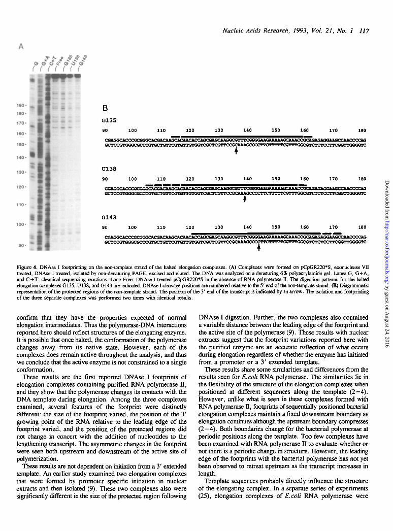
DNAse I footprinting of the non-template strand of elongationcomplexesUsing the three individual and separate elongation complexes thatwere shown to be stable, active, and isolable in homogeneousform, we probed the polymerase-DNA contacts with DNAse I(Fig. 4). The DNAse I footprints of the three separate complexeswere distinctly different in several respects. Surprisingly, theupstream and downstream boundaries of RNA polymerase H didnot move in concert relative to the template as the RNA increasedin length. Instead, the region of the template protected by RNApolymerase II increases upstream on the non-template strand whenthe polymerase elongates downstream from G135 to U138(Fig. 4A, lanes G135 and U138). The footprint then movessignificantly downstream, with both the upstream anddownstream boundaries of the polymerase advancing, when thepolymerase transcribes from U138 to G143 (Fig. 4A, lanes U138and G143).
In the G135 complex, the DNA template is protected from+ 113 to +161, a total of 48 bp, with the 3 bp on each edgeof this region being only partially protected (Fig. 4A, lane G135,and 4B). When the polymerase adds 3 additional nucleotides asit transcribes to position U138, the protected region of the DNAmoves backwards relative to the G135 complex (Fig. 4A laneU138 and 4B), giving a total of 55 bp protection. On the upstreamedge, this protection is only partial but distinct from +104 to+ 116, while on the downstream edge the footprint definitelyprotects two less bps to +159 even while the final 3bp are only
Figure 3. Analysis of RNA transcripts excised from non-denaturing gels. Theupper band for each complex as visualized in Figure 2 was excised from the non-denaturing gel and the RNA eluted as described in Methods. Eluted RNAs weredectrophoresed on a denaturing 6% polyacrylamide gd. The complex from whichthe RNA was eluted is indicated above the autoradiogram. All transcripts areof the expected sizes.
partially protected. After RNA polymerase II elongates a further5 nt to position G143, both edges of the footprint have moveddownstream on the DNA, protecting from +120 to +171(Fig. 4A, lane G143 and 4B). In this complex, the polymeraseprotects 51 bp with partial protection for the 7 bp on the leadingedge of the footprint. Surprisingly, although the transcript hasincreased in length by only 5 nt, the protected DNA region hasadvanced 16 bp on the lagging edge and 12 bp on the leadingedge relative to the U138 complex.
These asymmetric changes in the DNAse I footprint duringelongation result in modest changes in the total size of theprotected region. The polymerase fully protects 4 2 + / - 2 bp ofthe DNA. When the partial protection is included, this distanceextends to 51+/— 4 bp of protection.
The position of the RNA growing point within the DNAfootprint relative to the leading edge of the polymerase alsochanges during elongation and is distinct for each complex. Thisdistance changes from 26 bp to 21 bp to 28 bp respectively forthe G135, U138, and G143 complexes (Fig. 4B). These resultsalso suggest that the ternary complex is a dynamically changingstructure of multiple conformations.
DISCUSSION
We have used specifically halted elongation complexes of RNApolymerase II to investigate its interactions with the nascenttranscript and the template DNA. By monitoring the stability andactivity of the halted elongation complexes, we were able to
by guest on August 24, 2016
http://nar.oxfordjournals.org/D
ownloaded from
Nucleic Acids Research, 1993, Vol. 21, No. 1 117
190
180
170
160
150-
BG135
90 100 110 120 130 140 150 ieo 170 180
CGAOOC
120-
110-
100-
90 -
CCTCCOTOOGCGCCCGTOCTGrTCGTaTTaTaGTCQCTCaTTC
U138
9 0 100 110 120 130 140 130 160 170 180
«rrCCOTOOQCOCCCOTQCTOTTCO
G143
90 100 110 120 130 140 130 160 170 180
rGGGGTC
Figure 4. DNAse I footprinting on the non-template strand of the halted elongation complexes. (A) Complexes were formed on pCpGR220*S, exonuclease VIItreated, DNAse I treated, isolated by non-denaturing PAGE, excised and eluted. The DNA was analyzed on a denaturing 6% polyacrylamide gel. Lanes G, G+A,and C+T: chemical sequencing reactions. Lane Free: DNAse I treated pCpGR220*S in the absence of RNA polymerase II. The digestion patterns for the haltedelongation complexes G135, U138, andG143 are indicated. DNAse I cleavage positions are numbered relative to the 5' end of the non-template strand. (B) Diagrammaticrepresentation of the protected regions of the non-template strand. The position of the 3' end of the transcript is indicated by an arrow. The isolation and footprinringof the three separate complexes was performed two times with identical results.
confirm that they have the properties expected of normalelongation intermediates. Thus the polymerase-DNA interactionsreported here should reflect structures of the elongating enzyme.It is possible that once halted, the conformation of the polymerasechanges away from its native state. However, each of thecomplexes does remain active throughout the analysis, and thuswe conclude that the active enzyme is not constrained to a singleconformation.
These results are the first reported DNAse I footprints ofelongation complexes containing purified RNA polymerase II,and they show that the polymerase changes its contacts with theDNA template during elongation. Among the three complexesexamined, several features of the footprint were distinctlydifferent: the size of the footprint varied, the position of the 3'growing point of the RNA relative to the leading edge of thefootprint varied, and the position of the protected regions didnot change in concert with the addition of nucleotides to thelengthening transcript. The asymmetric changes in the footprintwere seen both upstream and downstream of the active site ofpolymerization.
These results are not dependent on initiation from a 3' extendedtemplate. An earlier study examined two elongation complexesthat were formed by promoter specific initiation in nuclearextracts and then isolated (9). These two complexes also weresignificantly different in the size of the protected region following
DNAse I digestion. Further, the two complexes also containeda variable distance between the leading edge of the footprint andthe active site of the polymerase (9). These results with nuclearextracts suggest that the footprint variations reported here withthe purified enzyme are an accurate reflection of what occursduring elongation regardless of whether the enzyme has initiatedfrom a promoter or a 3' extended template.
These results share some similarities and differences from theresults seen for E. coli RNA polymerase. The similarities lie inthe flexibility of the structure of the elongation complexes whenpositioned at different sequences along the template (2-4).However, unlike what is seen in these complexes formed withRNA polymerase n, footprints of sequentially positioned bacterialelongation complexes maintain a fixed downstream boundary aselongation continues although the upstream boundary compresses(2-4) . Both boundaries change for the bacterial polymerase atperiodic positions along the template. Too few complexes havebeen examined with RNA polymerase II to evaluate whether ornot there is a periodic change in structure. However, the leadingedge of the footprints with the bacterial polymerase has not yetbeen observed to retreat upstream as the transcript increases inlength.
Template sequences probably directly influence the structureof the elongating complex. In a separate series of experiments(25), elongation complexes of E.coli RNA polymerase were
by guest on August 24, 2016
http://nar.oxfordjournals.org/D
ownloaded from

118 Nucleic Acids Research, 1993, Vol. 21, No. 1
studied which were positioned over identical template sequencesalthough each complex contained a transcript of different length.In this case, the DNAse I footprints appeared identical for theindividual complexes. These results, and similar results withvaccinia RNA polymerase elongation complexes (26), suggestthat the template sequence (or structure) influences the structureof the polymerase during elongation. Such template influencesare important in intrinsic termination by mammalian RNApolymerase II (11), and template sequences have also been shownto be influential in regulating termination by E.coli RNApolymerase (27, 28).
For RNA polymerase n, the variations in the distance betweenthe active site and the leading edge of template protection suggestthat the polymerase does not translocate in simple monotonicsteps. Elongation by RNA polymerase II is apparently adiscontinuous process. Discontinuous movement also has beenreported for E.coli RNA polymerase (2). However, at this levelof resolution, it cannot be determined if the details of elongationare the same for both polymerases.
Discontinuous elongation indicates far more stuctural flexibilityof RNA polymerase than has been previously appreciated.Variation in the location of the active site within the dimensionsof the footprint suggests at least two forms of structural flexibility.The entire multisubunit enzyme complex may undergoconformational changes during elongation, or the active site itselfmay move within the enzyme relative to the template duringelongation.
The observed variation in polymerase conformation probablyreflects a basic element of transcript elongation. A structurallyflexible polymerase raises many possibilities for transcriptionregulation. Specifically during elongation when the enzymereaches a site that signals it to stop, the polymerase may takeon an alternate conformation that is stable yet incapable of furtherelongation. A factor that could alter thus conformation mightalleviate the elongation block and allow continuation oftranscription. Sn protein promotes such readthrough presumablyby interacting with stalled elongation complexes to promotecleavage of the nascent transcript prior to the resumption ofelongation (15-17). Alternatively, polymerases blocked duringelongation might be acted upon by termination factors whichpromote RNA release perhaps by modifying the conformationof a putative transcript binding site on the polymerase (21).Clearly the distinctive structures assumed by the ternary complexat different positions along the template during transcription mightmake the complexes more or less vulnerable to specific typesof factors. Structural analyses of many elongation complexespositioned at naturally occurring pauses as well as in the readilytranscribed sequences described here might highlight importantfeatures involved in the elongation control of such complexes.
6. Van Dyke, M. W., Roeder, R. G., and Sawadogo, M. (1988) Science 241,1335-1338.
7. Buratowslti, S., Hahn, S., Guarente, L., and Sharp, P. A. (1989) Cell 56,549-561.
8. Cai, H., and Luse, D. S. (1987) Molecular and Cell. Biol. 7, 3371 -3379.9. Linn, and Luse, D. S. (1991) Molecular and Cell. Biol. 11, 1508-1522.
10. Kerppola, T. K., and Kane, C. M. (1991) FASEB J. 5, 2833-2842.11. Kerppola, T. K., and Kane, C. M. (1990) Biochemistry 29, 269-278.12. Natori, S. (1982) Molec. Cell. Biocheni. 46, 173-187.13. Sawadogo, M., Huet, J., and Fromageot, P. (1980) Bkxhem. Biophys. Res.
Commun. 96, 258-264.14. Sluder, A. E., Greenleaf, A. L., and Price D. H. (1989) J. Biol. Chem.
264, 8963-8969.15. Reines, D. (1992) J. Biol. Chem. 267, 3795-3800.16. Reines, D., Ghanouni, P., Li, Q.-q., and Mote, J., Jr. (1992) J. Biol. Chem.
267, 15516-15522.17. Izban, M. G., and Luse, D. S. (1992) Genes and Dev. 6, 1342-1356.18. Kadesch, T. R., and Chamberlin, M. J. (1982) J. Biol. Chem. 27,
3187-3196.19. Kerppola, T. K., and Kane, C. M. (1988) Molecular and Cell. Biol. 8,
4389-4394.20. Hodo, H. G. I., and Blatri, S. P. (1977) Biochemistry 16, 2334-2343.21. Rice, G. A., Kane, C. M., and Chamberlin, M. J. (1991) Proc. Nat. Acad.
Sci. USA 88, 4245-4249.22. Dedrick, R. L., and M. J. Chamberlin (1985) Biochemistry 24, 2245-2253.23. Straney, D. C , and Crothers, D. M. (1985) Cell 43, 449-459.24. Maxam, A. M., and Gilbert, W. (1977) Proc. Nat. Acad. Sci. USA 74,
560-564.25. Metzger, W., Schickor, P., and Heumann, H. (1989) EMBO J. 8,
2745-2754.26. Hagler, J., and S. Shuman (1992), in press.27. Reynolds, R. R., and Chamberlin, M. J. (1991) J. Mol. Biol. 224, 53-63.28. Cheng, S. W. C , Lynch, E. C , Leason, K. R., Court, D. L., and Friedman,
D. (1991) Science 254, 1205-1207.
ACKNOWLEDGMENT
This work was supported by NIH grant GM34963.
REFERENCES
1. Levin, J. R., Krummel, B., and Chamberlin, M. J. (1987) J. Mol. Biol.1%, 85-100.
2. Krummd, B., and Chamberun, M. J. (1989) Biochemistry 28, 7829-7842.3. Krummd, B., and Chamberiin, M. J. (1992) J. Mol. Biol. 225, 221-237.4. Krummel, B., and Chamberiin, M. J. (1992) J. Mol. Biol. 225, 239-250.5. Cai, H., and Luse, D. S. (1987) J. Biol. Chem. 262, 298-304.
by guest on August 24, 2016
http://nar.oxfordjournals.org/D
ownloaded from