Glucocorticoid Hormones Decrease Proliferation of Embryonic Neural Stem Cells through...
9
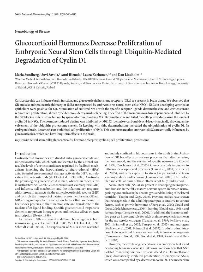
Neurobiology of Disease Glucocorticoid Hormones Decrease Proliferation of Embryonic Neural Stem Cells through Ubiquitin-Mediated Degradation of Cyclin D1 Maria Sundberg, 1 Suvi Savola, 1 Anni Hienola, 3 Laura Korhonen, 1,2 and Dan Lindholm 1,2 1 Minerva Medical Research Institute, Biomedicum Helsinki, FIN-00290 Helsinki, Finland, 2 Department of Neuroscience, Unit of Neurobiology, Uppsala University, Biomedical Centre, S-751 23 Uppsala, Sweden, and 3 Neuroscience Center, Department of Biosciences and Institute of Biotechnology, University of Helsinki, 00014 Helsinki, Finland Corticosteroids can influence brain function, and glucocorticoid hormone receptors (GRs) are present in brain tissue. We observed that GR and also mineralocorticoid receptor (MR) are expressed by embryonic rat neural stem cells (NSCs). NSCs in developing ventricular epithelium were positive for GR. Stimulation of cultured NSCs with the specific receptor ligands dexamethasone and corticosterone reduced cell proliferation, shown by 5-bromo-2-deoxy-uridine labeling. The effect of the hormones was dose dependent and inhibited by the GR blocker mifepristone but not by spironolactone, blocking MR. Dexamethasone inhibited the cell cycle by decreasing the levels of cyclin D1 in NSCs. The hormone-induced decline was inhibited by MG132 (benzyloxycarbonyl-leucyl-leucyl-leucinal), showing an in- volvement of the ubiquitin proteasome system, In keeping with this, dexamethasone increased the ubiquitination of cyclin D1. In embryonic brain, dexamethasone inhibited cell proliferation of NSCs. This demonstrates that embryonic NSCs are critically influenced by glucocorticoids, which can have long-term effects in the brain. Key words: neural stem cells; glucocorticoids; hormone receptor; cyclin D; cell proliferation; proteasome Introduction Corticosteroid hormones are divided into glucocorticoids and mineralocorticoids, which both are secreted by the adrenal cor- tex. The levels of corticosteroids are regulated by feedback mech- anisms involving the hypothalamic–pituitary–adrenal (HPA) axis. Stressful environmental changes activate the HPA axis ele- vating the corticosteroids (de Kloet et al., 1998, 2005). Cortisol is the physiological glucocorticoid in man, whereas in rodents this is corticosterone (Cort). Glucocorticoids act via receptors (GRs) and influence cell metabolism and the inflammatory response. Aldosterone in turn acts via the mineralocorticoid receptor (MR) and controls the transport of natrium across membranes. GR and MR are ligand-specific transcription factors that are bound to heat shock proteins in their inactive state and translocate to the nucleus after ligand binding. Specific glucocorticoid-responsive elements are present in target genes and mediate effects on gene transcription (Beato, 1989). In the brain, GRs are present in different brain regions in both neurons and glial cells (Fuxe et al., 1985; Van Eekelen et al., 1991; Schmidt et al., 2003). The expression of MR is more restricted and mainly confined to hippocampus in the adult brain. Activa- tion of GR has effects on various processes that alter behavior, memory, mood, and the survival of specific neurons (de Kloet et al., 1998; Crochemore et al., 2005). Glucocorticoids are known to influence developmental processes (Gass et al., 2001; de Kloet et al., 2005), and early exposure to stress has persistent effects on learning abilities and behavior (Lemaire et al., 2000). The molec- ular and cellular basis of these effects is not fully understood. Neural stem cells (NSCs) are present in developing neuroepithe- lium but also in the fully mature nervous system in certain neuro- genic regions, such as in the dentate gyrus and the lining of the lateral ventricles (Taupin and Gage, 2002). Previous studies have shown that neurogenesis in the adult hippocampus is sensitive to various factors, such as growth hormones (Åberg et al., 2000; Gould and Gross, 2002; Schanzer et al., 2004), learning (Gould et al., 1998), and various drugs (Lemaire et al., 2000). In addition, the hormonal mi- lieu plays an important role for adult brain neurogenesis, as shown for the sex steroids estrogens (Tanapat et al., 1999; Perfilieva et al., 2001; Bra ¨nnvall et al., 2002; Tanapat et al., 2005) and androgens (Perfilieva et al., 2001; Bra ¨nnvall et al., 2005). In adults, administra- tion of glucocorticoid hormones negatively influence neurogenesis (Cameron and Gould, 1994; Gould et al., 1998; Karishma and Her- bert, 2002). However, the effects of glucocorticoids in embryonic NSCs and developing brain are essentially unknown. We show here that NSC isolated from embryonic brain express GR and MR. Dexamethasone (Dex) dramatically inhibited proliferation of embryonic NSCs, which was accompanied by a decrease in cyclin D1. The mechanism Received Nov. 16, 2005; revised March 30, 2006; accepted April 2, 2006. This work was supported by the Medical Research Council, Minerva Foundation, Signe and Ane Gyllenberg Foundation, Liv och Ha ¨lsa, and Arvo and Lea Ylppo ¨ Foundation. We thank Mirkka Turunen for help with animals, Tuulikki Nyman for PCR, and Eeva Lehto and Johanna Ma ¨kela ¨ for skillful technical assistance. Correspondence should be addressed to Dan Lindholm, Department of Neuroscience, Biomedical Center, Box 587, S-75123 Uppsala, Sweden. E-mail: [email protected]. DOI:10.1523/JNEUROSCI.4906-05.2006 Copyright © 2006 Society for Neuroscience 0270-6474/06/265402-09$15.00/0 5402 • The Journal of Neuroscience, May 17, 2006 • 26(20):5402–5410
Transcript of Glucocorticoid Hormones Decrease Proliferation of Embryonic Neural Stem Cells through...
Neurobiology of Disease
Glucocorticoid Hormones Decrease Proliferation ofEmbryonic Neural Stem Cells through Ubiquitin-MediatedDegradation of Cyclin D1
Maria Sundberg,1 Suvi Savola,1 Anni Hienola,3 Laura Korhonen,1,2 and Dan Lindholm1,2
1Minerva Medical Research Institute, Biomedicum Helsinki, FIN-00290 Helsinki, Finland, 2Department of Neuroscience, Unit of Neurobiology, UppsalaUniversity, Biomedical Centre, S-751 23 Uppsala, Sweden, and 3Neuroscience Center, Department of Biosciences and Institute of Biotechnology, Universityof Helsinki, 00014 Helsinki, Finland
Corticosteroids can influence brain function, and glucocorticoid hormone receptors (GRs) are present in brain tissue. We observed thatGR and also mineralocorticoid receptor (MR) are expressed by embryonic rat neural stem cells (NSCs). NSCs in developing ventricularepithelium were positive for GR. Stimulation of cultured NSCs with the specific receptor ligands dexamethasone and corticosteronereduced cell proliferation, shown by 5�-bromo-2-deoxy-uridine labeling. The effect of the hormones was dose dependent and inhibited bythe GR blocker mifepristone but not by spironolactone, blocking MR. Dexamethasone inhibited the cell cycle by decreasing the levels ofcyclin D1 in NSCs. The hormone-induced decline was inhibited by MG132 (benzyloxycarbonyl-leucyl-leucyl-leucinal), showing an in-volvement of the ubiquitin proteasome system, In keeping with this, dexamethasone increased the ubiquitination of cyclin D1. Inembryonic brain, dexamethasone inhibited cell proliferation of NSCs. This demonstrates that embryonic NSCs are critically influenced byglucocorticoids, which can have long-term effects in the brain.
Key words: neural stem cells; glucocorticoids; hormone receptor; cyclin D; cell proliferation; proteasome
IntroductionCorticosteroid hormones are divided into glucocorticoids andmineralocorticoids, which both are secreted by the adrenal cor-tex. The levels of corticosteroids are regulated by feedback mech-anisms involving the hypothalamic–pituitary–adrenal (HPA)axis. Stressful environmental changes activate the HPA axis ele-vating the corticosteroids (de Kloet et al., 1998, 2005). Cortisol isthe physiological glucocorticoid in man, whereas in rodents thisis corticosterone (Cort). Glucocorticoids act via receptors (GRs)and influence cell metabolism and the inflammatory response.Aldosterone in turn acts via the mineralocorticoid receptor (MR)and controls the transport of natrium across membranes. GR andMR are ligand-specific transcription factors that are bound toheat shock proteins in their inactive state and translocate to thenucleus after ligand binding. Specific glucocorticoid-responsiveelements are present in target genes and mediate effects on genetranscription (Beato, 1989).
In the brain, GRs are present in different brain regions in bothneurons and glial cells (Fuxe et al., 1985; Van Eekelen et al., 1991;Schmidt et al., 2003). The expression of MR is more restricted
and mainly confined to hippocampus in the adult brain. Activa-tion of GR has effects on various processes that alter behavior,memory, mood, and the survival of specific neurons (de Kloet etal., 1998; Crochemore et al., 2005). Glucocorticoids are known toinfluence developmental processes (Gass et al., 2001; de Kloet etal., 2005), and early exposure to stress has persistent effects onlearning abilities and behavior (Lemaire et al., 2000). The molec-ular and cellular basis of these effects is not fully understood.
Neural stem cells (NSCs) are present in developing neuroepithe-lium but also in the fully mature nervous system in certain neuro-genic regions, such as in the dentate gyrus and the lining of the lateralventricles (Taupin and Gage, 2002). Previous studies have shownthat neurogenesis in the adult hippocampus is sensitive to variousfactors, such as growth hormones (Åberg et al., 2000; Gould andGross, 2002; Schanzer et al., 2004), learning (Gould et al., 1998), andvarious drugs (Lemaire et al., 2000). In addition, the hormonal mi-lieu plays an important role for adult brain neurogenesis, as shownfor the sex steroids estrogens (Tanapat et al., 1999; Perfilieva et al.,2001; Brannvall et al., 2002; Tanapat et al., 2005) and androgens(Perfilieva et al., 2001; Brannvall et al., 2005). In adults, administra-tion of glucocorticoid hormones negatively influence neurogenesis(Cameron and Gould, 1994; Gould et al., 1998; Karishma and Her-bert, 2002).
However, the effects of glucocorticoids in embryonic NSCs anddeveloping brain are essentially unknown. We show here that NSCisolated from embryonic brain express GR and MR. Dexamethasone(Dex) dramatically inhibited proliferation of embryonic NSCs,which was accompanied by a decrease in cyclin D1. The mechanism
Received Nov. 16, 2005; revised March 30, 2006; accepted April 2, 2006.This work was supported by the Medical Research Council, Minerva Foundation, Signe and Ane Gyllenberg
Foundation, Liv och Halsa, and Arvo and Lea Ylppo Foundation. We thank Mirkka Turunen for help with animals,Tuulikki Nyman for PCR, and Eeva Lehto and Johanna Makela for skillful technical assistance.
Correspondence should be addressed to Dan Lindholm, Department of Neuroscience, Biomedical Center, Box587, S-75123 Uppsala, Sweden. E-mail: [email protected].
DOI:10.1523/JNEUROSCI.4906-05.2006Copyright © 2006 Society for Neuroscience 0270-6474/06/265402-09$15.00/0
5402 • The Journal of Neuroscience, May 17, 2006 • 26(20):5402–5410
involved the proteasome-mediated degradation of cyclin D1 regu-lating the cell cycle. In vivo, dexamethasone reduced the proliferationof NSCs in embryonic brain, showing an important effect of thehormone on brain development.
Materials and MethodsAnimals. Wistar rats were obtained from Harlan (Horst, The Nether-lands) and housed at 12 h light/dark cycle with food available ad libitum.All experiments were approved by the local ethical committee and per-formed in accordance with the European Communities Council Direc-tive (86/609/EEC).
Preparation and dissociation of NSCs. Striatum was dissected from embry-onic day 17 (E17), and NSCs were prepared as described previously (Bran-nvall et al., 2002, 2005; Korhonen et al., 2003). Cells were incubated at�37°Cin 5% CO2 atmosphere in Corning (Helsinki, Finland) Suspension Culturedishes (5 � 106 cells per 10 cm dish; Corning) or in Corning Ultra LowAttachment dishes (Corning) in NSC medium containing 15 nM HEPES, pH7.5, 2.5 mM L-glutamine, 100 U/ml penicillin, 100 �g/ml streptomycin, 20ng/ml epidermal growth factor (EGF) (PeproTech, Rocky Hill, NJ), and B27supplement (Invitrogen, Espoo, Finland) in DMEM/F-12 (Invitrogen).Neurospheres were grown for 4–5 d, gently dissociated, and collected bycentrifugation for 5 min at 1500 rpm. The cells were resuspended into ap-propriate volume of medium containing EGF, hormones, and inhibitors asindicated.
NSC viability and proliferation assay. NSCs were cultured in 96-well cellculture dishes (70,000 cells per well; Costar 3599; Corning) in 100 �l ofserum-free medium in the presence of 20 ng/ml EGF and different concen-trations of Dex and Cort (both from Sigma, Helsinki, Finland) as indicatedin the figures. Mifepristone [RU-486 (17�-hydroxy-11�-(4-methylamino-phenyl)-17�-(1-propynyl) estra-4,9-dien-3 one-6–7)] and spironolactone[7�-(acetylthio)-17�-hydroxy-3oxopregn-4-ene-21carboxylic acid lac-tone] (both from Sigma) were used to inhibit GR and MR, respectively. Toestimate the viability of cells, we used the MTT [3-(4,5-dimethylthiazol-2-yl)-2,5-diphenyltetrazolium bromide (Sigma)] assay as described previously(Brannvall et al., 2002, 2005).
For 5�-bromo-2-deoxy-uridine (BrdU) labeling, NSCs were incubatedin 35 mm dishes (10 6 cells per dish, Corning Ultra Low Attachment) inculture medium in the presence of 1 �M Dex or 1 �M Cort. After 2 d,BrdU (10 �M; Sigma) was added, and the incubation was performed foran additional 24 h, after which cells were dissociated and plated onto 50�g/ml poly-DL-ornithine (Sigma)-coated coverslips (150,000 cells perwell; Costar 3524; Corning). After attachment, NSCs were fixed for 20min with 4% paraformaldehyde, washed three times with PBS, pH 7.4,and treated with 2 M HCl for 30 min at room temperature. Cells werewashed twice with PBS, blocked for 1 h in 3% BSA (Sigma), 0.1% TritonX-100/PBS, and primary rat anti-BrdU (diluted 1:200; Sigma). The nextday, cells were washed three times with PBS and incubated for 2 h withthe secondary anti-rat cyanine 3 (Cy3) antibody (diluted 1:200; JacksonImmunoResearch, Cambridgeshire, UK). Cells were washed three timeswith PBS and counterstained using Hoescht (4 �g/ml; Sigma) beforemounting in Gel Mount. The number of total and BrdU-positive cellswere counted using microscopy in four nonoverlapping fields per cover-slip. Experiments were repeated more than three times, and ANOVA wasused for statistical analysis.
To transfect NSCs, we used the Amaxa (Cologne, Germany) Nucleofectorkit and equipment and 8 �g of pDEST26 expression vector encoding humancyclin D1. Transfection with enhanced green fluorescent protein (GFP) wasused as control. At 24 h after transfection, half of the cells were treated for 2 dwith Dex, and cell proliferation was analyzed by BrdU labeling.
Cell differentiation. NSC were plated onto 50 �g/ml poly-DL-ornithine-coated 24-well culture dishes (150,000 cells per well; Costar 3524; Corning)and incubated for 5 d in the presence of Dex (Brannvall et al., 2002; Kor-honen et al., 2003). The cells were fixed for 20 min using 4% paraformalde-hyde, blocked in 1% BSA and 0.1% Triton X-100, washed by PBS, incubatedfor 30 min with 0.3% H2O2 to inhibit endogenous peroxidases, and blockedfor 1 h using 3% BSA in PBS/0.1% Triton X-100. The following primaryantibodies were used and added overnight at 4°C: monoclonal mouse anti-�-tubulin (diluted 1:200; BioSite, Helsinki, Finland), rabbit anti-glial fibril-
lary acidic protein (1:200; Sigma), and rabbit anti-�-nestin (1:1000; Chemi-con, Helsinki, Finland). Secondary Cy2 anti-mouse and Cy2 anti-rabbitantibodies (1:200; Jackson ImmunoResearch) were added for 1 h in PBS in1% BSA and 0.1% Triton X-100. The number of immunoreactive cells ineach well was counted using fluorescent microscopy in four independentfields. Statistical analysis was done as above.
Western blotting and immunohistochemistry. This was done essentially asdescribed previously (Korhonen et al., 2001; Brannvall et al., 2002, 2005).NSCs were incubated as above, and cells were lysed in radioimmunoprecipi-tation assay (RIPA) buffer containing 1% SDS and 10 mM Tris-HCl, pH 7.4to detect GR or using 50 mM Tris-HCl, pH 7.4, 150 mM NaCl, 1% NP-40,0.25% Na-deoxycholate, 1 mM EDTA, and a protease inhibitor cocktail(Roche, Espoo, Finland) to detect cell cycle proteins. In some experiments,MG123 (Calbiochem, Espoo, Finland) was used to inhibit the activity of theproteasome (Yu et al., 2003). Protein concentrations were determined byPierce (Helsinki, Finland) protein assay, and equal amounts of proteins wereloaded onto a 10% SDS-gel for separation. Loading was further controlled byPonceauS staining (Sigma). The gel was transferred onto a nitrocellulosemembrane (Amersham Biosciences, Helsinki, Finland) and incubated withprimary antibodies such as rabbit anti-GR antibody (1:250; Affinity BioRe-agents, Helsinki, Finland), mouse anti-MR antibody (1:100; Affinity BioRe-agents), mouse anti-cyclin D1 (1:700; Santa Cruz Biotechnology, Helsinki,Finland), mouse anti-p18 (1:400; BD Biosciences, Helsinki, Finland), mouseanti-p27 (1:800; BD Biosciences), mouse monoclonal anti-ubiquitin anti-body, P4G7 (1:1000; BioSite), or rabbit anti-actin (1:1000; Sigma). Afterwashing, the filter was incubated with horseradish peroxidase-conjugatedsecondary anti-rabbit/mouse antibodies (1:2500; Pierce) and detected usingthe ECL method. Actin was used as control. In some experiments, 100 �g ofubiquitin ladder (Affiniti Research, Exeter, UK) was analyzed to show thesize of the ubiquitinated protein.
To detect GR and MR in embryonic rat brain, specimens were embeddedin paraffin, sectioned using a microtome, and collected onto Superfrostslides. Sections were deparaffinated, and antigen retrieval was performed byboiling for 5 min twice in 0.01 M citric acid, pH 6.0. Sections were fixed for 1 hin 5% BSA and 0.1% Triton X-100/PBS and processed for staining andvisualization as described above, using anti-GR and anti-nestin antibodies(1:1400; Chemicon) for double staining.
Immunoprecipitation and ubiquitination of cyclin D1. For immunopre-cipitation cells were lysed in RIPA buffer (see above) supplemented withprotease inhibitor cocktail (Roche). The lysates were precleared withprotein G-agarose (Roche) for 1 h, followed by determination of proteinand an overnight incubation with primary mouse anti-cyclin D1 anti-body (Santa Cruz Biotechnology). A total of 40 �l of protein G-agarose(Roche) was added to each lysate for 2 h, followed by three washes withlysis buffer. Agarose beads were boiled, and proteins were separated asdescribed above. The anti-ubiquitin antibody P4G7 (1:300; BioSite) wasused to detect ubiquitinated cyclin D1.
PCR analysis. The presence of transcripts for cyclin D1, GR, and MR inNSCs was determined by reverse transcriptase (RT)-PCR. Total RNA wasextracted using GenElute Mammalian Total RNA Miniprep kit (Sigma)from cells according to the instructions of the manufacturer. A total of 1 �gof RNA was used for cDNA synthesis using 200 U of Moloney murine leu-kemia virus RT (Invitrogen) with 12.5 �g/ml oligo- dT primer (Promega,Helsinki, Finland) and appropriate ingredients and buffer (Invitrogen). PCRwas performed using the conditions of 94°C for 1 min, 55°C for 1 min, and72°C for 1 min for 30 cycles with a 10 min 72°C final extension. The specificoligonucleotides (Thermo Electron Corporation, Bremen, Germany) wereas follows: GR, 5�-GTCCATGGGGCTGTATATGG-3� (upstream) and 5�-TCCTCATTCGTGTTCCCTTC-3� (downstream) corresponding to nucle-otides 59–78 and 518–537 in the rat sequence; MR, 5�-TCAGACCTTG-GAGCGTTCTT-3� (upstream) and 5�-AGTGTGGAGGACCTGT-GACC-3� (downstream) corresponding to nucleotides 56–75 and 430–449.For cyclin D1, the oligonucleotides were as follows: 5�-AAGTAGTG-GCATCCGC-3� (upstream) and 5�-CCCGTCTCCCTATACTCAG-3�(downstream) corresponding to nucleotides 1041–1057 and 1253–1272 inthe rat sequence; �-actin, 5�-TGTTTGAGACCTTCAA-3� (upstream) and5�- TTGGCGTACAGGTCTTTGCGG-3� (downstream).
Sundberg et al. • Glucocorticoid Hormones Influence Embryonic Neural Stem Cells J. Neurosci., May 17, 2006 • 26(20):5402–5410 • 5403
In vivo experiments. Dex was injected intra-peritoneally (100 �g/kg) to timed-pregnantE14 rats for 3 d, and the controls received sa-line. BrdU (100 mg/kg, dissolved in 0.9% sa-line) was given for the last 2 d. Rats were anes-thetized and decapitated, and the brains of thepups were immediately removed, rinsed withPBS, and fixed for 24 h in 10% Formalin.Brains were stored up to 3 d in 70% ethanoland then dehydrated in an alcohol series andembedded in paraffin. Sections at 15 �m werecut using a Leitz (Wetzlar, Germany) mic-rotome and placed on SuperFrost Plus glassslides (Menzel-Glaser, Espoo, Finland).
Sections were deparaffinized with xyleneand incubated in ethanol and water. Antigenretrieval was done as above, and the sectionswere treated for 30 min at 67°C with 1 M HCland washed with PBS. Primary mouse anti-nestin and rat anti-BrdU antibodies wereadded overnight at 4°C, followed by washing ofsections with PBS. The additional incubationwas at room temperature for 1 h using second-ary AlexaFluor 488 anti-rat (1:2000) andAlexaFluor 568 anti-mouse (1:2000; Invitro-gen, Carlsbad, CA) antibodies. Sections werewashed and mounted with gel mounting me-dium (Gel Mount; Sigma).
Stereology. The number of BrdU–nestindouble-positive cells in the developing ratbrain was determined using unbiased stereol-ogy methods and the Stereo Investigator (Mi-croBrightField, Magdeburg, Germany) plat-form attached to an Olympus Optical (Tokyo,Japan) BX51 microscope. Cells were countedusing the optical fractionator method in com-bination with the dissector principle and unbi-ased counting rules (Mouton et al., 2002;Hienola et al., 2004). The method was opti-mized to give a coefficient of error �8% perindividual brain sample. Three individualbrains from Dex-treated and control animalswere analyzed. Every 10th section was selectedin a systematic random manner from a totalnumber of 52– 60 sections through the hip-pocampus of each brain, which generated fivesections per reference space per brain. Each ref-erence space was outlined at low power (10�),and cells were counted using a high-magnification (63�, 1.4 numerical apertureoil-immersion) objective. Cell number in theventricular zone of ganglion eminence wascounted from the same section. The referencevolume was estimated using Cavaleri dot grid(75 � 75 �m) to give the density in the ventric-ular zone. Statistical analyzes was done usingStudent’s t test.
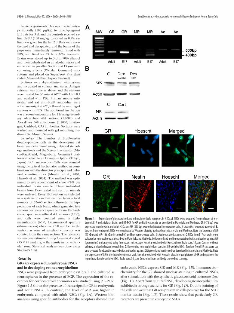
ResultsGRs are expressed in embryonic NSCsand in developing rat neuroepitheliumNSCs were prepared from embryonic rat brain and cultured asneurospheres in the presence of EGF. The expression of the re-ceptors for corticosteroid hormones was studied using RT-PCR.Figure 1A shows the presence of transcripts for GR in embryonicand adult NSCs. In contrast, the level of MR was higher inembryonic compared with adult NSCs (Fig. 1A). Western blotanalyses using specific antibodies for the receptors showed that
embryonic NSCs express GR and MR (Fig. 1B). Immunocyto-chemistry for the GR showed nuclear staining in cultured NSCsafter stimulation with the synthetic glucocorticoid hormone Dex(Fig. 1C). Apart from cultured NSC, developing neuroepitheliumexhibited a strong reactivity for GR (Fig. 1D). Double staining ofthe cells showed that GR was present in cells positive for the NSCmarker nestin (Fig. 1D). These results show that particularly GRreceptors are present in embryonic NSCs.
Figure 1. Expression of glucocorticoid and mineralocorticoid receptors in NSCs. A, NSCs were prepared from striatum of em-bryonic E17 and adult rat brain, and RT-PCR for GR and MR was made as described in Materials and Methods. GR (478 bp) wasexpressed in embryonic and adult NSCs, but MR (393 bp) was only detected in embryonic cells. �-Actin (Ac) was used as control. B,Lysates from embryonic NSCs were subjected to Western blotting as described in Materials and Methods. Note the presence of GR(97 kDa) and MR (116 kDa) in control (C) and hormone-treated cells. �-Actin was used as control. C, NSCs from E17 rat brain werecultured as neurospheres as described in Materials and Methods. Cells were fixed and immunostained with antibodies against GR(green color) and analyzed using fluorescent microscope. Nuclei are stained with Hoescht blue. Scale bars, 15 �m. Control withoutprimary antibody showed no staining. D, Developing neuroepithelium contains GR-positive NSCs. Sections from E17 rats were cuton a cryostat, fixed, and incubated with antibodies against GR (green) and nestin (red) as described in Materials and Methods. Notethe expression of GR in the lateral ventricular wall. Nuclei are stained with Hoescht blue. Merged pictures of GR and nestin on theright show double-positive NSCs. Scale bars, 30 �m. Control without antibody showed no staining.
5404 • J. Neurosci., May 17, 2006 • 26(20):5402–5410 Sundberg et al. • Glucocorticoid Hormones Influence Embryonic Neural Stem Cells

Dexamethasone decreases proliferation of NSCs inembryonic brainTo study whether glucocorticoids influence development ofNSCs in vivo, embryonic rats were treated with Dex for 3 d, fol-lowed by labeling of dividing cells using BrdU. NSCs were iden-tified by the marker nestin, and the number of BrdU–nestin,double-labeled cells were counted in control and hormone-treated animals using unbiased stereology techniques (see Mate-rials and Methods). Results on the double staining of cells indeveloping striatum and hippocampus are shown in Figure 2.Quantification of data showed that Dex treatment decreased thenumber of double-positive NSCs in developing neuroepitheliumfrom 1210.8 � 49 � 10 3 in controls to 752.8 � 31 cells � 10 3/mm 3 tissue in hormone-treated rats (reduction by 37.8 � 3.8%,means � SD; n � 3; p � 0.003). In the developing hippocampalanlage, the values for double-labeled cells were 66.8 � 3.9 � 10 3
in controls and 41.3 � 2.9 � 10 3 in hormone-treated rats. Thisamounts to a decrease in the number of dividing cells by �37.0 �4% (means � SD; n � 3; p � 0.003). This demonstrates that Dexreduces the proliferation of embryonic NSCs in vivo, which canhave profound effects on brain development.
Effects of glucocorticoids on the viability of embryonic NSCsin cultureTo study the role of glucocorticoids in more detail, embryonicNSCs were cultured as neurospheres in the presence of differentconcentrations of Dex or Cort. Figure 3A shows that 1 �M Dexand 1 �M Cort significantly, by �40 � 3%, reduced the viabilityof NSCs stimulated with EGF. To study which type of receptorsare mainly involved in this effect, we used specific blockers for theGR and MR. Mifepristone, a blocker of GR receptors, inhibitedthe Dex-mediated decrease in cell viability and partly counter-
Figure 2. Effect of dexamethasone administration on BrdU labeling of NSCs in embryonicbrain. Dex was injected into timed-pregnant, E14 rats in conjunction with BrdU as described inMaterials and Methods. Controls received an equal volume of saline. Brains sections were pre-pared, stained with antibodies for BrdU (green) and nestin (red), followed by counting of cellsusing unbiased stereology techniques (see Materials and Methods). Top, Neuroepithelium ofdeveloping striatum reveals double-labeled cells. Bottom, Hippocampal anlage is depicted inthe figure. A higher magnification of a double-labeled cell is shown in the inset to the right.Scale bar, 75 �m.
Figure 3. GR-dependent regulation of embryonic NSCs. NSC were prepared from E17 ratbrain and cultured with 20 ng/ml EGF in the absence or presence of various concentration ofdexamethasone (Dex) or corticosterone (Cort). Cell viability was measured by the MTT assay.Values represent mean � SEM (n � 9). A, NSCs were incubated for 3 d in the presence of 1 �M
Dex or 1 �M Cort. Mifepristone (MIFE) and spironolactone (SPIR) were at 2 �M. p � 0.001 forEGF versus Dex and EGF versus Cort and for Dex plus MIFE versus Dex. p � 0.005 for Cort plusMIFE versus Cort. B, Dose–response curve. NSCs were incubated for 2 d. C, Control. p � 0.001 forC versus 10 nM and higher Dex, and C versus 30 nM or higher Cort. C, Cultured NSCs were lysed,and equal amounts of proteins were subject to Western blotting using an antibody againstcleaved caspase-3 (17 kDa) as described in Materials and Methods. Note activation of caspase-3in the absence of EGF but no effect of the hormone treatment. D, Propidium iodine was added tothe cultures to assay cell viability. Number of cells taken up this compound is shown with nodifference between groups.
Sundberg et al. • Glucocorticoid Hormones Influence Embryonic Neural Stem Cells J. Neurosci., May 17, 2006 • 26(20):5402–5410 • 5405
acted the reduction observed with Cort (Fig. 3A). In contrast, theMR-specific blocker spironolactone had no effect on hormone-treated NSCs (Fig. 3A). Study of the dose–response curve showedthat 10 nM Dex was able to decrease viability of the embryonicNSCs (Fig. 3B). In contrast, the effect of Cort was discernible onlywith �30 nM of ligand (Fig. 3B). These results showed that theeffect of glucocorticoids occurs through activation of GR. Thesaturation of the GR is thought to occur with nanomolar concen-trations of ligand (de Kloet et al., 1998), suggesting that physio-logical concentrations of glucocorticoids can influence embry-onic NSCs.
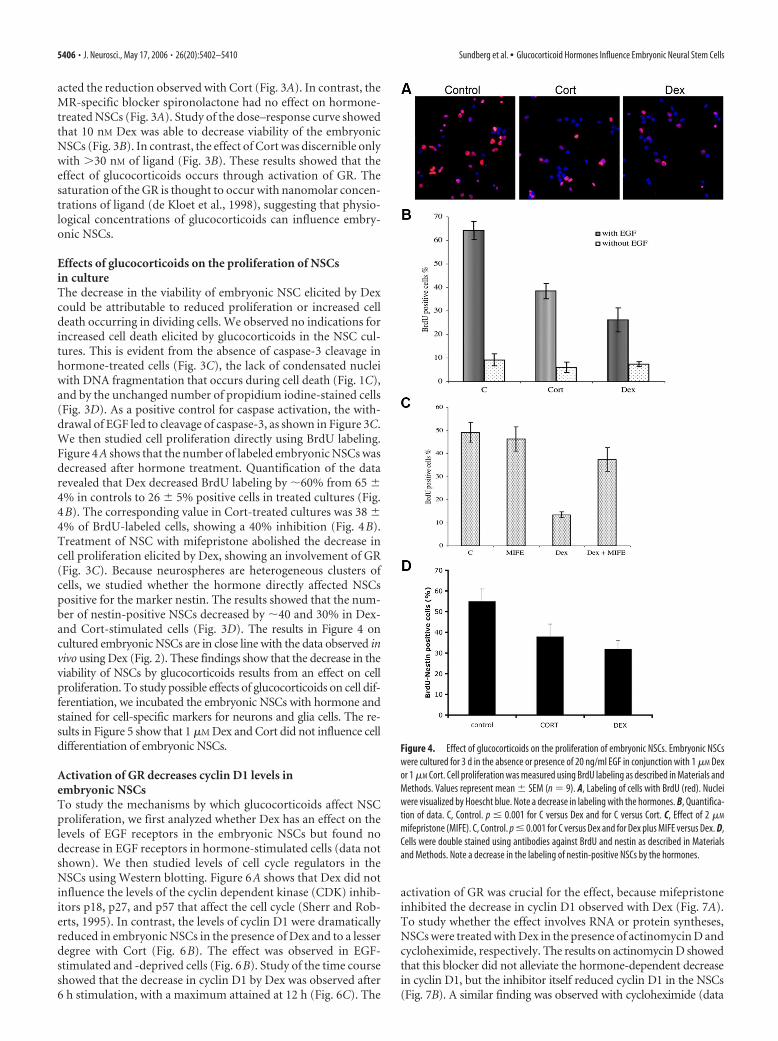
Effects of glucocorticoids on the proliferation of NSCsin cultureThe decrease in the viability of embryonic NSC elicited by Dexcould be attributable to reduced proliferation or increased celldeath occurring in dividing cells. We observed no indications forincreased cell death elicited by glucocorticoids in the NSC cul-tures. This is evident from the absence of caspase-3 cleavage inhormone-treated cells (Fig. 3C), the lack of condensated nucleiwith DNA fragmentation that occurs during cell death (Fig. 1C),and by the unchanged number of propidium iodine-stained cells(Fig. 3D). As a positive control for caspase activation, the with-drawal of EGF led to cleavage of caspase-3, as shown in Figure 3C.We then studied cell proliferation directly using BrdU labeling.Figure 4A shows that the number of labeled embryonic NSCs wasdecreased after hormone treatment. Quantification of the datarevealed that Dex decreased BrdU labeling by �60% from 65 �4% in controls to 26 � 5% positive cells in treated cultures (Fig.4B). The corresponding value in Cort-treated cultures was 38 �4% of BrdU-labeled cells, showing a 40% inhibition (Fig. 4B).Treatment of NSC with mifepristone abolished the decrease incell proliferation elicited by Dex, showing an involvement of GR(Fig. 3C). Because neurospheres are heterogeneous clusters ofcells, we studied whether the hormone directly affected NSCspositive for the marker nestin. The results showed that the num-ber of nestin-positive NSCs decreased by �40 and 30% in Dex-and Cort-stimulated cells (Fig. 3D). The results in Figure 4 oncultured embryonic NSCs are in close line with the data observed invivo using Dex (Fig. 2). These findings show that the decrease in theviability of NSCs by glucocorticoids results from an effect on cellproliferation. To study possible effects of glucocorticoids on cell dif-ferentiation, we incubated the embryonic NSCs with hormone andstained for cell-specific markers for neurons and glia cells. The re-sults in Figure 5 show that 1 �M Dex and Cort did not influence celldifferentiation of embryonic NSCs.
Activation of GR decreases cyclin D1 levels inembryonic NSCsTo study the mechanisms by which glucocorticoids affect NSCproliferation, we first analyzed whether Dex has an effect on thelevels of EGF receptors in the embryonic NSCs but found nodecrease in EGF receptors in hormone-stimulated cells (data notshown). We then studied levels of cell cycle regulators in theNSCs using Western blotting. Figure 6A shows that Dex did notinfluence the levels of the cyclin dependent kinase (CDK) inhib-itors p18, p27, and p57 that affect the cell cycle (Sherr and Rob-erts, 1995). In contrast, the levels of cyclin D1 were dramaticallyreduced in embryonic NSCs in the presence of Dex and to a lesserdegree with Cort (Fig. 6B). The effect was observed in EGF-stimulated and -deprived cells (Fig. 6B). Study of the time courseshowed that the decrease in cyclin D1 by Dex was observed after6 h stimulation, with a maximum attained at 12 h (Fig. 6C). The
activation of GR was crucial for the effect, because mifepristoneinhibited the decrease in cyclin D1 observed with Dex (Fig. 7A).To study whether the effect involves RNA or protein syntheses,NSCs were treated with Dex in the presence of actinomycin D andcycloheximide, respectively. The results on actinomycin D showedthat this blocker did not alleviate the hormone-dependent decreasein cyclin D1, but the inhibitor itself reduced cyclin D1 in the NSCs(Fig. 7B). A similar finding was observed with cycloheximide (data
Figure 4. Effect of glucocorticoids on the proliferation of embryonic NSCs. Embryonic NSCswere cultured for 3 d in the absence or presence of 20 ng/ml EGF in conjunction with 1 �M Dexor 1 �M Cort. Cell proliferation was measured using BrdU labeling as described in Materials andMethods. Values represent mean � SEM (n � 9). A, Labeling of cells with BrdU (red). Nucleiwere visualized by Hoescht blue. Note a decrease in labeling with the hormones. B, Quantifica-tion of data. C, Control. p � 0.001 for C versus Dex and for C versus Cort. C, Effect of 2 �M
mifepristone (MIFE). C, Control. p � 0.001 for C versus Dex and for Dex plus MIFE versus Dex. D,Cells were double stained using antibodies against BrdU and nestin as described in Materialsand Methods. Note a decrease in the labeling of nestin-positive NSCs by the hormones.
5406 • J. Neurosci., May 17, 2006 • 26(20):5402–5410 Sundberg et al. • Glucocorticoid Hormones Influence Embryonic Neural Stem Cells
not shown). This indicates that cyclin D1 is constantly turned over inthe NSCs, which depends on ongoing RNA and protein syntheses. Inaddition, RT-PCR revealed no change in mRNA levels for cyclin D1after treatment with Dex, indicating a posttranscriptional effect ofthe hormone (Fig. 6E).
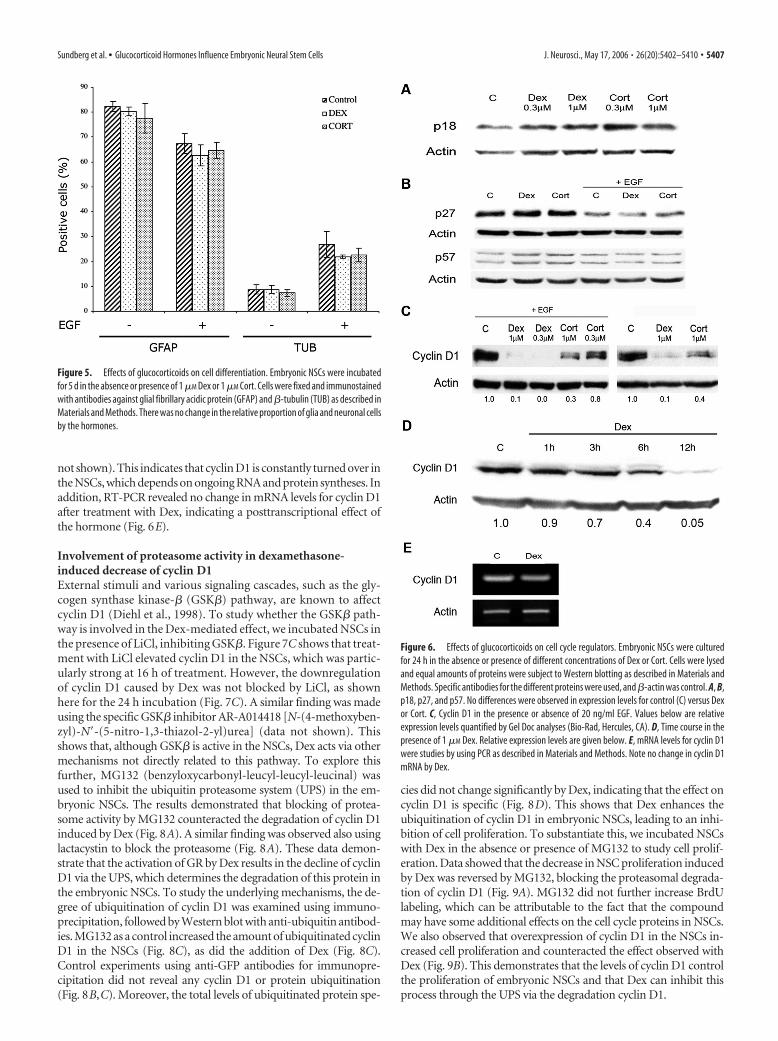
Involvement of proteasome activity in dexamethasone-induced decrease of cyclin D1External stimuli and various signaling cascades, such as the gly-cogen synthase kinase-� (GSK�) pathway, are known to affectcyclin D1 (Diehl et al., 1998). To study whether the GSK� path-way is involved in the Dex-mediated effect, we incubated NSCs inthe presence of LiCl, inhibiting GSK�. Figure 7C shows that treat-ment with LiCl elevated cyclin D1 in the NSCs, which was partic-ularly strong at 16 h of treatment. However, the downregulationof cyclin D1 caused by Dex was not blocked by LiCl, as shownhere for the 24 h incubation (Fig. 7C). A similar finding was madeusing the specific GSK� inhibitor AR-A014418 [N-(4-methoxyben-zyl)-N�-(5-nitro-1,3-thiazol-2-yl)urea] (data not shown). Thisshows that, although GSK� is active in the NSCs, Dex acts via othermechanisms not directly related to this pathway. To explore thisfurther, MG132 (benzyloxycarbonyl-leucyl-leucyl-leucinal) wasused to inhibit the ubiquitin proteasome system (UPS) in the em-bryonic NSCs. The results demonstrated that blocking of protea-some activity by MG132 counteracted the degradation of cyclin D1induced by Dex (Fig. 8A). A similar finding was observed also usinglactacystin to block the proteasome (Fig. 8A). These data demon-strate that the activation of GR by Dex results in the decline of cyclinD1 via the UPS, which determines the degradation of this protein inthe embryonic NSCs. To study the underlying mechanisms, the de-gree of ubiquitination of cyclin D1 was examined using immuno-precipitation, followed by Western blot with anti-ubiquitin antibod-ies. MG132 as a control increased the amount of ubiquitinated cyclinD1 in the NSCs (Fig. 8C), as did the addition of Dex (Fig. 8C).Control experiments using anti-GFP antibodies for immunopre-cipitation did not reveal any cyclin D1 or protein ubiquitination(Fig. 8B,C). Moreover, the total levels of ubiquitinated protein spe-
cies did not change significantly by Dex, indicating that the effect oncyclin D1 is specific (Fig. 8D). This shows that Dex enhances theubiquitination of cyclin D1 in embryonic NSCs, leading to an inhi-bition of cell proliferation. To substantiate this, we incubated NSCswith Dex in the absence or presence of MG132 to study cell prolif-eration. Data showed that the decrease in NSC proliferation inducedby Dex was reversed by MG132, blocking the proteasomal degrada-tion of cyclin D1 (Fig. 9A). MG132 did not further increase BrdUlabeling, which can be attributable to the fact that the compoundmay have some additional effects on the cell cycle proteins in NSCs.We also observed that overexpression of cyclin D1 in the NSCs in-creased cell proliferation and counteracted the effect observed withDex (Fig. 9B). This demonstrates that the levels of cyclin D1 controlthe proliferation of embryonic NSCs and that Dex can inhibit thisprocess through the UPS via the degradation cyclin D1.
Figure 5. Effects of glucocorticoids on cell differentiation. Embryonic NSCs were incubatedfor 5 d in the absence or presence of 1 �M Dex or 1 �M Cort. Cells were fixed and immunostainedwith antibodies against glial fibrillary acidic protein (GFAP) and �-tubulin (TUB) as described inMaterials and Methods. There was no change in the relative proportion of glia and neuronal cellsby the hormones.
Figure 6. Effects of glucocorticoids on cell cycle regulators. Embryonic NSCs were culturedfor 24 h in the absence or presence of different concentrations of Dex or Cort. Cells were lysedand equal amounts of proteins were subject to Western blotting as described in Materials andMethods. Specific antibodies for the different proteins were used, and �-actin was control. A, B,p18, p27, and p57. No differences were observed in expression levels for control (C) versus Dexor Cort. C, Cyclin D1 in the presence or absence of 20 ng/ml EGF. Values below are relativeexpression levels quantified by Gel Doc analyses (Bio-Rad, Hercules, CA). D, Time course in thepresence of 1 �M Dex. Relative expression levels are given below. E, mRNA levels for cyclin D1were studies by using PCR as described in Materials and Methods. Note no change in cyclin D1mRNA by Dex.
Sundberg et al. • Glucocorticoid Hormones Influence Embryonic Neural Stem Cells J. Neurosci., May 17, 2006 • 26(20):5402–5410 • 5407

DiscussionThe results in the present study demonstrate that glucocorticoidhormones influence proliferation of embryonic NSCs by activat-ing the cognate GRs. The mechanism was shown to involve ubiq-uitination of cyclin D1 and the degradation of the protein by theUPS. Cyclin D1 is rapidly turned over in the NSCs, and the levelsare regulated by the activity of GSK�, as shown previously inother cells (Diehl et al., 1998). Our data showed that inhibition ofGSK� increased cyclin D1 levels in control, but not in Dex-treated,NSCs. This suggested another pathway for Dex in the regulation ofcyclin D1 in these cells. We observed that the addition of Dex in-creased the ubiquitination of cyclin D1, which is a prerequisite for itsdegradation via the UPS. Inhibition of UPS by MG132 restored thelevels of cyclin D1 and counteracted the decrease in cell proliferationcaused by Dex. This demonstrates that Dex regulates proliferation ofNSCs through action on cyclin D1. Cyclin D1 has been shown pre-viously to undergo ubiquitination via the activity of the SKP1/SKP2/CUL-1 E3 ligase complex (Yu et al., 1998). The exact mechanisms bywhich Dex induces ubiquitination of cyclin D1 in the NSCs and therole of specific E3 ligases in this process remain to be studied in thefuture.
The crucial roles of type D cyclins in the regulation of NSCswas reported recently for adult NSCs in which lack of cyclin D2inhibited cell cycle and neurogenesis (Kowalczyk et al., 2004).The present findings show that activation of GR can profoundlydecrease cyclin D1 levels, leading to inhibition of cell prolifera-tion in embryonic NSCs. In preliminary experiments, we ob-served low levels of cyclin D2 in the embryonic NSCs, which werenot further decreased by Dex (data not shown). This suggests thatcyclin D1 is important for the regulation of NSCs proliferationduring embryonic life, whereas cyclin D2 is more crucial in theadult brain (Kowalczyk et al., 2004). Three type D cyclins havebeen cloned in mammals (Kozar et al., 2004). As shown by studiesin gene-deleted mice, the roles of the different type D cyclins in
the control of cell cycle vary between tissues and cells (Kozar etal., 2004). Mice lacking cyclin D1 show developmental neurolog-ical abnormalities and hypoplastic retinas, whereas cyclin D2-deficient animals have cerebellar defects (Fantl et al., 1995; Huardet al., 1999). In animals lacking all D-type cyclins, the proliferationof hematopoietic stem cells is severely affected during embryoniclife. Because of prenatal lethality, the nervous system of these animalshas so far not been analyzed. In the future, it is important to studywhether glucocorticoids can influence the development of otherstem cell populations than NSCs and whether this occurs throughregulation of cyclin D proteins.
In this context, it is interesting to note that the relative de-crease in cyclin D1 by Dex was greater than the degree of inhibi-tion of cell proliferation (compare Figs. 2B, 6C). The reason forthis is not clear at the moment, but it is known from studies offibroblasts lacking cyclin D that cell cycle progression can be driven
Figure 7. Effect of various inhibitors on the glucocorticoid-dependent regulation of cyclinD1. Embryonic NSCs were cultured for 16 or 24 h in the absence or presence of 1 �M Dex.Inhibitors were added together with the hormones as indicated. Western blots were performedand cyclin D1 levels were quantified as described in Materials and Methods. Relative expressionlevels are given below. A, Effect of 2 �M mifepristone (Mife). Incubation was for 24 h. B, Effectof 10 �M actinomycin D (AD), inhibiting gene transcription. Incubation was for 16 h. C, Effect of3 mM LiCl, inhibiting GSK�3. Incubation times as indicated. C, Control.
Figure 8. Role of the UPS in the degradation of cyclin D1 by glucocorticoids in NSCs. A,Embryonic NSCs were cultured for various time periods with 1 �M Dex in the absence or pres-ence of 10 �M MG132 (MG) to inhibit the proteasome. Western blots were performed and cyclinD1 levels were quantified as described in Materials and Methods. Relative expression levels aregiven below. Note restored cyclin D1 levels in the presence of MG132. Right shows experimentswith the proteasome inhibitor lactacystin, giving similar results. B, Cyclin D1 was immunopre-cipitated from NSCs by the cyclin D1 antibody (lane 1) but not by the irrelevant GFP antibody(lane 2). C, Cyclin D1 was immunoprecipitated (IP) from NSCs treated with 1 �M Dex or 10 �M
MG or both. Western blots were performed and the degree of cyclin D1 ubiquitination wasdetermined using the anti-ubiquitin antibody. Arrowheads show the presence of ubiquitinatedcyclin D1 at 36 kDa, and the upper band is the Ig heavy chain. Right shows control experimentusing the GFP antibody for immunoprecipitation and anti-ubiquitin antibodies for blotting.Note an increase in ubiquitination of cyclin D1 after Dex. Bottom shows total cyclin D1 levels inthe immunoprecipitate. Values below are the ratios of ubiquitinated to total cyclin D1. D, Totallevels of ubiquitination in NSC. Western blotting was performed using the ubiquitin antibodythat recognizes both mono-ubiquitin and poly-ubiquitin. The ladder to the right shows thelocalization of mono-ubiquitinated and poly-ubiquitinated species. Data indicates no generaleffect by Dex on the levels of mono-ubiquitin or poly-ubiquitin species in NSCs.
5408 • J. Neurosci., May 17, 2006 • 26(20):5402–5410 Sundberg et al. • Glucocorticoid Hormones Influence Embryonic Neural Stem Cells
by other proteins in the CDK2 complex (Kozar et al., 2004).Whether this is also the case for embryonic NSCs remains to bestudied.
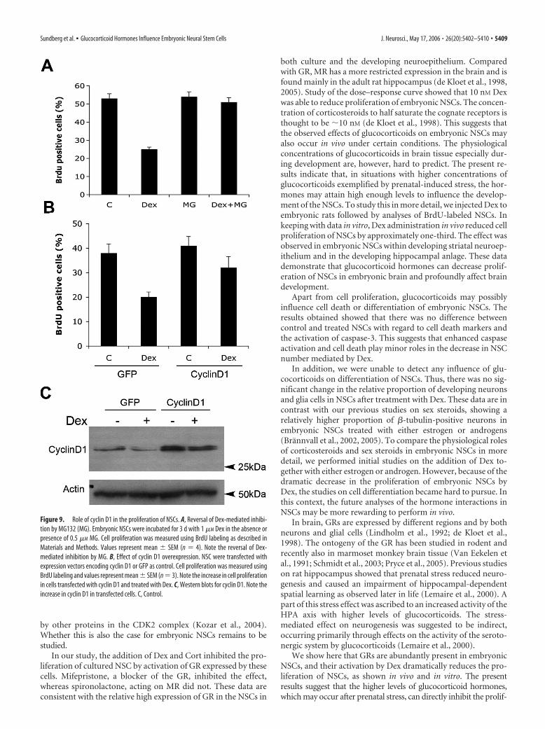
In our study, the addition of Dex and Cort inhibited the pro-liferation of cultured NSC by activation of GR expressed by thesecells. Mifepristone, a blocker of the GR, inhibited the effect,whereas spironolactone, acting on MR did not. These data areconsistent with the relative high expression of GR in the NSCs in
both culture and the developing neuroepithelium. Comparedwith GR, MR has a more restricted expression in the brain and isfound mainly in the adult rat hippocampus (de Kloet et al., 1998,2005). Study of the dose–response curve showed that 10 nM Dexwas able to reduce proliferation of embryonic NSCs. The concen-tration of corticosteroids to half saturate the cognate receptors isthought to be �10 nM (de Kloet et al., 1998). This suggests thatthe observed effects of glucocorticoids on embryonic NSCs mayalso occur in vivo under certain conditions. The physiologicalconcentrations of glucocorticoids in brain tissue especially dur-ing development are, however, hard to predict. The present re-sults indicate that, in situations with higher concentrations ofglucocorticoids exemplified by prenatal-induced stress, the hor-mones may attain high enough levels to influence the develop-ment of the NSCs. To study this in more detail, we injected Dex toembryonic rats followed by analyses of BrdU-labeled NSCs. Inkeeping with data in vitro, Dex administration in vivo reduced cellproliferation of NSCs by approximately one-third. The effect wasobserved in embryonic NSCs within developing striatal neuroep-ithelium and in the developing hippocampal anlage. These datademonstrate that glucocorticoid hormones can decrease prolif-eration of NSCs in embryonic brain and profoundly affect braindevelopment.
Apart from cell proliferation, glucocorticoids may possiblyinfluence cell death or differentiation of embryonic NSCs. Theresults obtained showed that there was no difference betweencontrol and treated NSCs with regard to cell death markers andthe activation of caspase-3. This suggests that enhanced caspaseactivation and cell death play minor roles in the decrease in NSCnumber mediated by Dex.
In addition, we were unable to detect any influence of glu-cocorticoids on differentiation of NSCs. Thus, there was no sig-nificant change in the relative proportion of developing neuronsand glia cells in NSCs after treatment with Dex. These data are incontrast with our previous studies on sex steroids, showing arelatively higher proportion of �-tubulin-positive neurons inembryonic NSCs treated with either estrogen or androgens(Brannvall et al., 2002, 2005). To compare the physiological rolesof corticosteroids and sex steroids in embryonic NSCs in moredetail, we performed initial studies on the addition of Dex to-gether with either estrogen or androgen. However, because of thedramatic decrease in the proliferation of embryonic NSCs byDex, the studies on cell differentiation became hard to pursue. Inthis context, the future analyses of the hormone interactions inNSCs may be more rewarding to perform in vivo.
In brain, GRs are expressed by different regions and by bothneurons and glial cells (Lindholm et al., 1992; de Kloet et al.,1998). The ontogeny of the GR has been studied in rodent andrecently also in marmoset monkey brain tissue (Van Eekelen etal., 1991; Schmidt et al., 2003; Pryce et al., 2005). Previous studieson rat hippocampus showed that prenatal stress reduced neuro-genesis and caused an impairment of hippocampal-dependentspatial learning as observed later in life (Lemaire et al., 2000). Apart of this stress effect was ascribed to an increased activity of theHPA axis with higher levels of glucocorticoids. The stress-mediated effect on neurogenesis was suggested to be indirect,occurring primarily through effects on the activity of the seroto-nergic system by glucocorticoids (Lemaire et al., 2000).
We show here that GRs are abundantly present in embryonicNSCs, and their activation by Dex dramatically reduces the pro-liferation of NSCs, as shown in vivo and in vitro. The presentresults suggest that the higher levels of glucocorticoid hormones,which may occur after prenatal stress, can directly inhibit the prolif-
Figure 9. Role of cyclin D1 in the proliferation of NSCs. A, Reversal of Dex-mediated inhibi-tion by MG132 (MG). Embryonic NSCs were incubated for 3 d with 1 �M Dex in the absence orpresence of 0.5 �M MG. Cell proliferation was measured using BrdU labeling as described inMaterials and Methods. Values represent mean � SEM (n � 4). Note the reversal of Dex-mediated inhibition by MG. B, Effect of cyclin D1 overexpression. NSC were transfected withexpression vectors encoding cyclin D1 or GFP as control. Cell proliferation was measured usingBrdU labeling and values represent mean � SEM (n � 3). Note the increase in cell proliferationin cells transfected with cyclin D1 and treated with Dex. C, Western blots for cyclin D1. Note theincrease in cyclin D1 in transfected cells. C, Control.
Sundberg et al. • Glucocorticoid Hormones Influence Embryonic Neural Stem Cells J. Neurosci., May 17, 2006 • 26(20):5402–5410 • 5409

eration of NSCs. The present data also show that embryonic NSCsare important targets for the action of glucocorticoids in developingbrain. The physiological and long-term consequences of the glu-cocorticoid-mediated regulation of the proliferation and number ofdeveloping NSCs warrant additional studies.
ReferencesÅberg MA, Åberg ND, Hedbacker H, Oscarsson J, Eriksson PS (2000) Pe-
ripheral infusion of IGF-I selectively induces neurogenesis in the adult rathippocampus. J Neurosci 20:2896 –2903.
Beato M (1989) Gene regulation by steroid hormones. Cell 56:335–344.Brannvall K, Korhonen L, Lindholm D (2002) Estrogen-receptor-
dependent regulation of neural stem cell proliferation and differentiation.Mol Cell Neurosci 21:512–520.
Brannvall K, Bogdanovic N, Korhonen L, Lindholm D (2005) 19-Nortestosterone influences neural stem cell proliferation and neurogen-esis in the rat brain. Eur J Neurosci 21:871– 878.
Cameron HA, Gould E (1994) Adult neurogenesis is regulated by adrenalsteroids in the dentate gyrus. Neuroscience 61:203–209.
Crochemore C, Lu J, Wu Y, Liposits Z, Sousa N, Holsboer F, Almeida OF(2005) Direct targeting of hippocampal neurons for apoptosis by glu-cocorticoids is reversible by mineralocorticoid receptor activation. MolPsychiatry 10:790 –798.
de Kloet ER, Vreugdenhil E, Oitzl MS, Joels M (1998) Brain corticosteroidreceptor balance in health and disease. Endocr Rev 19:269 –301.
de Kloet ER, Joels M, Holsboer F (2005) Stress and the brain: from adapta-tion to disease. Nat Rev Neurosci 6:463– 475.
Diehl JA, Cheng M, Roussel MF, Sherr CJ (1998) Glycogen synthase kinase-3beta regulates cyclin D1 proteolysis and subcellular localization. GenesDev 12:3499 –3511.
Fantl V, Stamp G, Andrews A, Rosewell I, Dickson C (1995) Mice lackingcyclin D1 are small and show defects in eye and mammary gland devel-opment. Genes Dev 9:2364 –2372.
Fuxe K, Wikstrom AC, Okret S, Agnati LF, Harfstrand A, Yu ZY, Granholm L,Zoli M, Vale W, Gustafsson JA (1985) Mapping of glucocorticoid recep-tor immunoreactive neurons in the rat tel-diencephalon using a mono-clonal antibody against rat liver glucocorticoid receptor. Endocrinology117:1803–1812.
Gass P, Reichardt HM, Strekalova T, Henn F, Tronche F (2001) Mice withtargeted mutations of glucocorticoid and mineralocorticoid receptors:models for depression and anxiety? Physiol Behav 73:811– 825.
Gould E, Gross CG (2002) Neurogenesis in adult mammals: some progressand problems. J Neurosci 22:619 – 623.
Gould E, Tanapat P, McEwen BS, Flugge G, Fuchs E (1998) Proliferation ofgranule cell precursors in the dentate gyrus of adult monkeys is dimin-ished by stress. Proc Natl Acad Sci USA 95:3168 –3171.
Hienola A, Pekkanen M, Raulo E, Vanttola P, Rauvala H (2004) HB-GAMinhibits proliferation and enhances differentiation of neural stem cells.Mol Cell Neurosci 26:75– 88.
Huard J, Forster C, Carter M, Sicinski P, Ross M (1999) Cerebellar histo-genesis is disturbed in mice lacking cyclin D2. Development126:1927–1935.
Karishma KK, Herbert J (2002) DHEA stimulates neurogenesis in the hip-pocampus of the rat, promotes survival of newly formed neurons andprevents corticosteroid-induced suppression. Eur J Neurosci 16:445– 454.
Korhonen L, Belluardo N, Lindholm D (2001) Regulation of X-chromosome-
linked inhibitor of apoptosis protein in kainic acid-induced neuronal deathin the rat hippocampus. Mol Cell Neurosci 17:364–372.
Korhonen L, Brannvall K, Skoglosa Y, Lindholm D (2003) Tumor suppres-sor gene BRCA-1 is expressed by embryonic and adult neural stem cellsand involved in cell proliferation. J Neurosci Res 71:769 –776.
Kowalczyk A, Filipkowski RK, Rylski M, Wilczynski GM, Konopacki FA,Jaworski J, Ciemerych MA, Sicinski P, Kaczmarek L (2004) The criticalrole of cyclin D2 in adult neurogenesis. J Cell Biol 167:209 –213.
Kozar K, Ciemerych MA, Rebel VI, Shigematsu H, Zagozdzon A, Sicinska E,Geng Y, Yu Q, Bhattacharya S, Bronson RT, Akashi K, Sicinski P (2004)Mouse development and cell proliferation in the absence of D-cyclins.Cell 118:477– 491.
Lemaire V, Koehl M, Le Moal M, Abrous DN (2000) Prenatal stress pro-duces learning deficits associated with an inhibition of neurogenesis in thehippocampus. Proc Natl Acad Sci USA 97:11032–11037.
Lindholm D, Castren E, Hengerer B, Zafra F, Berninger B, Thoenen H (1992)Differential regulation of nerve growth factor (NGF) synthesis in neuronsand astrocytes by glucocorticoid hormones. Eur J Neurosci 4:404 – 410.
Mouton PR, Long JM, Lei DL, Howard V, Jucker M, Calhoun ME, Ingram DK(2002) Age and gender effects on microglia and astrocyte numbers inbrains of mice. Brain Res 956:30 –35.
Perfilieva E, Risedal A, Nyberg J, Johansson BB, Eriksson PS (2001) Genderand strain influence on neurogenesis in dentate gyrus of young rats.J Cereb Blood Flow Metab 21:211–217.
Pryce CR, Feldon J, Fuchs E, Knuesel I, Oertle T, Sengstag C, Spengler M,Weber E, Weston A, Jongen-Relo A (2005) Postnatal ontogeny of hip-pocampal expression of the mineralocorticoid and glucocorticoid recep-tors in the common marmoset monkey. Eur J Neurosci 21:1521–1535.
Schanzer A, Wachs FP, Wilhelm D, Acker T, Cooper-Kuhn C, Beck H, Win-kler J, Aigner L, Plate KH, Kuhn HG (2004) Direct stimulation of adultneural stem cells in vitro and neurogenesis in vivo by vascular endothelialgrowth factor. Brain Pathol 14:237–248.
Schmidt M, Enthoven L, van der Mark M, Levine S, de Kloet ER, Oitzl MS(2003) The postnatal development of the hypothalamic-pituitary-adrenal axis in the mouse. Int J Dev Neurosci 21:125–132.
Sherr CJ, Roberts JM (1995) Inhibitors of mammalian G1 cyclin-dependentkinases. Genes Dev 9:1149 –1163.
Tanapat P, Hastings NB, Reeves AJ, Gould E (1999) Estrogen stimulates atransient increase in the number of new neurons in the dentate gyrus ofthe adult female rat. J Neurosci 19:5792–5801.
Tanapat P, Hastings NB, Gould E (2005) Ovarian steroids influence cellproliferation in the dentate gyrus of the adult female rat in a dose- andtime-dependent manner. J Comp Neurol 481:252–265.
Taupin P, Gage FH (2002) Adult neurogenesis and neural stem cells of thecentral nervous system in mammals. J Neurosci Res 69:745–749.
Van Eekelen JAM, Bohn MC, de Kloet ER (1991) Postnatal ontogeny ofmineralocorticoid and glucocorticoid receptor gene expression in regionsof the rat tel- and diencephalon. Dev Brain Res 61:33– 43.
Yu LY, Korhonen L, Martinez R, Jokitalo E, Chen Y, Arumae U, Lindholm D(2003) Regulation of sympathetic neuron and neuroblastoma cell deathby XIAP and its association with proteasomes in neural cells. Mol CellNeurosci 22:308 –318.
Yu ZK, Gervais JL, Zhang H (1998) Human CUL-1 associates with theSKP1/SKP2 complex and regulates p21 (CIP1/WAF1) and cyclin D pro-teins. Proc Natl Acad Sci USA 95:11324 –11329.
5410 • J. Neurosci., May 17, 2006 • 26(20):5402–5410 Sundberg et al. • Glucocorticoid Hormones Influence Embryonic Neural Stem Cells