Regulation of Pancreatic Exocrine Function by Islet Hormones
37
1 Regulation of Pancreatic Exocrine Function by Islet Hormones M. Dolors Sans 1 , Jason I.E. Bruce 2 , and John A. Williams 1 1 Department of Molecular and Integrative Physiology, University of Michigan, Ann Arbor, MI 2 Faculty of Biology, Medicine, and Health, School of Medical Sciences, University of Manchester, Manchester, UK e-mail: [email protected], [email protected], [email protected] Version 1.0 June 30 th , 2020 DOI: 10.3998/panc.2020.01 I. Introduction The exocrine pancreas is regulated by nerves, mainly the vagus, and gastrointestinal hormones, especially cholecystokinin (CCK) and secretin. A third specific source of regulation is by peptide hormones from the Islets of Langerhans. Islet hormones are relevant because of structural relations within the pancreas. Except for some fishes which have a single larger collection of endocrine cells, islets are dispersed within the pancreas and have the ability to regulate exocrine cells both by local diffusion which manifests as peri-insular halos of acinar cells and by a specific vascular relationship, the islet-acinar portal system. The latter results in acinar cells being exposed to high concentrations of islet hormones and other regulatory molecules. Of these the best defined relationship is with insulin produced in islet beta cells which serves as a trophic factor for the exocrine pancreas, as a short-term signal promoting digestive enzyme synthesis at the translational level, and as part of a long-term control system where dietary carbohydrate leads to the synthesis of the carbohydrate targeted digestive enzyme, pancreatic amylase. Insulin also facilitates digestive enzyme secretion and regulates the membrane transport of glucose and Ca 2+ . Earlier reviews of this topic include (12, 275, 330). Evidence for a direct effect of other islet peptides is less clear and some may exert an effect through regulating insulin secretion. II. Structure and Vascular Supply of the Pancreas A. Structure and distribution of Islets of the Pancreas Pancreatic islets are collections of endocrine cells scattered through the exocrine pancreas making up 2-3% of the gland. Islets are made up of five types of endocrine cells known as α, β, δ, ε, and PP/F cells that synthesize and secrete, respectively, glucagon, insulin, somatostatin, ghrelin, and pancreatic polypeptide, all of which are considered peptide hormones. Beta cells are the most numerous cell type (60-70%) and form the core in rodent islets but are scattered throughout the human islet. Alpha cells make up about 20% and along with δ cells (1-2%) form a mantle on the outside of rodent islets. Some of the cell types are geographically distributed with PP/F cells primarily in the ventral lobe of the pancreas. By contrast, ε cells are developmentally regulated making up about 10% of the fetal islet but then differentiate
-
Upload
khangminh22 -
Category
Documents
-
view
1 -
download
0
Transcript of Regulation of Pancreatic Exocrine Function by Islet Hormones

1
Regulation of Pancreatic Exocrine Function by Islet Hormones
M. Dolors Sans1, Jason I.E. Bruce2, and John A. Williams1
1Department of Molecular and Integrative Physiology, University of Michigan, Ann
Arbor, MI 2Faculty of Biology, Medicine, and Health, School of Medical Sciences,
University of Manchester, Manchester, UK
e-mail: [email protected], [email protected], [email protected]
Version 1.0 June 30th, 2020 DOI: 10.3998/panc.2020.01
I. Introduction
The exocrine pancreas is regulated by nerves,
mainly the vagus, and gastrointestinal hormones,
especially cholecystokinin (CCK) and secretin. A
third specific source of regulation is by peptide
hormones from the Islets of Langerhans. Islet
hormones are relevant because of structural
relations within the pancreas. Except for some
fishes which have a single larger collection of
endocrine cells, islets are dispersed within the
pancreas and have the ability to regulate exocrine
cells both by local diffusion which manifests as
peri-insular halos of acinar cells and by a specific
vascular relationship, the islet-acinar portal
system. The latter results in acinar cells being
exposed to high concentrations of islet hormones
and other regulatory molecules. Of these the best
defined relationship is with insulin produced in islet
beta cells which serves as a trophic factor for the
exocrine pancreas, as a short-term signal
promoting digestive enzyme synthesis at the
translational level, and as part of a long-term
control system where dietary carbohydrate leads to
the synthesis of the carbohydrate targeted
digestive enzyme, pancreatic amylase. Insulin
also facilitates digestive enzyme secretion and
regulates the membrane transport of glucose and
Ca2+. Earlier reviews of this topic include (12, 275,
330). Evidence for a direct effect of other islet
peptides is less clear and some may exert an effect
through regulating insulin secretion.
II. Structure and Vascular Supply of
the Pancreas
A. Structure and distribution of Islets of
the Pancreas
Pancreatic islets are collections of endocrine cells
scattered through the exocrine pancreas making
up 2-3% of the gland. Islets are made up of five
types of endocrine cells known as α, β, δ, ε, and
PP/F cells that synthesize and secrete,
respectively, glucagon, insulin, somatostatin,
ghrelin, and pancreatic polypeptide, all of which are
considered peptide hormones. Beta cells are the
most numerous cell type (60-70%) and form the
core in rodent islets but are scattered throughout
the human islet. Alpha cells make up about 20%
and along with δ cells (1-2%) form a mantle on the
outside of rodent islets. Some of the cell types are
geographically distributed with PP/F cells primarily
in the ventral lobe of the pancreas. By contrast, ε
cells are developmentally regulated making up
about 10% of the fetal islet but then differentiate
2
into α and β cells so that in the adult they are rare.
Some of the endocrine cells also express a second
biologically active peptide (12). Amylin and galanin
are synthesized in β cells and are co-secreted with
insulin. Pancreastatin is present in α, β, and δ cells
where it is derived from chromogranin, and
adrenomedulin is produced in PP/F cells. In
understanding the effects of these islet peptides on
the exocrine pancreas it is important to realize that
some (insulin, glucagon and PP) are essentially
unique to the islets but others like somatostatin are
more broadly distributed in the GI tract, which could
affect the exocrine pancreas locally or from
systemic sources. Because islet peptides can
regulate the other cells within the islet, effects of a
particular peptide could be mediated directly on
exocrine cells or indirectly by affecting the
secretion of insulin or another islet peptide.
Furthermore, some of these peptides can affect the
CNS and vagal regulation of the exocrine
pancreas. Receptors on exocrine pancreatic cells
along with direct in vitro effects of islet peptides
have been identified for insulin and somatostatin
but not for glucagon or PP. Further details with
references will be given in the sections on
individual peptides.
B. Islet – acinar portal system
Blood flow to the pancreas has been reported to be
0.4 to 1.1 ml per g weight per min with most data
from rats, rabbits, mice and dogs (288). Early
studies injecting dye or ink into arteries feeding the
pancreas showed rapid and intense perfusion of
glomerular structures that were identified as Islets
of Langerhans (68, 116, 198). Islet blood flow has
been most often quantitated using microspheres.
Such studies show that 5-20% of blood flow goes
to the islets which have a relative blood flow 5-10
times higher than the exocrine pancreas (130,
177). The regulation of blood flow to the two
portions of the pancreas is distinct with exocrine
secretagogues such as CCK and VIP increasing
blood flow to the exocrine pancreas (54, 129) and
glucose increasing islet blood flow (131). In a
study in rabbits, Lifson and colleagues concluded
that essentially all blood flow to the islets goes
subsequently to the acinar capillaries (175).
Considerable attention has also been paid to the
pathway of capillaries within the islets as it pertains
to which islet cells are upstream of the others (31,
183).
Early dye injection vascular perfusion studies also
showed that small capillary vessels exited the islets
and carried blood to the exocrine pancreas and
these have been named the insulo – acinar portal
system. This directional flow is facilitated by the
fact that islet capillaries are slightly larger in
diameter which should lead to higher capillary
pressure (117, 314). In some species, there are
also direct arterioles feeding the acinar tissue and
venules draining the islets so not all islet effluent
passes to the exocrine tissue. Similar results have
been seen for rabbits, rats, mice, guinea pigs, cats,
dogs and baboons (115). Evidence for the portal
system also comes from scanning electron
microscopy of vascular casts and from functional
studies. Vascular casts are made by injecting resin
or acrylate into the arteries and after
polymerization the residual tissue is digested away
leaving a three dimensional vascular cast which is
coated with gold and viewed in a scanning electron
microscope. Each islet has one or sometimes two
afferent vessels which break down into a glomus-
like capillary network. Efferent vessels similar in
size to capillaries leave the islet and connect to the
acinar capillaries (82, 83, 216, 225, 341). Similar
findings have been made in the rat, rabbit, monkey,
horse, baboon and human. Some controversy
exists over the extent of this phenomenon
especially in rats where Bonner-Weir reported that
a large extent of islet blood was drained directly
into venules (24) but this was subsequently
disputed (204). Lifson and Lassa also described
an acinar-ductal portal system in the rabbit
pancreas (176). In any case, all blood from
exocrine pancreas exits in venules which combine
to form the pancreatic veins.
A different type of evidence for the islet-acinar
portal system comes from physiological studies
particularly using the perfused rat pancreas.
Insulin added to the vascular perfusate enhances
3
the exocrine response to CCK and secretin (109,
137, 257, 258). Glucose, which by itself does not
affect exocrine secretion, when added to the
vascular perfusate enhances the release of insulin
and potentiates exocrine secretion (89, 258). Other
sugars that do not stimulate insulin secretion had
no effect, and epinephrine, which blocks insulin
secretion, blocked the potentiating effect of
glucose. Since insulin can only come from the
islets and would be carried away in the venous
drainage in the single pass perfusion system, an
effect on exocrine secretion can only come from
endogenous insulin reaching exocrine cells by the
portal system. Similar studies depleting
endogenous somatostatin with cysteamine have
also provided evidence for endogenous
somatostatin acting through the portal system
(238).
Based on the presence of the insulo-acinar portal
system, an important question is what is the
concentration of islet peptides in the exocrine
interstitial compartment. Two estimates indicate
that the concentration of insulin and somatostatin
is in the nanomolar range. The first comes from a
study evaluating saturable binding of iodinated
tracer for insulin and somatostatin in the rat
pancreas perfused anterograde or retrograde
(219). Binding was higher for retrograde perfusion
which was attributed to endogenous hormone
released during anterograde perfusion.
Displacement curves during retrograde perfusion
indicated an interstitial concentration of insulin
during anterograde perfusion with glucose of 7.5
nM and for somatostatin of 1.1 nM. A second study
used a microdialysis technique which showed a
unstimulated concentration of insulin of 0.4 nM
during both retrograde and anterograde perfusion
of dog pancreas (218). The response to
stimulation with glucose plus arginine was
markedly less during retrograde perfusion.
Although the stimulated concentrations did not
reach steady state, if insulin release went up 10
fold the interstitial exocrine concentration during
glucose stimulation would be about 4 nM.
Unfortunately, reported plasma insulin levels in
fasting rats and dogs are quite variable with most
ranging from 50-400 pM. Much better data is
available for humans where most fasting levels are
reported to be 25-150 pM.
In summary, although the islet-acinar portal system
does not account for all blood flow to exocrine
pancreas cells, it does allow a significant fraction
of exocrine acinar and duct cells to be exposed to
higher concentrations of islet hormones than is the
case for other organs. In addition, islet hormones
can act directly on acinar cells via specific
receptors or indirectly by affecting the release of
other islet hormones.
III. Action of insulin on the exocrine
pancreas
Insulin appears to be the most important of the
classical islet hormones as a regulator of the
exocrine pancreas and especially acinar cells.
Evidence exists from both human studies and
animal models and includes both physiology and
disease. Experimental studies have been carried
out at different levels of integration from the intact
organism to cellular and molecular studies of
acinar cells.
A. Clinical studies on the effects of
diabetes on exocrine pancreas
For over one hundred years the pancreas of
diabetic patients has been known to be smaller
with increased fibrosis consistent with pathology of
the entire pancreas and not just the islets (36).
Diabetes is now divided into Type 1 or insulin
dependent diabetes mellitus (IDDM) where
destruction of beta cells leads to lack of insulin,
Type 2 whose hallmark is insulin resistance and
often does not require additional insulin, and most
recently Type 3 which is due to exocrine pathology
such as chronic pancreatitis. It has long been
recognized that Type 1 and 2 patients may also
suffer exocrine abnormalities ranging from
subclinical to pancreatic exocrine insufficiency
(PEI).
1. Loss of pancreatic mass in diabetes
4
Autopsy studies of Type 1 diabetes have shown
decreased pancreatic weight and volume (91,
184). Methods have been developed to use CT
and MRI imaging to determine pancreatic volume
and distinguish fat within the pancreas. Ultrasound
has also been used but is not as quantitative and
is more operator dependent. Studies of diabetic
patients with CT and MRI have consistently shown
a decrease in pancreatic volume of 30-55% in Type
1 and 15-30% in Type 2 diabetes (96, 178, 188,
242, 323). Some studies have shown a
dependence on the duration of diabetes but others
have not. Several studies in which type 3 diabetes
or a history of alcoholism have been omitted have
shown a smaller loss in pancreatic volume. These
changes have been confirmed in a recent meta-
analysis of 17 studies (88). The authors noted,
however, that many of the individual studies were
small and classified as low to moderate quality
data. Only type 2 diabetes has shown increased
pancreatic fat which reduces the volume of
parenchymal tissue from the total volume (178,
188). In some studies, with functional as well as
imaging data, the decreased pancreatic volume
correlated with decreased function as measured by
fecal elastase (242). To date there are no
longitudinal studies although this could be done
with MRI since there is no ionizing radiation.
2. Effects of diabetes on pancreatic function
Direct measurement of pancreatic juice
bicarbonate ion and digestive enzymes in
response to secretin and CCK using multi-lumen
tube collection has been applied to diabetes.
Ewald and Hardt list 9 such studies from 1943 to
1996 (73). These studies all show decreased
exocrine pancreatic secretion with a bigger effect
seen in Type 1 diabetes. An effect of the duration
of diabetes was seen in some (162) but not in other
studies (80). Most studies show a bigger effect on
amylase than on other digestive enzymes and the
smallest effect is on bicarbonate.
Indirect pancreatic function testing on stool
samples have allowed study of greater number of
patients. The most commonly used test is fecal
elastase measured by an ELISA assay where
<200 µg/g is considered evidence for pancreatic
exocrine insufficiency (PEI) and <100 µg/g severe
deficiency. Elastase is used because it is more
resistant to protease digestion although some
studies have measured fecal trypsin or
chymotrypsin activity which yields similar results
relative to controls. In the first large study of more
than 1000 diabetics, 40.7% showed fecal elastase
levels of less than 200 µg/g with 22.9% being <100
µg/g (108). Other studies have reported similar
results (74, 163, 222) although the frequency of
PEI is lower when patients with Type 3 diabetes
were excluded (303, 316). Type 1 diabetics with
reduced fecal elastase show a higher frequency of
steatorrhea up to 60% although the amount of
stool fat is modest (35, 107) and clinical PEI
requiring enzyme supplementation is rare (38).
Overall, diabetes is accompanied by
decreased pancreatic mass and reduced
secretion of digestive enzymes along with a
diffuse pancreatic fibrosis characterized by
intra-acinar fibrosis whose pattern is distinct
from that of chronic pancreatitis. These
changes may be the result of reduced number
of acinar cells, loss of insulin as a trophic factor
or alteration in neural control (207). These
authors proposed that these changes be
recognized as distinct from chronic
pancreatitis and be termed “diabetic exocrine
pancreatopathy”.
B. Animal studies of the effects of insulin
and diabetes on the exocrine pancreas
1. Insulin receptors in the exocrine pancreas
Insulin receptors (IR) on pancreatic acinar cells
were first demonstrated by the binding of 125I-
insulin in a saturable manner to isolated acini from
mouse, rat and guinea pig (156, 263,
282). Scatchard-plot analysis of binding were
biphasic and showed high affinity sites with a Kd of
about 1.5 nM and capacity of about 10,000
receptors per cell; low affinity sites were much

5
more numerous with a Kd of 88 nM for mouse and
998 nM for rat. Addition of unlabeled insulin
accelerated dissociation from rat acini consistent
with negative cooperativity rather than two distinct
sites (263). The receptor bound insulin analogs
with varying potency (insulin > desdipeptide insulin
> proinsulin > desoctapeptide insulin) that was
similar to the ability of the analogs to increase
protein synthesis in acini from diabetic rats
(154). Guinea pig insulin is significantly different
from other species and guinea pig acinar receptors
bound bovine insulin with a higher affinity than
guinea pig insulin or porcine proinsulin
(282). Mouse acini were also shown to possess
distinct receptors for IGF-I and IGF-II (328).
The localization of insulin binding to acinar and
other cells in the pancreas has been demonstrated
by light and EM autoradiography. When 125I-
Insulin was injected intravenously, saturable
uptake was observed by the pancreas (19, 46,
261). EM autoradiographs showed silver grains
localized both over the plasma membrane and
intracellularly in acinar cells. Saturable binding
was also observed over duct cells (261). In an in
vitro study of 125I-Insulin binding to rat acini, silver
grains from 125I were primarily over the plasma
membrane at 3 min but by 30 min were primarily
intracellular (97). Studies in other cell types have
shown that both insulin and its receptor are
internalized although the function of internalized
insulin is unclear.
Insulin receptors on acinar cells are also subject to
regulation. Insulin down regulates its own receptor
in a variety of cells, including acinar cells, when
they are exposed to high concentrations of insulin
through the islet-acinar portal blood system (12,
324). Acini prepared from diabetic mice showed an
increased number of IRs compared to normal mice
and addition of insulin to 24 hour cultured acini
decreased insulin binding (209). Using pancreatic
AR42J cells, insulin decreased the biosynthesis of
insulin receptors in part by decreasing IR mRNA
levels (209, 226). The binding of insulin was also
affected by CCK and other secretagogues
(228). CCK, carbamylcholine, active phorbol ester
and calcium ionophore (A23187) all decreased
high affinity binding without affecting insulin
internalization or degradation.
The occupancy of insulin receptors has also been
related to the biological effects of insulin on acinar
cells. Insulin increased glucose transport (uptake)
by acini from normal and diabetic mice (156, 329)
with a greater effect on acini from diabetic animals
that was seen at lower concentrations of
insulin. This greater effect is due both to lower
contaminating insulin levels and IR
upregulation. In acini from diabetic rats and mice,
insulin increased protein synthesis as shown by
incorporation of radioactive amino acids (154). Of
likely relevance to protein synthesis, insulin rapidly
increased the phosphorylation of ribosomal protein
S6 which is associated with increased protein
synthesis (33, 290). The concentration
dependence and analog specificity of these actions
of insulin is similar to the data for receptor
occupancy (98).
More recently IR have been demonstrated in
pancreatic acini by immunoblotting of the beta
subunit of the receptor (265). The subunit had an
apparent size of about 95 kDa and was essentially
absent after deletion of the IR gene in acinar cells.
The biological response to insulin in the pancreas
of these mice was greatly reduced.
2. Insulin regulation of exocrine pancreas
biosynthetic and metabolic effects
Gene expression
Insulin is known to regulate both the amount and
activity of a number of anabolic processes and
specific metabolic enzymes. In the exocrine
pancreas the best studied tissue specific regulation
is that of the digestive enzyme amylase which acts
on dietary starch and glycogen (325). The
pancreatic amylase content is known to decrease
by over 90% in experimental diabetes in rats and
the fall can be reversed by administration of insulin.
This has been studied primarily in rats given
alloxan or streptozoticin which induce beta cell
death and diabetes (2, 15, 17, 58, 235, 283, 286).

6
At the same time other digestive enzymes
including trypsinogen, chymotrypsinogen and
lipase increase slightly and ribonuclease
decreases slightly. Amylase also decreases in
mouse pancreas in experimental diabetes but by
only about 50%. Pancreatic amylase also
decreases in the Zucker fatty rat and the ob/ob
mouse, well characterized models of insulin
resistance (304).
The decrease in pancreatic amylase in
experimental diabetes is accompanied by a
decrease in amylase synthesis (286). Moreover,
insulin increased amylase biosynthesis in rat
pancreatic derived AR42J cells (210). The major
cause of the decreased amylase synthesis and
tissue content is a decrease in amylase mRNA
which was first reported by Korc et al (60, 155).
The decrease in amylase mRNA could be reversed
by insulin. Chymotrypsinogen mRNA showed a
small change in the opposite direction and there
was essentially no change in salivary amylase or
its mRNA. Subsequently, Brannon and colleagues
showed that dietary glucose and insulin together
regulated pancreatic amylase (Amy2) gene
expression (306). More detailed information on the
mechanism of amylase gene regulation comes
from studies in mice. A 30-bp region in the
proximal amylase promoter overlapping with the
PTF1 binding site present in most pancreatic
digestive enzymes contains the insulin-dependent
element and can transfer insulin sensitivity to the
elastase promoter (134, 140). This regulatory
region is not present in all mouse amylase alleles
and this can explain why some mouse strains are
not as sensitive to diabetes and insulin (57).
Unfortunately, there has been little subsequent
work defining how insulin receptor signaling
regulates the insulin response element.
Membrane transport
Insulin is known to stimulate membrane transport
of many substances usually by effects on the
amount of or properties of specific transport
proteins. The best studied and probably the most
important physiologically is the uptake of glucose
into fat and muscle cells that is mediated by the
glucose transporter, GLUT4. GLUT4 is present at
rest in intracellular vesicles that translocate to the
plasma membrane in response to insulin,
increasing glucose uptake 5-20 fold. Most other
cell types contain GLUT1 that is regulated by
another mechanism but to a lesser extent. In
mouse pancreatic acini, insulin stimulated the
uptake of the nonmetabolizable sugar 2-deoxy-
glucose (2DG) about two fold (156). A more robust
effect of lower concentrations of insulin was seen
when acini were prepared from diabetic mice and
uptake of both 2DG and 3-O-methyl glucose, a non
metabolizable glucose analog, were increased
(329). Glucose uptake was not affected by
inhibitors of protein synthesis but was reduced by
the actin inhibitor, cytochalasin B.
In some tissues insulin also stimulates the uptake
of amino acids through effects on specific amino
acid transporters. Uptake of amino acids have
been studied in many pancreatic preparations.
One of the earliest studies using microdisected
pieces of mouse pancreas showed that amino
acids were taken up and oxidized in preference to
glucose (48). Neither CCK or insulin affected
uptake and transport did not appear to be
separated from protein synthesis. Latter studies
used perfused pancreas or isolated acini and
showed that the basolateral membranes of
pancreatic acinar cells possess 4-5 distinct
transporters as measured by physiological studies
or gene expression (190, 252). Insulin stimulation
studies in the perfused rat pancreas show effects
of exogenous insulin and diabetes on two Na+-
independent transporters, the Asc transporter used
by serine (193, 221) and a basic amino acid
transporter termed y+ for lysine/arginine (215) while
no effect was seen on the Na+ dependent
dependent transporters such as the system A
transporter for AIB and glutamine (192). Moreover,
increasing glucose in the perfusate which released
endogenous insulin had no effect (221). Studies in
isolated rat pancreatic acini also showed no effect
of insulin on AIB and cycloleucine uptake which are
not used for protein synthesis although it
stimulated incorporation of leucine into protein

7
(154). In a more recent study of the mouse
pancreas, Rooman et al analized the expression of
37 genes encoding transporters and found
expression of a number with the highest
expression of slc7a8 and slc3a2 and confirmed the
expression of five by western blot (252).
Protein synthesis
Insulin is known to stimulate protein synthesis by
translational effects in many tissues (132). Early
studies in the pancreas involved in vivo studies of
the incorporation of radioactive amino acids into
total protein or specific enzymes of normal and
diabetic rodents (45, 61, 157). While most of the
studies showed a positive effect of insulin the
results are complicated by changes in the
precursor pool or changes in the levels of mRNAs.
When protein synthesis in mice was quantitated by
autoradiography, there was more incorporation
into peri-insular acinar cells then into tele-insular
acinar cells (7). This difference was lost following
treatment with streptozotocin and the authors
ascribed it to insulin. One study overcame some
of these issues by changing plasma insulin and
glucose in vivo but measuring protein synthesis in
vitro using pancreatic lobules incubated in a
constant medium (2). It showed that either glucose
infusion in vivo or treatment with a sulfonylurea
drug both of which stimulate insulin release
increased protein synthesis by 25 – 40%. When
Zucker fatty rats were studied as a model of insulin
resistance, overall pancreatic protein synthesis
was reduced by nearly 50% (305). There was
considerable difference in synthesis between
different digestive enzymes separated by 2-D-gel
electrophoresis. A morphological study of
prolonged diabetes in rats showed gross
abnormality in the secretory pathway 28 days after
STZ treatment that was partially reversed by insulin
administration (333). However, shorter studies
have not shown significant structural alterations 1
week after STZ other than the appearance of
cytoplasmic lipid droplets (154).
More recent studies have been able to overcome
some of the methodological issues by measuring
the effects of insulin over short times to prevent
changes in mRNA and under conditions where the
precursor pool is large and constant (264). In vitro
studies have used isolated pancreatic acini under
dilute incubation to keep the precursor pool
constant. Insulin stimulates the incorporation of
multiple different amino acids (leucine, methionine,
phenylalanine) in isolated acini from diabetic rats
(153, 154, 160, 227). Similarly, insulin increased
methionine incorporation into total protein and
immunoprecipitated amylase in rat pancreatic
derived AR42J cells (210). The effect of insulin on
both cell types occurred over concentrations from
30 pM to 100 nM and was mediated by the insulin
receptor as insulin analogs stimulated in parallel to
their ability to bind to the insulin receptor (154,
227). Insulin was also shown to have nonparallel
effects on different digestive enzymes and
structural proteins such as myosin and LDH (227).
Although the mechanism of insulin signaling is well
studied in a number of target tissues especially
liver, fat and muscle, only a few studies have been
carried out in the exocrine pancreas. The main
signaling pathway regulating protein synthesis is
the mTOR pathway and this pathway has been
documented in pancreatic acinar cells primarily in
mediating the actions of CCK and acetylcholine to
stimulate digestive enzyme synthesis (266, 326).
Insulin has been shown to activate S6 kinase and
S6 phosphorylation downstream of TORC1 in rat
and mouse acini (33, 290). Akt is upstream of
mTOR and insulin activates the phosphorylation of
Akt on S473 and T308 in rat acini (20). This action
appeared mediated by PI3K. That these actions
are important physiologically is shown by over a
50% reduction in mTOR pathway activation after
feeding in mice without acinar cell insulin receptors
(265).
Secretion
The effects of insulin administration and diabetes
on pancreatic exocrine secretion have been
studied in a number of species and systems

8
ranging from intact awake animals down to isolated
acinar cells. The more isolated systems have
better defined parameters but lack neural control
and other integrative aspects found in the intact
organism.
In vivo studies have largely been carried out in rats
with some studies in dogs and other species and
often have compared streptozotocin (STZ) treated
and intact animals. STZ and alloxan treatment
damage islets thus lowering insulin levels and
leading to hyperglycemia. In STZ-diabetic rats
pancreatic juice flow has been reported to be
moderately increased (18, 241, 285). In two studies
this effect was reversed by transplanting syngeneic
islets into the liver (53, 168). However, in mice STZ
treatment or genetic models of diabetes leads to
reduced juice flow (265), and in sheep, alloxan
treatment led to a substantial decrease that could
be restored by insulin treatment (243). In all of
these studies the effects of the diabetic state on
plasma glucose and lipids is not clear. The effects
of CCK on digestive enzyme secretion in diabetes
are complicated by changes in the pancreatic
content of specific digestive enzymes. The
concentration of secreted amylase is decreased
and trypsin increased although both respond to
CCK (18, 285). The most valid results may be in
animals with chronic pancreatic fistulas where juice
can be collected in the unanaesthetized state but
such studies have not been carried out in diabetes.
Other studies have focused on the effects of insulin
in normal animals with intact islets. When a high
concentration of insulin (4 U per 100 gm) was
injected into the femoral vein, subsequent
pancreatic stimulation by CCK was potentiated
(137). In another study, insulin (1U as a bolus)
induced hypoglycemia, increased fluid flow and
increased amylase and total protein output; this
effect was blocked by atropine and ascribed to
hypoglycemia-induced vagal cholinergic activation
(241). In a third study an intravenous infusion of
insulin also increased pancreatic secretion (76).
Not all studies however have reported this effect
(284). In a different approach using
unanaesthetized rats, IV injection of rabbit anti-
insulin serum but not control serum blocked the
pancreatic response to a liquid meal and to
exogenous CCK or secretin stimulation (167). In a
study in conscious dogs which secrete a high
bicarbonate pancreatic juice, most of which comes
from the ducts, insulin administration diminished
secretin stimulated bicarbonate secretion and this
effect was blocked by prior pancreatic denervation
(21). This study illustrates the complexity of
administering insulin in vivo and show that some of
the effects of high dose insulin in vivo are mediated
by neural pathways. However, overall they
suggest a positive effect of insulin on pancreatic
exocrine secretion.
Studying an in vitro pancreas preparation removes
the effects of plasma glucose and the brain initiated
neural control. While some studies have been
carried out with isolated pancreas segments from
animals with thin pancreas, the perfused pancreas
provides better oxygenation and when single pass
perfusion is used the secretion of both endocrine
and exocrine secretions can be separately
measured. Because the islets are present in their
normal position, they can be stimulated
independent of the exocrine pancreas and the islet
hormones will reach the exocrine cells by local
diffusion and even more importantly by the islet-
acinar portal system. Most studies of the perfused
normal rat pancreas or isolated pancreatic
segments incubated with normal glucose have
shown that exogenous insulin has little or no effect
on basal fluid and protein secretion but potentiates
the secretory effect of CCK, ACh, or secretin (89,
109, 135, 137, 169, 240, 257, 258, 277). In STZ
induced diabetic animals with high plasma
glucose, the in vitro response to secretin, and CCK
was reduced compared to normal rats (229, 230).
This change could be partially reversed by
treatment with insulin. To study the effect of
endogenous insulin, investigators have used a low
plasma glucose background and then a pulse of
high glucose to stimulate insulin release; this
endogenous insulin which must reach the acinar
cells by the islet-acinar portal system increased
9
secretion of fluid, protein and amylase in response
to CCK or ACh (89, 257, 258). This effect was
induced only by sugars that stimulate insulin
secretion and could be blocked with epinephrine or
somatostatin which block glucose induced insulin
release. In an alternative approach, Chey and
colleagues added anti-insulin antibody to the
perfusate in the rat or dog perfused pancreas and
showed that this resulted in reduced juice,
bicarbonate and protein secretion (165-167). All
of this data is consistent with insulin having an
action on acinar and duct cells to facilitate
secretion. However, other studies have reported
that both high glucose and exogenous insulin
inhibits CCK stimulated amylase secretion in the
mouse (47) or caerulein-stimulated perfused rat
pancreas (32). In the perfused cat pancreas,
insulin had no effect on the basal fluid or enzyme
secretion (332). While a stimulatory effect of
insulin on acinar cells is consistent with the
presence of insulin receptors on acinar and duct
cells and the ability of insulin to act on isolated
pancreatic acini (see below), other studies have
suggested effects within the islet can explain much
of the actions of insulin. In the studies of
immunoneutralization of insulin in perfused rat and
dog pancreas, Chey’s group showed that insulin
antibody increased the concentration of
somatostatin and pancreatic polypeptide in the
venous effluent. In their rat study, addition of a
antisomatostatin antibody partially reversed the
effect of the anti-insulin and the addition of
somatostatin partially reversed the effect of
combined stimulation by CCK and secretin (166,
169). Using a perfused canine pancreas, insulin
antiserum blocked secretion but when SS and PP
antibodies were added together, the effect on fluid
and bicarbonate was fully reversed and the
inhibition of protein secretion was partially
reversed (165). These investigators concluded that
the inhibitory effect of insulin antisera was
mediated by the local release of and action on the
exocrine pancreas of somatostatin and PP (165).
In a related study, Nakagawa et al concluded that
the net effect of islet peptides on the exocrine
pancreas was negative because exocrine
secretion was greater with retrograde than
anterograde perfusion (219). Thus the action of
insulin could be mediated at least in part by
suppressing the secretion of inhibitory islet
peptides. One study of duct function showed that
insulin increased pancreatic juice secretion and
that this effect was blocked by ouabain (109).
Isolated pancreatic acini and ducts can be used to
study cellular function in the absence of neural,
hormonal and islet control. Isolated acini are
exquisitely sensitive to CCK, acetylcholine, and
bombesin, and respond through an increase in
intracellular Ca2+ to increase digestive enzyme
secretion. Acini also respond through an increase
in cyclic AMP to secretin and VIP which can
increase enzyme secretion or potentiate the
actions of agonists that mobilize Ca2+. Isolated
acini from STZ induced diabetic rats show reduced
secretion of amylase and ribonuclease in response
to CCK and cholinergic agonists (233-235, 239).
This decreased secretion is also seen with alloxan-
induced diabetes and can be reversed by
treatment of the animals with insulin. One cause
of the decreased secretion is a decrease in the
synthesis and content of digestive enzymes. When
secretion is normalized to content and expressed
as per cent of content released per 30 min,
maximal secretion is similar but the dose response
to CCK is shifted to the right while the response to
carbachol is similar. Thus, there is also an effect
on CCK receptors which was confirmed by
radioactive binding of CCK showing changes in
affinity and capacity of the two affinity states (233).
Interestingly, RT-PCR of the CCK1 receptors
showed no change in pancreatic receptor mRNA in
rat STZ induced diabetes (255). Acinar cell post
receptor signaling is also altered in diabetes with
changes in CCK-induced IP3 formation (40, 149,
255). This is accompanied by changes in the
amount and phosphorylation of IP3 receptors. The
increase in intracellular free Ca2+ measured in
suspension or single cells is also reduced in
diabetes (239, 255).

10
An effect of insulin in vitro on both diabetic and
normal rat acini has also been demonstrated. An
effect of insulin to increase amylase release was
seen as early as 30 min and increased to a
maximum at 2 h (234). Over this time the number
and affinity of CCK receptors was also changed. In
another study in acini from normal rats, insulin after
90 min potentiated the secretion in response to
CCK plus secretin but had no effect on either
alone; this potentiating effect of insulin was
inhibited by the Na+ - K+ ATPase inhibitor, ouabain
(197). Insulin also has been shown to directly
activate the plasma membrane Ca2+ ATPase
(PMCA) in rat (191).
Figure 1. Sites of insulin action on pancreatic acinar cells. Insulin has receptor dependent actions to regulate protein synthesis at the translational level, gene expression at the transcriptional level, membrane transport of glucose and calcium, and potentiation of digestive enzyme secretion. Not all of the steps between receptor occupancy and the end biological effect are known and most of the data is from rodent pancreas. (See text for details and references).
These studies along with other metabolic effects of
insulin on isolated acinar cells indicate that insulin
can directly affect acinar cell secretion. However,
the mechanism of these effects are not fully
established. Moreover, all studies have been
carried out in rats and rat tissue and the results
need to be extended to other species. The cellular
sites where insulin acts on pancreatic acinar cells
are shown in Figure 1. Finally, data is needed
from isolated ducts to establish whether there is a
direct effect of insulin on ductal bicarbonate
secretion.
C. Effects of insulin on pancreatitis and
other pancreatic diseases
1. Clinical and animal studies linking diabetes
with pancreatitis
There is emerging evidence from clinical studies
(93, 99, 106, 202, 250, 271) and animal studies
(111, 114, 338) that pre-existing diabetes may pre-
dispose, increase the risk or make pancreatitis
more severe. This implies that endogenous insulin
may have a protective role during pancreatitis.
Type-2 diabetics have an ~3 fold increased risk of
developing acute pancreatitis (AP) (93, 220, 250,
271), which could be explained by a loss of direct
protection of insulin on pancreatic acinar cells, due
to insulin resistance. Pre-existing diabetes
increases the severity of acute pancreatitis (202)
and diabetes increases the mortality in patients
with chronic pancreatitis (148, 172). Around half of
type-1 diabetic patients exhibit lesions within the
exocrine pancreas that are reminiscent of chronic
pancreatitis (106).
Moreover, acute pancreatic patients with
hyperglycemia are at higher risk of multiple organ
failure (199). Hyperglycemia frequently
accompanies severe acute pancreatitis and is
used in the Ranson score as a predictor of disease
severity (246). Furthermore, the incidence of AP is
higher among type-2 diabetics compared to the
normal population and the risk of AP was reduced
among diabetic patients treated with insulin or
metformin compared to those treated with other
drugs such as sulphonylurea based antidiabetic
drugs (99).

11
For many years there have been numerous
anecdotal histological observations of the
pancreas in animals that the acinar cells
surrounding the islets (peri-islet acinar cells) are
morphologically distinct from acinar cells distant
from the islets. This was further investigated in L-
arginine-induced experimental animal models of
pancreatitis in which peri-islet acinar cells
remained relatively intact compared to distal
injured acinar cells, (111, 113, 114, 296). This peri-
islet acinar cell protection was abolished in
streptozotocin (STZ)-induced diabetic rats, with
impaired insulin secretion (111, 112, 114). These
studies suggest that insulin release has a paracrine
protective role in the pancreas. Moreover, in
addition to the acute injury, the regeneration of
exocrine pancreatic tissue was abolished in
diabetic rats and restored following the
administration of exogenous insulin (111, 113, 114,
296). This was further investigated in the seminal
study by Zechener et al 2012 (338), in which
caerulein-induced pancreatitis was aggravated in
streptozotocin (STZ)-induced type-1 diabetic mice
(338). Specifically, they showed that numerous
markers of the acute phase of pancreatitis injury
(within 24 hours) were markedly potentiated;
including plasma amylase, lipase and trypsinogen,
pancreatic edema (wet/dry weight ratio),
pancreatic histological tissue injury score (H&E),
inflammation and cell death. In addition,
regeneration of the pancreas (7 days later) was
delayed (338). Moreover, this diabetic-induced
potentiation of pancreatitis phenotype was partially
corrected by exogenous administration of insulin
(slow release pellet implant). They also suggested
that the mechanism for this more severe
pancreatitis phenotype in STZ-induced diabetic
mice was due at least in part to depleted pancreatic
regenerating islet-derived 3β (REG3β) peptide,
which has important anti-inflammatory and
antimicrobial properties. REG3β is upregulated
during pancreatic injury and is suggested to act as
an acute emergency program to circumvent the
inflammatory response during pancreatitis (43, 94,
312, 340). However, REG3β expression was found
to be severely blunted in the pancreas of STZ-
induced diabetic mice, suggesting that this may
contribute to the more severe pancreatitis
phenotype (338).
2. Insulin protection during cellular models of
pancreatitis
From a clinical perspective, and to some extent the
diabetic animal studies, it is very difficult to
separate the confounding effects of hyperglycemia
or reduced systemic effects of insulin from a loss
of direct insulin protection of acinar cells. This is
important because hyperglycemia is associated
with a more severe pancreatitis, by providing a pro-
inflammatory environment and by facilitating
sepsis (251). Moreover, insulin itself exhibits anti-
inflammatory properties (159), therefore the
systemic loss of insulin secretion or insulin
effectiveness would be predicted to promote
systemic inflammation. However, studies using
acutely isolated pancreatic acinar cells provide
compelling evidence that insulin directly protects
acinar cells from cellular injury induced by
pancreatitis-inducing agents, such as oxidative
stress (H2O2) or the alcohol/fatty acid metabolite,
palmitoleic acid (POA) (cellular models of acute
pancreatitis) (191, 262). Specifically, insulin
markedly attenuated ATP depletion, the inhibition
of plasma membrane calcium ATPase (PMCA)
which transports Ca2+ out of cells, cytotoxic Ca2+
overload and necrotic cell death. This protection
was partially PI3K/Akt-dependent and due to an
acute metabolic switch from mitochondrial to
glycolytic metabolism (191, 262). Consistent with
this, insulin enhanced PMCA inhibition by
glycolytic inhibitors and abolished PMCA inhibition
by mitochondrial inhibitors (191). Therefore, this
switch to glycolysis appears to maintain cytosolic
ATP concentration sufficiently to fuel the PMCA
and thus prevent cytotoxic Ca2+ overload, even in
the face of impaired mitochondrial function.
The most likely downstream signaling pathways
responsible for insulin’s protective effects during
pancreatitis are likely to be mediated by tyrosine
kinase or PI3K/Akt. It’s also interesting to note that
12
several related growth factors (GF), that also
couple to tyrosine kinase and PI3K/Akt , similar to
insulin, are also reported to be protective in animal
models of pancreatitis (50, 318, 319). These
include insulin-like growth factor (IGF-1), fibroblast
growth (FGF), hepatocyte growth factor (HGF) and
epidermal growth factor (EGF). Although, altered
expression of signaling proteins and metabolic
enzymes may contribute to insulins protection in
vivo, most of the protective effects of insulin
observed in acinar cells occurred over a relatively
short-term (15-30 minutes), suggesting a more
rapid effect of post-translational effects, such as
tyrosine kinase or PI3K/Akt phosphorylation.
Arguably, the most important mechanism for
insulin’s protection is the regulation of glycolytic
ATP. Glycolytic flux is primarily regulated by the
activity of phosphofructokinase-1 (PFK1), which
catalyses the conversion of fructose-6-phosphate
(F6P) to fructose-1,6-bisphosphate (F1,6BP) and
represents the first rate-limiting irreversible step in
glycolysis. Insulin has been shown to lead to the
Akt-mediated phosphorylation and direct activation
of phosphofructokinase-2 (PFK2); otherwise
known as phosphofructokinase-fructose
bisphosphatase (PFKFB). PFKFB consists of four
separate bi-functional glycolytic enzymes
(PFKFB1-4), with varying catalytic and functional
activities (51). PFKFB catalyses the conversion of
F6P to fructose-2,6-bisphosphate (F2,6BP), via
their kinase activity, but also the reverse reaction,
by converting F2,6BP back to F6P, via their
bisphosphatase activity (51). F2,6BP is a potent
positive allosteric activator of PFK-1, which
maintains high glycolytic flux (14). Therefore, Akt-
mediated phosphorylation of PFKFB2 may
represent the major molecular mechanism by
which insulin increases glycolytic ATP supply in the
face of impaired mitochondrial metabolism during
acute pancreatitis.
3. The effect of insulin on the gut microbiome-
pancreatitis link
Severe acute pancreatitis is frequently
accompanied by infected pancreatic necrosis and
systemic bacteremia or sepsis (37). The source of
the bacteria is thought to be from “leaky” gut,
particularly the colon, which houses a large
reservoir of pathogenic and commensal bacteria
(317). This caused by gut dysbiosis, inflammation,
altered mucus secretion and the consequent loss
of gut barrier function and bacterial translocation
into the blood (37). The gut microbiome consists of
a delicate balance of trillions of bacteria from
thousands of different bacterial species that can be
broadly categorized into “good” (anti-inflammatory)
or “bad” (pro-inflammatory). Moreover, bacteria
can aid in the breakdown and absorption of
undigested carbohydrates and produce numerous
beneficial anti-inflammatory metabolites, such as
the short chain fatty acids (SCFAs) butyrate,
acetate and proprionate that promote epithelial
integrity and gut barrier function (297).
This delicate balance of the gut microbiome and
bacterial diversity is maintained by numerous anti-
microbial peptides (AMPs) secreted mainly from
the Paneth cells within the crypts of Lieberkuhn of
the small intestine (8). However, several studies
show that AMPs are secreted from pancreatic
acinar cells, which may contribute to maintaining a
healthy gut microbiome. These include the related
C-type lectin family members; lithostathine
(REG1α/β) (49), REG3 family members
(REG3α/β/γ) - sometimes referred to as
pancreatitis-associated peptide (PAP)/
hepatocarcinoma-intestine-pancreas (HIP) (43, 94,
312, 340), cathelicidin-related AMP (CRAMP (4)),
and defensins (268).
PAP was originally discovered in pancreas
homogenates, secretory granules and in the
pancreatic juice during experimentally-induced
acute pancreatitis in rats, but was absent in control
rats (139). PAP expression was also increased in
pancreatic acinar cells in response to both acute
and chronic pancreatitis (232). Moreover,
PAP/REG3β expression is regulated by dietary
carbohydrates (64) and is blunted in diabetic mice

13
(338), suggesting that insulin regulates the
expression of PAP/REG3β. Therefore, during
pancreatitis the loss of PAP/REG3β secretion into
the gut may accentuate the gut dysbiosis,
inflammation and loss of barrier function and
bacterial translocation, thereby further contributing
to the severity of acute pancreatitis.
It is also possible that gut dysbiosis in diabetics
may be caused by reduced pancreatic secretion of
amylase. Amylase is a highly abundant digestive
enzyme that breaks down starch in the gut and its
expression and secretion is controlled by insulin
and high carbohydrate diet (248) and is reduced in
diabetic animals (154). Such loss of amylase
secretion may also alter the gut microbiome, due
to huge amounts of undigested starch entering the
colon leading to overgrowth of bacteria that break
down un-digested starch (Bacteroides spp).
However, this has never been fully and specifically
investigated and it remains unclear whether
reduced amylase per se in the gut would be
beneficial or detrimental. Even though pancreatic
enzyme insufficiency (PEI) leads to numerous co-
morbidities that might be explained by an altered
gut microbiome, disentangling the specific effects
of reduced amylase secretion from the loss of other
digestive enzymes would be difficult.
It is also interesting that the gut microbiome is able
to signal to and thus contribute to the maintenance
of a healthy inflammatory and immune landscape
within the pancreas, and when disrupted may
ultimately leading to autoimmune type-1 diabetes
(289). Specifically, SCFAs, such as butyrate,
released from gut bacteria are able to leak into the
circulation and reach sufficient concentration to
promote the secretion of CRAMP from pancreatic
β cells. CRAMP induced a positive
immunoregulatory phenotype in pancreatic
resident macrophages by promoting the production
of anti-inflammatory cytokine, TGFβ, to maintain
immune homeostasis via Treg induction. However,
the loss of SCFAs following gut dysbiosis disrupts
this process, leading to reduced CRAMP secretion
which promotes a pro-inflammatory environment in
which activated macrophages secrete TNFα and
the ultimate autoimmune destruction of pancreatic
β cells and the consequent Type-1 diabetes (289).
This therefore suggests a complex reciprocal
regulation between the gut microbiome, the
exocrine and the endocrine pancreas
4. Insulin as a therapy to treat acute
pancreatitis
Insulin therapy has been used to specifically treat
hypertriglyceridemia (HTG)-induced pancreatitis
with promising outcomes (125, 247). HTG occurs
when plasma lipids exceeds 1,000 mg/dL (normal
range, 101-150 mg/dL) (182). HTG-induced
pancreatitis is relatively rare, accounting for 2.3%
to 10% of all cases of acute pancreatitis, but is a
well-documented etiological risk factor leading to
severe disease (307). The rationale for using
insulin therapy to treat HTG-induced pancreatitis is
that it lowers plasma triglycerides, by activating
lipoprotein lipase (convert triglycerides into free
fatty acids) and inhibiting the hormone-sensitive
lipase (liberates adipocyte triglyceride), thereby
limiting inflammation (66). However, given the
evidence presented above, it’s entirely possible
that the beneficial effects of insulin therapy in HTG-
induced pancreatitis patients could be due to a
direct protection of acinar cells.
Insulin is also used as the standard of care for all
critical care patients, including those with severe
acute pancreatitis, with the aim of targeting
hyperglycemia associated with the acute phase of
injury, which facilitates inflammation and sepsis
(287). There have been numerous clinical trials
and meta-analyses testing the effectiveness of
intensive insulin therapy in critical care patients
and some studies question whether there is any
overall patient benefit (101, 181, 322, 334-336).
However, this is likely because in patient groups
receiving insulin, fewer patients die from
septicemia, but more die from the complications of
inadvertent hypoglycemia, an independent risk
factor of mortality (67, 126, 158, 310).

14
However, given the direct protective effect of
insulin on acinar cells described above there is a
strong rationale to test high dose insulin infusion
with very tight moment-to-moment glucose control
to specifically target the acinar injury. This could be
achieved using the hyperinsulinemic euglycemic
clamp with automated insulin minipumps combined
with continuous closed loop subcutaneous glucose
monitoring devices (170). It could be argued that
there is a greater requirement for high dose insulin
because pancreatic acinar cells receive a portal
blood flow (175) with ~10 times higher insulin
concentration than the systemic circulation (218,
219). Furthermore, stress hormones, such as
adrenaline, cortisol and glucagon, and
inflammatory cytokines (TNFα and IL-1β) reduce
tissue sensitivity of insulin, so a higher dose of
insulin may be necessary to overcome this.
Moreover, high dose insulin infusion (8 mU/kg/min;
adjusted for surface area to weight ratio) with tight
physiological glucose control has been tested in
healthy human volunteers during endurance
exercise studies with no reported adverse effects
(189).
IV. Action of other islet
hormones/peptides on the exocrine
pancreas
A. Glucagon
Glucagon was originally identified in pancreatic
extracts as a hyperglycemic-glycogenolytic factor
that came from the α cells of the pancreatic islets
(102, 293). Foa and colleagues showed by cross
circulation experiments in dogs that hypoglycemia
induced by insulin triggered glucagon release
which causes hyperglycemia in the recipient
animal (78). The subsequent development of a
radioimmunoassay for glucagon by Unger made
more detailed studies of the physiology and
pathophysiology of glucagon possible (309).
Glucagon is a 29 amino acid peptide derived from
proglucagon in islet α cells through the tissue
specific processing by prohormone convertase 2
(PCSK2) (87). By contrast, in intestinal
enteroendocrine cells proglucagon is processed to
GLP-1, GLP-2, oxyntomodulin, and glicentin (171).
The major role of glucagon is to antagonize the
effects of insulin and maintain plasma glucose
homeostasis by promoting hepatic
gluconeogenesis and glycogenolysis and inhibiting
glycogen synthesis (25). The major regulators of
glucagon secretion by islet alpha cells are glucose
which inhibits and amino acids as well as
parasympathetic and sympathetic nerves which
stimulate glucagon release. The suppression by
high glucose, however, is not direct but mediated
by insulin/GABA secreted by islet beta cells.
Somatostatin also suppresses glucagon secretion.
The effect of exogenous glucagon on exocrine
pancreatic secretion has been studied in vivo with
a consistent effect of high concentrations of
purified crystalline glucagon to inhibit exocrine
secretion of fluid, bicarbonate and protein
stimulated by food, CCK or secretin in dogs (65,
123, 152, 276), cats (151), rats (1, 22, 272), and
humans (42, 52, 105). The mechanism of this
inhibition is unclear in these studies. In vitro,
studies have been carried out in the perfused
pancreas, pancreatic segments and lobules with
mixed results (25). With the development of
isolated pancreatic acini and acinar cells, natural
purified glucagon was shown to stimulate amylase
secretion and increase cyclic AMP in rat, mouse
and guinea pig acini (237, 278, 279). However, the
stimulatory principle did not elute with synthetic
glucagon so its nature is unknown. Most
importantly, synthetic glucagon has no effect on
amylase secretion by isolated acini (10, 237).
Glucagon receptor mRNA has been identified in
the pancreas but not in isolated acini or ducts (63,
104). Thus, these studies do not support a direct
effect of glucagon on acinar cells. Whether the
islets or the nervous system is involved is not yet
established.
B. Somatostatin
Somatostatin (SS) was originally identified in
hypothalamic extracts as a factor able to inhibit

15
growth hormone release and was subsequently
purified as a cyclic peptide containing 14 amino
acids (SS-14) (27). A second form extended at the
amino terminal and containing 28 amino acids (SS-
28) was later characterized (244). Subsequently,
somatostatin was found to be widely distributed in
the body including other regions of the brain, the
small intestine, pancreatic islets and the stomach.
For information on the mRNA sequence and the
somatostatin precursor see (208). Outside the
nervous system, including islets, somatostatin is
produced in D cells and is now considered to be
both a hormone and a paracrine regulator and
generally acts as an inhibitor of specific
physiological processes such as gastrin secretion
in the stomach and insulin secretion in the islet.
The effect of somatostatin is mediated by specific
receptors which most often act to inhibit adenylyl
cyclase but can also regulate ion channels and
activate tyrosine phosphatase (249).
In all species studied plasma somatostatin goes up
after a meal; in humans the basal concentration is
about 10 pM and this doubles after eating (103,
343). The majority of circulating SS comes from
the gut and not the islets (295). However,
somatostatin released from islet D cells can have
direct effects on other islet cells and pancreatic
exocrine cells. Somatostatin release is controlled
by cholinergic and adrenergic nerves as well as
gastrointestinal hormones including CCK, secretin
and VIP (208). The half-life of SS-14 in plasma is
1-2 min while that of SS-28 is 3-4 min (313).
Pancreatic exocrine secretion of fluid and enzymes
is inhibited by in vivo administration of SS-14 or
SS-28 in conjunction with stimulation by meal
feeding or administration of CCK and/or secretin in
dogs (23, 138, 147, 291, 292, 331), rats (41, 79),
cats (6), rabbits (203), and humans (56, 69, 120).
Additional studies using long acting SS analogs
such as Octreotide (SMS201-995) and RC160
have shown similar results (141, 150, 194, 294).
That an effect of endogenous SS release on the
pancreas may have physiological importance is
suggested by the fact that exogenous SS
mimicking postprandial levels is able to inhibit
insulin and amylase secretion (103). Moreover,
immunoneutralization of circulating SS leads to an
enhancement of CCK stimulated pancreatic
amylase secretion (311).
The site at which SS inhibits exocrine pancreatic
secretion and its physiological importance is
unclear. Suggested sites include the central or
peripheral nervous system, within the islet through
effects on insulin secretion and at the level of
pancreatic exocrine cells. It is of course possible
that SS acts at more than one site. Because this
review focuses on regulation of exocrine pancreas
by islet peptides and because somatostatin
receptors have been identified in the exocrine
pancreas that possibility will be considered first.
Using iodinated analogs of SS-14 and SS-28, high
affinity binding sites with the characteristics of
receptors have been identified in isolated
pancreatic acini, pancreatic membranes and
purified plasma membranes (71, 259, 298, 339).
Covalent crosslinking studies showed that the
binding protein was a 90 kDa glycoprotein (259).
Saturable binding sites were also demonstrated in
the perfused rat pancreas with autoradiography
showing uptake by islets and acinar cells with the
acini showing the highest density (89). Molecular
cloning has identified 6 different SS receptors,
SSRT1- SSRT5 with SSRT2 having two subtypes
(249). All of these are 7 transmembrane proteins
that are G protein coupled whose action is
mediated by Gi/Go (164). SSR2 and SSR5 appear
to be the main SS receptor isoforms in acinar and
islet cells although further work is needed (26,
124).
Studies directed at actions of somatostatin on
pancreatic exocrine cells have primarily been
carried out using isolated pancreatic acini which
have been separated from islets. SS-14, SS-28
and synthetic analogs have consistently been
shown to inhibit cAMP formation through a
pertussis toxin sensitive mechanism and in most
cases to inhibit amylase secretion stimulated by

16
VIP or secretin which act through cAMP (72, 127,
196, 260, 281). However, most studies reported
no inhibition by somatostatin of amylase release
stimulated by CCK or cholinergic analogues (72,
127, 214, 280). Rather SS inhibited the
potentiating effect of secretin or VIP on CCK
stimulation (196, 224). This effect whose size is
species-dependent may explain a portion of SS
inhibition in vivo but it does not appear to be a
major effect. Moreover, it is not clear if it involves
islet SS or systemic SS.
The major in vitro technique that allows study of
islet SS on the exocrine pancreas is the isolated
perfused pancreas where secretion of both islet
hormones and exocrine secretion can be
measured. Insulin in the perfused pancreas
potentiates CCK stimulated exocrine secretion and
somatostatin has been established to inhibit both
exocrine secretion and insulin secretion (70, 89).
In the perfused rat pancreas, high glucose is
known to induce secretion of both insulin and SS,
and a SS antagonist enhanced amylase secretion
induced by glucose plus CCK (238). Depleting SS
with cysteamine pretreatment also enhanced CCK
stimulated secretion which could be inhibited with
exogenous SS. Interestingly, endogenous SS did
not modify exocrine secretion stimulated by CCK
alone (213, 238). In the perfused pancreas SS
also appears to inhibit duct function as fluid
secretion stimulated by secretin was potentiated by
insulin and inhibited by somatostatin in a manner
dependent on Na+- K+ ATPase function (109).
The other proposed locus of somatostatin action to
inhibit the exocrine pancreas is at the level of
efferent neural control which is initiated in the
dorsal motor nucleus of the vagus (DMV) and
travels through the vagus nerve to reach
pancreatic parenchymal cells. Studies suggesting
inhibition at a central vagal site include the finding
in rats that somatostatin inhibits the action of 2-
deoxyglucose, a known central vagal stimulator
(173) and that microinjection of somatostatin into
the DMV inhibits pancreatic secretion evoked by
CCK or 2DG (174). However, in dogs and rats,
other investigators have reported that somatostatin
still blocked CCK stimulated secretion after
complete pancreatic denervation (29, 269). These
and other authors concluded that the inhibitory
action of somatostatin is not dependent on the
extrinsic nervous system but that the intrinsic
nervous system could be involved (214).
C. Pancreatic Polypeptide
Pancreatic polypeptide (PP) was discovered as a
contaminant in the purification of insulin in 1975
(144). It contains 36 amino acids and is part of a
family of peptides that includes peptide YY (PYY)
and neuropeptide Y (NPY) and has about 50%
homology to these other peptides (301). The
biologically active part of the molecule is in the
carboxyl terminal and this part of the molecule
extends out from the main globular portion (95).
PP is immunogenic and immunohistochemistry
has localized it in the islets to a specific cell type
originally known as F cells, and now referred to as
PP cells (77). These cells are most abundant in
the original ventral lobe of the pancreas and some
are present outside the islet in the duct epithelium.
Small amounts of PP are found in other tissues
including the brain (122).
In humans fasting pancreatic polypeptide plasma
levels are 10-30 pM, they increase rapidly after
feeding and remain elevated for 4-5 hours. The
vagal nerve is the main stimulator of PP secretion
and secretion can be blocked with atropine.
Electrical stimulation of the vagus, sham feeding,
2-deoxyglucose and insulin-induced hypoglycemia
all stimulate PP secretion in a vagal dependent
manner (270, 327). The half-life of PP in plasma is
about 6 minutes. Reported actions of PP include
effects on the GI tract, metabolism, most notably
reversal of hepatic insulin resistance, and as a
satiety factor (327). The latter is based in part on
Prader-Willi syndrome where loss of PP secretion
is associated with obesity (342). The actions of PP
are mediated by specific receptors that belong to a
family of receptors that bind NPY, PYY and PP
(200). The various receptors are denoted by a
capital Y with a numerical subscript. The Y4

17
receptor has specificity for PP with a high affinity
and a hundred-fold lower affinity for PYY.
Purified bovine PP was shown by Lin et al in 1977
to reduce exocrine pancreatic secretion in dogs
with pancreatic fistulas (179). This was confirmed
by Taylor et al who showed that inhibition of
exocrine secretion occurred at doses of porcine PP
that raised plasma levels less than seen after a
meat meal (302). Similar actions in dogs have
been reported by others (16, 39, 185). Shiratori et
al. showed a similar effect of human synthetic PP
to inhibit canine pancreatic secretion and also
showed that immunoneutralization of endogenous
PP enhanced both interdigestive and postprandial
secretion (274). Similar effects of exogenous PP
have been reported in humans (3, 100) and in rats
(186, 245).
Although PP acts to inhibit pancreatic secretion in
vivo, this effect appears to be indirect as
exogenous PP had no effect on amylase release
from isolated rat or mouse pancreatic acini (92,
186), the perfused cat pancreas (143), or
incubated uncinate pancreas of young rats or
pancreatic fragments (143, 186). Binding studies
with 125I-PP also failed to reveal high affinity binding
sites on rat pancreatic acini. Although this lack of
in vitro effects is generally accepted (301), there
are several differing reports all using isolated rat
pancreatic acini showing a small amount of
inhibition (133), stimulation by high concentrations
of human PP (59), and inhibition of carbachol but
not CCK stimulation by bovine PP (236). Some of
these effects could possibly have been due to
contaminants in purified PP.
More recent studies have focused on a neural
locus for the action of PP to inhibit pancreatic
exocrine secretion. Most studies indicate a site of
action in the brain stem. Receptors for PP are
present in the area postrema, the nuceus tractus
solitarius (NTS) and the dorsal motor nucleus of
the vagus (320, 321) and intravenous PP inhibits
pancreatic enzyme secretion in the rat stimulated
with 2-deoxyglucose which acts centrally (245).
More definitively, PP microinjected into the DMV in
a site specific manner inhibited pancreatic
secretion (231). PP directly spritzed on individual
DMV neurons revealed a subset where PP
reduced postsynaptic currents (30). This finding
suggest that PP in the circulation gains access to
the brain through the area postrema and reaches
the adjacent DMV where it inhibits vagal excitatory
output to the pancreas (217). As an alternative
neural site for PP inhibition, Jung et al presented
data that rat PP inhibited potassium stimulated
amylase release and the presynaptic release of
acetylcholine in rat pancreatic slices (136). They
suggested that PP acts on postganglionic
cholinergic neurons to prevent acetylcholine
release. The function of inhibition of pancreatic
secretion by PP is unclear although several
authors have suggested it is to prevent
overstimulation of the pancreas.
D. Adrenomedulin
Adrenomedulin (AM) is a 52 amino acid peptide
that was isolated from a human
pheochromocytoma and has slight primary
homology but stronger tertiary resemblance to
calcitonin gene related peptide (CGRP). When
injected intravenously, AM causes a potent and
long lasting hypotensive effect in anesthetized rats
(145, 146). AM is present in various organs but
most abundantly in the adrenal medulla. It is
present diffusely in endocrine cells of the
pancreatic islet but most abundantly colocalizes
with PP cells where it is present in secretory
granules (195). These authors also showed that it
is a strong inhibitor of insulin secretion both in vivo
and with isolated islets. The AM receptor is made
up of a previously orphan receptor, the calcitonin
receptor-like receptor (CTL) combined with a
receptor-activity-modifying protein (RAMP) (211).
There are two AM receptors, AM1 which includes
CTLR and RAMP2 and AM2 which is made up of
CTLR and RAMP3; the CGRP receptor is CTLR
plus RAMP1 (110). In most cells that respond to
AM, there is an increase in cAMP (121). In aortic
endothelial cells, AM stimulates two signal

18
transduction pathways that increase cAMP and
mobilize Ca2+ (273).
Because AM released from PP cells might affect
the exocrine pancreas via local blood flow,
Tsuchida et al studied the effect of AM on
pancreatic acini (308). Radio-iodinated AM was
shown to bind to pancreatic acini in a manner
inhibited by picomolar concentrations of AM but not
by CGRP. AM at concentrations of 1 pM to 1 nM
inhibited CCK-stimulated amylase secretion. AM
did not affect CCK binding to acini. In contrast to
some other cell types, AM did not affect
intracellular cAMP or the rise in Ca2+ induced by
CCK. Rather, the effect of AM was blocked by
pertussis toxin (PTX) suggesting the participation
of a PTX sensitive G protein in the action of AM.
Additional work on this effect of AM would seem
warranted. More recent work on AM in the
pancreas has focused on AM produced by
pancreatic cancer cells as a mediator of diabetes
that often associates with pancreatic cancer,
possibly by inhibiting insulin secretion (256).
E. Amylin
Amylin or islet amyloid polypeptide (IAP) was
isolated from amyloid rich pancreatic extracts of
type 2 diabetic pancreas and shown to be a 37
amino acid peptide that can form oligomers, fibrils
and amyloid deposits (44). The amyloid deposits
are injurious to the pancreas and are also seen in
the brain where they resemble the plaques of
Alzheimer’s disease (81). Amyloid is a term for
protein aggregation state in which the proteins form
a β-sheet. The amyloid deposits are injurious to
the pancreas and are also seen in the brain of type
2 diabetics (34). Although amylin is not
homologous to the Alzheimer Aβ protein, both can
interact with the Amylin receptor which is widely
distributed in the brain.
Amylin is produced by islet beta cells, has been
localized to the secretory granules (187) and is
secreted along with insulin at a ratio of 1:10 to
1:100 (34). Amylin in vivo can inhibit glucagon
secretion that occurs at low glucose concentrations
and inhibits glucagon secretion from isolated
mouse islets (5). The amylin receptor was
identified as a heterodimer of a calcitonin seven
transmembrane receptor and a single
transmembrane receptor activity modifying protein
or RAMP (212). These receptors are widely
distributed in the brain. Amylin receptor antagonist
increased glucagon secretion (90). The acute
effects on insulin secretion are more complex as
amylin at low concentrations enhanced insulin
release while high concentrations inhibited insulin
release from mouse islets (5).
A few reports have evaluated the effect of amylin
on the exocrine pancreas. In one report amylin had
a small effect to increase basal pancreatic
secretion in conscious rats (84). In a study in
anesthetized rats, Young et al found no effect on
basal secretion but a dose-dependent inhibition of
CCK stimulated fluid, amylase and lipase secretion
up to 67% (337). This effect was seen at low
concentrations of amylin (ED50 was 0.11 µg)
comparable to effects reported on gastric
emptying. However, multiple studies have shown
no effect of amylin on secretion by isolated
pancreatic acini or AR42J cells (75, 142, 337).
Thus, there does not appear to be a direct effect of
amylin on pancreatic exocrine cells and the in vivo
effects may be on the CNS. There is also no
information available on whether amylin receptors
exist in the exocrine pancreas.
F. Pancreastatin
Pancreastatin (PST) was first isolated from pig
pancreas as a result of screening for peptides with
a C-terminal amide structure and shown to inhibit
stimulated insulin release (299). While porcine
PST is a 49 amino acid peptide, the biological
activity resides in the carboxyl half and requires the
C-terminal glycine-amide. PST is believed to be
derived from chromogranin A as the result of
further processing by prohormone convertase 1.
The 52 amino acid sequence predicted by the gene
structure of human chromogranin A is homologous
to porcine PST and when synthesized the full
length peptide and its 29 amino acid carboxy-

19
terminal had similar activity to porcine PST to
inhibit insulin release (85). Subsequent studies
confirmed the inhibition of insulin secretion in vivo,
in the perfused pancreas and in isolated pancreatic
islets. For reviews see (267, 315). By
immunocytochemistry, pancreastatin was shown
to be present in the pituitary, adrenal glands,
pancreas and throughout the gastrointestinal tract
in cells known to contain chromogranin. In the
pancreas, PST colocalizes with all four major
endocrine cell types and elsewhere is found in
neuroendocrine cells (28, 267). Pancreastatin can
be measured in plasma by RIA and may serve as
a predictive marker for neuroendocrine tumors
where PST is elevated (223, 254).
Along with the inhibition of insulin secretion,
multiple natural and synthetic forms of PST
(human, porcine, bovine, rat) have been shown to
inhibit pancreatic digestive enzyme secretion
stimulated by CCK in vivo in rats (85, 86, 119) and
dogs (55). PST also inhibited pancreatic exocrine
secretion stimulated by 2-deoxy-D-Glucose, a
central vagal activator (205). By contrast to CCK,
pancreatic exocrine secretion stimulated by the
cholinergic analog bethanechol in vivo was not
blocked by PST. Studies of isolated rat pancreatic
acini showed no inhibitory action of PST on CCK
stimulation (55, 86, 119, 205, 206) although there
is one report of inhibition of CCK stimulation in
guinea pig acini (128). Because PST inhibited
exocrine pancreatic secretion in vivo but not on
isolated cells, the effect of PST is presumed to be
indirect.
One possible site of action is on pancreatic blood
flow as shown in anesthetized rats using the
hydrogen clearance method (201). These
investigators found that caerulein enhanced
pancreatic blood flow and this increase was dose
dependently inhibited by PST at 100 to 500
pmol/kg per h. However, another study in
anesthetized dogs showed no effect of PST at
similar concentrations using the laser Doppler
flowmeter method although protein and amylase
secretion were inhibited (55). The other possible
site is on the neural stimulatory pathway. Herzig et
al showed that when pancreatic lobules which
contained neural elements were incubated in vitro,
high K+ concentrations stimulated the release of
acetylcholine and amylase secretion; both of which
were partially inhibited by PST (119). Further
studies are necessary to establish pancreastatin
as a regulatory peptide and to follow up these
potential mechanisms of action. It is also not clear
whether PST from a pancreatic or systemic source
plays a physiological role in the regulation of
exocrine pancreatic function. Absent at present is
any information about a PST receptor molecule or
PST sensitive ion channel. Moreover, it is not clear
what is a physiological concentration of PST.
Finally, there is little new information carried out in
the last 20 years beyond using PST levels as a
tumor marker.
G. Galanin
Galaninin is a 29 amino acid peptide isolated from
porcine intestine in 1983 and named for its amino
terminal glycine and carboxyl terminal alanine
(300). It is considered to be a neuropeptide and is
found throughout the body with the highest
concentrations in the brain, spinal cord and enteric
nervous system (11). Galanin is often co-
expressed with other peptides or neurotansmitters
including norepinephrine, serotonin, GABA and
acetylcholine. Galanin acts via 3 major G protein
coupled receptors, GalR1, GalR2 and GalR3
(161). In the pancreas, galanin has been localized
mainly to nerves present in both the exocrine and
endocrine components but also in some studies to
islet cells where colocalization with insulin has
been observed (9, 62, 180). In dogs, rodents and
humans galanin suppresses insulin release both in
vivo and in vitro (161).
A number of studies have evaluated the effect of
galanin on isolated rodent acini and the results are
presented in the review by Barreto (11). Most
show no effect or inhibition. In a study of the
isolated, perfused rat pancreas, galanin at low
concentrations enhanced both insulin secretion
and CCK stimulated amylase secretion (253). On

20
the other hand, using isolated mouse pancreas
lobules, galanin had no effect on carbachol
stimulation but inhibited CCK stimulated release
suggesting an effect on cholinergic transmitter
release (13). Galanin also inhibited acetylcholine
release stimulated by veratridine in rat pancreatic
lobules (118). Overall, these studies suggest
galanin is more likely to affect the neural control of
exocrine secretion rather than directly affecting
acinar cells. Galanin could also affect amylase
secretion by blocking somatostatin release (11).
V. References
1. Adler G. Effect of glucagon on the secretory process in the rat exocrine pancreas. Cell Tissue Res 182: 193-204, 1977. PMID: 902302.
2. Adler G, and Kern HF. Regulation of exocrine pancreatic secretory process by insulin in vivo. Horm Metab Res 7: 290-296, 1975. PMID: 807510.
3. Adrian TE, Besterman HS, Mallinson CN, Greenberg GR, and Bloom SR. Inhibition of secretin stimulated pancreatic secretion by pancreatic polypeptide. Gut 20: 37-40, 1979. PMID: 761835.
4. Ahuja M, Schwartz DM, Tandon M, Son A, Zeng M, Swaim W, Eckhaus M, Hoffman V, Cui Y, Xiao B, Worley PF, and Muallem S. Orai1-Mediated Antimicrobial Secretion from Pancreatic Acini Shapes the Gut Microbiome and Regulates Gut Innate Immunity. Cell Metab 25: 635-646, 2017. PMID: 28273482.
5. Akesson B, Panagiotidis G, Westermark P, and Lundquist I. Islet amyloid polypeptide inhibits glucagon release and exerts a dual action on insulin release from isolated islets. Regul Pept 111: 55-60, 2003. PMID: 12609749.
6. Albinus M, Blair EL, Case RM, Coy DH, Gomez-Pan A, Hirst BH, Reed JD, Schally AV, Shaw B, Smith PA, and Smy JR. Comparison of the effect of somatostatin on gastrointestinal function in the conscious and anaesthetized cat and on the isolated cat pancreas. J Physiol 269: 77-91, 1977. PMID: 330838.
7. Aughsteen AA, and Kataoka K. Quantitative Radioautographic Study on 3H-Leucine Uptake of Peri- and Tele-Insular Acinar Cells of the Pancreas in Normal and Streptozotocin-Diabetic Mice. Acta Histochem Cytochem 27: 67-74, 1994. DOI: 10.1267/ahc.27.67.
8. Ayabe T, Satchell DP, Wilson CL, Parks WC, Selsted ME, and Ouellette AJ. Secretion of microbicidal alpha-defensins by intestinal Paneth cells in response to bacteria. Nat Immunol 1: 113-118, 2000. PMID: 11248802.
9. Baltazar ET, Kitamura N, Hondo E, Narreto EC, and Yamada J. Galanin-like immunoreactive endocrine cells in bovine pancreas. J Anat 196 ( Pt 2): 285-291, 2000. PMID: 10739025.
10. Bandisode MS, and Singh M. Amylase secretion from isolated pure acinar cells. Biochem Biophys Res Commun 129: 63-69, 1985. PMID: 2408620.
11. Barreto SG. Galanin. Pancreapedia: Exocrine Pancreas Knowledge Base, 2015. DOI: 10.3998/panc.2015.21
12. Barreto SG, Carati CJ, Toouli J, and Saccone GT. The islet-acinar axis of the pancreas: more than just insulin. Am J Physiol Gastrointest Liver Physiol 299: G10-22, 2010. PMID: 20395539.
13. Barreto SG, Woods CM, Carati CJ, Schloithe AC, Jaya SR, Toouli J, and Saccone GT. Galanin inhibits caerulein-stimulated pancreatic amylase secretion via cholinergic nerves and insulin. Am J Physiol Gastrointest Liver Physiol 297: G333-339, 2009. PMID: 19497960.
14. Bartrons R, Simon-Molas H, Rodriguez-Garcia A, Castano E, Navarro-Sabate A, Manzano A, and Martinez-Outschoorn UE. Fructose 2,6-Bisphosphate in Cancer Cell Metabolism. Front Oncol 8: 331, 2018. PMID: 30234009.
15. Bazin R, and Lavau M. Diet composition and insulin effect on amylase to lipase ratio in pancreas of diabetic rats. Digestion 19: 386-391, 1979. PMID: 94021.
16. Beglinger C, Taylor IL, Grossman MI, and Solomon TE. Pancreatic polypeptide inhibits exocrine pancreatic responses to six stimulants. Am J Physiol Gastrointest Liver Physiol 246: G286-291, 1984. PMID: 6142655.
21
17. Ben Abdeljlil A, Palla JC, and Desnuelle P. Effect of Insulin on Pancreatic Amylase and Chymotrypsinogen. Biochem Biophys Res Commun 18: 71-75, 1965. PMID: 14265760.
18. Berg T, Johansen L, and Brekke IB. Insulin potentiates cholecystokinin (CCK)-induced secretion of pancreatic kallikrein. Acta Physiol Scand 123: 89-95, 1985. PMID: 2578723.
19. Bergeron JJ, Rachubinski R, Searle N, Sikstrom R, Borts D, Bastian P, and Posner BI. Radioautographic visualization of in vivo insulin binding to the exocrine pancreas. Endocrinology 107: 1069-1080, 1980. PMID: 6997018.
20. Berna MJ, Tapia JA, Sancho V, Thill M, Pace A, Hoffmann KM, Gonzalez-Fernandez L, and Jensen RT. Gastrointestinal growth factors and hormones have divergent effects on Akt activation. Cell Signal 21: 622-638, 2009. PMID: 19166928.
21. Berry SM, and Fink AS. Insulin inhibits secretin-stimulated pancreatic bicarbonate output by a dose-dependent neurally mediated mechanism. Am J Physiol Gastrointest Liver Physiol 270: G163-170, 1996. PMID: 8772514.
22. Biedzinski TM, Bataille D, Devaux MA, and Sarles H. The effect of oxyntomodulin (glucagon-37) and glucagon on exocrine pancreatic secretion in the conscious rat. Peptides 8: 967-972, 1987. PMID: 3441447.
23. Boden G, Sivitz MC, Owen OE, Essa-Koumar N, and Landor JH. Somatostatin suppresses secretin and pancreatic exocrine secretion. Science 190: 163-165, 1975. PMID: 1166308.
24. Bonner-Weir S, and Orci L. New perspectives on the microvasculature of the islets of Langerhans in the rat. Diabetes 31: 883-889, 1982. PMID: 6759221.
25. Bozadjieva N, Williams JA, and Bernal-Mizrachi E. Glucagon. Pancreapedia: Exocrine Pancreas Knowledge base, 2013. DOI: 10.3998/panc.2013.23.
26. Braun M. The somatostatin receptor in human pancreatic beta-cells. Vitam Horm 95: 165-193, 2014. PMID: 24559918.
27. Brazeau P, Vale W, Burgus R, Ling N, Butcher M, Rivier J, and Guillemin R. Hypothalamic polypeptide that inhibits the secretion of immunoreactive pituitary growth hormone. Science 179: 77-79, 1973. PMID: 4682131.
28. Bretherton-Watt D, Ghatei MA, Bishop AE, Facer P, Fahey M, Hedges M, Williams G, Valentino KL, Tatemoto K, Roth K, Polak JM, and Bloom SR. Pancreastatin distribution and plasma levels in the pig. Peptides 9: 1005-1014, 1988. PMID: 3244555.
29. Brodish RJ, Kuvshinoff BW, McFadden DW, and Fink AS. Somatostatin inhibits cholecystokinin-induced pancreatic protein secretion via cholinergic pathways. Pancreas 10: 401-406, 1995. PMID: 7792297.
30. Browning KN, Coleman FH, and Travagli RA. Effects of pancreatic polypeptide on pancreas-projecting rat dorsal motor nucleus of the vagus neurons. Am J Physiol Gastrointest Liver Physiol 289: G209-219, 2005. PMID: 15817809.
31. Brunicardi FC, Stagner J, Bonner-Weir S, Wayland H, Kleinman R, Livingston E, Guth P, Menger M, McCuskey R, Intaglietta M, Charles A, Ashley S, Cheung A, Ipp E, Gilman S, Howard T, and Passaro E, Jr. Microcirculation of the islets of Langerhans. Long Beach Veterans Administration Regional Medical Education Center Symposium. Diabetes 45: 385-392, 1996. PMID: 8603757.
32. Bruzzone R, Trimble ER, Gjinovci A, and Renold AE. Glucose-insulin interactions on exocrine secretion from the perfused rat pancreas. Gastroenterology 87: 1305-1312, 1984. PMID: 6208076.
33. Burnham DB, and Williams JA. Effects of carbachol, cholecystokinin, and insulin on protein phosphorylation in isolated pancreatic acini. J Biol Chem 257: 10523-10528, 1982. PMID: 7050109.
34. Caillon L, Hoffmann AR, Botz A, and Khemtemourian L. Molecular Structure, Membrane Interactions, and Toxicity of the Islet Amyloid Polypeptide in Type 2 Diabetes Mellitus. J Diabetes Res 2016: 5639875, 2016. PMID: 26636105.
35. Cavalot F, Bonomo K, Fiora E, Bacillo E, Salacone P, Chirio M, Gaia E, and Trovati M. Does pancreatic elastase-1 in stools predict steatorrhea in type 1 diabetes? Diabetes Care 29: 719-721, 2006. PMID: 16505538.
36. Cecil RL. A Study of the Pathological Anatomy of the Pancreas in Ninety Cases of Diabetes Mellitus. J Exp Med 11: 266-290, 1909. PMID: 19867248.

22
37. Cen ME, Wang F, Su Y, Zhang WJ, Sun B, and Wang G. Gastrointestinal microecology: a crucial and potential target in acute pancreatitis. Apoptosis 23: 377-387, 2018. PMID: 29926313.
38. Chakraborty PP, and Chowdhury S. A Look Inside the Pancreas: The "Endocrine-Exocrine Cross-talk". Endocrinol Metab Synd 4: 1-4, 2015. DOI: 10.4172/ 2161.1017.1000160.
39. Chance RE, Cieszkowski M, Jaworek J, Konturek SJ, Swierczek J, and Tasler J. Effect of pancreatic polypeptide and its C-terminal hexapeptide on meal and secretin induced pancreatic secretion in dogs. J Physiol 314: 1-9, 1981. PMID: 7310683.
40. Chandrasekar B, and Korc M. Alteration of cholecystokinin-mediated phosphatidylinositol hydrolysis in pancreatic acini from insulin-deficient rats. Evidence for defective G protein activation. Diabetes 40: 1282-1291, 1991. PMID: 1936591.
41. Chariot J, Roze C, Vaille C, and Debray C. Effects of somatostatin on the external secretion of the pancreas of the rat. Gastroenterology 75: 832-837, 1978. PMID: 700325.
42. Clain JE, Barbezat GO, Waterworth MM, and Bank S. Glucagon inhibition of secretin and combined secretin and cholecystokinin stimulated pancreatic exocrine secretion in health and disease. Digestion 17: 11-17, 1978. PMID: 627317.
43. Closa D, Motoo Y, and Iovanna JL. Pancreatitis-associated protein: from a lectin to an anti-inflammatory cytokine. World J Gastroenterol 13: 170-174, 2007. PMID: 17226896.
44. Cooper GJ, Willis AC, Clark A, Turner RC, Sim RB, and Reid KB. Purification and characterization of a peptide from amyloid-rich pancreases of type 2 diabetic patients. Proc Natl Acad Sci U S A 84: 8628-8632, 1987. PMID: 3317417.
45. Couture Y, Dunnigan J, and Morisset. Stimulation of pancreatic amylase secretion and protein synthesis by insulin. Scand J Gastroenterol 7: 257-263, 1972. PMID: 5034533.
46. Cruz J, Posner BI, and Bergeron JJ. Receptor-mediated endocytosis of [125I]insulin into pancreatic acinar cells in vivo. Endocrinology 115: 1996-2008, 1984. PMID: 6386447.
47. Danielsson A. Effects of glucose, insulin and glucagon on amylase secretion from incubated mouse pancreas. Pflugers Arch 348: 333-342, 1974. PMID: 4857978.
48. Danielsson A, and Sehlin J. Transport and oxidation of amino acids and glucose in the isolated exocrine mouse pancreas: effects of insulin and pancreozymin. Acta Physiol Scand 91: 557-565, 1974. PMID: 4432765.
49. De Reggi M, and Gharib B. Protein-X, Pancreatic Stone-, Pancreatic thread-, reg-protein, P19, lithostathine, and now what? Characterization, structural analysis and putative function(s) of the major non-enzymatic protein of pancreatic secretions. Curr Protein Pept Sci 2: 19-42, 2001. PMID: 12369899.
50. Dembinski A, Warzecha Z, Ceranowicz P, Cieszkowski J, Pawlik WW, Tomaszewska R, Kusnierz-Cabala B, Naskalski JW, Kuwahara A, and Kato I. Role of growth hormone and insulin-like growth factor-1 in the protective effect of ghrelin in ischemia/reperfusion-induced acute pancreatitis. Growth Horm IGF Res 16: 348-356, 2006. PMID: 17084100.
51. Deprez J, Vertommen D, Alessi DR, Hue L, and Rider MH. Phosphorylation and activation of heart 6-phosphofructo-2-kinase by protein kinase B and other protein kinases of the insulin signaling cascades. J Biol Chem 272: 17269-17275, 1997. PMID: 9211863.
52. DiMagno EP, Go VL, and Summerskill HJ. Intraluminal and postabsorptive effects of amino acids on pancreatic enzyme secretion. J Lab Clin Med 82: 241-248, 1973. PMID: 4721379.
53. Dodi G, Militello C, Pedrazzoli S, Zannini G, and Lise M. Exocrine pancreatic function in diabetic rats treated with intraportal islet transplantation. Eur Surg Res 16: 9-14, 1984. PMID: 6321193.
54. Doi R, Hosotani R, Inoue K, Kogire M, Sumi S, Fujii N, Yajima H, Rayford PL, and Tobe T. Effect of synthetic human cholecystokinin-33 on pancreatic blood flow in dogs. Pancreas 5: 615-620, 1990. PMID: 2235971.
55. Doi R, Inoue K, Hosotani R, Higashide S, Takaori K, Funakoshi S, Yajima H, Rayford PL, and Tobe T. Effects of synthetic human pancreastatin on pancreatic secretion and blood flow in rats and dogs. Peptides 12: 499-502, 1991. PMID: 1717953.
56. Dollinger HC, Raptis S, and Pfeiffer EF. Effects of somatostatin on exocrine and endocrine pancreatic function stimulated by intestinal hormones in man. Horm Metab Res 8: 74-78, 1976. PMID: 765255.
23
57. Dranginis A, Morley M, Nesbitt M, Rosenblum BB, and Meisler MH. Independent regulation of nonallelic pancreatic amylase genes in diabetic mice. J Biol Chem 259: 12216-12219, 1984. PMID: 6207174.
58. Duan RD, and Erlanson-Albertsson C. Pancreatic lipase and colipase activity increase in pancreatic acinar tissue of diabetic rats. Pancreas 4: 329-334, 1989. PMID: 2471969.
59. Duan RD, and Erlanson-Albertsson C. Stimulatory effects of human pancreatic polypeptide on rat pancreatic acini. Regul Pept 12: 215-222, 1985. PMID: 3878538.
60. Duan RD, Poensgen J, Wicker C, Westrom B, and Erlanson-Albertsson C. Increase in pancreatic lipase and trypsin activity and their mRNA levels in streptozotocin-induced diabetic rats. Dig Dis Sci 34: 1243-1248, 1989. PMID: 2473868.
61. Duan RD, Wicker C, and Erlanson-Albertsson C. Effect of insulin administration on contents, secretion, and synthesis of pancreatic lipase and colipase in rats. Pancreas 6: 595-602, 1991. PMID: 1719525.
62. Dunning BE, Ahren B, Veith RC, Bottcher G, Sundler F, and Taborsky GJ, Jr. Galanin: a novel pancreatic neuropeptide. Am J Physiol Endocrinol Metab 251: E127-133, 1986. PMID: 2425633.
63. Dunphy JL, Taylor RG, and Fuller PJ. Tissue distribution of rat glucagon receptor and GLP-1 receptor gene expression. Mol Cell Endocrinol 141: 179-186, 1998. PMID: 9723898.
64. Dusetti NJ, Frigerio JM, Keim V, Dagorn JC, and Iovanna JL. Structural organization of the gene encoding the rat pancreatitis-associated protein. Analysis of its evolutionary history reveals an ancient divergence from the other carbohydrate-recognition domain-containing genes. J Biol Chem 268: 14470-14475, 1993. PMID: 8314803.
65. Dyck WP, Rudick J, Hoexter B, and Janowitz HD. Influence of glucagon on pancreatic exocrine secretion. Gastroenterology 56: 531-537, 1969. PMID: 5766909.
66. Eckel RH. Lipoprotein lipase. A multifunctional enzyme relevant to common metabolic diseases. N Engl J Med 320: 1060-1068, 1989. PMID: 2648155.
67. Egi M, Bellomo R, Stachowski E, French CJ, Hart GK, Taori G, Hegarty C, and Bailey M. Hypoglycemia and outcome in critically ill patients. Mayo Clin Proc 85: 217-224, 2010. PMID: 20176928.
68. El-Gohary Y, and Gittes G. Structure of Islets and Vascular Relationship to the Exocrine Pancreas. Pancreapedia: Exocrine Pancreas Knowledge Base, 2017. DOI: 10.3998/panc.2017.10.
69. Emoto T, Miyata M, Izukura M, Yumiba T, Mizutani S, Sakamoto T, and Matsuda H. Simultaneous observation of endocrine and exocrine functions of the pancreas responding to somatostatin in man. Regul Pept 68: 1-8, 1997. PMID: 9094748.
70. Ensinck JW, Laschansky EC, Vogel RE, and D'Alessio DA. Effect of somatostatin-28 on dynamics of insulin secretion in perfused rat pancreas. Diabetes 40: 1163-1169, 1991. PMID: 1682197.
71. Esteve JP, Susini C, Vaysse N, Antoniotti H, Wunsch E, Berthon G, and Ribet A. Binding of somatostatin to pancreatic acinar cells. Am J Physiol Gastrointest Liver Physiol 247: G62-69, 1984. PMID: 6146265.
72. Esteve JP, Vaysse N, Susini C, Kunsch JM, Fourmy D, Pradayrol L, Wunsch E, Moroder L, and Ribet A. Bimodal regulation of pancreatic exocrine function in vitro by somatostatin-28. Am J Physiol Gastrointest Liver Physiol 245: G208-216, 1983. PMID: 6192725.
73. Ewald N, and Hardt PD. Alterations in Exocrine Pancreatic Function in Diabetes Mellitus. Pancreapedia: Exocrine Pancreas Knowledge Base, 2015. DOI: 10.3998/panc/2015.7
74. Ewald N, Raspe A, Kaufmann C, Bretzel RG, Kloer HU, and Hardt PD. Determinants of Exocrine Pancreatic Function as Measured by Fecal Elastase-1 Concentrations (FEC) in Patients with Diabetes mellitus. Eur J Med Res 14: 118-122, 2009. PMID: 19380282.
75. Fehmann HC, Weber V, Goke R, Goke B, Eissele R, and Arnold R. Islet amyloid polypeptide (IAPP;amylin) influences the endocrine but not the exocrine rat pancreas. Biochem Biophys Res Commun 167: 1102-1108, 1990. PMID: 1690993.
76. Ferrer R, Medrano J, Diego M, Calpena R, Graells L, Molto M, Perez T, Perez F, and Salido G. Effect of exogenous insulin and glucagon on exocrine pancreatic secretion in rats in vivo. Int J Pancreatol 28: 67-75, 2000. PMID: 11185712.
24
77. Fiocca R, Sessa F, Tenti P, Usellini L, Capella C, O'Hare MM, and Solcia E. Pancreatic polypeptide (PP) cells in the PP-rich lobe of the human pancreas are identified ultrastructurally and immunocytochemically as F cells. Histochemistry 77: 511-523, 1983. PMID: 6345484.
78. Foa PP, Santamaria L, Weinstein HR, Berger S, and Smith JA. Secretion of the hyperglycemic-glycogenolytic factor in normal dogs. Am J Physiol 171: 32-36, 1952. PMID: 12985958.
79. Folsch UR, Lankisch PG, and Creutzfeldt W. Effect of somatostatin on basal and stimulated pancreatic secretion in the rat. Digestion 17: 194-203, 1978. PMID: 640271.
80. Frier BM, Saunders JH, Wormsley KG, and Bouchier IA. Exocrine pancreatic function in juvenile-onset diabetes mellitus. Gut 17: 685-691, 1976. PMID: 976808.
81. Fu W, Patel A, and Jhamandas JH. Amylin receptor: a common pathophysiological target in Alzheimer's disease and diabetes mellitus. Front Aging Neurosci 5: 42, 2013. PMID: 23966942.
82. Fujita T. Insulo-acinar portal system in the horse pancreas. Arch Histol Jpn 35: 161-171, 1973. PMID: 4573520.
83. Fujita T, and Murakami T. Microcirculation of monkey pancreas with special reference to the insulo-acinar portal system. A scanning electron microscope study of vascular casts. Arch Histol Jpn 35: 255-263, 1973. PMID: 4198293.
84. Funakoshi A, Miyasaka K, Kitani K, Nakamura J, Funakoshi S, Fukuda H, and Fujii N. Stimulatory effects of islet amyloid polypeptide (amylin) on exocrine pancreas and gastrin release in conscious rats. Regul Pept 38: 135-143, 1992. PMID: 1574608.
85. Funakoshi A, Miyasaka K, Nakamura R, Kitani K, Funakoshi S, Tamamura H, Fujii N, and Yajima H. Bioactivity of synthetic human pancreastatin on exocrine pancreas. Biochem Biophys Res Commun 156: 1237-1242, 1988. PMID: 3190702.
86. Funakoshi A, Miyasaka K, Nakamura R, Kitani K, and Tatemoto K. Inhibitory effect of pancreastatin on pancreatic exocrine secretion in the conscious rat. Regul Pept 25: 157-166, 1989. PMID: 2474177.
87. Furuta M, Zhou A, Webb G, Carroll R, Ravazzola M, Orci L, and Steiner DF. Severe defect in proglucagon processing in islet A-cells of prohormone convertase 2 null mice. J Biol Chem 276: 27197-27202, 2001. PMID: 11356850.
88. Garcia TS, Rech TH, and Leitao CB. Pancreatic size and fat content in diabetes: A systematic review and meta-analysis of imaging studies. PLoS One 12: e0180911, 2017. PMID: 28742102.
89. Garry DJ, Garry MG, Williams JA, Mahoney WC, and Sorenson RL. Effects of islet hormones on amylase secretion and localization of somatostatin binding sites. Am J Physiol Gastrointest Liver Physiol 256: G897-904, 1989. PMID: 2470260.
90. Gedulin BR, Jodka CM, Herrmann K, and Young AA. Role of endogenous amylin in glucagon secretion and gastric emptying in rats demonstrated with the selective antagonist, AC187. Regul Pept 137: 121-127, 2006. PMID: 16914214.
91. Gepts W. Pathologic anatomy of the pancreas in juvenile diabetes mellitus. Diabetes 14: 619-633, 1965. PMID: 5318831.
92. Gettys TW, Tanaka I, and Taylor IL. Modulation of pancreatic exocrine function in rodents by treatment with pancreatic polypeptide. Pancreas 7: 705-711, 1992. PMID: 1280366.
93. Girman CJ, Kou TD, Cai B, Alexander CM, O'Neill EA, Williams-Herman DE, and Katz L. Patients with type 2 diabetes mellitus have higher risk for acute pancreatitis compared with those without diabetes. Diabetes Obes Metab 12: 766-771, 2010. PMID: 20649628.
94. Gironella M, Folch-Puy E, LeGoffic A, Garcia S, Christa L, Smith A, Tebar L, Hunt SP, Bayne R, Smith AJ, Dagorn JC, Closa D, and Iovanna JL. Experimental acute pancreatitis in PAP/HIP knock-out mice. Gut 56: 1091-1097, 2007. PMID: 17409121.
95. Glover ID, Barlow DJ, Pitts JE, Wood SP, Tickle IJ, Blundell TL, Tatemoto K, Kimmel JR, Wollmer A, Strassburger W, and Zhang Y-S. Conformational studies on the pancreatic polypeptide hormone family. Eur J Biochem 142: 379-385, 1984. PMID: 6745282.
96. Goda K, Sasaki E, Nagata K, Fukai M, Ohsawa N, and Hahafusa T. Pancreatic volume in type 1 and type 2 diabetes mellitus. Acta Diabetol 38: 145-149, 2001. PMID: 11827436.

25
97. Goldfine ID, Kriz BM, Wong KY, Hradek G, Jones AL, and Williams JA. Insulin action in pancreatic acini from streptozotocin-treated rats. III. Electron microscope autoradiography of 125I-insulin. Am J Physiol Gastrointest Liver Physiol 240: G69-75, 1981. PMID: 7006419.
98. Goldfine ID, and Williams JA. Receptors for insulin and CCK in the acinar pancreas: relationship to hormone action. Int Rev Cytol 85: 1-38, 1983. PMID: 6198304.
99. Gonzalez-Perez A, Schlienger RG, and Rodriguez LA. Acute pancreatitis in association with type 2 diabetes and antidiabetic drugs: a population-based cohort study. Diabetes Care 33: 2580-2585, 2010. PMID: 20833867.
100. Greenberg GR, McCloy RF, Adrian TE, Chadwick VS, Baron JH, and Bloom SR. Inhibition of pancreas and gallbladder by pancreatic polypeptide. Lancet 2: 1280-1282, 1978. PMID: 82783.
101. Griesdale DE, de Souza RJ, van Dam RM, Heyland DK, Cook DJ, Malhotra A, Dhaliwal R, Henderson WR, Chittock DR, Finfer S, and Talmor D. Intensive insulin therapy and mortality among critically ill patients: a meta-analysis including NICE-SUGAR study data. CMAJ 180: 821-827, 2009. PMID: 19318387.
102. Gromada J, Franklin I, and Wollheim CB. Alpha-cells of the endocrine pancreas: 35 years of research but the enigma remains. Endocr Rev 28: 84-116, 2007. PMID: 17261637.
103. Gyr K, Beglinger C, Kohler E, Trautzl U, Keller U, and Bloom SR. Circulating somatostatin. Physiological regulator of pancreatic function? J Clin Invest 79: 1595-1600, 1987. PMID: 2884233.
104. Hansen LH, Abrahamsen N, and Nishimura E. Glucagon receptor mRNA distribution in rat tissues. Peptides 16: 1163-1166, 1995. PMID: 8532603.
105. Harada H, Kochi F, Hanafusa E, Kobayashi T, Oka H, and Kimura I. Studies on the effect of glucagon on human pancreatic secretion by analysis of endoscopically obtained pure pancreatic juice. Gastroenterol Jpn 20: 28-36, 1985. PMID: 4018495.
106. Hardt PD, Brendel MD, Kloer HU, and Bretzel RG. Is pancreatic diabetes (type 3c diabetes) underdiagnosed and misdiagnosed? Diabetes Care 31 Suppl 2: S165-169, 2008. PMID: 18227480.
107. Hardt PD, Hauenschild A, Jaeger C, Teichmann J, Bretzel RG, Kloer HU, and Group SSS. High prevalence of steatorrhea in 101 diabetic patients likely to suffer from exocrine pancreatic insufficiency according to low fecal elastase 1 concentrations: a prospective multicenter study. Dig Dis Sci 48: 1688-1692, 2003. PMID: 14560984.
108. Hardt PD, Hauenschild A, Nalop J, Marzeion AM, Jaeger C, Teichmann J, Bretzel RG, Hollenhorst M, Kloer HU, and Group SSS. High prevalence of exocrine pancreatic insufficiency in diabetes mellitus. A multicenter study screening fecal elastase 1 concentrations in 1,021 diabetic patients. Pancreatology 3: 395-402, 2003. PMID: 14526149.
109. Hasegawa H, Okabayashi Y, Koide M, Kido Y, Okutani T, Matsushita K, Otsuki M, and Kasuga M. Effect of islet hormones on secretin-stimulated exocrine secretion in isolated perfused rat pancreas. Dig Dis Sci 38: 1278-1283, 1993. PMID: 8325188.
110. Hay DL, Garelja ML, Poyner DR, and Walker CS. Update on the pharmacology of calcitonin/CGRP family of peptides: IUPHAR Review 25. Br J Pharmacol 175: 3-17, 2018. PMID: 29059473.
111. Hegyi P, Rakonczay-Jr Z, Sari R, Czako L, Farkas N, Gog C, Nemeth J, Lonovics J, and Takacs T. Insulin is necessary for the hypertrophic effect of cholecystokinin-octapeptide following acute necrotizing experimental pancreatitis. World J Gastroenterol 10: 2275-2277, 2004. PMID: 15259081.
112. Hegyi P, Rakonczay Z, Jr., Sari R, Gog C, Lonovics J, Takacs T, and Czako L. L-arginine-induced experimental pancreatitis. World J Gastroenterol 10: 2003-2009, 2004. PMID: 15237423.
113. Hegyi P, Takacs T, Jarmay K, Nagy I, Czako L, and Lonovics J. Spontaneous and cholecystokinin-octapeptide-promoted regeneration of the pancreas following L-arginine-induced pancreatitis in rat. Int J Pancreatol 22: 193-200, 1997. PMID: 9444550.
114. Hegyi P, Takacs T, Tiszlavicz L, Czako L, and Lonovics J. Recovery of exocrine pancreas six months following pancreatitis induction with L-arginine in streptozotocin-diabetic rats. J Physiol Paris 94: 51-55, 2000. PMID: 10761689

26
115. Henderson JR, and Daniel PM. A comparative study of the portal vessels connecting the endocrine and exocrine pancreas, with a discussion of some functional implications. Q J Exp Physiol 64: 267-275, 1979. PMID: 118478.
116. Henderson JR, Daniel PM, and Fraser PA. The pancreas as a single organ: the influence of the endocrine upon the exocrine part of the gland. Gut 22: 158-167, 1981. PMID: 6111521.
117. Henderson JR, and Moss MC. A morphometric study of the endocrine and exocrine capillaries of the pancreas. Q J Exp Physiol 70: 347-356, 1985. PMID: 3898188.
118. Herzig KH, Brunke G, Schon I, Schaffer M, and Folsch UR. Mechanism of galanin's inhibitory action on pancreatic enzyme secretion: modulation of cholinergic transmission--studies in vivo and in vitro. Gut 34: 1616-1621, 1993. PMID: 7694889.
119. Herzig KH, Louie DS, Tatemoto K, and Owyang C. Pancreastatin inhibits pancreatic enzyme secretion by presynaptic modulation of acetylcholine release. Am J Physiol Gastrointest Liver Physiol 262: G113-117, 1992. PMID: 1370747.
120. Hildebrand P, Ensinck JW, Gyr K, Mossi S, Leuppi J, Eggenberger C, and Beglinger C. Evidence for hormonal inhibition of exocrine pancreatic function by somatostatin 28 in humans. Gastroenterology 103: 240-247, 1992. PMID: 1351858.
121. Hinson JP, Kapas S, and Smith DM. Adrenomedullin, a multifunctional regulatory peptide. Endocr Rev 21: 138-167, 2000. PMID: 10782362.
122. Holzer P, Reichmann F, and Farzi A. Neuropeptide Y, peptide YY and pancreatic polypeptide in the gut-brain axis. Neuropeptides 46: 261-274, 2012. PMID: 22979996.
123. Horiguchi Y. Interaction of secretin and glucagon on exocrine pancreatic secretion. Gastroenterol Jpn 14: 63-73, 1979. PMID: 446988.
124. Hunyady B, Hipkin RW, Schonbrunn A, and Mezey E. Immunohistochemical localization of somatostatin receptor SST2A in the rat pancreas. Endocrinology 138: 2632-2635, 1997. PMID: 9165058.
125. Inayat F, Zafar F, Riaz I, Younus F, Baig AS, and Imran Z. Hypertriglyceridemic Pancreatitis: Is Insulin Monotherapy A Feasible Therapeutic Option? Cureus 10: e3461, 2018. PMID: 30564540.
126. Investigators COIITSS Study; Annane D, Cariou A, Maxime V, Azoulay E, D'Honneur G, Timsit JF, Cohen Y, Wolf M, Fartoukh M, Adrie C, Santre C, Bollaert PE, Mathonet A, Amathieu R, Tabah A, Clec'h C, Mayaux J, Lejeune J, and Chevret S. Corticosteroid treatment and intensive insulin therapy for septic shock in adults: a randomized controlled trial. JAMA 303: 341-348, 2010. PMID: 20103758.
127. Ishiguro H, Hayakawa T, Kondo T, Shibata T, Kitagawa M, Sakai Y, Sobajima H, Nakae Y, and Tanikawa M. The effect of somatostatin analogue octreotide on amylase secretion from mouse pancreatic acini. Digestion 54: 207-212, 1993. PMID: 7694882.
128. Ishizuka J, Asada I, Poston GJ, Lluis F, Tatemoto K, Greeley GH, Jr., and Thompson JC. Effect of pancreastatin on pancreatic endocrine and exocrine secretion. Pancreas 4: 277-281, 1989. PMID: 2471966.
129. Jansson L. Vasoactive intestinal polypeptide increases whole pancreatic blood flow but does not affect islet blood flow in the rat. Acta Diabetol 31: 103-106, 1994. PMID: 7949220
130. Jansson L, Barbu A, Bodin B, Drott CJ, Espes D, Gao X, Grapensparr L, Kallskog O, Lau J, Liljeback H, Palm F, Quach M, Sandberg M, Stromberg V, Ullsten S, and Carlsson PO. Pancreatic islet blood flow and its measurement. Ups J Med Sci 121: 81-95, 2016. PMID: 27124642.
131. Jansson L, and Hellerstrom C. Stimulation by glucose of the blood flow to the pancreatic islets of the rat. Diabetologia 25: 45-50, 1983. PMID: 6350083.
132. Jefferson LS. Lilly Lecture 1979: role of insulin in the regulation of protein synthesis. Diabetes 29: 487-496, 1980. PMID: 6991336.
133. Joehl RJ, and DeJoseph MR. Pancreatic polypeptide inhibits amylase release by rat pancreatic acini. J Surg Res 40: 310-314, 1986. PMID: 2422440.
134. Johnson TM, Rosenberg MP, and Meisler MH. An insulin-responsive element in the pancreatic enhancer of the amylase gene. J Biol Chem 268: 464-468, 1993. PMID: 7678001.
135. Juma LM, Singh J, Pallot DJ, Salido GM, and Adeghate E. Interactions of islet hormones with acetylcholine in the isolated rat pancreas. Peptides 18: 1415-1422, 1997. PMID: 9392845.
27
136. Jung G, Louie DS, and Owyang C. Pancreatic polypeptide inhibits pancreatic enzyme secretion via a cholinergic pathway. Am J Physiol Gastrointest Liver Physiol 253: G706-710, 1987. PMID: 2446510.
137. Kanno T, and Saito A. The potentiating influences of insulin on pancreozymin-induced hyperpolarization and amylase release in the pancreatic acinar cell. J Physiol 261: 505-521, 1976. PMID: 978585.
138. Kayasseh L, Gyr K, Stalder GA, Rittmann WW, and Girard J. Effect of somatostatin on exocrine pancreatic secretion stimulated by pancreozymin-secretin or by a test meal in the dog. Horm Res 9: 176-184, 1978. PMID: 640582.
139. Keim V, Rohr G, Stockert HG, and Haberich FJ. An additional secretory protein in the rat pancreas. Digestion 29: 242-249, 1984. PMID: 6468771.
140. Keller SA, Rosenberg MP, Johnson TM, Howard G, and Meisler MH. Regulation of amylase gene expression in diabetic mice is mediated by a cis-acting upstream element close to the pancreas-specific enhancer. Genes Dev 4: 1316-1321, 1990. PMID: 1699843.
141. Kemmer TP, Malfertheiner P, Buchler M, Friess H, Meschenmoser L, and Ditschuneit H. Inhibition of human exocrine pancreatic secretion by the long-acting somatostatin analogue octreotide (SMS 201-995). Aliment Pharmacol Ther 6: 41-50, 1992. PMID: 1371938.
142. Kikuchi Y, Koizumi M, Shimosegawa T, Kashimura J, Suzuki S, and Toyota T. Islet amyloid polypeptide has no effect on amylase release from rat pancreatic acini stimulated by CCK-8, secretin, carbachol and VIP. Tohoku J Exp Med 165: 41-48, 1991. PMID: 1724710.
143. Kim KH, and Case RM. Effects of pancreatic polypeptide on the secretion of enzymes and electrolytes by in vitro preparations of rat and cat pancreas. Yonsei Med J 21: 99-105, 1980. PMID: 6171935.
144. Kimmel JR, Hayden LJ, and Pollock HG. Isolation and characterization of a new pancreatic polypeptide hormone. J Biol Chem 250: 9369-9376, 1975. PMID: 1194289.
145. Kitamura K, Kangawa K, Kawamoto M, Ichiki Y, Nakamura S, Matsuo H, and Eto T. Adrenomedullin: a novel hypotensive peptide isolated from human pheochromocytoma. Biochem Biophys Res Commun 192: 553-560, 1993. PMID: 8387282.
146. Kitamura K, Sakata J, Kangawa K, Kojima M, Matsuo H, and Eto T. Cloning and characterization of cDNA encoding a precursor for human adrenomedullin. Biochem Biophys Res Commun 194: 720-725, 1993. PMID: 7688224.
147. Kohler E, Beglinger C, Ribes G, Grotzinger U, Loubatieres-Mariani MM, and Gyr K. Effect of circulating somatostatin on exocrine pancreatic secretion in conscious dogs. Pancreas 1: 455-459, 1986. PMID: 2882502.
148. Koizumi M, Yoshida Y, Abe N, Shimosegawa T, and Toyota T. Pancreatic diabetes in Japan. Pancreas 16: 385-391, 1998. PMID: 9548683.
149. Komabayashi T, Sawada H, Izawa T, and Kogo H. Altered intracellular Ca2+ regulation in pancreatic acinar cells from acute streptozotocin-induced diabetic rats. Eur J Pharmacol 298: 299-306, 1996. PMID: 8846830.
150. Konturek SJ, Bilski J, Jaworek J, Tasler J, and Schally AV. Comparison of somatostatin and its highly potent hexa- and octapeptide analogs on exocrine and endocrine pancreatic secretion. Proc Soc Exp Biol Med 187: 241-249, 1988. PMID: 2448802.
151. Konturek SJ, Demitrescu T, Radecki T, Thor P, and Pucher A. Effect of glucagon on gastric and pancreatic secretion and peptic ulcer formation in cats. Am J Dig Dis 19: 557-564, 1974. PMID: 4597820.
152. Konturek SJ, Tasler J, and Obtulowicz W. Characteristics of inhibition of pancreatic secretion by glucagon. Digestion 10: 138-149, 1974. PMID: 4841910.
153. Korc M, Bailey AC, and Williams JA. Regulation of protein synthesis in normal and diabetic rat pancreas by cholecystokinin. Am J Physiol Gastrointest Liver Physiol 241: G116-121, 1981. PMID: 6791509.
154. Korc M, Iwamoto Y, Sankaran H, Williams JA, and Goldfine ID. Insulin action in pancreatic acini from streptozotocin-treated rats. I. Stimulation of protein synthesis. Am J Physiol Gastrointest Liver Physiol 240: G56-62, 1981. PMID: 6161541.

28
155. Korc M, Owerbach D, Quinto C, and Rutter WJ. Pancreatic islet-acinar cell interaction: amylase messenger RNA levels ar determined by insulin. Science 213: 351-353, 1981. PMID: 6166044.
156. Korc M, Sankaran H, Wong KY, Williams JA, and Goldfine ID. Insulin receptors in isolated mouse pancreatic acini. Biochem Biophys Res Commun 84: 293-299, 1978. PMID: 363126.
157. Kramer MF, and Poort C. The effect of insulin on amino acid incorporation into exocrine pancreatic cells of the rat. Horm Metab Res 7: 389-393, 1975. PMID: 1183917.
158. Krinsley JS, Egi M, Kiss A, Devendra AN, Schuetz P, Maurer PM, Schultz MJ, van Hooijdonk RT, Kiyoshi M, Mackenzie IM, Annane D, Stow P, Nasraway SA, Holewinski S, Holzinger U, Preiser JC, Vincent JL, and Bellomo R. Diabetic status and the relation of the three domains of glycemic control to mortality in critically ill patients: an international multicenter cohort study. Crit Care 17: R37, 2013. PMID: 23452622.
159. Krogh-Madsen R, Moller K, Dela F, Kronborg G, Jauffred S, and Pedersen BK. Effect of hyperglycemia and hyperinsulinemia on the response of IL-6, TNF-alpha, and FFAs to low-dose endotoxemia in humans. Am J Physiol 286: E766-772, 2004. PMID: 14722028.
160. Lahaie RG. Translational control of protein synthesis in isolated pancreatic acini: role of CCK8, carbachol, and insulin. Pancreas 1: 403-410, 1986. PMID: 2436215.
161. Lang R, Gundlach AL, and Kofler B. The galanin peptide family: receptor pharmacology, pleiotropic biological actions, and implications in health and disease. Pharmacol Ther 115: 177-207, 2007. PMID: 17604107.
162. Lankisch PG, Manthey G, Otto J, Koop H, Talaulicar M, Willms B, and Creutzfeldt W. Exocrine pancreatic function in insulin-dependent diabetes mellitus. Digestion 25: 211-216, 1982. PMID: 6186557.
163. Larger E, Philippe MF, Barbot-Trystram L, Radu A, Rotariu M, Nobecourt E, and Boitard C. Pancreatic exocrine function in patients with diabetes. Diabet Med 29: 1047-1054, 2012. PMID: 22273174.
164. Law SF, Manning D, and Reisine T. Identification of the subunits of GTP-binding proteins coupled to somatostatin receptors. J Biol Chem 266: 17885-17897, 1991. PMID: 1655730.
165. Lee KY, Krusch D, Zhou L, Song Y, Chang TM, and Chey WY. Effect of endogenous insulin on pancreatic exocrine secretion in perfused dog pancreas. Pancreas 11: 190-195, 1995. PMID: 7479678.
166. Lee KY, Lee YL, Kim CD, Chang TM, and Chey WY. Mechanism of action of insulin on pancreatic exocrine secretion in perfused rat pancreas. Am J Physiol Gastrointest Liver Physiol 267: G207-212, 1994. PMID: 7915495.
167. Lee KY, Zhou L, Ren XS, Chang TM, and Chey WY. An important role of endogenous insulin on exocrine pancreatic secretion in rats. Am J Physiol Gastrointest Liver Physiol 258: G268-274, 1990. PMID: 1689549.
168. Lee PC, Jordan M, Pieper GM, and Roza AM. Normalization of pancreatic exocrine enzymes by islet transplantation in diabetic rats. Biochem Cell Biol 73: 269-273, 1995. PMID: 8829373.
169. Lee YL, Kwon HY, Park HS, Lee TH, and Park HJ. The role of insulin in the interaction of secretin and cholecystokinin in exocrine secretion of the isolated perfused rat pancreas. Pancreas 12: 58-63, 1996. PMID: 8927620.
170. Leelarathna L, English SW, Thabit H, Caldwell K, Allen JM, Kumareswaran K, Wilinska ME, Nodale M, Mangat J, Evans ML, Burnstein R, and Hovorka R. Feasibility of fully automated closed-loop glucose control using continuous subcutaneous glucose measurements in critical illness: a randomized controlled trial. Crit Care 17: R159, 2013. PMID: 23883613.
171. Lefebvre PJ. Glucagon and its family revisited. Diabetes Care 18: 715-730, 1995. PMID: 8586014. 172. Levy P, Milan C, Pignon JP, Baetz A, and Bernades P. Mortality factors associated with chronic
pancreatitis. Unidimensional and multidimensional analysis of a medical-surgical series of 240 patients. Gastroenterology 96: 1165-1172, 1989. PMID: 2925060.
173. Li Y, and Owyang C. Somatostatin inhibits pancreatic enzyme secretion at a central vagal site. Am J Physiol Gastrointest Liver Physiol 265: G251-257, 1993. PMID: 8103634.
29
174. Liao Z, Li ZS, Lu Y, and Wang WZ. Microinjection of exogenous somatostatin in the dorsal vagal complex inhibits pancreatic secretion via somatostatin receptor-2 in rats. Am J Physiol 292: G746-752, 2007. PMID: 17138968.
175. Lifson N, Kramlinger KG, Mayrand RR, and Lender EJ. Blood flow to the rabbit pancreas with special reference to the islets of Langerhans. Gastroenterology 79: 466-473, 1980. PMID: 7000613.
176. Lifson N, and Lassa CV. Note on the blood supply of the ducts of the rabbit pancreas. Microvasc Res 22: 171-176, 1981. PMID: 7033729.
177. Lifson N, Lassa CV, and Dixit PK. Relation between blood flow and morphology in islet organ of rat pancreas. Am J Physiol Endocrinol Metab 249: E43-48, 1985. PMID: 2409813.
178. Lim S, Bae JH, Chun EJ, Kim H, Kim SY, Kim KM, Choi SH, Park KS, Florez JC, and Jang HC. Differences in pancreatic volume, fat content, and fat density measured by multidetector-row computed tomography according to the duration of diabetes. Acta Diabetol 51: 739-748, 2014. PMID: 24671510.
179. Lin TM, Evans DC, Chance RE, and Spray GF. Bovine pancreatic peptide: action on gastric and pancreatic secretion in dogs. Am J Physiol 232: E311-315, 1977. PMID: 842664.
180. Lindskog S, Ahren B, Dunning BE, and Sundler F. Galanin-immunoreactive nerves in the mouse and rat pancreas. Cell Tissue Res 264: 363-368, 1991. PMID: 1715243.
181. Ling Y, Li X, and Gao X. Intensive versus conventional glucose control in critically ill patients: a meta-analysis of randomized controlled trials. Eur J Intern Med 23: 564-574, 2012. PMID: 22863436.
182. Lithell H, Vessby B, Walldius G, and Carlson LA. Hypertriglyceridemia--acute pancreatitis--ischemic heart disease. A case study in a pair of monozygotic twins. Acta Med Scand 221: 311-316, 1987. PMID: 3591470.
183. Liu YM, Guth PH, Kaneko K, Livingston EH, and Brunicardi FC. Dynamic in vivo observation of rat islet microcirculation. Pancreas 8: 15-21, 1993. PMID: 8419903.
184. Lohr M, and Kloppel G. Residual insulin positivity and pancreatic atrophy in relation to duration of chronic type 1 (insulin-dependent) diabetes mellitus and microangiopathy. Diabetologia 30: 757-762, 1987. PMID: 3322901.
185. Lonovics J, Guzman S, Devitt PG, Hejtmancik KE, Suddith RL, Rayford PL, and Thompson JC. Action of pancreatic polypeptide on exocrine pancreas and on release of cholecystokinin and secretin. Endocrinology 108: 1925-1930, 1981. PMID: 7215307.
186. Louie DS, Williams JA, and Owyang C. Action of pancreatic polypeptide on rat pancreatic secretion: in vivo and in vitro. Am J Physiol Gastrointest Liver Physiol 249: G489-495, 1985. PMID: 2413769.
187. Lukinius A, Wilander E, Westermark GT, Engstrom U, and Westermark P. Co-localization of islet amyloid polypeptide and insulin in the B cell secretory granules of the human pancreatic islets. Diabetologia 32: 240-244, 1989. PMID: 2668077.
188. Macauley M, Percival K, Thelwall PE, Hollingsworth KG, and Taylor R. Altered volume, morphology and composition of the pancreas in type 2 diabetes. PLoS One 10: e0126825, 2015. PMID: 25950180.
189. Maclaren DP, Mohebbi H, Nirmalan M, Keegan MA, Best CT, Perera D, Harvie MN, and Campbell IT. Effect of a 2-h hyperglycemic-hyperinsulinemic glucose clamp to promote glucose storage on endurance exercise performance. Eur J Appl Physiol 111: 2105-2114, 2011. PMID: 21286922.
190. Mailliard ME, Stevens BR, and Mann GE. Amino acid transport by small intestinal, hepatic, and pancreatic epithelia. Gastroenterology 108: 888-910, 1995. PMID: 7875494.
191. Mankad P, James A, Siriwardena AK, Elliott AC, and Bruce JI. Insulin protects pancreatic acinar cells from cytosolic calcium overload and inhibition of the plasma membrane calcium pump. J Biol Chem 287: 1823-1836, 2012. PMID: 22128146.
192. Mann GE, Habara Y, and Peran S. Characteristics of L-glutamine transport in the perfused rat exocrine pancreas: lack of sensitivity to insulin and streptozotocin-induced experimental diabetes. Pancreas 1: 239-245, 1986. PMID: 3106960.

30
193. Mann GE, and Norman PS. Regulatory effects of insulin and experimental diabetes on neutral amino acid transport in the perfused rat exocrine pancreas. Kinetics of unidirectional L-serine influx and efflux at the basolateral plasma membrane. Biochim Biophys Acta 778: 618-622, 1984. PMID: 6439248.
194. Maouyo D, and Morisset J. Modulation of pancreatic secretion of individual digestive enzymes in octreotide (SMS 201-995)-infused rats. Pancreas 14: 47-57, 1997. PMID: 8981507.
195. Martinez A, Weaver C, Lopez J, Bhathena SJ, Elsasser TH, Miller MJ, Moody TW, Unsworth EJ, and Cuttitta F. Regulation of insulin secretion and blood glucose metabolism by adrenomedullin. Endocrinology 137: 2626-2632, 1996. PMID: 8641217.
196. Matsushita K, Okabayashi Y, Hasegawa H, Koide M, Kido Y, Okutani T, Sugimoto Y, and Kasuga M. In vitro inhibitory effect of somatostatin on secretin action in exocrine pancreas of rats. Gastroenterology 104: 1146-1152, 1993. PMID: 7681794.
197. Matsushita K, Okabayashi Y, Koide M, Hasegawa H, Otsuki M, and Kasuga M. Potentiating effect of insulin on exocrine secretory function in isolated rat pancreatic acini. Gastroenterology 106: 200-206, 1994. PMID: 7506218.
198. McCuskey RS, and Chapman TM. Microscopy of the living pancreas in situ. Am J Anat 126: 395-407, 1969. PMID: 5369107.
199. Mentula P, Kylanpaa ML, Kemppainen E, and Puolakkainen P. Obesity correlates with early hyperglycemia in patients with acute pancreatitis who developed organ failure. Pancreas 36: e21-25, 2008. PMID: 18192869.
200. Michel MC, Beck-Sickinger A, Cox H, Doods HN, Herzog H, Larhammar D, Quirion R, Schwartz T, and Westfall T. XVI. International Union of Pharmacology recommendations for the nomenclature of neuropeptide Y, peptide YY, and pancreatic polypeptide receptors. Pharmacol Rev 50: 143-150, 1998. PMID: 9549761.
201. Migita Y, Nakano I, Goto M, Ito T, and Nawata H. Effect of pancreastatin on cerulein-stimulated pancreatic blood flow and exocrine secretion in anaesthetized rats. J Gastroenterol Hepatol 14: 583-587, 1999. PMID: 10385069.
202. Miko A, Farkas N, Garami A, Szabo I, Vincze A, Veres G, Bajor J, Alizadeh H, Rakonczay Z, Jr., Vigh E, Marta K, Kiss Z, Hegyi P, and Czako L. Preexisting Diabetes Elevates Risk of Local and Systemic Complications in Acute Pancreatitis: Systematic Review and Meta-analysis. Pancreas 47: 917-923, 2018. PMID: 30113426.
203. Miller TA, Tepperman FS, Fang WF, and Jacobson ED. Effect of somatostatin on pancreatic protein secretion induced by cholecystokinin. J Surg Res 26: 488-493, 1979. PMID: 439879.
204. Miyake T, Murakami T, and Ohtsuka A. Incomplete vascular casting for a scanning electron microscope study of the microcirculatory patterns in the rat pancreas. Arch Histol Cytol 55: 397-406, 1992. PMID: 1482604.
205. Miyasaka K, Funakoshi A, Kitani K, Tamamura H, Funakoshi S, and Fujii N. Inhibitory effect of pancreastatin on pancreatic exocrine secretions. Pancreastatin inhibits central vagal nerve stimulation. Gastroenterology 99: 1751-1756, 1990. PMID: 1977653.
206. Miyasaka K, Funakoshi A, Yasunami Y, Nakamura R, Kitani K, Tamamura H, Funakoshi S, and Fujii N. Rat pancreastatin inhibits both pancreatic exocrine and endocrine secretions in rats. Regul Pept 28: 189-198, 1990. PMID: 1693005.
207. Mohapatra S, Majumder S, Smyrk TC, Zhang L, Matveyenko A, Kudva YC, and Chari ST. Diabetes Mellitus Is Associated With an Exocrine Pancreatopathy: Conclusions From a Review of Literature. Pancreas 45: 1104-1110, 2016. PMID: 26918874.
208. Morisset J. Somatostatin. Pancreapedia: Exocrine Pancreas Knowledge base, 2015. DOI: 10.3998/ 2015.43.
209. Mossner J, Logsdon CD, Goldfine ID, and Williams JA. Regulation of pancreatic acinar cell insulin receptors by insulin. Am J Physiol Gastrointest Liver Physiol 247: G155-160, 1984. PMID: 6380310.
210. Mossner J, Logsdon CD, Williams JA, and Goldfine ID. Insulin, via its own receptor, regulates growth and amylase synthesis in pancreatic acinar AR42J cells. Diabetes 34: 891-897, 1985. PMID: 2411617.

31
211. Muff R, Born W, and Fischer JA. Adrenomedullin and related peptides: receptors and accessory proteins. Peptides 22: 1765-1772, 2001. PMID: 11754962.
212. Muff R, Buhlmann N, Fischer JA, and Born W. An amylin receptor is revealed following co-transfection of a calcitonin receptor with receptor activity modifying proteins-1 or -3. Endocrinology 140: 2924-2927, 1999. PMID: 10342886.
213. Muller MK, Kessel B, Hutt T, Kath R, Layer P, and Goebell H. Effects of somatostatin-14 on gastric and pancreatic responses to hormonal and neural stimulation using an isolated perfused rat stomach and pancreas preparation. Pancreas 3: 303-310, 1988. PMID: 2455292.
214. Mulvihill SJ, Bunnett NW, Goto Y, and Debas HT. Somatostatin inhibits pancreatic exocrine secretion via a neural mechanism. Metabolism 39: 143-148, 1990. PMID: 1698248.
215. Munoz M, Sweiry JH, and Mann GE. Insulin stimulates cationic amino acid transport activity in the isolated perfused rat pancreas. Exp Physiol 80: 745-753, 1995. PMID: 8546864.
216. Murakami T, Fujita T, Taguchi T, Nonaka Y, and Orita K. The blood vascular bed of the human pancreas, with special reference to the insulo-acinar portal system. Scanning electron microscopy of corrosion casts. Arch Histol Cytol 55: 381-395, 1992. PMID: 1482603.
217. Mussa BM, and Verberne AJ. The dorsal motor nucleus of the vagus and regulation of pancreatic secretory function. Exp Physiol 98: 25-37, 2013. PMID: 22660814.
218. Nakagawa A, Samols E, and Stagner JI. Exocrine interstitial insulin and somatostatin in the perfused dog pancreas. Am J Physiol Gastrointest Liver Physiol 264: G728-734, 1993. PMID: 8097377.
219. Nakagawa A, Stagner JI, and Samols E. In situ binding of islet hormones in the isolated perfused rat pancreas: evidence for local high concentrations of islet hormones via the islet-acinar axis. Diabetologia 38: 262-268, 1995. PMID: 7758870.
220. Noel RA, Braun DK, Patterson RE, and Bloomgren GL. Increased risk of acute pancreatitis and biliary disease observed in patients with type 2 diabetes: a retrospective cohort study. Diabetes Care 32: 834-838, 2009. PMID: 19208917.
221. Norman PS, Habara Y, and Mann GE. Paradoxical effects of endogenous and exogenous insulin on amino acid transport activity in the isolated rat pancreas: somatostatin-14 inhibits insulin action. Diabetologia 32: 177-184, 1989. PMID: 2568959.
222. Nunes AC, Pontes JM, Rosa A, Gomes L, Carvalheiro M, and Freitas D. Screening for pancreatic exocrine insufficiency in patients with diabetes mellitus. Am J Gastroenterol 98: 2672-2675, 2003. PMID: 14687815.
223. O'Dorisio TM, Krutzik SR, Woltering EA, Lindholm E, Joseph S, Gandolfi AE, Wang YZ, Boudreaux JP, Vinik AI, Go VL, Howe JR, Halfdanarson T, O'Dorisio MS, and Mamikunian G. Development of a highly sensitive and specific carboxy-terminal human pancreastatin assay to monitor neuroendocrine tumor behavior. Pancreas 39: 611-616, 2010. PMID: 20124939.
224. Ohnishi H, Mine T, and Kojima I. Inhibition by somatostatin of amylase secretion induced by calcium and cyclic AMP in rat pancreatic acini. Biochem J 304 ( Pt 2): 531-536, 1994. PMID: 7528010.
225. Ohtani O, Ushiki T, Kanazawa H, and Fujita T. Microcirculation of the pancreas in the rat and rabbit with special reference to the insulo-acinar portal system and emissary vein of the islet. Arch Histol Jpn 49: 45-60, 1986. PMID: 3524504.
226. Okabayashi Y, Maddux BA, McDonald AR, Logsdon CD, Williams JA, and Goldfine ID. Mechanisms of insulin-induced insulin-receptor downregulation. Decrease of receptor biosynthesis and mRNA levels. Diabetes 38: 182-187, 1989. PMID: 2644141.
227. Okabayashi Y, Moessner J, Logsdon CD, Goldfine ID, and Williams JA. Insulin and other stimulants have nonparallel translational effects on protein synthesis. Diabetes 36: 1054-1060, 1987. PMID: 3301474.
228. Okabayashi Y, Otsuki M, Nakamura T, Koide M, Hasegawa H, Okutani T, and Kido Y. Regulatory effect of cholecystokinin on subsequent insulin binding to pancreatic acini. Am J Physiol Endocrinol Metab 258: E562-568, 1990. PMID: 2110419.
32
229. Okabayashi Y, Otsuki M, Ohki A, Nakamura T, Tani S, and Baba S. Secretin-induced exocrine secretion in perfused pancreas isolated from diabetic rats. Diabetes 37: 1173-1180, 1988. PMID: 2457529.
230. Okabayashi Y, Otsuki M, Ohki A, Suehiro I, and Baba S. Effect of diabetes mellitus on pancreatic exocrine secretion from isolated perfused pancreas in rats. Dig Dis Sci 33: 711-717, 1988. PMID: 3286155.
231. Okumura T, Pappas TN, and Taylor IL. Pancreatic polypeptide microinjection into the dorsal motor nucleus inhibits pancreatic secretion in rats. Gastroenterology 108: 1517-1525, 1995. PMID: 7729645.
232. Orelle B, Keim V, Masciotra L, Dagorn JC, and Iovanna JL. Human pancreatitis-associated protein. Messenger RNA cloning and expression in pancreatic diseases. J Clin Invest 90: 2284-2291, 1992. PMID: 1469087.
233. Otsuki M, Goldfine ID, and Williams JA. Diabetes in the rat is associated with a reversible postreceptor defect in cholecystokinin action. Gastroenterology 87: 882-887, 1984. PMID: 6205933.
234. Otsuki M, and Williams JA. Direct modulation of pancreatic CCK receptors and enzyme secretion by insulin in isolated pancreatic acini from diabetic rats. Diabetes 32: 241-246, 1983. PMID: 6186556.
235. Otsuki M, and Williams JA. Effect of diabetes mellitus on the regulation of enzyme secretion by isolated rat pancreatic acini. J Clin Invest 70: 148-156, 1982. PMID: 6177717.
236. Pan GZ, Lu L, Qian JM, and Xue BG. Bovine pancreatic polypeptide as an antagonist of muscarinic cholinergic receptors. Am J Physiol Gastrointest Liver Physiol 252: G384-391, 1987. PMID: 2435168.
237. Pandol SJ, Sutliff VE, Jones SW, Charlton CG, O'Donohue TL, Gardner JD, and Jensen RT. Action of natural glucagon on pancreatic acini: due to contamination by previously undescribed secretagogues. Am J Physiol Gastrointest Liver Physiol 245: G703-710, 1983. PMID: 6195929.
238. Park HS, Yoon HS, Park YD, Cui ZY, Lee YL, and Park HJ. Endogenous somatostatin inhibits interaction of insulin and cholecystokinin on exocrine secretion of isolated, perfused rat pancreas. Pancreas 24: 373-379, 2002. PMID: 11961490.
239. Patel R, Pariente JA, Martinez MA, Salido GM, and Singh J. Effect of insulin on acetylcholine-evoked amylase release and calcium mobilization in streptozotocin-induced diabetic rat pancreatic acinar cells. Ann N Y Acad Sci 1084: 58-70, 2006. PMID: 17151293.
240. Patel R, Shervington A, Pariente JA, Martinez-Burgos MA, Salido GM, Adeghate E, and Singh J. Mechanism of exocrine pancreatic insufficiency in streptozotocin-induced type 1 diabetes mellitus. Ann N Y Acad Sci 1084: 71-88, 2006. PMID: 17151294.
241. Patel R, Singh J, Yago MD, Vilchez JR, Martinez-Victoria E, and Manas M. Effect of insulin on exocrine pancreatic secretion in healthy and diabetic anaesthetised rats. Mol Cell Biochem 261: 105-110, 2004. PMDI: 15362492.
242. Philippe MF, Benabadji S, Barbot-Trystram L, Vadrot D, Boitard C, and Larger E. Pancreatic volume and endocrine and exocrine functions in patients with diabetes. Pancreas 40: 359-363, 2011. PMID: 21283038.
243. Pierzynowski S, and Barej W. The dependence of exocrine pancreatic secretion on insulin in sheep. Q J Exp Physiol 69: 35-39, 1984. PMID: 6201945.
244. Pradayrol L, Jornvall H, Mutt V, and Ribet A. N-terminally extended somatostatin: the primary structure of somatostatin-28. FEBS Lett 109: 55-58, 1980. PMID: 7353633.
245. Putnam WS, Liddle RA, and Williams JA. Inhibitory regulation of rat exocrine pancreas by peptide YY and pancreatic polypeptide. Am J Physiol Gastrointest Liver Physiol 256: G698-703, 1989. PMID: 2565088.
246. Rajaratnam SG, and Martin IG. Admission serum glucose level: an accurate predictor of outcome in gallstone pancreatitis. Pancreas 33: 27-30, 2006. PMID: 16804409.
247. Rawla P, Sunkara T, Thandra KC, and Gaduputi V. Hypertriglyceridemia-induced pancreatitis: updated review of current treatment and preventive strategies. Clin J Gastroenterol 11: 441-448, 2018. PMID: 29923163.

33
248. Reboud JP, Marchis-Mouren G, Cozzone A, and Desnuelle P. Variations in the biosynthesis rate of pancreatic amylase and chymotrypsinogen in response to a starch-rich or a protein-rich diet. Biochem Biophys Res Commun 22: 94-99, 1966. PMID: 5937345.
249. Reisine T, and Bell GI. Molecular properties of somatostatin receptors. Neuroscience 67: 777-790, 1995. PMID: 7675204.
250. Renner IG, Savage WT, 3rd, Pantoja JL, and Renner VJ. Death due to acute pancreatitis. A retrospective analysis of 405 autopsy cases. Dig Dis Sci 30: 1005-1018, 1985. PMID: 3896700.
251. Roberts SR, and Hamedani B. Benefits and methods of achieving strict glycemic control in the ICU. Critical Care Nursing Clinics 16: 537-545, 2004. PMID: 15571942.
252. Rooman I, Lutz C, Pinho AV, Huggel K, Reding T, Lahoutte T, Verrey F, Graf R, and Camargo SM. Amino acid transporters expression in acinar cells is changed during acute pancreatitis. Pancreatology 13: 475-485, 2013. PMID: 24075511.
253. Runzi M, Muller MK, Schmid P, von Schonfeld J, and Goebell H. Stimulatory and inhibitory effects of galanin on exocrine and endocrine rat pancreas. Pancreas 7: 619-623, 1992. PMID: 1280360.
254. Rustagi S, Warner RR, and Divino CM. Serum pancreastatin: the next predictive neuroendocrine tumor marker. J Surg Oncol 108: 126-128, 2013. PMID: 23775817.
255. Ryu GR, Sung CH, Kim MJ, Sung JH, Lee KH, Park DW, Sim SS, Min DS, Rhie DJ, Yoon SH, Hahn SJ, Kim MS, and Jo AY. Changes in IP3 receptor are associated with altered calcium response to cholecystokinin in diabetic rat pancreatic acini. Pancreas 29: e106-112, 2004. PMID: 15502636.
256. Sah RP, Nagpal SJ, Mukhopadhyay D, and Chari ST. New insights into pancreatic cancer-induced paraneoplastic diabetes. Nat Rev Gastroenterol Hepatol 10: 423-433, 2013. PMID: 23528347.
257. Saito A, Williams JA, and Kanno T. Potentiation by insulin of the acetylcholine-induced secretory response of the perfused rat pancreas. Biomedical Research 1: 101-103, 1980. DOI: 10.2220/biomedres.1.101.
258. Saito A, Williams JA, and Kanno T. Potentiation of cholecystokinin-induced exocrine secretion by both exogenous and endogenous insulin in isolated and perfused rat pancreata. J Clin Invest 65: 777-782, 1980. PMID: 6987265.
259. Sakamoto C, Goldfine ID, and Williams JA. The somatostatin receptor on isolated pancreatic acinar cell plasma membranes. Identification of subunit structure and direct regulation by cholecystokinin. J Biol Chem 259: 9623-9627, 1984. PMID: 6146617.
260. Sakamoto C, Matozaki T, Nagao M, and Baba S. Coupling of guanine nucleotide inhibitory protein to somatostatin receptors on pancreatic acinar membranes. Am J Physiol Gastrointest Liver Physiol 253: G308-314, 1987. PMID: 2888315.
261. Sakamoto C, Williams JA, Roach E, and Goldfine ID. In vivo localization of insulin binding to cells of the rat pancreas. Proc Soc Exp Biol Med 175: 497-502, 1984. PMID: 6369335.
262. Samad A, James A, Wong J, Mankad P, Whitehouse J, Patel W, Alves-Simoes M, Siriwardena AK, and Bruce JI. Insulin protects pancreatic acinar cells from palmitoleic acid-induced cellular injury. J Biol Chem 289: 23582-23595, 2014. PMID: 24993827.
263. Sankaran H, Iwamoto Y, Korc M, Williams JA, and Goldfine ID. Insulin action in pancreatic acini from streptozotocin-treated rats. II. Binding of 125I-insulin to receptors. Am J Physiol Gastrointest Liver Physiol 240: G63-68, 1981. PMID: 7006418.
264. Sans MD. Measurement of pancreatic protein synthesis. Pancreapedia: Exocrine Pancreas Knowledge base, 2010. DOI: 10.3998/panc.2010.16.
265. Sans MD, Amin RK, Vogel NL, D'Alecy LG, Kahn RC, and Williams JA. Specific deletion of insulin receptors on pancreatic acinar cells defines the insulin-acinar axis: implications for pancreatic insufficiency in diabetes. Gastroenterology 140: Suppl.1:A-233, 2011. ISSN: 0016-5085.
266. Sans MD, and Williams JA. The mTOR Signaling Pathway and Regulation of Pancreatic Function. Pancreapedia: Exocrine Pancreas Knowledge base, 2017. DOI: 10.3998/panc.2017.08.
267. Schmidt WE, and Creutzfeldt W. Pancreastatin--a novel regulatory peptide? Acta Oncol 30: 441-449, 1991. PMID: 1854501.

34
268. Schnapp D, Reid CJ, and Harris A. Localization of expression of human beta defensin-1 in the pancreas and kidney. J Pathol 186: 99-103, 1998. PMID: 9875146.
269. Schonfeld JV, Muller MK, Demirtas B, Soukup J, Runzi M, and Goebell H. Effect of neural blockade on somatostatin-induced inhibition of exocrine pancreatic secretion. Digestion 43: 81-86, 1989. PMID: 2572497.
270. Schwartz TW. Pancreatic polypeptide: a hormone under vagal control. Gastroenterology 85: 1411-1425, 1983. PMID: 6138294.
271. Seicean A, Tantau M, Grigorescu M, Mocan T, Seicean R, and Pop T. Mortality risk factors in chronic pancreatitis. J Gastrointestin Liver Dis 15: 21-26, 2006. PMID: 16680228.
272. Shaw HM, and Heath TJ. The effect of glucagon on the formation of pancreatic juice and bile in the rat. Can J Physiol Pharmacol 51: 1-5, 1973. PMID: 4692196.
273. Shimekake Y, Nagata K, Ohta S, Kambayashi Y, Teraoka H, Kitamura K, Eto T, Kangawa K, and Matsuo H. Adrenomedullin stimulates two signal transduction pathways, cAMP accumulation and Ca2+ mobilization, in bovine aortic endothelial cells. J Biol Chem 270: 4412-4417, 1995. PMID: 7876206.
274. Shiratori K, Lee KY, Chang TM, Jo YH, Coy DH, and Chey WY. Role of pancreatic polypeptide in the regulation of pancreatic exocrine secretion in dogs. Am J Physiol Gastrointest Liver Physiol 255: G535-541, 1988. PMID: 3189545.
275. Shiratori K, and Shimizu K. Insulo-Acinar Relationship. In: The Pancreas: An Integrated Textbook of Basic Science, Medicine, and Surgery, edited by Beger HG, Warshaw AL, Hruban RH, Buchler MW, Lerch MM, Neoptolemos JP, Shimosegawa T, and Whitcomb DCJohn Wiley & Sons Ltd, p. 123-131, 2018. ISBN: 978-1-119-18839-1.
276. Singer MV, Tiscornia OM, Mendes de Oliveiro JP, Demol P, Levesque D, and Sarles H. Effect of glucagon on canine exocrine pancreatic secretion stimulated by a test meal. Can J Physiol Pharmacol 56: 1-6, 1978. PMID: 638847.
277. Singh J. Mechanism of action of insulin on acetylcholine-evoked amylase secretion in the mouse pancreas. J Physiol 358: 469-482, 1985. PMID: 2580088.
278. Singh M. Amylase release from dissociated mouse pancreatic acinar cells stimulated by glucagon: effect of membrane stabilizers. J Physiol 309: 81-91, 1980. PMID: 6166745.
279. Singh M. Effect of glucagon on digestive enzyme synthesis, transport and secretion in mouse pancreatic acinar cells. J Physiol 306: 307-322, 1980. PMID: 6162027.
280. Singh M. Effect of somatostatin on amylase secretion from in vivo and in vitro rat pancreas. Dig Dis Sci 31: 506-512, 1986. PMID: 2421987.
281. Singh P, Asada I, Owlia A, Collins TJ, and Thompson JC. Somatostatin inhibits VIP-stimulated amylase release from perifused guinea pig pancreatic acini. Am J Physiol Gastrointest Liver Physiol 254: G217-223, 1988. PMID: 2450469.
282. Sjodin L, Holmberg K, and Lyden A. Insulin receptors on pancreatic acinar cells in guinea pigs. Endocrinology 115: 1102-1109, 1984. PMID: 6086285.
283. Snook JT. Effect of diet, adrenalectomy, diabetes, and actinomycin D on exocrine pancreas. Am J Physiol 215: 1329-1333, 1968. PMID: 5722992.
284. Sofrankova A. Effect of exogenous and endogenous insulin on the secretory response of the pancreas to the octapeptide of cholecystokinin (CCK8) in normal rats. Physiol Bohemoslov 33: 391-398, 1984. PMID: 6095348.
285. Sofrankova A, and Dockray GJ. Cholecystokinin- and secretin-induced pancreatic secretion in normal and diabetic rats. Am J Physiol Gastrointest Liver Physiol 244: G370-374, 1983. PMID: 6301289.
286. Soling HD, and Unger KO. The role of insulin in the regulation of -amylase synthesis in the rat pancreas. Eur J Clin Invest 2: 199-212, 1972. PMID: 5053831.
287. Song F, Zhong LJ, Han L, Xie GH, Xiao C, Zhao B, Hu YQ, Wang SY, Qin CJ, Zhang Y, Lai DM, Cui P, and Fang XM. Intensive insulin therapy for septic patients: a meta-analysis of randomized controlled trials. Biomed Res Int 2014: 698265, 2014. PMID: 25136614.
288. Studley JG, Mathie RT, and Blumgart LH. Blood flow measurement in the canine pancreas. J Surg Res 42: 101-115, 1987. PMID: 3543497.
35
289. Sun J, Furio L, Mecheri R, van der Does AM, Lundeberg E, Saveanu L, Chen Y, van Endert P, Agerberth B, and Diana J. Pancreatic beta-Cells Limit Autoimmune Diabetes via an Immunoregulatory Antimicrobial Peptide Expressed under the Influence of the Gut Microbiota. Immunity 43: 304-317, 2015. PMID: 26253786.
290. Sung CK, and Williams JA. Insulin and ribosomal protein S6 kinase in rat pancreatic acini. Diabetes 38: 544-549, 1989. PMID: 2653925.
291. Susini C, Esteve JP, Bommelaer G, Vaysse N, and Ribet A. Inhibition of exocrine pancreatic secretion by somatostatin in dogs. Digestion 18: 384-393, 1978. PMID: 750262.
292. Susini C, Esteve JP, Vaysse N, Pradayrol L, and Ribet A. Somatostatin 28: effects on exocrine pancreatic secretion in conscious dogs. Gastroenterology 79: 720-724, 1980. PMID: 7409390.
293. Sutherland EW, and De Duve C. Origin and distribution of the hyperglycemic-glycogenolytic factor of the pancreas. J Biol Chem 175: 663-674, 1948. PMID: 18880761.
294. Suzuki T, Naruse S, and Yanaihara N. The inhibitory effect of octreotide on exocrine pancreatic secretion in conscious dogs. Pancreas 8: 226-232, 1993. PMID: 8096338.
295. Taborsky GJ, Jr., and Ensinck JW. Contribution of the pancreas to circulating somatostatin-like immunoreactivity in the normal dog. J Clin Invest 73: 216-223, 1984. PMID: 6140271.
296. Takacs T, Hegyi P, Jarmay K, Czako L, Gog C, Rakonczay Z, Jr., Nemeth J, and Lonovics J. Cholecystokinin fails to promote pancreatic regeneration in diabetic rats following the induction of experimental pancreatitis. Pharmacol Res 44: 363-372, 2001. PMID: 11712866.
297. Tan J, McKenzie C, Potamitis M, Thorburn AN, Mackay CR, and Macia L. The role of short-chain fatty acids in health and disease. Adv Immunol 121: 91-119, 2014. PMID: 24388214.
298. Taparel D, Esteve JP, Susini C, Vaysse N, Balas D, Berthon G, Wunsch E, and Ribet A. Binding of somatostatin to guinea-pig pancreatic membranes: regulation by ions. Biochem Biophys Res Commun 115: 827-833, 1983. PMID: 6138038.
299. Tatemoto K, Efendic S, Mutt V, Makk G, Feistner GJ, and Barchas JD. Pancreastatin, a novel pancreatic peptide that inhibits insulin secretion. Nature 324: 476-478, 1986. PMID: 3537810.
300. Tatemoto K, Rokaeus A, Jornvall H, McDonald TJ, and Mutt V. Galanin - a novel biologically active peptide from porcine intestine. FEBS Lett 164: 124-128, 1983. PMID: 6197320.
301. Taylor IL. Pancreatic polypeptide family: pancreatic polypeptide, neuropeptide Y, and peptide YY. In: Handbook of Physiology: The Gastrointestinal System II Section VI, Vol 2. 1989.
302. Taylor IL, Solomon TE, Walsh JH, and Grossman MI. Pancreatic polypeptide. Metabolism and effect on pancreatic secretion in dogs. Gastroenterology 76: 524-528, 1979. PMID: 428706.
303. Terzin V, Varkonyi T, Szabolcs A, Lengyel C, Takacs T, Zsori G, Stajer A, Palko A, Wittmann T, Palinkas A, and Czako L. Prevalence of exocrine pancreatic insufficiency in type 2 diabetes mellitus with poor glycemic control. Pancreatology 14: 356-360, 2014. PMID: 25278304.
304. Trimble ER, Bruzzone R, and Belin D. Insulin resistance is accompanied by impairment of amylase-gene expression in the exocrine pancreas of the obese Zucker rat. Biochem J 237: 807-812, 1986. PMID: 2432875.
305. Trimble ER, Rausch U, and Kern HF. Changes in individual rates of pancreatic enzyme and isoenzyme biosynthesis in the obese Zucker rat. Biochem J 248: 771-777, 1987. PMID: 3325041.
306. Tsai A, Cowan MR, Johnson DG, and Brannon PM. Regulation of pancreatic amylase and lipase gene expression by diet and insulin in diabetic rats. Am J Physiol Gastrointest Liver Physiol 267: G575-583, 1994. PMID: 7524347.
307. Tsuang W, Navaneethan U, Ruiz L, Palascak JB, and Gelrud A. Hypertriglyceridemic pancreatitis: presentation and management. Am J Gastroenterol 104: 984-991, 2009. PMID: 19293788.
308. Tsuchida T, Ohnishi H, Tanaka Y, Mine T, and Fujita T. Inhibition of stimulated amylase secretion by adrenomedullin in rat pancreatic acini. Endocrinology 140: 865-870, 1999. PMID: 9927317.
309. Unger RH, Eisentraut AM, McCall M, and Madison LL. Glucagon antibodies and an immunoassay for glucagon. J Clin Invest 40: 1280-1289, 1961. PMID: 13779204.
310. van den Berghe G, Wouters P, Weekers F, Verwaest C, Bruyninckx F, Schetz M, Vlasselaers D, Ferdinande P, Lauwers P, and Bouillon R. Intensive insulin therapy in critically ill patients. N Engl J Med 345: 1359-1367, 2001. PMID: 11794168.
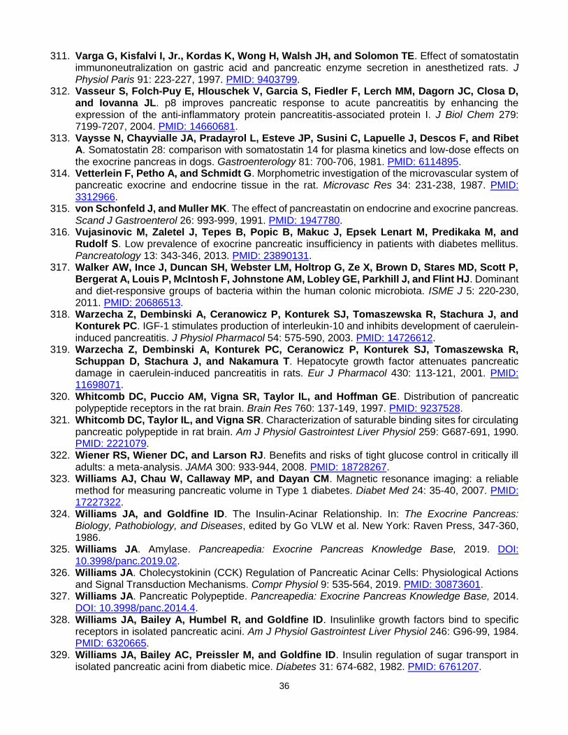
36
311. Varga G, Kisfalvi I, Jr., Kordas K, Wong H, Walsh JH, and Solomon TE. Effect of somatostatin immunoneutralization on gastric acid and pancreatic enzyme secretion in anesthetized rats. J Physiol Paris 91: 223-227, 1997. PMID: 9403799.
312. Vasseur S, Folch-Puy E, Hlouschek V, Garcia S, Fiedler F, Lerch MM, Dagorn JC, Closa D, and Iovanna JL. p8 improves pancreatic response to acute pancreatitis by enhancing the expression of the anti-inflammatory protein pancreatitis-associated protein I. J Biol Chem 279: 7199-7207, 2004. PMID: 14660681.
313. Vaysse N, Chayvialle JA, Pradayrol L, Esteve JP, Susini C, Lapuelle J, Descos F, and Ribet A. Somatostatin 28: comparison with somatostatin 14 for plasma kinetics and low-dose effects on the exocrine pancreas in dogs. Gastroenterology 81: 700-706, 1981. PMID: 6114895.
314. Vetterlein F, Petho A, and Schmidt G. Morphometric investigation of the microvascular system of pancreatic exocrine and endocrine tissue in the rat. Microvasc Res 34: 231-238, 1987. PMID: 3312966.
315. von Schonfeld J, and Muller MK. The effect of pancreastatin on endocrine and exocrine pancreas. Scand J Gastroenterol 26: 993-999, 1991. PMID: 1947780.
316. Vujasinovic M, Zaletel J, Tepes B, Popic B, Makuc J, Epsek Lenart M, Predikaka M, and Rudolf S. Low prevalence of exocrine pancreatic insufficiency in patients with diabetes mellitus. Pancreatology 13: 343-346, 2013. PMID: 23890131.
317. Walker AW, Ince J, Duncan SH, Webster LM, Holtrop G, Ze X, Brown D, Stares MD, Scott P, Bergerat A, Louis P, McIntosh F, Johnstone AM, Lobley GE, Parkhill J, and Flint HJ. Dominant and diet-responsive groups of bacteria within the human colonic microbiota. ISME J 5: 220-230, 2011. PMID: 20686513.
318. Warzecha Z, Dembinski A, Ceranowicz P, Konturek SJ, Tomaszewska R, Stachura J, and Konturek PC. IGF-1 stimulates production of interleukin-10 and inhibits development of caerulein-induced pancreatitis. J Physiol Pharmacol 54: 575-590, 2003. PMID: 14726612.
319. Warzecha Z, Dembinski A, Konturek PC, Ceranowicz P, Konturek SJ, Tomaszewska R, Schuppan D, Stachura J, and Nakamura T. Hepatocyte growth factor attenuates pancreatic damage in caerulein-induced pancreatitis in rats. Eur J Pharmacol 430: 113-121, 2001. PMID: 11698071.
320. Whitcomb DC, Puccio AM, Vigna SR, Taylor IL, and Hoffman GE. Distribution of pancreatic polypeptide receptors in the rat brain. Brain Res 760: 137-149, 1997. PMID: 9237528.
321. Whitcomb DC, Taylor IL, and Vigna SR. Characterization of saturable binding sites for circulating pancreatic polypeptide in rat brain. Am J Physiol Gastrointest Liver Physiol 259: G687-691, 1990. PMID: 2221079.
322. Wiener RS, Wiener DC, and Larson RJ. Benefits and risks of tight glucose control in critically ill adults: a meta-analysis. JAMA 300: 933-944, 2008. PMID: 18728267.
323. Williams AJ, Chau W, Callaway MP, and Dayan CM. Magnetic resonance imaging: a reliable method for measuring pancreatic volume in Type 1 diabetes. Diabet Med 24: 35-40, 2007. PMID: 17227322.
324. Williams JA, and Goldfine ID. The Insulin-Acinar Relationship. In: The Exocrine Pancreas: Biology, Pathobiology, and Diseases, edited by Go VLW et al. New York: Raven Press, 347-360, 1986.
325. Williams JA. Amylase. Pancreapedia: Exocrine Pancreas Knowledge Base, 2019. DOI: 10.3998/panc.2019.02.
326. Williams JA. Cholecystokinin (CCK) Regulation of Pancreatic Acinar Cells: Physiological Actions and Signal Transduction Mechanisms. Compr Physiol 9: 535-564, 2019. PMID: 30873601.
327. Williams JA. Pancreatic Polypeptide. Pancreapedia: Exocrine Pancreas Knowledge Base, 2014. DOI: 10.3998/panc.2014.4.
328. Williams JA, Bailey A, Humbel R, and Goldfine ID. Insulinlike growth factors bind to specific receptors in isolated pancreatic acini. Am J Physiol Gastrointest Liver Physiol 246: G96-99, 1984. PMID: 6320665.
329. Williams JA, Bailey AC, Preissler M, and Goldfine ID. Insulin regulation of sugar transport in isolated pancreatic acini from diabetic mice. Diabetes 31: 674-682, 1982. PMID: 6761207.

37
330. Williams JA, and Goldfine ID. The insulin-pancreatic acinar axis. Diabetes 34: 980-986, 1985. PMID: 2412919.
331. Wilson RM, Boden G, Shore LS, and Essa-Koumar N. Effect of somatostatin on meal-stimulated pancreatic exocrine secretions in dogs. Diabetes 26: 7-10, 1977. PMID: 830567.
332. Wizemann V, Weppler P, and Mahrt R. Effect of glucagon and insulin on the isolated exocrine pancreas. Digestion 11: 432-435, 1974. PMID: 4463136.
333. Yagihashi S. Exocrine pancreas of streptozotocin-diabetes rats treated with insulin. Tohoku J Exp Med 120: 31-42, 1976. PMID: 134471.
334. Yamada T, Shojima N, Hara K, Noma H, Yamauchi T, and Kadowaki T. Glycemic control, mortality, secondary infection, and hypoglycemia in critically ill pediatric patients: a systematic review and network meta-analysis of randomized controlled trials. Intensive Care Med 43: 1427-1429, 2017. PMID: 28424848.
335. Yamada T, Shojima N, Noma H, Yamauchi T, and Kadowaki T. Glycemic control, mortality, and hypoglycemia in critically ill patients: a systematic review and network meta-analysis of randomized controlled trials. Intensive Care Med 43: 1-15, 2017. PMID: 27637719.
336. Yatabe T, Inoue S, Sakaguchi M, and Egi M. The optimal target for acute glycemic control in critically ill patients: a network meta-analysis. Intensive Care Med 43: 16-28, 2017. PMID: 27686353.
337. Young AA, Jodka C, Pittner R, Parkes D, and Gedulin BR. Dose-response for inhibition by amylin of cholecystokinin-stimulated secretion of amylase and lipase in rats. Regul Pept 130: 19-26, 2005. PMID: 15982756.
338. Zechner D, Spitzner M, Bobrowski A, Knapp N, Kuhla A, and Vollmar B. Diabetes aggravates acute pancreatitis and inhibits pancreas regeneration in mice. Diabetologia 55: 1526-1534, 2012. PMID: 22327285.
339. Zeggari M, Viguerie N, Susini C, Esteve JP, Vaysse N, Rivier J, Wunsch E, and Ribet A. Characterization of pancreatic somatostatin binding sites with a 125I-somatostatin 28 analog. Peptides 7: 953-959, 1986. PMID: 2882495.
340. Zhang H, Kandil E, Lin YY, Levi G, and Zenilman ME. Targeted inhibition of gene expression of pancreatitis-associated proteins exacerbates the severity of acute pancreatitis in rats. Scand J Gastroenterol 39: 870-881, 2004. PMID: 15513386.
341. Zhou ZG, Gao XH, Wayand WU, Xiao LJ, and Du Y. Pancreatic microcirculation in the monkey with special reference to the blood drainage system of Langerhans islets: light and scanning electron microscopic study. Clin Anat 9: 1-9, 1996. PMID: 8838272.
342. Zipf WB, O'Dorisio TM, Cataland S, and Sotos J. Blunted pancreatic polypeptide responses in children with obesity of Prader-Willi syndrome. J Clin Endocrinol Metab 52: 1264-1266, 1981. PMID: 7014602.
343. Zyznar ES, Pietri AO, Harris V, and Unger RH. Evidence for the hormonal status of
somatostatin in man. Diabetes 30: 883-886, 1981. PMID: 6115787.