Regulation of Th2 responses by different cell types expressing ...
Upload
independentCategory
view
1download
0
IntroductionKeratins are epithelia-specific intermediate-filament (IF)proteins encoded by a large multigene family; they are clas-sified as type I or type II, based upon their charge, immuno-logical relatedness, and sequence similarity. Because ker-atin filament assembly begins with the formation of a typeI-type II heterodimer, all epithelial cells express at least 1member of each keratin type (reviewed in refs. 1, 2).
The cellular function of these proteins is largelyunknown. Keratin expression is tightly regulated, how-ever, both in terms of the pattern and the amount ofpolypeptides found in the diverse types of epithelial cells.This specific expression is thought to create different IFnetworks tailored to particular functions and pheno-typic features. In this regard, point mutations in humanepidermal and mucosal keratins, such as K5/K14,K1/K10, K9, K6/K16/K17, or K4/K13, are associatedwith several inherited epithelial diseases — in particular,skin fragility diseases (reviewed in refs. 3, 4).
Keratin K8 and its partner, keratin K18, are found typ-ically in simple or single-layered epithelial tissues (1).They are the first IF proteins to be expressed duringmurine embryogenesis and are found in the trophecto-derm at the blastocyst stage (5–7). It has been shownrecently that transgenic mice expressing a point-mutat-
ed K18 keratin manifest liver inflammation and necro-sis in association with hepatocyte fragility (8). These ani-mals exhibit an increased sensitivity to toxin-inducedliver injury (9). Hepatic lesions, colorectal hyperplasiaand inflammation, female sterility, and embryoniclethality were also noted in K8-null mice, depending onthe mouse strain (10, 11). These results indicate that theK8/K18 pair plays an important role in the function ofdifferent simple epithelia, such as liver, gastrointestinaltract, and, possibly, reproductive tract.
Although there are several well-known pathologiesthat involve abnormally elevated expression of keratins— for example, high K8/K18 levels in malignant cells(12–15) — to date, the physiological consequences ofthe overaccumulation of keratin filaments in a cell arenot clear. In transgenic mice, overexpression of other IFproteins, such as neurofilaments (expressed in neu-ronal cells) and vimentin (in cells of mesenchymal ori-gin), results in morphological and functional alter-ations such as progressive neuronopathy resemblingamyotrophic lateral sclerosis (ALS) (16, 17) and alteredlens cell differentiation (18).
To address the importance of the correct amount ofthe appropriate keratin filament complement for epithe-lial function and development, we used transgenic mice
The Journal of Clinical Investigation | June 1999 | Volume 103 | Number 11 1587
Exocrine pancreatic disorders in transsgenic mice expressing human keratin 8
M. Llanos Casanova,1 Ana Bravo,2 Angel Ramírez,1 Gabriela Morreale de Escobar,3
Felipe Were,4 Glenn Merlino,5 Miguel Vidal,4 and José L. Jorcano1
1Cell and Molecular Biology, Centro de Investigaciones Energéticas, Medioambíentales y Technológicas, E-28040 Madrid, Spain2Department of Animal Pathology, Veterinary School, University of Santiago de Compostela, E-27002 Lugo, Spain3Unidad de Endocrinología Molecular, Instituto de Investigaciones Biomédicas, E-28029 Madrid, Spain4Department of Developmental and Cell Biology, Centro de Investigaciones Biológicas, E-28006 Madrid, Spain5Laboratory of Molecular Biology, National Cancer Institute, National Institutes of Health, Bethesda, Maryland 20892, USA
Address correspondence to: José L. Jorcano, Cell and Molecular Biology, Centro de Investigaciones Energéticas, Medioambíentales y Technológicas, Av. Complutense 22, E-28040 Madrid, Spain. Phone: 34-91-346-65-98; Fax: 34-91-346-63-93; E-mail: jorcano @ciemat.es
Received for publication September 25, 1998, and accepted in revised form May 4, 1999.
Keratins K8 and K18 are the major components of the intermediate-filament cytoskeleton of simple epithe-lia. Increased levels of these keratins have been correlated with various tumor cell characteristics, includ-ing progression to malignancy, invasive behavior, and drug sensitivity, although a role for K8/K18 intumorigenesis has not yet been demonstrated. To examine the function of these keratins, we generatedmice expressing the human K8 (hk8) gene, which leads to a moderate keratin-content increase in their sim-ple epithelia. These mice displayed progressive exocrine pancreas alterations, including dysplasia and lossof acinar architecture, redifferentiation of acinar to ductal cells, inflammation, fibrosis, and substitutionof exocrine by adipose tissue, as well as increased cell proliferation and apoptosis. Histological changeswere not observed in other simple epithelia, such as the liver. Electron microscopy showed that transgenicacinar cells have keratins organized in abundant filament bundles dispersed throughout the cytoplasm,in contrast to control acinar cells, which have scarce and apically concentrated filaments. The phenotypefound was very similar to that reported for transgenic mice expressing a dominant-negative mutant TGF-β type II receptor (TGFβRII mice). We show that these TGFβRII mutant mice also have elevated K8/K18levels. These results indicate that simple epithelial keratins play a relevant role in the regulation of exocrinepancreas homeostasis and support the idea that disruption of mechanisms that normally regulate keratinexpression in vivo could be related to inflammatory and neoplastic pancreatic disorders.
J. Clin. Invest.103:1587–1595 (1999).

to force simple epithelial cells to increase K8 expression.We reported previously that transgenic mice containingthe hk8 gene express the transgene in a tissue-specific,copy number–dependent fashion (19). Here we showthat the lines with the higher copy number present pro-found structural and functional alterations in their aci-nar cells, eventually leading to tissue destruction andadipose atrophy of the pancreas in aging animals. Weconclude that the phenotype of these animals may be rel-evant to understanding the origin of inflammatory andneoplastic pathologies of the exocrine pancreas.
MethodsGenomic DNA and generation of transgenic mice. Construct HK8-12, containing the entire hk8 gene, is described in Casanova etal. (19). Construct 3′∆HK8 was derived from construct HK8-12 by deleting a 2.6-kb XbaI-SalI fragment containing 3′ flank-ing sequences. DNA fragments were isolated and microinject-ed into C57BL/10 × BALB/c-F2 (construct HK8-12) orC57BL/6 × CBA-F2 (construct 3′∆HK8) mouse embryos, asdescribed (19). Eight transgenic lines (TGK8-1 to TGK8-8)were obtained for the first construct and 4 lines (TGK8-9 toTGK8-12) for the second. Lines TGK8-1 to TGK8-4 corre-spond to lines 1864, 1861, 1918, and 1865, respectively (19).Copy number determinations were carried out as described(19), except that hybridization signals were quantified by phos-phorimaging. We observed no variation in the phenotypeattributable to the different genetic backgrounds used.
Mice were housed in a facility with controlled temperature,humidity, and light/dark cycles, under standard conditions.Their sanitary status was strictly controlled for both internaland external parasites, as well as for bacterial and viral infec-tions. They were weaned at 3 weeks of age. Nontransgenic lit-termates were used as controls.
Preparation and analysis of RNA. Total RNA from different tissueswas purified by the acid phenol method (20). K8 and K18 keratintranscripts were detected by Northern blot analysis of 10–15 µgof total RNA using random-primed, 32P-labeled DNA probes.Probes for hk8 and mK8 were as described previously (19). MouseK18 was detected with a 1.0-kbp EcoRI-PstI fragment of the mouseK18 cDNA. As a loading control, the filters were stripped andrehybridized with a rat β-actin or a ribosomal 7S probe.
Histological and immunohistochemical analysis. For histology andimmunohistochemical analysis, freshly collected tissues werefixed immediately in 10% formaldehyde or 70% ethanol, respec-tively, embedded in paraffin, sectioned, and stained with hema-toxylin/eosin. The antibodies used were the rat mAb TROMA-1 (5), which recognizes mouse and human K8; LE61, a mousemAb that recognizes mouse K18 (21); or CAM 5.2 (SigmaChemical Co., St. Louis, Missouri, USA), a mouse mAb that rec-ognizes human K8. Sections were incubated with a biotinylat-ed anti-mouse or anti-rat antibody, and then with streptavidinconjugated to horseradish peroxidase (DAKO A/S, Glostrup,Denmark). Control immunostainings using the secondaryantibody in the absence of the primary antibody were routine-ly performed. Antibody localization was determined using 3,3-diaminobenzidine (DAB) in PBS.
Enzymatic functional analysis. Fecal trypsin activity was deter-mined as described by Fraser (22). Feces were added to 180 µLsodium bicarbonate solution to bring the total to 200 µL, andthen were placed on unprocessed autoradiographic film andincubated at 37°C for 1 hour. The film was then washed, and thelevel of gelatin emulsion degradation was measured by densito-metry. Fecal fat content was stimated using the Sudan III semi-quantitative-staining method (23), with 8-week-old mice fed anolive oil–rich diet for 2 days. Sudan III was added to an extensionof feces, and fat drops were counted by microscopy.
Electron microscopy analysis. Pancreata from 2-month-old con-trol and TGK8-4 transgenic mice were fixed in 2.5% glu-taraldehyde in 0.1 M phosphate buffer (pH 7.5) and postfixedin 1% osmium tetroxide prior to dehydration and embeddingin Epon 812 resin. Semithin sections were stained with 1% tolu-idine blue for field selection. Ultrathin sections were stainedwith uranyl acetate and lead citrate.
Protein extraction and analysis by electrophoresis and immunoblot-ting. Keratins from tissues and cultured cells were isolated usinghigh-salt extractions, as described (24). Total proteins were iso-lated as described by Ku et al. (8). Protein concentrations weredetermined using the protein assay from Bio-Rad LaboratoriesInc. (Hercules, California, USA). Loading was also confirmedby Coomassie blue staining of duplicate gels. After elec-trophoresis in 8.5% SDS-PAGE gels, proteins were transferredto Immobilon-P membranes (Millipore Corp., Bedford, Mass-achusetts, USA). The membranes were incubated for 1 hourwith rat mAb TROMA-1 (5), with M20 mouse anti-HK8 mAb(Sigma Chemical Co.), or with rabbit polyclonal anti-K18 anti-
1588 The Journal of Clinical Investigation | June 1999 | Volume 103 | Number 11
Figure 1Phenotype of transgenic mice carrying HK8 constructs. (a) DNA constructs injected. Arrows indicate the position of the transcriptional start site;exons are denoted by filled boxes. RI represents EcoRI restriction site and is included for orientation. (b) Dwarfism of transgenic HK8 mice. Eight-week-old transgenic animal (right) and nontransgenic littermate (left). (c) Reduction of pancreas size. Pancreata from 8-week-old control (left) andtransgenic mice (TGK8-4; right) were photographed. The transgenic pancreas is about 30% the size of that in nontransgenic littermates. D, duode-num; S, stomach; P, pancreas. Scale bar: 3 mm.
body 1589 (25), followed by a peroxidase-coupled anti-rat, anti-mouse, or anti-rabbit IgG (Jackson ImmunoResearch Labora-tories, West Grove, Pennsylvania, USA). Keratin bands wereviewed using enhanced chemiluminescence (ECL; AmershamLife Sciences Inc., Arlington Heights, Illinois, USA).
In vivo proliferation and apoptosis studies. For in vivo proliferationassays, control and transgenic mice received an intraperitonealinjection of bromodeoxyuridine (BrdU; 120 mg/kg bodyweight) (Boehringer Mannheim GmbH, Mannheim, Germany)in 0.9% NaCl; animals were sacrificed 2 hours later. Pancreatawere fixed in 70% ethanol for 72 hours, embedded in paraffin,sectioned, and sequentially incubated for 1 hour with an anti-BrdU rat mAb (kindly provided by S. Mittnacht, Chester Beat-ty Laboratories, London, United Kingdom) and an FITC-labeled secondary anti-rat antibody (Jackson ImmunoResearchLaboratories) to label S-phase cells.
Apoptotic cells were detected using different methods. (a)Terminal deoxynucleotidyl transferase–mediated dUTP nick-end labeling (TUNEL) assays (Boehringer Mannheim GmbH)were performed following the manufacturer’s instructions. (b)Analyses of hematoxylin/eosin– and toluidine blue–stainedsemithin sections from pancreata of 2- to 7-month-old micewere performed. Cells displaying nuclear and cytoplasmic con-densation or nuclear/cellular fragmentation were consideredapoptotic. (c) Nuclei of pancreas sections were stained as fol-lows: ethanol-fixed histological preparations were deparaf-finized, treated with RNase A (200 µg/mL) for 30 minutes at37°C, washed, mounted with Micromount (Surgipath Canada
Inc., Winnipeg, Manitoba, Canada) containing propidiumiodide (PI; 50 µg/mL), and then examined by confocalmicroscopy. Cells with condensed chromatin or nuclear frag-mentation were considered apoptotic. (d) DNA content wasdetermined by flow cytometry. Nuclei were isolated by homog-enizing pancreata in a buffer containing 10 mM Tris (pH 7.4),10 mM NaCl, 3 mM Cl2Mg, 0.1 mM PMSF, and 5% NP40, fol-lowed by centrifugation at 400 g for 10 minutes. The nuclearpellet was resuspended, fixed in ethanol, treated with RNase A(200 µg/mL) citrate buffer, and stained with PI (50 µg/mL),according to Darzynkiewicz and Juan (26). DNA content wasanalyzed in an Epics XL flow cytometer (Coulter ElectronicsLtd., Hialeah, Florida, USA). For each analysis, a total of 10,000nuclei were analyzed. Off-line analysis was done using theWinMDI version 2.7 software (a kind gift of J. Trotter, ScrippsResearch Institute, La Jolla, California, USA). Integrated andpeak DNA signals were used for aggregate discrimination. Toavoid cellular debris, events were gated out 1 log below the 2-NDNA peak. The number of BrdU- and TUNEL-positive cellswere scored in a blind fashion by 2 persons.
ResultsGeneration of HK8 transgenic mice. We previously reportedthe generation of 4 transgenic lines (TGK8-1 to TGK8-4) that contain the unrearranged HK8-12 construct (Fig-ure 1a). As a result of further microinjections, weobtained 4 additional lines expressing this construct(TGK8-5 to TGK8-8) and 4 lines expressing construct3′∆HK8-12 (TGK8-9 to TGK8-12; Figure 1a). All theselines express human K8 (HK8) mRNA proportionally tothe integrated transgene copy number and in a tissue-specific pattern similar to the endogenous mouse K8(mK8) gene (ref. 19 and results not shown).
Mice carrying more than 17 transgene copies (hemizy-gous TGK8-4, TGK8-8, TGK8-12, and homozygousTGK8-11) had somewhat erect hair and reduced growth(Figure 1b); their weight was approximately 70% that of
The Journal of Clinical Investigation | June 1999 | Volume 103 | Number 11 1589
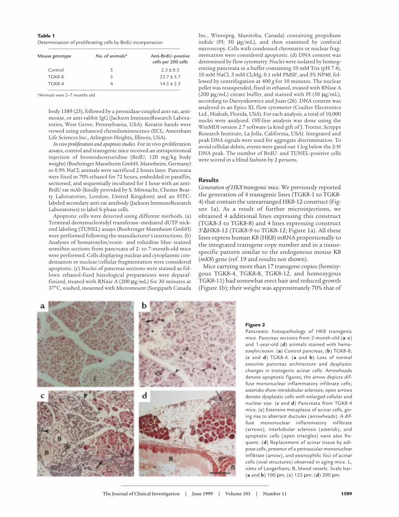
Table 1Determination of proliferating cells by BrdU incorporation
Mouse genotype No. of animalsA Anti-BrdU–positivecells per 200 cells
Control 5 2.3 ± 0.5TGK8-8 5 22.7 ± 3.7TGK8-4 4 14.5 ± 2.5
AAnimals were 2–7 months old.
Figure 2Pancreatic histopathology of HK8 transgenicmice. Pancreas sections from 2-month-old (a–c)and 1-year-old (d) animals stained with hema-toxylin/eosin. (a) Control pancreas; (b) TGK8-8;(c and d) TGK8-4. (a and b) Loss of normalexocrine pancreas architecture and dysplasticchanges in transgenic acinar cells. Arrowheadsdenote apoptotic figures; the arrow depicts dif-fuse mononuclear inflammatory infiltrate cells;asterisks show intralobular sclerosis; open arrowsdenote dysplastic cells with enlarged cellular andnuclear size. (c and d) Pancreata from TGK8-4mice. (c) Extensive metaplasia of acinar cells, giv-ing rise to aberrant ductules (arrowheads). A dif-fuse mononuclear inflammatory infiltrate(arrows), interlobular sclerosis (asterisk), andapoptotic cells (open triangles) were also fre-quent. (d) Replacement of acinar tissue by adi-pose cells, presence of a perivascular mononuclearinfiltrate (arrow), and eosinophilic foci of acinarcells (oval structures) observed in aging mice. L,islets of Langerhans; B, blood vessels. Scale bar:(a and b) 100 µm; (c) 125 µm; (d) 200 µm.

nontransgenic littermates. The life span of these micewas normal, although we occasionally observed earlymortality in some hemizygous animals. When hemizy-gous mice of lines TGK8-4 and TGK8-8 were mated, veryfew homozygous mice were born, and most of these diedwithin a few days of birth. Unless otherwise specified, theresults that follow were obtained in hemizygous animalsfrom these 2 lines, the most extensively studied.
HK8 transgenic mice have exocrine pancreas alterations. Todetermine the cause(s) underlying the dwarf phenotype,8-week-old transgenic mice and nontransgenic litter-mates were dissected. The only remarkable macroscopicdifference found was a dramatic reduction in the size ofthe transgenic pancreas, whose weight was only approx-imately 30% that of the nontransgenic mouse pancreas(Figure 1c and results not shown).
Histological examination of the pancreas sectionsshowed that the exocrine component was altered (Figure2). The abnormalities were more severe and occurred at anearlier age in line TGK8-4 than in line TGK8-8, althoughin adulthood (7 months) both lines developed the samelesions. The acini had lost their typical lobular organiza-tion of acinar cells, with the apical region oriented towardthe central lumen (Figure 2, compare a and b). In addition,as a consequence of acinar cell redifferentiation, a largepart of the acinar lobules were replaced by ductal cells,resulting in extensive areas of tubular complex appearance(Figure 2 c; see also inset in Figure 3e). Acinar cells werealso observed showing dysplastic changes characterized byenlarged cellular and nuclear size, cellular pleomorphism,and loss of the basal nuclear polarity typical of normal aci-
nar cells (Figure 2b). The number of these cells increasedwith age (results not shown). Intra- and interlobular fibro-sis was also seen (Figure 2, b and c). Inflammation waspresent as diffuse mononuclear infiltrates in the con-junctive stroma of pancreata in young animals (Figure 2,b and c), a pathology reminiscent of human chronic pan-creatitis. A progressive substitution of acinar epitheliumby adipose tissue (adipose atrophy) was observed, as wellas the presence of persistent focal infiltrates of mononu-clear inflammatory cells around isolated interacinar ductsand blood vessels (Figure 2d). Eosinophilic foci of acinarcells, considered a focal hyperplasia marker in rats (27),were observed in the pancreata of phenotypic mice at thisage (Figure 2d).
To determine whether these histological abnormalitiesaffected pancreatic function, we assayed fecal trypsin activ-ity (22) and found that it was drastically reduced in trans-genic mice (30–50 times in TGK8-8 and at least 100 timesin TGK8-4 mice; results not shown). We also found abun-dant fat drops (steatorrhea) in feces of transgenic mice feda fat-enriched diet, indicating fat malabsorption as a con-sequence of lipase deficiency. Together, these results showthat HK8 mice have a severe pancreatic insufficiency.
Although loss of normal acinar architecture and dys-plasia of the exocrine pancreas are characteristic of a pre-neoplastic state (28, 29), we have not observed pancreat-ic neoplasia in transgenic mice under 2 years of age. Onlythe exocrine pancreas seemed to be affected, because theislets of Langerhans and other simple epithelia, such asliver, lung, intestine, and stomach, showed no histolog-ical differences between control and transgenic mice.
1590 The Journal of Clinical Investigation | June 1999 | Volume 103 | Number 11
Figure 3Analysis of HK8 protein and mRNA in transgenic mouse pancreas. (a) Western blot analysis of totalprotein extracts (10 µg) from pancreata of 6-week-old nontransgenic mice (Co), nonphenotypic trans-genic mice (line TGK8-6), and phenotypic transgenic (line TGK8-4) mice. Triplicate gels wereimmunoblotted using anti-HK8 mAb M20 (top); rat mAb TROMA-1, which, under these conditions,recognizes mouse K8 strongly (double asterisk) and human K8 weakly (single asterisk) (middle); or theanti-K18 polyclonal antibody 1589 (bottom). (b) Northern blot analysis using 15 µg of total RNA frompancreata of 6-week-old animals bearing different hk8 transgene copy numbers. The filters were sequen-tially hybridized for HK8 (top), mK18 (middle), and 7S (bottom). (c) Coomassie blue–stained gel. Equalamounts of cytoskeletal extracts from the same amount (in grams) of pancreata and livers from 6-week-old control and transgenic mice were loaded. (d and e) Immunodetection of K8 in pancreas sections.Paraffin-embedded sections from 2-month-old control (d) and transgenic (e) pancreata. Control pan-creas was stained with the TROMA-1 mAb to view endogenous mK8. The signal is restricted to the api-cal region of acinar cells and to ductal cells. Transgenic pancreas sections were labeled with the HK8-specific CAM 5.2 antibody. The staining is stronger and appears throughout the cytoplasm of acinarcells. The inset shows an area of ductules reacting with antibody CAM 5.2; the arrow depicts an acinusundergoing dedifferentiation into a tubular complex. L, islet of Langerhans. Scale bar: 60 µm.

Regarding the time at which pancreatic lesions appear,exocrine cells appeared normal both in 15.5-day post-coitum embryos and newborn transgenic mice, indicatingthat the changes described develop after birth. In TGK8-4 mice, histological anomalies were first detected in 2-week-old animals, when weight differences between con-trol and transgenic mice also become apparent (notshown). At this time, pancreas abnormalities were limitedto some disorganization of the acinar lobules. The acinar-ductal conversion was first detected in 3-week-old animalsand was clearly present 1 week later. The adipose conver-sion took place only in adult animals (2–4 months old;results not shown).
To test whether dwarfism was hormonal in origin, wemeasured hypophyseal and thyroid hormone levels,because of their well-known implication in animal devel-opment and because of the HK8 expression in hypophy-seal and thyroid glands. No statistically significant dif-ferences were found between control and transgenic micein the amount of circulating T4, T4 and T3 in the liver orbrain, or T4 and T3 contents in the thyroid gland, eitherincorporated into protein or ready for secretion (“freefraction”); nor were there differences in thyroid weight orin pituitary thyroid-stimulating hormone (TSH) orgrowth hormone (GH) contents (results not shown).
Pancreatic lesions correlate with increased keratin levels. West-ern blot analysis of total protein extracts (10 µg; Figure 3a)or enriched cytoskeletal fractions (results not shown)demonstrated that transgenic pancreata expressed humanK8, because they contained a 42.5-kDa protein that react-ed specifically with an anti-HK8 antibody (Figure 3a, top)and comigrated on SDS-PAGE gels with the band corre-sponding to HK8 from HeLa cells (result not shown). Inline TGK8-4 pancreata, HK8 was strongly expressed, ascompared with nonphenotypic transgenic mice (Figure3a, top; compare the intensity of the band correspondingto TGK8-4 with that of TGK8-6. This radiograph was
overexposed to show that control pancreata were com-pletely negative for HK8). As a result of the presence oftransgenic HK8, endogenous mK8 was concomitantlyreduced or absent (Figure 3a, middle), whereas, in con-trast, the amount of mK18 (the partner of K8) wasincreased (Figure 3a, bottom). Similar changes in theamount of pancreatic cytoskeletal proteins were alsoobserved in Coomassie blue–stained gels of purified IFproteins (Figure 3c). By densitometric analysis, theamount of K8/K18 was estimated to be approximately 3-fold higher in phenotypic mice than in nontransgenic ornonphenotypic transgenic mice.
We found that the increase in K18 also occurred at themRNA level (Figure 3b). In pancreata from mice progres-sively overexpressing transgenic HK8 mRNA (linesTGK8-3, TGK8-8, and TGK8-4; Figure 3b, top), theamount of mK18 mRNA increases concomitantly (Figure3b, middle). A similar increase in mK18 mRNA was alsofound in liver, stomach, and thymus from these mice(results not shown).
The relationship between morphological changes andtransgene expression at the cellular level was studied byimmunohistochemical staining. Control pancreas sec-tions stained with TROMA-1, which detects endogenousK8, gave a weak signal, localized mainly at the apicalregion of the acinar cells (Figure 3d) and in ductal cells.Transgenic pancreas sections treated with the HK8-spe-cific mAb CAM 5.2 showed strong, filamentous stainingthroughout the cytoplasm of acinar and ductal cells (Fig-ure 3e). Similarly, immunostaining experiments using theLE61 mAb showed that whereas mK18 is barely detectedin the nontransgenic pancreas, a prominent mK18 signalwas detected in acinar and ductal cells of transgenic mice(results not shown). Collectively, these biochemical andimmunohistochemical results demonstrate a correlationbetween the alterations observed in transgenic mouse aci-nar cells and the presence in these cells of increased
The Journal of Clinical Investigation | June 1999 | Volume 103 | Number 11 1591
Figure 4Increased apoptosis in the transgenic pancreas. (a and b) Toluidine blue staining of semithin sections (1 µm). (c and d) Confocal microscopy of PI-stained nuclei. (e and f) DNA content determination by flow cytometry. Integrated and peak DNA signals were used for aggregated discrimination.To avoid cellular debris, events were gated out 1 log below the 2-N DNA peak. (a, c, and e) Control pancreas; (b, d, and f) transgenic pancreas.Arrowheads and asterisks denote apoptotic and normal nuclei, respectively. Scale bar: (a and b) 10 µm; (c and d) 30µm.

amounts of K8 and K18 keratins organized in a cytoskele-ton dispersed throughout the cytoplasm, in contrast tothe lower amounts and apical localization of these ker-atins in acinar cells from control animals.
Acinar cells from transgenic pancreata show increased prolif-eration and apoptosis. DNA synthesis was measured in thepancreata of 2- to 7-month-old mice using an anti-BrdUantibody to estimate BrdU incorporation. The number ofacinar cells synthesizing DNA was 7–11 times higher intransgenic mice (Table 1). We also analyzed apoptosis inpancreas sections of 2- to 7-month-old control and trans-genic mice (from both TGK8-4 and TGK8-8) by differentmethods. Using the TUNEL assay, we found that apop-tosis was clearly increased in transgenic animals (Table 2).Similarly increased numbers of apoptotic cells were alsodetected in transgenic pancreas sections stained withhematoxylin/eosin (Figure 2, b and c) and in semithinsections stained with toluidine blue (Figure 4, a and b). Ahigh proportion of acinar cells with fragmented or per-inuclearly collapsed chromatin were also detected by con-focal microscopy of PI-stained transgenic pancreas sec-tions (Figure 4, c and d). Finally, we also determined thenumber of pancreas nuclei with hypodiploid DNA con-tent using flow cytometry. In control animals, 2.46% ofthe total events counted had hypodiploid DNA, whereasthis proportion increased to 35.03% and to 32.78% inTGK8-4 and TGK8-8 mice, respectively (Figure 4, e and f;and data not shown).
These results show that, in addition to the alterations indifferentiation, increased keratin expression also disturbsthe processes of proliferation and apoptosis in acinar cells.
Ultrastructural studies. Normal acinar cells containsecretory granules (zymogen) concentrated at the apicalregion, as seen in toluidine blue–stained semithin sec-tions (Figure 4a). In contrast, acinar cells of pathologi-cal pancreata showed a larger number of zymogen gran-ules dispersed throughout the cytoplasm (Figure 4b).
These results were corroborated by electron microscopystudies (Figure 5, a and b). These pictures also show thattransgenic acinar cells frequently manifest nuclei thatare fragmented or that have indented profiles andgreater electron density than control nuclei, in con-junction with a swollen endoplasmic reticulum, char-acteristics of apoptotic cells. At higher magnifications,control acinar cells showed few IFs, and these weremainly anchored to desmosomes at the apical region ofthe cytoplasm (Figure 5c). In contrast, transgenic acinarcells had abundant filament bundles of normal appear-ance, located not only at the secretory apical region butalso dispersed throughout the cytoplasm (Figure 5, dand e). These results corroborate the immunostainingdata (Figure 3) and demonstrate that the increasedamounts of keratin IF, containing both the transgenicand the endogenous proteins, were properly assembledfrom a structural point of view, although the resultantcytoskeleton was more uniformly distributed through-out the cytoplasm than in normal cells.
Increased K8/K18 levels are also found in the altered exocrinepancreata of transgenic mice expressing a mutant TGF-β type IIreceptor. Analyses of the exocrine pancreata of transgenicmice expressing a dominant-negative mutant form ofthe TGF-β type II receptor, generated by Böttinger et al.(30), showed that these animals manifest a pancreaticphenotype very similar to those described above, includ-ing reduced organ weight, acino-ductular metaplasia,fibrosis, and adipose replacement, as well as increasedproliferation and apoptosis (30). As observed in HK8mice, these alterations also appear progressively with agein TGFβRII mice.
In view of these similarities, we asked if TGFβRII micealso have altered K8/K18 levels. Immunohistochemicalanalysis using anti-K8 and anti-K18 antibodies (Figure6, a and b; and results not shown), as well as Westernblots and Coomassie blue–stained gels (Figure 6c),
1592 The Journal of Clinical Investigation | June 1999 | Volume 103 | Number 11
Figure 5Ultrastructural analysis of pancreatic acinar transgenic cells. (aand b) Low-magnification electron microscopy survey of control(a) and TGK8-4 (b) acinar cells from 2-month-old littermates.Arrowheads mark the lumen in the acini of control cells. Notethe accumulation and loss of the apical localization of zymogengranules in transgenic cells. Apoptotic transgenic cells with con-densed chromatin, indented nuclei, and swollen endoplasmicreticulum are also frequent. (c) Electron micrograph showing theapical region of 2 cells around the acinar lumen in the controlpancreas. IFs, frequently anchored to desmosomes, are scarceand are restricted to this region in these cells. (d and e) Bundlesof IFs are abundant and found dispersed throughout the cyto-plasm in transgenic acinar cells. S, secretory granules; D, desmo-some. Scale bar: (a and b) 5 µm; (c–e) 250 nm.
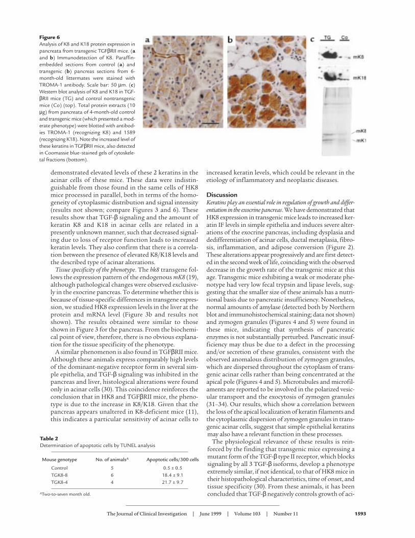
demonstrated elevated levels of these 2 keratins in theacinar cells of these mice. These data were indistin-guishable from those found in the same cells of HK8mice processed in parallel, both in terms of the homo-geneity of cytoplasmic distribution and signal intensity(results not shown; compare Figures 3 and 6). Theseresults show that TGF-β signaling and the amount ofkeratin K8 and K18 in acinar cells are related in apresently unknown manner, such that decreased signal-ing due to loss of receptor function leads to increasedkeratin levels. They also confirm that there is a correla-tion between the presence of elevated K8/K18 levels andthe described type of acinar alterations.
Tissue specificity of the phenotype. The hk8 transgene fol-lows the expression pattern of the endogenous mK8 (19),although pathological changes were observed exclusive-ly in the exocrine pancreas. To determine whether this isbecause of tissue-specific differences in transgene expres-sion, we studied HK8 expression levels in the liver at theprotein and mRNA level (Figure 3b and results notshown). The results obtained were similar to thoseshown in Figure 3 for the pancreas. From the biochemi-cal point of view, therefore, there is no obvious explana-tion for the tissue specificity of the phenotype.
A similar phenomenon is also found in TGFβRII mice.Although these animals express comparably high levelsof the dominant-negative receptor form in several sim-ple epithelia, and TGF-β signaling was inhibited in thepancreas and liver, histological alterations were foundonly in acinar cells (30). This coincidence reinforces theconclusion that in HK8 and TGFβRII mice, the pheno-type is due to the increase in K8/K18. Given that thepancreas appears unaltered in K8-deficient mice (11),this indicates a particular sensitivity of acinar cells to
increased keratin levels, which could be relevant in theetiology of inflammatory and neoplastic diseases.
DiscussionKeratins play an essential role in regulation of growth and differ-entiation in the exocrine pancreas. We have demonstrated thatHK8 expression in transgenic mice leads to increased ker-atin IF levels in simple epithelia and induces severe alter-ations of the exocrine pancreas, including dysplasia anddedifferentiation of acinar cells, ductal metaplasia, fibro-sis, inflammation, and adipose conversion (Figure 2).These alterations appear progressively and are first detect-ed in the second week of life, coinciding with the observeddecrease in the growth rate of the transgenic mice at thisage. Transgenic mice exhibiting a weak or moderate phe-notype had very low fecal trypsin and lipase levels, sug-gesting that the smaller size of these animals has a nutri-tional basis due to pancreatic insufficiency. Nonetheless,normal amounts of amylase (detected both by Northernblot and immunohistochemical staining; data not shown)and zymogen granules (Figures 4 and 5) were found inthese mice, indicating that synthesis of pancreaticenzymes is not substantially perturbed. Pancreatic insuf-ficiency may thus be due to a defect in the processingand/or secretion of these granules, consistent with theobserved anomalous distribution of zymogen granules,which are dispersed throughout the cytoplasm of trans-genic acinar cells rather than being concentrated at theapical pole (Figures 4 and 5). Microtubules and microfil-aments are reported to be involved in the polarized vesic-ular transport and the exocytosis of zymogen granules(31–34). Our results, which show a correlation betweenthe loss of the apical localization of keratin filaments andthe cytoplasmic dispersion of zymogen granules in trans-genic acinar cells, suggest that simple epithelial keratinsmay also have a relevant function in these processes.
The physiological relevance of these results is rein-forced by the finding that transgenic mice expressing amutant form of the TGF-β type II receptor, which blockssignaling by all 3 TGF-β isoforms, develop a phenotypeextremely similar, if not identical, to that of HK8 mice intheir histopathological characteristics, time of onset, andtissue specificity (30). From these animals, it has beenconcluded that TGF-β negatively controls growth of aci-
The Journal of Clinical Investigation | June 1999 | Volume 103 | Number 11 1593
Figure 6Analysis of K8 and K18 protein expression inpancreata from transgenic TGFβRII mice. (aand b) Immunodetection of K8. Paraffin-embedded sections from control (a) andtransgenic (b) pancreas sections from 6-month-old littermates were stained withTROMA-1 antibody. Scale bar: 50 µm. (c)Western blot analysis of K8 and K18 in TGF-βRII mice (TG) and control nontransgenicmice (Co) (top). Total protein extracts (10µg) from pancreata of 4-month-old controland transgenic mice (which presented a mod-erate phenotype) were blotted with antibod-ies TROMA-1 (recognizing K8) and 1589(recognizing K18). Note the increased level ofthese keratins in TGFβRII mice, also detectedin Coomassie blue–stained gels of cytoskele-tal fractions (bottom).
Table 2Determination of apoptotic cells by TUNEL analysis
Mouse genotype No. of animalsA Apoptotic cells/300 cells
Control 5 0.5 ± 0.5TGK8-8 6 18.4 ± 9.1TGK8-4 4 21.7 ± 9.7
ATwo-to-seven month old.
nar cells and is essential for the maintenance of a differ-entiated acinar phenotype in the exocrine pancreas. Wefound that TGFβRII mice, exactly as HK8 mice, have ele-vated levels and homogeneous cytoplasmic distributionof K8/K18 in acinar cells (Figure 6). These results strong-ly suggest that these keratins are involved in the TGF-βsignaling mechanism, a possibility that we are presentlyinvestigating. The function of keratins has been largelyunknown, although they have been related to providingresistance for cells (3, 4, 8). We recently demonstrated thatepidermal keratins K10 and K16 are involved in the con-trol of cell proliferation and differentiation in vitro (35);the data presented here extend this function to simplekeratins in vivo. This involvement is also supported bythe observation that K8/K18 bind members of the 14-3-3 protein family (known to interact with a number ofkinases and phosphates) and bind to the heat shock pro-tein 70 (HSP-70), and a protein kinase Cε–related kinase(reviewed in ref. 7).
Implications for human disease: chronic pancreatitis and pan-creatic cancer. There appears to be some consistency in theresponse of the pancreas to a variety of insults. Tubularcomplex formation, interstitial fibrosis, and mucoid cellmetaplasia have all been associated, to some extent, withductal obstruction–induced atrophy, adenocarcinoma,and chronic pancreatitis (36–38). All these characteris-tics are present in HK8 mice. Aging HK8 mice show adi-pose atrophy of the pancreas and chronic inflammation,also present in human chronic pancreatitis. These ani-mals, therefore, recapitulate many features characteris-tic of human chronic pancreatitis and may constitute avaluable model for this disease.
Our results may also have implications for pancreaticcarcinogenesis. The morphological aspect of mostexocrine pancreas cancers is reminiscent of ductal cells,as has long been recognized by pathologists (36, 39),although ductal cells constitute a minor cell fraction innormal tissue. In addition, pancreatic cancer cellsexpress differentiation markers characteristic of ductalcells (40). When human exocrine pancreas cells are cul-tured in vitro, a phenotypic switch takes place from aci-nar to ductal characteristics (41), which is reversibleupon cessation of growth (42). Because preneoplasticlesions have not been clearly identified in the humanpancreas, considerable controversy exists on the acinaror ductal origin of pancreatic cancers (43).
The first signs of anomaly in HK8 mice are loss of aci-nar architecture, acinar cell dysplasia, and increasedproliferation, all characteristics of early neoplasticstages; they later undergo acino-ductal dedifferentia-tion. These features support the hypothesis that acinarcells are, in fact, the origin of pancreatic cancer and thatthey dedifferentiate to the ductal phenotype observedin most tumors during the carcinogenic process (43,44). Transgenic mice expressing oncogenes (c-myc andSV40 T antigen; refs. 28, 45) and growth factors (TGF-α; refs. 46–48), or that are deficient in TGF-β signaling(30) in acinar cells, also reinforce this model. Thesecharacteristics suggest that the HK8 mouse pancreascould represent the early stages in the pathway to pan-creatic neoplasia. That these lesions do not progress toneoplasia may be a consequence of the elevated apop-
tosis observed and an indication of the requirement foradditional defects in other genes (e.g., p53) for carcino-genesis to proceed. Carcinogenesis experiments are cur-rently under way, and the HK8 mice are being crossedwith p53-null mice to test this hypothesis.
Other studies also suggest the relevance of keratinsK8/K18 in carcinogenesis; for instance, these keratinsare involved in aspects of tumorigenic cell behavior,such as cell migration and invasiveness (49–51). It hasalso been reported that K8/K18 are induced by v-ras intransformed epidermal keratinocytes (52, 53) and thatthere is a positive correlation between the expression ofthese keratins and epidermal cell malignancy both invitro (54) and in vivo (12, 13). In transitional cell carci-noma, increased K8 and K18 levels have been detectedat the tumor invasion front, and there is a relationshipbetween the expression of these keratins and tumormalignancy (14, 15). It is thus possible that, under cer-tain circumstances, K8/K18 may provide a permissivecellular environment for induction of malignancy.
These data, together with those from our transgenicmice, suggest that a relationship exists between deregu-lated K8/K18 expression and disease, and that the eluci-dation of the cellular and molecular mechanisms bywhich HK8 mice develop pathological features in pan-creatic acinar cells could contribute to our understand-ing of the etiology of inflammatory and neoplastichuman diseases.
AcknowledgmentsWe thank Werner Franke, Rolf Kemler, E. Birgit Lane, CarlosCaulin, R.G. Oshima, Lalage M. Wakefield, and Sybille Mittnacht for providing materials, and Francisco X. Real fordiscussion and advice. We are particularly grateful to IsabelFabregat, Alberto Alvarez, and Jose Carlos Segovia for theirexpert help in the analysis of apoptosis; to Jesús Martínez-Pala-cios for mouse husbandry; to Claudia Pérez and M. Isabel delos Santos for invaluable assistance in histology and immunos-taining; to Soledad Moreno for photographic work; and to C.Mark for expert editorial assistance. This work was supportedin part by grant PB 94-1230 of the Spanish Dirección Generalde Investigación Científica y Tecnológica.
1. Moll, R., Franke, W.W., and Schiller, D. 1982. The catalog of humancytokeratins: patterns of expression in normal epithelia, tumors and cul-tured cells[review]. Cell. 31:11–24.
2. Fuchs, E., and Weber, K. 1994. Intermediate filaments: structure, dynam-ics, function and disease. Annu. Rev. Biochem. 63:345–382.
3. McLean, W.H., and Lane, E.B. 1995. Intermediate filaments indisease[review]. Curr. Opin. Cell Biol. 7:118–125.
4. Fuchs, E. 1996. The cytoskeleton and disease: genetic disorders of IFs.Annu. Rev. Genet. 30:197–231.
5. Brûlet, P., Babinet, C., Kemler, R., and Jacob, F. 1980. Monoclonal anti-bodies against trophectoderm-specific markers during mouse blastocystformation. Proc. Natl. Acad. Sci. USA. 77:4113–4117.
6. Jackson, B.W., Grund, C., Winter, S., Franke, W.W., and Illmensee, K.1981. Formation of cytoskeletal elements during mouse embryogenesis:epithelial differentiation and intermediate-sized filaments in earlypostimplantation embryos. Differentiation. 20:203–216.
7. Oshima, R.G., Baribault, H., and Caulín, C. 1996. Oncogenic regulationand function of keratins 8 and 18. Cancer Metastasis Rev. 15:445–471.
8. Ku, N.O., Michie, S., Oshima, R.G., and Omary, M.B. 1995. Chronic hep-atitis, hepatocyte fragility, and increased soluble phosphoglycokeratinsin transgenic mice expressing a keratin 18 conserved arginine mutant. J.Cell Biol. 5:1303–1314.
9. Ku, N.O., et al. 1996. Susceptibility of hepatotoxicity in transgenic micethat express a dominant-negative human keratin 18 mutant. J. Clin.Invest. 98:1034–1046.
10. Baribault, H., Price, J., Miyai, K., and Oshima, R.G. 1993. Mid-gestation-
1594 The Journal of Clinical Investigation | June 1999 | Volume 103 | Number 11

al lethality in mice lacking keratin 8. Genes Dev. 7:1191–1202.11. Baribault, H., Penner, J., Iozzo, R.V., and Wilson-Heiner, M. 1994. Col-
orectal hyperplasia and inflammation in keratin 8-deficient FVB/Nmice. Genes Dev. 8:2964–2973.
12. Markey, A.C., et al. 1991. Expression of simple epithelial keratins 8 and18 in epidermal neoplasia. J. Invest. Dermatol. 97:763–770.
13. Larcher, F., et al. 1992. Aberrant expression of the simple epithelial typeII keratin 8 by mouse skin carcinomas but not papillomas. Mol. Carcinog.6:112–121.
14. Schaafsma, H.E., et al. Cytokeratin expression patterns in metastatictransitional cell carcinoma of the urinary tract. An immunohistochem-ical study comparing local and autologous metastases. Am. J. Pathol.1389–1400.
15. Schaafsma, H.E., et al. 1993. Increased expression of cytokeratins 8, 18and vimentin in the invasion front of mucosal squamous cell carcino-ma. J. Pathol. 170:77–86.
16. Xu, Z., Cork, L.C., Griffin, J.W., and Cleveland, D.W. 1993. Increasedexpression of neurofilament subunit NF-L produces morphologicalalterations that resemble the pathology of human motor neuron disease.Cell. 73:23–33.
17. Côte, F., Collard, F.J., and Julien, J.P. 1993. Progressive neuropathy intransgenic mice expressing the human neurofilament heavy gene: amouse model of amyotrophic lateral sclerosis. Cell. 73:35–47.
18. Capetanaki, Y., Smith, S., and Heath, J.P. 1989. Overexpression of thevimentin gene in transgenic mice inhibits normal lens cell differentia-tion. J. Cell Biol. 109:1653–1664.
19. Casanova, L., et al. 1995. Tissue-specific and efficient expression of thehuman simple epithelial keratin 8 gene in transgenic mice. J. Cell Sci.108:811–820.
20. Chomczynski, P., and Sacchi, N. 1987. Single-step method of RNA iso-lation by acid guanidinium thiocyanate-phenol-chloroform extraction.Anal. Biochem. 162:156–159.
21. Lane, E.B. 1982. Monoclonal antibodies provide specific intramolecularmarkers for the study of epithelial tonofilament organization. J. Cell Biol.92:665–673.
22. Fraser, C.M. 1988. Valores y procedimientos químicos. In Manual deMerck de Veterinaria. Tercera edición. C.M. Fraser et al., editors. Centrum.Barcelona, Spain. 1026–1027.
23. Nelson D.A., Tomar, R.H., and Washington, J.A. 1988. Examenmicroscópico de las heces. In Todd-Sanford-Davidsohn Diagnóstico ytratamiento clínicos por el laboratorio. D.A. Nelson, J.A. Washington, W.W.McLendon, B.E. Statland, and R.H. Tomar, editors. Salvat Editores S.A.Barcelona, Spain. 710 pp.
24. Achtstaetter, T., Hatzfeld, M., Quinlan, R.A., Parmelee, D.C., and Franke,W.W. 1986. Separation of cytokeratin polypeptides by gel electrophoreticand chromatographic techniques and their identification byimmunoblotting. Methods Enzymol. 134:355–371.
25. Oshima, R.G. 1981. Identification and immunoprecipitation ofcytoskeletal proteins from extraembryonic endodermal cells. J. Biol.Chem. 256:8124–8133.
26. Darzynkiewicz, Z., and Juan, G. 1997. DNA content measurement forDNA ploidy and cell cycle analysis. In Current protocols in cytometry. J.P.Robinson. et al., editors. John Wiley & Sons. New York, NY. 7.5.2.
27. Eustis, S.L., Boorman, G.A., and Hayashi, Y. 1990. Exocrine pancreas. InPathology of the Fischer rat. G.A. Boorman, S.L. Eustis, M.R. Elwell, C.A.Montgomery, and W.F. MacKenzie, editors. Academic Press. San Diego,CA. 95–108.
28. Ornitz, D.M., Hammer, R.E., Messing, A., Palmiter, R.D., and Brinster,R.L. 1987. Pancreatic neoplasia induced by SV40-T antigen expressionin acinar cells of transgenic mice. Science. 238:188–193.
29. Quaife, C.J., Pinkert, C.A., Ornitz, D.M., Palmiter, R.D., and Brinster, R.L.1987. Pancreatic neoplasia induced by ras expression in acinar cells oftransgenic mice. Cell. 48:1023–1034.
30. Böttinger, E.P., et al. 1997. Expression of a dominant-negative mutantTGF-β type II receptor in transgenic mice reveals essential roles for TGF-β in regulation of growth and differentiation in the exocrine pancreas.EMBO J. 16:2621–2633.
31. Achler, C., Filmer, D., Merte, C., and Drenckhahn, D. 1989. Role ofmicrotubules in polarized delivery of apical membrane proteins to the
brush border of the intestinal epithelium. J. Cell Biol. 109:179–189.32. Parczyk, K., Haase, W., and Kondor-Koch, C. 1989. Microtubules are
involved in the secretion of proteins at the apical cell surface of the polar-ized epithelial cell, Madin-Darby canine kidney. J. Biol. Chem.264:16837–16846.
33. Drenckhahn, D., and Mannherz, H.G. 1983. Distribution of actin andthe actin-associated proteins myosin, tropomyosin, alpha-actin, vin-culin, and villin in rat and bovine exocrine glands. Eur. J. Cell Biol.30:167–176.
34. Jungermann, J., et al. 1995. Disassembly of rat pancreatic acinar cellcytoskeleton during supramaximal secretagogue stimulation. Am. J. Phys-iol. 268:G328–G338.
35. Paramio, J.M., et al. 1999. Modulation of cell proliferation by cytoker-atins K10 and K16. Mol. Cell. Biol. 19:3086–3094.
36. Klöppel, G. 1993. Pathology of nonendocrine pancreatic tumors. In Thepancreas. Biology, pathobiology, and disease. V.L.W. Go, et al., editors. RavenPres. New York, NY. 871–897.
37. Oertel, J.E. 1989. The pancreas: non-neoplastic alterations. Am. J. Surg.Pathol. 13:50–65.
38. Bockman, D.E., Boydston W.R., and Anderson M.C. 1982. Origin of duc-tular complexes in human chronic pancreatitis. Am. J. Surg. 144:243–249.
39. Bockman, D.E. 1981. Cells of origin of pancreatic cancer: experimentalanimal tumors related to human pancreas. Cancer. 47(Suppl.6):1528–1534.
40. Schüssler, M.M., Skoudy, A., Ramaekers, F., and Real, F.X. 1992. Inter-mediate filaments as differentiation markers of normal pancreas andpancreas cancer. Am. J. Pathol. 140:559–568.
41. Vilá, M.R., Lloreta, J., and Real, F.X. 1994. Normal human pancreas cul-tures display functional ductal characteristics. Lab. Invest. 71:423–431.
42. De Lisle, R.C., and Logsdon, C.D. 1990. Pancreatic acinar cells in culture:expression of acinar and ductal antigens in a growth-related manner.Eur. J. Cell Biol. 51:64–75.
43. Scarpelli, D.G., Rao, M.S., and Reddy, J.K. 1991. Are acinar cells involvedin the pathogenesis of ductal adenocarcinoma of the pancreas? CancerCells. 3:275–277.
44. Logsdon, C.D. 1995. Pancreatic duct cell cultures: there is more to ductsthan salty water. Gastroenterology. 109:1005–1009.
45. Sandgren, E.P., Quaife, C.J., Paulovich, A.C., Palmiter, R.D., and Brinster,R.L. 1991. Pancreatic tumor pathogenesis reflects the causative geneticlesion. Proc. Natl. Acad. Sci. USA. 88:93–97.
46. Sandgren, E.P, Luetteke, N.C., Palmiter, R.D., Brinster, R.L., and Lee, D.C.1990. Overexpression of TGF-alpha in transgenic mice: induction ofepithelial hyperplasia, pancreatic metaplasia and carcinoma of thebreast. Cell. 61:1121–1135.
47. Jhappan, C., et al. 1990. TGF-α overexpression in transgenic miceinduces liver neoplasia and abnormal development of the mammarygland and pancreas. Cell. 61:1137–1146.
48. Bockman, D.E., and Merlino, G. 1992. Cytological changes in the pan-creas of transgenic mice overexpressing transforming growth factoralpha. Gastroenterology. 103:1883–1892.
49. Chu, W.W., Runyan, R.B., Oshima, R.G., and Hendrix, M.J.C. 1993.Expression of complete keratin filaments in mouse L cells augments cellmigration and invasion. Proc. Natl. Acad. Sci. USA. 90:4261–4265.
50. Chu, Y.W., Seftor, E.A., Romer, L.H., and Hendrix, M.J.C. 1996. Experi-mental coexpression of vimentin and keratin intermediate filaments inhuman melanoma cells augments motility. Am. J. Pathol. 148:63–69.
51. Hendrix, M.J.C., et al. 1992. Coexpression of vimentin and keratins byhuman melanoma tumor cells: correlation with invasive and metastaticpotential. J. Natl. Cancer Inst. 84:165–174.
52. Cheng, C., Kilkenny, A., Roop, D., and Yuspa, S. 1990. The v-ras onco-gene inhibits the expression of differentiation markers and facilitatesexpression of cytokeratins 8 and 18 in mouse keratinocytes. Mol. Car-cinog. 3:363–373.
53. Díaz-Guerra, M., et al. 1992. Expression of simple epithelial cytokeratinsin mouse epidermal keratinocytes harboring Harvey ras gene alterations.Cancer Res. 52:680–687.
54. Caulín, C., Bauluz, C., Gandarillas, A., Cano, A., and Quintanilla, M.1993. Changes in keratin expression during malignant progression oftransformed mouse epidermal keratinocytes. Exp. Cell Res. 204:11–21.
The Journal of Clinical Investigation | June 1999 | Volume 103 | Number 11 1595
Copyright © 2022 FDOKUMEN