The role of keratin subfamilies and keratin pairs in the formation of human epidermal intermediate...
11
The Role of Keratin Subfamilies and Keratin Pairs in the Formation of Human Epidermal Intermediate Filaments Riva Eichner,* Tung-Tien Sun, * and Ueli Aebi* • Departments of Dermatology and Cell Biology and Anatomy, The Johns Hopkins University School of Medicine, Baltimore, Maryland 21205, and *Departments of Dermatology and Pharmacology, New York University School of Medicine, New York 10016 Abstract. The four major keratins of normal human epidermis (molecular mass 50, 56.5, 58, and 65-67 kD) can be subdivided on the basis of charge into two subfamilies (acidic 50-kD and 56.5-kD keratins vs. relatively basic 58-kD and 65-67-kD keratins) or sub- divided on the basis of co-expression into two "pairs" (50-kD/58-kD keratin pair synthesized by basal cells vs. 56.5-kD/65-67-kD keratin pair expressed in supra- basal cells). Acidic and basic subfamilies were sepa- rated by ion exchange chromatography in 8.5 M urea and tested for their ability to reassemble into 10-nm filaments in vitro. The two keratins in either subfam- ily did not reassemble into 10-nm filaments unless combined with members of the other subfamily, While electron microscopy of acidic and basic keratins equilibrated in 4.5 M urea showed that keratins within each subfamily formed distinct oligomeric structures, possibly representing precursors in filament assembly, chemical cross-linking followed by gel analysis re- vealed dimers and larger oligomers only when subfamilies were combined. In addition, among the four major keratins, the acidic 50-kD and basic 58-kD keratins showed preferential association even in 8.5 M urea, enabling us to isolate a 50-kD/58-kD keratin complex by gel filtration. This isolated 50-kD/58-kD keratin pair readily formed 10-nm filaments in vitro. These results demonstrate that in tissues containing multiple keratins, two keratins are sufficient for fila- ment assembly, but one keratin from each subfamily is required. More importantly, these data provide the first evidence for the structural significance of specific co-expressed acidic/basic keratin pairs in the forma- tion of epithelial 10-nm filaments. T HE intermediate-sized filaments (10-nm diameter) of epithelial cells are composed of a family of water- insoluble proteins termed keratins (molecular mass 40-70 kD). Although ~20 different human keratin polypep- tides have been identified, data from monoclonal antikeratin antibody reactivity (4, 6, 11, 35), nucleic acid hybridization (9, 13), peptide mapping (25), and two-dimensional gel elec- trophoresis (17, 32) have demonstrated that all keratins can be subdivided into two subfamilies: acidic keratins (isoelectric point < pH 5.5) and relatively basic keratins (isoelectric point > pH 6.0). Detailed analysis of the keratins in various epithelia has shown that only 2-10 keratins are expressed in any individual tissue, but at least one member of each subfamily is always represented (4, 17, 25, 32, 35, 41). More recently, Franke and co-workers have demonstrated the association of specific acidic and basic keratins as "complexes" in polyac- rylamide gels containing 4.0-9.5 M urea (7). In rat hepato- cytes, which contain only a relatively basic 55-kD keratin (A) and an acidic 49-kD keratin (D), the complex observed in 4 M urea gels was shown to represent heterotypic tetramers which could reassemble in vitro into 10-nm filaments (22). Taken together with earlier data suggesting that at least two different keratins are required for keratin filament reconsti- tution (14, 16, 29), these results have led to the general assumption that one member from each keratin subfamily may be necessary for filament assembly. In epithelia contain- ing more than two keratins, however, the role of multiple keratins within each subfamily in the formation and function of 10-nm filaments remains unclear. To elucidate the structural and functional significance of keratin heterogeneity, we have studied the in vitro interaction of the four major keratins expressed in normal human epi- dermis (molecular mass 50, 56.5, 58, and 65-67 kD" Moll catalog Nos. 14 and 15, 10, 5. and I and 2. respectively) (3, 6. 17, 18, 26, 39). These four keratins can be subdivided into two keratins of the acidic subfamily (50 and 56.5 kD) and two keratins of the basic subfamily (58 and 65-67 kD) (6). Alternatively, based on co-expression data, the four epidermal keratins can be grouped into two "keratin pairs": the acidic 50-kD/basic 58-kD "pair," which are the only keratins syn- thesized by epidermal basal cells, and the acidic 56.5-kD/ basic 65-67-kD "pair," which are co-expressed only in the suprabasal layers of keratinizing epithelia (26, 32, 39). In the present study, we have separated the keratins of the two © The Rockefeller Universily Press, 0021-9525/86/05/1767/11 $1.00 The Journal of Cell Biology, Volume 102, May 1986 1767-1777 1767 on October 5, 2013 jcb.rupress.org Downloaded from Published May 1, 1986 on October 5, 2013 jcb.rupress.org Downloaded from Published May 1, 1986 on October 5, 2013 jcb.rupress.org Downloaded from Published May 1, 1986 on October 5, 2013 jcb.rupress.org Downloaded from Published May 1, 1986 on October 5, 2013 jcb.rupress.org Downloaded from Published May 1, 1986 on October 5, 2013 jcb.rupress.org Downloaded from Published May 1, 1986 on October 5, 2013 jcb.rupress.org Downloaded from Published May 1, 1986 on October 5, 2013 jcb.rupress.org Downloaded from Published May 1, 1986 on October 5, 2013 jcb.rupress.org Downloaded from Published May 1, 1986 on October 5, 2013 jcb.rupress.org Downloaded from Published May 1, 1986 on October 5, 2013 jcb.rupress.org Downloaded from Published May 1, 1986
-
Upload
independent -
Category
Documents
-
view
0 -
download
0
Transcript of The role of keratin subfamilies and keratin pairs in the formation of human epidermal intermediate...

The Role of Keratin Subfamilies and Keratin Pairs in the Formation of Human Epidermal Intermediate Filaments Riva E ichne r ,* T u n g - T i e n Sun , * and Uel i Aebi*
• Departments of Dermatology and Cell Biology and Anatomy, The Johns Hopkins University School of Medicine, Baltimore, Maryland 21205, and *Departments of Dermatology and Pharmacology, New York University School of Medicine, New York 10016
Abstract. The four major keratins of normal human epidermis (molecular mass 50, 56.5, 58, and 65-67 kD) can be subdivided on the basis of charge into two subfamilies (acidic 50-kD and 56.5-kD keratins vs. relatively basic 58-kD and 65-67-kD keratins) or sub- divided on the basis of co-expression into two "pairs" (50-kD/58-kD keratin pair synthesized by basal cells vs. 56.5-kD/65-67-kD keratin pair expressed in supra- basal cells). Acidic and basic subfamilies were sepa- rated by ion exchange chromatography in 8.5 M urea and tested for their ability to reassemble into 10-nm filaments in vitro. The two keratins in either subfam- ily did not reassemble into 10-nm filaments unless combined with members of the other subfamily, While electron microscopy of acidic and basic keratins equilibrated in 4.5 M urea showed that keratins within each subfamily formed distinct oligomeric structures,
possibly representing precursors in filament assembly, chemical cross-linking followed by gel analysis re- vealed dimers and larger oligomers only when subfamilies were combined. In addition, among the four major keratins, the acidic 50-kD and basic 58-kD keratins showed preferential association even in 8.5 M urea, enabling us to isolate a 50-kD/58-kD keratin complex by gel filtration. This isolated 50-kD/58-kD keratin pair readily formed 10-nm filaments in vitro.
These results demonstrate that in tissues containing multiple keratins, two keratins are sufficient for fila- ment assembly, but one keratin from each subfamily is required. More importantly, these data provide the first evidence for the structural significance of specific co-expressed acidic/basic keratin pairs in the forma- tion of epithelial 10-nm filaments.
T HE intermediate-sized filaments (10-nm diameter) of epithelial cells are composed of a family of water- insoluble proteins termed keratins (molecular mass
40-70 kD). Although ~20 different human keratin polypep- tides have been identified, data from monoclonal antikeratin antibody reactivity (4, 6, 11, 35), nucleic acid hybridization (9, 13), peptide mapping (25), and two-dimensional gel elec- trophoresis (17, 32) have demonstrated that all keratins can be subdivided into two subfamilies: acidic keratins (isoelectric point < pH 5.5) and relatively basic keratins (isoelectric point > pH 6.0). Detailed analysis of the keratins in various epithelia has shown that only 2-10 keratins are expressed in any individual tissue, but at least one member of each subfamily is always represented (4, 17, 25, 32, 35, 41). More recently, Franke and co-workers have demonstrated the association of specific acidic and basic keratins as "complexes" in polyac- rylamide gels containing 4.0-9.5 M urea (7). In rat hepato- cytes, which contain only a relatively basic 55-kD keratin (A) and an acidic 49-kD keratin (D), the complex observed in 4 M urea gels was shown to represent heterotypic tetramers which could reassemble in vitro into 10-nm filaments (22). Taken together with earlier data suggesting that at least two
different keratins are required for keratin filament reconsti- tution (14, 16, 29), these results have led to the general assumption that one member from each keratin subfamily may be necessary for filament assembly. In epithelia contain- ing more than two keratins, however, the role of multiple keratins within each subfamily in the formation and function of 10-nm filaments remains unclear.
To elucidate the structural and functional significance of keratin heterogeneity, we have studied the in vitro interaction of the four major keratins expressed in normal human epi- dermis (molecular mass 50, 56.5, 58, and 65-67 kD" Moll catalog Nos. 14 and 15, 10, 5. and I and 2. respectively) (3, 6. 17, 18, 26, 39). These four keratins can be subdivided into two keratins of the acidic subfamily (50 and 56.5 kD) and two keratins of the basic subfamily (58 and 65-67 kD) (6). Alternatively, based on co-expression data, the four epidermal keratins can be grouped into two "keratin pairs": the acidic 50-kD/basic 58-kD "pair," which are the only keratins syn- thesized by epidermal basal cells, and the acidic 56.5-kD/ basic 65-67-kD "pair," which are co-expressed only in the suprabasal layers of keratinizing epithelia (26, 32, 39). In the present study, we have separated the keratins of the two
© The Rockefeller Universily Press, 0021-9525/86/05/1767/11 $1.00 The Journal of Cell Biology, Volume 102, May 1986 1767-1777 1767
on October 5, 2013
jcb.rupress.orgD
ownloaded from
Published May 1, 1986
on October 5, 2013
jcb.rupress.orgD
ownloaded from
Published May 1, 1986
on October 5, 2013
jcb.rupress.orgD
ownloaded from
Published May 1, 1986
on October 5, 2013
jcb.rupress.orgD
ownloaded from
Published May 1, 1986
on October 5, 2013
jcb.rupress.orgD
ownloaded from
Published May 1, 1986
on October 5, 2013
jcb.rupress.orgD
ownloaded from
Published May 1, 1986
on October 5, 2013
jcb.rupress.orgD
ownloaded from
Published May 1, 1986
on October 5, 2013
jcb.rupress.orgD
ownloaded from
Published May 1, 1986
on October 5, 2013
jcb.rupress.orgD
ownloaded from
Published May 1, 1986
on October 5, 2013
jcb.rupress.orgD
ownloaded from
Published May 1, 1986
on October 5, 2013
jcb.rupress.orgD
ownloaded from
Published May 1, 1986
subfamilies by ion exchange chromatography and have inves- tigated their association during filament reconstitution in vitro using electron microscopy and chemical cross-linking. Neither the two acidic nor the two basic keratins formed filaments in the absence of the other subfamily. In contrast, when a 50-kD/58-kD keratin complex was isolated by gel filtration, the two keratins in this "pair" readily formed 10- nm filaments. Although individual filaments of differing poly- peptide composition appeared morphologically similar by electron microscopy, the 50-kD/58-kD keratin filaments ex- hibited no significant filament-filament interaction, whereas the 56.5-kD/65-67-kD keratin-containing filaments tended to laterally aggregate and intertwine, reminiscent of the dense filaments in cornified cells. Our results indicate that, not only are two keratins representing both subfamilies required for filament assembly, but that co-expressed members of each subfamily, i.e., keratin pairs, may specifically associate during filament formation. Furthermore, these data suggest that al- though different keratin pairs reassemble in vitro into mor- phologically similar 10-nm filaments, they may form fila- ments with differing physical properties.
Materials and Methods
Keratin Extraction and Identification by Gel Electrophoresis and Immunoblot Analysis Keratins were extracted from the living layers of normal human epidermis labdominal skin obtained at autopsy or breast skin obtained after reduction mammoplasty) in 9.5 M urea as described (6). Keratins were routinely analyzed on one-dimensional 12.5% polyaerylamide/SDS gels. Gradient gels of 6-15% ac~lamide were used where noted to resolve proteins after chemical cross- linking. Two-dimensional gels using nonequilibrium pH gradient (NEpHG) j electrophoresis in the first dimension were run as described by O'Farrell et al. 120). except that the urea concentration in the first dimension was varied from 4.0 to 9.5 M as designated. For immunoblot analysis, proteins from unstained gels were transferred electrophoretically to nitrocellulose sheets (33), stained with Fast Green to visualize total proteins, and then stained with monoclonal antikeratin antibodies AE3 and AEI by the peroxidase-antiperoxidase tech- nique (39).
Column Chromatography Ion Exchange Chromatography. A 20 cm x 2.6 cm column of DEAE-cellulose I Whatman Chemical Separation, Inc.. Clifton, N J) was equilibrated in 25 mM !h~drox~methyl)aminoethane, pH adjusted with HCI (Tris), 8.5 M urea, pH ~.2 at room temperature. (Urea was first deionized by passage through a mixed bed resin column, AG 501-X8 D, Bio-Rad Laboratories, Richmond, CA). Keratin extracts (<0.5 mg/ml) were dialyzed at room temperature against the column buffer containing 0.5 mM dithiothreitol (DTT), centrifuged at 100,000 ,,, for 30 min. and then incubated for l h at 37"C. After loading the sample, the column was washed at a rate of 40 ml/h with 150 ml buffer before during bound proteins with a 500-ml gradient of 0-0.2 M KCI. 5-ml fractions were collected, and absorbance at 280 nm and conductivity were measured.
Gel Filtration. A 55 x 2.6 cm column of Sephacryl S-400 (Pharmacia Fine Chemicals, Piscataway. N J) was equilibrated in 50 mM Tris. 8.5 M urea Ipreviousl,v deionized: see above), pH 7.5. Keratin extracts ( 1.5-2 mg/ml) were dialyzed at room temperature against the column buffer containing 1 mM DTT. incubated 1 h at 37"C. and then centrifuged for 30 min at 100,000 g. The sample was pumped onto the column and eluted at a rate of 18 ml/h. 2- ml fractions were collected and absorbance at 280 nm was determined.
Peak fractions eluted from both ion exchange chromatography and gel filtration columns were analyzed on SDS gels. When necessary, fractions were concentrated by suction dialysis (Schleicher & Schuell, Inc., Keene. NH)against 10 mM Tris, 8.5 M urea. pH 7.5.
1. Abbreviations used in this paper: BSOCOES, bis[2-(succinimido oxycarbon- yloxy) ethyl]sulfone; DTT, dithiothreitol; EGS, ethylene glycol bis (succinimi- dylsuccinate); NEphG, nonequilibrium pH gradient; Tris, Tris (hydroxy- methyl) aminoethane, pH adjusted with HCI.
Chemical Cross-linking Keratin extracts, or keratins isolated after ion exchange chromatography, were dialyzed against 10 mM sodium phosphate. 1 mM DTT, pH 8.0 containing 4-5 M urea. Ethylene glycol bis(succinimidyl-succinate) (EGS, Pierce Chemi- cal Co., Rockford, IL) or bis[2-(succinimidooxycarbonyloxy)ethyl]sulfone (BSOCOES, Pierce Chemical Co.) was dissolved at 200 mg/ml in dimethy[- formamide before each experiment, and then further diluted with phosphate/ urea buffer to the desired concentrations. EGS, BSOCOES, or dimethylform- amide diluted with phosphate/urea buffer as a control, waS added to the dialyzed keratins and incubated for 60 min at room temperature. The reaction was terminated by precipitating the proteins with 10% TCA for 1 h on ice. The precipitates were collected by centrifugation, washed twice with 90% acetone/ 10% H:O, and then air dried (22). Pellets were resuspended in SDS gel sample buffer, heated in boiling water for 5 rain, and then analyzed on 6-15% polyacrylamide gradient gels,
Reconstitution of Keratin Filaments Under our standard conditions for filament reconstitution, keratin extracts or chromatographically purified keratins were incubated in 10 mM Tris, 8.5 M urea, 50 mM DTT, pH 7.5 for 3 h at 37"C. centrifuged for 10 min at 100,000 g. and then supernates were dialyzed against 5 mM Tris, 1 mM DTT, pH 7.5 (standard filament assembly buffer) for 5-16 h at 4"C. Some samples were reassembled into filaments by stepwise dialysis against 5 mM Tris, 1 mM DTT, pH 7.5 containing 6.5 M urea (2-3 h), 4.5 M urea (2-3 h), and 3.5 M urea (2- 3 h) at room temperature before final dialysis against standard filament assem- bly buffer. In one set of experiments, samples were dialyzed for the in vitro reconstitution of nonepithelial intermediate filaments (desmin, vimentin), i.e., 5 mM Tris, 1 mM DTT, 170 mM NaCl, pH 7.4 (28).
Specimen Preparation and Electron Microscopy Carbon-coated electron microscope grids were glow-discharged and pretreated with cytochrome c (0.2 mg/ml cylochrome c in 0.1% 3-methyl-l-butanol) immediately before applying the specimen. Samples were diluted with buffer to 0.1 mg/ml and a 3-#1 drop was adsorbed onto a pretreated grid for 60 s, blotted, washed for 30 s on several drops of water, and negatively stained for 30 s with 0.75% uranyl formate, pH 4.25. Excess stain was blotted offand then further removed by suction with a capillary before air drying. Specimens were examined in a Zeiss EM 10C electron microscope operated at an acceleration voltage of 80 kV. Electron micrographs were recorded at either 25,000x or 50,000x (nominal magnifications) under minimal dose conditions on either Kodak 4489 or Kodak SO-163 electron image film, and developed for 4 rain in 3x diluted Kodak D-19 developer. Magnification was calibrated using negatively stained catalase crystals, as described previously (40).
Results
Separation of Human Epidermal Keratin Subfamilies Cytoskeletal proteins extracted from normal human epider- mis (containing 50-, 56.5-, 58-, and 65-67-kD keratins) were fractionated by ion exchange chromatography in 8.5 M urea. An elution profile at OD2so revealed four major peaks (Fig. I a). SDS gel analysis showed that the first two peaks repre- sented a number of minor proteins (Fig. t b, lanes 1 and 2), the third peak consisted of the 58- and 65-67-kD keratins (Fig. I b, lane 3) and the fourth peak contained the 50- and 56.5-kD keratins (Fig. 1 b, lane 4). Immunoblot analysis with monoclonal antikeratin antibodies AE3 (Fig, l c) and AE1 (Fig. I d) confirmed that peak 3 contained only the 58- and 65-67-kD keratins, and peak 4 contained the 50- and 56.5- kD keratins, with small amounts of the two larger keratins which were not detectable by Coomassie Blue staining (see Fig. 1 b). Two-dimensional gel electrophoresis has previously shown that the AE3-positive 58- and 65-67-kD keratins are members of the subfamily of relatively basic keratins and the AEl-positive 50- and 56.5-kD keratins belong to the acidic subfamily (6; see also Fig. 5). Thus, the four major keratins of normal human epidermis were separated into their respec-
Jhc Journal of Cell Biolog}. Volume 102. 1986 1 "/68
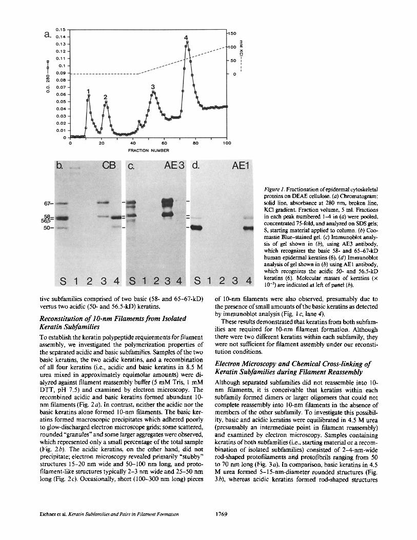
tive subfamilies comprised of two basic (58- and 65-67-kD) versus two acidic (50- and 56.5-kD) keratins.
Reconstitution o f l O-rim Filaments from Isolated Keratin Subfamilies
To establish the keratin polypeptide requirements for filament assembly, we investigated the polymerization properties of the separated acidic and basic subfamilies. Samples of the two basic keratins, the two acidic keratins, and a recombination of all four keratins (i.e., acidic and basic keratins in 8.5 M urea mixed in approximately equimolar amounts) were di- alyzed against filament reassembly buffer (5 mM Tris, 1 mM DTT, pH 7.5) and examined by electron microscopy. The recombined acidic and basic keratins formed abundant 10- nm filaments (Fig. 2a). In contrast, neither the acidic nor the basic keratins alone formed 10-nm filaments. The basic ker- atins formed macroscopic precipitates which adhered poorly to glow-discharged electron microscope grids; some scattered, rounded "granules" and some larger aggregates were observed, which represented only a small percentage of the total sample (Fig. 2b). The acidic keratins, on the other hand, did not precipitate; electron microscopy revealed primarily "stubby" structures 15-20 nm wide and 50-100 nm long, and proto- filament-like structures typically 2-3 nm wide and 25-50 nm long (Fig. 2 c). Occasionally, short (100-300 nm long) pieces
Figure 1. Fractionation of epidermal cytoskeletal proteins on DEAE cellulose. (a) Chromatogram; solid line, absorbance at 280 nm, broken line, KCI gradient. Fraction volume, 5 ml. Fractions in each peak numbered 1-4 in (a) were pooled, concentrated 75-fold, and analyzed on SDS gels; S, starting material applied to column. (b) Coo- massie Blue-stained gel. (c) Immunobiot analy- sis of gel shown in (b), using AE3 antibody, which recognizes the basic 58- and 65-67-kD human epidermal keratins (6). (d) Immunobiot analysis of gel shown in (b) using AE 1 antibody, which recognizes the acidic 50- and 56.5-kD keratins (6). Molecular masses of keratins (x 10 -3) are indicated at left of panel (b).
of 10-nm filaments were also observed, presumably due to the presence of small amounts of the basic keratins as detected by immunoblot analysis (Fig. I c, lane 4).
These results demonstrated that keratins from both subfam- ilies are required for 10-nm filament formation. Although there were two different keratins within each subfamily, they were not sufficient for filament assembly under our reconsti- tution conditions.
Electron Microscopy and Chemical Cross-linking o f Keratin Subfamilies during Filament Reassembly
Although separated subfamilies did not reassemble into 10- nm filaments, it is conceivable that keratins within each subfamily formed dimers or larger oligomers that could not complete reassembly into 10-nm filaments in the absence of members of the other subfamily. To investigate this possibil- ity, basic and acidic keratins were equilibrated in 4.5 M urea (presumably an intermediate point in filament reassembly) and examined by electron microscopy. Samples containing keratins of both subfamilies (i.e., starting material or a recom- bination of isolated subfamilies) consisted of 2-4-nm-wide rod-shaped protofilaments aild protofibrils ranging from 50 to 70 nm long (Fig. 3a). In comparison, basic keratins in 4.5 M urea formed 5-15-nm-diameter rounded structures (Fig. 3b), whereas acidic keratins formed rod-shaped structures
Eichner et al. Keratin Subfamilies and Pairs in Filament Formation 1769

Figure 2. Electron microscopy of epidermal keratin subfamilies isolated by ion exchange chromatography, dialyzed against standard filament reassembly buffer (5 mM Tris, 1 mM DTT, pH 7.5), and contrasted by negative staining. (a) Basic and acidic keratins (recombined in 8.5 M urea). (b) Basic keratins alone. (c) Acidic keratins alone. The sparse, short pieces of 10-nm filaments in c presumably result from the slight contamination of basic keratins, as detected by antibody AE3 in Fig. I c, lane 4. Scale bars, 100 nm (a-c).
more similar to the recombined subfamilies (2-4 nm wide and 40-50 nm long; Fig. 3 c). Although basic or acidic keratins alone formed structures that could be distinguished from the protofilaments and protofibrils typical of samples containing all four keratins, the discrete structures observed suggested that keratins within each subfamily may interact specifically. Furthermore, since acidic and basic keratins recombined in 4.5 M urea were still able to reassemble into 10-nm filaments, these structures could represent "competent" filament precur- sors rather than "dead end" structures.
The possibility of intra-subfamily associations during fila- ment assembly was also investigated using chemical cross- linking in the presence of 4.5 M urea. Basic and acidic keratins were dialyzed against 4.5 M urea, separately and in combi- nation, and reacted with EGS or BSOCOES, two amino group cross-linkers ( l, 42). SDS gel analysis revealed that significant amounts of cross-linked oligomers were present only when acidic and basic keratins were combined (Fig. 4). The apparent mobilities of the cross-linked species on 6-15% polyacryl- amide gradient gels were similar to bands previously identified
by Quinlan et al. as dimers and tetramers of intermediate filament polypeptides (22, 23), with an additional band in the 5% stacking gel (possibly an octamer). These cross-linked species were consistently observed, although the relative in- tensities of the bands varied depending on keratin and cross- linker concentration. No larger aggregates were detected. Thus, in contrast to the microscopic observations suggesting that keratins within a subfamily formed specific associations in 4.5 M urea, keratins from both basic and acidic subfamilies were necessary for intermolecular cross-linking under our experimental conditions.
Isolation o f a 50-kD/ 58-kD "Keratin Pair" Complex
To investigate the interactions between specific acidic and basic keratins, we performed "urea melting" experiments similar to those described by Franke et al. (7). Fig. 5 shows two-dimensional gels of human epidermal keratins in which the urea concentration in the first dimension NEpHG gels was varied between 9.5 and 4 M. In 9.5 M urea gels, acidic and basic epidermal keratins are generally well-resolved at
The Journal of Cell Biology, Volume 102, 1986 1770
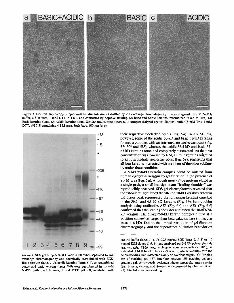
Figure 3. Electron microscopy of epidermal keratin subfamilies isolated by ion exchange chromatography, dialyzed against I0 mM NaPO4 buffer, 4.5 M urea, 1 mM DTT, pH 8.0, and contrasted by negative staining. (a) Basic and acidic keratins (recombined in 8.5 M urea). (b) Basic keratins alone. (c) Acidic keratins alone. Similar results were observed in samples dialyzed against filament buffer (5 mM "Iris, 1 mM DTT, pH 7.5) containing 4.5 M urea. Scale bars, 100 nm (a-c).
their respective isoelectric points (Fig. 5a). In 8.5 M urea, however, some of the acidic 50-kD and basic 58-kD keratins formed a complex with an intermediate isoelectric point (Fig. 5 b, 50* and 58"), whereas the acidic 56.5-kD and basic 65- 67-kD keratins remained completely dissociated. As the urea concentration was lowered to 4 M, all four keratins migrated to an intermediate isoelectric point (Fig. 5 c), suggesting that all four keratins interacted with members of the other subfam- ily under these conditins.
A 50-kD/58-kD keratin complex could be isolated from human epidermal keratins by gel filtration in the presence of 8.5 M urea (Fig. 6a). Although most of the proteins eluted as a single peak, a small but significant "leading shoulder" was reproducibly observed. SDS gel electrophoresis revealed that the "shoulder" contained the 50- and 58-kD keratins, whereas the major peak represented the remaining keratins enriched in the 56.5- and 65-67-kD keratins (Fig. 6b). Immunoblot analysis using antibodies AE3 (Fig. 6c) and AEI (Fig. 6d) confirmed that the leading shoulder contained the 50-kD/58- kD keratins. The 50-kD/58-kD keratin complex eluted at a position somewhat larger than beta-galactosidase (molecular mass 116 kD). Due to the limited resolution of gel filtration chromatography, and the dependence of elution behavior on
Figure 4. SDS gel of epidermal keratin subfamilies separated by ion exchange chromatography and chemically cross-linked with EGS. Basic keratins (lanes 1-3), acidic keratins (lanes 4-6), or recombined acidic and basic keratins (lanes 7-9) were equilibrated in 10 mM NaPO4 buffer, 4.5 M urea, 1 mM DTT, pH 8.0, incubated with
control buffer (lanes 1, 4, 7), 0.25 mg/ml EGS (lanes 2, 5, 8) or 1.0 mg/ml EGS (lanes 3, 6, 9), and analyzed on 6-15% polyacrylamide gradient gels. Right lane, molecular mass standards (x 10-3), as indicated. 43-kD band in lanes 4-9 is actin, which co-elutes with the acidic keratins, but is detectable only on overloaded gels. "O" (origin), top of stacking gel; "S', interface between 5% stacking gel and gradient gel. Arrowheads designate higher molecular mass species (i.e., 2-mers, 4-mers, and 8-mers, as determined by Quinlan et al., 22) detected after cross-linking.
Eichner et al. Keratin Subfamilies and Pairs in Filament Formation 1771
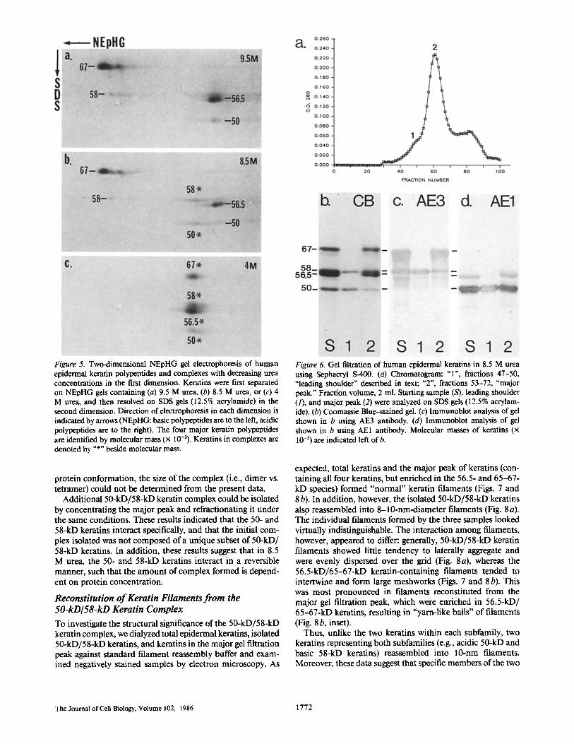
Figure 5. Two-dimensional NEpHG gel electrophoresis of human epidermal keratin polypeptides and complexes with decreasing urea concentrations in the first dimension. Keratins were first separated on NEpHG gels containing (a) 9.5 M urea, (b) 8.5 M urea, or (c) 4 M urea, and then resolved on SDS gels (12.5% acrylamide) in the second dimension. Direction of electrophoresis in each dimension is indicated by arrows (NEpHG: basic polypeptides are to the left, acidic polypeptides are to the right). The four major keratin polypeptides are identified by molecular mass (x 10-3). Keratins in complexes are denoted by "*" beside molecular mass.
protein conformation, the size of the complex (i.e., dimer vs. tetramer) could not be determined from the present data.
Additional 50-kD/58-kD keratin complex could be isolated by concentrating the major peak and refractionating it under the same conditions. These results indicated that the 50- and 58-kD keratins interact specifically, and that the initial com- plex isolated was not composed of a unique subset of 50-kD/ 58-kD keratins. In addition, these results suggest that in 8.5 M urea, the 50- and 58-kD keratins interact in a reversible manner, such that the amount of complex formed is depend- ent on protein concentration.
Reconstitution o f Keratin Filaments from the 50-kD/ 58-kD Keratin Complex
To investigate the structural significance of the 50-kD/58-kD keratin complex, we dialyzed total epidermal keratins, isolated 50-kD/58-kD keratins, and keratins in the major gel filtration peak against standard filament reassembly buffer and exam- ined negatively stained samples by electron microscopy. As
Figure 6. Gel filtration of human epidermal keratins in 8.5 M urea using Sephacryl S-400. (a) Chromatogram: " l ' , fractions 47-50, "leading shoulder" described in text; "2", fractions 53-72, "major peak." Fraction volume, 2 ml. Starting sample (S), leading shoulder (1), and major peak (2) were analyzed on SDS gels 02.5% acrylam- ide). (b) Coomassie Blue-stained gel. (c) Immunoblot analysis of gel shown in b using AE3 antibody. (d) Immunohlot analysis of gel shown in b using AE1 antibody. Molecular masses of keratins (x l0 -3) are indicated left of b.
expected, total keratins and the major peak of keratins (con- mining all four keratins, but enriched in the 56.5- and 65-67- kD species) formed "normal" keratin filaments (Figs. 7 and 8 b). In addition, however, the isolated 50-kD/58-kD keratins also reassembled into 8-10-nm-diameter filaments (Fig. 8 a). The individual filaments formed by the three samples looked virtually indistinguishable. The interaction among filaments, however, appeared to differ: generally, 50-kD/58-kD keratin filaments showed little tendency to laterally aggregate and were evenly dispersed over the grid (Fig. 8 a), whereas the 56.5-kD/65-67-kD keratin-containing filaments tended to intertwine and form large meshworks (Figs. 7 and 8 b). This was most pronounced in filaments reconstituted from the major gel filtration peak, which were enriched in 56.5-kD/ 65-67-kD keratins, resulting in "yarn-like balls" of filaments (Fig. 8 b, inset).
Thus, unlike the two keratins within each subfamily, two keratins representing both subfamilies (e.g., acidic 50-kD and basic 58-kD keratins) reassembled into 10-rim filaments. Moreover, these data suggest that specific members of the two
The Journal of Cell Biology, Volume 102, 1986 1772
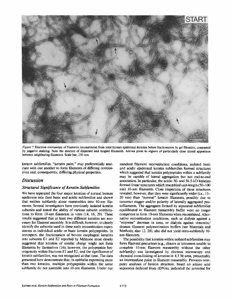
Figure 7. Electron microscopy of filaments reconstituted from total human epidermal keratins before fractionation by gel filtration, contrasted by negative staining. Note the mixture of dispersed and tangled filaments. Arrows point to regions of particularly close lateral apposition between neighboring filaments. Scale bar, 250 nm.
keratin subfamilies, "keratin pairs," may preferentially asso- ciate with one another to form filaments of differing compos- ition and, consequently, differing physical properties.
Discussion
Structural Significance of Keratin Subfamilies We have separated the four major keratins of normal human epidermis into their basic and acidic subfamilies and shown that neither subfamily alone reassembles into 10-nm fila- ments. Several investigators have previously isolated keratin subunits and tested the ability of various subunit combina- tions to form 10-rim filaments in vitro (14, 16, 29). These results suggested that at least two different keratins are nec- essary for filament assembly. It is difficult, however, to clearly identify the subunits used in these early reconstitution exper- iments as individual acidic or basic keratin polypeptides. In retrospect, the fractionation of bovine esophageal keratins into subunits E1 and E2 reported by Milstone most closely suggested that keratins of similar charge might not form filaments by themselves (16); however, the polypeptide het- erogeneity within fractions E 1 and E2, and the significance of keratin subfamilies, was not recognized at that time. The data presented here demonstrate that, in epithelia expressing more than two keratins, multiple polypeptides within the same subfamily do not assemble into 10-nm filaments. Under our
standard filament reconstitution conditions, isolated basic and acidic epidermal keratin subfamilies formed structures which suggested that keratin polypeptides within a subfamily may be capable of lateral aggregation but not end-to-end association. In particular, the acidic 50- and 56.5-kD keratins formed linear structures which resembled unit-length (50- 100 nm) 10-nm filaments. Close inspection of these structures revealed, however, that they were significantly wider (i.e., 15- 20 nm) than "normal" keratin filaments, possibly due to incorrect stagger and/or polarity of laterally aggregated pro- tofilaments. The aggregates formed by separated subfamilies equilibrated in filament reassembly buffer were no longer competent to form 10-nm filaments when recombine& Alter- native reconstitution conditions, such as dialysis against a "stepwise" decrease in urea, or dialysis against vimentin/ desmin filament polymerization buffers (see Materials and Methods; also 12, 28), also did not yield intra-subfamily 10- nm filaments.
The possibility that keratins within isolated subfamilies may form filament precursors (e.g., dimers or tetramers unable to complete 10-nm filament reassembly without the other subfamily) was investigated by electron microscopy and chemical cross-linking of keratins in 4.5 M urea, presumably an intermediate point in filament reassembly. Previous com- puter analyses of keratin structure, based on amino acid sequences deduced from cDNAs, indicated the potential for
Eichne r et al. Keratin Subfamilies and Pairs in Filament Formation 1773
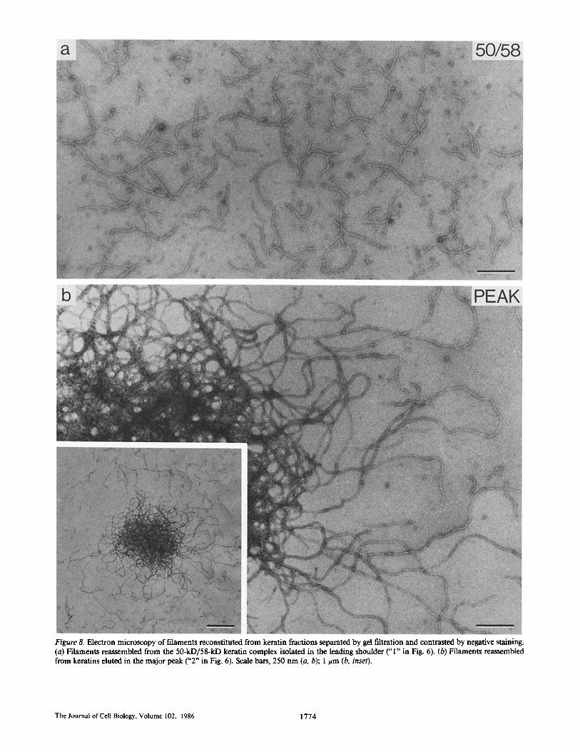
Figure 8. Electron microscopy of filaments reconstituted from keratin fractions separated by gel filtration and contrasted by negative staining. (a) Filaments reassembled from the 50-kD/58-kD keratin complex isolated in the leading shoulder ("1" in Fig. 6). (b) Filaments reassembled from keratins eluted in the major peak ("2 ~ in Fig. 6). Scale bars, 250 nm (a, b); 1 t=m (b, inset).
The Journal of Cell Biology, Volume 102, 1986 1774
intra-subfamily polypeptides to form at least coiled-coil di- mers (31). Electron microscopy of isolated acidic or basic keratins in 4.5 M urea revealed distinct oligomeric structures which could result from specific intra-subfamily interactions. Although both of these structures differed from protofilaments or protofibrils containing all four keratins, these structures were still competent to reassemble into 10-nm filaments when combined, and thus could represent intra-subfamily dimer or tetramer precursors in filament assembly. Alternatively, how- ever, the observed acidic and basic structures could represent either intermediates in an alternative assembly pathway not leading to filament formation (which dissociate and rearrange when mixed with the other subfamily), or structures resulting from nonspecific polypeptide associations. Our present data cannot distinguish between these possibilities.
Cross-linking of keratin subfamilies in 4.5 M urea with EGS or BSOCOES revealed dimers and larger oligomers only when both acidic and basic keratins were present (Fig. 4). These results are in agreement with the cross-linking data of Quinlan et al. which indicated that the two keratins of rat hepatocyte 10-nm filaments form heterotetrameric subunits of two acidic and two basic keratins (22). Our results are also supported by the conclusions of Parry et al. derived from partial amino acid sequences of trypsin-generated keratin filament "parti- cles", indicating that acidic and basic keratins form hetero- typic two-chain coiled coils (21). However, we cannot distin- guish whether the dimers observed after cross-linking acidic and basic keratins with EGS or BSOCOES represent cross- links within a coiled-coil heterodimer, or across coiled-coil homodimers composing heterotetramers. Although we chose to use amino group cross-linking reagents primarily because all keratin polypeptides sequenced to date contain several lysines (e.g., see 12, 15, 30, 31, 36), we cannot exclude the possibility that keratins within either one or both subfamilies form dimers (e.g., either intra-subfamily homo- or heterodi- mers, as suggested by electron microscopy) which are not amenable to cross-linking by EGS or BSOCOES.
Structural Significance of Keratin Pairs Based upon the patterns of keratin expression in epithelial cells under normal, pathological, and culture conditions, we have previously proposed that coexpressed acidic and basic keratins form specific "keratin pairs" (4, 32). In human epi- dermal cells, for example, the 50-kD/58-kD keratin pair represents the only keratins synthesized by basal cells, the 56.5-kD/65-67-kD pair is expressed only in suprabasal cells undergoing keratinization, and the 48-kD/56-kD keratin pair is expressed by hyperproliferative keratinocytes in culture and in various epidermal diseases (6, 32, 38, 39). Our results disagree with a recent suggestion that the acidic 50-kD keratin and the basic 56-kD keratin represent a coexpressed pair in human epidermal basal cells (36), which was based primarily on the coexistence of these two keratins in cultured human epidermal cells.
The data presented here provide evidence for the structural significance of specific keratin pairs in the formation of ker- atin filaments. Taking advantage of the stronger affinity of the 50-kD/58-kD keratin pair compared with the affinity of the 56.5-kD/65-67-kD pair, we were able to isolate by gel filtration a 50-kD/58-kD keratin complex from human epi- dermal keratin extracts. In contrast to the two keratins within
each subfamily, the two keratins within the pair formed "normal-looking" 10-nm filaments, thus demonstrating that two specific keratins from a tissue containing multiple kera- tins can preferentially associate and form filaments. Since the amino acid sequences of the central helical domains of kera- tins within each subfamily are similar (12, 30), specificity between keratins within the 50-kD/58-kD keratin pair prob- ably resides in the nonhelical end domains. The importance of the nonhelical extensions for in vitro reconstitution of 10- nm filaments has been previously documented (19, 24). The rapid assembly of keratin polypeptides into filaments in vivo (8) further suggests the possibility that pair recognition site(s) may be on the amino termini, providing a means for proper polypeptide alignment and initiation of polymerization dur- ing translation, as suggested in the biosynthesis of procollagen (37).
The term "keratin pair," as used here, refers to co-expressed acidic and basic keratins and does not a priori define a specific level of fibrillar substructure (e.g., dimer, tetramer, octamer, etc.). It is not clear at what stage of fdament assembly keratins of a co-expressed pair no longer preferentially associate and start to integrate with other keratins or keratin pairs. Since expression of the 50-kD/58-kD keratin pair in epidermal basal cells is temporally separated from the synthesis of the 56.6- kD/65-67-kD pair in suprabasal cells, keratin pairs could easily segregate into separate filaments in epidermal cells. In view of the dynamic rearrangements of keratin filaments observed in certain cells (8, 10), it is also possible that "basal cell filaments" can unravel and repolymerize with "suprabasal keratins" to form heterotypic filaments integrating both pairs. The strong, specific affinity between the 50- and 58-kD ker- atins, however, may provide a mechanism for maintaining a certain level (e.g., tetramer, octamer, etc.) of segregation throughout epidermal differentiation.
One of our long term goals is to elucidate the structural and functional significance of keratin heterogeneity. It seems most logical that different keratin pairs may form filaments with differing physical properties (see reference 32). In the present study, we noted that 10-nm filaments composed of the 50-kD/58-kD keratin pair showed little tendency toward filament-filament interactions (e.g., lateral aggregation and/ or intertwining as observed by electron microscopy of nega- tively stained samples), whereas filaments enriched in the 56.5-kD/65-67-kD keratin pair had a marked propensity to form dense filament tangles. Although we cannot eliminate the possibility that this aggregation resulted from the presence of small amounts of filaggrin, a keratin filament aggregating protein (27), filaggrin could not be detected with a monoclonal antibody that recognizes human filaggrin in immunoblot analysis (5). In addition, the observed filament tangles did not resemble the more aligned filaments typical of filaggrin-kera- tin filament macrofibrils (27).
The observed differences in filament-filament association correlates well with the expression and potential function of these keratin pairs in filaments in vivo. The 50-kD/58-kD keratin pair is found in living cells of stratified squamous epithelia, where filaments are presumably in a flexible, dy- namic state, and filament "bundling" might be lethal. Expres- sion of the 56.5-kD/65-67-kD keratin pair, on the other hand, has been correlated with the formation of anucleate, cornified cells in normal epidermis and vitamin A-deficient epithelia,
Eichner et al. Keratin Subfamilies and Pairs in Filament Formation 1775
w h e r e de nse ly p a c k e d f i l a m e n t s are t h e u l t i m a t e p r o d u c t (34, 39). T h e s e o b s e r v a t i o n s sugges t t h a t a l t h o u g h d i f f e r en t ke r a t i n c o m b i n a t i o n s m a y f o r m m o r p h o l o g i c a l l y s imi l a r l ook i ng 10- n m f i l amen t s , ke r a t i n he t e rogene i t y , by i t se l f o r t h r o u g h in- t e r a c t i o n wi th i n t e r m e d i a t e f i l amen t - spec i f i c m o l e c u l e s , m a y p r o v i d e a bas i s for h igh ly spec ia l i zed f u n c t i o n s w i t h i n t h e cell.
We thank Dr. Marc Kahn for critical reading o f the manuscript. This work was supported by grants from the National Institutes of
Health (NIH) (AM 34462 to R. Eichner, EY 04722 and AM 25140 to T.-T. Sun, and G M 31940 to U. Aebi), the Gillette Company, and Estee Lauder. T.-T. Sun is the recipeint o f an NIH Research Career Development Award (EY 0125) and a Monique Weill-Caulier Career Scientist Award. U. Aebi is also the recipient o f a research award from the Maufice-Muller-Foundation o f Switzerland.
Received for publication 21 August 1985, and in revised form 28 January 1986.
Note Added in Proof. After we submitted this manuscript, Hatzfeld and Franke published similar findings on the association of acidic and basic keratins during in vitro reconstitution (Pair formation and promiscuity of cytokeratins: formation in vitro of heterotypic com- plexes and intermediate-sized fdaments by homologous and heterol- ogous recombinations of purified polypeptides, 1985, J. Cell Biol., l01: 1826-184 l). Our data are in close agreement, although we found that under our filament reassembly conditions, basic (Type II) kera- tins formed macroscopic precipitates with some small granules (rather than 2 -3 -nm protofilaments, 10-30-nm granules, and short cylinders) and acidic (Type I) keratins formed 15-20-nm structures and 2-3- nm protofilaments (rather than primarily 2 -3 -nm protofilaments). In addition, although they found that 50-kD/58-kD keratins show sim- ilar "urea melting" characteristics in acrylamide gels as 56.5-kD/58- kD keratins, our data suggest that, in solution, the 50-kD/58-kD keratin pair displays a stronger specific affinity than other combina- tions of epidermal keratins.
References
1. Abdella, P. M., P. K. Smith, and G. P. Royer. 1979. A new cleavable reagent for cross-linking and reversible immobilization of proteins. Biochem. Biophys. Res. Commun. 87:734-742.
2. Aebi, U., W. Fowler, P. Rew, and T.-T. Sun. 1983. The fibrillar substruc- ture of keratin filaments unraveled. J. Cell Biol. 97:1131-1143.
3. Bowden, P. E., and W. J. Cunliffe. 1981. Modifications of human prekeratin during epidermal differentiation. Biochem. J. 199:145-154.
4. Cooper, D., and T.-T. Sun. 1986. Monoclonal antibody analysis of cow epithelial keratins: keratin subfamilies and pairs. J. BioL Chem. In press.
5. Dale, B. A., and T.-T. Sun. 1983. Filaggrin, 56.5kd and 67kd keratins share an antigenic determinant as defined by AE2 monoclonal antibody. £ BioL Chem. 97:228a. (Abstr.)
6. Eichner, R., P. Bonitz, and T.-T. Sun. 1984. Classification of epidermal keratins according to their immunoreactivity, isoelectric point, and mode of expression..L Cell Biol. 98:1388-1396.
7. Franke, W. W., D. L. Schiller, M. Hatzfeld, and S. Winter. 1983. Protein complexes of intermediate-sized filaments: melting of eytokeratin complexes in urea reveals different polypeptide separation characteristics. Proc. NatL Acad. Sci. USA. 80:7113-7117.
8. Franke, W. W., E. Schmid, S. Mittnacht, C. Grund, and J. L. Jorcano. 1984. Integration of different keratins into the same filament system after microinjection of mRNA for epidermal keratins into kidney epithelial cells. Cell. 36:813-825.
9. Fuchs, E., S. M. Coppock, H. Green, and D. W. Cleveland. 1981. Two distinct classes of keratin genes and their evolutionary significance. Cell. 27:75- 84.
10. Geiger, B., T. E. Kreis, O. Gigi, E. Schmid, S. Mittnacht, J. L. Jorcano, D. B. von Bassewitz, and W. W. Franke. 1984. Dynamic rearrangements of cytokeratins in living cells. In Cancer Cells. Vol. 1. The Transformed Pheno- type. A. Levine, W. Topp, G. Vande Woude, and J. D. Watson, editors. Cold Spring Harbor Laboratory, Cold Spring Harbor, New York. 169-176.
I I. Gigi, O., B. Creiger, Z. Eshhar, R. Moll, E. Sehmid, S. Winter, D. L. Schiller, and W. W. Franke. 1982. Detection of a cytokeratin determinant common to diverse epithelial cells by a broadly cross-reacting monoclonal antibody. EMBO (Eur. Mol. Biol. Organ.) J. 1:1429-1437.
12. Jorcano, J. L., M. Rieger, J. K. Franz, D. L. Schiller, R. Moll, and W. W. Franke. 1984. Identification of two types of keratin polypeptides within acidic cytokeratin subfamily I. J. Mol. Biol. 179:257-281.
13. Kim, K. H., J. Rbeinwald, and E. V. Fuchs. 1983. Tissue specificity of epithelial keratins: differential expression of mRNAs from two multigene families. Mol. Cell. Biol. 3:495-502.
14. Lee, L. D., and H. P. Baden. 1976. Organization of the polypeptide chains in mammalian keratin. Nature (Lond.). 264:377-379.
15. Marchuk, D., S. McCrohon, and E. Fuchs. 1984. Remarkable conser- vation of structure among intermediate filament genes. Cell. 39:49 I--498.
16. Milstone, L. 1981. Isolation and characterization of two polypeptides that form intermediate filaments in bovine esophageal epithelium. J. Cell Biol. 88:317-322.
17. Moll, R., W. W. Franke, D. L. Schiller, B. Geiger, and R. Krepler. 1982. The catalog of human cytokeratins: patterns of expression in normal epithelia, tumors and cultured cells. Cell. 31:11-24.
18. Moll, R., I. Moll, and W. W. Franke. 1984. Differences of expression of cytokeratin polypeptides in various epithelial skin tumors. Arch. Dermatol. Res. 276:349-363.
19. Nelson, W. J., and P. Traub. 1983. Proteolysis of vimentin and desmin by the Ca++-activated proteinase specific for these intermediate proteins. Mol. Cell. Biol. 3:1146-1156.
20. O'Farrell, P. Z., H. M. Goodman, and P. H. O'Farrell. 1977. High resolution two-dimensional eleetrophoresis of basic as well as acidic proteins. Cell. 12:1133-1142.
21. Parry, D. A. D., A. C. Steven, and P. M. Steinert. 1985. The coiled-coil molecules of intermediate filaments consist of two parallel chains in exact axial register. Biochem. Biophys. Res. Commun. 127:1012-1018.
22. Quinlan, R. A., J. A. Cohlberg, D. L. Schiller, M. Hatzfeld, and W. W. Franke. 1984. Heterotypic tetramer (A2D2) complexes of non-epidermal kera- tins isolated from cytoskeletons of rat hepatocytes and hepatoma cells. J. Mol. Biol. 178:365-388.
23. Quinlan, R. A., and W. W. Franke. 1982. Heteropolymer filaments of vimentin and desmin in vascular smooth muscle tissue and cultured baby hamster kidney cells demonstrated by chemical crosslinking. Proc. Natl. Acad. Sci. USA. 79:3452-3456.
24. Sauk, J. J., M. Krumweide, D. Cocking-Johnson, and J. G. White. 1984. Reconstitution of cytokeratin filaments in vitro: further evidence for the role on nonhelical peptides in filament assembly. J. Cell Biol. 99:1590-1597.
25. Schiller, D. L., W. W. Franke, and B. Geiger. 1982. A subfamily of relatively large and basic cytokeratin polypeptides as defined by peptide map- ping is represented by one or several polypeptides in epithelial cells. EMBO (Eur. Mol. Biol. Organ.)J. 1:761-769.
26. Skerrow, D., and C. J. Skerrow. 1983. Tonofilament differentiation in human epidermis: isolation and polypeptide chain composition of keratinocyte subpopulations. Exp. Cell Res. 143:27-35.
27. Steinert, P. M., J. S. Cantieri, D. C. Teller, J. D. Lonsdale-Eccles, and B. A. Dale. 1981. Characterization of a class of cationic proteins that specifically interact with intermediate filaments. Proc. Natl. Acad. Sci. USA. 78:4097- 4101.
28. Steinert, P. M., W. W. Idler, F. Cabral, M. M. Gottesman, and R. D. Goldman. 1981. In vitro assembly of homopolymer and copolymer filaments from intermediate filament subunits of muscle and fibroblastic cells. Proc. Natl. Acad. Sci. USA. 78:3692-3696.
29. Steinert, P. M., W. W. Idler, and S. B. Zimmerman. 1976. Self-assembly of bovine epidermal keratin filaments in vitro. J. Mol. Biol. 108:547-567.
30. Steinert, P. M., D. A. D. Parry, W. W. Idler, L. D. Johnson, A. C. Steven, and D. R. Roop. 1985. Amino acid sequences of mouse and human epidermal type II keratins of Mr 67,000 provide a systematic basis for the structural and functional diversity of the end domains of keratin intermediate filament sub- units. J. Biol. Chem. 260:7142-7149.
31. Steinert, P. M., D. A. D. Parry, E. L. Racoosin, W. W. Idler, A. C. Steven, B. L. Trus, and D. R. Roop. 1984. The complete cDNA and deduced amino acid sequence of a type II mouse epidermal keratin of 60,000 Da: analysis of sequence differences between type I and type II keratins. Proc. Natl. Acad. Sci. USA. 81: 5709-5713.
32. Sun, T.-T., R. Eichner, A. Schermer, D. Cooper, W. G. Nelson, and R. A. Weiss. 1984. Classification, expression, and possible mechanisms of evolu- tion of mammalian epithelial keratins: a unifying model. In Cancer Ceils. Vol. 1. The Transformed Phenotype. A. Levine, W. Topp, G. Vande Woude, and J. D. Watson, editors. Cold Spring Harbor Laboratory, Cold Spring Harbor, New York. 169-176.
33. Towbin, H., T. Staehelin, and J. Gordon. 1979. Electrophoretic transfer of proteins from polyacrylamide gels to nitrocellulose sheets: procedure and some applications. Proc. Natl. Acad. Sci. USA. 76:4350-4354.
34. Tseng, S. C. G., D. Hatchell, N. Tierney, A. J.-W. Huang, and T.-T. Sun. 1984. Expression of specific keratin markers by rabbit corneal, conjunc- tival, and esophageal epithelia during vitamin A deficiency..L Cell Biol. 99:2279-2286.
35. Tseng, S. C. G., M. J. Jarvinen, W. G. Nelson, J.-W. Huang, J. Wood- cock-Mitchell, and T.-T. Sun. 1982. Correlation of specific keratins with different types of epithelial differentiation: monoclonal antibody studies. Cell. 30:361-372.
The Journal of Cell Biology, Volume 102, 1986 1776

36. Tyner, A. L., M. J. Eichman, and E. Fuchs. 1985. The sequence of a type II keratin gene expressed in human skin: conservation of structure among all intermediate filaments. Proc. Natl. Acad. Sci. USA. 82:4683--4687.
37. Veis, A., S. J. Leibovich, J. Evans, and T. Z. Kirk. 1985. Supramolecular assemblies of mRNA direct the coordinated synthesis of type I procollagen chains. Proc. Natl. Acad. Sci. USA. 82:3693-3697.
38. Weiss, R. A., R. Eichner, and T.-T. Sun. 1984. Monoclonal antibody analysis of keratin expression in epidermal diseases: a 48- and 56-kdalton keratin as molecular markers for hyperproliferative keratinocytes. J. Cell BioL 98:1397-1 406,
39. Woodcock-Mitchell, J., R. Eichner, W. G. Nelson, and T.-T. Sun. 1982.
Immunolocalization of keratin polypeptides in human epidermis using mono- clonal antibodies. J. CellBiol. 95:580-588.
40. Wrigley, N. E. 1968. The lattice spacing of crystalline catalasc as an internal standard of length in electron microscopy. J. Ultrastruct. Res. 24:454- 464.
41. Wu, Y.-J., L. M. Parker, N. E. Binder, M. A. Beekett, J. H. Sinard, C. T. Griffiths, and J. G. Rheinwald. 1982. The mesothelial keratins: a new family of cytoskeletal proteins identified in cultured mesothelial ceils and nonkeratin- izing epithelia. Cell. 31:693-703.
42. Zarling, D. A., A. Watson, and F. H. Bach. 1980. Mapping of lymphocyte surface polypeptide antigens with BSOCOES. J. Immunol. 124:913-920.
Eichner et al. Keratin Subfamilies and Pairs in Filament Formation 1777